plos one 2013
TRANSCRIPT

FcStuA from Fusarium culmorum Controls Wheat Footand Root Rot in a Toxin Dispensable MannerMatias Pasquali1*., Francesca Spanu2., Barbara Scherm2, Virgilio Balmas2, Lucien Hoffmann1,
Kim E. Hammond-Kosack3, Marco Beyer1, Quirico Migheli2,4
1 Environment and Agro-biotechnologies Department - Centre de Recherche Public - Gabriel Lippmann, Belvaux, Luxembourg, 2 Dipartimento di Agraria - Sezione di
Patologia vegetale ed entomologia and Unita di ricerca Istituto Nazionale di Biostrutture e Biosistemi, Universita degli Studi di Sassari, Sassari, Italy, 3 Wheat
Pathogenomics, Plant Biology and Crop Science Department, Rothamsted Research, Harpenden, Herts, United Kingdom, 4 Centro interdisciplinare per lo sviluppo della
ricerca biotecnologica e per lo studio della biodiversita della Sardegna e dell’area mediterranea, Universita degli Studi di Sassari, Sassari, Italy
Abstract
Fusarium culmorum is one of the most harmful pathogens of durum wheat and is the causal agent of foot and root rot (FRR)disease. F. culmorum produces the mycotoxin deoxynivalenol (DON) that is involved in the pathogenic process. The role ofthe gene FcStuA, a StuA ortholog protein with an APSES domain sharing 98.5% homology to the FgStuA protein(FGSG10129), was determined by functional characterisation of deletion mutants obtained from two F. culmorum wild-typestrains, FcUk99 (a highly pathogenic DON producer) and Fc233B (unable to produce toxin and with a mild pathogenicbehavior). The DFcStuA mutants originating from both strains showed common phenotypic characters including stuntedvegetative growth, loss of hydrophobicity of the mycelium, altered pigmentation, decreased activity of polygalacturonicenzymes and catalases, altered and reduced conidiation, delayed conidial germination patterns and complete loss ofpathogenicity towards wheat stem base/root tissue. Glycolytic process efficiency [measured as growth on glucose as solecarbon (C) source] was strongly impaired and growth was partially restored on glutamic acid. Growth on pectin-like sourcesranked in between glucose and glutamic acid with the following order (the lowest to the highest growth): beechwoodxylan, sugarbeet arabinan, polygalacturonic acid, citrus pectin, apple pectin, potato azogalactan. DON production in themutants originating from FcUK99 strain was significantly decreased (295%) in vitro. Moreover, both sets of mutants wereunable to colonise non-cereal plant tissues, i.e. apple and tomato fruits and potato tubers. No differences between mutants,ectopic and wild-type strains were observed concerning the level of resistance towards four fungicides belonging to threeclasses, the demethylase inhibitors epoxiconazole and tebuconzole, the succinate dehydrogenase inhibitor isopyrazam andthe cytochrome bc1 inhibitor trifloxystrobin. StuA, given its multiple functions in cell regulation and pathogenicity control, isproposed as a potential target for novel disease management strategies.
Citation: Pasquali M, Spanu F, Scherm B, Balmas V, Hoffmann L, et al. (2013) FcStuA from Fusarium culmorum Controls Wheat Foot and Root Rot in a ToxinDispensable Manner. PLoS ONE 8(2): e57429. doi:10.1371/journal.pone.0057429
Editor: Yin-Won Lee, Seoul National University, Republic of Korea
Received August 31, 2012; Accepted January 22, 2013; Published February 22, 2013
Copyright: � 2013 Pasquali et al. This is an open-access article distributed under the terms of the Creative Commons Attribution License, which permitsunrestricted use, distribution, and reproduction in any medium, provided the original author and source are credited.
Funding: This work was funded by the Ministry of University and Research, by Regione Autonoma della Sardegna (Legge Regionale 7 agosto 2007, n. 7‘‘Promozione della ricerca scientifica e dell’innovazione tecnologica in Sardegna’’) and by Qatar National Research Fund (National Priorities Research ProgramGrant number 4 - 259 - 2 - 083) and by ‘Administration des Services Techniques de l’Agriculture du Luxembourg’. FS acknowledges receipt of a PhD fellowship(XXIV cycle) sponsored by AGRIS SARDEGNA – Agenzia per la Ricerca in Agricoltura. MP acknowledges the AM2c program of the National Research Fund ofLuxembourg. KHK is supported by the Biotechnology and Biological Sciences Research Council (BBSRC) of the United Kingdom through the Institute StrategicProgramme Grant 20:20 WheatH. The funders had no role in study design, data collection and analysis, decision to publish, or preparation of the manuscript.
Competing Interests: The authors have declared that no competing interests exist.
* E-mail: [email protected]
. These authors contributed equally to this work.
Introduction
In ascomycetous fungi, APSES proteins are an important class
of transcription factors involved in the control of the main
developmental processes and in the regulation of the cell-cycle. All
members that belong to the family of APSES proteins have a
,100-residue sequence-specific basic helix-loop-helix DNA-bind-
ing domain [1,2,3,4]. APSES proteins are similar to viral KilA-N-
domains implying probably a viral origin due to an ancestral
infection of a fungal cell [5]. Asm-1 (Neurospora crassa [6]), Phd1 and
Sok2 (Saccharomyces cerevisiae [7]), Efg1 and Efh1 (Candida albicans [3]),
StuA (Aspergillus nidulans [8]) are the main identified genes belonging
to the APSES protein group. In S. cerevisiae and in C. albicans
Phd1p, Sok2p, Efg1p, and Efh1p regulate genetic physiological
and biochemical mechanisms responsible for dimorphic transition
[7,9,10]. Asm-1 controls sexual and asexual reproduction. In
general, StuA homologues regulate sporulation mechanisms,
cellular differentiation, morphogenetic processes, mycelial growth,
and virulence, but their role changes according to the fungal
species.
F. culmorum is an important pathogen on cereals, distributed
worldwide and able to produce a range of mycotoxins that are
harmful to human and animal health [11,12]. The main
mycotoxins produced belong to the type B trichothecenes.
Chemically, the trichothecenes are sesquiterpenoid compounds
able to inhibit protein synthesis in eukaryotic cells and to induce
apoptosis [13]. Moreover, the production of some trichothecenes,
for example deoxynivalenol (DON), plays an important role
PLOS ONE | www.plosone.org 1 February 2013 | Volume 8 | Issue 2 | e57429

during Fusarium graminearum infection of some host plant species,
notably wheat floral tissue [14,15,16]. An analysis conducted by
confocal laser scanner microscopy showed the infection processes
and mechanisms of penetration by F. culmorum in foot and root rot
diseases [17]. Initially, the fungal hyphae follow an apoplastic
intercellular pathway and then colonise the cortex via a symplastic
intercellular pathway. Nevertheless, F. culmorum does not penetrate
into the root stele. Instead, section analysis of the stem showed that
F. culmorum penetrates through the stomata of the leaf sheaths that
wrap around the stem base [18].
Despite the economic importance of F. culmorum incited disease,
knowledge on pathogenicity factors in F. culmorum is limited.
Therefore, as a starting point, a study of the effect of potential key
regulators in the genome is warranted. It has been already shown
that DON production may play a role in wheat foot and root rot
severity [19,20]. Here we identified and characterised the role of
FcStuA in F. culmorum aiming at understanding its role in foot and
root rot and head blight pathogenicity, in the biosynthesis of
pectolytic enzymes such as polygalacturonase and in the metabolic
or morphological processes.
Materials and Methods
Strains and culture conditionsTransformation experiments were done with two F. culmorum
wild-type strains: FcUK99 (Rothamsted Research, UK), a DON-
producer (NRRL54111) isolated from an infected wheat plant
[21], and Fc233B (NRRL54905), also isolated from an infected
wheat plant [22], previously characterised as being unable to
produce toxin in vitro (Pasquali et al., unpublished). Toxin
production of strain Fc233B was verified according to the
analytical procedure described by [22] in media derived from
[23], (sugar source sucrose) and from [24], following cultural
conditions as described by [25].
Morphological characterisation of wild-type and mutant strains
was carried out on different substrates. Potato dextrose agar (PDA;
Sigma-Aldrich, St. Louis, MO), synthetic low-nutrient agar
medium (SNA, [26]) in weight/volume consisting of: 0.1%
KH2PO4, 0.1% KNO3, 0.05% MgSO4 7H2O, 0.05% KCl,
0.02%, glucose, 0.02% sucrose, and 2% agar, carboxy-methyl
cellulose liquid medium (CMC, [27]), Czapek-Dox broth (C1551;
Sigma-Aldrich) were used. Lyophilised mycelium for DNA
extraction was obtained by growing fungi in liquid complete
medium (CM) [28]. For polygalacturonase (PG) activity induction,
the Szecsi medium was used [29]: NH4H2PO4 0.09% (w/v), (NH4)
2HPO4 0.2%, MgSO4 7H2O 0.01%, KCl 0.05%, pectin ICN 1%.
The cup plate analysis was done to evaluate polygalacturonase
expression in 100 mM acetate buffer pH 4.0, 0.5% polygalac-
turonic acid, 0.8% agarose [30]. To compare the abilities of
carbon (C) source utilisation, minimal medium (pH 6) containing
as a sole C source 1% w/v one of the following compounds: xylan
from beechwood, red arabinogalactan from sugar beet, pectin
from citrus peel, pectin from apple, azo-galactan from potato,
polygalacturonic acid (all from Sigma-Aldrich) were used as
described in [31]. To verify the impairment of the glycolytic
process in the mutants 30 mM of glucose or of glutamic acid
monosodic (all media were adjusted to pH 6.0) was also used in the
comparative growth test at 25 C in the dark. Growth was
measured after 5 days on biological triplicates.
Split-marker recombination and collection of FcStuAmutants
Conserved regions of the StuA gene in the three Fusarium species
so far sequenced (F. graminearum PH-1, Fusarium oxysporum 4287 and
Fusarium verticillioides 7600, available at: www.broadinstitute.org/
annotation/genome/fusarium_group/MultiHome.html) were
identified by Muscle alignments search done using the CLC Main
Workbench v 6.01 software (CLC bio Aarhus, Denmark) aligning
both DNA sequences and protein-derived sequences. The primers
were designed by the same software to obtain the corresponding
StuA gene sequence in F. culmorum. The upstream gene region was
obtained from the genome sequencing project of F. culmorum strain
UK99 (Hammond-Kosack, Antoniw, Urban et al., unpublished).
Split-marker recombination [32] was used for FcStuA deletion in
FcUK99 and in Fc233B (Fig. S1) using the primers listed in
Table 1.
Fungal protoplasts were obtained from macroconidia germinat-
ed on PDA. Typically, 12–14 plates of PDA medium were overlaid
with a disc of sterile cellophane (model 583 Gel Dryer; Bio-Rad
Hercules, CA, USA) and inoculated with 106 conidia of F.
culmorum FcUK99 or Fc233B and incubated at 25uC for about 16–
18 hours. Then, the young mycelium was scraped from the
cellophane surface using a sterile spatula and transferred into 2
Petri dishes (90 mm of diameter) each one containing 10 mL of
lysis solution consisting of 10 mg/mL of lysing enzymes (L1492,
Sigma-Aldrich, St. Louis, MO, USA) dissolved in 1.2 M MgSO4
(pH 5.8).
After 3–4 hours of incubation at room temperature and slow
(50–60 rpm) agitation, the protoplasts were purified according to
[33] and used directly in fungal transformation as described by
[34].
Identification of FcStuA deletion mutantsTo verify whether in the 20 transformants obtained from F.
culmorum strains FcUK99 and Fc233B, FcStuA was correctly deleted
by the hph gene (which confers resistance to hygromycin B), a
screening on selective medium (PDA amended with 200 mg/mL of
hygromycin B) was performed. Monosporic transformants were
further checked by direct specific PCR analysis [35] and only 9
transformants (S2-S9-S11-S12-S13-S16-S17-S21-S22) from
FcUK99 and two (S19–S20) from Fc233B were selected for
Southern blot analysis. Primer sequences used are listed in
Table 1.
Genomic DNA was obtained by a standard extraction method
[36] from mycelium grown in 50–70 mL of CM at 25uC with
gentle shaking (100–120 rpm) for 5 days. Then, the mycelium was
filtered with Miracloth membrane (475855 Calbiochem, Merck
Darmstadt, Germany) and freeze-dried for 3 days. For Southern
blot analysis, a total of 5 mg of each genomic DNA was digested at
37uC for 16–18 hours with 50 units of EcoRV (New England
Biolabs) in a final volume of 100 mL. Subsequently, digested DNA
was separated by electrophoresis on a 1.2% agarose gel and
transferred to a nylon membrane (Hybond-N Amersham Biosci-
ences, GE Healthcare, USA) using a vacuum blotter (Model 785,
Bio-Rad) according to standard procedures [37]. The membrane
was then hybridised and the specific probes were detected with
Dig High Prime DNA labelling kit and Detection Starter II (Roche
Applied Science, Basel, Switzerland) as described in the manu-
facturer’s protocols. To identify FcStuA gene deletion, the
membrane was labelled by a left flank probe (806 bp) and after
stripping the membrane was labelled by a partial gene StuA probe
(401 bp).
The left flank probe was amplified using the TopTaqTM DNA
Polymerase kit (QiagenS.p.A., Milan, Italy) in 100 mL of total
volume containing: 10 mL of TopTaqTM DNA Polymerase
amplification buffer, 0.2 mM dNTPs, 0.5 mM of primer stuA
1F, 0.5 mM of primer stuA 2R and 2.5 U of TopTaqTM DNA
Polymerase, 50 ng of FcUK99 DNA. The reaction was done
FcStuA Gene Characterisation in Fusarium culmorum
PLOS ONE | www.plosone.org 2 February 2013 | Volume 8 | Issue 2 | e57429

according to the following protocol: 94uC for 3 min, then 35 cycles
of 94uC for 20 sec, 53uC for 20 sec and 72uC for 1 min, followed
by a final elongation step at 72uC for 5 min. The partial StuA gene
probe was obtained from DNA of FcUK99 as template with
primers stuA NF-stuA NR and PhusionH High-Fidelity PCR
Master Mix with HF Buffer (New England Biolabs) at the
following conditions: 98uC for 2 min, then 33 cycles of 98uC for
20 sec, 60uC for 30 sec and 72uC for 50 sec, followed by a final
elongation step at 72uC for 5 min.
Pathogenicity testsFcStuA deletion mutants confirmed by Southern blot analysis
were tested on durum wheat seedlings to evaluate the role of
FcStuA in the pathogenic process by the soil-borne fungus F.
culmorum.
Mycelium plugs bearing one seed of durum wheat (Triticum
durum cv. Claudio, kindly provided by Unita di Ricerca per la
Valorizzazione Qualitativa dei Cereali, CRA-QCE, Rome, Italy)
were placed into a plastic sowing pot and covered by sterile soil.
Pathogenicity tests were conducted according to [38] in a
greenhouse at 25u C and three weeks after inoculation the severity
of disease was assessed using the McKinney index [39].
Head Blight symptoms were evaluated on Triticum durum cv
Simeto plants (kindly provided by CRA-QCE, Rome, Italy)
inoculated according to the procedure described in [40]. Plants
were observed every 3 days and final evaluation was carried out at
14 days after inoculation. Score indices ranged from 0 to 10, which
is equivalent to the number of spikelets infected above the
inoculation point.
In addition, the ability of DFcStuA mutants to colonise other
plant tissues was evaluated. This included the inoculation of apple
fruit slices [41], tomato fruits [42] and potato tuber slices with the
respective wild-type and ectopic transformants. Thus, apple (cv.
Golden Delicious), potato (cv. Spunta olandese) and tomato (cv.
Altavilla) were washed under tap water and then disinfected within
a sterile hood with a 2% sodium hypochlorite solution for 5
minutes and rinsed in sterile water at least twice. Later, plant
tissues (except tomato) were cut into slices about 1 cm thick. Slices
were placed in sterile Petri dishes and 10 mL of a concentrated
spore suspension containing 105 conidia/mL were pipetted on the
surface. Inoculated samples were incubated in a dark room at
25uC and monitored daily for fungal growth. Tomato fruit were
inoculated by direct deposition of the spore suspension on the fruit
after creating a superficial damage with a pipetting tip. They were
incubated in sterile plastic containers at the same modalities as
described above. These experiments were performed three times.
Microscopic observations of durum wheat kernelsTo analyse the development of fungal hyphae during the first
steps of colonisation, pathogenicity test conditions were repro-
duced in vitro. Ten mycelium discs (from the same set of strains as
indicated before) bearing one seed were placed into a Petri dish
and incubated 3 days in the dark.
The observations were conducted by a scanning electron
microscope ZEISS EVO LS with environmental control in low
vacuum conditions (pressure 600 Pa, temperature 2uC, humidity
85%). To investigate whether the impaired seedling germination
was caused by seed death or by an inhibition of seed germination
induced by surrounding fungal hyphae, seeds (washed with 1%
NaClO 3 times for 1 minute, then washed with sterile water) were
incubated in a water agar plate for 3 days in the dark after being in
contact for three days with the mycelium. Germination of the
seeds was then verified. The experiment was done in triplicate.
Morphological analysisFrom each of the F. culmorum FcUK99 and Fc233B wild-type
strains, respectively, single-spore FcStuA deletion mutants (S12–
S13 and S19) and an ectopic transformant (S2 and S20) were
chosen for morphological characterisation. To evaluate mycelium
hydrophobicity, a droplet (20 mL) of sterile H2O was deposited on
the surface of colonies grown on PDA for 5 days. The time (in
seconds) elapsed to absorb the drop by FcStuA deletion mutants
and by ectopic transformants was compared to the respective wild-
type strain. Hyphal structures, sporodochial development, con-
idiogenesis, as well as the time and extent of spore germination
were observed by a light microscope (Olympus BX41) after growth
in CMC liquid medium or SNA solid substrate. These experi-
ments were replicated three times.
Briefly, three mycelium plugs were inoculated into 50–70 mL of
liquid CMC medium in the dark at room temperature and
120 rpm shaking for 5 days. The spore suspension was filtered
with a MiraclothH membrane and then washed twice with sterile
water. The filtrate was centrifuged to collect conidia and
subsequently the concentration was estimated using a Burker-
Turk hemacytometer. The growth experiment was done on SNA
at 25 uC with a photoperiod of 24 hours for 18 days [43] to check
sporodochial production. To determine the timing of spore
germination, 1 mL of spore suspension (106 conidia/mL) was
inoculated into Erlenmeyer flasks containing 30 mL of Czapek-
Dox broth. The evaluation was made after 0, 2, 4,and 8 h of
incubation at 25 uC in the dark with slow shaking (100 rpm).
Table 1. Primer sequences used to obtain the transformingconstructs and to identify mutants by PCR and Southernblotting.
Split-markerrecombination Primer sequence
HY1F 59-GGCTTGGCTGGAGCTAGTGGAGGTCAA-39
HY2R 59-GCCGAACCCGCTCGTCTGGCTAAGA-39
YG1F 59-GATGTAGGAGGGCGTGGATATGTCCT-39
YG2R 59-GAACCCGCGGTCGGCATCTACTCTAT-39
StuA 1F 59-CCGTTCTTAAACTTTGAAGCTCTATT-39
StuA 2R 59-TTGACCTCCACTAGCTCCAGCCAAGCCGAAAGCAGTCGTGATAAATGAAGAT-39
StuA 3F 59-GAATAGAGTAGATGCCGACCGCGGGTTCTATGCTTGCGAAATTGTAGATCAT-39
StuA 4R 59-GTGAGTCGAGGGAGTTACTGATTT-39
Identification ofmutants by PCR
Primer sequence
StuA NF 59-CTTGATAAACGGCAGTGGAGA-39
StuA NR 59-GAATCTGTTCCGAACTCATCATT-39
StuA 1F 59-CCGTTCTTAAACTTTGAAGCTCTATT-39
StuA 4R 59-GTGAGTCGAGGGAGTTACTGATTT-39
ITS1 59-TCCGTAGGTGAACCTGCGG-39
ITS4 59-TCCTCCGCTTATTGATATGC-39
Probes Primer sequence
StuA NF 59-CTTGATAAACGGCAGTGGAGA-39
StuA NR 59-GAATCTGTTCCGAACTCATCATT-39
doi:10.1371/journal.pone.0057429.t001
FcStuA Gene Characterisation in Fusarium culmorum
PLOS ONE | www.plosone.org 3 February 2013 | Volume 8 | Issue 2 | e57429

Oxidative stress effectsTo evaluate the ability of FcStuA mutants to grow in oxidative
stress conditions, strains were cultured for 5 days at room
temperature on CM plates amended with 3 mM H2O2, or with
50 mM potassium persulphate or with 10 mM methyl viologen. In
addition, vegetative growth and pigment production were
evaluated on PDA. Ten mL of a spore suspension (106 conidia/
mL) were spotted in the middle of each plate. After 5 days of
incubation at room temperature the radial development was
measured.
The effect of oxidative stress on the mutants was measured by
comparing the ratios of growth (colony diameter at 5 days) of each
strain to the respective wild type strain. A series of hydrogen
peroxide concentrations (3- 4- 5- 6- 7 mM) were used to determine
if the mutant and wild type fungal colonies differed in their
sensitivity to high oxidative stress.
To analyse catalase activity in macroconidia, spores (16105 mL21)
of FcUK99. Fc233B and DFgStuA S12 and S19 mutants were
suspended in 3.5 mM H2O2. The absorbance at 240 nm was
measured every minute for 80 minutes with a Lambda 35
spectrophotometer (Perkin Elmer). The linear part of the degradation
curve was compared and the resulting rate of degradation calculated as
described in [45].
Toxin analysisThe mycelium production and toxin inducing conditions were
realised as previously described [25]. The test was conducted on
FcUK99 wild-type, the ectopic S2 and the deletion mutants S12,
S13. DON measurement was done using the AgraQuantH DON
test kit (Romerlab, Tulln, Austria) on medium after filtering out
mycelium according to manufacturer’s procedures. The absor-
bance of each well was determined at 630 and 450 nm using a
Genious reader (Tecan Group Ltd., Mannedorf, Switzerland).
To fit the toxin range sensitivity (i.e. the standard curve) of the
kit, the wild-type and ectopic growth media were diluted in water
20 times. Values were calculated per g of dried mycelium mass.
Three biological replicates were examined.
‘‘Cup-plate’’ analysisThe diffusion assay on agarose gel - referred to as ‘‘cup-plate’’ -
allows a visual estimate of polygalacturonase (PG) content
produced by fungi [30]. For this analysis the following strains
were chosen: FcUK99, S12, S13, S2 and Fc233B, S19, S20. Five
mycelium plugs (5 mm of diameter) for each strain were
inoculated in a 250 mL Erlenmeyer flask with 50 mL of Szecsi
medium [28] at 25uC with gentle shaking (100–120 rpm) for 3
days to induce PG activity. Then, the cultures were filtered by two
layers of filter paper and 100 mL of solution were inoculated into
the wells of 0.8 mm of diameter which had previously been made
on the surface of the agarose plate. The agarose gel Petri dishes
were incubated at 30uC for 20 hours in the dark. After incubation
with 6–10 mL of 6N HCl for 15 minutes the plates were washed
with distilled water. The enzyme activity was measured according
to the size of the inhibition halo formed around the holes as a
result of polygalacturonic matrix degradation by polygalacturo-
nase enzymes.
Fungicide sensitivity assaysThe sensitivity of the wild type strains FcUK99 and Fc233B, of
the two ectopics S2 and S20, as well as of the FcStuA deletion
mutants S9, S12, S13 and S19 towards the fungicides epoxico-
nazole, isopyrazam, tebuconazole and trifloxystrobin was evalu-
ated in vitro using the method described in [44].
Statistical analysisThe descriptive statistics and the one-way ANOVA, followed by
multiple comparisons applying the post hoc Tukey test, were done
using the statistical analysis software package SPSS 19.0. We
evaluated the percentage of seedling emergence, the disease
severity, fungicides dose response curves, the vegetative growth on
CM amended with oxidative stress inducers, and the polygalac-
turonase production. Whenever fungicides were able to inhibit
fungal growth by more than 50%, the concentration inhibiting
fungal growth by 50% (EC50) was calculated by fitting a curve
y~100
1ze{
x{EC50b
� � ,
(where y = optical density of the fungal culture (% of fungicide
free control), x = concentration of the fungicide (mM) and b =
slope parameter).
The DFcStuA mutants and the ectopic co-transformants were
compared with the respective wild-type strains as a control. In the
case of toxin production, values were log transformed and
significance was assayed using the Tukey test.
For carbon source effects, fungal colony diameter measurements
after 5 days were subjected to a 2-factorial analysis of variance,
where the different carbon sources in the medium was factor 1 and
the fungal strain was factor 2. Subsequently, a multiple compar-
ison procedure according to Tukey was applied.
The confidence level for all tests was set to 95%.
Results
Gene characteristics and mutant generationThe FcStuA gene sequence was obtained by amplification of
FcUK99 DNA using conserved primers designed on the aligned F.
culmorum genomic sequences available (Fig. S2). Manual inspec-
tion of the annotated FcStuA gene sequence revealed that the
corresponding protein has a predicted 60.88 KDa weight with an
isoelectric point of 8.71. It is composed of 553 amino acids, with
98.5% homology towards the FgStuA protein from F. graminearum.
By using the split marker PCR approach (Fig. S1), 20
hygromycin B-resistant transformants were obtained, 17 showing
a FcStuA deletion and 3 being ectopic transformants. These PCR
results were confirmed by Southern blot analysis of 11 randomly
selected transformants. Overall, 16 FcStuA deletion mutants and 2
ectopic transformants were obtained from FcUK99, while one
FcStuA deletion mutant and a single ectopic transformant were
obtained from Fc233B.
Deletion of FcStuA results in loss of pathogenicity inFusarium culmorum
The pathogenicity of the wild-type strains FcUK99 and Fc233 B
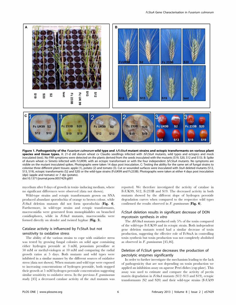
was confirmed to differ by 50% (Table 2, Fig. 1). The deletion of
the FcStuA gene from both strains caused a complete impairment
in pathogenicity. Statistical analysis indicated highly significant
(P, 0.001) reduction of FRR symptoms on durum wheat seedlings
for all 10 mutants tested, whilst the ectopic transformants did not
differ significantly from the wild-type reference strains (Table 2;
Fig. 1A). Also, in the floral infection tests, as previously reported
in F. graminearum [45], the FcStuA deletion caused the complete
absence of symptoms in the plant (Fig. 1B). Symptoms caused by
the non-toxigenic strains Fc233B were very mild but significantly
stronger than the respective mutant (Fig. 1B). In addition, the
FcStuA deletion mutants showed a reduced ability to colonise fruit
FcStuA Gene Characterisation in Fusarium culmorum
PLOS ONE | www.plosone.org 4 February 2013 | Volume 8 | Issue 2 | e57429

and tuber tissues of the non-cereal host plants, namely apple
tomato and potato (Fig. 1C). Collectively, these results further
strengthen the hypothesis that the disruption of the FcStuA gene
has a major impact on fungal virulence in F. culmorum.
To further investigate the lack of pathogenic behaviour, the
pathogenicity test used for FRR was simulated in vitro and observed
with electron scanning microscopy. As shown in Fig. 2, mycelium
of the wild-type strain entirely surrounded the seed by forming a
complex network of thick and sturdy hyphae, thus preventing
germination (Fig. 2A–2B), while the developing hyphae of
DFcStuA mutants did not hinder the emergence of the primary
root from the caryopsis (Fig. 2C–2D), suggesting that the
infection leading to FRR in our experimental setting is blocking
seed germination before coleoptile growth. This method based on
seed emergence in vitro gave identical results when compared to in
planta experiments proving to be as reliable as in planta experiments
at least for FcStuA deletions, suggesting a potential role for general
use of in vitro testing of FRR-impaired mutants. The DFcStuA
mutants did not kill the seeds and interesting in this assay, the two
wild types strains maintained the different level of aggressiveness,
namely FcUK99, the highly pathogenic isolate, killing most seeds,
and Fc233B, the less pathogenic isolate, killing ,50% of the seeds
(Fig. 3). While the wild type hyphae penetrated the seeds, the
mutants were unable to kill the seed and were washed away,
allowing the seed to germinate freely.
We also observed differences between mutant and wild-type
strains in the structure of the mycelium, especially concerning the
robustness of hyphae and the capability of the hyphae to colonise a
single seed of durum wheat. In addition, compared to both the
wild-type and ectopic strains, the DFcStuA mutants produced fewer
aerial mycelium and the hyphae were thin, winding and stunted
(Fig. 2).
FcStuA deletion induces phenotypic and morphologicalchanges in F. culmorum
FcStuA deletion mutants grown on PDA showed a phenotype
similar to F. graminearum mutants [45]. White pigment, sparse
mycelium, stunted growth and mycelium embedded in the solid
medium were typical phenotypic features of DFcStuA mutants.
Instead, ectopic transformants, that were resistant to hygromycin
B, presented a phenotype similar to the respective wild-type strain,
namely rapid growth, abundant aerial mycelium, presence of
white to yellow or pale orange pigmentation in the media which
became brown to red-brown in older cultures.
The mycelial surface of DFcStuA colonies was characterised by
low hydrophobicity unlike wild-type and ectopic strains: the ratio
between the time required to absorb a 20 mL drop of water by wild
types compared to their respective DFcStuA strains was approxi-
mately 10 for FcUK99 and 3 for Fc233B. In both cases water
penetrated into the mycelial surface more quickly in the gene-
deletion mutants (Table 2).
FcStuA deletion mutants grown in liquid CMC produced fewer
macroconidia/mL than ectopic transformants and wild-type
strains: conidiogenesis of DFcStuA mutants was reduced by 5 to
10 times (Table 2) when compared to original wild-types. DFcStuA
mutants showed a delayed germination of conidia compared to the
respective ectopic transformants and wild-type strains at 4 h.
However, the effect was minimal at 8 h, with all conidia
germinating at a similar level (Table 2). The delayed germination
of DFcStuA conidia was not linked to the ability of mycelium to
grow. In fact, fungal biomass of the mutants in pectolytic medium
was higher when compared to wild types and ectopics
(P›,›0.05). The phenomenon was not observed in cultured
Ta
ble
2.
Ph
en
oty
pic
me
asu
res
of
the
stra
ins
use
din
this
stu
dy.
FR
Rse
ve
rity
aF
RR
em
erg
en
ceb
FH
Bp
ath
og
en
icit
yc
Hy
dro
ph
ob
icit
yo
fth
em
yce
liu
md
Co
nid
iap
rod
uct
ion
at
8d
ay
se
Co
nid
iag
erm
ina
tio
na
t4
hrs
f
Co
nid
iag
erm
ina
tio
na
t8
hrs
g
DO
Np
rod
uct
ion
in(m
gp
er
mg
of
dry
we
igh
t)h
Po
lyg
ala
ctu
ron
ice
nz
ym
ep
rod
uct
ion
i
Stra
in
FcU
K9
9(w
ildty
pe
)1
00
.006
0.0
03
.36
5.8
9.5
86
0.7
2.
60
10
.026
2.3
97
36
69
6.3
36
2.0
56
.496
3.3
32
0.5
96
1.2
7
S2(e
cto
pic
)1
00
.006
0.0
00
.08
.666
1.2
1.
60
9.0
66
3.3
48
16
2.6
96
.006
2.9
49
.076
4.4
32
1.4
46
0.4
9
S12
(mu
tan
t)1
0.0
06
10
.00
*9
0.0
61
0.0
*06
0*
90
.946
0.7
4*
416
5.7
*9
4.0
06
2.9
40
.186
0.0
5*
17
.316
0.9
8*
S13
(mu
tan
t)1
0.0
06
10
.00
*9
0.0
61
0.0
*06
0*
90
.766
0.1
8*
556
2*
91
.676
2.3
60
.266
0.0
3*
17
.136
1.3
2*
Fc2
33
B(w
ildty
pe
)7
0.8
36
15
.07
43
.36
15
.33
.146
0.7
55
5.4
76
1.0
29
3,3
63
93
.006
2.8
3N
ot
me
asu
rab
le2
8.8
56
2.6
2
S20
(ect
op
ic)
69
.26
19
.45
3.3
61
5.3
2.5
86
0.2
21
37
.866
1.8
29
56
28
8.6
76
6.3
4N
ot
me
asu
rab
le2
8.4
66
2.0
7
S19
(mu
tan
t)6
.676
5.7
7*
93
.36
5.8
*06
0*
14
1.4
16
0.1
6*
49
.76
2.9
*8
8.6
76
5.7
3N
ot
me
asu
rab
le2
1.5
36
1.0
9*
Foo
tan
dro
ot
rot
seve
rity
me
asu
red
wit
hM
cKin
ne
yin
de
x(0
–1
00
)a
and
em
erg
en
ceb
test
ed
on
du
rum
wh
eat
cvC
lau
dio
;nu
mb
er
of
spik
es
infe
cte
dat
14
day
sab
ove
the
po
int
of
ino
cula
tio
n[3
8]c
;hyd
rop
ho
bic
ity
of
the
myc
eliu
mm
eas
ure
din
seco
nd
sn
ee
de
db
efo
reab
sorp
tio
no
fa
20
ml
wat
er
dro
pd
;mill
ion
so
fco
nid
iap
rod
uce
din
CM
Cm
ed
ium
ino
cula
ted
wit
h1
03
con
idia
afte
r5
day
sg
row
the;p
erc
en
tag
eo
fg
erm
inat
ion
of
con
idia
afte
r4
ho
urs
on
CM
f ;p
erc
en
tag
eo
fg
erm
inat
ing
con
idia
at8
hrs
(all
mu
tan
tssh
ow
ed
ad
ela
yed
ge
rmin
atio
nb
ut
no
sig
nif
ican
td
iffe
ren
cein
the
nu
mb
er
of
ge
rmin
atin
gco
nid
iaw
aso
bse
rve
d)g
;D
ON
pro
du
ctio
nin
toxi
nin
du
cin
gliq
uid
me
diu
mco
rre
cte
dfo
rm
go
fd
ryw
eig
ht
afte
r8
day
scu
ltu
rin
gh;p
oly
gal
actu
ron
ase
en
zym
eac
tivi
tym
eas
ure
dw
ith
ad
iffu
sio
nas
say
on
agar
ose
ge
l(‘‘
cup
pla
te’’)
;me
asu
rem
en
tso
fp
ect
oly
tic
en
zym
eac
tivi
tyin
dia
me
ter
(mm
)co
rre
cte
db
yd
ryfu
ng
alb
iom
ass
(mg
)i .*
P,
0.0
1b
yT
uke
y’s
po
st-h
oc
test
.A
llva
lue
sar
efo
llow
ed
by
SD.
Sig
nif
ican
td
iffe
ren
ces
com
par
ed
tore
spe
ctiv
ew
ildty
pe
are
mar
ked
wit
han
aste
risk
.d
oi:1
0.1
37
1/j
ou
rnal
.po
ne
.00
57
42
9.t
00
2
FcStuA Gene Characterisation in Fusarium culmorum
PLOS ONE | www.plosone.org 5 February 2013 | Volume 8 | Issue 2 | e57429
mycelium after 8 days of growth in toxin- inducing medium, where
no significant differences were observed (data not shown).
Wild-type strains and ectopic transformants grown on SNA
produced abundant sporodochia of orange to brown colour, while
FcStuA deletion mutants did not form sporodochia (Fig. 4).
Furthermore, in wild-type strains and ectopic transformants,
macroconidia were generated from monophialides on branched
conidiophores, while in FcStuA mutants, macroconidia were
formed directly on slender and twisted hyphae (Fig. 5).
Catalase activity is influenced by FcStuA but notsensitivity to oxidative stress
The ability of the various strains to cope with oxidative stress
was tested by growing fungal colonies on solid agar containing
either hydrogen peroxide at 3 mM, potassium persulfate at
50 mM or methyl-viologen at 10 mM and comparing the radial
growth ratios at 5 days. Both mutants and wild types were
inhibited in a similar manner by the different sources of oxidative
stress (data not shown). When mutants and wild type were exposed
to increasing concentrations of hydrogen peroxide, both stopped
their growth at 5 mM hydrogen peroxide concentration suggesting
similar sensitivity to oxidative stress. In the previous F. graminearum
study [45] a decreased catalase activity of the stuA mutants was
reported. We therefore investigated the activity of catalase in
FcUK99, S12, Fc233B and S19. The decreased activity in both
mutants showed by the different slope of hydrogen peroxide
degradation curves when compared to the respective wild types
confirmed the results observed in F. graminearum (Fig. 6).
FcStuA deletion results in significant decrease of DONmycotoxin synthesis in vitro
The DFcStuA mutants produced only 5% of the toxin compared
of the wild-type FcUK99 and its ectopic strain. Both independent
gene deletion mutants tested had a similar decrease of toxin
production, suggesting the effective role of FcStuA in controlling
toxin synthesis but toxin production was not completely abolished
as observed in F. graminearum [45,46].
Deletion of FcStuA gene decreases the production ofpectolytic enzymes significantly
In order to further investigate the mechanism leading to the lack
of pathogenicity that are not depending on toxin production we
applied an inhibition assay on agarose Petri dishes (cup plate). This
assay was used to estimate and compare the activity of pectin
matrix degradation in FcStuA mutants (S12–S13 and S19), ectopic
transformants (S2 and S20) and their wild-type strains (FcUk99
Figure 1. Pathogenicity of the Fusarium culmorum wild type and DFcStuA mutant strains and ectopic transformants on various plantspecies and tissue types. A: 21-d old durum wheat cv Claudio seedlings infected with DFcStuA mutants, wild types and ectopics and mockinoculated (test). No FRR symptoms were detected on the plants derived from the seeds inoculated with the mutants (S19, S20, S12 and S13). B: Spikeof durum wheat cv Simeto infected with FcUK99, with an ectopic transformant or with the four independent DFcStuA mutants. No symptoms arevisible on the mutant inoculated spikes. Photographs were taken 14 days post inoculation. C: Testing the ability for the same set of fungal strains tocolonise three different plant tissues: apple (1), potato (2) and tomato (3). Cut or wounded surfaces were inoculated with StuA deleted mutants (S12,S13, S19), ectopic transformants (S2 and S20) or the wild-type strains (FcUK99 and Fc233B). Photographs were taken at either 4 days post inoculation(dpi) (apple and tomato) or 7 dpi (potato).doi:10.1371/journal.pone.0057429.g001
FcStuA Gene Characterisation in Fusarium culmorum
PLOS ONE | www.plosone.org 6 February 2013 | Volume 8 | Issue 2 | e57429

Figure 2. Scanning electron microscope analysis of the infection of durum wheat seeds by Fusarium culmorum wild-type and DFcStuAmutant strains. These ZEISS EVO LS images reveal the appearance of the surface of a seed of durum wheat placed over a mycelium disk of eitherwild-type Fusarium culmorum FcUK99 (A and B) or its DFcStuA mutant (C and D), 3 days after incubation at 25uC. During observation the sampleswere subjected to the following conditions: temperature 2uC, humidity 85% and pressure 600 Pa. A and C panel show an apical seed view. B and Dshow a detail of the mycelium surrounding the seed.doi:10.1371/journal.pone.0057429.g002
Figure 3. Fusarium culmorum wild types and ectopic transformants penetrate into wheat seeds and kill the seedling while DFcStuAmutants are unable to penetrate the seed. Seed viability after 3 days in the dark at 25uC on water agar (germination) after previous pluginoculation for 3 days with the two wild types (FcUK99 and Fc233B), the mutants (S12, S13 and S19), the ectopic transformants (S2 and S20) and amock control inoculated with 10 mL water (W). Seeds were washed with NaClO before plating to eliminate external mycelium.doi:10.1371/journal.pone.0057429.g003
FcStuA Gene Characterisation in Fusarium culmorum
PLOS ONE | www.plosone.org 7 February 2013 | Volume 8 | Issue 2 | e57429

and Fc233B). The polygalacturonase enzyme activity was found to
be significantly different between the various non-pathogenic
DFcStuA mutants and their respective virulent wild-type control
strains (Table 2). This suggests that the FcStuA gene is involved in
the mechanisms of polygalacturonic enzyme secretion during
infection and colonisation of the host.
DFcStuA mutants have an altered glycolytic metabolismand are able to degrade various carbon sources todifferent extent
To further verify that the impairment in polygalacturonase
enzyme production was due to the activity of StuA controlled
genes, different substrates from various plant origins were used as
the sole carbon source. Growth diameter after 5 days ranged from
6 mm to 56 mm depending on the strain and the carbon source
(Fig 7). Highly significant (p,0.0001) effects of type of carbon
source and fungal strain as well as a highly significant interaction
of carbon source x fungal strain on fungal colony diameter were
observed. The growth of all mutants was reduced to about 80% of
the wild type strain when glutamic acid was the sole carbon source,
whereas the growth of the corresponding ectopic mutants was not
significantly different from the wild type (Fig. 7). The use of potato
azo-galactan, apple pectin and citrus pectin resulted in a
comparable growth similar to that observed with glutamic acid.
The growth of all mutants was reduced to about 55% of the wild
type strain when glucose was the sole carbon source, whereas the
growth of the ectopics was again not significantly different from
the wild type (Fig. 7). Beechwood xylan had a similar effect to
glucose. The response of the mutants towards sugarbeet arabinan
and polygalacturonic acid was variable and somewhere between
the low relative inhibition level observed for glutamic acid and the
high relative inhibition level observed for glucose (Fig. 7).
Fungicide sensitivity of DFcStuA mutants is not alteredsignificantly
The demethylase inhibitors epoxiconazole and tebuconzole
inhibited F. culmorum FcUK99 by 50% at concentrations of 0.0142
6 0.0030 and 0.0041 6 0.0012 mM, respectively (Fig. 8A, C). By
contrast, the complex II and complex III respiration inhibitors
isopyrazam and trifloxystrobin were unable to inhibit F. culmorum
FcUK99 and Fc233B up to a concentration of 1 mM (Fig. 8B,D). The ectopic strains S2 and S20 expressed the same fungicide
sensitivity profile as the wild type strains (Fig. 9). The fungicide
sensitivity of FcStuA deficient mutants was similar to the sensitivity
of the wild type strain and the ectopic transformants (Fig. 8, 9).
Discussion
This work reports on the identification of a pathogenicity factor
in F. culmorum that adds to the very few other genes known to play
a role in the pathogenic behaviour of this species [47,48]. Besides
the genes shown to contribute to trichothecene production [19],
this is only the second functionally characterised pathogenicity
gene involved in foot and root rot disease caused by a Fusarium
species [42].
FcStuA gene affects a large set of phenotypic charactersAPSES proteins, homologues of StuAp from Aspergillus nidulans
[49], were reported to control metabolic, morphological and
developmental stages in various fungal species (for details see:
[50]). This was confirmed in F. culmorum where FcStuA controls
both conidial germination confirming that budding factors are
influenced by this gene [51] and spore production, influences the
growth of aerial mycelium, the hydrophobicity of the mycelium
and its thickness, having overall a significant negative effect on the
fitness of the strains lacking the gene. The StuA protein is
necessary for the production of conidiophores and phialides as
shown previously [45,51,52]. The direct conidiation from hyphae
is probably the main reason for the reduced number of conidia
produced by the mutants.
The FcStuA gene sequence has a very high homology to the F.
graminearum FgStuA gene. The lack of significant amino acid
differences between the two sequences indicated an identical
structure of the two proteins. When comparing the similarity of
FgStuA and FcStuA to a general level of similarity between the two
species, at least with partial data obtained from a transposon
tagging approach [53], the level of conservation seems to be higher
than average: 98.5% vs 96%. It is therefore suggested that FcStuA
is under strong selective pressure having a crucial role in cell
regulation and therefore being highly conserved between the two
closely related species.
The morphological effects of the deletion of StuA in the two
species were found to be highly overlapping. However, in this
study and another [48], specific characteristics were identified that
differed between these two species. Identifying differences and
then their underlying genome basis will help to elucidate the genes
which play a role in the adaptation and fitness of the two
phytopathogens. For example, the overall reduction of conidia
production in the StuA mutants was quantitatively different
between F. graminearum (1:10000 drop) and F. culmorum (1:10
drop). This difference suggests that in F. culmorum compensation
Figure 4. DFcStuA mutants of Fusarium culmorum are unable to produce sporodochia. Sporodochia formation on SNA after 18 daysgrowth. A: FcUK99, B: Fc233B; C: S12 (mutant obtained from A); D: S19 (mutant obtained from D).doi:10.1371/journal.pone.0057429.g004
FcStuA Gene Characterisation in Fusarium culmorum
PLOS ONE | www.plosone.org 8 February 2013 | Volume 8 | Issue 2 | e57429

mechanisms and / or an alternative signalling pathway(s) for spore
production exist.
Few regulators are known to control secondary metabolites
production in fungi [54]. FcStuA was confirmed to regulate
secondary metabolite production in F. culmorum. While pigment
production was absent in both species suggesting that aurofusarin
and other pigments were not produced in the StuA mutants, the
level of deoxynivalenol (DON) mycotoxin production in vitro was
reduced differently between F. culmorum and F. graminerum. In F.
graminearum toxin production was either zero or below 1% of wild-
type levels [45,46] while in the present study F. culmorum mutants
produced 5% of DON, when compared to wild-type. The F.
graminearum strain PH-1 is a DON and 15ADON producer,
whereas FcUK99 is a DON and 3ADON producer [21]. It is
possible that these chemotype differences and/ or the media used
to induce DON production in vitro conditions may partially explain
the species differences observed between the two studies.
Nonetheless, overall FcStuA gene deletion seems to have milder
effects compared to the FgStuA gene deletion on DON mycotoxin
production in vitro.
Despite numerous attempts, it was impossible to complement
the wild-type gene back into the DFcStuA mutants. Direct co-
transformation with the gene and a plasmid carrying geneticin
resistance [34] was unsuccessful as well as the attempt to clone the
gene in a bacterial strain. The same phenomenon was observed on
FgStuA gene of F. graminearum [45], as well as for U. maydis [55] and
Stagonospora nodorum [56]. Despite the lack of available StuA
complemented F. culmorum strains for comparative purposes, we
Figure 5. An in vitro comparison of the asexual development of the wild-type and DFcStuA mutant strains of Fusarium culmorum.Conidiogenesis by StuA deletion mutants, ectopic transformants and wild-type strains was observed with a light microscope (OLYMPUS BX41).Macroconidia of the wild-type FcUK99 strain (A) and the ectopic S20 strain (B) are formed from monophialides on branched conidiophores.Macroconidia of the DFcStuA mutant strains spores are generated directly from hyphae as in S12 and S13 (StuA mutants). (B) The same observationswere made for FcStuA deletion mutant S19 obtained from strain Fc233B. Photos were taken with MOTICAM 2500 5.O MP live resolution (Motic).Hyphal diameter is smaller in the mutant.doi:10.1371/journal.pone.0057429.g005
FcStuA Gene Characterisation in Fusarium culmorum
PLOS ONE | www.plosone.org 9 February 2013 | Volume 8 | Issue 2 | e57429

are confident that all the effects presented here are solely due to
the lack of the FcStuA transcription factor for the following two
reasons: (1) all mutants showed the same phenotype, while both
ectopic strains were similar to respective wild-types, (2) the effects
of FcStuA gene loss were confirmed in the genetic background of
two F. culmorum strains with different geographic origins and
different mycotoxin chemotypes.
How could FcStuA control pathogenicity on wheat?The identification and functional characterisation of a tran-
scription factor containing a highly conserved bHLH-like APSES
domain [6] in F. culmorum was carried out in order to investigate
the role of the gene in a previously untested pathogenicity
mechanism during seed germination/initial seedling plant estab-
lishment. The gene is known to be a pathogenicity factor for many
fungal species, including F. graminearum [45] and wheat heads,
Ustilago maydis and maize leaves [55], Glomerella cingulata and apple
fruit [57] and Stagonospora nodorum and wheat leaves [56]. These
fungi are not taxonomically closely related and also have diverse in
planta lifestyle including biotrophy, hemibiotrophy and necrotro-
phy.
In comparison, in the taxonomically more closely related species
F. oxysporum f. sp. lycopersici, causal agent of root rot of tomato, the
FoStuA deletion mutant was not affected in its virulence towards
tomato plants and root and stem tissues [52]. Given the different
role(s) played by StuA homologues in F. graminearum and F.
oxysporum (the two most closely related organisms to F. culmorum) we
investigated the effect of the gene in F. culmorum combining two
different cereal pathosystems (head infection and foot and root rot
infection of wheat) and three non-cereal hosts (tomato fruit, apple
fruit and potato tuber). The lack of symptoms following
inoculation with each of the DFcStuA mutants tested proved that
FcStuA is essential for colonisation of the tissues in all five plant
cases. By comparing the pathogenic behaviour of FcStuA mutants
obtained from a toxin producer and a non-toxin producer strain it
was possible to infer a role for DON toxin in pathogenicity assays.
In both genetic backgrounds, a loss of pathogenicity was obtained
when FcStuA was deleted and mild symptoms were caused by the
wild type non-toxin producer strain on wheat heads and on wheat
seedlings confirming that DON production may not be essential
for causing FRR symptoms [20]. Interestingly, the potential ability
to produce small amounts of toxin in the FcUK99 DFcStuA mutant
Figure 6. Catalase activity is reduced in the DFcStuA mutants of Fusarium culmorum. Linear phase of degradation of H2O2 over time inferredby reduced light absorbance at 240 nm. Ten microliter of a spore suspension (106 mL21) of FcUK99 and DFcStuA S12 (A) and Fc233B and DFcStuA S19were suspended in a solution of 3.5 mM H2O2. Absorbance of the solution was read every minute for a total of 80 min. Results show that wild typescatalyse H2O2 more efficiently than DFcStuA respective mutants.doi:10.1371/journal.pone.0057429.g006
FcStuA Gene Characterisation in Fusarium culmorum
PLOS ONE | www.plosone.org 10 February 2013 | Volume 8 | Issue 2 | e57429

did not induce significant symptoms, further suggesting that the
lack of pathogenicity towards both germinating seeds and wheat
floral tissue is stopped by other mechanisms directly and / or
indirectly controlled by the FcStuA protein. This conclusion is
further supported the environmental Scanning Electron Micro-
scope (envSEM) observations. The images obtained revealed the
inability of the DFcStuA mutant to consistently attach on the seed
Figure 7. Growth of wild types, mutants and ectopic strains on different carbon sources obtained from various plant origins. Colonydiameter in mm and standard deviation are indicated on the left and refer to the average of 3 biological replicates. The graph indicates the effect ofcarbon source on the growth ratio (mutant / wild type) after 5 days of growth on Petri dishes containing glucose, beechwood xylan, sugar beetarabinan, polygalacturonic acid, citrus pectin, apple pectin, potato azo-galactan or glutamic acid as a sole carbon source. Bars labelled with the sameletter are not significantly different according to Tukey’s post hoc test p,0.05. Bars represent SDs of the ratios (within each strain) of three biologicalreplicates.doi:10.1371/journal.pone.0057429.g007
FcStuA Gene Characterisation in Fusarium culmorum
PLOS ONE | www.plosone.org 11 February 2013 | Volume 8 | Issue 2 | e57429

and inhibit seed germination while it is known that toxin is
important during the later stage of infection.
Virulence is a highly complex character [58] that can be
influenced by different molecular mechanisms. Therefore we
attempted to decipher which mechanism could be controlled by
FcStuA. Several morphological differences caused by deletion of
FcStuA that may impact on disease severity were observed. These
included slow growth on solid medium as well as reduced
colonisation of the cut surfaces of apple, potato and tomato tissues
and on the wheat seed surface. On the contrary growth rate was
not decreased in liquid medium. Reduced expression of hydro-
phobins [59] as observed in F. graminearum and Glomerella cingulata
[45,57] may indeed explain the difference of growth in different
media and have a direct effect on the ability to cause disease [60].
To further investigate the possible cause(s) for the inability of
DFcStuA mutants to colonise wheat seeds and colonise wheat stem
base tissue efficiently in both parental backgrounds, polygalactu-
ronase enzyme production was investigated. In several other
pathosystems, this enzyme activity is considered to play a crucial
role in penetration of plant cells and therefore to influence early
pathogenic behaviour [61,62,63,64,65]. By calculating polygalac-
turonase production per unit dry mass of mycelium, we could
correlate total polygalacturonase activity to the amount produced
per gram of mycelium. Both types of DFcStuA mutants produced a
lower amount of enzyme. However, a reduced polygalacturonases
activity in the mutants is unlikely to be the main factor
determining decreased pathogenicity. For example, efficient
growth of the DFcStuA mutants was observed in vitro on the
different pectin-like carbon sources from apple and citrus as well as
on azo-galactan from potato whereas the mutants were unable to
efficiently colonise apples and potato slices. While we could
confirm that glycolytic process is inhibited in the StuA mutants,
pectolitic enzymes necessary to degrade the complex set of pectin –
like compounds [66], as tested in this study, are only partially
regulated (directly or indirectly) by FcStuA gene such as xylan
degrading enzymes. Therefore it is confirmed that pectin enzyme
sets are controlled by a diverse set of regulatory circuits [67].
Catalase activity was also significantly decreased in the DFcStuA
mutant tested here confirming previous results produced for the
corresponding FgStuA mutants [45]. DFcStuA mutants response to
the oxidative stress does not differ from the wild type excluding
that, during plant infection and colonisation, the fungal cells
lacking StuA cope less efficiently with oxidative stress resulting
from plant defense mechanisms. Therefore it is probable that the
decreased catalase activity does not have an influence on the
decreased pathogenicity of the mutants. Due to the reduced DON
production by the DFcStuA mutants, minimisation of the induction
of plant defense responses, via inhibition of host protein
translation, is likely to be low. It can be that the reduced catalase
activity and the inability to produce toxin belong to the same
Figure 8. The sensitivity of the highly pathogenic wild-type FcUK99, ectopic and FcStuA deletion mutants to various fungicides. Theoptical density of liquid cultures of the Fusarium culmorum wild type strain FcUK99, an ectopic and FcStuA deficient strains (S9, S12, S13) growing in200 mL of 12.5% (w/w) potato dextrose broth when augmented with different concentrations of fungicides (A) epoxiconazole, (B) isopyrazam, (C)tebuconazole, or (D) trifloxystrobin. The optical density measurements were made after 5 days of incubation at 120 rpm and 22uC in the dark. Anoptical density close to 100% indicates no sensitivity to the fungicide, whilst an optical density close to 0% full inhibition. Error bars represent thestandard error of 3 replicates.doi:10.1371/journal.pone.0057429.g008
FcStuA Gene Characterisation in Fusarium culmorum
PLOS ONE | www.plosone.org 12 February 2013 | Volume 8 | Issue 2 | e57429

molecular pathway linking environmental sensing and toxin
production [68,69,70] in the fungus and that FcStuA is controlling
genes upstream in this pathway.
It is not possible to pinpoint which compromised mechanism or
combination of compromised mechanisms is responsible for the
initial lack of seed penetration and / or young seedling
colonisation. However, from the phenotypic data presented here,
the lack of the StuA protein in F. culmorum causes the very early
arrest of the infection process.
FcStuA as a potential target for novel chemicalsThe functional characterisation of genes controlling pathoge-
nicity and developmental stages on fungal plant pathogens is an
important strategy for potential development of novel molecules
able to control pathogen growth and infection. Indeed, cellular
controllers of fungal fitness, possibly specific to the fungal domain
as APSES proteins [50], are ideal targets for the design of new
fungicides [71].
As shown here, FcStuA is controlling pathogenicity via a set of
different mechanisms that are not exclusively linked to the toxin
production making it a perfect target for novel molecules with a
fungistatic activity combined with a toxin blocking production
activity. Given the emergence of resistance phenomena, the use of
novel fungicide combinations and the adoption of multiple active
compounds, is the most efficient solution at the moment [72]. It
was therefore of interest to know if putative molecules that target
FcStuA transcription factors, simulated here via the complete
elimination of the gene, may have any negative effect on the onset
of inhibition to specific classes of fungicides [73]. Therefore we
studied the level of resistance by the mutants to three classes of
fungicides widely used in agriculture. Fusarium species are partially
sensitive towards fungicides belonging to the group of demethylase
inhibitors such as epoxiconazole and tebuconazole, but are
intrinsically resistant towards complex III respiration inhibitors
such as trifloxystrobin [44]. We observed no significant effect (in
both direction: i.e. resistance or susceptibility) caused by the
deletion of the FcStuA transcription factor suggesting that the
protein is not directly involved in controlling the processes of
intrinsic fungicide resistance to strobilurins or partial susceptibility
to azoles. It is noteworthy that the two wild type F. culmorum strains
tested for growth in the presence of the respiration inhibitor
isopyrazam, a recently introduced compound revealed that this
species is also intrinsically resistant to complex II inhibitors as
already observed in F. graminearum [74].
Our result suggests that a putative molecule able to target the
specificities of the FcStuA protein, would not increase the risk of
resistance against other known fungicides. Given the restricted
spectrum of available molecules against Fusarium species it seems
promising to suggest APSES proteins as novel targets for
developing new pathogen controlling measures.
Conclusions
Our work presented evidence, for the first time in F. culmorum,
that FcStuA controls several pathways within the fungal cell that
Figure 9. The sensitivity of the mildly pathogenic wild-type Fc233B, ectopic and FcStuA deficient strains to various fungicides.Optical density of the Fusarium culmorum wild-type strain Fc233B, an ectopic (S20) and a FcStuA deficient strain (S19) in liquid cultures (200 mL,medium: 12.5% (w/w) potato dextrose broth) as affected by epoxiconazole (A), isopyrazam (B), tebuconazole (C), or trifloxystrobin (D) concentrationafter 5 days of incubation at 120 rpm and 22uC in the dark. An optical density close to 100% indicates no sensitivity to the fungicide, whilst an opticaldensity close to 0% full inhibition. Error bars represent the standard error of 3 replicates.doi:10.1371/journal.pone.0057429.g009
FcStuA Gene Characterisation in Fusarium culmorum
PLOS ONE | www.plosone.org 13 February 2013 | Volume 8 | Issue 2 | e57429
directly or indirectly regulate morphological development, the
glycolytic process, catalase activity, exopectinase and pectin like
metabolism and trichothecene mycotoxin production. By using
two strains differing for virulence and toxin production we were
able to show that pathogenic behaviour is not simply determined
by the lack of toxin production in DFcStuA mutants. As in other
plant pathogenic fungi able to produce toxins that are involved in
the pathogenic process (F. graminearum, S. nodorum, U. maydis)
pathogenicity was impaired, while in F. oxysporum - a species not yet
known to produce toxin-like pathogenicity factors - the pathogenic
process was not altered. We therefore investigated the role of toxin
production in determining F. culmorum DFcStuA pathogenicity. Our
data suggest that the toxinogenic process is not the determinant of
pathogenicity impairment in the deletion mutant. We showed that
mechanisms that may determine the lack of pathogenicity in
FcStuA mutants result in early blocking of the infection process.
Decreased conidial germination efficiency (hence decreasing the
inoculum) and reduced robustness of mycelium probably deter-
mine the inability to overcome plant defense mechanisms by the
fungus.
Future studies focusing on the interaction of this transcription
factor with other regulatory proteins may shed light on the
regulatory pathways that are involved in shaping the different
aspects of the fungual phenotype. As a molecule targeting the StuA
protein would induce very low selective pressure (no biocide
activity) but would decrease virulence and toxin production
without increasing the risk of resistance to other known fungicides,
the exploration of compounds able to interact with this protein are
of interest for developing novel control strategies.
Supporting Information
Figure S1 (A) FcStuA gene deletion procedure. The name and
location of each primer used in split marker recombination are
marked (scheme adapted from [30]). (B) Agarose gel electropho-
resis of PCR products obtained from StuA mutant strain (S19),
ectopic transformant (S20) and wild-type strain (Fc233B). Primers
stuA NF and stuA NR were used to verify the FcStuA gene
deletion. The DFcStuA mutants lacked the PCR band of 401 bp
corresponding to the endogenous FcStuA gene, while this fragment
was amplified in the wild-type strain (pink box) and in ectopic
transformant (green box). The primers ITS1 and ITS4 were used
as an internal control for DNA quality. The gel on the right shows
results of PCR analysis with the following primer pairs: StuA 1F-
StuA 4R, StuA 1F-StuA NR, StuA NF-StuA 4R used to confirm the
deletion of StuA gene. (C) Independent confirmation of the specific
deletion of the FcStuA gene sequence was achieved by Southern
blot analysis of 11 StuA deleted transformants. Genomic DNAs
were digested with EcoRV and after transfer of the DNA to the
membrane, the hybridisation was done by labelling a partial gene
stuA specific probe (401 bp). The fragment sizes expected were:
3,400 bp and 2,240 bp, respectively, for DFcStuA mutants and for
control (wild-type strains and ectopic transformants).
(TIF)
Figure S2 Alignment of FcStuA protein with homologuesobtained from three other Fusarium species. The
sequence is 98.5% identical to F. graminearum protein
(FGSG_10129) with the 2 amino acid differences not linked to
any particular functional role. The blue arrow shows the
conserved APSES domain.
(TIF)
Acknowledgments
Dr. Salvatore Marceddu is acknowledged for the assistance with the
Scanning Electron Microscopy and Dr. Daniele Sanna for spectrophoto-
metric measurements. We thank Dr. John Antoniw for screening the
unassembled UK99 F. culmorum genome for sequences homologous to
FgStuA. Angela Marcello is acknowledged for excellent technical assistance.
Author Contributions
Conceived and designed the experiments: MP QM MB. Performed the
experiments: FS MP MB VB BS. Analyzed the data: MP FS MB VB QM.
Contributed reagents/materials/analysis tools: QM BS LH KHK. Wrote
the paper: MP FS KHK QM MB LH.
References
1. Miller KY, Toennis TM, Adams TH, Miller BL (1991) Isolation and
transcriptional characterization of a morphological modifier: the Aspergillus
nidulans stunted (stuA) gene. Mol Gen Genet 227: 285–292.
2. Dutton JR, Johns S, Miller BL (1997) StuAp is a sequence-specific transcription
factor that regulates developmental complexity in Aspergillus nidulans. EMBO J
16: 5710–5721.
3. Stoldt VR, Sonneborn A, Leuker C, Ernst JF (1997) Efg1, an essential regulator
of morphogenesis of the human pathogen Candida albicans, is a member of a
conserved class of bHLH proteins regulating morphogenetic processes in fungi.
EMBO J 16: 1982–1991.
4. Massari ME, Murre C (2000) Helix-loop-helix proteins: regulators of
transcription in eucaryotic organisms. Mol Cell Biol 20: 429–440.
5. Iyer LM, Koonin EV, Aravind L (2002) Extensive domain shuffling in
transcription regulators of DNA viruses and implications for the origin of
fungal APSES transcription factors. Genome Biol 3: 1–11.
6. Aramayo R, Peleg Y, Addison R, Metzenberg R (1996) Asm-1, a Neurospora crassa
gene related to transcriptional regulators of fungal development. Genetics 144:
991–1003.
7. Gimeno CJ, Fink GR (1994) Induction of pseudohyphal growth by
overexpression of PHD1, a Saccharomyces cerevisiae gene related to transcriptional
regulators of fungal development. Mol Cell Biol 14: 2100–2112.
8. Wu J, Miller BL (1997) Aspergillus asexual reproduction and sexual reproduction
are differentially affected by transcriptional and translational mechanisms
regulating stunted gene expression. Mol Cell Biol 17: 6191–6201.
9. Ward MP, Gimeno CJ, Fink GR, Garrett S (1995) SOK2 may regulate cyclic
AMP-dependent protein kinase-stimulated growth and pseudohyphal develop-
ment by repressing transcription. Mol Cell Biol 15: 6854–6863.
10. Merson-Davies LA, Odds FC (1992) Expansion of the Candida albicans cell
envelope in different morphological forms of the fungus. J Gen Microbiol 138:
461–466.
11. Scherm B, Balmas V, Spanu F, Pani G, Delogu G, et al. (2013) Fusarium
culmorum: the causal agent of foot and root rot and head blight on wheat. Mol
Plant Pathol doi:10.1111/mpp.12011
12. Wagacha JM, Muthomi JW (2007) Fusarium culmorum: infection process,
mechanisms of mycotoxin production and their role in pathogenesis in wheat.
Crop Prot 26: 877–885.
13. Marasas WFO, Nelson PE, Toussoun TA (1984) Toxigenic Fusarium species:
identity and mycotoxicology. The Pennsylvania State University Press. 328 p.
14. Proctor RH, Hohn TM, McCormick SP (1995) Reduced virulence of Gibberella
zeae caused by disruption of a trichothecene toxin biosynthetic gene. Mol Plant-
Microbe Interact 8: 593–601.
15. Maier FJ, Miedaner T, Hadeler B, Felk A, Salomon S, et al. (2006) Involvement
of trichothecenes in fusarioses of wheat, barley and maize evaluated by gene
disruption of the trichodiene synthase (Tri5) gene in three field isolates of
different chemotype and virulence. Mol Plant Pathol 7: 449–461.
16. Cuzick A, Urban M, Hammond-Kosack KE (2008) Fusarium graminearum gene
deletion mutants map1 and tri5 reveal similarities and differences in the
pathogenicity requirements to cause disease on Arabidopsis and wheat floral tissue.
New Phytol 177: 990–1000.
17. Beccari G, Covarelli L, Nicholson P (2011) Infection processes and soft wheat
response to root rot and crown rot caused by Fusarium culmorum. Plant Pathol 60:
671–684.
18. Malalasekera RAP, Sanderson FR, Colhoun J (1973) Fusarium diseases of cereals
XI. Penetration and invasion of wheat seedlings by Fusarium culmorum and F.
nivale. Trans Brit Mycol Soc 60: 453–462.
19. Scherm B, Orru M, Balmas V, Spanu F, Azara E, et al. (2011) Altered
trichothecene biosynthesis in TRI6-silenced transformants of Fusarium culmorum
influences the severity of crown and foot rot on durum wheat seedlings. Mol
Plant Pathol 12: 759–771.
20. Mudge AM, Dill-Macky R, Dong Y, Gardiner DM, White RG, et al. (2006) A
role for the mycotoxin deoxynivalenol in stem colonisation during crown rot
FcStuA Gene Characterisation in Fusarium culmorum
PLOS ONE | www.plosone.org 14 February 2013 | Volume 8 | Issue 2 | e57429

disease of wheat caused by Fusarium graminearum and Fusarium pseudograminearum.
Physiol Mol Plant Pathol 69: 73–85.21. Lowe RGT, Allwood JW, Galster A, Urban M, Daudi A, et al. (2010) A
combined H NMR and ESI-MS analysis to understand the basal metabolism of
plant pathogenic Fusarium species. Mol Plant-Microbe Interact 23: 1605–1618.22. Giraud F, Pasquali M, El Jarroudi M, Vrancken C, Brochot C, et al. (2010)
Fusarium head blight and associated mycotoxin occurrence on winter wheat inLuxembourg in 2007/2008. Food Addit Contam Part A Chem Anal Control
Expo Risk Assess 27: 825–835.
23. Jiao F, Kawakami A, Nakajima T (2008) Effects of different carbon sources ontrichothecene production and Tri gene expression by Fusarium graminearum in
liquid culture. FEMS Microbiol Lett 285: 212–219.24. Gardiner DM, Kazan K, Manners JM (2009) Novel genes of Fusarium
graminearum that negatively regulate deoxynivalenol production and virulence.Mol Plant-Microbe Interact 22: 1588–1600.
25. Pasquali M, Giraud F, Lasserre JP, Planchon S, Hoffmann L, et al. (2010) Toxin
induction and protein extraction from Fusarium spp. cultures for proteomicstudies. J Vis Exp doi:10.3791/1690
26. Nirenberg HI (1990) Recent advances in the taxonomy of Fusarium. Stud Mycol32: 91–101.
27. Cappellini RA, Peterson JL (1965) Macroconidium formation in submerged
cultures by a non-sporulating strain of Gibberella zeae. Mycologia 57: 962–966.28. Correll JC, Klittich CJR, Leslie JF (1987) Nitrate non-utilizing mutants of
Fusarium oxysporum and their use in vegetative compatibility tests. Phytopathology77: 1640–1646.
29. Szecsi A (1990) Analysis of pectic enzyme zymograms of Fusarium species II.Comparison of polygalacturonase zymograms of Fusarium culmorum and Fusarium
graminearum. J Phytopathol 130: 188–196.
30. Taylor RJ, Secor GA (1998) An improved diffusion assay for quantifying thepolygalacturonase content of Erwinia culture filtrates. Phytopathology 78: 1101–
1103.31. Benoit I, Coutinho P, Schols H, Gerlach J, Henrissat B, et al. (2012)
Degradation of different pectins by fungi: correlations and contrasts between
the pectinolytic enzyme sets identified in genomes and the growth on pectins ofdifferent origin. BMC Genomics 13: 321.
32. Catlett NL, Lee BN, Yoder OC, Turgeon BG (2003) Split-marker recombina-tion for efficient targeted deletion of fungal genes. Fungal Genet Newsl 50: 9–11.
33. Langin T, Daboussi MJ, Gerlinger C, Brygoo Y (1990) Influence of biologicalparameters and gene transfer technique on transformation of Fusarium oxysporum.
Curr Genet 17: 313–319.
34. Breakspear A, Pasquali M, Broz K, Dong Y, Kistler HC (2011) Npc1 is involvedin sterol trafficking in the filamentous fungus Fusarium graminearum. Fungal Genet
Biol 48: 725–730.35. Pasquali M, Giraud F, Brochot C, Cocco E, Hoffmann L, et al. (2010) Genetic
Fusarium chemotyping as a useful tool for predicting nivalenol contamination in
winter wheat. Int J Food Microbiol 137: 246–253.36. Migheli Q, Friard O, Del Tedesco D, Musso MR, Gullino ML (1996) Stability of
transformed antagonistic Fusarium oxysporum strains in vitro and in soil microcosms.Mol Ecol 5: 641–649.
37. Sambrook J, Fritsch EF, Maniatis T (1989) Molecular Cloning: A LaboratoryManual 2nd edition. Cold Spring Harbor Laboratory Press.
38. Balmas V, Delogu G, Esposito S, Rau D, Migheli Q (2006) Use of a
complexation of tebuconazole with b-cyclodextrin for controlling foot and crownrot of durum wheat incited by Fusarium culmorum. J Agric Food Chem 54: 480–
484.39. McKinney HH (1923) Influence of soil temperature and moisture on infection of
wheat seedlings by Helminthosporium sativum. J Agric Res 26: 195–217.
40. Seong KY, Pasquali M, Zhou X, Song J, Hilburn K, et al. (2009) Global generegulation by Fusarium transcription factors Tri6 and Tri10 reveals adaptations
for toxin biosynthesis. Mol Microbiol 72: 354–367.41. Di Pietro A, Garcıa-MacEira FI, Meglecz E, Roncero MI (2001) A MAP kinase
of the vascular wilt fungus Fusarium oxysporum is essential for root penetration and
pathogenesis. Mol Microbiol 39: 1140–1152.42. Urban M, Mott E, Farley T, Hammond-Kosack KE (2003) The Fusarium
graminearum MAP1 gene is essential for pathogenicity and development ofperithecia. Mol Plant Pathol 4: 347–359.
43. Skovgaard K, Rosendahl S, O’Donnell K, Nirenberg HI (2003) Fusarium commune
is a new species identified by morphological and molecular phylogenetic data.
Mycologia 95: 630–636.
44. Dubos T, Pasquali M, Pogoda F, Hoffmann L, Beyer M (2011) Evidence fornatural resistance towards trifloxystrobin in Fusarium graminearum. Eur J Plant
Pathol 130: 239–248.45. Lysøe E, Pasquali M, Breakspear A, Kistler HC (2011) The transcription factor
FgStuAp influences spore development, pathogenicity, and secondary metabo-
lism in Fusarium graminearum. Mol Plant-Microbe Interact 24: 54–67.46. Son H, Seo YS, Min K, Park AR, Lee J et al. (2011) A phenome-based
functional analysis of transcription factors in the cereal head blight fungus,Fusarium graminearum. PLoS Pathog 7: e1002310.
47. Skov J, Lemmens M, Giese H (2004) Role of a Fusarium culmorum ABCtransporter (FcABC1) during infection of wheat and barley. Physiol Mol Plant
Pathol 64: 245–254.
48. Baldwin TK, Urban M, Brown N, Hammond-Kosac K (2010) A role fortopoisomerase I in Fusarium graminearum and F. culmorum pathogenesis and
sporulation. Mol Plant-Microbe Interact 23: 566–577.
49. Miller KY, Wu JG, Miller BL (1992) Stua is required for cell pattern formation in
Aspergillus. Genes Dev 6: 1770–1782.
50. Ramirez-Zavala B, Dominguez A (2008) Evolution and phylogenetic relation-ships of APSES proteins from Hemiascomycetes. FEMS 8: 511–519.
51. Borneman AR, Hynes MJ, Andrianopoulos A (2002) A basic helix-loop-helix
protein with similarity to the fungal morphological regulators, Phd1p, Efg1p and
StuA, controls conidiation but not dimorphic growth in Penicillium marneffei. MolMicrobiol 44: 621–631.
52. Ohara T, Tsuge T (2004) FoSTUA, encoding a basic helix-loop-helix protein,
differentially regulates development of three kinds of asexual spores, macroco-nidia, microconidia, and chlamydospores, in the fungal plant pathogen Fusarium
oxysporum. Eukaryot Cell 3: 1412–1422.
53. Spanu F, Pasquali M, Scherm B, Balmas V, Marcello A, et al. (2012)
Transposition of the miniature inverted-repeat transposable element mimp1 inthe wheat pathogen Fusarium culmorum. Mol Plant Pathol 13: 1149–1155.
54. Yin W, Keller NP (2011) Transcriptional regulatory elements in fungal
secondary metabolism. J Microbiol 49: 329–339.
55. Garcıa-Pedrajas MD, Baeza-Montanez L, Gold SE (2010) Regulation of Ustilago
maydis dimorphism, sporulation, and pathogenic development by a transcriptionfactor with a highly conserved APSES domain. Mol Plant-Microbe Interact 23:
211–222.
56. IpCho SVS, Tan K-C, Koh G, Gummer J, Oliver RP, et al. (2010) Thetranscription factor stuA regulates central carbon metabolism, mycotoxin
production, and effector gene expression in the wheat pathogen Stagonospora
nodorum. Eukaryot Cell 9: 1100–1108.
57. Tong XZ, Zhang XW, Plummer KM, Stowell KM, Sullivan PA, et al. (2007)GcSTUA, an APSES transcription factor, is required for generation of
appressorial turgor pressure and full pathogenicity of Glomerella cingulata. MolPlant-Microbe Interact 20: 1102–1111.
58. Sacristan S, Garcıa-Arenal F (2008) The evolution of virulence andpathogenicity in plant pathogen populations. Mol Plant Pathol 9: 369–384.
59. Teertstra WR, Deelstra HJ, Vranes M, Bohlmann R, Kahmann R, et al. (2006)
Repellents have functionally replaced hydrophobins in mediating attachment toa hydrophobic surface and in formation of hydrophobic aerial hyphae in Ustilago
maydis. Microbiology 152: 3607–3612.
60. Kim S, Ahn IP, Rho HS, Lee YH (2005) MHP1, a Magnaporthe grisea
hydrophobin gene, is required for fungal development and plant colonization.Mol Microbiol 57: 1224–1237.
61. Kang Z, Buchenauer H (2000) Ultrastructural and cyto-chemical studies on
cellulose, xylan and pectin degradation in wheat spikes infected by Fusarium
culmorum. J Phytopathol 148: 263–275.
62. Jaroszuk-Scisel J, Kurek E, Slomka A, Janczarek M, Rodzik B (2011) Activitiesof cell wall degrading enzymes in autolyzing cultures of three Fusarium culmorum
isolates: growth-promoting, deleterious and pathogenic to rye (Secale cereale).Mycologia 103: 929–945.
63. Aleandri MP, Magro P, Chilosi G (2007) Modulation of host pH during wheat-
Fusarium culmorum interaction and its influence on pectolytic enzymes production
and activity. Plant Pathol 56: 517–525.
64. Aleandri MP, Magro P, Chilosi G (2008) Influence of environmental pHmodulation on efficiency of apoplastic PR proteins during Fusarium culmorum-
wheat seedling interaction. Plant Pathol 57: 1017–1025.
65. Oeser B, Heidrich PM, Muller U, Tudzynski P, Tenberge KB (2002)
Polygalacturonase is a pathogenicity factor in the Claviceps purpurea/ ryeinteraction. Fungal Genet Biol 36: 176–186.
66. van den Brink J, de Vries RP (2011) Fungal enzyme sets for plant polysaccharide
degradation. Appl Microbiol Biotechnol 91: 1477–1492.
67. De Groot MJL, van de Vondervoort PJI, de Vries RP, van Kuyk P, Ruijter GJG,
et al. (2003) Isolation and characterization of two specific regulatory Aspergillus
niger mutants shows antagonistic regulation of arabinan and xylan metabolism.
Microbiology 149: 1183–1191.
68. Lee BN, Kroken S, Chou DY, Robbertse B, Yoder OC, et al. (2005) Functionalanalysis of all nonribosomal peptide synthetases in Cochliobolus heterostrophus
reveals a factor, NPS6, involved in virulence and resistance to oxidative stress.
Eukaryot Cell 4: 545–555.
69. Ponts N, Couedelo L, Pinson-Gadais L, Verdal-Bonnin MN, Barreau C, et al.(2009) Fusarium response to oxidative stress by H2O2 is trichothecene chemotype-
dependent. FEMS Microbiol Lett 293: 255–262.
70. Reverberi M, Ricelli A, Zjalic S, Fabbri AA, Fanelli C (2010) Natural functionsof mycotoxins and control of their biosynthesis in fungi. Appl Microbiol
Biotechnol 87: 899–911.
71. Fernandez Acero FJ, Carbu M, El-Akhal MR, Garrido C, Gonzalez-Rodrıguez
VE, et al. (2011) Development of proteomics-based fungicides: new strategies forenvironmentally friendly control of fungal plant diseases. Int J Mol Sci 12: 795–
816.
72. Cools HJ, Hammond-Kosack KE (2012) Exploitation of genomics in fungicide
research: current status and future perspectives. Mol Plant Pathol 14:197–210.
73. Kretschmer M, Leroch M, Mosbach A, Walker AS, Fillinger S, et al. (2009)Fungicide-driven evolution and molecular basis of multidrug resistance in field
populations of the grey mould fungus Botrytis cinerea. PLoS Pathog 5: e1000696.
74. Dubos T, Pasquali M, Pogoda F, Casanova A, Hoffmann L, et al. (2013)
Differences between the succinate dehydrogenase sequences of isopyrazamsensitive Zymoseptoria tritici and insensitive Fusarium graminearum strains. Pest
Biochem Physiol: 10.1016/j.pestbp.2012.11.004.
FcStuA Gene Characterisation in Fusarium culmorum
PLOS ONE | www.plosone.org 15 February 2013 | Volume 8 | Issue 2 | e57429