enhanced photocatalytic degradation of tetracycline ... - plos
TRANSCRIPT
RESEARCH ARTICLE
Enhanced photocatalytic degradation of
tetracycline hydrochloride over Au-doped
BiOBr nanosheets under visible light
irradiation
Chu-Ya WangID*, Xin Fang, Qi Zeng, Heng-Deng Zhou, Yongze LuID
School of Energy and Environment, Southeast University, Nanjing, China
Abstract
Bismuth(III) oxybromide (BiOBr) is a typical photocatalyst with a unique layered structure.
However, the response of BiOBr to visible light is not strong enough for practical application.
Moreover, the charge separation efficiency of BiOBr still needs to be improved. In this study,
series of Au-doped BiOBr photocatalysts was prepared through a facile one-step hydrother-
mal method. The as-prepared Au0.3-BiOBr nanosheets exhibited an excellent electrochemi-
cal performance. The charge separation efficiency of Au0.3-BiOBr nanosheets was
enhanced by 18.5 times compared with that of BiOBr. The intrinsic photocatalytic activity of
Au0.3-BiOBr nanosheets in the degradation of tetracycline hydrochloride was approximately
twice higher than that of BiOBr under visible light irradiation. In addition, three pathways
were identified for the photocatalytic degradation and mineralization of tetracycline hydro-
chloride, which involve four reactions: hydroxylation, demethylation, ring opening and miner-
alization. Accordingly, this study proposes a feasible and effective Au-doped BiOBr
photocatalyst, and describes a promising strategy for the design and synthesis of high-per-
formance photocatalysts.
Introduction
Due to the rapid population and economic growth worldwide, energy shortages and environ-
mental pollution have become increasingly serious. Photocatalytic technology has a great
potential application in environmental protection and energy development. Photocatalytic
degradation of organic pollutants is an advanced oxidation technology, in which clean and sus-
tainable solar energy could be used as the driving force to completely mineralize organic pol-
lutants into CO2 and H2O [1–5]. Among various semiconductor photocatalysts, bismuth(III)
oxybromide (BiOBr) is a promising photocatalyst with a unique layered structure, composed
of stacked [Bi2O2]2+ layers and interlaced halogen ions [Br2]2- [6–9]. The layered structure has
a large internal space, which can effectively promote the polarization of related atoms and
orbitals. In addition, the electronic field between [Bi2O2]2+ and [Br2]2- layers can promote the
separation and transfer of photogenerated electron-hole pairs, and thus improve the
PLOS ONE
PLOS ONE | https://doi.org/10.1371/journal.pone.0273169 August 26, 2022 1 / 18
a1111111111
a1111111111
a1111111111
a1111111111
a1111111111
OPEN ACCESS
Citation: Wang C-Y, Fang X, Zeng Q, Zhou H-D, Lu
Y (2022) Enhanced photocatalytic degradation of
tetracycline hydrochloride over Au-doped BiOBr
nanosheets under visible light irradiation. PLoS
ONE 17(8): e0273169. https://doi.org/10.1371/
journal.pone.0273169
Editor: Van-Huy Nguyen, Shoolini University,
INDIA
Received: April 22, 2022
Accepted: August 3, 2022
Published: August 26, 2022
Copyright: © 2022 Wang et al. This is an open
access article distributed under the terms of the
Creative Commons Attribution License, which
permits unrestricted use, distribution, and
reproduction in any medium, provided the original
author and source are credited.
Data Availability Statement: All relevant data are
within the manuscript and its Supporting
Information files.
Funding: This study has received funding from the
National Natural Science Foundation of China
(21906021). The funders had no role in study
design, data collection and analysis, decision to
publish, or preparation of the manuscript.
Competing interests: The authors have declared
that no competing interests exist.

photocatalytic effect [10–12]. Although BiOBr has a suitable band gap of 2.7 eV, its response to
visible light is not strong enough for practical applications [8]. Moreover, there is also a need
to further improve the charge separation efficiency of BiOBr.
In recent decades, many research efforts have been focused on how to improve the light
absorption of BiOBr. An effective strategy is to enhance the visible light photocatalytic activity
by developing a BiOBr-based heterojunction, such as ZnO/BiOBr [13], TiO2/BiOBr [14],
BiOBr/BiMoO6 [15] and Ag2CO3/BiOBr/CdS [16]. However, it is difficult to achieve a highly
homogeneous distribution of constituents when constructing a heterojunction, which makes
the preparation of a catalyst more complicated. In addition, introducing oxygen vacancies as
lattice defects and varying the morphology have been undertaken to improve the photocataly-
tic performance of BiOBr [17–20]. However, many shortcomings remain to be overcome. For
example, tailoring the morphology does not change the intrinsic light absorption property of
BiOBr, and oxygen vacancies might result in a long reaction times [21]. In addition, the
enhancement of the charge separation efficiency of BiOBr is also important for its photocataly-
tic activity.
Doping modification is a promising strategy to tailor the band structure of a semiconductor
without substantially changing the host crystal structure. Previous studies reported the suc-
cessful synthesis of Ag-doped BiOBr with flower-like microsphere structure by a simple sol-
vothermal method [22]. The doping modification of BiOBr with Ag improved light absorption
and charge separation efficiency of the catalyst, which resulted in a higher photocatalytic activ-
ity. Moreover, the doping modification of BiOBr with self-assembled hollow-microsphere
structure with Fe could also improve the photocatalytic ability [23]. Based on these studies,
and considering the excellent electrochemical performance of Au, it is theoretically possible
that doping modification with Au might improve the charge separation efficiency and photo-
catalytic performance of BiOBr.
With the rapid development of photocatalysts, an important problem has been studied, that
is, the separation of catalysts from the treated system after reaction, and this problem greatly
restricts their wide application. It has been reported in the past that introducing magnetic
materials into the photocatalyst will facilitate recovery using an external magnetic field [24,
25]. Therefore, the non-magnetic Au-doped BiOBr photocatalyst in this work needs to be
combined with magnetic materials in future studies to improve the application of BiOBr-
based photocatalytic nanomaterials.
In this study, a series of Au-doped BiOBr nanosheets were synthesized through a simple
hydrothermal route. The morphology, crystal structure and energy band structure of Au-
doped BiOBr were systematically characterized by various physicochemical techniques. Then,
the photoelectric characteristics of Au-doped BiOBr were systematically analyzed. In addition,
the photocatalytic performance without dye-sensitization was investigated using tetracycline
hydrochloride (TH), a typical non-dye antibiotic pollutant, as the target pollutant. Also, the
photocatalytic activity of Au-doped BiOBr under visible light irradiation was investigated.
Additionally, the main active species in the degradation process were identified. Furthermore,
the intermediates of TH degradation were also identified, and the degradation pathway of TH
was elucidated. Based on these results, an effective strategy for the design and synthesis of
high-performance visible photosensitive catalysts is proposed.
Materials and methods
Materials
Ethylene glycol and gold(III) chloride trihydrate (HAuCl4�3H2O) were purchased from Sino-
pharm Chemical Reagent Co., Ltd. (Shanghai, China) and Shanghai Macklin Biochemical Co.,
PLOS ONE Photocatalytic degradation of tetracycline hydrochloride over Au-doped BiOBr nanosheets
PLOS ONE | https://doi.org/10.1371/journal.pone.0273169 August 26, 2022 2 / 18

Ltd. (Shanghai, China), respectively, Bismuth nitrate pentahydrate (Bi(NO3)3�5H2O), ammo-
nium bromide (NH4Br), distilled water, and other reagents were obtained from Aladdin
Reagent Co., Ltd. (Shanghai, China). The purchased chlorauric acid solids (HAuCl4�3H2O)
were formulated into a reserve solution at a concentration of 30 g/ L. All chemicals used in this
study were of analytical-grade and were used directly without any further purification.
Synthesis of Au-doped BiOBr photocatalyst
The Au-doped BiOBr nanomaterial was synthesized by a typical hydrothermal procedure, as
follows. First, 0.970 g (2 mmol) of Bi(NO3)3�5H2O was added into 5 mL ethylene glycol, and
mixed by continuous ultrasonication until a homogeneous solution was obtained. Addition-
ally, 0.196 g (2 mmol) of NH4Br was dissolved into 30 mL of distilled water, and the mixture
was continuously stirred for 5 min to obtain a clear suspension. Then, different amounts of
HAuCl4 reserve solution (2.65×10⁻², 5.3×10⁻² and 7.95×10⁻² mmol) were added into the
above-mentioned suspension, and the obtained solutions were designated as Au0.3-BiOBr,
Au0.6-BiOBr and Au0.9-BiOBr, respectively. Afterwards, the mixture was transferred into a
50-mL Teflon-lined reactor, and heated at 160˚C for 12 h. Subsequently, after cooling to room
temperature naturally, the product powder was collected by centrifugation and each samples
was washed three times with distilled water and anhydrous ethanol to remove the residuals
and impurities. Eventually, the samples were dried in vacuum at 80˚C for 10 h. For pure
BiOBr, the synthetic procedure was the same as that for Au-doped BiOBr, but without adding
HAuCl4 solution.
Characterization
The crystallinity of the samples was determined by X-ray diffraction (XRD) using a Bruker D8
Advance diffractometer (Bruker AXS GmbH, Karlsruhe, German) equipped with a monochro-
matized Cu Kα radiation (λ = 1.541874 Å) source. The morphology of the samples was mexa-
mined by (SEM), using a JSM-700F scanning electron microscope (JEOL Ltd., Tokyo, Japan)
and the elements were confirmed by energy-dispersive X-ray spectroscopy (EDS). The micro-
scopic morphology of the product was characterized by transmission electron microscopy
(TEM), using a JEM 2100F transmission electron microscope (JEOL Ltd.). X-ray photoelec-
tron spectroscopy (XPS) was performed on a Thermo Scientific K-Alpha X-ray Photoelectron
Spectrometer system (Thermo Fisher Scientific Inc., Waltham, MA, USA) to determine the
chemical composition and the valence potential of the as-prepared samples. The optical band
gap of the samples was measured by diffuse reflectance spectroscopy (DRS) using a Shimadzu
UV-3600i Plus ultraviolet/visible/near infrared (UV/Vis/NIR) spectrometer (Shimadzu Cor-
poration, Kyoto, Japan). The Au content in the sample was analyzed by inductively coupled
plasma emission spectroscopy (ICP-MS), Agilent 7800 ICP-MS system (Agilent Technologies
Inc., Santa Clara, CA, USA). The surface area was measured by gas absorption on a Micro-
meritics APSP 2460 surface area and porosity analyzer (Micromeritics Corporation, Norcross,
GA, USA), using the Brunauer-Emmett-Teller (BET) method. TH degradation products were
identified by high performance liquid chromatography-mass spectrometry (HPLC-MS) using
the UltimateTM 3000 HPLC—Q Exactive System (Thermo Fisher Scientific Inc.).
Electrochemical measurements
All electrochemical characterizations were performed on a CHI760E electrochemical worksta-
tion (CH Instrument Co., Shanghai, China) using a three-electrode system. A Pt wire and an
Ag/AgCl (KCl, 3 M) electrode were used as the counter electrode and the reference electrode,
respectively. Quartz glass was used in the photocurrent experiment, and other parts were
PLOS ONE Photocatalytic degradation of tetracycline hydrochloride over Au-doped BiOBr nanosheets
PLOS ONE | https://doi.org/10.1371/journal.pone.0273169 August 26, 2022 3 / 18

ordinary glass products. The working electrodes used in the Electrochemical impedance spec-
troscopy (EIS) measurements, Mott-Schottky plots and photocurrent responses tests were pre-
pared as follows: 5 mg of catalyst was ultrasonically dispersed in 1 mL of methanol, and 10 μL
of Nafion solution was added, mixed well and dripped onto both the glassy carbon electrode
and F-doped SnO2 (FTO) glass. The EIS measurements were performed in 0.05 M K3[Fe
(CN)6] and K4[Fe(CN)6] electrolyte solution at an alternating current frequency of 1~106 Hz
and a voltage amplitude of 5 mV. Mott-Schottky plots were performed in 0.1 M Na2SO4 elec-
trolyte solution at a frequency of 1,000 Hz and an alternating current voltage amplitude of 5
mV. Photocurrent measurements were performed in 0.1 M Na2SO4 electrolyte solution with a
bias voltage of 0.5 V.
Photocatalytic activity evaluation
The photocatalytic degradation activity of the samples was measured using a CHF-XM500,
500 W Xenon light source (PerfectLight, Beijing, China) with a 420 nm cutoff filter at room
temperature. The target pollutant was 50 mg/L of TH solution. In a typical degradation process
for each experiment, 10 mg of photocatalyst powder was added into 50 mL of the TH solution,
and the mixture was continuously stirred for 20 min in the dark to achieve adsorption-desorp-
tion equilibrium. At a specific time interval, 0.5 mL of the solution was taken out from the
reaction system and immediately centrifuged. After that, the TH concentration of the obtained
samples was measured by HPLC using a Hitachi Primaide HPLC system (Hitachi Ltd.). The
temperature of the chromatographic column was 30˚C. The mobile phase was composed of
deionized water (containing 0.1% formic acid) and acetonitrile with a volume ratio of 2:3, and
the flow rate was set at 0.5 mL/min. All experiments were performed in duplicate.
Results and discussion
Characterizations
The phase of the as-prepared products was determined by XRD analysis. All the diffraction
peaks in the XRD spectra (Fig 1) could be indexed to BiOBr (JCPDS No. 09–0393). The sharp
diffraction peaks and absence of unknown peaks in the XRD spectrum of BiOBr demonstrate
its high crystallinity and purity. Moreover, the diffraction peaks of Au-BiOBr are also similar
to those of pure BiOBr, and only a weak peak, at 38.2˚, found in the spectrum of the Au0.9-
sample, belongs to the (111) facet of Au (S1 Fig). This finding indicates that the doped Au ele-
ment does not affect the crystal phase of BiOBr frame, and Au might be highly dispersed in the
host crystal. The peak of (001) facet at 10.9˚ is attributed to the periodic stacking structure
among [Br-Bi-O-Bi-Br] layers along the c-axis, and the peak at 32.2˚ belongs to the (110) facet,
which is perpendicular to (001) plane. For Au-doped BiOBr, the peak intensity of (001) facet
increases while that of (110) facet decreases, indicating that the (110) facets were suppressed by
Au doping.
The morphology of the samples was observed by SEM and TEM (Fig 2). BiOBr and Au-
doped BiOBr display a large-scale sheet-like structure. No impurities were observed in the
images, indicating the high purity of the product, which is consistent with the conclusion from
the XRD analysis. The products before and after modification by doping with Au had a mean
size of 1 μm and a thickness of approximately 50 nm. This result implied that the doping of Au
does not substantially alter the morphology of the products. Moreover, Au particles were not
observed on the surface of the sheets in SEM and TEM images, which also indicated that Au
might be doped into BiOBr crystal.
The crystal structure of the as-prepared Au-doped BiOBr sheets was further characterized
by high-resolution TEM (HRTEM) as shown in Fig 3. The image in Fig 3A shows the clear
PLOS ONE Photocatalytic degradation of tetracycline hydrochloride over Au-doped BiOBr nanosheets
PLOS ONE | https://doi.org/10.1371/journal.pone.0273169 August 26, 2022 4 / 18

and continuous lattice fringes of the Au0.9-BiOBr sample. Also, as indicated in Fig 3B, the lat-
tice fringes with an interplanar lattice spacing of 0.28 nm match well with the (110) atomic
plane of BiOBr, whose diffraction peak in the XRD pattern occurred at 32.2˚. In addition, the
corresponding selected area electron diffraction (SAED) pattern (insert in Fig 3B) reveals the
single-crystalline nature of BiOBr sheets and orthogonal to (110) and (1�10) planes.
Fig 1. XRD patterns of BiOBr and Au-modified BiOBr, respectively.
https://doi.org/10.1371/journal.pone.0273169.g001
Fig 2. SEM and TEM images of (a1, a2) BiOBr, (b1, b2) Au0.3-BiOBr, (c1, c2) Au0.6-BiOBr and (d1, d2) Au0.9-BiOBr, respectively.
https://doi.org/10.1371/journal.pone.0273169.g002
PLOS ONE Photocatalytic degradation of tetracycline hydrochloride over Au-doped BiOBr nanosheets
PLOS ONE | https://doi.org/10.1371/journal.pone.0273169 August 26, 2022 5 / 18

Furthermore, the EDS mapping images (Fig 3C) revealed the highly homogeneous distribution
of Bi, O, Br and Au elements in the as-prepared product, and the prsence of a very small
amount of Au element in the crystal of BiOBr. Further analysis of the content of Au element in
the product was by ICP-MS showed that the molar ratio of Au:Bi was approximately 0.01, 0.02
and 0.03 in Au0.3-BiOBr, Au0.6-BiOBr and Au0.9-BiOBr sheets, respectively. Thus, according
to the above results, Au element was successfully introduced into the host crystal of BiOBr
through the aforementioned one-pot hydrothermal procedure.
Characterization of the elemental composition and chemical states of the sample surface by
XPS analysis, using the standard value of the C 1s peak (284.60 eV) to calibrate all samples,
revealed that the spectra (Fig 4A) included the XPS peaks of Bi, O, Br and Au. The fine spec-
trum of Bi 4f (Fig 4B) contains two main peaks with binding energies of 158.5 and 163.8 eV,
and with the splitting energy of 5.3 eV, which are consistent with the theoretical values of 4f7/2
and 4f5/2 of Bi3+ [9, 26, 27]. The XPS spectrum of O 1s (Fig 4C) shows that the strong peak
position is at 529.3 eV, which corresponds to that of O2- from the Bi-O bond [6, 28, 29]. Two
peaks in the Br 3d XPS spectrum (Fig 4D) could be assigned to Br 3d5/2 (67.6 eV) and Br 3d3/2
(68.6 eV) of Br- [9, 30]. The high-resolution Au 4f spectra are displayed in Fig 5. The two dou-
blets at 83.2 and 86.8 eV are consistent with the binding energies of metallic Au 4f7/2 and Au
4f5/2, respectively, which are caused by metallic Au0 [31–33]. Therefore, all the aforementioned
results demonstrate that the as-prepared product was an Au-doped BiOBr nanosheet with
highly exposed (001) facet, and the amount of doped Au element in the host crystal was
tunable.
Band structures and electrochemical properties
The optical properties of the samples were characterized using UV-Vis diffuse reflectance
spectroscopy (UV-Vis DRS), as shown in Fig 6A. The materials exhibit very similar absorption
spectra. The absorption edges of BiOBr, Au0.3-BiOBr, Au0.6-BiOBr and Au0.9-BiOBr samples
were 434, 439, 445 and 445 nm, respectively. The absorption edge of Au-doped BiOBr
Fig 3. (a, b) HRTEM and (c) EDS mapping images of Au0.9-BiOBr. The insert in (b) is the corresponding SAED pattern.
https://doi.org/10.1371/journal.pone.0273169.g003
PLOS ONE Photocatalytic degradation of tetracycline hydrochloride over Au-doped BiOBr nanosheets
PLOS ONE | https://doi.org/10.1371/journal.pone.0273169 August 26, 2022 6 / 18

redshifted slightly compared to that of BiOBr, demonstrating their narrowed band gaps after
Au-doping modification and higher absorption efficiency of visible light. Additionally, in the
UV-Vis DRS spectra, no trailing were observed at about 550 nm, which indicates that the
doped Au did not produce localized surface plasmon resonance (LSPR) effect. Specifically, the
doped Au was highly dispersed in the BiOBr crystal, rather than forming heterojunctions on
the surface of the BiOBr nanosheets.
The Tauc plot was used to determine the band gap energy of the semiconductor (Fig 6B),
which was calculated by plotting the Tauc curve based on the relationship between light absor-
bance and band gap energy describe by Eq (1) [9]:
ahu ¼ Aðhu � EgÞn=2
ð1Þ
where α, hυ, A and Eg are the absorption coefficient, incident photon energy, a constant and
the energy of the band gap, respectively. Considering that BiOBr is a typical indirect band gap
semiconductor, the n value is 4. The band gap energy of BiOBr, Au0.3-BiOBr, Au0.6-BiOBr and
Au0.9-BiOBr were 2.73, 2.69, 2.56 and 2.53 eV, respectively. These findings indicate that after
doping with Au the band gap of BiOBr became narrower, which is more suitable to harvest vis-
ible light.
Fig 4. (a) XPS survey spectra, (b) Bi 4f, (c) O 1s, (d) Br 4f of BiOBr and Au-doped BiOBr.
https://doi.org/10.1371/journal.pone.0273169.g004
PLOS ONE Photocatalytic degradation of tetracycline hydrochloride over Au-doped BiOBr nanosheets
PLOS ONE | https://doi.org/10.1371/journal.pone.0273169 August 26, 2022 7 / 18

The valance band (VB) top potential energy of the sample was measured using the XPS VB
spectra (Fig 6C). The VB top of BiOBr was determined to be 1.32 eV. Coincidentally, samples
doped with Au shared the same VB value of 1.26 eV. According to the semiconductor band
gap energy and VB top potential energy, the CB bottom (ECB) of each sample was calculated
using Eq (2) [26]:
Eg ¼ EVB � ECB ð2Þ
where Eg, EVB and ECB are the band gap energy, the VB top energy and the CB bottom energy,
respectively. The band structure of each sample was determined, and the results are shown in
Fig 6D. Clearly, after doping with Au, the band gap of the semiconductor was slightly nar-
rowed. Also, the CB bottom energy would become less negative when more Au element was
doped into the BiOBr crystal.
Fig 5. XPS spectra of Au 4f in (a) BiOBr, (b) Au0.3-BiOBr, (c) Au0.6-BiOBr and (d) Au0.9-BiOBr, respectively.
https://doi.org/10.1371/journal.pone.0273169.g005
PLOS ONE Photocatalytic degradation of tetracycline hydrochloride over Au-doped BiOBr nanosheets
PLOS ONE | https://doi.org/10.1371/journal.pone.0273169 August 26, 2022 8 / 18

The electrochemical property is considered as another important factor that determine the
performance of semiconductor photocatalysts. The EIS tests were performed to measure the
efficiency of charge carrier separation and the transportation of the samples. As shown in Fig
7A, the curvature radius of the curves of the samples had the following order: Au0.9-
BiOBr> Au0.6-BiOBr > Au0.3-BiOBr> BiOBr. These results showed that the curvature radius
increases with the increase of the Au doping concentration. Specifically, modification by dop-
ing with Au enhanced the resistance of BiOBr.
Furthermore, Mott-Schottky plots (Fig 7B) were used to determine the density of charge
carriers of the samples using Eq (3) [6]:
Nd ¼2
e0εε0
� �dðC2Þ
dV
� �� 1
ð3Þ
where e0, ε and ε0 are the electron charge, the dielectric constant of the sample and the permit-
tivity of the vacuum, respectively; [d(C2)/dV]-1 is the slope of the Mott-Schottky plots. Clearly,
Fig 6. (a) UV-Vis DRS, (b) Tauc plots, (c) valence band and (d) band structure diagrams of BiOBr and Au-doped BiOBr, respectively.
https://doi.org/10.1371/journal.pone.0273169.g006
PLOS ONE Photocatalytic degradation of tetracycline hydrochloride over Au-doped BiOBr nanosheets
PLOS ONE | https://doi.org/10.1371/journal.pone.0273169 August 26, 2022 9 / 18

the linear part of the Mott-Schottky curve had a positive slope, revealing that all the as-pre-
pared products were n-type semiconductors. The slopes of these samples were in the following
order: Au0.3-BiOBr > BiOBr > Au0.6-BiOBr > Au0.9-BiOBr. Given that the carrier concentra-
tion is inversely proportional to the slope of the linear part of the Mott-Schottky curve, the car-
rier concentration of Au0.3-BiOBr slightly decreased by 7% compared with that of BiOBr.
When more Au element was introduced, the carrier concentration of the semiconductor was
substantially increased.
In addition, transient photocurrent response measurements (Fig 7C) were performed to
investigate the charge injection properties of the as-prepared photocatalysts. Considering that
BiOBr only weakly absorbs visible light, UV light was used in this experiment to compare the
charge injection characteristics of Au-doped BiOBr and BiOBr. The order of the photocurrent
response intensity of these photocatalysts was as follows: Au0.3-BiOBr> Au0.6-BiOBr > Au0.9-
BiOBr> BiOBr. This order reveals that Au0.3-BiOBr nanosheets showed the highest photo-
current, which was 2.2 times higher than that of BiOBr. Therefore, according to the results of
carrier concentration, the charge separation efficiency of Au0.3-BiOBr was 2.3 times higher
than that of BiOBr, demonstrating that modification by doping with Au could substantially
improve the charge separation efficiency of the catalyst.
Photocatalytic degradation of TH
The degradation efficiency of TH was investigated to evaluate the photocatalytic performance
of various catalysts under visible light irradiation. The degradation trend of TH in the presence
of different catalysts during 90 min of irradiation is shown in Fig 8A. To achieve the adsorp-
tion/resolution equilibrium of the photocatalyst, the reaction system was stirred in the dark for
20 minutes. In the absence of photocatalyst, the decomposition of TH was negligible, indicat-
ing that TH is very stable under visible light irradiation. The degradation efficiency of TH by
TiO2 and BiOBr was only 21.5 and 44.8%, respectively, indicating that the photocatalytic deg-
radation of TH by TiO2 and BiOBr under visible light irradiation were limited. However, after
modification by doping with Au, the TH degradation efficiency for Au0.3-BiOBr, Au0.6-BiOBr
and Au0.9-BiOBr was 88, 67.5 and 62.4%, respectively. It is evident that Au0.3-BiOBr exhibited
the highest rate of degradation of TH under visible light irradiation and thus had the best per-
formance among these photocatalysts. A table is presented to compare organics removal over
different systems and makes a comparison with this work (S1 Table) [9, 34–36]. Combined
with the electrochemical characteristics of these samples, these results revealed that modifica-
tion by doping with Au mainly reduces the band gap of the semiconductor and increases the
carrier concentration, but it also increases the impedance, which is not conducive to carrier
Fig 7. (a) EIS spectrum, (b) Mott-Schottky plots and (c) photocurrent response test plots of BiOBr and Au-doped BiOBr.
https://doi.org/10.1371/journal.pone.0273169.g007
PLOS ONE Photocatalytic degradation of tetracycline hydrochloride over Au-doped BiOBr nanosheets
PLOS ONE | https://doi.org/10.1371/journal.pone.0273169 August 26, 2022 10 / 18

migration. As a result, the Au0.9-BiOBr nanosheets with the narrowest band gap did not have
the highest photocatalytic efficiency due to its low charge separation efficiency. However, in
the case of Au0.3-BiOBr nanosheets, the highest photocatalytic degradation efficiency was
achieved due to the substantially enhanced visible light absorption efficiency and charge sepa-
ration efficiency of Au0.3-BiOBr compared to those of BiOBr.
The following pseudo first-order kinetics Eq (Eq 4) was used to quantitatively describe the
photocatalytic efficiency of these samples [9]:
� lnðCt=C0Þ ¼ kt ð4Þ
where Ct, C0, k and t are the concentration at the given time t, initial concentration, the kinetic
constant and time, respectively. The corresponding first order dynamics diagram is shown in
Fig 8B. The k values for TiO2, BiOBr, Au0.3-BiOBr, Au0.6-BiOBr and Au0.9-BiOBr were 0.0025,
0.0055, 0.0216, 0.0116 and 0.0109 min-1, respectively. The photocatalytic efficiency of Au0.3-
BiOBr was 3.9 times higher than that of BiOBr. In addition, the specific values of the BET sur-
face area for BiOBr, Au0.3-BiOBr, Au0.6-BiOBr and Au0.9-BiOBr were 2.15, 4.31, 2.65 and 2.65
m2 g-1, respectively (S2 Fig). Additionally, the surface-area-normalized kinetic constant (k/
SBET) was calculated to eliminate differences in the exposure of active sites. The k/SBET values
of BiOBr, Au0.3-BiOBr, Au0.6-BiOBr and Au0.9-BiOBr were 2.56, 5.01, 4.38 and 4.11 mg m-2
min-1, respectively. Thus, the intrinsic photocatalytic efficiency of Au0.3-BiOBr was approxi-
mately twice higher than that of BiOBr, indicating that modification by doping with Au could
enhance the intrinsic photocatalytic activity of BiOBr by improving the charge separation effi-
ciency under visible light irradiation. Furthermore, S3 Fig shows the stability of Au0.3-BiOBr
nanosheets in the photocatalytic degradation of TH under visible light irradiation. After 4 cir-
culations of TH degradation, the photocatalytic activity of Au0.3-BiOBr nanosheets retained
about 95.2%, confirming the high stability of the photocatalytic activity. Moreover, to further
demonstrate the stability of the catalyst itself, the XRD and SEM were used to analyze the
phase and morphology of the Au0.3-BiOBr nanosheets after reaction (S4 Fig). The XRD pattern
of sample after reaction was almost unchanged compared to the unused Au0.3-BiOBr
nanosheets, which indicates the high stability of crystal structure.
Fig 8. (a) Photocatalytic degradation curves and (b) corresponding kinetic curves of the BiOBr and Au-doped BiOBr, respectively.
https://doi.org/10.1371/journal.pone.0273169.g008
PLOS ONE Photocatalytic degradation of tetracycline hydrochloride over Au-doped BiOBr nanosheets
PLOS ONE | https://doi.org/10.1371/journal.pone.0273169 August 26, 2022 11 / 18

Mechanism of TH photocatalytic degradation
Due to the strong oxidation capacity of reactive oxygen species (ROS) such as �O2-, they usually
play an important role in the photocatalytic degradation process. Therefore, to verify the pres-
ence of �O2-, nitroblue tetrazolium (NBT) was chosen to trap the �O2
- generated in the visible
light irradiation over the Au0.3-BiOBr nanosheets, and the result was shown in S5 Fig. The
maximum absorbance of NBT declined prominently, indicating that abundant �O2- has been
generated rapidly under visible light [29]. Furthermore, electron paramagnetic resonance
(EPR) spectroscopy analysis was performed to investigate the active substances produced dur-
ing the photocatalytic degradation of TH. The corresponding EPR spectra, shown in Fig 9,
reveal that, under visible light irradiation, �O2- was generated in samples with Au0.3-BiOBr and
BiOBr. The signal of �O2- generated with Au0.3-BiOBr was slightly stronger than that with
BiOBr, indicating that more �O2- was generated with Au0.3-BiOBr during the photocatalytic
degradation process.
The difference of the effects of the main active substances on the photocatalytic degradation
of TH was investigated through a series of free radical quenching experiments using the scav-
engers N2, ascorbic acid, Na2C2O4 and tert-butyl alcohol (TBA), to remove dissolved O2, �O2-,
h+ and �OH, respectively. As is shown in Fig 10A, these four scavengers inhibited the degrada-
tion of TH to different degrees. In particular, N2 purging and ascorbic acid had a strong effect
on the degradation of TH. The kinetic constants of TH degradation with the scavengers N2,
ascorbic acid, Na2C2O4 and TBA were 0.005, 0.0056, 0.009 and 0.0101, respectively (Fig 10B).
Compared with the kinetic constants of TH degradation without scavengers, these scavengers
Fig 9. EPR spectrum of �O2-.
https://doi.org/10.1371/journal.pone.0273169.g009
PLOS ONE Photocatalytic degradation of tetracycline hydrochloride over Au-doped BiOBr nanosheets
PLOS ONE | https://doi.org/10.1371/journal.pone.0273169 August 26, 2022 12 / 18

reduced the kinetic constants by 77, 74, 58 and 53%, respectively, confirming that �O2- was the
main active species in the TH degradation process. However, the contribution of h+ and �OH
during the TH degradation process should not be neglected. The generated �O2- could be
transformed into �OH via the following reactions [4, 6, 9]:
O2 þ e� ! �O2� ð5Þ
�O2� þH2O! �OOHþOH� ð6Þ
�OOHþH2Oþ 2e� ! �OHþ 2OH� ð7Þ
The analysis of the intermediates formed in the degradation process by HPLC-MS revealed
the pathways for the photocatalytic degradation of TH. According to the results of the analysis
of intermediates, the TH degradation process could be divided into four stages, namely
hydroxylation, demethylation, ring opening and mineralization. Some functional groups in
the TH molecule, such as double bond, phenol group and amine group, have a high electron
density and are susceptible to ROS during the degradation process, resulting in a series of
intermediates [37, 38]. Therefore, as illustrated in Fig 11, three pathways for TH degradation
were identified. The initial m/z value of the original TH was 445. Through the hydroxylation
reaction, intermediates with m/z of 459 and 477 were generated. Then, due to the continuous
attack by ROS, demethylation occurs and breaks the amino, methyl and hydroxyl groups,
which result in the generation of intermediates with m/z of 340, 362 and 371. Through aro-
matic ring-opening reactions, the intermediates with m/z of 171, 227, 274 and 277 were gener-
ated followed by intermediate with m/z of 340, 362 and 371. As a result of the occurrence of
further aromatic ring-opening reactions and the absence of certain functional groups, some
intermediates were further degraded into small molecule compounds with m/z of 111, 149,
114, 118 and 102. Ultimately, these small compounds were continuously decomposed into
even smaller compounds until completely mineralized to CO2 and H2O.
Based on the above results, the elucidated mechanism of photocatalytic TH degradation by
Au-doped BiOBr nanosheets under visible light irradiation can be summarized as follows.
Fig 10. (a) photocatalytic degradation curves of TH with added scavengers and (b) the corresponding kinetic constants.
https://doi.org/10.1371/journal.pone.0273169.g010
PLOS ONE Photocatalytic degradation of tetracycline hydrochloride over Au-doped BiOBr nanosheets
PLOS ONE | https://doi.org/10.1371/journal.pone.0273169 August 26, 2022 13 / 18
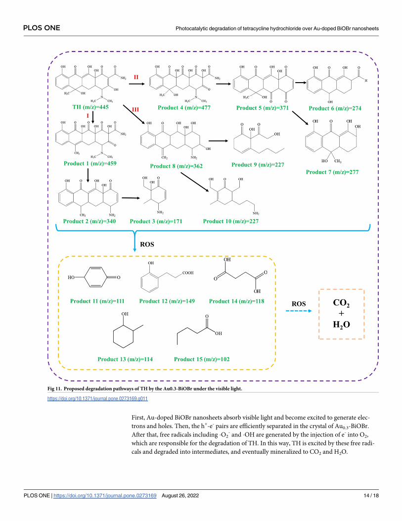
First, Au-doped BiOBr nanosheets absorb visible light and become excited to generate elec-
trons and holes. Then, the h+-e- pairs are efficiently separated in the crystal of Au0.3-BiOBr.
After that, free radicals including �O2- and �OH are generated by the injection of e- into O2,
which are responsible for the degradation of TH. In this way, TH is excited by these free radi-
cals and degraded into intermediates, and eventually mineralized to CO2 and H2O.
Fig 11. Proposed degradation pathways of TH by the Au0.3-BiOBr under the visible light.
https://doi.org/10.1371/journal.pone.0273169.g011
PLOS ONE Photocatalytic degradation of tetracycline hydrochloride over Au-doped BiOBr nanosheets
PLOS ONE | https://doi.org/10.1371/journal.pone.0273169 August 26, 2022 14 / 18

Conclusions
In this study, Au-doped BiOBr nanosheets were successfully synthesized through a simple
one-pot hydrothermal method, and the Au atoms were found to be highly dispersed in the
BiOBr host crystal. Au0.3-BiOBr nanosheets had a narrower band gap and a higher charge sep-
aration efficiency than those of BiOBr. Specifically, the charge separation efficiency of Au0.3-
BiOBr was 2.3 times higher than that of BiOBr. Therefore, Au0.3-BiOBr nanosheets exhibited
the highest efficiency in the degradation of TH under visible light irradiation, which was twice
higher than that of BiOBr. During the photocatalytic process, �O2- was the main active species
while other ROS also contributed to the degradation of TH. Furthermore, the degradation
pathways of TH were elucidated, and shown to involve hydroxylation, demethylation, ring
opening and mineralization. Therefore, modification by doping with Au is proposed as a
promising strategy for the modification of BiOBr to improve the charge separation efficiency
and enhance the photocatalytic degradation efficiency for organic pollutants under visible
light irradiation.
Supporting information
S1 Fig. XRD patterns of Au0.9-BiOBr.
(TIF)
S2 Fig. Nitrogen adsorption/desorption isotherms of the BiOBr and Au-doped BiOBr,
respectively.
(TIF)
S3 Fig. Recycling properties of the photocatalytic degradation of TH over the Au0.3-BiOBr
nanosheets.
(TIF)
S4 Fig. (a) XRD spectra and (b) SEM image after several cycles of photocatalytic degradation
of TH over the Au0.3-BiOBr nanosheets.
(TIF)
S5 Fig. Absorbance of NBT curve with time over Au0.3-BiOBr nanosheets.
(TIF)
S1 Table. Comparison of degradation capacities of some catalysts as described in litera-
ture.
(DOCX)
S1 Graphical abstract.
(TIF)
Author Contributions
Conceptualization: Chu-Ya Wang.
Data curation: Xin Fang.
Funding acquisition: Chu-Ya Wang.
Investigation: Yongze Lu.
Methodology: Xin Fang.
Project administration: Qi Zeng.
PLOS ONE Photocatalytic degradation of tetracycline hydrochloride over Au-doped BiOBr nanosheets
PLOS ONE | https://doi.org/10.1371/journal.pone.0273169 August 26, 2022 15 / 18

Resources: Chu-Ya Wang, Yongze Lu.
Software: Xin Fang, Qi Zeng.
Supervision: Qi Zeng, Yongze Lu.
Validation: Qi Zeng, Heng-Deng Zhou.
Visualization: Heng-Deng Zhou.
Writing – original draft: Chu-Ya Wang, Xin Fang.
References1. Byrne C, Subramanian G, Pillai SC. Recent advances in photocatalysis for environmental applications.
Journal of Environmental Chemical Engineering. 2018; 6(3):3531–55. https://doi.org/10.1016/j.jece.
2017.07.080
2. Patial S, Raizada P, Hasija V, Singh P, Thakur VK, Nguyen VH. Recent advances in photocatalytic mul-
tivariate metal organic frameworks-based nanostructures toward renewable energy and the removal of
environmental pollutants. Materials Today Energy. 2021;19. https://doi.org/10.1016/j.mtener.2020.
100589
3. Chen Y, Yang J, Zeng L, Zhu M. Recent progress on the removal of antibiotic pollutants using photoca-
talytic oxidation process. Critical Reviews in Environmental Science and Technology. 2021; 52
(8):1401–48. https://doi.org/10.1080/10643389.2020.1859289
4. Xiong L, Tang J. Strategies and Challenges on Selectivity of Photocatalytic Oxidation of Organic Sub-
stances. Advanced Energy Materials. 2021; 11(8). https://doi.org/10.1002/aenm.202003216
5. Sharma S, Dutta V, Raizada P, Hosseini-Bandegharaei A, Singh P, Nguyen V-H. Tailoring cadmium
sulfide-based photocatalytic nanomaterials for water decontamination: a review. Environmental Chem-
istry Letters. 2020; 19(1):271–306. https://doi.org/10.1007/s10311-020-01066-x
6. Wang C-Y, Zhang X, Qiu H-B, Huang G-X, Yu H-Q. Bi24O31Br10 nanosheets with controllable thickness
for visible-light-driven catalytic degradation of tetracycline hydrochloride. Applied Catalysis B: Environ-
mental. 2017; 205:615–23. https://doi.org/10.1016/j.apcatb.2017.01.015
7. Gao P, Yang Y, Yin Z, Kang F, Fan W, Sheng J, et al. A critical review on bismuth oxyhalide based
photocatalysis for pharmaceutical active compounds degradation: Modifications, reactive sites, and
challenges. J Hazard Mater. 2021; 412:125186. Epub 20210121. https://doi.org/10.1016/j.jhazmat.
2021.125186 PMID: 33516110.
8. Zhao G-Q, Hu J, Zou J, Long X, Jiao F-P. Modulation of BiOBr-based photocatalysts for energy and
environmental application: A critical review. Journal of Environmental Chemical Engineering. 2022; 10
(2). https://doi.org/10.1016/j.jece.2022.107226
9. Wang CY, Zeng Q, Zhu G. Novel S-doped BiOBr nanosheets for the enhanced photocatalytic degrada-
tion of bisphenol A under visible light irradiation. Chemosphere. 2021; 268:128854. Epub 20201103.
https://doi.org/10.1016/j.chemosphere.2020.128854 PMID: 33220984.
10. Xue X, Chen R, Chen H, Hu Y, Ding Q, Liu Z, et al. Oxygen Vacancy Engineering Promoted Photocata-
lytic Ammonia Synthesis on Ultrathin Two-Dimensional Bismuth Oxybromide Nanosheets. Nano Lett.
2018; 18(11):7372–7. Epub 20181015. https://doi.org/10.1021/acs.nanolett.8b03655 PMID: 30350657.
11. Li J, Sun S, Qian C, He L, Chen KK, Zhang T, et al. The role of adsorption in photocatalytic degradation
of ibuprofen under visible light irradiation by BiOBr microspheres. Chemical Engineering Journal. 2016;
297:139–47. https://doi.org/10.1016/j.cej.2016.03.145
12. Li H, Hu T, Du N, Zhang R, Liu J, Hou W. Wavelength-dependent differences in photocatalytic perfor-
mance between BiOBr nanosheets with dominant exposed (001) and (010) facets. Applied Catalysis B:
Environmental. 2016; 187:342–9. https://doi.org/10.1016/j.apcatb.2016.01.053
13. Ma Z, He Y, Li X, Zhou C, Deng L. Ultrasonic-assisted efficient degradation of tetracycline over ZnO/
BiOBr heterojunctions: Synergistic effect and role of oxidative species. Materials Research Bulletin.
2022;146. https://doi.org/10.1016/j.materresbull.2021.111591
14. Juntrapirom S, Tantraviwat D, Thongsook O, Anuchai S, Pornsuwan S, Channei D, et al. Natural sun-
light driven photocatalytic coupling of primary amines over TiO2/BiOBr heterojunction. Applied Surface
Science. 2021;545. https://doi.org/10.1016/j.apsusc.2021.149015
15. Hu T, Yang Y, Dai K, Zhang J, Liang C. A novel Z-scheme Bi2MoO6/BiOBr photocatalyst for enhanced
photocatalytic activity under visible light irradiation. Applied Surface Science. 2018; 456:473–81.
https://doi.org/10.1016/j.apsusc.2018.06.186
PLOS ONE Photocatalytic degradation of tetracycline hydrochloride over Au-doped BiOBr nanosheets
PLOS ONE | https://doi.org/10.1371/journal.pone.0273169 August 26, 2022 16 / 18

16. Perumal K, Shanavas S, Ahamad T, Karthigeyan A, Murugakoothan P. Construction of Ag2CO3/BiOBr/
CdS ternary composite photocatalyst with improved visible-light photocatalytic activity on tetracycline
molecule degradation. Journal of Environmental Sciences. 2023; 125:47–60. https://doi.org/10.1016/j.
jes.2021.10.021
17. Li H, Shang J, Ai Z, Zhang L. Efficient Visible Light Nitrogen Fixation with BiOBr Nanosheets of Oxygen
Vacancies on the Exposed {001} Facets. J Am Chem Soc. 2015; 137(19):6393–9. Epub 20150503.
https://doi.org/10.1021/jacs.5b03105 PMID: 25874655.
18. Wang H, Yong D, Chen S, Jiang S, Zhang X, Shao W, et al. Oxygen-Vacancy-Mediated Exciton Disso-
ciation in BiOBr for Boosting Charge-Carrier-Involved Molecular Oxygen Activation. J Am Chem Soc.
2018; 140(5):1760–6. Epub 20180124. https://doi.org/10.1021/jacs.7b10997 PMID: 29319310.
19. Liu D, Chen D, Li N, Xu Q, Li H, He J, et al. Surface Engineering of g-C3N4 by stacked biobr sheets rich
in oxygen vacancies for boosting photocatalytic performance. Angewandte Chemie International Edi-
tion. 2020; 59(11):4519–24. https://doi.org/10.1002/anie.201914949 PMID: 31876040
20. Chen X, Zhang X, Li Y-H, Qi M-Y, Li J-Y, Tang Z-R, et al. Transition metal doping BiOBr nanosheets
with oxygen vacancy and exposed {102} facets for visible light nitrogen fixation. Applied Catalysis B:
Environmental. 2021;281. https://doi.org/10.1016/j.apcatb.2020.119516
21. Li H, Li J, Ai Z, Jia F, Zhang L. Oxygen vacancy-mediated photocatalysis of BiOCl: reactivity, selectivity,
and perspectives. Angew Chem Int Ed Engl. 2018; 57(1):122–38. Epub 20171124. https://doi.org/10.
1002/anie.201705628 PMID: 28635079.
22. Jiang G, Wang R, Wang X, Xi X, Hu R, Zhou Y, et al. Novel highly active visible-light-induced photocata-
lysts based on BiOBr with Ti doping and Ag decorating. ACS Appl Mater Interfaces. 2012; 4(9):4440–4.
Epub 20120824. https://doi.org/10.1021/am301177k PMID: 22913293.
23. Jiang G, Wang X, Wei Z, Li X, Xi X, Hu R, et al. Photocatalytic properties of hierarchical structures
based on Fe-doped BiOBr hollow microspheres. Journal of Materials Chemistry A. 2013;1(7). https://
doi.org/10.1039/c2ta00942k
24. Shekofteh-Gohari M, Habibi-Yangjeh A, Abitorabi M, Rouhi A. Magnetically separable nanocomposites
based on ZnO and their applications in photocatalytic processes: A review. Critical Reviews in Environ-
mental Science and Technology. 2018; 48(10–12):806–57. https://doi.org/10.1080/10643389.2018.
1487227
25. Asadzadeh-Khaneghah S, Habibi-Yangjeh A. g-C3N4/carbon dot-based nanocomposites serve as effi-
cacious photocatalysts for environmental purification and energy generation: A review. Journal of
Cleaner Production. 2020;276. https://doi.org/10.1016/j.jclepro.2020.124319
26. Wang C-Y, Zhang X, Zhang Y-J, Chen J-J, Huang G-X, Jiang J, et al. Direct generation of hydroxyl radi-
cals over bismuth oxybromide nanobelts with tuned band structure for photocatalytic pollutant degrada-
tion under visible light irradiation. Applied Catalysis B: Environmental. 2018; 237:464–72. https://doi.
org/10.1016/j.apcatb.2018.06.015
27. Wang C-Y, Zhang Y-J, Wang W-K, Pei D-N, Huang G-X, Chen J-J, et al. Enhanced photocatalytic deg-
radation of bisphenol A by Co-doped BiOCl nanosheets under visible light irradiation. Applied Catalysis
B: Environmental. 2018; 221:320–8. https://doi.org/10.1016/j.apcatb.2017.09.036
28. Liu C, Mao S, Wang H, Wu Y, Wang F, Xia M, et al. Peroxymonosulfate-assisted for facilitating photoca-
talytic degradation performance of 2D/2D WO3/BiOBr S-scheme heterojunction. Chemical Engineering
Journal. 2022;430. https://doi.org/10.1016/j.cej.2021.132806
29. Wang C-Y, Zhang X, Qiu H-B, Wang W-K, Huang G-X, Jiang J, et al. Photocatalytic degradation of bis-
phenol A by oxygen-rich and highly visible-light responsive Bi12O17Cl2 nanobelts. Applied Catalysis B:
Environmental. 2017; 200:659–65. https://doi.org/10.1016/j.apcatb.2016.07.054
30. Wu Y, Ji H, Liu Q, Sun Z, Li P, Ding P, et al. Visible light photocatalytic degradation of sulfanilamide
enhanced by Mo doping of BiOBr nanoflowers. J Hazard Mater. 2022; 424(Pt C):127563. Epub
20211024. https://doi.org/10.1016/j.jhazmat.2021.127563 PMID: 34736201.
31. Wang P, Cao Y, Xu S, Yu H. Boosting the H2-evolution performance of TiO2/Au photocatalyst by the
facile addition of thiourea molecules. Applied Surface Science. 2020;532. https://doi.org/10.1016/j.
apsusc.2020.147420
32. Zhang H, Zhang Y, Zhong Y, Ding J. Novel strategies for 2,8-dichlorodibenzo-p-dioxin degradation
using ternary Au-modified iron doped TiO2 catalysts under UV-vis light illumination. Chemosphere.
2022; 291(Pt 2):132826. Epub 20211111. https://doi.org/10.1016/j.chemosphere.2021.132826 PMID:
34774912.
33. Misra M, Chowdhury SR, Lee TI. Sunlight driven decomposition of toxic organic compound, coumarin,
p-nitrophenol, and photo reduction of Cr(VI) ions, using a bridge structure of Au@CNT@TiO2 nanocom-
posite. Applied Catalysis B: Environmental. 2020;272. https://doi.org/10.1016/j.apcatb.2020.118991
34. Wang H, Liao B, Lu T, Ai Y, Liu G. Enhanced visible-light photocatalytic degradation of tetracycline by a
novel hollow BiOCl@CeO2 heterostructured microspheres: Structural characterization and reaction
PLOS ONE Photocatalytic degradation of tetracycline hydrochloride over Au-doped BiOBr nanosheets
PLOS ONE | https://doi.org/10.1371/journal.pone.0273169 August 26, 2022 17 / 18

mechanism. J Hazard Mater. 2020; 385:121552. Epub 20191103. https://doi.org/10.1016/j.jhazmat.
2019.121552 PMID: 31733996.
35. Chen P, Dong N, Zhang J, Wang W, Tan F, Wang X, et al. Investigation on visible-light photocatalytic
performance and mechanism of zinc peroxide for tetracycline degradation and Escherichia coli inactiva-
tion. J Colloid Interface Sci. 2022; 624:137–49. Epub 20220527. https://doi.org/10.1016/j.jcis.2022.05.
134 PMID: 35660882.
36. Zhang J, Chen P, Gao W, Wang W, Tan F, Wang X, et al. Melamine-cyanurate supramolecule induced
graphitic N-rich graphene for singlet oxygen-dominated peroxymonosulfate activation to efficiently
degrade organic pollutants. Separation and Purification Technology. 2021;265. https://doi.org/10.1016/
j.seppur.2021.118474
37. Huang H, Guo T, Wang K, Li Y, Zhang G. Efficient activation of persulfate by a magnetic recyclable
rape straw biochar catalyst for the degradation of tetracycline hydrochloride in water. Sci Total Environ.
2021; 758:143957. Epub 20201204. https://doi.org/10.1016/j.scitotenv.2020.143957 PMID: 33333296.
38. Liu C, Dai H, Tan C, Pan Q, Hu F, Peng X. Photo-Fenton degradation of tetracycline over Z-scheme Fe-
g-C3N4/Bi2WO6 heterojunctions: Mechanism insight, degradation pathways and DFT calculation.
Applied Catalysis B: Environmental. 2022;310. https://doi.org/10.1016/j.apcatb.2022.121326
PLOS ONE Photocatalytic degradation of tetracycline hydrochloride over Au-doped BiOBr nanosheets
PLOS ONE | https://doi.org/10.1371/journal.pone.0273169 August 26, 2022 18 / 18