paper maira plos one
TRANSCRIPT
Anti-CD25 Treatment Depletes Treg Cells and DecreasesDisease Severity in Susceptible and Resistant MiceInfected with Paracoccidioides brasiliensisMaıra Felonato, Adriana Pina, Eliseu Frank de Araujo, Flavio V. Loures, Silvia B. Bazan, Claudia Feriotti,
Vera L. G. Calich*
Departamento de Imunologia, Instituto de Ciencias Biomedicas, Universidade de Sao Paulo, Sao Paulo, Sao Paulo, Brazil
Abstract
Regulatory T (Treg) cells are fundamental in the control of immunity and excessive tissue pathology. Inparacoccidioidomycosis, an endemic mycosis of Latin America, the immunoregulatory mechanisms that control theprogressive and regressive forms of this infection are poorly known. Due to its modulatory activity on Treg cells, weinvestigated the effects of anti-CD25 treatment over the course of pulmonary infection in resistant (A/J) and susceptible(B10.A) mice infected with Paracoccidioides brasiliensis. We verified that the resistant A/J mice developed higher numbersand more potent Treg cells than susceptible B10.A mice. Compared to B10.A cells, the CD4+CD25+Foxp3+ Treg cells of A/Jmice expressed higher levels of CD25, CTLA4, GITR, Foxp3, LAP and intracellular IL-10 and TGF-b. In both resistant andsusceptible mice, anti-CD25 treatment decreased the CD4+CD25+Foxp3+ Treg cell number, impaired indoleamine 2,3-dioxygenase expression and resulted in decreased fungal loads in the lungs, liver and spleen. In A/J mice, anti-CD25treatment led to an early increase in T cell immunity, demonstrated by the augmented influx of activated CD4+ and CD8+ Tcells, macrophages and dendritic cells to the lungs. At a later phase, the mild infection was associated with decreasedinflammatory reactions and increased Th1/Th2/Th17 cytokine production. In B10.A mice, anti-CD25 treatment did not alterthe inflammatory reactions but increased the fungicidal mechanisms and late secretion of Th1/Th2/Th17 cytokines.Importantly, in both mouse strains, the early depletion of CD25+ cells resulted in less severe tissue pathology and abolishedthe enhanced mortality observed in susceptible mice. In conclusion, this study is the first to demonstrate that anti-CD25treatment is beneficial to the progressive and regressive forms of paracoccidioidomycosis, potentially due to the anti-CD25-mediated reduction of Treg cells, as these cells have suppressive effects on the early T cell response in resistant mice andthe clearance mechanisms of fungal cells in susceptible mice.
Citation: Felonato M, Pina A, Araujo EFd, Loures FV, Bazan SB, et al. (2012) Anti-CD25 Treatment Depletes Treg Cells and Decreases Disease Severity inSusceptible and Resistant Mice Infected with Paracoccidioides brasiliensis. PLoS ONE 7(11): e51071. doi:10.1371/journal.pone.0051071
Editor: Derya Unutmaz, New York University, United States of America
Received May 31, 2012; Accepted October 30, 2012; Published November 30, 2012
Copyright: � 2012 Felonato et al. This is an open-access article distributed under the terms of the Creative Commons Attribution License, which permitsunrestricted use, distribution, and reproduction in any medium, provided the original author and source are credited.
Funding: This work was supported by Fundacao de Amparo a Pesquisa do Estado de Sao Paulo (FAPESP) and Conselho Nacional de Pesquisas (CNPq). Thefunders had no role in study design, data collection and analysis, decision to publish, or preparation of the manuscript.
Competing Interests: The authors have declared that no competing interests exist.
* E-mail: [email protected]
Introduction
Paracoccidioidomycosis (PCM), the most important systemic
mycosis in Latin America, is caused by the dimorphic fungus
Paracoccidioides brasiliensis [1]. The inhalation of conidia usually
leads to an asymptomatic infection but a few infected individuals
evolve to overt disease.The infection and the benign forms of the
disease are associated prevalent Th1 immunity whereas the severe
forms with suppressed DTH responses and prevalent Th2/Th3
immunity [2,3]. In a genetic model of infection, A/J mice was
shown to be resistant while B10.A mice susceptible to the i.p and
i.t. routes of infection [4,5]. In the pulmonary model, A/J mice are
initially tolerant to fungal growth but lately develop efficient
cellular immunity with prevalent IFN-c secretion, macrophage
activation and regressive disease [6,7]. In contrast, B10.A mice are
able to control the initial infection but soon develop T cell anergy
and disseminated disease [5,8,9]. Both, CD4+ and CD8+T cells
were shown to be immunoprotective against P. brasiliensis infection.
Resistant mice develop mixedTh1/Th2 responses concomitant
with protective CD8+ T cells that synthesize large amounts of IFN-
c [7,8,10]. The relative protection of susceptible mice is mediated
by CD8+ T cells which, however, are not able to compensate the
CD4+ T cell anergy induced by excessive nitric oxide (NO)
production [7,11] and indoleamine-2,3 dioxygenase (IDO) activity
[Araujo et al., manuscript in preparation].
CD4+CD25+ regulatory T cells (Treg) have been shown to
control the afferent and efferent arms of immune responses, and
play an essential role in the control of autoimmune diseases,
transplantation and infectious processes [12–15]. Natural Treg
cells develop in the thymus, have a CD4+CD25+ phenotype, and
are seeded to peripheral lymphoid organs where they control
autoimmunity and excessive inflammatory responses against
endogenous and exogenous aggressions [16,17]. At the periphery,
naıve CD4+ T cells can also acquire a suppressive phenotype and
ability to control excessive immunity [17,18]. In addition to CD25
(the alpha chain of IL-2R), Treg cells express other activation
markers such as CTLA-4 (CD152, cytotoxic T lymphocyte-
associated antigen 4), GITR (glucocorticoid-induced tumor
PLOS ONE | www.plosone.org 1 November 2012 | Volume 7 | Issue 11 | e51071
necrosis factor-receptor-related protein), OX40 (CD134), and L-
selectin or CD62 ligand (CD62L) [19–21]. Several transcription
factors were shown to control Treg cells development and activity,
but Foxp3 has been described as the crucial factor for the
suppressive function of these cells [22–25]. The suppressive
activity of Tregs depends on cell contact and/or the activity of
several inhibitory molecules such as IL-10, TGF-b, IL-35, CTLA-
4, IDO, and granzyme/perforin [18,22,26]. Although Tregs are
likely to use multiple mechanisms to suppress immune responses,
CTLA-4 may have a dominant role [27–29]. There is increasing
evidence that Treg cells and, in particular, natural CD4+CD25+
Treg cells play a key role in the control of infectious processes. The
presence of Treg cells has been associated with many chronic
infectious diseases where they facilitate the maintenance of
a residual number of microorganisms and immunological memory
[14,17,30]. Treg cells were shown to increase fungal loads in mice
infected with Candida albicans, Pneumocystis carinii and Aspergillus
fumigatus. In these experimental models, while maintaining in-
fection, Treg cells limited the associated immunopathology [31–
34]. There are few reports in the literature on the participation of
Treg cells in the control of paracoccidioidomycosis, but increased
numbers of these cells have been associated with disease severity.
Thus, a higher number of Foxp3+ Treg cells was demonstrated in
the lesions and peripheral blood of patients with active disease
when compared with treated patients or healthy controls. These
cells produced high levels of TGF-b and IL-10 and expressed
increased levels of characteristic surface markers of Tregs (GITR,
CD38, CD95L, CTLA-4, TLR-2 and LAP) [35,36]. Furthermore,
in CCR5-deficient C57BL/6 mice, which have intermediate
susceptibility to P. brasiliensis infection, the survival of yeast cells
and the severe immunosuppression of hosts were shown to be
mediated by Treg cells [37]. In the pulmonary model of murine
PCM, our group recently showed that the development of Treg
cells was associated with CD28, TLR2 and TLR4 expression. [38–
40]. In addition, the adaptor protein MyD88 was also shown to be
involved in the control of Treg cells differentiation [41].
In this study we explored the presence, phenotype and function
of CD4+CD25+Foxp3+ Treg cells in resistant A/J and susceptible
B10.A mice to P. brasiliensis infection. Subsequently, the severity of
the disease was studied at an early and late periods of infection
using anti-CD25-treated and untreated mice. Interestingly, un-
infected and infected resistant mice presented higher numbers and
more potent Treg cells than susceptible mice. The early depletion
of CD25+ cells by monoclonal antibodies led to a less severe
infection in both mouse strains, but only in resistant mice the early
migration of inflammatory cells to the site of infection was
restored. Antibody-mediated depletion of CD25+ T cells of
susceptible did not alter the migration of inflammatory T cells,
but recued these animals from progressive disease and precocious
mortality. Importantly, anti-CD25 treatment did not induce sterile
immunity, but significantly reduced organ pathology. In conclu-
sion, ours results showed for the first time regulatory T cells exert
detrimental effects to resistant and susceptible mice to P. brasiliensis
infection, and their modulation by anti-CD25 treatment can bring
beneficial effects to both, the progressive and regressive forms of
this chronic fungal disease.
Materials and Methods
Ethics StatementAnimal experiments were performed in strict accordance with
the Brazilian Federal Law 11,794 establishing procedures for the
scientific use of animals, and the State Law establishing the Animal
Protection Code of the State of Sao Paulo. All efforts were made to
minimize suffering, and all animal procedures were approved by
the Ethics Committee on Animal Experiments of the Institute of
Biomedical Sciences of University of Sao Paulo (Proc.76/04/
CEEA).
MiceA/J (resistant), B10.A (susceptible), and Foxp3tm1Kuch C57BL/6
(intermediate susceptibility) mouse strains were bred at the
University of Sao Paulo animal facilities under specific-patho-
gen-free (SPF) conditions in closed-top cages. The Foxp3GFP
reporter allele C57BL/6 mouse strain was kindly donated by
Dr.Vijay K. Kuchroo, from Harvard University. Clean food and
water were given ad libitum. Mice were 8 to 11 weeks of age at the
time of infection, and procedures involving animals and their care
were approved by the Ethics Committee on Animal Experiments
from Instituto de Ciencias Biomedicas, Universidade de Sao
Paulo.
Fungus and Mice InfectionP. brasiliensis 18 isolate (Pb18), which is highly virulent, was used
throughout the study. To ensure the maintenance of its virulence,
the isolate was used after three serial animal passages [42]. Pb18
yeast cells were then maintained by weekly subcultivation in
semisolid Fava Netto culture medium [43] at 35uC and used on
the seventh day of culture. The fungal cells were washed in
phosphate-buffered saline (PBS; pH 7.2) and counted in a hemo-
cytometer and the suspension was adjusted to 206106 fungal -
cells/mL. The viability of fungal suspension, determined by Janus
Green B vital dye (Merk, Darmstadt, Germany), was always higher
than 85%. Mice were anesthetized and submitted to i.t. P.
brasiliensis infection as previously described [5]. Briefly, after
intraperitoneal anesthesia the animals were infected with
16106 Pb18 yeast cells, contained in 50 mL of PBS, by surgical
i.t. inoculation, which allowed dispensing of the fungal cells
directly into the lungs. The skins of the animals were then sutured,
and the mice were allowed to recover under a heat lamp. Mice
were studied 2 and 10 weeks after infection. Two to three
experiments were performed separately.
In vivo Depletion of CD25+ T cellsB10.A, A/J mice and Foxp3GFP C57BL/6 mice were given i.p.
injections of 0.5 mg of an anti-CD25 (clone PC61) or a control rat
IgG obtained from BioXcell (USA). Antibodies were administered
3 days before and 3 days after infection with P. brasiliensis yeasts.
Foxp3GFP C57BL/6 mice were studied at week 2 post-infection.
A/J and B10.A mice were sacrificed 2 and 10 weeks after
infection, and organs analyzed for CFU counts, histopathology,
inflammatory reactions and levels of cytokines. PC61 administra-
tion promoted long-lasting depletion/neutralization of CD25
expression (longer than 2weeks) in naıve mice, as previously
demonstrated [44,45].
Assay for Organ Colony Forming Units (CFU)The number of viable microorganisms in infected organs (lung,
liver and spleen) from experimental and control mice were
determined by counting the number of CFU. Animals (n = 6–8)
from each group were sacrificed, and the enumeration of viable
organisms was done as previously described [46]. Briefly, aliquots
(100 mL) of the cellular suspensions and serial dilutions were plated
on brain heart infusion agar (Difco, Detroit, MI) supplemented
with 4% (vol/vol) horse serum (Instituto Butantan, Sao Paulo,
Brazil) and 5% P. brasiliensis 192 culture filtrate, the latter
constituting a source of growth-promoting factor. The plates were
Anti-CD25 Antibody Improves Paracoccidioidomycosis
PLOS ONE | www.plosone.org 2 November 2012 | Volume 7 | Issue 11 | e51071
incubated at 35uC, and colonies were counted daily until no
increase in counts was observed. The number (log10) of viable
P. brasiliensis colonies per gram of tissue were expressed as means
6 standard errors (SE).
Mortality RatesMortality studies were performed with anti-CD25-treated and
control A/J and B10.A mice inoculated i.t. with 16106 yeast cells
or PBS (n = 9–11). Deaths were registered daily for a 200-day
period and experiments were repeated twice.
Lung and Liver Leukocytes IsolationLungs from each mouse were excised, washed in PBS, minced,
and digested enzymatically for 1 hour in 15 mL/lung of digestion
buffer [RPMI, 5% fetal calf serum, 1 mg/mL collagenase (Sigma
Aldrich Inc.H), and 30 mg/mL DNase]. Livers from individual
mice were obtained, submitted to organ perfusion using 10.0 ml of
warm PBS via portal vein and organ fragments were pressed
through 70 mm cell strainer (Becton Dickson). After erythrocyte
lysis using NH4Cl buffer, cells were washed, resuspended in
complete media, and centrifuged for 30 minutes at 1,2006g in
presence of 20% Percoll (Sigma) to separate leukocytes from cell
debris and epithelial cells. Total leukocyte numbers were assessed
in the presence of trypan blue using a hemocytometer; viability
was always higher than 85%. The absolute number of a leukocyte
subset was equal to the percentage of that cell subset multiplied by
the total number of leukocytes recovered from the digested organ/
100.
Purification of CellsMagnetic sorting of various splenic and lung cell types were
performed with the AutoMACS (MiltenyiBiotec, Germany),
according to the manufacturer’s instructions. Splenic effector
CD4+CD252 T cells from naive B10.A and A/J mice were sorted
after labeling cells with the CD4+ selection MACS kit. Suppressor
CD4+CD25+ T cells were obtained from lung cell suspensions
previously prepared as above described. Using the CD4+CD25+
isolation kit, non-CD4+ cells were depleted first using antibodies
targeting CD8+ T cells, B cells, macrophages, NK cells and
erythrocytes. CD25+ cells were then positively selected using PE-
labeled anti-CD25 and anti-PE microbeads.
Coculture Proliferation-suppression AssayResponder CD4+ T cells from naıve B10.A and A/J mice were
stained with CFSE (Invitrogen). To perform the cell staining,
56106 cells were incubated with 10 mM CFSE in RPMI contain-
ing 5% FBS for 15 min at 37uC in the dark. Cells were then
washed twice and resuspended in RPMI containing 10% FBS for
seeding. CD4+CD252 T cells (56104/well) were seeded in U-
bottom, 96 well plate in RPMI with 10% FBS, containing 2 mM
glutamine, 100 IU/penicillin, 100 mg/ml streptomycin, 10 mM
Hepes, 1 mM sodium pyruvate, and 50 mM 2-ME, with or
without 26105 cell/well T-cell depleted, irradiated spleen cells,
plus 0.5 mg/ml soluble anti-CD3 antibody. CD4+CD25+ T cells
from infected lungs were added to the well at a ratio (CD4+CD252
T cell: CD4+CD25+ T cell) of 1:1, 1:0.33 and 1:0.11. Cells were
cultivated for 5 days and the proliferative response of CFSE-
labeled cells was measured by flow cytometry (FacsCanto). The
proliferation index (PI) was calculated dividing the geometric
mean of fluorescence from non-stimulated CD4+CD252 T cells by
the geometric mean of fluorescence from stimulated CD4+CD252
T cells in the presence or absence of CD4+CD25+ T cells.
Flow Cytometry AnalysisFor surface staining alone, lung or liver leukocytes were washed
and resuspended at a concentration of 16106 cells/mL in staining
buffer (PBS 1x, 2% FBS and 0,5% NaN3). Fc receptors were
blocked by the addition of unlabeled anti-CD16/32 (Fc block; BD
Pharmingen, San Diego, CA). The leukocytes were then stained
for 20 min at 4uC with the optimal dilution of each antibody
labeled with the adequate fluorochrome (BD, Pharmingen). The
following antibodies were used: anti-CD11b, CD11c, F4/80,
GR1, Iak, CD3, CD4, CD8, CD44, CD62L, CD25, CTLA-4,
GITR, LAP, CD19, and B220. Cells were washed twice with
staining buffer, resuspended in 100 ml, and an equal volume of 2%
formalin was added to fix the cells. The stained cells were analyzed
immediately on a FACSCanto flow cytometer (BD Biosciences,
CA) using the FACSDiva software (BD Biosciences) gating on
macrophages or lymphocytes as judged from forward and side
light scatter. Twenty thousand cells were counted and the data
expressed as the percentage or the absolute number of positive
cells which was calculated trough the percentage obtained by
FACS and the number of cells determined in Neubauer chambers.
The intracellular detection of Foxp3, the X-linked forkhead/
winged helix transcription factor, in leukocytes obtained from
infected lung or liver was performed in fixed and permeabilized
cells using Cytofix/Cytoperm (BD Biosciences). Initially, the cells
were labeled with antibodies for cell surface molecules such as
FITC-conjugated anti-CD4 and PE-Cy7 conjugated anti-CD25.
Next, the cells were fixed, permeabilized, and stained with PE-
conjugated anti-Foxp3, for 90 min at 4uC. In selected experi-
ments, Foxp3+ Treg cells were also labeled for membrane LAP,
CTLA4 and GITR expression. For intracellular cytokines, after
Foxp3 labeling (PE or APC), pacific blue anti IL-10 and PE anti-
TGF-b were added to fixed and permeabilized cells. Cells were
then washed twice with staining buffer, resuspended in 100 ml, and
an equal volume of 2% formalin was added to fix the cells. A
minimum of 100,000 events were acquired on FACSCanto flow
cytometer using the FACSDiva software, as described above.
Surface staining of CD25 and intracellular Foxp3 expression were
back-gated on the CD4 T cell population.
Measurement of CytokinesMice were infected i.t. with P. brasiliensis (n = 6–8), and their
right lung was aseptically removed and individually disrupted in
5.0 mL of PBS. Supernatants were separated from cell debris by
centrifugation at 2,0006g for 15 min, passed through 0.22 mm
pore-size filters (Millipore, Bedford, MA), and stored at 270uC.
The levels of IL-2, IL-12, IFN-c, TNF-a, IL-4, IL-10, GM-CSF,
IL-6, IL-23, IL-17 and TGF-b were measured by capture enzyme-
linked immunosorbent assay (ELISA) with antibodies pairs
purchased from Pharmingen. The ELISA procedure was per-
formed according to the manufacture’s protocol. The concentra-
tions of cytokines were determined with reference to a standard
curve for several twofold dilutions of murine recombinant
cytokines.
Histopathologic and Morphometrical AnalyzesGroups of anti-CD25 treated and untreated B10.A and A/J
mice were killed at the second and tenth weeks post-infection.
Lungs and livers were collected, fixed in 10% formalin and
embedded in paraffin. Five-micrometer sections were stained by
the hematoxilin-eosin (H&E) for an analysis of the lesions, silver
stained for fungal evaluation. Pathological changes were analyzed
based on the size, morphology and cell composition of granulo-
matous lesions, presence of fungi and intensity of the inflammatory
infiltrates. Morphometrical analysis was performed using a Nikon
Anti-CD25 Antibody Improves Paracoccidioidomycosis
PLOS ONE | www.plosone.org 3 November 2012 | Volume 7 | Issue 11 | e51071
DXM 1200c digital camera (magnification of 10x), and Nikon NIS
Elements AR 2.30 software. The area of lesions was measured (in
mm2) in 10 microscopic fields per slide (n = 4–6). Results were
expressed as the mean 6 standard error of the mean (SEM) of
total area of lesions for each animal.
Quantitative Analysis of 2,3 Indoleamine Dioxygenase(IDO) mRNA Expression
RNA was extracted from normal and infected lungs using Trizol
reagent (Invitrogen,Calrsbad, CA), and cDNA was synthesized
from 1 mg RNA using High Capacity RNA-to-cDNA kit (Applied
Biosystems) according to manufacturer’s instructions. IDO mRNA
expression was quantified relative to glyceraldehyde-3-phosphate
dehydrogenase (GAPDH) using assay-on-demand primers and
probes, Taqman Universal Master Mix, and ABI Prism 7000
apparatus (Applied Biosystems).
Determination of IDO Enzymatic ActivityTo monitor IDO enzymatic activity, kynurenine was detected
using a modified spectrophotometric assay. The amount of 50 ml
of 30% trichloroacetic acid was added to 100 ml of lung
homogenates, vortexed, and centrifuged at 800 g for 5 min. A
volume of 75 ml of the supernatant was then added to an equal
volume of Ehrlich reagent (100 mg P-dimethylbenzaldehyde, 5 ml
glacial acetic acid) in a 96 wells microtiter plate. Optical density
was measured at 492 nm, using a Multiskan MS (Labsystems,
Helsinki, Finland) microplate reader. A standard curve of defined
kynurenine concentrations (0–100 mM) was used to determine
unknown kynurenine concentrations.
Statistical AnalysisData are expressed as the mean 6 SEM. Differences between
groups were analyzed by Student’s t test or analysis of variance
(ANOVA) followed by the Bonferroni test. Differences between
survival times were determined with the LogRank test. Data were
analyzed using GraphPad Prism 5.01 software for Windows
(GraphPad, San Diego, CA). P values #0.05 were considered
significant.
Results
Early in the Infection, Resistant Mice Develop a MoreSevere Infection and Present a Higher Number of TregCells in the Lungs
First we characterized the severity of P.brasiliensis infection in
resistant (A/J and susceptible mice (B10.A) infected i.t. with
16106 viable yeast cells. This control was necessary because there
is no adequate virulence marker for P. brasiliensis whose infectivity
is maintained by in vivo passages, resulting in virulence fluctua-
tion. The data here obtained are consistent with those previously
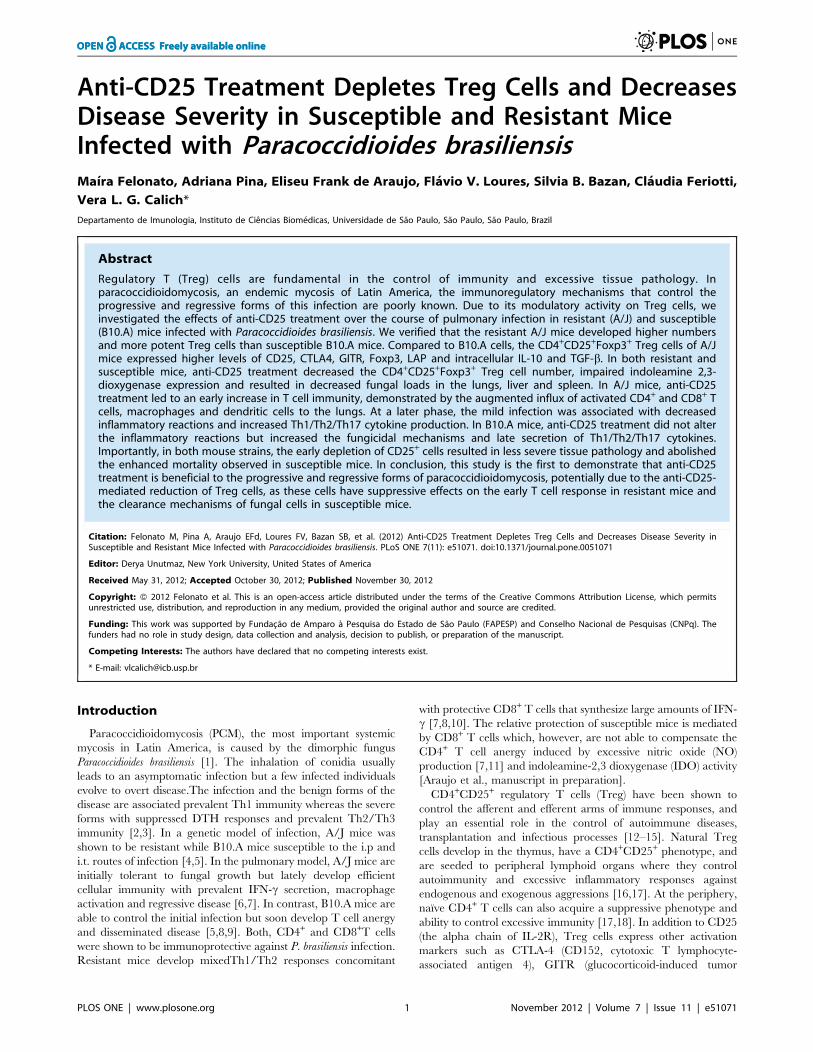
described [5]. At week 2, the pulmonary infection of A/J mice is
slightly higher than in B10.A mice, and allows an early
dissemination and enhanced fungal growth in the liver (Fig. 1A).
At week 10, the infection is regressive in A/J mice and progressive
in B10.Amice. At this late period, while A/J mice control the
fungal growth in the liver, B10.A mice show increased numbers of
viable yeasts in the liver and spleen (Fig. 1B). These data
reproduce our earlier findings in the pulmonary model of
infection, and demonstrate that, early in the infection, A/J mice
are more permissive to fungal growth and dissemination than the
susceptible strain. However, at the late phase A/J mice develop an
adequate control of the infection whereas a progressive disease is
established in B10.A mice.
Figure 1. In the course of P.brasiliensis infection resistant (A/J)mice develop higher numbers of Treg cells than susceptible(B10.A) mice. Groups (n = 6) of A/J and B10.A mice were infected i.t.
Anti-CD25 Antibody Improves Paracoccidioidomycosis
PLOS ONE | www.plosone.org 4 November 2012 | Volume 7 | Issue 11 | e51071
As Treg cells were shown to be involved in the control of several
infectious processes [14,17,30–34], we asked whether these cells
could be associated with the resistant and susceptible phenotype of
A/J and B10.A mice. We have then assessed the presence of
regulatory T cells in the lungs of A/J and B10.A mice infected with
1 million fungal cells by the pulmonary route. In the course of
infection, both mouse strains increased the numbers of pulmonary
CD4+CD25+Foxp3+ Treg cells. However, compared with B10.A
mice, and at both post-infection periods, higher numbers of
CD4+CD25+Foxp3+ Treg cells were found in A/J mice (Fig. 1C).
Interestingly, the total number of mononuclear leukocytes present
in the lungs of uninfected B10.A mice was 1.8 fold higher than
those of A/J mice (44.865.26106 cells X 24.562.76106 cells).
However, even uninfected A/J mice (day zero) showed an
augmented number (0.3660.046106 X 0.1060.026106 cells)
and frequency (1.4660.16 X 0.260.02) of pulmonary Treg cells
than susceptible B10.A mice.
Number and Phenotype of Regulatory T cells in theCourse of P. brasiliensis Infection of Resistant andSusceptible Mice
To better characterize CD4+CD25+Foxp3+ Treg cells, the
expression of membrane TGF-b (LAP), CTLA4, GITR and
intracellular TGF-b and IL-10 were evaluated. As shown in
(Fig. 1A,B), A/J mice showed increased numbers of Treg cells
expressing all studied activation markers, besides increased
presence of intracellular IL-10 and TGF-b (Fig. 1 C). As the
expression of Foxp3 and CTLA4 have been implicated in the
suppressive potency of Treg cells (16,29) we analyzed the mean
fluorescence intensity of these molecules expressed by Treg cells.
As shown in Fig. 2D–F, the intensity of all these three molecules
increased in the course of infection, and was always higher in A/J
than in B10.A Treg cells.
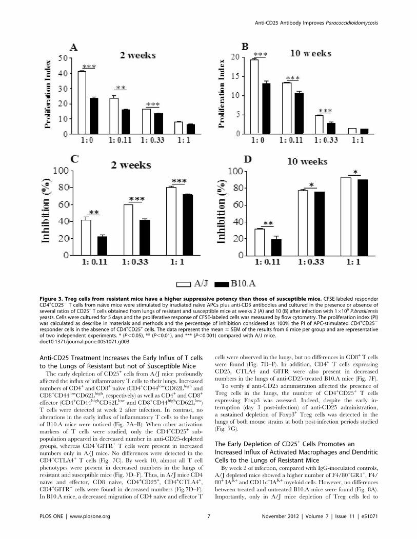
Regulatory T cells from Resistant Mice have a MorePotent Suppressive Activity
Following verifying that resistant mice had higher numbers of
Treg cells expressing higher levels of Foxp3 and CTLA4 in their
lungs than B10.A mice, we asked whether these cells had
equivalent suppressive activity. Then, naıve CD4+CD252splenic
T cells from uninfected mice were stimulated by irradiated APCs
plus anti-CD3 antibodies in the presence or absence of
CD4+CD25+ T cells isolated from the lungsof B10.A and A/J
mice at weeks 2 and 10 after infection. CD4+CD252 cells were
previously labeled with CFSE, stimulated for 5 days, and the
proliferative response analyzed by flow cytometry. The results are
expressed as proliferation index (PI) and percentage of inhibition.
At both infection periods, naıve spleen cells from A/J mice showed
an increased proliferative response, and the addition of several
proportions of Treg cells led to decreased proliferation, which was
maximal at the T effector:Treg ratio of 1:1 (Fig. 3A, B). When the
percentage of inhibition was calculated, we could see that Treg
cells from A/J mice exerted a superior suppressive activity on
lymphocyte proliferation than B10.A Tregs. Importantly, this
difference between strains was most marked at the second than at
the tenth week after infection. At week 10, however, Treg cells
appear to be more suppressive than those obtained at week 2, and
this fact could be seen with the higher proportions of Treg cells
employed (Fig. 3 C,D). As a whole, these findings demonstrated
that in the course of infection resistant and susceptible mice
develop increased numbers of Treg cells with increased suppres-
sive potency. However, Tregs from resistant mice have a more
potent suppressive activity on the proliferation of naıve T cells
than those of B10.A mice.
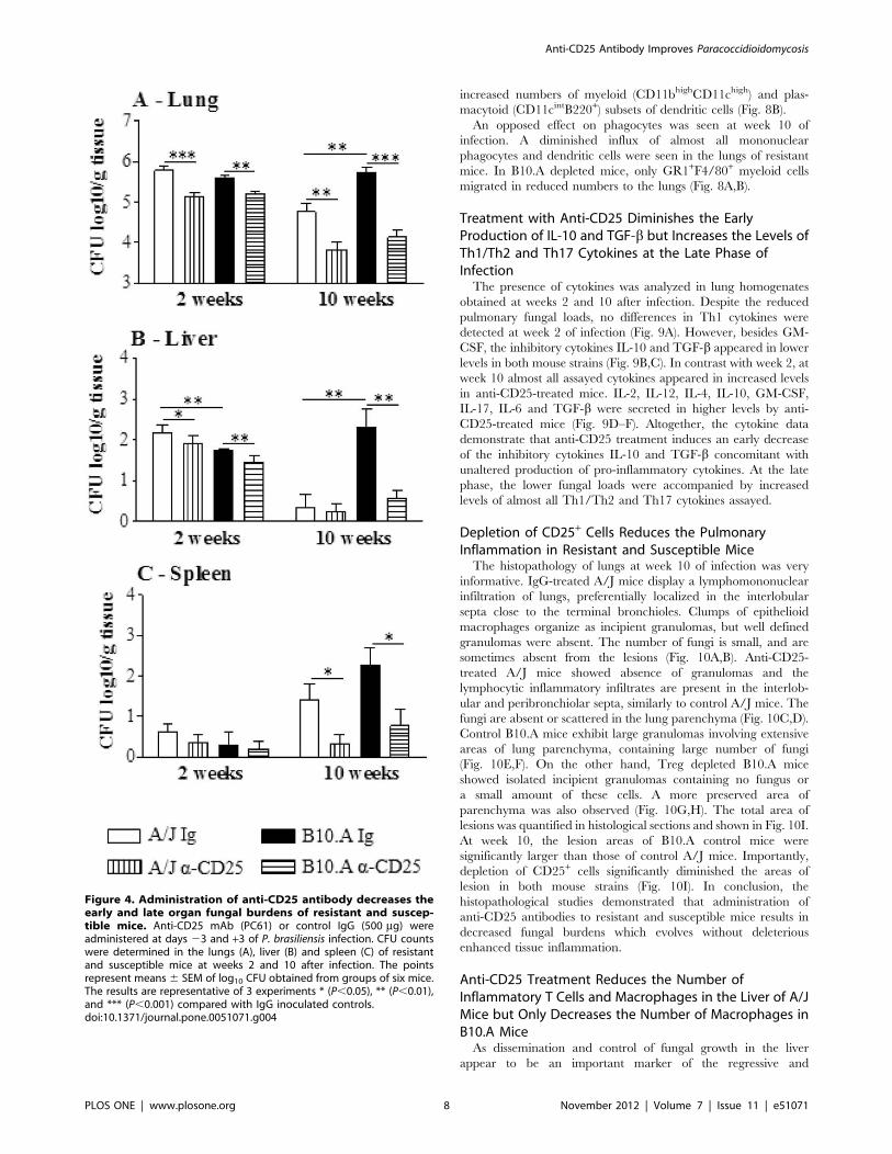
In Resistant and Susceptible Mice, Depletion of CD25+
T Cells Promotes an Enhanced Control of FungalGrowth.
We next asked whether anti-CD25 treatment of A/J and B10.A
mice would change the severity of fungal infection. This treatment
was chosen because it induces a consistent and persistent (2 weeks)
depletion of Treg cells [44]. Then, groups (n = 6–7) of susceptible
and resistant mice were treated i.p. with 500 mg of anti-CD25
monoclonal antibody (PC61) at days 23 and +3 of P.brasiliensis
infection. Control mice were injected with an equivalent amount
of normal rat IgG. In both mouse strains, at both post-infection
periods, a decreased fungal burden was observed. Of note was the
effect anti-CD25 administration at the late period of infection of
susceptible mice. Besides the lowered CFU counts in the lungs, the
livers and spleens of B10.A mice showed an impressive reduction
in the number of viable yeast cells (Fig. 4A–C). I.
In an attempt to better characterize the effect of anti-CD25
(PC61) treatment on normal and P. brasiliensis-infected mice, we
performed some experiments using Foxp3GFP C57BL/6 mice.
The groups of uninfected and infected mice (16106 P. brasiliensis
injected through the i.t. route) were treated or not treated with
anti-CD25 (PC61) mAb using the same protocol that was used to
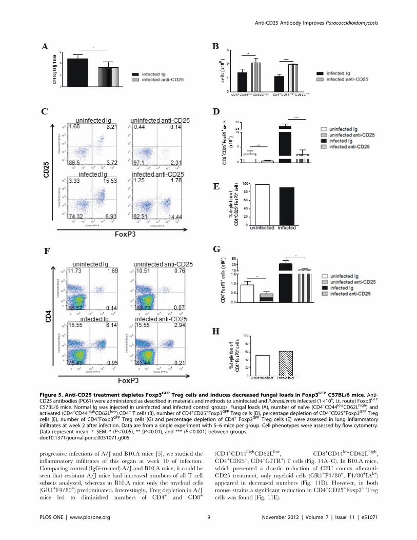
treat the A/J and B10.A mice. As shown in Figure 5, decreased
fungal burdens (Fig. 5A) and Foxp3+ Treg cells
(CD4+CD25+Foxp3+), which were associated with increased
numbers of effector CD4 T (CD44highCD62low) cells, were
detected at week 2 post-infection (Fig. 5B–E). In addition, the
FoxP3+ cells were characterized by the expression of GFP and not
by the use of anti-CD25 antibodies. Although less extreme than
that observed with the anti-CD25 antibodies, a decreased number
of FoxP3+ cells was observed (Fig. 5F–H). Therefore, the anti-
CD25 antibodies induced a consistent decrease in Foxp3GFP Treg
cells that was associated with an increase (not a decrease) in
effector T cells. These data brought consistent evidence that
treatment with PC61 antibodies effectively depletes Treg cells and
increases effector T cells in P. brasiliensis infected mice.
Depletion of CD25+ Cells Affects the Number but not theFrequency of Pulmonary Leukocytes
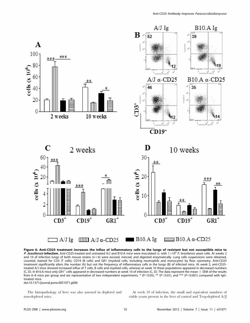
In the lungs of A/J mice, anti-CD25 treatment increased the
number of inflammatory cells at week 2, but led to diminished
numbers at week 10 of infection. In contrast, in B10.A mice
a reduced inflammation was seen only at week 10 (Fig. 6A).When
the frequency of T cells, B cells and GR1+ cells (myeloid cells,
including cells of the granulocytic lineage such as neutrophils and
the monocytic lineage) was characterized no important differences
between treated and untreated groups were observed (Fig. 6B);
however, A/J mice showed all leucocyte subsets in increased
numbers at week 2 and reduced at week 10 of infection. In B10.A
mice, only GR1+ cells were seen in decreased numbers at the late
phase of infection (Fig. 6C, D).
with 16106 yeasts cells of P.brasiliensis. Organ CFU counts weremeasured at weeks 2 (A) and 10 (B) after infection. At these timepoints, lungs were removed, leukocytes obtained and the number ofCD4+CD25+Foxp3+ cells analyzed by flow cytometry. The data representthe mean 6 SEM of the results from 6 mice per group and arerepresentative of two independent experiments. * (P,0.05), **(P,0.01), and *** (P,0.001) compared with A/J mice.doi:10.1371/journal.pone.0051071.g001
Anti-CD25 Antibody Improves Paracoccidioidomycosis
PLOS ONE | www.plosone.org 5 November 2012 | Volume 7 | Issue 11 | e51071
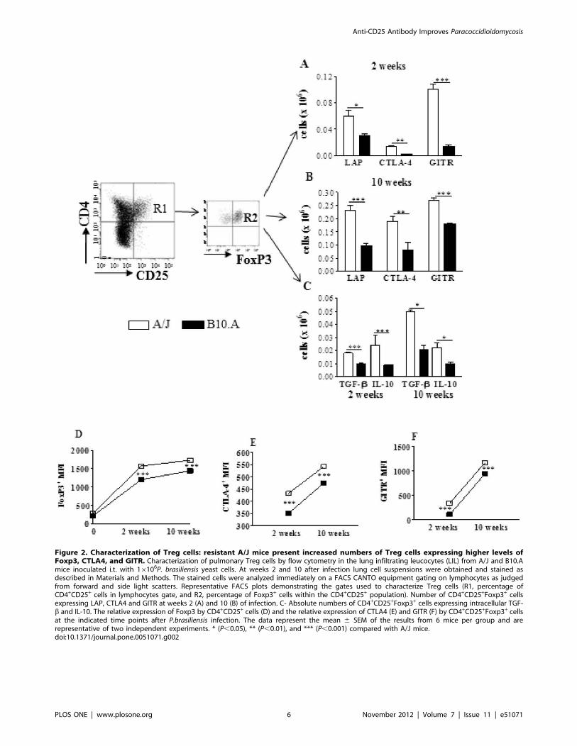
Figure 2. Characterization of Treg cells: resistant A/J mice present increased numbers of Treg cells expressing higher levels ofFoxp3, CTLA4, and GITR. Characterization of pulmonary Treg cells by flow cytometry in the lung infiltrating leucocytes (LIL) from A/J and B10.Amice inoculated i.t. with 16106P. brasiliensis yeast cells. At weeks 2 and 10 after infection lung cell suspensions were obtained and stained asdescribed in Materials and Methods. The stained cells were analyzed immediately on a FACS CANTO equipment gating on lymphocytes as judgedfrom forward and side light scatters. Representative FACS plots demonstrating the gates used to characterize Treg cells (R1, percentage ofCD4+CD25+ cells in lymphocytes gate, and R2, percentage of Foxp3+ cells within the CD4+CD25+ population). Number of CD4+CD25+Foxp3+ cellsexpressing LAP, CTLA4 and GITR at weeks 2 (A) and 10 (B) of infection. C- Absolute numbers of CD4+CD25+Foxp3+ cells expressing intracellular TGF-b and IL-10. The relative expression of Foxp3 by CD4+CD25+ cells (D) and the relative expression of CTLA4 (E) and GITR (F) by CD4+CD25+Foxp3+ cellsat the indicated time points after P.brasiliensis infection. The data represent the mean 6 SEM of the results from 6 mice per group and arerepresentative of two independent experiments. * (P,0.05), ** (P,0.01), and *** (P,0.001) compared with A/J mice.doi:10.1371/journal.pone.0051071.g002
Anti-CD25 Antibody Improves Paracoccidioidomycosis
PLOS ONE | www.plosone.org 6 November 2012 | Volume 7 | Issue 11 | e51071
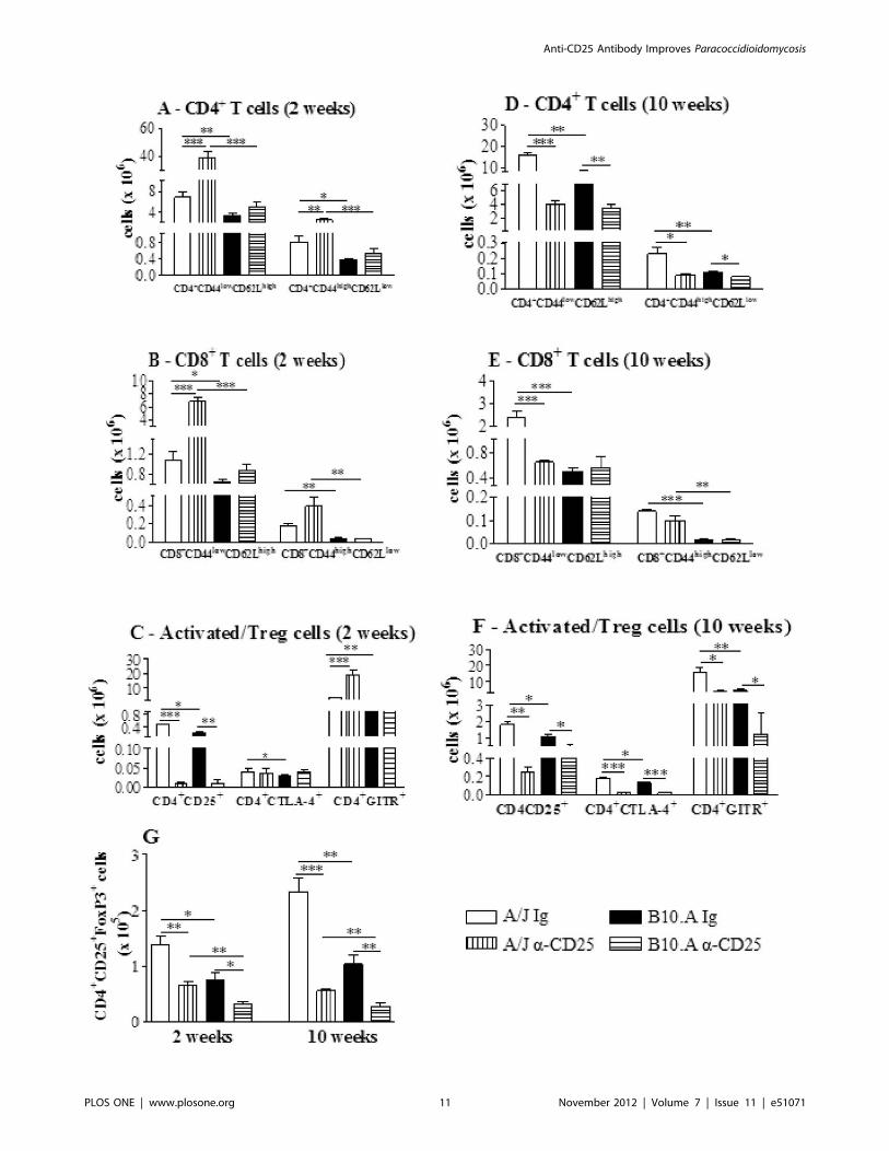
Anti-CD25 Treatment Increases the Early Influx of T cellsto the Lungs of Resistant but not of Susceptible Mice
The early depletion of CD25+ cells from A/J mice profoundly
affected the influx of inflammatory T cells to their lungs. Increased
numbers of CD4+ and CD8+ naıve (CD4+CD44lowCD62Lhigh and
CD8+CD44lowCD62Lhigh, respectively) as well as CD4+ and CD8+
effector (CD4+CD44highCD62Llow and CD8+CD44highCD62Llow)
T cells were detected at week 2 after infection. In contrast, no
alterations in the early influx of inflammatory T cells to the lungs
of B10.A mice were noticed (Fig. 7A–B). When other activation
markers of T cells were studied, only the CD4+CD25+ sub-
population appeared in decreased number in anti-CD25-depleted
groups, whereas CD4+GITR+ T cells were present in increased
numbers only in A/J mice. No differences were detected in the
CD4+CTLA4+ T cells (Fig. 7C). By week 10, almost all T cell
phenotypes were present in decreased numbers in the lungs of
resistant and susceptible mice (Fig. 7D–F). Thus, in A/J mice CD4
naıve and effector, CD8 naive, CD4+CD25+, CD4+CTLA4+,
CD4+GITR+ cells were found in decreased numbers (Fig.7D–F).
In B10.A mice, a decreased migration of CD4 naıve and effector T
cells were observed in the lungs, but no differences in CD8+ T cells
were found (Fig. 7D–F). In addition, CD4+ T cells expressing
CD25, CTLA4 and GITR were also present in decreased
numbers in the lungs of anti-CD25-treated B10.A mice (Fig. 7F).
To verify if anti-CD25 administration affected the presence of
Treg cells in the lungs, the number of CD4+CD25+ T cells
expressing Foxp3 was assessed. Indeed, despite the early in-
terruption (day 3 post-infection) of anti-CD25 administration,
a sustained depletion of Foxp3+ Treg cells was detected in the
lungs of both mouse strains at both post-infection periods studied
(Fig. 7G).
The Early Depletion of CD25+ Cells Promotes anIncreased Influx of Activated Macrophages and DendriticCells to the Lungs of Resistant Mice
By week 2 of infection, compared with IgG-inoculated controls,
A/J depleted mice showed a higher number of F4/80+GR1+, F4/
80+ IAK+ and CD11c+IAK+ myeloid cells. However, no differences
between treated and untreated B10.A mice were found (Fig. 8A).
Importantly, only in A/J mice depletion of Treg cells led to
Figure 3. Treg cells from resistant mice have a higher suppressive potency than those of susceptible mice. CFSE-labeled responderCD4+CD252 T cells from naıve mice were stimulated by irradiated naıve APCs plus anti-CD3 antibodies and cultured in the presence or absence ofseveral ratios of CD25+ T cells obtained from lungs of resistant and susceptible mice at weeks 2 (A) and 10 (B) after infection with 16106 P.brasiliensisyeasts. Cells were cultured for 5 days and the proliferative response of CFSE-labeled cells was measured by flow cytometry. The proliferation index (PI)was calculated as describe in materials and methods and the percentage of inhibition considered as 100% the PI of APC-stimulated CD4+CD252
responder cells in the absence of CD4+CD25+ cells. The data represent the mean 6 SEM of the results from 6 mice per group and are representativeof two independent experiments. * (P,0.05), ** (P,0.01), and *** (P,0.001) compared with A/J mice.doi:10.1371/journal.pone.0051071.g003
Anti-CD25 Antibody Improves Paracoccidioidomycosis
PLOS ONE | www.plosone.org 7 November 2012 | Volume 7 | Issue 11 | e51071
increased numbers of myeloid (CD11bhighCD11chigh) and plas-
macytoid (CD11cintB220+) subsets of dendritic cells (Fig. 8B).
An opposed effect on phagocytes was seen at week 10 of
infection. A diminished influx of almost all mononuclear
phagocytes and dendritic cells were seen in the lungs of resistant
mice. In B10.A depleted mice, only GR1+F4/80+ myeloid cells
migrated in reduced numbers to the lungs (Fig. 8A,B).
Treatment with Anti-CD25 Diminishes the EarlyProduction of IL-10 and TGF-b but Increases the Levels ofTh1/Th2 and Th17 Cytokines at the Late Phase ofInfection
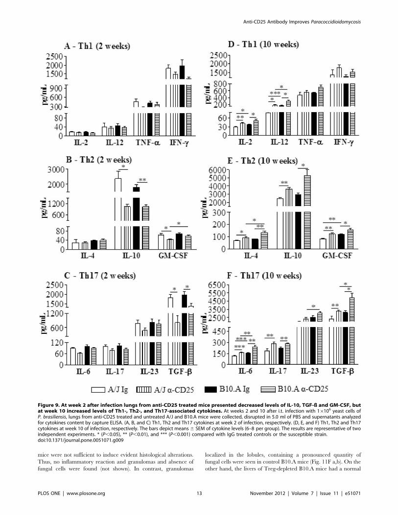
The presence of cytokines was analyzed in lung homogenates
obtained at weeks 2 and 10 after infection. Despite the reduced
pulmonary fungal loads, no differences in Th1 cytokines were
detected at week 2 of infection (Fig. 9A). However, besides GM-
CSF, the inhibitory cytokines IL-10 and TGF-b appeared in lower
levels in both mouse strains (Fig. 9B,C). In contrast with week 2, at
week 10 almost all assayed cytokines appeared in increased levels
in anti-CD25-treated mice. IL-2, IL-12, IL-4, IL-10, GM-CSF,
IL-17, IL-6 and TGF-b were secreted in higher levels by anti-
CD25-treated mice (Fig. 9D–F). Altogether, the cytokine data
demonstrate that anti-CD25 treatment induces an early decrease
of the inhibitory cytokines IL-10 and TGF-b concomitant with
unaltered production of pro-inflammatory cytokines. At the late
phase, the lower fungal loads were accompanied by increased
levels of almost all Th1/Th2 and Th17 cytokines assayed.
Depletion of CD25+ Cells Reduces the PulmonaryInflammation in Resistant and Susceptible Mice
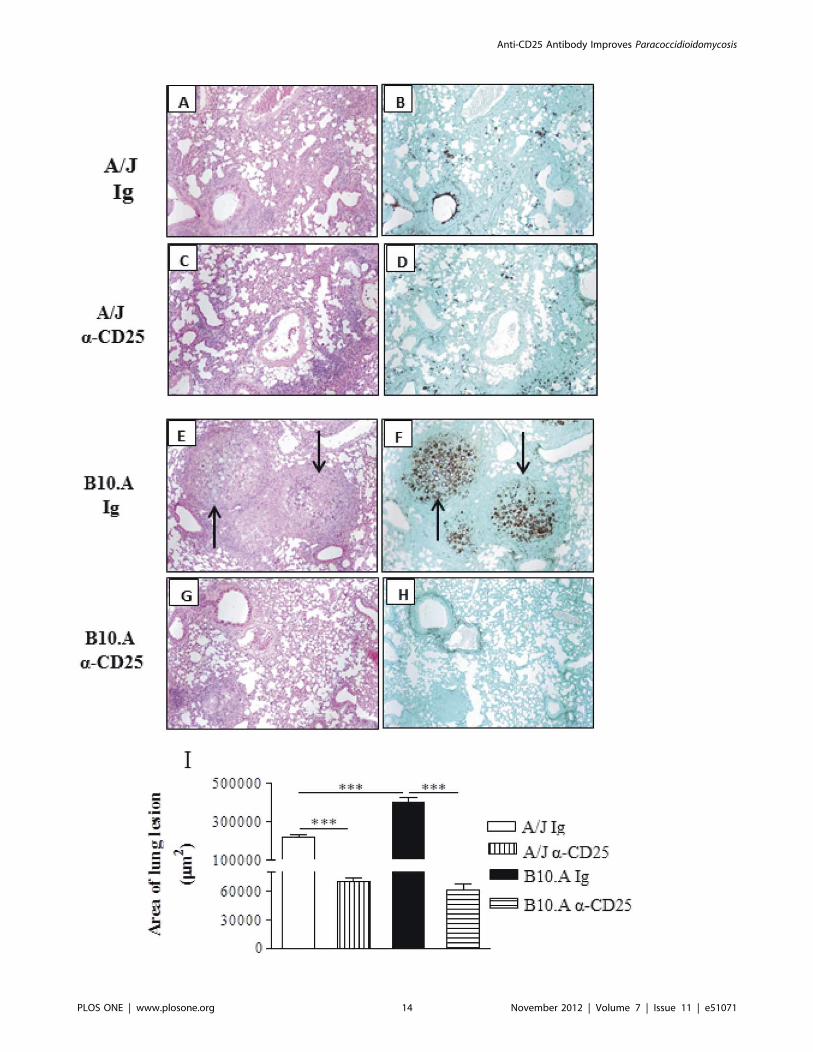
The histopathology of lungs at week 10 of infection was very
informative. IgG-treated A/J mice display a lymphomononuclear
infiltration of lungs, preferentially localized in the interlobular
septa close to the terminal bronchioles. Clumps of epithelioid
macrophages organize as incipient granulomas, but well defined
granulomas were absent. The number of fungi is small, and are
sometimes absent from the lesions (Fig. 10A,B). Anti-CD25-
treated A/J mice showed absence of granulomas and the
lymphocytic inflammatory infiltrates are present in the interlob-
ular and peribronchiolar septa, similarly to control A/J mice. The
fungi are absent or scattered in the lung parenchyma (Fig. 10C,D).
Control B10.A mice exhibit large granulomas involving extensive
areas of lung parenchyma, containing large number of fungi
(Fig. 10E,F). On the other hand, Treg depleted B10.A mice
showed isolated incipient granulomas containing no fungus or
a small amount of these cells. A more preserved area of
parenchyma was also observed (Fig. 10G,H). The total area of
lesions was quantified in histological sections and shown in Fig. 10I.
At week 10, the lesion areas of B10.A control mice were
significantly larger than those of control A/J mice. Importantly,
depletion of CD25+ cells significantly diminished the areas of
lesion in both mouse strains (Fig. 10I). In conclusion, the
histopathological studies demonstrated that administration of
anti-CD25 antibodies to resistant and susceptible mice results in
decreased fungal burdens which evolves without deleterious
enhanced tissue inflammation.
Anti-CD25 Treatment Reduces the Number ofInflammatory T Cells and Macrophages in the Liver of A/JMice but Only Decreases the Number of Macrophages inB10.A Mice
As dissemination and control of fungal growth in the liver
appear to be an important marker of the regressive and
Figure 4. Administration of anti-CD25 antibody decreases theearly and late organ fungal burdens of resistant and suscep-tible mice. Anti-CD25 mAb (PC61) or control IgG (500 mg) wereadministered at days 23 and +3 of P. brasiliensis infection. CFU countswere determined in the lungs (A), liver (B) and spleen (C) of resistantand susceptible mice at weeks 2 and 10 after infection. The pointsrepresent means 6 SEM of log10 CFU obtained from groups of six mice.The results are representative of 3 experiments * (P,0.05), ** (P,0.01),and *** (P,0.001) compared with IgG inoculated controls.doi:10.1371/journal.pone.0051071.g004
Anti-CD25 Antibody Improves Paracoccidioidomycosis
PLOS ONE | www.plosone.org 8 November 2012 | Volume 7 | Issue 11 | e51071
progressive infections of A/J and B10.A mice [5], we studied the
inflammatory infiltrates of this organ at week 10 of infection.
Comparing control (IgG-treated) A/J and B10.A mice, it could be
seen that resistant A/J mice had increased numbers of all T cell
subsets analyzed, whereas in B10.A mice only the myeloid cells
(GR1+F4/80+) predominated. Interestingly, Treg depletion in A/J
mice led to diminished numbers of CD4+ and CD8+
(CD4+CD44highCD62Llow, CD8+CD44lowCD62Lhigh,
CD4+CD25+, CD4+GITR+) T cells (Fig. 11A–C). In B10.A mice,
which presented a drastic reduction of CFU counts afteranti-
CD25 treatment, only myeloid cells (GR1+F4/80+, F4/80+IAk+)
appeared in decreased numbers (Fig. 11D). However, in both
mouse strains a significant reduction in CD4+CD25+Foxp3+ Treg
cells was found (Fig. 11E).
Figure 5. Anti-CD25 treatment depletes Foxp3GFP Treg cells and induces decreased fungal loads in Foxp3GFP C57BL/6 mice. Anti-CD25 antibodies (PC61) were administered as described in materials and methods to uninfected and P.brasiliensis infected (16106, i.t. route) Foxp3GFP
C57BL/6 mice. Normal Ig was injected in uninfected and infected control groups. Fungal loads (A), number of naıve (CD4+CD44lowCD62Lhigh) andactivated (CD4+CD44highCD62Llow) CD4+ T cells (B), number of CD4+CD25+Foxp3GFP Treg cells (D), percentage depletion of CD4+CD25+Foxp3GFP Tregcells (E), number of CD4+Foxp3GFP Treg cells (G) and percentage depletion of CD4+ Foxp3GFP Treg cells (E) were assessed in lung inflammatoryinfiltrates at week 2 after infection. Data are from a single experiment with 5–6 mice per group. Cell phenotypes were assessed by flow cytometry.Data represent mean 6 SEM. * (P,0.05), ** (P,0.01), and *** (P,0.001) between groups.doi:10.1371/journal.pone.0051071.g005
Anti-CD25 Antibody Improves Paracoccidioidomycosis
PLOS ONE | www.plosone.org 9 November 2012 | Volume 7 | Issue 11 | e51071
The histopathology of liver was also assessed in depleted and
non-depleted mice.
At week 10 of infection, the small and equivalent numbers of
viable yeasts present in the liver of control and Treg-depleted A/J
Figure 6. Anti-CD25 treatment increases the influx of inflammatory cells to the lungs of resistant but not susceptible mice toP. brasiliensis infection. Anti-CD25-treated and untreated A/J and B10.A mice were inoculated i.t. with 16106 P. brasiliensis yeast cells. At weeks 2and 10 of infection lungs of both mouse strains (n = 6) were excised, minced, and digested enzymatically. Lung cells suspensions were obtained,counted, stained for CD3 (T cells), CD19 (B cells) and GR1 (myeloid cells, including neutrophils and monocytes) by flow cytometry. Anti-CD25treatment significantly alters the number (A) but not the frequency of inflammatory cells in the lungs (B) of infected mice. At week 2, anti-CD25-treated A/J mice showed increased influx of T cells, B cells and myeloid cells, whereas at week 10 these populations appeared in decreased numbers(C, D). In B10.A mice only GR1+ cells appeared in decreased numbers at week 10 of infection (C, D). The data represent the mean 6 SEM of the resultsfrom 6–8 mice per group and are representative of two independent experiments. * (P,0.05), ** (P,0.01), and *** (P,0.001) compared with IgG-treated mice.doi:10.1371/journal.pone.0051071.g006
Anti-CD25 Antibody Improves Paracoccidioidomycosis
PLOS ONE | www.plosone.org 10 November 2012 | Volume 7 | Issue 11 | e51071
Anti-CD25 Antibody Improves Paracoccidioidomycosis
PLOS ONE | www.plosone.org 11 November 2012 | Volume 7 | Issue 11 | e51071
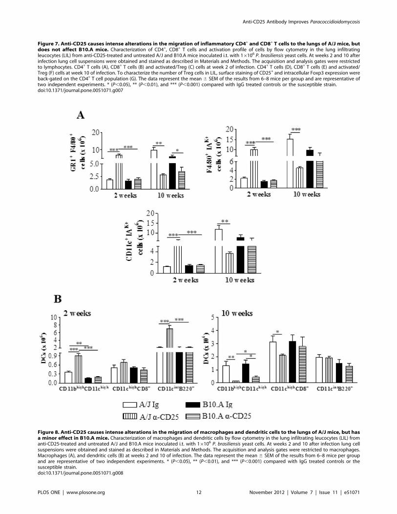
Figure 7. Anti-CD25 causes intense alterations in the migration of inflammatory CD4+ and CD8+ T cells to the lungs of A/J mice, butdoes not affect B10.A mice. Characterization of CD4+, CD8+ T cells and activation profile of cells by flow cytometry in the lung infiltratingleucocytes (LIL) from anti-CD25-treated and untreated A/J and B10.A mice inoculated i.t. with 16106 P. brasiliensis yeast cells. At weeks 2 and 10 afterinfection lung cell suspensions were obtained and stained as described in Materials and Methods. The acquisition and analysis gates were restrictedto lymphocytes. CD4+ T cells (A), CD8+ T cells (B) and activated/Treg (C) cells at week 2 of infection. CD4+ T cells (D), CD8+ T cells (E) and activated/Treg (F) cells at week 10 of infection. To characterize the number of Treg cells in LIL, surface staining of CD25+ and intracellular Foxp3 expression wereback-gated on the CD4+ T cell population (G). The data represent the mean 6 SEM of the results from 6–8 mice per group and are representative oftwo independent experiments. * (P,0.05), ** (P,0.01), and *** (P,0.001) compared with IgG treated controls or the susceptible strain.doi:10.1371/journal.pone.0051071.g007
Figure 8. Anti-CD25 causes intense alterations in the migration of macrophages and dendritic cells to the lungs of A/J mice, but hasa minor effect in B10.A mice. Characterization of macrophages and dendritic cells by flow cytometry in the lung infiltrating leucocytes (LIL) fromanti-CD25-treated and untreated A/J and B10.A mice inoculated i.t. with 16106 P. brasiliensis yeast cells. At weeks 2 and 10 after infection lung cellsuspensions were obtained and stained as described in Materials and Methods. The acquisition and analysis gates were restricted to macrophages.Macrophages (A), and dendritic cells (B) at weeks 2 and 10 of infection. The data represent the mean 6 SEM of the results from 6–8 mice per groupand are representative of two independent experiments. * (P,0.05), ** (P,0.01), and *** (P,0.001) compared with IgG treated controls or thesusceptible strain.doi:10.1371/journal.pone.0051071.g008
Anti-CD25 Antibody Improves Paracoccidioidomycosis
PLOS ONE | www.plosone.org 12 November 2012 | Volume 7 | Issue 11 | e51071
mice were not sufficient to induce evident histological alterations.
Thus, no inflammatory reaction and granulomas and absence of
fungal cells were found (not shown). In contrast, granulomas
localized in the lobules, containing a pronounced quantity of
fungal cells were seen in control B10.A mice (Fig. 11F a,b). On the
other hand, the livers of Treg-depleted B10.A mice had a normal
Figure 9. At week 2 after infection lungs from anti-CD25 treated mice presented decreased levels of IL-10, TGF-ß and GM-CSF, butat week 10 increased levels of Th1-, Th2-, and Th17-associated cytokines. At weeks 2 and 10 after i.t. infection with 16106 yeast cells ofP. brasiliensis, lungs from anti-CD25 treated and untreated A/J and B10.A mice were collected, disrupted in 5.0 ml of PBS and supernatants analyzedfor cytokines content by capture ELISA. (A, B, and C) Th1, Th2 and Th17 cytokines at week 2 of infection, respectively. (D, E, and F) Th1, Th2 and Th17cytokines at week 10 of infection, respectively. The bars depict means 6 SEM of cytokine levels (6–8 per group). The results are representative of twoindependent experiments. * (P,0.05), ** (P,0.01), and *** (P,0.001) compared with IgG treated controls or the susceptible strain.doi:10.1371/journal.pone.0051071.g009
Anti-CD25 Antibody Improves Paracoccidioidomycosis
PLOS ONE | www.plosone.org 13 November 2012 | Volume 7 | Issue 11 | e51071
Anti-CD25 Antibody Improves Paracoccidioidomycosis
PLOS ONE | www.plosone.org 14 November 2012 | Volume 7 | Issue 11 | e51071
aspect and did not present inflammatory reactions or fungal cells
(Fig. 11F c,d).
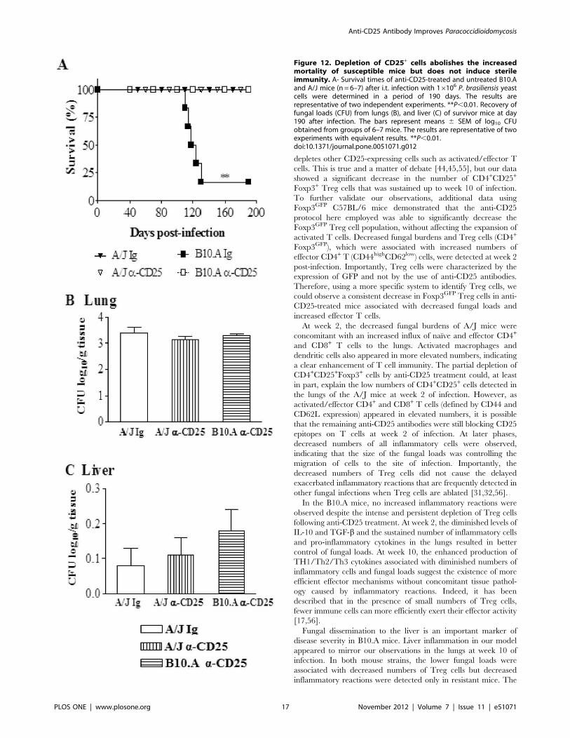
Anti-CD25 Treatment Abolishes the High Mortality ofSusceptible Mice but does not Induce Sterile Immunity
To assess the influence of anti-CD25 treatment on the disease
outcome, mortality of P.brasiliensis infected A/J and B10.A mice
previously treated or not with anti-CD25 antibodies (n = 6–7) was
registered daily after infection with 16106 yeast cells. As shown in
Fig. 12A, at day 130 after infection 5/6 (83%) of control B10.A
mice were dead. In the same period, all Treg-depleted B10.A and
A/J mice, besides control A/J mice, were still alive.
To assess if the early depletion of CD25+ cells led to the sterile
cure of mice, the presence of viable P. brasiliensis was analyzed in
the organs of survivor mice at day 190 post-infection. As shown in
Fig. 112B, low and equivalent numbers of viable yeast cells were
recovered from the lungs of all studied groups. Furthermore, a very
small number of yeast cells was still present in the livers (Fig. 12C)
but no viable P. brasiliensis were detected in the spleens. Altogether,
these data demonstrate that anti-CD25 treatment rescue suscep-
tible mice from increased mortality caused by the progressive
P.brasiliensis infection. In addition, this treatment did not induce
a sterile cure of infection, but eliminated the differences in the
disease outcome between susceptible and resistant mice to this
fungal pathogen.
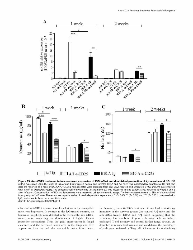
IDO mRNA Expression, Kynurenine and NO Productionare Reduced by Anti-CD25 Treatment of Infected Mice
Indoleamine 2,3 dioxygenase is an enzyme involved in
tryptophan catabolism. Its role in antimicrobial resistance by
depleting tryptophan, essential for the growth of microorganisms,
is well described [33,47–50]. In addition, IDO has an important
role in regulating T cell activation and preventing tissue pathology
due to excessive immunological inflammation. Due to their
capacity to induce Tregs and inhibit Th17, IDO and kynurenines
contribute to define the subsets of T cells which are activated
during fungal infections [47–50]. Our study demonstrated that
anti-CD25 treatment depleted Treg cells and was associated with
the activation of cytokines of the Th1/Th17 pathways. Thus we
decided to investigate the role of anti-CD25 treatment on the
expression of IDO and production of kynurenines. As shown in
Fig. 13A, P. brasiliensis infection induced a high expression of IDO
mRNA in the lungs of both, B10.A and A/J mice. This expression
was higher at week 1 than week 2 of infection, and was higher in
the lungs of A/J than in B10.A mice. In both mouse strains, anti-
CD25 treatment led to decreased expression of IDO. Consistent
with those findings, decreased levels of kynurenine were observed
in the lungs of CD25-depleted mice (Fig. 13B).
Because NO has been described as the major anti-fungal
compound in paracoccidioidomycosis [11,51], we have also
assessed the levels of nitrite in the lung homogenates of mice.
Interestingly, an inverse correlation with kynurenines was found
(Fig. 13C). At week 1 of infection, low levels of NO were
concomitant with high levels of kynurenines; at week 2 an inverse
picture was detected. Furthermore, by week 2 of infection,
susceptible mice produced higher levels of NO than resistant
mice and only in B10.A mice depletion of CD25+ cells resulted in
decreased levels of pulmonary NO.
Discussion
The murine model of paracoccidioidomycosis has some
peculiarities that help to explain the results here observed. The
resistance and susceptibility patterns of A/J and B10.A mice are
manifested late in the course of P.brasiliensis infection. As reported
before [5] and confirmed here, the initial infection is more severe
in the resistant A/J mice, which allows for precocious and more
intense dissemination of fungal cells than in B10.A mice. This
behavior appears to be governed, at least in part, by innate
immune cells. The alveolar macrophages and dendritic cells of A/J
mice are permissive to fungal growth and synthesize large amounts
of TGF-b, TNF-a and IL-6, followed by the late development of
CD4+ and CD8+ effector T cells ([7,52], Pina et al.submitted).
During the chronic phase, cell-mediated immunity predominates
and the infection is regressive in the lungs and in the dissemination
organs. In contrast, the infection in B10.A mice is initially well
controlled and characterized by early secretion of IL-12 and NO
by alveolar macrophages and DCs ([5,11,52], Pina et al.sub-
mitted). Excessive production of NO, however, has been shown to
cause T cell anergy, progressive infection and death in susceptible
mice [9,11].
The ex vivo analysis of Treg cells revealed some clues that help
us understand the differential roles of Treg cells in resistant and
susceptible mice. First, normal, uninfected A/J mice have higher
baseline numbers of Treg cells in their lungs than B10.A mice.
However, Foxp3+ Treg cells from both mouse strains express
membrane LAP, CTLA4, GITR and intracellular IL-10 and
TGF-b suggesting equivalent phenotypes and possibly mechan-
isms of action. During the course of infection, the numbers of Treg
cells were always greater in the resistant mice. The elevated
synthesis of TGF-b by alveolar macrophages and DCs of these
mice ([52], Pina et al submitted), likely contributed to the greater
number of Treg cells. The increased potency of A/J Tregs was
concomitant with an elevated expression of Foxp3, CTLA4 and
IDO which are markers of the suppressive ability of these cells
[27–29]. Interestingly, in several other infectious processes, strong
Treg responses also correlate with disease resistance. Resistance to
persistent salmonellosis depends on the early development of
highly suppressive Treg cells [28], and in invasive pneumococcal
pneumonia, the early immune response in resistant mice is
characterized by an increased synthesis of TGF-b and a rapid
increase in the number of lung Treg cells [53]. However, in
experimental leishmaniasis, higher suppressive activity of Treg
cells was observed in susceptible BALB/c mice [54].
This study demonstrated that anti-CD25 treatment of resistant
mice and susceptible mice resulted in persistent depletion of
CD4+CD25+Foxp3+ Treg cells. At this point it can be argued that
the use of anti-CD25 antibodies is not adequate to characterize
Treg cells function, since this antibody also neutralizes and/or
Figure 10. Histopathology of pulmonary lesions of anti-CD25-treated and untreated A/J and B10.A mice at week 10 post-infectionwith 16106 P. brasiliensis yeasts. IgG-treated (A, B) and anti-CD25-treated (C, D) A/J mice showed equivalent diffuse inflammatory reactionscharacterized by a small number of yeasts in the presence of elevated number of macrophages, lymphocytes and plasma cells; small portions of lungtissue were preserved, with limited signs of inflammatory cell recruitment. IgG-treated B10.A mice (E, F) presented an elevated number of well-defined, confluent, necrotic, granulomas of various sizes (E) containing an elevated number of fungal cells (arrows in F); these lesions occupy a largearea of lung tissue (E, F). Compared with control B10.A mice, anti-CD25-treated B10.A mice showed significantly smaller lesions (G) containing a fewnumber of yeasts (H). A, C, E, G, (HE, X 100); B, D, F, H (Groccot X 100). I- Total area of lung lesions of mice (n = 6) at week 10 after infection. ** (P,0.01),and *** (P,0.001) compared with IgG-treated controls or the susceptible strain.doi:10.1371/journal.pone.0051071.g010
Anti-CD25 Antibody Improves Paracoccidioidomycosis
PLOS ONE | www.plosone.org 15 November 2012 | Volume 7 | Issue 11 | e51071
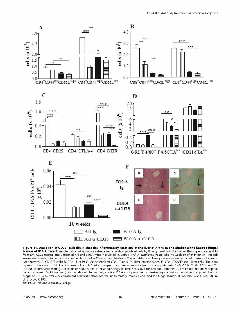
Figure 11. Depletion of CD25+ cells diminishes the inflammatory reactions in the liver of A/J mice and abolishes the hepatic fungallesions of B10.A mice. Characterization of leukocyte subsets and activation profile of cells by flow cytometry in the liver infiltrating leucocytes (LIL)from anti-CD25-treated and untreated A/J and B10.A mice inoculated i.t. with 16106 P. brasiliensis yeast cells. At week 10 after infection liver cellsuspensions were obtained and stained as described in Materials and Methods. The acquisition and analysis gates were restricted to macrophages orlymphocytes. A, CD4+ T cells; B, CD8+ T cells; C- Activated/Treg CD4+ T cells. D- Liver macrophages. E- CD4+CD25+Foxp3+ Treg cells. The datarepresent the mean 6 SEM of the results from 5–6 mice per group and are representative of two experiments. * (P,0.05), ** (P,0.01), and ***(P,0.001) compared with IgG controls or B10.A strain. F- Histopathology of liver. Anti-CD25 treated and untreated A/J mice did not show hepaticlesions at week 10 of infection (data not shown). In contrast, control B10.A mice presented extensive hepatic lesions containing large numbers offungal cells (F, a,b). Anti-CD25 treatment practically abolished the inflammatory lesions (F, c,d) and the fungal loads of B10.A mice. a, c (HE, X 100); b,d (Groccot X 100).doi:10.1371/journal.pone.0051071.g011
Anti-CD25 Antibody Improves Paracoccidioidomycosis
PLOS ONE | www.plosone.org 16 November 2012 | Volume 7 | Issue 11 | e51071
depletes other CD25-expressing cells such as activated/effector T
cells. This is true and a matter of debate [44,45,55], but our data
showed a significant decrease in the number of CD4+CD25+
Foxp3+ Treg cells that was sustained up to week 10 of infection.
To further validate our observations, additional data using
Foxp3GFP C57BL/6 mice demonstrated that the anti-CD25
protocol here employed was able to significantly decrease the
Foxp3GFP Treg cell population, without affecting the expansion of
activated T cells. Decreased fungal burdens and Treg cells (CD4+
Foxp3GFP), which were associated with increased numbers of
effector CD4+ T (CD44highCD62low) cells, were detected at week 2
post-infection. Importantly, Treg cells were characterized by the
expression of GFP and not by the use of anti-CD25 antibodies.
Therefore, using a more specific system to identify Treg cells, we
could observe a consistent decrease in Foxp3GFP Treg cells in anti-
CD25-treated mice associated with decreased fungal loads and
increased effector T cells.
At week 2, the decreased fungal burdens of A/J mice were
concomitant with an increased influx of naıve and effector CD4+
and CD8+ T cells to the lungs. Activated macrophages and
dendritic cells also appeared in more elevated numbers, indicating
a clear enhancement of T cell immunity. The partial depletion of
CD4+CD25+Foxp3+ cells by anti-CD25 treatment could, at least
in part, explain the low numbers of CD4+CD25+ cells detected in
the lungs of the A/J mice at week 2 of infection. However, as
activated/effector CD4+ and CD8+ T cells (defined by CD44 and
CD62L expression) appeared in elevated numbers, it is possible
that the remaining anti-CD25 antibodies were still blocking CD25
epitopes on T cells at week 2 of infection. At later phases,
decreased numbers of all inflammatory cells were observed,
indicating that the size of the fungal loads was controlling the
migration of cells to the site of infection. Importantly, the
decreased numbers of Treg cells did not cause the delayed
exacerbated inflammatory reactions that are frequently detected in
other fungal infections when Treg cells are ablated [31,32,56].
In the B10.A mice, no increased inflammatory reactions were
observed despite the intense and persistent depletion of Treg cells
following anti-CD25 treatment. At week 2, the diminished levels of
IL-10 and TGF-b and the sustained number of inflammatory cells
and pro-inflammatory cytokines in the lungs resulted in better
control of fungal loads. At week 10, the enhanced production of
TH1/Th2/Th3 cytokines associated with diminished numbers of
inflammatory cells and fungal loads suggest the existence of more
efficient effector mechanisms without concomitant tissue pathol-
ogy caused by inflammatory reactions. Indeed, it has been
described that in the presence of small numbers of Treg cells,
fewer immune cells can more efficiently exert their effector activity
[17,56].
Fungal dissemination to the liver is an important marker of
disease severity in B10.A mice. Liver inflammation in our model
appeared to mirror our observations in the lungs at week 10 of
infection. In both mouse strains, the lower fungal loads were
associated with decreased numbers of Treg cells but decreased
inflammatory reactions were detected only in resistant mice. The
Figure 12. Depletion of CD25+ cells abolishes the increasedmortality of susceptible mice but does not induce sterileimmunity. A- Survival times of anti-CD25-treated and untreated B10.Aand A/J mice (n = 6–7) after i.t. infection with 16106 P. brasiliensis yeastcells were determined in a period of 190 days. The results arerepresentative of two independent experiments. **P,0.01. Recovery offungal loads (CFU) from lungs (B), and liver (C) of survivor mice at day190 after infection. The bars represent means 6 SEM of log10 CFUobtained from groups of 6–7 mice. The results are representative of twoexperiments with equivalent results. **P,0.01.doi:10.1371/journal.pone.0051071.g012
Anti-CD25 Antibody Improves Paracoccidioidomycosis
PLOS ONE | www.plosone.org 17 November 2012 | Volume 7 | Issue 11 | e51071
effects of anti-CD25 treatment on liver lesions in the susceptible
mice were impressive. In contrast to the IgG-treated controls, no
lesions or fungal cells were detected in the livers of the anti-CD25-
treated mice, suggesting the development of highly efficient
protective mechanisms. Thus, the great improvement in fungal
clearance and the decreased lesion area in the lungs and liver
appear to have rescued the susceptible mice from death.
Furthermore, the anti-CD25 treatment did not lead to sterilizing
immunity in the survivor groups (the control A/J mice and the
anti-CD25 treated B10.A and A/J mice), suggesting that the
remaining low numbers of yeast cells were able to induce
prolonged T cell memory and control further fungal growth. As
described in murine leishmaniasis and candidiasis, the persistence
of pathogens conferred by Treg cells is important for maintaining
Figure 13. Anti-CD25 treatment induces reduced expression of IDO mRNA and diminished production of kynurenine and NO. IDOmRNA expression (A) in the lungs of IgG or anti-CD25 treated normal and infected B10.A and A/J mice was monitored by quantitative RT-PCR. Thedata are reported as a ratio of IDO/GAPDH. Lung homogenates were obtained from anti-CD25 treated and untreated B10.A and A/J mice infectedwith 16106 P. brasiliensis yeasts. The concentration of kynurenine (B) and nitrite (C) was measured in lung supernatants obtained at weeks 1 and 2after infection. Concentrations of NO and kynurenine were measured using colorimetric assays. The bars represent means 6 SEM of data obtainedfrom groups of 6–7 mice. The results are representative of two independent experiments. * (P,0.05), ** (P,0.01), and *** (P,0.001) compared withIgG treated controls or the susceptible strain.doi:10.1371/journal.pone.0051071.g013
Anti-CD25 Antibody Improves Paracoccidioidomycosis
PLOS ONE | www.plosone.org 18 November 2012 | Volume 7 | Issue 11 | e51071
memory cells and protective immunity that control further
microbial challenges [30,31].
The anti-inflammatory cytokine TGF-b drives the differentia-
tion of naıve CD4+CD25+ T cells to CD4+CD25+ regulatory T
cells by inducing Foxp3 [57]. In contrast, IL-6 prevents the
induction of Foxp3 by TGF-b and instead directs T cells towards
the Th17 lineage by inducing the RORct and RORa transcrip-
tion factors [58,59]. Our cytokine data at week 10 of infection
demonstrated increased levels of Th17-associated cytokines
without increased lung pathology. The increased levels of IL-10
also present in lung homogenates may have controlled the
pathogenicity of IL-17-producing cells without inhibiting their
protective function [59]. Moreover, anti-CD25 treatment of
susceptible and resistant mice led to reduced numbers of Treg
cells, IDO expression and kynurenines production. Thus, the
modulatory activity of anti-CD25 antibodies on Treg cells appears
to have potentiated the development of Th17 cells, possibly
contributing to the control of fungal burdens. Indeed, our previous
studies with C57BL/6 TLR2 deficient mice demonstrated that
inflammatory PMNs induced by enhanced Th17 immunity are
protective in paracoccidioidomycosis. However, the impaired
production of IL-10 by TLR2-deficient mice allowed increased
inflammatory reactions and lung pathology [38]. In another study,
we demonstrated that in TLR4-deficient mice, reduced numbers
of IL-17+ CD4+ T cells paralleled enhanced differentiation of Treg
cells, demonstrating the opposing development of Treg and Th17
cells in paracoccoccidioidomycosis [39].
The data obtained in this study demonstrate that the number
and activity of Treg cells, as well as the levels of IDO mRNA and
kynurenines, are modulated by anti-CD25 treatment. Our pre-
vious results suggested that the immunoregulatory mechanisms of
resistant mice are mainly governed by TGF-b, whereas pro-
inflammatory mediators, such as those induced by the IL-12/IFN-
c axis, are prevalent in susceptible mice [9,52]. In A/J mice, the
elevated levels of TGF-b secreted by alveolar macrophages and
DCs ([52], Pina et al. submitted), the high number of Treg cells
and the increased IDO activity indicate the existence of an initial
tolerogenic loop that is mediated by these cells and mediators. In
fact, it has been shown that Treg cells utilize TGF-b to maintain
the tolerogenic function of dendritic cells by inducing the ITIM
(immunoreceptor tyrosine-based inhibitory motif) mediated sig-
naling properties of IDO molecules. However, the IFN-c-induced
enzymatic activity of IDO leads to tryptophan starvation and the
generation of kynurenines, which are immunoregulatory catabo-
lites that promote the conversion of naıve T cells into Foxp3+ Treg
cells [60–62]. During the early phase of the infection, the relative
control of fungal growth in B10.A and A/J mice is partially IDO-
mediated [Frank de Araujo, manuscript in preparation]. However,
as was observed in this study, the enhanced migration of activated
T cells and macrophages to the site of infection, which was
observed only in resistant mice, was associated with anti-CD25
treatment and reduced numbers of Foxp3+ Treg cells. In other
words, the early immunosuppression observed in A/J mice
appears to be tightly regulated through a TGF-b-Treg-centered
mechanism that utilizes IDO to clear fungal cells and induce
tolerogenic dendritic cells. Moreover, anti-CD25 treatment
appears to block this suppressive loop, such that T cells can
expand and migrate to the site of infection.
Albeit at lower levels, B10.A mice also produced significant
levels of IDO and kynurenines and developed elevated numbers of
Treg cells after one week of infection. However, the reduced
production of IDO and kynurenines induced by anti-CD25
treatment was not able to restore the impaired T cell migration to
the lungs. Our previous studies showed that, in response to P.
brasiliensis infection, the alveolar macrophages and DCs in B10.A
mice secrete high levels of IL-12 and activate the secretion of IFN-
c by innate immune cells [7,10,52]. These pro-inflammatory
mediators induce the excessive production of NO and IDO, which
controls fungal growth and exerts a prominent role in the
immunosuppression of susceptible mice [9,11]. Consistent with
these observations, the in vivo depletion of NO [11] and the
inhibition of IDO activity by 1-methyl tryptophan [Frank de
Araujo, manuscript in preparation] restored T cell immunity and
the early migration of T cells and macrophages to the lungs of
B10.A mice. Thus, the early immunity generated in susceptible
mice appears to be governed by pro-inflammatory mediators (e.g.,
IL-12, IFN-c and NO) that likely regulate the IDO-mediated
control of Treg cells. In the present study, anti-CD25 treatment
also led to diminished levels of IL-10 and TGF-b, which reinforced
the suppressive pro-inflammatory milieu in the lungs of B10.A
mice [9,11]. However, this immunological balance that reduced
fungal loads without causing excessive tissue damage was able to
rescue susceptible mice from precocious death.
In conclusion, our studies are the first to demonstrate that Treg
cells exert deleterious effects on the mild and severe forms of
paracoccidioidomycosis. These results highlight new perspectives
for understanding the immunopathology of this infection. For
example, previous studies generally associated the presence of
Treg cells with severe cases of paracoccidioidomycosis [35–39].
Importantly, our data demonstrate that anti-CD25 treatment
causes a partial depletion of Treg cells and abolishes the
differences between the severe and mild forms of paracoccidioi-
domycosis, which indicates that the manipulation of Treg cells
may prove to be a new potential therapy against this important
systemic mycosis.
Acknowledgments
We are grateful to Tania A. Costa and Paulo Albee for their invaluable
technical assistance.
Author Contributions
Conceived and designed the experiments: VLGC MF. Performed the
experiments: MF AP EFA FVL SBB CF. Analyzed the data: MF AP EFA
FVL SBB CF. Wrote the paper: VLGC MF.
References
1. Franco M, Montenegro MR, Mendes RP, Marques SA, Dillon NL, et al. (1987)
Paracoccidioidomycosis: a recently proposed classification on its clinical forms.
Soc Bras Med Trop 20: 129–133.
2. Mamoni RL, Blotta MH (2005) Kinetics of cytokines and chemokines gene
expression distinguishes Paracoccidioides brasiliensis infection from disease. Cytokine
32: 20–29.
3. Benard G, Romano CC, Cacere CR, Juvenale M, Mendes-Giannini MJ, et al.
(2001) Imbalance of IL-2, IFN-gamma and IL-10 secretion in the immunosup-
pression associated with human paracoccidioidomycosis. Cytokine 13: 248–252.
4. Calich VLG, Singer-Vermes LM, Burger E (1985) Susceptibility and resistance
of inbred mice to Paracoccidioides brasiliensis. Br J Exp Pathol 66: 585–594.
5. Cano LE, Singer-Vermes LM, Vaz CAC, Russo M, Calich VLG (1995)
Pulmonary paracoccidioidomycosis in resistant and susceptible mice: relation-
ship among progression of infection, bronchoalveolar cell activation, cellular
immune response and specific isotype patterns. Infect Immun 63: 1777–1783.
6. Cano LE, Kashino SS, Arruda C, Andre DC, Xidieh CF, et al. (1998) Protective
role of gamma-interferon in experimental pulmonary paracoccidioidomycosis.
Infec Immun 66: 800–806.
7. Chiarella AP, Arruda C, Pina A, Costa TA, Ferreira RC, et al. (2007) The
relative importance of CD4+ and CD8+ T cell in immunity to pulmonary
paracoccidioidomycosis. Microbes Infect 9: 1078–1088.
Anti-CD25 Antibody Improves Paracoccidioidomycosis
PLOS ONE | www.plosone.org 19 November 2012 | Volume 7 | Issue 11 | e51071
8. Calich VLG, Blotta MHSL (2005) Pulmonary paracoccidioidomycosis. In
Fungal Immunology: From an Organ Perspective. Fidel, PL, Huffnagle, GB,
editors. Springer, New York. 201–228.
9. Calich VL, Costa TA, Felonato M, Arruda C, Bernardino S, et al. (2008) Innate
immunity to Paracoccidioides brasiliensis. Mycopathologia 165: 223–236.
10. Cano LE, Singer-Vermes LM, Costa TA, Mengel JO, Xidieh CF, et al. (2000)
Depletion of CD8+ T cells in vivo impairs host defense of mice resistant and
susceptible to pulmonary paracoccidioidomycosis. Infect Immun 68: 352–359.
11. Nascimento FRF, Calich VLG, Rodrigues D, Russo M (2002) Dual role for
nitric oxide in Paracoccidioidomycosis: essential for resistance, but over-
production associated with susceptibility. J Immunol 168: 4593–4600.
12. McHugh RS, Shevach EM (2002) The role of suppressor T cells in regulation of
immune responses. J Allergy Clin Immunol 110: 693–702.
13. Sakaguchi S, Sakaguchi N, Shimizu J, Yamazaki S, Sakihama T, et al. (2001)
Immunologic tolerance maintained by CD25+CD4+ regulatory T cells: their
common role in controlling autoimmunity, tumor immunity, and transplantation
tolerance. Immunol Rev 182: 18–32.
14. Belkaid Y, Rouse BT (2005) Natural regulatory T cells in infectious diseases.
Nature Immunol 6: 353–360.
15. Rudensky AY (2011) Regulatory T cells and Foxp3. Immunol Rev 241: 260–
268.
16. Sakaguchi S (2005) Naturally arising Foxp3-expressing CD25+CD4+ regulatory
T cells in immunological tolerance to self and non-self. Nat Immunol 6: 345–
352.
17. Belkaid Y, Tarbell K (2009) Regulatory T cells in the control of host-
microorganism interactions. Annu Rev Immunol 27: 551–589.
18. Miyara M, Sakaguchi S (2007) Natural regulatory T cells: mechanisms of
suppression. Trends Mol Med 13: 108–116.
19. Yamaguchi T, Hirota K, Nagahama K, Ohkawa K, Takahashi T, et al. (2007)
Control of immune responses by antigen-specific regulatory T cells expressing
the folate receptor. Immunity 24: 145–159.
20. Shevach EM, Stephens GL (2006) The GITR-GITRL interaction: co-
stimulation or contrasuppression of regulatory activity? Nat Rev Immunol 6:
613–618.
21. Deaglio S, Dwyer KM, Gao W, Friedman D, Usheva A, et al. (2007) Adenosine
generation catalyzed by CD39 and CD73 expressed on regulatory T cells
mediates immune suppression. J Exp Med 204: 1257–1265.
22. Ohkura N, Sakaguchi S (2011) Maturation of regulatory T cells. Nat Immunol
12: 283–284.
23. Hori S, Nomura T, Sakaguchi S (2003) Control of regulatory T cell development
by the transcription factor Foxp3. Science 299: 1057–1061.
24. Sakaguchi S (2003) The origin of Foxp3-expressing CD4+ regulatory T cells:
thymus or periphery. J Clin Invest 112: 1310–1312.
25. Fontenot JD, Dooley JL, Farr AG, Rudensky AY (2005) Developmental
regulation of Foxp3 expression during ontogeny. J Exp Med 202: 901–906.
26. Jonuleit H, Schmitt E (2003) The regulatory T cell family: distinct subsets and
their interrelations. J Immunol 171: 6323–6327.
27. Fontenot JD, Gavin MA, Rudensky AY (2003) Foxp3 programs the
development and function of CD4+CD25+ regulatory T cells. Nat Immunol 4:
330–336.
28. Johanns TM, Ertelt JM, Rowe JH, Way SS (2010) Regulatory T cell suppressive
potency dictates the balance between bacterial proliferation and clearance
during persistent Salmonella infection. PLoS Pathog 6: e1001043.
29. Wing K, Onishi Y, Prieto-Martin P, Yamaguchi T, Miyara M, et al. (2008)
CTLA-4 control over Foxp3+ regulatory T cell function. Science 322: 271–275.
30. Belkaid Y, Piccirilo AC, Mendez S, Shevack E, Sacks DL (2002) CD4+CD25+
regulatory T cells control Leishmania major persistence and immunity. Nature 420:
502–507.
31. Montagnoli C, Bacci A, Bozza S, Gaziano R, Mosci P, et al. (2002) B7/CD28-
dependent CD4+CD25+ regulatory T cells are essential components of the
memory-protective immunity to Candida albicans. J Immunol 169: 6298–6308.
32. Hori S, Carvalho TL, Demengeot J (2002) CD25+CD4+ regulatory suppress
CD4+ T cell-mediated pulmonary hyperinflammation driven by Pneumocystis
carinii in immunodeficient mice. Eur J Immunol 32: 1282–1291.
33. Montagnoli C, Fallarino F, Gaziano R, Bozza S, Bellocchio S, et al. (2006)
Immunity and tolerance to Aspergillus involve functionally distinct regulatory T
cells and tryptophan catabolism. 2006. J Immunol 176: 1712–1723.
34. Kroetz DN, Deepe GS (2010) CCR5 dictates the equilibrium of proinflamma-
tory IL-17+ and regulatory Foxp3+ T cells in fungal infection. J Immunol 184:
5224–5231.
35. Cavassani KA, Campanelli AP, Moreira AP, Vancim JO, Vitali LH, et al. (2006)
Systemic and local characterization of regulatory T cells in a chronic fungal
infection in humans. J Immunol 177: 5811–5818.
36. Ferreira MC, de Oliveira RT, da Silva RM, Blotta MH, Mamoni RL (2010)
Involvement of regulatory T cells in the immunosuppression characteristic of
patients with paracoccidioidomycosis. Infect Immun 78: 4392–4401.
37. Moreira AP, Cavassani KA, Massafera-Tristao FS, Campanelli AP, Martinez R,
et al. (2008) CCR5-dependent regulatory T cell migration mediates fungalsurvival and severe immunosuppression. J Immunol 180: 3049–3056.
38. Loures FV, Pina A, Felonato M, Calich VL (2009) TLR2 is a negative regulator
of Th17 cells and tissue pathology in a pulmonary model of fungal infection.J Immunol 183: 1279–1290.
39. Loures FV, Pina A, Felonato M, Araujo EF, Leite KR, et al. (2010) Toll-likereceptor 4 signaling leads to severe fungal infection associated with enhanced
proinflammatory immunity and impaired expansion of regulatory T cells. Infect
Immun 78: 1078–1088.40. Felonato M, Pina A, Bernardino S, Loures FV, de Araujo EF, et al. (2010) CD28
exerts protective and detrimental effects in a pulmonary model of paracocci-dioidomycosis. Infect Immun 78: 4922–4935.
41. Loures FV, Pina A, Felonato M, Feriotti C, de Araujo EF, et al. (2011) MyD88signaling is required for efficient innate and adaptive immune responses to
Paracoccidioides brasiliensis infection. Infect Immun 79: 2470–2480.
42. Kashino SS, Singer-Vermes LM, Calich VLG, Burger E (1990) Alterations inthe pathogenicity of one Paracoccidioides brasiliensis isolate do not correlate with its
in vitro growth. Mycopathology 111: 173–180.43. Fava Netto C, Vegas VS, Sciannamea IM, Guarnieri EDB (1969) Antıgeno
polissacarıdico do Paracoccidioides brasiliensis. Estudo do tempo de cultivo do P.
brasiliensis necessario ao preparo do antıgeno. Rev Inst Med Trop Sao Paulo 11:177–181.
44. Zelenay S, Demengeot J (2006) Comment on ‘‘Cutting edge: anti-CD25monoclonal antibody injection results in the functional inactivation, not
depletion, of CD4+CD25+ T regulatory cells’’. J Immunol 177: 2036–2037.45. Couper KN, Blount DG, de Souza JB, Suffia I, Belkaid Y, et al. (2007)
Incomplete depletion and rapid regeneration of Foxp3+ regulatory T cells
following anti-CD25 treatment in malaria-infected mice. J Immunol 178: 4136–4146.
46. Singer-Vermes LM, Ciavaglia MC, Kashino SS, Burger E, Calich VLG (1992)The source of the growth-promoting factor(s) affects the plating efficiency of
Paracoccidioides brasiliensis. J Med Vet Mycol 30: 261–264.
47. Bonifazi P, Zelante T, D’Angelo C, De Luca A, Moretti S, et al. (2009)Balancing inflammation and tolerance in vivo through dendritic cells by the
commensal Candida albicans. Mucosal Immunol 2: 362–374.48. Baban B, Chandler PR, Sharma MD, Pihkala J, Koni PA, et al. (2009) IDO
activates regulatory T cells and blocks their conversion into Th17-like T cells.J Immunol 183: 2475–2483.
49. De Luca A, Montagnoli C, Zelante T, Bonifazi P, Bozza S, et al. (2007)
Functional yet balanced reactivity to Candida albicans requires TRIF, MyD88,and IDO-dependent inhibition of Rorc. J Immunol 179: 5999–6008.
50. De Luca A, Bozza S, Zelante T, Zagarella S, D’Angelo C, et al. (2010) Non-hematopoietic cells contribute to protective tolerance to Aspergillus fumigatus via
a TRIF pathway converging on IDO. Cell Mol Immunol 7: 459–470.
51. Brummer E (1994) Interaction of Paracoccidioides brasiliensis with host defense cells.In: Franco M, Lacaz, CS, Restrepo, A, del Negro G, editors. Paracoccidioi-
domycosis. Boca Raton, Florida: CRC Press. 213–224.52. Pina A, Bernardino S, Calich VLG (2008) Alveolar macrophages from
susceptible mice are more competent than those of resistant mice to controlinitial Paracoccidioides brasiliensis infection. J Leukoc Biol 83: 1088–1099.
53. Neill DR, Fernandes VE, Wisby L, Haynes AR, Ferreira DM, et al. (2012) T
Regulatory cells control susceptibility to invasive pneumococcal pneumonia inmice. PloS Pathog 8: e1002660.
54. Chen X, Oppenheim JJ, Howard OMZ. (2005) BALB/c mice have moreCD4+CD25+ T regulatory cells and show greater susceptibility to suppression of
their CD4+CD252 responder than C57BL/6 mice. J Leukoc Biol 78: 114–121.
55. Rowe JH, Ertelt JM, Way SS (2011) Foxp3+ regulatory T cells, immunestimulation and host defense against infection. Immunol 136: 1–10.
56. McKinley L, Logar AJ, McAllister F, Zheng M, Steele C, et al. (2006)Regulatory T cells dampen pulmonary inflammation and lung injury in an
animal model of pneumocystis pneumonia. J Immunol 177: 6215–6226.
57. Chen W, Jin W, Hardegen N, Lei KJ, Li L, et al. (2003) Conversion ofperipheral CD4+CD252 naive T cells to CD4+CD25+ regulatory T cells by
TGF-beta induction of transcription factor Foxp3. J Exp Med 198: 1875–86.58. Bettelli EY, Carrier W, Gao T, Korn TB, Strom M, et al. (2006) Reciprocal
developmental pathways for the generation of pathogenic effector Th17 andregulatory T cells. Nature 441: 235–238.
59. McGeachy MJ, Bak-Jensen KS, Chen Y, Tato CM, Blumenschein W, et al.
(2007) TGF-beta and IL-6 drive the production of IL-17 and IL-10 by T cellsand restrain T(H)-17 cell-mediates pathology. Nat Immunol 8: 1390–1397.
60. Pallota MT, Orabona C, Volpi C, Vacca C, D’Angelo C, et al. (2011)Indoleamine 2,3-dioxygenase is a signaling protein in long term tolerance by
dendritic cells. Nat Immun. 12: 870–878.
61. Fallarino F. Grohmann U, Puccetti P (2012) Indoleamine 2,3-dioxygenase:fromcatalyst to signaling function. Eur J Immunol 42: 1932–1937.
62. Romani L. (2011) Immunity to fungal infections. Nat Rev Immunol 11: 275–288.
Anti-CD25 Antibody Improves Paracoccidioidomycosis
PLOS ONE | www.plosone.org 20 November 2012 | Volume 7 | Issue 11 | e51071