pneumothorax in connective tissue disease - plos
TRANSCRIPT
RESEARCH ARTICLE
Pneumothorax in connective tissue disease-
associated interstitial lung disease
Koji Nishimoto1, Tomoyuki FujisawaID1*, Katsuhiro Yoshimura1, Yasunori Enomoto1,
Hideki Yasui1, Hironao Hozumi1, Masato Karayama1, Yuzo Suzuki1, Kazuki Furuhashi1,
Noriyuki Enomoto1, Yutaro Nakamura1, Naoki Inui1,2, Hiromitsu Sumikawa3,
Takeshi Johkoh4, Takafumi Suda1
1 Second Division, Department of Internal Medicine, Hamamatsu University School of Medicine,
Hamamatsu, Japan, 2 Department of Clinical Pharmacology and Therapeutics, Hamamatsu University
School of Medicine, Hamamatsu, Japan, 3 Department of Radiology, Sakai City Medical Center, Sakai,
Japan, 4 Department of Radiology, Kansai Rosai Hospital, Amagasaki, Japan
Abstract
Background
Spontaneous pneumothorax is a complication that occurs in patients with connective tissue
disease-associated interstitial lung disease (CTD-ILD); however, few studies on the clinical
implications of pneumothorax for patients with CTD-ILD have been performed.
Objectives
This study aimed to investigate the incidence and prognostic significance of pneumothorax
and the risk factors for its onset in patients with CTD-ILD.
Methods
This study included 140 consecutive patients with CTD-ILD. Clinical characteristics, labora-
tory findings, pulmonary function test results, and chest high-resolution computed tomogra-
phy (HRCT) images were retrospectively evaluated.
Results
A total of 18 patients (12.9%) developed pneumothorax during their clinical course. The
cumulative incidence of pneumothorax from the time of CTD-ILD diagnosis was 6.5%,
8.7%, and 11.3% at 1, 3, and 5 years, respectively. The 10-year survival rate was signifi-
cantly lower in patients with pneumothorax (29.6%) than that in those without pneumothorax
(81.3%). The development of pneumothorax was significantly associated with poor progno-
sis (HR 22.0; p < 0.010). Furthermore, a lower body mass index, greater extent of reticular
abnormalities on HRCT, and administration of methylprednisolone pulse therapy were sig-
nificantly associated with the development of pneumothorax.
PLOS ONE
PLOS ONE | https://doi.org/10.1371/journal.pone.0235624 July 7, 2020 1 / 12
a1111111111
a1111111111
a1111111111
a1111111111
a1111111111
OPEN ACCESS
Citation: Nishimoto K, Fujisawa T, Yoshimura K,
Enomoto Y, Yasui H, Hozumi H, et al. (2020)
Pneumothorax in connective tissue disease-
associated interstitial lung disease. PLoS ONE
15(7): e0235624. https://doi.org/10.1371/
journal.pone.0235624
Editor: Atsushi Miyamoto, Toranomon Hospital,
JAPAN
Received: April 6, 2020
Accepted: June 18, 2020
Published: July 7, 2020
Copyright: © 2020 Nishimoto et al. This is an open
access article distributed under the terms of the
Creative Commons Attribution License, which
permits unrestricted use, distribution, and
reproduction in any medium, provided the original
author and source are credited.
Data Availability Statement: All relevant data are
within the paper and its Supporting Information
files.
Funding: The authors received no specific funding
for this work.
Competing interests: The authors have declared
that no competing interests exist.
Conclusion
Pneumothorax is a serious complication in the clinical course of patients with CTD-ILD and
the onset of pneumothorax predicts a poor outcome.
Introduction
Interstitial lung disease (ILD) is one of the common complications in patients with connective
tissue disease (CTD), such as rheumatoid arthritis (RA), polymyositis (PM)/dermatomyositis
(DM), Sjogren’s syndrome (SjS), and systemic scleroderma (SSc) [1]. ILD is associated with
impaired respiratory function and leads to an increased risk of mortality in various CTDs [2];
thus, it is clinically important to determine the factors associated with poor outcomes in
patients with CTD-associated ILD (CTD-ILD). Several studies have demonstrated the prog-
nostic significance of clinical data, including older age, male sex, and lower level of forced vital
capacity (FVC), in patients with CTD-ILD [3–7].
Pneumothorax sometimes occurs in patients with CTD-ILD during their clinical course.
Generally, secondary spontaneous pneumothorax induces respiratory failure more frequently
than primary spontaneous pneumothorax [8–12]. Furthermore, the recurrence rate of second-
ary spontaneous pneumothorax is considered to be higher than that of primary spontaneous
pneumothorax [10, 12]. We have previously reported that pneumothorax developed in 20.2%
of patients with idiopathic pulmonary fibrosis (IPF) during their clinical courses, and the
onset of pneumothorax was significantly associated with a poor outcome [13]. The onset of
pneumothorax may also affect the clinical course and prognosis of patients with CTD-ILD;
however, there have been no studies on the incidence and clinical importance of pneumotho-
rax in patients with CTD-ILD. In addition, few studies have assessed the risk factors for the
onset of pneumothorax in patients with CTD-ILD. In general, the male sex, a tall and thin stat-
ure, a history of smoking, and the presence of subpleural bullae are considered to be risk fac-
tors of primary spontaneous pneumothorax [8, 12, 14]. However, whether these factors are
associated with the onset of pneumothorax in patients with CTD-ILD remains uncertain, as
pre-existing diseases and drugs used for treatment may affect the conditions. In the present
study, we investigated the incidence and prognostic significance of pneumothorax in patients
with CTD-ILD. In addition, risk factors for the onset of pneumothorax were assessed in
patients with CTD-ILD.
Methods
Study subjects
Clinical records were retrospectively reviewed for 140 consecutive patients with CTD-ILD,
who were evaluated at Hamamatsu University Hospital (Hamamatsu, Japan) between January
2000 and December 2014. All data were fully anonymized before we accessed them. Medical
records were accessed between May 2018 and May 2019. The diagnoses of CTDs were based
on accepted criteria[15–18], according to clinical findings. For RA or SjS, we have reassessed
according to the criteria [15, 18] and confirmed all the patients with RA or SjS in this study
were fulfilled the criteria. The diagnosis of ILD was based on the presence of respiratory symp-
toms, physical examination findings, chest high-resolution computed tomography (HRCT)
findings, and pulmonary function tests. Other diseases, such as infection, drug-induced pneu-
monia, aspiration-induced lung injury, and heart failure, were excluded. No patients had
PLOS ONE Pneumothorax in CTD-ILD
PLOS ONE | https://doi.org/10.1371/journal.pone.0235624 July 7, 2020 2 / 12
concurrent advanced cancer at the time of CTD-ILD diagnosis. This study was conducted in
accordance with the ethical standards of the Declaration of Helsinki and approved by the Insti-
tutional Review Board of Hamamatsu University School of Medicine (approval no. 15–197).
The Institutional Review Board waived patient approval or informed consent as the study
involved a retrospective review of patient records.
Clinical data collection
We retrospectively reviewed the patients’ medical records, laboratory data, pulmonary func-
tion data, and lung HRCT image findings obtained at the time of ILD diagnosis. We also
reviewed treatments for CTD-ILD, such as the use of prednisolone and immunosuppressive
agents (e.g., cyclophosphamide, cyclosporin, tacrolimus, azathioprine, and methotrexate), dur-
ing the observation period.
Radiological analysis
HRCT images were obtained from all the patients on initial diagnosis of ILD and were
reviewed by two expert thoracic radiologists with 30- and 15-years, respectively, who had no
access to the patients’ clinical data. The slice thickness of the HRCT was 1.0 or 1.5-mm and the
section interval was 10-mm. The HRCT findings, including the presence of honeycombing,
reticular pattern, emphysema, ground-glass opacity (GGO), and consolidation, were inter-
preted according to the Fleischner Society criteria [19]. The extent of reticular abnormalities
(i.e., reticular pattern and honeycombing), emphysema, GGO, and consolidation were semi-
quantitatively scored as follows: Grade 0 (0%), Grade 1 (<25%), Grade 2 (25–50%), Grade 3
(50–75%), and Grade 4 (>75%). During the observation period, the patients underwent chest
radiography or HRCT at least every 6 months. The clinical records and all radiological images
(chest radiographs or HRCT images) were reviewed by three pulmonologists to confirm the
onset of pneumothorax.
Statistical analysis
Data are presented as the number (percentage) or median (interquartile ranges). Either Fish-
er’s exact test or the Mann-Whitney U test was used for between-group comparisons. The
cumulative incidence of pneumothorax was evaluated using Gray’s test, considering any cause
of death as an important competing factor. The observation period for survival was calculated
from the date of initial ILD diagnosis to the final date of contact or time of death. Univariate
and multivariate Cox proportional hazards model analyses were performed to determine the
predictive factors affecting the prognosis and onset of pneumothorax. The onset of pneumo-
thorax, use of prednisolone, immunosuppressive agents and methylprednisolone pulse therapy
were analyzed as time-dependent covariates. The Kaplan–Meier method was used to produce
survival curves, and the survival rates between the groups of patients were compared with a
log-rank test. A p-value of<0.05 was considered statistically significant for all analyses. All sta-
tistical analyses were performed with EZR (Saitama Medical Centre, Jichi Medical University,
Saitama, Japan), which is a graphical user interface for R software (version 2.13.0, The R Foun-
dation for Statistical Computing, Vienna, Austria) [20].
Results
Clinical characteristics
The baseline characteristics of the study subjects at the time of CTD-ILD diagnosis are shown
in Table 1. The median age was 62.5 years, and 61 patients (43.6%) were male. The median
PLOS ONE Pneumothorax in CTD-ILD
PLOS ONE | https://doi.org/10.1371/journal.pone.0235624 July 7, 2020 3 / 12
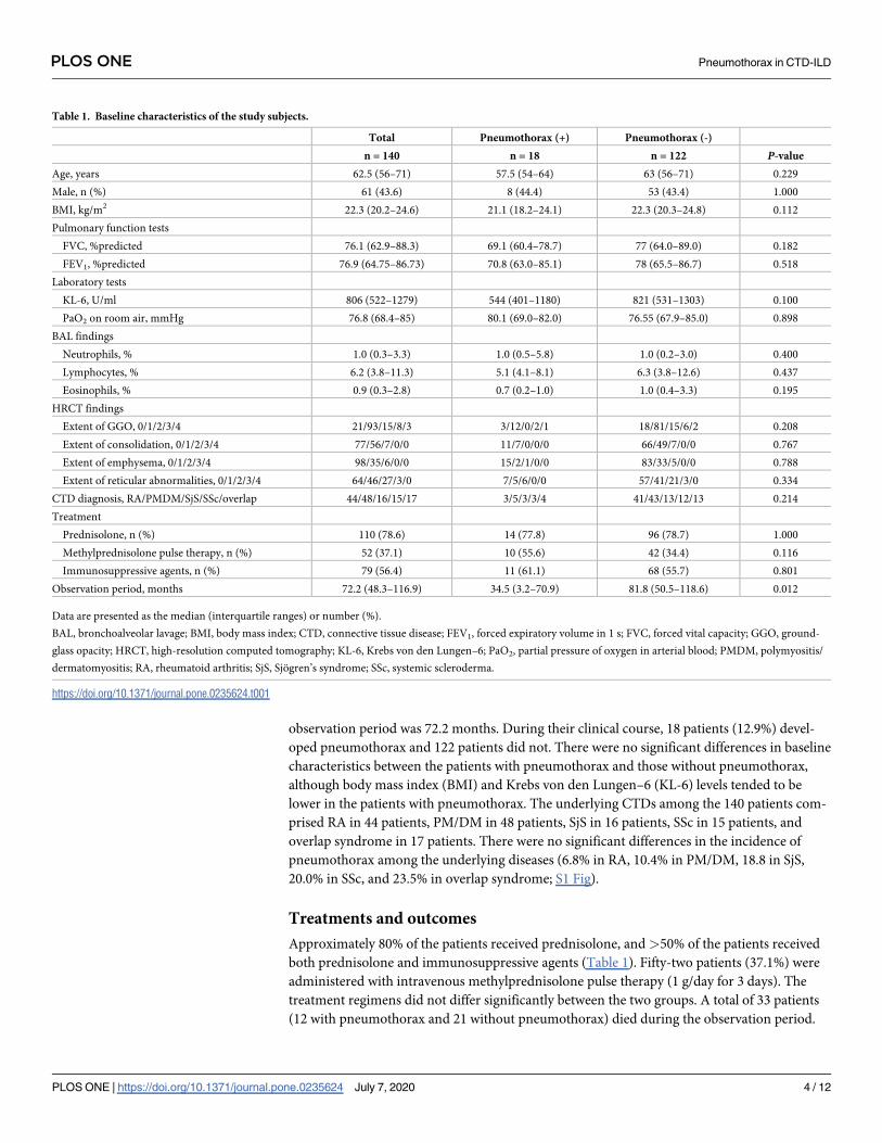
observation period was 72.2 months. During their clinical course, 18 patients (12.9%) devel-
oped pneumothorax and 122 patients did not. There were no significant differences in baseline
characteristics between the patients with pneumothorax and those without pneumothorax,
although body mass index (BMI) and Krebs von den Lungen–6 (KL-6) levels tended to be
lower in the patients with pneumothorax. The underlying CTDs among the 140 patients com-
prised RA in 44 patients, PM/DM in 48 patients, SjS in 16 patients, SSc in 15 patients, and
overlap syndrome in 17 patients. There were no significant differences in the incidence of
pneumothorax among the underlying diseases (6.8% in RA, 10.4% in PM/DM, 18.8 in SjS,
20.0% in SSc, and 23.5% in overlap syndrome; S1 Fig).
Treatments and outcomes
Approximately 80% of the patients received prednisolone, and >50% of the patients received
both prednisolone and immunosuppressive agents (Table 1). Fifty-two patients (37.1%) were
administered with intravenous methylprednisolone pulse therapy (1 g/day for 3 days). The
treatment regimens did not differ significantly between the two groups. A total of 33 patients
(12 with pneumothorax and 21 without pneumothorax) died during the observation period.
Table 1. Baseline characteristics of the study subjects.
Total Pneumothorax (+) Pneumothorax (-)
n = 140 n = 18 n = 122 P-value
Age, years 62.5 (56–71) 57.5 (54–64) 63 (56–71) 0.229
Male, n (%) 61 (43.6) 8 (44.4) 53 (43.4) 1.000
BMI, kg/m2 22.3 (20.2–24.6) 21.1 (18.2–24.1) 22.3 (20.3–24.8) 0.112
Pulmonary function tests
FVC, %predicted 76.1 (62.9–88.3) 69.1 (60.4–78.7) 77 (64.0–89.0) 0.182
FEV1, %predicted 76.9 (64.75–86.73) 70.8 (63.0–85.1) 78 (65.5–86.7) 0.518
Laboratory tests
KL-6, U/ml 806 (522–1279) 544 (401–1180) 821 (531–1303) 0.100
PaO2 on room air, mmHg 76.8 (68.4–85) 80.1 (69.0–82.0) 76.55 (67.9–85.0) 0.898
BAL findings
Neutrophils, % 1.0 (0.3–3.3) 1.0 (0.5–5.8) 1.0 (0.2–3.0) 0.400
Lymphocytes, % 6.2 (3.8–11.3) 5.1 (4.1–8.1) 6.3 (3.8–12.6) 0.437
Eosinophils, % 0.9 (0.3–2.8) 0.7 (0.2–1.0) 1.0 (0.4–3.3) 0.195
HRCT findings
Extent of GGO, 0/1/2/3/4 21/93/15/8/3 3/12/0/2/1 18/81/15/6/2 0.208
Extent of consolidation, 0/1/2/3/4 77/56/7/0/0 11/7/0/0/0 66/49/7/0/0 0.767
Extent of emphysema, 0/1/2/3/4 98/35/6/0/0 15/2/1/0/0 83/33/5/0/0 0.788
Extent of reticular abnormalities, 0/1/2/3/4 64/46/27/3/0 7/5/6/0/0 57/41/21/3/0 0.334
CTD diagnosis, RA/PMDM/SjS/SSc/overlap 44/48/16/15/17 3/5/3/3/4 41/43/13/12/13 0.214
Treatment
Prednisolone, n (%) 110 (78.6) 14 (77.8) 96 (78.7) 1.000
Methylprednisolone pulse therapy, n (%) 52 (37.1) 10 (55.6) 42 (34.4) 0.116
Immunosuppressive agents, n (%) 79 (56.4) 11 (61.1) 68 (55.7) 0.801
Observation period, months 72.2 (48.3–116.9) 34.5 (3.2–70.9) 81.8 (50.5–118.6) 0.012
Data are presented as the median (interquartile ranges) or number (%).
BAL, bronchoalveolar lavage; BMI, body mass index; CTD, connective tissue disease; FEV1, forced expiratory volume in 1 s; FVC, forced vital capacity; GGO, ground-
glass opacity; HRCT, high-resolution computed tomography; KL-6, Krebs von den Lungen–6; PaO2, partial pressure of oxygen in arterial blood; PMDM, polymyositis/
dermatomyositis; RA, rheumatoid arthritis; SjS, Sjogren’s syndrome; SSc, systemic scleroderma.
https://doi.org/10.1371/journal.pone.0235624.t001
PLOS ONE Pneumothorax in CTD-ILD
PLOS ONE | https://doi.org/10.1371/journal.pone.0235624 July 7, 2020 4 / 12
The mortality rate was significantly higher in patients with pneumothorax than in those with-
out (66.7% vs. 17.2%, respectively; p< 0.001). The mortality rate was similar among the under-
lying diseases (20.5% for RA, 20.8% for PM/DM, 31.3% for SjS, 26.7% for SSc, and 29.4% for
overlap (S1 Table). In all CTDs, the mortality rate was higher in patients with pneumothorax
than in those without pneumothorax (S1 Table).
Cumulative incidence of pneumothorax in CTD-ILD
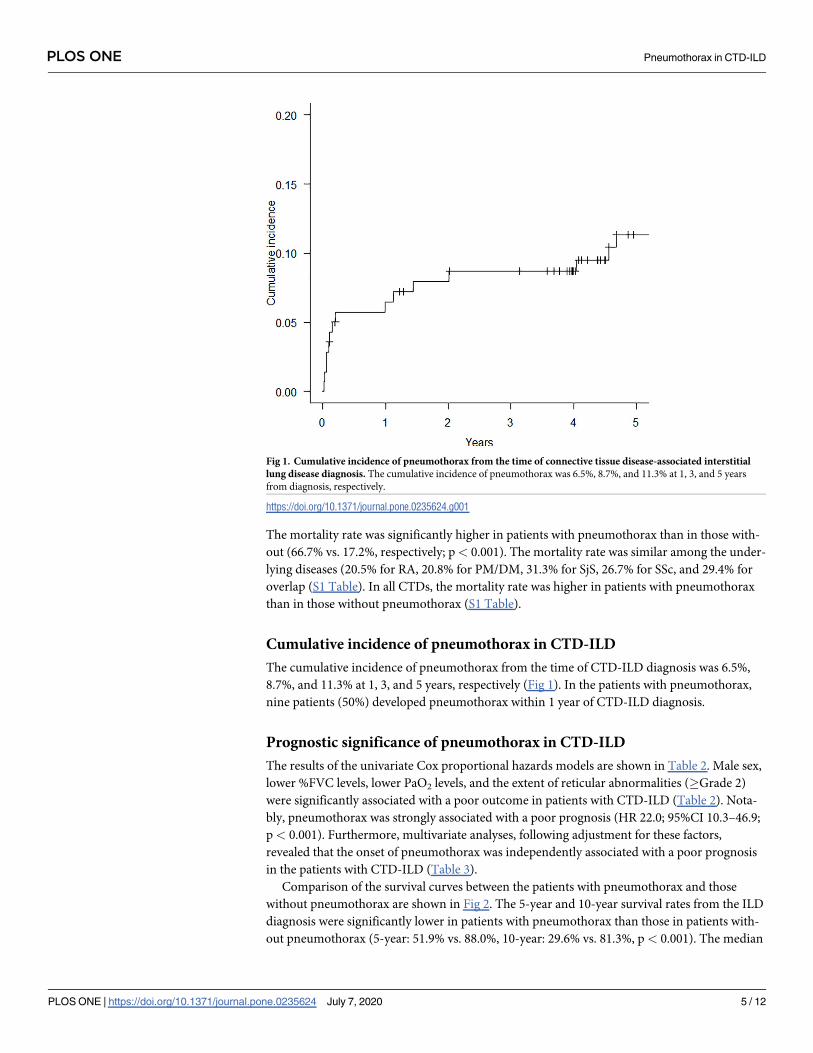
The cumulative incidence of pneumothorax from the time of CTD-ILD diagnosis was 6.5%,
8.7%, and 11.3% at 1, 3, and 5 years, respectively (Fig 1). In the patients with pneumothorax,
nine patients (50%) developed pneumothorax within 1 year of CTD-ILD diagnosis.
Prognostic significance of pneumothorax in CTD-ILD
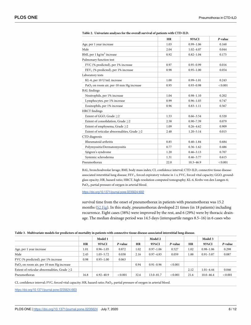
The results of the univariate Cox proportional hazards models are shown in Table 2. Male sex,
lower %FVC levels, lower PaO2 levels, and the extent of reticular abnormalities (�Grade 2)
were significantly associated with a poor outcome in patients with CTD-ILD (Table 2). Nota-
bly, pneumothorax was strongly associated with a poor prognosis (HR 22.0; 95%CI 10.3–46.9;
p< 0.001). Furthermore, multivariate analyses, following adjustment for these factors,
revealed that the onset of pneumothorax was independently associated with a poor prognosis
in the patients with CTD-ILD (Table 3).
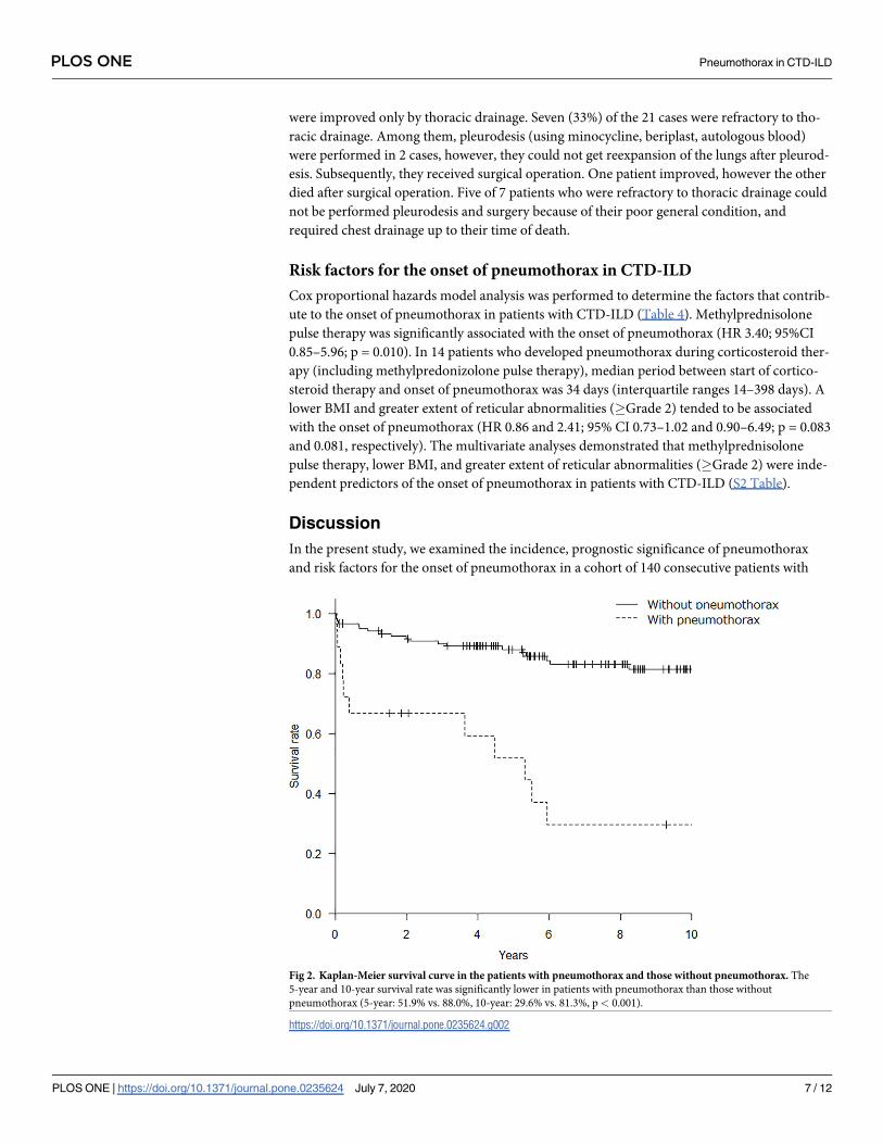
Comparison of the survival curves between the patients with pneumothorax and those
without pneumothorax are shown in Fig 2. The 5-year and 10-year survival rates from the ILD
diagnosis were significantly lower in patients with pneumothorax than those in patients with-
out pneumothorax (5-year: 51.9% vs. 88.0%, 10-year: 29.6% vs. 81.3%, p < 0.001). The median
Fig 1. Cumulative incidence of pneumothorax from the time of connective tissue disease-associated interstitial
lung disease diagnosis. The cumulative incidence of pneumothorax was 6.5%, 8.7%, and 11.3% at 1, 3, and 5 years
from diagnosis, respectively.
https://doi.org/10.1371/journal.pone.0235624.g001
PLOS ONE Pneumothorax in CTD-ILD
PLOS ONE | https://doi.org/10.1371/journal.pone.0235624 July 7, 2020 5 / 12
survival time from the onset of pneumothorax in patients with pneumothorax was 15.2
months (S2 Fig). In this study, pneumothorax developed 21 times (in 18 patients) including
recurrence. Eight cases (38%) were improved by the rest, and 6 (29%) were by thoracic drain-
age. The median drainage period was 14.5 days (interquartile ranges 8.5–16) in 6 cases who
Table 2. Univariate analyses for the overall survival of patients with CTD-ILD.
HR 95%CI P-value
Age, per 1 year increase 1.03 0.99–1.06 0.160
Male 2.04 1.02–4.07 0.044
BMI, per 1 kg/m2 increase 0.92 0.82–1.04 0.175
Pulmonary function test
FVC (% predicted), per 1% increase 0.97 0.95–0.99 0.016
FEV1 (% predicted), per 1% increase 0.98 0.95–1.00 0.054
Laboratory tests
KL-6, per 10 U/mL increase 1.00 0.99–1.01 0.243
PaO2 on room air, per-10 mm Hg increase 0.95 0.93–0.98 <0.001
BAL findings
Neutrophils, per 1% increase 1.04 0.98–1.10 0.202
Lymphocytes, per 1% increase 0.99 0.96–1.03 0.747
Eosinophils, per 1% increase 0.96 0.83–1.11 0.567
HRCT findings
Extent of GGO, Grade�2 1.53 0.66–3.54 0.320
Extent of consolidation, Grade�2 2.58 0.90–7.39 0.079
Extent of emphysema, Grade�2 1.09 0.26–4.62 0.909
Extent of reticular abnormalities, Grade�2 2.48 1.20–5.14 0.015
CTD diagnosis
Rheumatoid arthritis 0.85 0.40–1.84 0.684
Polymyositis/Dermatomyositis 0.77 0.36–1.62 0.486
Sjogren’s syndrome 1.20 0.46–3.13 0.707
Systemic scleroderma 1.31 0.46–3.77 0.615
Pneumothorax 22.0 10.3–46.9 <0.001
BAL, bronchoalveolar lavage; BMI, body mass index; CI, confidence interval; CTD-ILD, connective tissue disease-
associated interstitial lung disease; FEV1, forced expiratory volume in 1 s; FVC, forced vital capacity; GGO, ground-
glass opacity; HR, hazard ratio; HRCT, high-resolution computed tomography; KL-6, Krebs von den Lungen-6;
PaO2, partial pressure of oxygen in arterial blood.
https://doi.org/10.1371/journal.pone.0235624.t002
Table 3. Multivariate models for predictors of mortality in patients with connective tissue disease-associated interstitial lung disease.
Model 1 Model 2 Model 3
HR 95%CI P-value HR 95%CI P-value HR 95%CI P-value
Age, per 1 year increase 1.01 0.96–1.05 0.872 1.02 0.97–1.06 0.527 1.02 0.98–1.06 0.298
Male 2.45 1.05–5.72 0.038 2.16 0.97–4.83 0.059 1.88 0.91–3.87 0.087
FVC (% predicted), per 1% increase 0.98 0.95–1.00 0.063
PaO2 on room air, per 10 mm Hg increase 0.94 0.91–0.96 <0.001
Extent of reticular abnormalities, Grade�2 2.12 1.01–4.44 0.046
Pneumothorax 16.8 6.92–40.9 <0.001 32.6 13.0–81.7 <0.001 21.6 10.0–46.4 <0.001
CI, confidence interval; FVC, forced vital capacity; HR, hazard ratio; PaO2, partial pressure of oxygen in arterial blood.
https://doi.org/10.1371/journal.pone.0235624.t003
PLOS ONE Pneumothorax in CTD-ILD
PLOS ONE | https://doi.org/10.1371/journal.pone.0235624 July 7, 2020 6 / 12
were improved only by thoracic drainage. Seven (33%) of the 21 cases were refractory to tho-
racic drainage. Among them, pleurodesis (using minocycline, beriplast, autologous blood)
were performed in 2 cases, however, they could not get reexpansion of the lungs after pleurod-
esis. Subsequently, they received surgical operation. One patient improved, however the other
died after surgical operation. Five of 7 patients who were refractory to thoracic drainage could
not be performed pleurodesis and surgery because of their poor general condition, and
required chest drainage up to their time of death.
Risk factors for the onset of pneumothorax in CTD-ILD
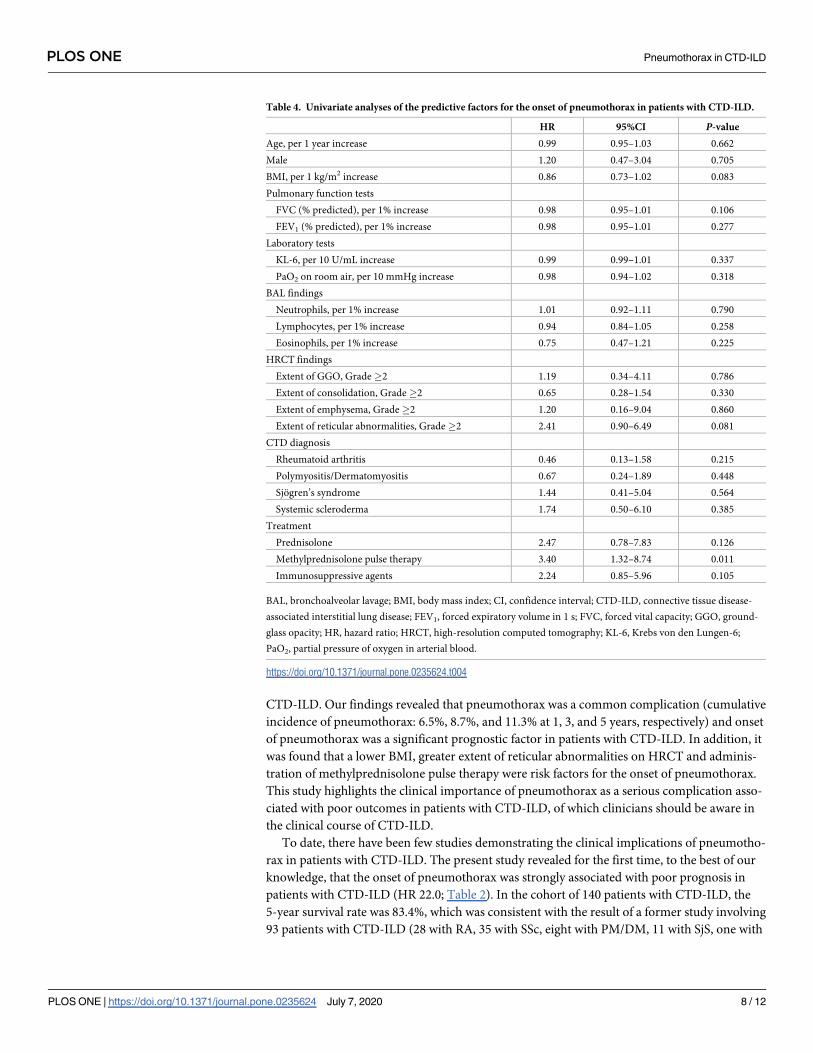
Cox proportional hazards model analysis was performed to determine the factors that contrib-
ute to the onset of pneumothorax in patients with CTD-ILD (Table 4). Methylprednisolone
pulse therapy was significantly associated with the onset of pneumothorax (HR 3.40; 95%CI
0.85–5.96; p = 0.010). In 14 patients who developed pneumothorax during corticosteroid ther-
apy (including methylpredonizolone pulse therapy), median period between start of cortico-
steroid therapy and onset of pneumothorax was 34 days (interquartile ranges 14–398 days). A
lower BMI and greater extent of reticular abnormalities (�Grade 2) tended to be associated
with the onset of pneumothorax (HR 0.86 and 2.41; 95% CI 0.73–1.02 and 0.90–6.49; p = 0.083
and 0.081, respectively). The multivariate analyses demonstrated that methylprednisolone
pulse therapy, lower BMI, and greater extent of reticular abnormalities (�Grade 2) were inde-
pendent predictors of the onset of pneumothorax in patients with CTD-ILD (S2 Table).
Discussion
In the present study, we examined the incidence, prognostic significance of pneumothorax
and risk factors for the onset of pneumothorax in a cohort of 140 consecutive patients with
Fig 2. Kaplan-Meier survival curve in the patients with pneumothorax and those without pneumothorax. The
5-year and 10-year survival rate was significantly lower in patients with pneumothorax than those without
pneumothorax (5-year: 51.9% vs. 88.0%, 10-year: 29.6% vs. 81.3%, p< 0.001).
https://doi.org/10.1371/journal.pone.0235624.g002
PLOS ONE Pneumothorax in CTD-ILD
PLOS ONE | https://doi.org/10.1371/journal.pone.0235624 July 7, 2020 7 / 12
CTD-ILD. Our findings revealed that pneumothorax was a common complication (cumulative
incidence of pneumothorax: 6.5%, 8.7%, and 11.3% at 1, 3, and 5 years, respectively) and onset
of pneumothorax was a significant prognostic factor in patients with CTD-ILD. In addition, it
was found that a lower BMI, greater extent of reticular abnormalities on HRCT and adminis-
tration of methylprednisolone pulse therapy were risk factors for the onset of pneumothorax.
This study highlights the clinical importance of pneumothorax as a serious complication asso-
ciated with poor outcomes in patients with CTD-ILD, of which clinicians should be aware in
the clinical course of CTD-ILD.
To date, there have been few studies demonstrating the clinical implications of pneumotho-
rax in patients with CTD-ILD. The present study revealed for the first time, to the best of our
knowledge, that the onset of pneumothorax was strongly associated with poor prognosis in
patients with CTD-ILD (HR 22.0; Table 2). In the cohort of 140 patients with CTD-ILD, the
5-year survival rate was 83.4%, which was consistent with the result of a former study involving
93 patients with CTD-ILD (28 with RA, 35 with SSc, eight with PM/DM, 11 with SjS, one with
Table 4. Univariate analyses of the predictive factors for the onset of pneumothorax in patients with CTD-ILD.
HR 95%CI P-value
Age, per 1 year increase 0.99 0.95–1.03 0.662
Male 1.20 0.47–3.04 0.705
BMI, per 1 kg/m2 increase 0.86 0.73–1.02 0.083
Pulmonary function tests
FVC (% predicted), per 1% increase 0.98 0.95–1.01 0.106
FEV1 (% predicted), per 1% increase 0.98 0.95–1.01 0.277
Laboratory tests
KL-6, per 10 U/mL increase 0.99 0.99–1.01 0.337
PaO2 on room air, per 10 mmHg increase 0.98 0.94–1.02 0.318
BAL findings
Neutrophils, per 1% increase 1.01 0.92–1.11 0.790
Lymphocytes, per 1% increase 0.94 0.84–1.05 0.258
Eosinophils, per 1% increase 0.75 0.47–1.21 0.225
HRCT findings
Extent of GGO, Grade�2 1.19 0.34–4.11 0.786
Extent of consolidation, Grade�2 0.65 0.28–1.54 0.330
Extent of emphysema, Grade�2 1.20 0.16–9.04 0.860
Extent of reticular abnormalities, Grade�2 2.41 0.90–6.49 0.081
CTD diagnosis
Rheumatoid arthritis 0.46 0.13–1.58 0.215
Polymyositis/Dermatomyositis 0.67 0.24–1.89 0.448
Sjogren’s syndrome 1.44 0.41–5.04 0.564
Systemic scleroderma 1.74 0.50–6.10 0.385
Treatment
Prednisolone 2.47 0.78–7.83 0.126
Methylprednisolone pulse therapy 3.40 1.32–8.74 0.011
Immunosuppressive agents 2.24 0.85–5.96 0.105
BAL, bronchoalveolar lavage; BMI, body mass index; CI, confidence interval; CTD-ILD, connective tissue disease-
associated interstitial lung disease; FEV1, forced expiratory volume in 1 s; FVC, forced vital capacity; GGO, ground-
glass opacity; HR, hazard ratio; HRCT, high-resolution computed tomography; KL-6, Krebs von den Lungen-6;
PaO2, partial pressure of oxygen in arterial blood.
https://doi.org/10.1371/journal.pone.0235624.t004
PLOS ONE Pneumothorax in CTD-ILD
PLOS ONE | https://doi.org/10.1371/journal.pone.0235624 July 7, 2020 8 / 12
systemic lupus erythematosus, five with mixed connective tissue disease, and five with “other”)
[21]. However, the 5-year survival rate of patients with pneumothorax in the present study was
only 51.9%, which was significantly lower than that of the patients without pneumothorax
(88.0%). In addition, our data demonstrated that the survival time from the onset of pneumo-
thorax was relatively short (median survival time: 15.2 months), which was almost identical
for the survival time from the onset of pneumothorax in patients with IPF in our previous
study (median survival time: 13.3 months). Therefore, clinicians should pay attention to the
onset of pneumothorax in the clinical course of patients with CTD-ILD, as its onset highly
influences patient prognosis.
At present, the incidence of pneumothorax in patients with CTD-ILD remains unclear. The
present study demonstrated that the cumulative incidence of pneumothorax from the time of
CTD-ILD diagnosis was 6.5%, 8.7%, and 11.3% at 1, 3, and 5 years, respectively. Previous stud-
ies have reported the incidence of pneumothorax in patients with IPF as 6.4–34% [13, 22, 23].
We have previously reported that the cumulative incidence of pneumothorax in patients with
IPF was 8.5% and 17.7% at 1 and 3 years, respectively. Based on the findings of former studies
and the present study, although the incidence of pneumothorax in CTD-ILD is likely to be
marginally lower than that in IPF, pneumothorax is a common and serious complication in
patients with CTD-ILD.
It was important to elucidate the predictive factors for the onset of pneumothorax as our
findings demonstrated that pneumothorax was significantly associated with poorer outcomes
in patients with CTD-ILD. In the present study, we found that a lower BMI was associated
with the onset of pneumothorax in patients with CTD-ILD, which is consistent with previous
reports regarding primary spontaneous pneumothorax [8, 12, 14] and secondary pneumotho-
rax in patients with IPF [13]. A lower BMI is considered to be a risk factor for the development
of blebs, one of the causes of pneumothorax [24]. In addition, an association was found
between reticular abnormalities on HRCT and pneumothorax in the patients with CTD-ILD.
These findings are also consistent with the previous reports regarding secondary pneumotho-
rax in patients with IPF [13, 23].
The present study also showed that methylprednisolone pulse therapy was associated with
the onset of pneumothorax in patients with CTD-ILD. In the real-world, corticosteroid ther-
apy, including methylprednisolone pulse therapy, is often required for patients with CTD-ILD
in their clinical courses, although corticosteroid may increase a risk of pneumothorax. In this
viewpoint, clinician should pay more attention to the onset of pneumothorax in patients with
CTD-ILD, especially those who have risk factors for the development of pneumothorax in
their clinical findings (e.g. a lower body mass index, greater extent of reticular abnormalities
on HRCT, and administration of methylprednisolone pulse therapy). In general, the etiological
factors of primary spontaneous pneumothorax, such as emphysema-like changes and pleural
porosity, are known to contribute to air leakage into the pleural space [12, 25–28]. Inflamma-
tory processes accelerate these etiologies, suggesting that a persistent inflammatory process in
the lung causes the development of pneumothorax in CTD-ILD. As patients administered
methylprednisolone pulse therapy usually have higher disease activity, pneumothorax may be
more likely in these patients. In addition, corticosteroids impair the healing process of pneu-
mothorax by decreasing vascular permeability and suppressing the migration of inflammatory
cells [29]. Therefore, corticosteroid use is likely to be associated with refractory pneumothorax;
however, the association between corticosteroid use and the onset of pneumothorax remains
unclear. Further investigation is required to determine whether methylprednisolone pulse
therapy itself affects the onset of pneumothorax or disease activity.
In this cohort, 33% of the cases with pneumothorax in CTD-ILD were refractory to thoracic
drainage, and most of them could not be performed pleurodesis and/or surgery because of
PLOS ONE Pneumothorax in CTD-ILD
PLOS ONE | https://doi.org/10.1371/journal.pone.0235624 July 7, 2020 9 / 12
their poor general condition. Picado et al. reported that it was not easy to get reexpansion of
the lungs for patients with pneumothorax in ILD even though thoracic drainage were per-
formed [30], which is consistent with the findings in the present study. Alternative therapeutic
approach should be considered for patients with pneumothorax refractory to thoracic drain-
age. Recently, the efficacy of Endobronchial Watanabe Spigot (EWS) has been reported for
patients with refractory pneumothorax in ILD [31]. The combination of such treatments
might be effective for patients with refractory pneumothorax in CTD-ILD, although further
studies will be required.
There were limitations to this study. First, the study was retrospective, and the observation
period differed among patients, which may affect the incidence of pneumothorax. Although
this bias exists, the median observation period of the study subjects was 72.2 months (Table 1),
which was considered long enough to evaluate the incidence of pneumothorax in their clinical
course. Second, the sample size of each CTD was relatively small, therefore, it was not possible
to perform detailed analysis based on CTD diagnosis. However, the results revealed no obvious
difference in the incidence of pneumothorax among CTDs (Table 1; S1 Fig), and the patients
with pneumothorax tended to have a poorer prognosis than those without pneumothorax for
each CTD (S1 Table). In addition, none of the CTDs were associated with the onset of pneu-
mothorax in patients with CTD-ILD (Table 4). Taken together, the results of this study
revealed that pneumothorax was a complication indicative of a poor prognosis for any type of
CTD-ILD. Larger prospective observational studies are required to validate these findings.
In conclusion, pneumothorax is a serious complication in the clinical course of CTD-ILD,
which is strongly associated with a poor survival rate. The incidence of pneumothorax in
patients with CTD-ILD was relatively high, and clinicians should pay attention to the onset of
pneumothorax, particularly in the patients who have risk factors for its onset.
Supporting information
S1 Fig. Incidence rate of pneumothorax for each patient with CTD.
(TIF)
S2 Fig. Kaplan-Meier survival curve from the onset of pneumothorax in patients with
pneumothorax. The median survival time was 15.2 months.
(TIFF)
S1 Table. Mortality rate for each patient with CTD.
(DOCX)
S2 Table. Multivariate analysis of the predictive factors for the onset of pneumothorax in
patients with CTD-ILD.
(DOCX)
Author Contributions
Conceptualization: Koji Nishimoto, Tomoyuki Fujisawa, Noriyuki Enomoto, Takafumi Suda.
Data curation: Koji Nishimoto, Katsuhiro Yoshimura, Yasunori Enomoto, Hiromitsu Sumi-
kawa, Takeshi Johkoh.
Formal analysis: Koji Nishimoto, Tomoyuki Fujisawa.
Investigation: Koji Nishimoto, Tomoyuki Fujisawa, Yasunori Enomoto, Hideki Yasui, Hiro-
nao Hozumi, Masato Karayama, Yuzo Suzuki, Kazuki Furuhashi, Noriyuki Enomoto,
Yutaro Nakamura, Naoki Inui, Hiromitsu Sumikawa.
PLOS ONE Pneumothorax in CTD-ILD
PLOS ONE | https://doi.org/10.1371/journal.pone.0235624 July 7, 2020 10 / 12
Methodology: Koji Nishimoto, Tomoyuki Fujisawa, Yasunori Enomoto, Hiromitsu Sumi-
kawa, Takeshi Johkoh.
Project administration: Tomoyuki Fujisawa.
Supervision: Takafumi Suda.
Validation: Tomoyuki Fujisawa, Hiromitsu Sumikawa, Takeshi Johkoh.
Writing – original draft: Koji Nishimoto, Tomoyuki Fujisawa.
Writing – review & editing: Yasunori Enomoto, Hideki Yasui, Hironao Hozumi, Masato Kar-
ayama, Yuzo Suzuki, Kazuki Furuhashi, Noriyuki Enomoto, Yutaro Nakamura, Naoki Inui,
Hiromitsu Sumikawa, Takeshi Johkoh, Takafumi Suda.
References
1. Mathai SC, Danoff SK. Management of interstitial lung disease associated with connective tissue dis-
ease. BMJ. 2016; 352: h6819. https://doi.org/10.1136/bmj.h6819 PMID: 26912511
2. Fischer A, du Bois R. Interstitial Lung Disease in the Connective Tissue Diseases. Lancet. 2012; 28:
689–698.
3. Assayag D, Lubin M, Lee JS, King TE, Collard HR, Ryerson CJ. Predictors of mortality in rheumatoid
arthritis-related interstitial lung disease. Respirology. 2014; 19: 493–500. https://doi.org/10.1111/resp.
12234 PMID: 24372981
4. Winstone T, Assayag D, Wilcox P, Dunne J, Hague C, Leipsic J, et al. Predictors of mortality and pro-
gression in scleroderma-associated interstitial lung disease: a systematic review. Chest. 2014; 146:
422–436. https://doi.org/10.1378/chest.13-2626 PMID: 24576924
5. Goh NSL, Desai SR, Veeraraghavan S, Hansell DM, Copley SJ, Maher TM, et al. Interstitial Lung Dis-
ease in Systemic Sclerosis A Simple Staging System. Am J Respir Crit Care Med. 2008; 177: 1248–
1254. https://doi.org/10.1164/rccm.200706-877OC PMID: 18369202
6. Fujisawa T, Hozumi H, Kono M, Enomoto N, Hashimoto D, Nakamura Y, et al. Prognostic Factors for
Myositis-Associated Interstitial Lung Disease. PLoS ONE. 2014; 9: e98824. https://doi.org/10.1371/
journal.pone.0098824 PMID: 24905449
7. Fujisawa T, Hozumi H, Kono M, Enomoto N, Nakamura Y, Inui N, et al. Predictive factors for long-term
outcome in polymyositis / dermatomyositis-associated interstitial lung diseases. Respir Investig. 2017;
55: 130–137. https://doi.org/10.1016/j.resinv.2016.09.006 PMID: 28274528
8. Sahn S, Heffener J. Spontaneous pneumothorax. N Engl J Med. 2000; 342: 868–874. https://doi.org/
10.1056/NEJM200003233421207 PMID: 10727592
9. Bobbio A, Dechartres A, Bouam S, Damotte D, Rabbat A, Regnard J, et al. Epidemiology of spontane-
ous pneumothorax: gender-related differences. Thorax. 2015; 70: 653–658. https://doi.org/10.1136/
thoraxjnl-2014-206577 PMID: 25918121
10. Baumann M, Noppen M. Pneumothorax. Respirology. 2004; 9: 157–164. https://doi.org/10.1111/j.
1440-1843.2004.00577.x PMID: 15182264
11. Baumann M, Strange C, Heffner J, Light R, Kirby T, Klein J, et al. Management of Pneumothorax.
Chest. 2001; 119: 590–602. https://doi.org/10.1378/chest.119.2.590 PMID: 11171742
12. Noppen M, Keukeleire T De. Pneumothorax. Respiration. 2008; 76: 121–127. https://doi.org/10.1159/
000135932 PMID: 18708734
13. Nishimoto K, Fujisawa T, Yoshimura K, Enomoto Y, Enomoto N, Nakamura Y, et al. The prognostic sig-
nificance of pneumothorax in patients with idiopathic pulmonary fibrosis. Respirology. 2018; 23: 519–
525. https://doi.org/10.1111/resp.13219 PMID: 29130562
14. Huang T, Lee S, Cheng Y. Contralateral Recurrence of Primary Spontaneous Pneumothorax. Chest.
2007; 132: 1146–1150. https://doi.org/10.1378/chest.06-2772 PMID: 17550937
15. Aletaha D, Neogi T, Silman AJ, Funovits J, Felson DT, Bingham CO, et al. 2010 Rheumatoid Arthritis
Classification Criteria: An American College of Rheumatology/European League Against Rheumatism
Collaborative Initiative. Ann Rheum Dis. 2010; 62: 2569–2581.
16. Kim J, Lee J, Ryu Y, Chang J. Preliminary criteria for the classification of systemic sclerosis (sclero-
derma). Subcommittee for scleroderma criteria of the American Rheumatism Association Diagnostic
and Therapeutic Criteria Committee. Arthritis Rheum. 1980; 23: 581–590. https://doi.org/10.1002/art.
1780230510 PMID: 7378088
PLOS ONE Pneumothorax in CTD-ILD
PLOS ONE | https://doi.org/10.1371/journal.pone.0235624 July 7, 2020 11 / 12
17. Bohan A, Peter J. Polymyositis and dermatomyositis (first of two parts). N Engl J Med. 1975; 292: 344–
347. https://doi.org/10.1056/NEJM197502132920706 PMID: 1090839
18. Shiboski S, Shiboski C, Criswell L, Baer A, Challacombe S, Lanfranchi H, et al. American College of
Rheumatology classification criteria for Sjogren’s syndrome: a data-driven, expert consensus approach
in the Sjogren’s International Collaborative Clinical Alliance cohort. Arthritis Care Res. 2012; 64: 475–
487.
19. Hansell DM, Bankier AA, Mcloud TC, Mu NL, Remy J. Fleischner Society: Glossary of terms for thoracic
imaging. Radiology. 2008; 246: 697–722. https://doi.org/10.1148/radiol.2462070712 PMID: 18195376
20. Kanda Y. Investigation of the freely available easy-to-use software ‘EZR’ for medical statistics. Bone
Marrow Transplant. 2013; 48: 452–458. https://doi.org/10.1038/bmt.2012.244 PMID: 23208313
21. Park JH, Kim DS, Park IN, Jang SJ, Kitaichi M, Nicholson AG, et al. Prognosis of Fibrotic Interstitial
Pneumonia Idiopathic versus Collagen Vascular Disease–related Subtypes. Am J Respir Crit Care
Med. 2007; 175: 705–711. https://doi.org/10.1164/rccm.200607-912OC PMID: 17218621
22. Franquet T. Spontaneous pneumothorax and pneumomediatinum in IPF. Eur Radiol. 2000; 10: 108–
113. https://doi.org/10.1007/s003300050014 PMID: 10663725
23. Iwasawa T, Ogura T, Takahashi H. Pneumothorax and idiopathic pulmonary fibrosis. Jpn J Radiol.
2010; 28: 672–679. https://doi.org/10.1007/s11604-010-0494-1 PMID: 21113751
24. Amjadi K, Alvarez G, Vanderhelst E, Velkeniers B, Lam M, Noppen M. The prevalence of blebs or bullae
among young healthy adults: a thoracoscopic investigation. Chest. 2007; 132: 1140–1145. https://doi.
org/10.1378/chest.07-0029 PMID: 17890475
25. Macduff A, Arnold A, Harvey J, Pleural BTS. Management of spontaneous pneumothorax: British Tho-
racic Society pleural disease guideline 2010. Thorax. 2010; 65: ii18–ii31. https://doi.org/10.1136/thx.
2010.136986 PMID: 20696690
26. Noppen M, Stratakos G, Verbanck S, D’Haese J, Meysman M, Vincken W. Fluorescein-enhanced Auto-
fluorescence Thoracoscopy in Primary Spontaneous Pneumothorax. Am J Respir Crit Care Med. 2004;
170: 680–682. https://doi.org/10.1164/rccm.200404-438CR PMID: 15215153
27. Haynes D, Baumann M. Pleural controversy: Aetiology of pneumothorax. Respirology. 2011; 16: 604–
610. https://doi.org/10.1111/j.1440-1843.2011.01968.x PMID: 21401800
28. Noppen M, Dekeukeleire T, Hanon S, Stratakos G, Amjadi K, Madsen P, et al. Fluorescein-enhanced
Autofluorescence Thoracoscopy in Patients with Primary Spontaneous Pneumothorax and Normal
Subjects. Am J Respir Crit Care Med. 2006; 174: 26–30. https://doi.org/10.1164/rccm.200602-259OC
PMID: 16627865
29. Eastridge CE, Hamman JL. Pneumothorax Complicated by Chronic Steroid Treatment. Am J Surg.
1973; 126: 784–787. https://doi.org/10.1016/s0002-9610(73)80071-6 PMID: 4758799
30. Picado C, Gomez de Almeida R, Xaubet A, Montserrat J, Letang E, Sanchez-Lloret J. Spontaneous
pneumothorax in cryptogenic fibrosing alveolitis. Respiration. 1985; 48: 77–80. https://doi.org/10.1159/
000194803 PMID: 4023442
31. Hayama M, Sato S, Shiroyama T, Nishida T, Nishihara T, Okamoto N. Endoscopic bronchial occlusion
with silicone spigots under virtual bronchoscopic navigation. Respirol Case Rep. 2016; 4: e00157.
https://doi.org/10.1002/rcr2.157 PMID: 27512560
PLOS ONE Pneumothorax in CTD-ILD
PLOS ONE | https://doi.org/10.1371/journal.pone.0235624 July 7, 2020 12 / 12