recombinant expression, purification, and characterization of thmd, the oxidoreductase component of...
TRANSCRIPT
Archives of Biochemistry and Biophysics 496 (2010) 123–131
Contents lists available at ScienceDirect
Archives of Biochemistry and Biophysics
journal homepage: www.elsevier .com/ locate/yabbi
Recombinant expression, purification, and characterization of ThmD,the oxidoreductase component of tetrahydrofuran monooxygenase q
Michelle Oppenheimer a, Brad S. Pierce b, Joshua A. Crawford b, Keith Ray a, Richard F. Helm a,Pablo Sobrado a,*
a Department of Biochemistry, Virginia Tech., Blacksburg, VA 24061, USAb Department of Chemistry and Biochemistry, University of Texas at Arlington, TX 76019, USA
a r t i c l e i n f o
Article history:Received 22 December 2009and in revised form 9 February 2010Available online 14 February 2010
Keywords:Tetrahydrofuran monooxygenaseOxidoreductaseFlavoenzymeFlavin-binding domainCovalent flavinCytochrome c reductaseIron–sulfur center
0003-9861/$ - see front matter Published by Elsevierdoi:10.1016/j.abb.2010.02.006
q Funded in part by a grant from ACS-Petroleum Re* Corresponding author. Fax: +1 540 231 9070.
E-mail address: [email protected] (P. Sobrado).1 Abbreviations used: BDR, benzoate 1,2-dioxygenase
indophenol; DEAE, diethyl amino ethyl; ICP, inductimmobilized metal affinity chromatography; IPTG, isoside; MBP, maltose binding protein; MMOR, methanehydroxylase; T4MOF, toluene monooxygenase.
a b s t r a c t
Tetrahydrofuran monooxygenase (Thm) catalyzes the NADH-and oxygen-dependent hydroxylation of tet-rahydrofuran to 2-hydroxytetrahydrofuran. Thm is composed of a hydroxylase enzyme, a regulatory sub-unit, and an oxidoreductase named ThmD. ThmD was expressed in Escherichia coli as a fusion to maltose-binding protein (MBP) and isolated to homogeneity after removal of the MBP. Purified ThmD containscovalently bound FAD, [2Fe–2S] center, and was shown to use ferricyanide, cytochrome c, 2,6-dichloroindo-phenol, and to a lesser extent, oxygen as surrogate electron acceptors. ThmD displays 160-fold preferencefor NADH over NADPH and functions as a monomer. The flavin-binding domain of ThmD (ThmD-FD) waspurified and characterized. ThmD-FD displayed similar activity as the full-length ThmD and showed aunique flavin spectrum with a major peak at 463 nm and a small peak at 396 nm. Computational modelingand mutagenesis analyses suggest a novel three-dimensional fold or covalent flavin attachment in ThmD.
Published by Elsevier Inc.
Introduction
Bacterial multicomponent monooxygenases (BMM)1 are diiron-containing enzymes that hydroxylate a variety of hydrocarbons[1,2]. The diiron cluster ligands are provided by the side chainsof residues that make up the sequence motif (D/E)EX2-
H(X)�100�EX2H(D/E)[3]. BMMs consist of, at least, an NADH oxido-reductase component, a hydroxylase component, which houses thediiron center, and a small protein regulatory component [2]. TheBMM family has been divided into five groups based on componentcomposition and operon organization [4]. Because these enzymesare capable of tuning the diiron center to perform C–H bond cleav-age and hydroxylation in a variety of substrates, they are believedto have a potential application in bioremediation [4,5]. There is aplethora of work regarding the mechanism of oxygen activationand several intermediates have been directly observed by usingtransient kinetic approaches and spectroscopy [1,6–8]. Similarly,there are several structures of hydroxylases in the reduced and
Inc.
search Fund to P.S.
reductase; DCIP, 2,6-dichoro-ively coupled plasma; IMAC,propryl-b-thio galactopyrano-
monoxygenase; PHR, phenol
oxidized states and in the presence and absence of substrates[9–13]. More recently, structures of toluene 4-monooxygenase incomplex with its regulatory protein have provided informationabout the mechanism of regulation in this group of enzymes. Itwas shown that binding of the regulatory protein causes a seriesof changes extending �25 Å from the active site along several heli-ces on the toluene 4-monooxygenase hydroxylase component,poising the enzyme for oxygen activation [14].
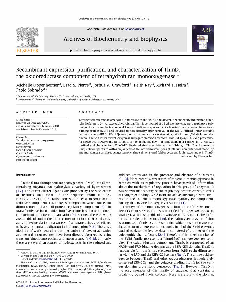
Tetrahydrofuran monooxygenase (Thm) is one of the two mem-bers of Group 5 BMM. Thm was identified from Pseudonocardia sp.strain K1, which is capable of growing aerobically on tetrahydrofu-ran as the sole carbon source [15]. The hydroxylase enzyme of Thmis composed of only a and b subunits, which in solution are pre-dicted to form a heterotetramer, (ab)2. In all of the BMM enzymesstudied to date, the hydroxylase is composed of a dimer of threepolypeptide chains, (abc)2 [2,4]. Therefore, this novel member ofthe BMM family represents a ‘‘simplified” monooxygenase com-plex. The oxidoreductase component, ThmD, is composed of anNADH-and FAD-binding domain and a [2Fe–2S] domain. ThmD isresponsible for transferring electrons from NADH to the diiron cen-ter via the FAD and the [2Fe–2S] center (Fig. 1). The amino acid se-quence between ThmD and other oxidoreductases is moderatelyconserved (30–60%) and the cofactor-binding motifs for the vari-ous domains are strictly conserved (Fig. 2). However, ThmD isthe only member of this family of enzymes that contains acovalently bound flavin cofactor. Here we present the cloning,
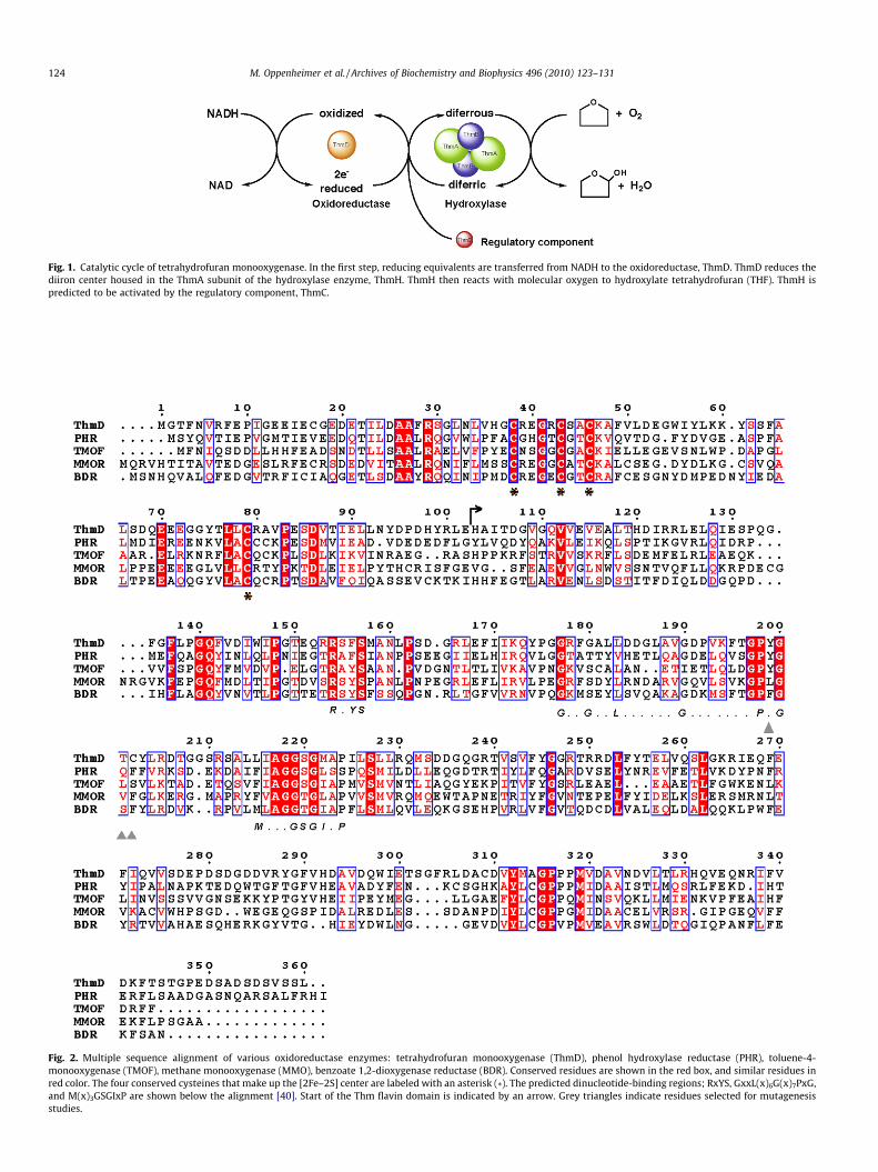
Fig. 1. Catalytic cycle of tetrahydrofuran monooxygenase. In the first step, reducing equivalents are transferred from NADH to the oxidoreductase, ThmD. ThmD reduces thediiron center housed in the ThmA subunit of the hydroxylase enzyme, ThmH. ThmH then reacts with molecular oxygen to hydroxylate tetrahydrofuran (THF). ThmH ispredicted to be activated by the regulatory component, ThmC.
Fig. 2. Multiple sequence alignment of various oxidoreductase enzymes: tetrahydrofuran monooxygenase (ThmD), phenol hydroxylase reductase (PHR), toluene-4-monooxygenase (TMOF), methane monooxygenase (MMO), benzoate 1,2-dioxygenase reductase (BDR). Conserved residues are shown in the red box, and similar residues inred color. The four conserved cysteines that make up the [2Fe–2S] center are labeled with an asterisk (*). The predicted dinucleotide-binding regions; RxYS, GxxL(x)6G(x)7PxG,and M(x)3GSGIxP are shown below the alignment [40]. Start of the Thm flavin domain is indicated by an arrow. Grey triangles indicate residues selected for mutagenesisstudies.
124 M. Oppenheimer et al. / Archives of Biochemistry and Biophysics 496 (2010) 123–131
M. Oppenheimer et al. / Archives of Biochemistry and Biophysics 496 (2010) 123–131 125
expression, purification, and characterization of the full-lengthThmD enzyme and its flavin domain (ThmD-FD) and computa-tional and mutagenesis studies that suggest that the mode of flavinattachment in ThmD appears to be novel.
Materials and methods
Material
All buffers and media components were obtained from FisherScientific (Pittsburgh, PA). BL21TIR chemical competent cells,NADH, NADPH, and cytochrome c (cyt c) were obtained fromSigma–Aldrich (St. Louis, MO). DNA primers were synthesized byIntegrated DNA Technologies (Coralville, IA). Isopropyl b-thio-galactopyranoside (IPTG) was obtained from Gold biotechnology(St. Louis, MO) and DNA gel extraction and plasmid DNA purifica-tion kits were from Qiagen (Valencia, CA). Escherichia coli TOP-10chemically competent cells were from Invitrogen (Carlsbad, CA).PmeI/SgfI enzyme blend was from Promega (Madison, WI). Chro-matographic columns were obtained from GE Healthcare.
Bioinformatics analysis
Amino acid sequence alignment was done using the Clustal Wprogram included in the DNAstar program package (Madison,WI). Fig. 2 was created using the program Espript 2.2 (http://espript.ibcp.fr/ESPript/ESPript/). The program Pyre (http://www.sbg.bio.ic.ac.uk/phyre/) was used to create the threading-based three-dimensional model of ThmD [16].
Cloning the full-length ThmD
The gene coding for ThmD was amplified by polymerasechain reaction (PCR) directly from Pseudonocardia sp. strain K1genomic DNA (generous gift of Prof. B. Theimer) using the ThmD(F) and ThmD (R) primers (Table 1). A single PCR product of�1300 bp was observed on a 0.8% agarose gel, correspondingto the expected size of the ThmD gene (1355 bp). The DNA bandwas excised from the gel, purified, and digested with PmeI/SgfIrestriction enzyme blend at 37 �C for 40 min. The reaction wasstopped by heat denaturation at 65 �C for 30 min. This samplewas ligated into the pVP56 K plasmid, which was previously di-gested with PmeI and SgfI using T4 ligase. This plasmid allowsthe expression of ThmD as a fusion to an 8xHis-maltose-bindingprotein (8xHis-MBP) and carries a kanamycin resistant marker. Aligation reaction was also performed with the pVP55A plasmidfor the expression of ThmD as a fusion with an 8xHis tag. Thisplasmid carries an ampicillin resistant marker (both plasmidswhere obtained from the Center for Eukaryotic StructuralGenomics, University of Wisconsin, Madison) [17]. Ligation reac-tions were transformed into chemically competent TOP-10 cellsand plated in Luria–Bertani (LB) agar supplemented with thecorresponding antibiotic and incubated overnight at 37 �C. Fivecolonies from each ligation reaction were used to inoculate five10 mL LB cultures supplemented with the appropriate antibiotic
Table 1Primers used for cloning ThmD and its truncated forms containing the flavin domain and
Primer Restriction site Amino acid
ThmD (F) SgfI 1–360ThmD (R) PmeI 1–360ThmD-FD (F) SgfI 102–360ThmD-[2Fe–2S] (R) PmeI 1–101
a F and R are the forward and reverse primers, respectively.
and grown overnight at 37 �C. Plasmids were isolated using aQiagen Kit and those plasmids containing the ThmD gene wereidentified by PCR using gene specific primers. The complete cod-ing region was sequenced to ensure that no unwanted mutationswere incorporated during the PCR reactions.
Engineering of ThmD-FD and ThmD-[2Fe–2S] domain
Amino acid sequence alignment of several members of theBMM oxidoreductase family was used to delineate the various do-mains in ThmD (Fig. 2). The [2Fe–2S]-binding domain corre-sponded to the first �100 amino acids of the N-terminus. Thisregion contains all four essential cysteine residues that bind the ir-ons [18–20]. To clone this domain, the ThmD-[2Fe–2S] (R) andThmD (F) primers were used in a PCR reaction using the wild-typeThmD gene as a template (Table 1). Primers ThmD-FD (F) andThmD (R) were used to obtain the DNA fragment coding for resi-dues 102–360, which correspond to the flavin and the NADH-bind-ing domains. The truncated forms of ThmD were cloned into thepVP55A plasmid in frame with an N-terminus 8xHis tag as de-scribed above for the ThmD full-length protein.
Site-directed mutagenesis
Site-directed mutagenesis was performed using the Quik-Change protocol (Stratagene, CA). Residues were selected for muta-genesis studies based of their proximity to the flavin cofactor in themodel structure of ThmD (Fig. S1).
Expression of ThmD
Expression of ThmD fused to an 8xHis tag (in the pVP55A plasmid)was performed in LB medium supplemented with 200 lg mL�1 ofampicillin and incubated at 37 �C with agitation set at 250 rpm.Clones were transformed into BL21TIR cells and grown on LB agarplates. After 24 h incubation, a single colony was used to inoculatea 50 mL LB starter culture and incubated overnight. Four cultureseach of 1.5 L of LB media were inoculated with 10 mL of the overnightculture and incubated until the optical density measured at 600 nm(OD600) reached a value of �0.8, at which point protein expressionwas induced by addition of 0.5 mM IPTG. After 4 h induction, the cul-tures were harvested by centrifugation at 5000g for 20 min and thecell pellet stored at �80 �C. ThmD expressed well as a fusion to anN-terminal 8xHis tag; however, this protein was completely insolu-ble. After modifying the expression and purification procedures, wewere unable to obtain soluble enzyme in this form. ThmD was ob-tained in a partially soluble form when expressed as a fusion toMBP using the pVP56 K plasmid. Throughout the expression of ThmDin pVP56 K, the LB media was supplemented with 50 lg mL�1 kana-mycin with agitation set at 250 rpm. A single colony of BL21TIR trans-formed with pVP56 K ThmD was used to inoculate a 50 mL LBculture. This culture was incubated at 37 �C. The next day, 6 cultureflasks containing 1.5 L of LB supplemented with 0. 5 M NaCl wereeach inoculated with 7 mL of the overnight culture. These cultureswere grown at 37 �C until the OD600 reached a value of�0.6. The incu-
[2Fe–2S] centersa.
s Sequence
50-GGTTGCGATCGCATGGGAACCTTCAACGTAAGGTTCG-30
50- AAAAGTTTAAACAAGCGACGATACAGAATCGG-30
50-TTCGGCGATCGCCCATGCAATTACCGATGGAGTTGGCC-30
50-AGCAGTTTAAACTGCATGCTCTAGCCGATAGTGGTCAGG-30
126 M. Oppenheimer et al. / Archives of Biochemistry and Biophysics 496 (2010) 123–131
bation temperature was increased to 47 �C for 30 min. After this heatshock period, protein expression was induced by addition of 300 lMIPTG and the media was supplemented with 150 mg ferric citrate,150 mg ammonium ferric citrate, and 363 mg L-cysteine [21]. Thecultures were incubated overnight at 10 �C. Cells were harvestedby centrifugation at 5000g for 20 min and the cell pellet stored at�80 �C. This procedure regularly yielded 20 g of cell paste.
Expression of ThmD-FD and ThmD-[2Fe–2S] domain
ThmD-FD and ThmD-[2Fe–2S] domains were expressed usingthe plasmid pVP55A, which produced the recombinant protein asan N-terminal fusion to an 8xHis tag. Recombinant ThmD-[2Fe–2S] domain was found to be completely insoluble. Thus, no furtherexperiments were performed with this truncated form of ThmD.For the ThmD-FD, a single colony of BL21TIR containing thepVP55AThmD-FD plasmid was used to inoculate a 50 mL LB cul-ture and incubated at 37 �C. The next day, 4 flasks, each containing1.5 L of LB medium were inoculated with 10 mL of overnight cul-ture. These cultures were incubated at 37 �C until the OD600
reached a value of 0.6. Protein expression was induced by additionof 0.5 mM IPTG. After 4 h of incubation at 37 �C, the cells were har-vested by centrifugation at 5000g for 20 min. This protocol nor-mally yielded �30 g of cell paste. All mutants of ThmD-FD wereexpressed following the same procedure as wild-type ThmD-FD.
Purification of ThmD
Cell pellets (�20 g) were resuspended in 90 mL 25 mM HEPES,at pH 6.5, 420 lM tris(2-carboxyethyl)phosphine (TCEP),
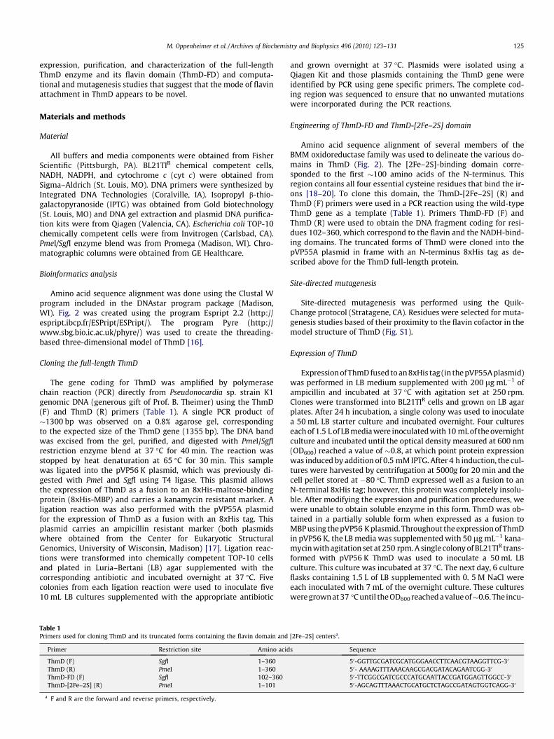
Fig. 3. (A) Coomassie stained SDS–PAGE analysis of ThmD from different steps of purificaafter treatment with Tev protease; lane 4, after nickel IMAC; lane 5, after second DEAE chstained SDS–PAGE showing ThmD-FD after size exclusion chromatography. (C) Right pAspergillus fumigatus (AfUGM), ThmD, and Thm-FD. Left panel shows the Western-blotattached flavin. The negative control, AfUGM, does not. The numbers on the left are the
0.75 mM phenylmethyl sulfonylfluoride (PMSF), 15 lg/mL DNase,15 lg/mL RNase, and 15 lg/mL lysozyme, and stirred at 4 �C for20 min. Cells were lysed by sonication for 5 min (5 s on, 10 s off).Insoluble fractions and unlysed cells were precipitated by centrifu-gation at 30,000g for 1 h at 4 �C. The resulting supernatant wasloaded onto a diethyl amino ethyl (DEAE) column previously equil-ibrated in 25 mM HEPES at pH 6.5. After washing the column with100 mL of 25 mM HEPES at pH 6.5 containing 140 mM NaCl, a125 mL gradient of 140–400 mM NaCl was used to elute theMBP–ThmD fusion protein from the column. Fractions containingMBP–ThmD were identified by sodium dodecyl sulfate polyacryl-amide gel electrophoresis (SDS–PAGE) and only those with thespectrum characteristic of a [2Fe–2S] cluster were pooled. Tobaccoetch virus (Tev) protease was added (1:10) to liberate ThmD fromMBP. The reaction was incubated at 4 �C overnight and the solutionwas centrifuged at 30,000g for 20 min to remove precipitated pro-teins before being loaded onto a nickel immobilized metal affinitychromatography (IMAC) column. Since both MBP and Tev proteinshave an 8xHis tag these proteins remained bound, and the flowthrough containing free ThmD was collected. This sample was di-luted 10-fold and loaded onto a second DEAE column. The columnwas washed with 50 mL of 25 mM HEPES, 150 mM NaCl, pH 7.5,followed by a 350 mL gradient from 150 to 400 mM NaCl in thesame buffer. Fractions containing ThmD were identified by SDS–PAGE and by spectra analyses. Those fractions that were brownin color and showed spectra similar to Fig. 4 were pooled concen-trated and loaded onto a S-200 Sephadex column previously equil-ibrated in 25 mM HEPES, 125 mM NaCl, pH 7.5. This step separatedThmD from some high molecular weight contaminants yieldingmore than 95% pure protein. The final sample was stored at �80 �C.
tion. Lane 1, lysate supernatant; lane 2, after DEAE chromatography at pH 6.5; lane 3,romatography at pH 7.5; lane 6, after size exclusion chromatography. (B) Coomassieanel shows the SDS–PAGE of the flavoenzyme UDP-galactopyranose mutase fromusing anti-flavin antibody. Recombinant ThmD and ThmD-FD contain covalentlymolecular weight standards.
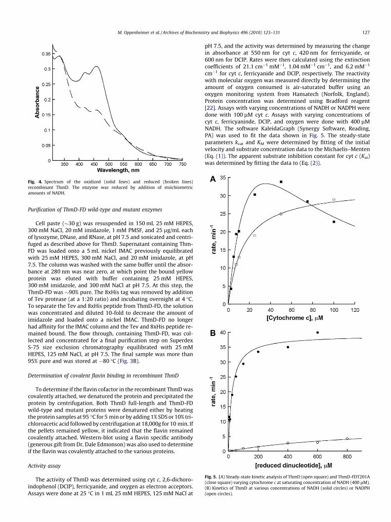
Fig. 4. Spectrum of the oxidized (solid lines) and reduced (broken lines)recombinant ThmD. The enzyme was reduced by addition of stoichiometricamounts of NADH.
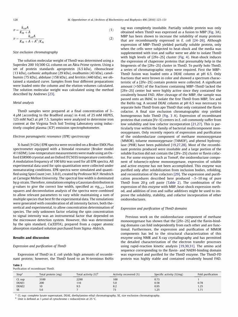
Fig. 5. (A) Steady-state kinetic analysis of ThmD (open square) and ThmD-FDT201A(close square) varying cytochrome c at saturating concentration of NADH (400 lM).(B) Kinetics of ThmD at various concentrations of NADH (solid circles) or NADPH(open circles).
M. Oppenheimer et al. / Archives of Biochemistry and Biophysics 496 (2010) 123–131 127
Purification of ThmD-FD wild-type and mutant enzymes
Cell paste (�30 g) was resuspended in 150 mL 25 mM HEPES,300 mM NaCl, 20 mM imidazole, 1 mM PMSF, and 25 lg/mL eachof lysozyme, DNase, and RNase, at pH 7.5 and sonicated and centri-fuged as described above for ThmD. Supernatant containing Thm-FD was loaded onto a 5 mL nickel IMAC previously equilibratedwith 25 mM HEPES, 300 mM NaCl, and 20 mM imidazole, at pH7.5. The column was washed with the same buffer until the absor-bance at 280 nm was near zero, at which point the bound yellowprotein was eluted with buffer containing 25 mM HEPES,300 mM imidazole, and 300 mM NaCl at pH 7.5. At this step, theThmD-FD was �90% pure. The 8xHis tag was removed by additionof Tev protease (at a 1:20 ratio) and incubating overnight at 4 �C.To separate the Tev and 8xHis peptide from ThmD-FD, the solutionwas concentrated and diluted 10-fold to decrease the amount ofimidazole and loaded onto a nickel IMAC. ThmD-FD no longerhad affinity for the IMAC column and the Tev and 8xHis peptide re-mained bound. The flow through, containing ThmD-FD, was col-lected and concentrated for a final purification step on SuperdexS-75 size exclusion chromatography equilibrated with 25 mMHEPES, 125 mM NaCl, at pH 7.5. The final sample was more than95% pure and was stored at �80 �C (Fig. 3B).
Determination of covalent flavin binding in recombinant ThmD
To determine if the flavin cofactor in the recombinant ThmD wascovalently attached, we denatured the protein and precipitated theprotein by centrifugation. Both ThmD full-length and ThmD-FDwild-type and mutant proteins were denatured either by heatingthe protein samples at 95 �C for 5 min or by adding 1% SDS or 10% tri-chloroacetic acid followed by centrifugation at 18,000g for 10 min. Ifthe pellets remained yellow, it indicated that the flavin remainedcovalently attached. Western-blot using a flavin specific antibody(generous gift from Dr. Dale Edmonson) was also used to determineif the flavin was covalently attached to the various proteins.
Activity assay
The activity of ThmD was determined using cyt c, 2,6-dichoro-indophenol (DCIP), ferricyanide, and oxygen as electron acceptors.Assays were done at 25 �C in 1 mL 25 mM HEPES, 125 mM NaCl at
pH 7.5, and the activity was determined by measuring the changein absorbance at 550 nm for cyt c, 420 nm for ferricyanide, or600 nm for DCIP. Rates were then calculated using the extinctioncoefficients of 21.1 cm�1 mM�1, 1.04 mM�1 cm�1, and 6.2 mM�1
cm�1 for cyt c, ferricyanide and DCIP, respectively. The reactivitywith molecular oxygen was measured directly by determining theamount of oxygen consumed is air-saturated buffer using anoxygen monitoring system from Hansatech (Norfolk, England).Protein concentration was determined using Bradford reagent[22]. Assays with varying concentrations of NADH or NADPH weredone with 100 lM cyt c. Assays with varying concentrations ofcyt c, ferricyaninde, DCIP, and oxygen were done with 400 lMNADH. The software KaleidaGraph (Synergy Software, Reading,PA) was used to fit the data shown in Fig. 5. The steady-stateparameters kcat and KM were determined by fitting of the initialvelocity and substrate concentration data to the Michaelis–Menten(Eq. (1)). The apparent substrate inhibition constant for cyt c (Kai)was determined by fitting the data to (Eq. (2)).
128 M. Oppenheimer et al. / Archives of Biochemistry and Biophysics 496 (2010) 123–131
v ¼ kcatAKM þ A
ð1Þ
v ¼ kcatA
KM þ Aþ A2=KAi
ð2Þ
Size exclusion chromatography
The solution molecular weight of ThmD was determined using aSuperdex 200 10/300 GL column on an Äkta Prime system. Using aset of protein standards aproprotein (6.5 kDa), ribonuclease(13 kDa), carbonic anhydrase (29 kDa), ovalbumin (43 kDa), canol-bumin (75 kDa), aldolase (158 kDa), and ferritin (440 kDa), we ob-tained a standard curve. Samples from four different preparationswere loaded onto the column and the elution volumes calculated.The solution molecular weight was calculated using the methoddescribed by Andrews [23].
Metal analysis
ThmD samples were prepared at a final concentration of 3–4 lM (according to the Bradford assay) in 4 mL of 25 mM HEPES,125 mM NaCl at pH 7.5. Samples were analyzed to determine ironcontent at the Virginia Tech Soil Testing Laboratory using induc-tively coupled plasma (ICP) emission spectrophotometer.
Electron paramagnetic resonance (EPR) spectroscopy
X-band (9 GHz) EPR spectra were recorded on a Bruker EMX Plusspectrometer equipped with a bimodal resonator (Bruker model4116DM). Low-temperature measurements were made using an Ox-ford ESR900 cryostat and an Oxford ITC503S temperature controller.A modulation frequency of 100 kHz was used for all EPR spectra. Allexperimental data used for spin-quantitation were collected undernonsaturating conditions. EPR spectra were simulated and quanti-fied using Spin Count (ver. 3.0.0), created by Professor M.P. Hendrichat Carnegie Mellon University. The spectral line width is dominatedby g-strain. Therefore, simulations employ a Gaussian distribution ing-values to give the correct line width, specified as rgx,y,z. Leastsquares and deconvolution analysis of the spectra were combinedto allow relevant parameters to vary while maintaining a sum ofmultiple species that best fit the experimental data. The simulationswere generated with consideration of all intensity factors, both the-oretical and experimental, to allow concentration determination ofspecies. The only unknown factor relating the spin concentrationto signal intensity was an instrumental factor that depended onthe microwave detection system. However, this was determinedby the spin standard, Cu(EDTA), prepared from a copper atomicabsorption standard solution purchased from Sigma–Aldrich.
Results and discussion
Expression and purification of ThmD
Expression of ThmD in E. coli yields high amounts of recombi-nant protein; however, the ThmD fused to an N-terminus 8xHis
Table 2Purification of recombinant ThmD.
Stepa Total protein (mg) Total activity (U)b Activi
CL sup 3100 2290 100DEAE1 200 116 5.0DEAE2 10 9.5 8.2SE 5.6 7 73
a CL sup: complete lysate supernatant, DEAE, diethylamino ethyl chromatography, SEb Unit is defined as 1 lmol of cytochrome c reduced/min at 25 �C.
tag was completely insoluble. Partially soluble protein was onlyobtained when ThmD was expressed as a fusion to MBP (Fig. 3A).MBP has been shown to increase the solubility of many proteinsthat are recombinantly expressed in E. coli [24–26]. Althoughexpression of MBP–ThmD yielded partially soluble protein, onlywhen the cells were subjected to heat-shock and the media wassupplemented with iron and sulfur were we able to isolate ThmDwith high levels of [2Fe–2S] cluster (Fig. 4). Heat-shock inducesthe expression of chaperone proteins that presumably help in thebiogenesis of the [2Fe–2S] cluster in ThmD. To purify holo ThmD,a series of chromatographic steps were required. First the MBP–ThmD fusion was loaded onto a DEAE column at pH 6.5. Onlyfractions that were brown in color and showed a spectrum charac-teristic of a [2Fe–2S] contain protein were collected. A significantamount (>50%) of the fractions containing MBP–ThmD lacked the[2Fe–2S] center but were highly active since they contained thecovalently bound FAD. After cleavage of the MBP, the sample waspassed onto an IMAC to isolate the free ThmD from MBP, Tev andthe 8xHis tag. A second DEAE column at pH 6.5 was necessary toseparate holo ThmD from apo ThmD that only contained the flavincofactor. A final size exclusion chromatographic step yieldedhomogeneous holo ThmD (Fig. 3 A). Expression of recombinantproteins that contain [Fe–S] centers in E. coli commonly suffer fromlow solubility and low cofactor incorporation [21,27]. This is par-ticularly true within the family of bacterial multicomponent mon-ooxygenases. Only recently reports of expression and purificationof the oxidoreductase component of methane monooxygenase(MMOR), toluene monooxygenase (T4MOF), and phenol hydroxy-lase (PHR) have been published [19,27,28]. Most of the recombi-nant proteins produced were insoluble and a large portion of thesoluble fraction did not contain the [2Fe–2S] cluster or flavin cofac-tor. For some enzymes such as TomoF, the oxidoreductase compo-nent of toluene/o-xylene monooxygenase, expression of solubleand active enzyme has not been accomplished. This enzyme waspurified only after solubilization from inclusion bodies, refolding,and reconstitution of the cofactors [29]. The expression and purifi-cation procedures described here produced �5–10 mg of pureThmD from 20 g cell paste (Table 2). The combination of theexpression of this enzyme with MBP, heat-shock expression meth-od, and addition of iron and sulfur additives might be used to im-prove the solubility, stability, and cofactor incorporation of otheroxidoreductases.
Expression and purification of ThmD domains
Previous work on the oxidoreductase component of methanemonooxygenase has shown that the [2Fe–2S] and the flavin-bind-ing domains can fold independently from each other and are func-tional. Furthermore, the expression and purification of MMORcomponents has led to the structural characterization of thisenzyme using NMR and X-ray crystallography and has permittedthe detailed characterization of the electron transfer processesusing rapid-reaction kinetic analysis [19,30,31]. The amino acidsequence corresponding to the flavin- and NADH-binding domainwas expressed and purified for the ThmD enzyme. The ThmD-FDprotein was highly stable and contained covalently bound FAD.
ty recovered (%) Specific activity (U/mg) Fold purification
0.73 10.58 0.780.95 1.251.25 1.71
, size exclusion chromatography.
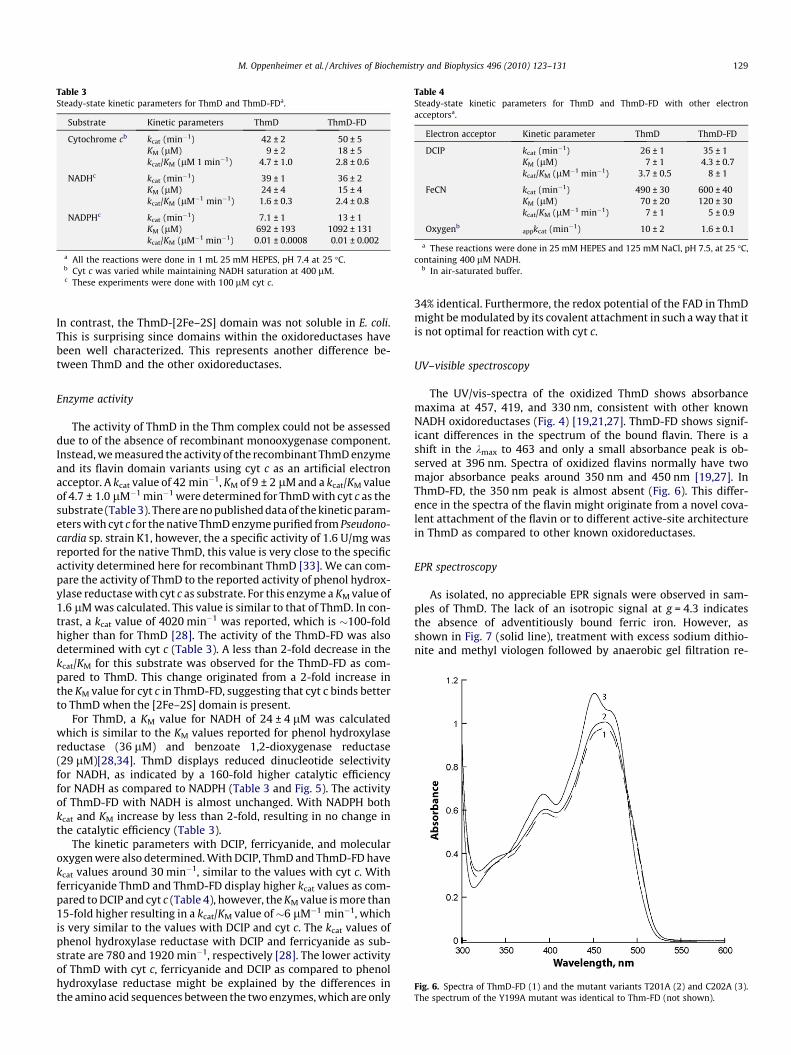
Table 3Steady-state kinetic parameters for ThmD and ThmD-FDa.
Substrate Kinetic parameters ThmD ThmD-FD
Cytochrome cb kcat (min�1) 42 ± 2 50 ± 5KM (lM) 9 ± 2 18 ± 5kcat/KM (lM 1 min�1) 4.7 ± 1.0 2.8 ± 0.6
NADHc kcat (min�1) 39 ± 1 36 ± 2KM (lM) 24 ± 4 15 ± 4kcat/KM (lM�1 min�1) 1.6 ± 0.3 2.4 ± 0.8
NADPHc kcat (min�1) 7.1 ± 1 13 ± 1KM (lM) 692 ± 193 1092 ± 131kcat/KM (lM�1 min�1) 0.01 ± 0.0008 0.01 ± 0.002
a All the reactions were done in 1 mL 25 mM HEPES, pH 7.4 at 25 �C.b Cyt c was varied while maintaining NADH saturation at 400 lM.c These experiments were done with 100 lM cyt c.
Table 4Steady-state kinetic parameters for ThmD and ThmD-FD with other electronacceptorsa.
Electron acceptor Kinetic parameter ThmD ThmD-FD
DCIP kcat (min�1) 26 ± 1 35 ± 1KM (lM) 7 ± 1 4.3 ± 0.7kcat/KM (lM�1 min�1) 3.7 ± 0.5 8 ± 1
FeCN kcat (min�1) 490 ± 30 600 ± 40KM (lM) 70 ± 20 120 ± 30kcat/KM (lM�1 min�1) 7 ± 1 5 ± 0.9
Oxygenbappkcat (min�1) 10 ± 2 1.6 ± 0.1
a These reactions were done in 25 mM HEPES and 125 mM NaCl, pH 7.5, at 25 �C,containing 400 lM NADH.
b In air-saturated buffer.
M. Oppenheimer et al. / Archives of Biochemistry and Biophysics 496 (2010) 123–131 129
In contrast, the ThmD-[2Fe–2S] domain was not soluble in E. coli.This is surprising since domains within the oxidoreductases havebeen well characterized. This represents another difference be-tween ThmD and the other oxidoreductases.
Fig. 6. Spectra of ThmD-FD (1) and the mutant variants T201A (2) and C202A (3).The spectrum of the Y199A mutant was identical to Thm-FD (not shown).
Enzyme activity
The activity of ThmD in the Thm complex could not be assesseddue to of the absence of recombinant monooxygenase component.Instead, we measured the activity of the recombinant ThmD enzymeand its flavin domain variants using cyt c as an artificial electronacceptor. A kcat value of 42 min�1, KM of 9 ± 2 lM and a kcat/KM valueof 4.7 ± 1.0 lM�1 min�1 were determined for ThmD with cyt c as thesubstrate (Table 3). There are no published data of the kinetic param-eters with cyt c for the native ThmD enzyme purified from Pseudono-cardia sp. strain K1, however, the a specific activity of 1.6 U/mg wasreported for the native ThmD, this value is very close to the specificactivity determined here for recombinant ThmD [33]. We can com-pare the activity of ThmD to the reported activity of phenol hydrox-ylase reductase with cyt c as substrate. For this enzyme a KM value of1.6 lM was calculated. This value is similar to that of ThmD. In con-trast, a kcat value of 4020 min�1 was reported, which is �100-foldhigher than for ThmD [28]. The activity of the ThmD-FD was alsodetermined with cyt c (Table 3). A less than 2-fold decrease in thekcat/KM for this substrate was observed for the ThmD-FD as com-pared to ThmD. This change originated from a 2-fold increase inthe KM value for cyt c in ThmD-FD, suggesting that cyt c binds betterto ThmD when the [2Fe–2S] domain is present.
For ThmD, a KM value for NADH of 24 ± 4 lM was calculatedwhich is similar to the KM values reported for phenol hydroxylasereductase (36 lM) and benzoate 1,2-dioxygenase reductase(29 lM)[28,34]. ThmD displays reduced dinucleotide selectivityfor NADH, as indicated by a 160-fold higher catalytic efficiencyfor NADH as compared to NADPH (Table 3 and Fig. 5). The activityof ThmD-FD with NADH is almost unchanged. With NADPH bothkcat and KM increase by less than 2-fold, resulting in no change inthe catalytic efficiency (Table 3).
The kinetic parameters with DCIP, ferricyanide, and molecularoxygen were also determined. With DCIP, ThmD and ThmD-FD havekcat values around 30 min�1, similar to the values with cyt c. Withferricyanide ThmD and ThmD-FD display higher kcat values as com-pared to DCIP and cyt c (Table 4), however, the KM value is more than15-fold higher resulting in a kcat/KM value of�6 lM�1 min�1, whichis very similar to the values with DCIP and cyt c. The kcat values ofphenol hydroxylase reductase with DCIP and ferricyanide as sub-strate are 780 and 1920 min�1, respectively [28]. The lower activityof ThmD with cyt c, ferricyanide and DCIP as compared to phenolhydroxylase reductase might be explained by the differences inthe amino acid sequences between the two enzymes, which are only
34% identical. Furthermore, the redox potential of the FAD in ThmDmight be modulated by its covalent attachment in such a way that itis not optimal for reaction with cyt c.
UV–visible spectroscopy
The UV/vis-spectra of the oxidized ThmD shows absorbancemaxima at 457, 419, and 330 nm, consistent with other knownNADH oxidoreductases (Fig. 4) [19,21,27]. ThmD-FD shows signif-icant differences in the spectrum of the bound flavin. There is ashift in the kmax to 463 and only a small absorbance peak is ob-served at 396 nm. Spectra of oxidized flavins normally have twomajor absorbance peaks around 350 nm and 450 nm [19,27]. InThmD-FD, the 350 nm peak is almost absent (Fig. 6). This differ-ence in the spectra of the flavin might originate from a novel cova-lent attachment of the flavin or to different active-site architecturein ThmD as compared to other known oxidoreductases.
EPR spectroscopy
As isolated, no appreciable EPR signals were observed in sam-ples of ThmD. The lack of an isotropic signal at g = 4.3 indicatesthe absence of adventitiously bound ferric iron. However, asshown in Fig. 7 (solid line), treatment with excess sodium dithio-nite and methyl viologen followed by anaerobic gel filtration re-
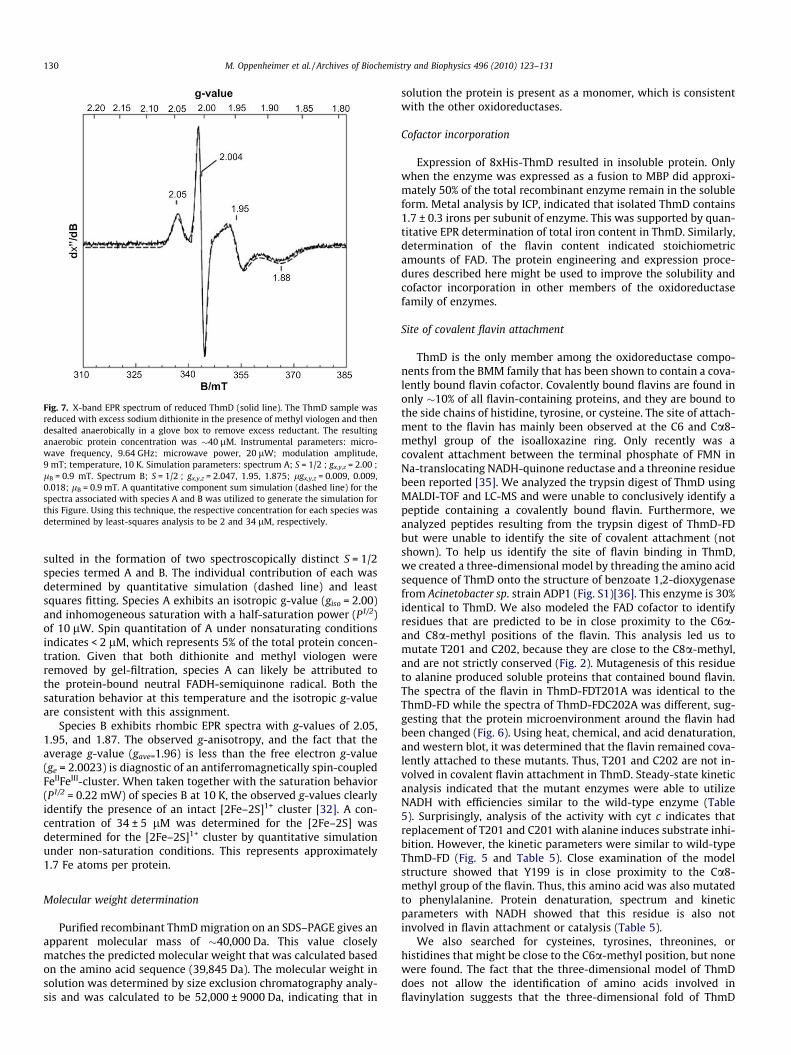
Fig. 7. X-band EPR spectrum of reduced ThmD (solid line). The ThmD sample wasreduced with excess sodium dithionite in the presence of methyl viologen and thendesalted anaerobically in a glove box to remove excess reductant. The resultinganaerobic protein concentration was �40 lM. Instrumental parameters: micro-wave frequency, 9.64 GHz; microwave power, 20 lW; modulation amplitude,9 mT; temperature, 10 K. Simulation parameters: spectrum A; S = 1/2 ; gx,y,z = 2.00 ;lB = 0.9 mT. Spectrum B; S = 1/2 ; gx,y,z = 2.047, 1.95, 1.875; lgx,y,z = 0.009, 0.009,0.018; lB = 0.9 mT. A quantitative component sum simulation (dashed line) for thespectra associated with species A and B was utilized to generate the simulation forthis Figure. Using this technique, the respective concentration for each species wasdetermined by least-squares analysis to be 2 and 34 lM, respectively.
130 M. Oppenheimer et al. / Archives of Biochemistry and Biophysics 496 (2010) 123–131
sulted in the formation of two spectroscopically distinct S = 1/2species termed A and B. The individual contribution of each wasdetermined by quantitative simulation (dashed line) and leastsquares fitting. Species A exhibits an isotropic g-value (giso = 2.00)and inhomogeneous saturation with a half-saturation power (P1/2)of 10 lW. Spin quantitation of A under nonsaturating conditionsindicates < 2 lM, which represents 5% of the total protein concen-tration. Given that both dithionite and methyl viologen wereremoved by gel-filtration, species A can likely be attributed tothe protein-bound neutral FADH-semiquinone radical. Both thesaturation behavior at this temperature and the isotropic g-valueare consistent with this assignment.
Species B exhibits rhombic EPR spectra with g-values of 2.05,1.95, and 1.87. The observed g-anisotropy, and the fact that theaverage g-value (gave=1.96) is less than the free electron g-value(ge = 2.0023) is diagnostic of an antiferromagnetically spin-coupledFeIIFeIII-cluster. When taken together with the saturation behavior(P1/2 = 0.22 mW) of species B at 10 K, the observed g-values clearlyidentify the presence of an intact [2Fe–2S]1+ cluster [32]. A con-centration of 34 ± 5 lM was determined for the [2Fe–2S] wasdetermined for the [2Fe–2S]1+ cluster by quantitative simulationunder non-saturation conditions. This represents approximately1.7 Fe atoms per protein.
Molecular weight determination
Purified recombinant ThmD migration on an SDS–PAGE gives anapparent molecular mass of �40,000 Da. This value closelymatches the predicted molecular weight that was calculated basedon the amino acid sequence (39,845 Da). The molecular weight insolution was determined by size exclusion chromatography analy-sis and was calculated to be 52,000 ± 9000 Da, indicating that in
solution the protein is present as a monomer, which is consistentwith the other oxidoreductases.
Cofactor incorporation
Expression of 8xHis-ThmD resulted in insoluble protein. Onlywhen the enzyme was expressed as a fusion to MBP did approxi-mately 50% of the total recombinant enzyme remain in the solubleform. Metal analysis by ICP, indicated that isolated ThmD contains1.7 ± 0.3 irons per subunit of enzyme. This was supported by quan-titative EPR determination of total iron content in ThmD. Similarly,determination of the flavin content indicated stoichiometricamounts of FAD. The protein engineering and expression proce-dures described here might be used to improve the solubility andcofactor incorporation in other members of the oxidoreductasefamily of enzymes.
Site of covalent flavin attachment
ThmD is the only member among the oxidoreductase compo-nents from the BMM family that has been shown to contain a cova-lently bound flavin cofactor. Covalently bound flavins are found inonly �10% of all flavin-containing proteins, and they are bound tothe side chains of histidine, tyrosine, or cysteine. The site of attach-ment to the flavin has mainly been observed at the C6 and Ca8-methyl group of the isoalloxazine ring. Only recently was acovalent attachment between the terminal phosphate of FMN inNa-translocating NADH-quinone reductase and a threonine residuebeen reported [35]. We analyzed the trypsin digest of ThmD usingMALDI-TOF and LC-MS and were unable to conclusively identify apeptide containing a covalently bound flavin. Furthermore, weanalyzed peptides resulting from the trypsin digest of ThmD-FDbut were unable to identify the site of covalent attachment (notshown). To help us identify the site of flavin binding in ThmD,we created a three-dimensional model by threading the amino acidsequence of ThmD onto the structure of benzoate 1,2-dioxygenasefrom Acinetobacter sp. strain ADP1 (Fig. S1)[36]. This enzyme is 30%identical to ThmD. We also modeled the FAD cofactor to identifyresidues that are predicted to be in close proximity to the C6a-and C8a-methyl positions of the flavin. This analysis led us tomutate T201 and C202, because they are close to the C8a-methyl,and are not strictly conserved (Fig. 2). Mutagenesis of this residueto alanine produced soluble proteins that contained bound flavin.The spectra of the flavin in ThmD-FDT201A was identical to theThmD-FD while the spectra of ThmD-FDC202A was different, sug-gesting that the protein microenvironment around the flavin hadbeen changed (Fig. 6). Using heat, chemical, and acid denaturation,and western blot, it was determined that the flavin remained cova-lently attached to these mutants. Thus, T201 and C202 are not in-volved in covalent flavin attachment in ThmD. Steady-state kineticanalysis indicated that the mutant enzymes were able to utilizeNADH with efficiencies similar to the wild-type enzyme (Table5). Surprisingly, analysis of the activity with cyt c indicates thatreplacement of T201 and C201 with alanine induces substrate inhi-bition. However, the kinetic parameters were similar to wild-typeThmD-FD (Fig. 5 and Table 5). Close examination of the modelstructure showed that Y199 is in close proximity to the Ca8-methyl group of the flavin. Thus, this amino acid was also mutatedto phenylalanine. Protein denaturation, spectrum and kineticparameters with NADH showed that this residue is also notinvolved in flavin attachment or catalysis (Table 5).
We also searched for cysteines, tyrosines, threonines, orhistidines that might be close to the C6a-methyl position, but nonewere found. The fact that the three-dimensional model of ThmDdoes not allow the identification of amino acids involved inflavinylation suggests that the three-dimensional fold of ThmD

Table 5Steady-state kinetic parameters for ThmD-FD mutant enzymesa.
Substrate Kinetic parameters Y199F T201A C202A
Cytochrome cb kcat (min�1) 51 ± 14 94 ± 38 106 ± 68KM (lM) 15 ± 6 32 ± 7 44 ± 35kcat/K (lM�1min�1) 3.4 ± 0.6 3.2 ± 0.6 3.1 ± 0.9Ki (lM) 104 ± 65 38 ± 23 25 ± 22
NADHc kcat (min�1) 31 ± 2 32 ± 1 29 ± 1KM (lM) 9 ± 2 10 ± 2 4 ± 1kcat/KM (lM�1min�1) 3.6 ± 0.9 3.2 ± 0.6 6 ± 1
a All the reactions were done in 1 mL 25 mM HEPES, pH 7.4 at 25 �C.b Cyt c was varied while maintaining NADH saturation at 400 lM.c These experiments were done at 50 lM cyt c to prevent substrate inhibition.
M. Oppenheimer et al. / Archives of Biochemistry and Biophysics 496 (2010) 123–131 131
might be different than other members of the oxidoreductase fam-ily of enzymes. It might also be possible that the site of covalentattachment is novel or perhaps there is a bi-covalent attachmentof the FAD in ThmD. Recently, attachment of FAD cofactors at posi-tions C6- and C8-alpha have been reported for the berberine bridgeand chito-oligosaccharide oxidase enzymes [37,38]. Ultimately, thethree-dimensional structure of ThmD or Thm-FD will provide clearevidence of the mode of covalent flavin attachment in this novelenzyme. Our protein expression and purification efforts will allowfor the screening of conditions for crystallization of ThmD and thiswork is currently in progress.
Conclusions
Thm is a recently identified member of the BMM family of en-zymes and the enzymes in this enzyme complex are different fromother well-characterized systems such as methane monooxygen-ase and toluene 4-monooxygenase [14,39]. In the Thm operon,ThmA and ThmB, code for the hydroxylase component. These pro-teins are homologous to the a and b subunits of all other BMOhydroxylases, however, Thm does not contain a c subunit likethe other proteins. The oxidoreductase component in Thm also dif-fers from all other BMOs. Among the members of the BMO oxido-reductases, ThmD is the only enzyme with a covalently boundflavin. In this work, we presented the cloning, expression, and puri-fication of the recombinant form of ThmD. We showed that byexpressing this enzyme as a fusion to MBP and in the presence ofextra iron and sulfur in the growth medium, a soluble holo enzymecould be isolated. Heat-shock of the E. coli cells was also necessary,which suggests that chaperones might be necessary for the properfolding of recombinant ThmD. The resulting enzyme contained fullcofactor incorporation, was active, and very stable. The flavin-bind-ing domain containing covalently bound flavin was also cloned andisolated. This domain retained wild-type ThmD NADH oxidase andcyt c reductase activities. Modeling studies combined with site-di-rected mutagenesis failed to identify the site of covalent flavinattachment in ThmD-FD suggesting that the three-dimensionalstructure or the nature of the residue(s) involved in covalentattachment might be novel. This hypothesis is supported by thevery peculiar flavin spectrum observed in the ThmD-FD enzyme.The work presented here sets the stage for future in vitro studiesof the role of covalent flavin attachment in this unique enzymeby rapid kinetic analysis and X-ray structural determination.
Acknowledgments
The authors thank Dr. Dale Edmonson (Emory University,Atlanta, GA) for providing the anti-flavin antibody and Dr. BarbaraTheimer (Martin-Luther-Universität Halle, Germany) for providingthe Pseudonocardia sp. strain K1 genomic DNA. The authors also
thanks Dr. M.P. Hendrich (Carnegie Mellon University) for provid-ing the EPR analysis and simulation software (Spin Count) and theUTA Center for Nanostructured Materials (http://www.uta.edu/cos/cnm/) for the use of EPR instrumentation.
Appendix A. Supplementary data
Supplementary data associated with this article can be found, inthe online version, at doi:10.1016/j.abb.2010.02.006.
References
[1] B.J. Wallar, J.D. Lipscomb, Chem. Rev. 96 (1996) 2625–2658.[2] J.G. Leahy, P.J. Batchelor, S.M. Morcomb, FEMS Microbiol. Rev. 27 (2003) 449–
479.[3] B.G. Fox, J. Shanklin, J. Ai, T.M. Loehr, J. Sanders-Loehr, Biochemistry 33 (1994)
12776–12786.[4] E. Notomista, A. Lahm, A. Di Donato, A. Tramontano, J. Mol. Evol. 56 (2003)
435–445.[5] M.H. Sazinsky, J. Bard, J. Biol. Chem. 279 (2004) 30600–30610.[6] E.Y. Tshuva, D. Lee, W. Bu, S.J. Lippard, J. Am. Chem. Soc. 124 (2002) 2416–
2417.[7] V. Guallar, B.F. Gherman, W.H. Miller, S.J. Lippard, R.A. Friesner, J. Am. Chem.
Soc. 124 (2002) 3377–3384.[8] J.A. Broadwater, C. Achim, E. Munck, B.G. Fox, Biochemistry 38 (1999) 12197–
12204.[9] A.C. Rosenzweig, P. Nordlund, P.M. Takahara, C.A. Frederick, S.J. Lippard, Chem.
Biol. 2 (1995) 409–418.[10] L.J. Murray, S.J. Lippard, Acc. Chem. Res. 40 (2007) 466–474.[11] M.H. Sazinsky, P.W. Dunten, M.S. McCormick, A. Didonato, S.J. Lippard,
Biochemistry 45 (2006) 15392–15404.[12] L.J. Bailey, B.G. Fox, Biochemistry 48 (2009) 8932–8939.[13] N.L. Elsen, L.J. Bailey, A.D. Hauser, B.G. Fox, Biochemistry 48 (2009) 3838–
3846.[14] L.J. Bailey, J.G. McCoy, G.N. Phillips Jr., Proc. Natl. Acad. Sci. USA 105 (2008)
19194–19198.[15] B. Thiemer, J.R. Andreesen, T. Schrader, Arch. Microbiol. 179 (2003) 266–
277.[16] L. Kelly, M.J.E. Sternberg, Nat. Protoc. 4 (2009) 363–371.[17] P.G. Blommel, P.A. Martin, K.D. Seder, R.L. Wrobel, B.G. Fox, Methods Mol. Biol.
498 (2009) 55–73.[18] J. Muller, A.A. Lugovskoy, G. Wagner, S.J. Lippard, Biochemistry 41 (2002) 42–
51.[19] J.L. Blazyk, S.J. Lippard, Biochemistry 41 (2002) 15780–15794.[20] J.L. Blazyk, S.J. Lippard, J. Biol. Chem. 279 (2004) 5630–5640.[21] S. Jaganaman, A. Pinto, M. Tarasev, D.P. Ballou, Protein Expression Purif. 52
(2007) 273–279.[22] M.M. Bradford, Anal. Biochem. 72 (1976) 248–254.[23] P. Andrews, Biochem. J. 91 (1964) 222–233.[24] P. Sobrado, M.A. Goren, D. James, C.K. Amundson, B.G. Fox, Protein Expr. Purif.
58 (2008) 229–241.[25] W.B. Jeon, D.J. Aceti, C.A. Bingman, F.C. Vojtik, A.C. Olson, J.M. Ellefson, J.E.
McCombs, H.K. Sreenath, P.G. Blommel, K.D. Seder, B.T. Burns, H.V. Geetha, A.C.Harms, G. Sabat, M.R. Sussman, B.G. Fox, G.N. Phillips Jr., J. Struct. Func.Genomics 6 (2005) 143–147.
[26] B.P. Austin, S. Nallamsetty, D.S. Waugh, N.J 498 (2009) 157–172.[27] L.J. Bailey, N.L. Elsen, B.S. Pierce, B.G. Fox, Protein Expr. Purif. 57 (2008) 9–16.[28] E. Pessione, S. Divari, E. Griva, M. Cavaletto, G.L. Rossi, G. Gilardi, C. Giunta,
FEBS 265 (1999) 549–555.[29] V. Cafaro, R. Scognamiglio, A. Viggiani, V. Izzo, I. Passaro, E. Notomista, F.D.
Piaz, A. Amoresano, A. Casbarra, P. Pucci, A. Di Donato, FEBS 269 (2002) 5689–5699.
[30] L.L. Chatwood, J. Muller, J.D. Gross, G. Wagner, S.J. Lippard, Biochemistry 43(2004) 11983–11991.
[31] D.A. Kopp, G.T. Gassner, J.L. Blazyk, S.J. Lippard, Biochemistry 40 (2001)14932–14941.
[32] S.J.A.B. Lippard, J.M. Berg, Principles of Bioinorganic Chemistry, UniversityScience Books, 1994.
[33] B. Thiemer, J.R. Andreesen, T. Schrader, FEBS 268 (2001) 3774–3782.[34] M. Yamaguchi, H. Fujisawa, J. Biol. Chem. 253 (1978) 8848–8853.[35] M. Hayashi, Y. Nakayama, M. Yasui, M. Maeda, K. Furuishi, T. Unemoto, FEBS
Lett. 488 (2001) 5–8.[36] G.T. Gassner, M.L. Ludwig, D.L. Gatti, C.C. Correll, D.P. Ballou, FASEB J. 9 (1995)
1411–1418.[37] A. Winkler, F. Hartner, T.M. Kutchan, A. Glieder, P. Macheroux, J. Biol. Chem.
281 (2006) 21276–21285.[38] C.H. Huang, W.L. Lai, M.H. Lee, C.J. Chen, A. Vasella, Y.C. Tsai, S.H. Liaw, J. Biol.
Chem. 280 (2005) 38831–38838.[39] M. Merkx, D.A. Kopp, M.H. Sazinsky, J.L. Blazyk, J. Muller, S.J. Lippard, Angew.
Chem. Int. Ed. Engl. 40 (2001) 2782–2807.[40] O. Dym, D. Eisenberg, Protein Sci. 10 (2001) 1712–1728.