microbiological deterioration of organic materials - govinfo.gov
TRANSCRIPT
Microbiological Deterioration of
Organic Materials:
Its Prevention and Methods of Test
United States Department of Commerce
National Bureau of Standards
Miscellaneous Publication 188
UNITED STATES DEPARTMENT OF COMMERCE • Charles Sawyer. Secretary
NATION AL BUREAU OF STANDARDS • E. U. Condon, Director
Microbiological Deterioration of
Organic Materials:
Its Prevention and Methods of Test
by Edward Abrams
National Bureau of Standards Miscellaneous Publication 188
Issued November 1, 1948
For sale by the Superintendent of Documents, U. S. Government Printing Office, Washington 25, D. C.
Price 25 cents
PREFACE
The deterioration of textiles, leather, and other organic materials by mildew
and rot is a major problem in the tropics, and is also important in many parts
of the continental United States. Accordingly, it has seemed worth while to
present the information on the subject that has been accumulated by the
National Bureau of Standards in the past several years, and to describe the
methods currently used to ascertain the susceptibility of materials to mildew
growth and to evaluate protective treatments. Part I has been written prima-
rily for persons looking for general information on the cause and prevention of
mildew damage. Part II is addressed more particularly to the technologist and
the laboratory worker.
E. U. Condon, Director.
Contents
Part I. Review of the LiteraturePage
Preface nI. Introduction 1
II. Microbiology ^ 1
III. Enzyme act|on 4
IV. Organic materials utilized by microorganisms 5
1. Cellulosic materials 8
2. Proteinaceous materials 10
3. Leather 10
4. Miscellaneous materials 10
(a) Rubber 10
(b) Plastics 11
V. Prevention of deterioration 11
VI. Testing for fungicidal efficiency 14
Part 2. Investigation of Microbiological Tests
VII. Development of mildew testing at Bureau 18
VIII. Representative problems 22
IX. Test methods _ 25
1. Pure-culture biological methods 25
(a) Rot-resistance tests with Chaetomium globosum 25
(1) Direct-inoculation—sterilized specimen method. 25
(2) Mycelial mat method. _ 26
(b) Mildew-resistance tests with Aspergillus niger 26
(1) Mycelial mat method 26
(2) Sugar-free medium method 26
2. Mixed-culture biological test methods 28
(a) Soil-burial test 28
(b) Soil-suspension test 28
3. Accelerated weathering test 28
X. Experimental results and discussion 29
XI. Summary and conclusions 38
XII. References 39
Microbiological Deterioration of Organic Materials
:
Its Prevention and Methods of Test
By Edward Abrams *
This paper presents a detailed review of the literature on the microbiological deterioration of organic and fibrous
materials. Also, representative problems incidental to the development of test methods for the evaluation of mildewresistance and the development of mildew resistant materials are given. The most widely recognized test methodsfor the evaluation of mildew-resistance and rot-resistance are listed. A wide variety of fungicides for different usesis classified according to effectiveness. Plastics and plasticizers and related materials are rated according to their suscepti-bility to support mold growth.
Part 1. Review of the Literature
I. Introduction
The deterioration of organic materials bymicroorganisms in nature is of vital importanceto man. Everyone is familiar with the accumula-tion of leaves and plant material in the fall.
However, by the following spring practically all
of this debris becomes a part of the soil. In this
way nature renews her store of energy, the micro-organisms completing the carbon, nitrogen, andsulfur cycles. Unfortunately, microorganisms arenot discriminating in selecting their nutrimentand are, therefore, a source of great loss when theyattack organic materials of economic value.From ancient times man has been plagued by
mildew. As no method of control was known,it has been regarded as a necessary evil. A list
of materials attacked by mildew would cover the
entire field of organic materials. Because of
ignorance and inertia a great waste of materials
has been tolerated through the ages. Underfavorable conditions of moisture and temperaturemany substances are attacked by molds. Thepresent paper is restricted to the consideration
of cellulosic and proteinaceous materials, rubber,
and plastics. Foodstuffs are not included becausetheir treatment presents an entirely different
problem.
II. Microbiology
The terms microorganisms, molds, and mildewshave been used interchangeably. Strictly speak-ing, in this field the term microorganisms shouldalso include bacteria. However, this discussionis restricted to the effects of molds and mildews(the terms are synonymous) as the moisture level
required for deterioration by bacteria is rarelyencountered except in specialized fields, such asthe maritime industry.Molds are filamentous, branching forms of
plant life. They belong to the subphylum Fungiof the phylum Thallophyta. According to defini-
tion they are plants that do not have roots, stems,or leaves. They are free of chlorophyll and requirepreformed organic materials and certain inorganicsubstances for their growth. Most molds areprolific spore producers. The spores serve to
' Present address: Institute of Textile Technology, Charlottesville, Va.
propagate the individual as do the seeds of higherplants. These spores are formed either at thetips of mycelial branches or are borne in special
bodies known as sporangia or perithecia. Theyare liberated readily in vast numbers and float
easily in the air. In this condition they are able
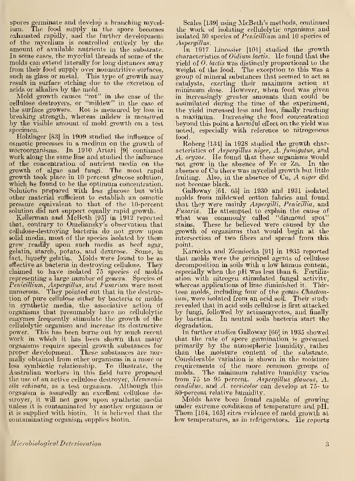
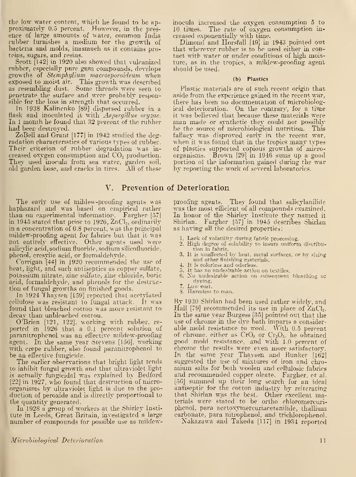
to survive for long periods of time and underextreme conditions of humidity and temperature.Figure 1 shows the sporehead of Aspergillus niger
magnified 1,000 times.
The number of genera and species of molds thathave been isolated and classified runs into thethousands. They vary greatly in then morphol-ogy, physiology, and biochemical activities. Mostof them are aerobic. Thev thrive at temperaturesbetween 10° and 37° C and in the pH range from2 to 8, with optimum growth at pH 4 to 7.
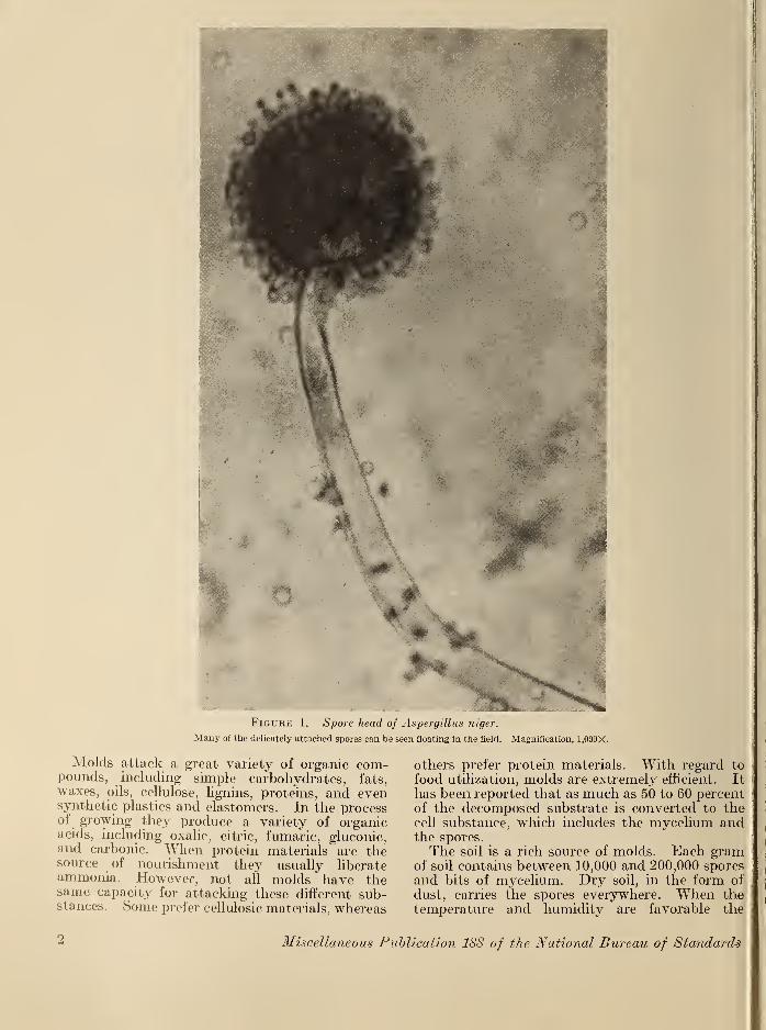
FiGUBic 1, Spore head of Aspergillus niger.
Many of the delicately attached spores can be seen floating in the field. Magnification, 1.000X.
Molds attack a great variety of organic com-pounds, including simple carbohydrates, fats,waxes, oils, cellulose, lignins, proteins, and evensynthetic plastics and elastomers. In the processof growing they produce a variety of organicacids, including oxalic, citric, fumaric, gluconic,and carbonic. When protein materials are thesource of nourishment they usually liberateammonia. However, not all molds have thesame capacity for attacking these different sub-stances. Some prefer cellulosic materials, whereas
others prefer protein materials. With regard to
food utilization, molds are extremely efficient. It
has been reported that as much as 50 to 60 percentof the decomposed substrate is converted to thecell substance, which includes the mycelium andthe spores.
The soil is a rich source of molds. Each gramof soil contains between 10,000 and 200,000 sporesand bits of mycelium. Dry soil, in the form of
dust, carries the spores everywhere. When thetemperature and humidity are favorable the
2 Miscellaneous Publication 188 of the National Bureau of Standards
spores germinate and develop a branching mycel-ium. The food supply in the spore becomesexhausted rapidly, and the further developmentof the mycelium is controlled entirely by theamount of available nutrients in the substrate.In some cases, the mycelial threads of some of themolds can extend laterally for long distances awayfrom their food supply over normutritive surfaces,
such as glass or metal. This type of growth mayresult in surface etching due to the excretion ofacids or alkalies by the mold.Mold growth causes "rot" in the case of the
cellulose destroyers, or "mildew" in the case of
the surface growers. Rot is measured by loss in
breaking strength, whereas mildew is measuredby the visible amount of mold growth on a test
specimen.Holzinger [83] in 1909 studied the influence of
osmotic processes in a medium on the growth of
microorganisms. In 1910 Artari [9] continuedwork along the same line and studied the influence
of the concentration of nutrient media on thegrowth of algae and fungi. The most rapidgrowth took place in 10 percent glucose solution,
which he found to be the optimum concentration.Solutions prepared with less glucose but withother material sufficient to establish an osmoticpressure equivalent to that of the 10-percentsolution did not support equally rapid growth.
Kellerman and McBeth [95] in 1912 reportedthat, contrary to Omeliansky's observation thatcellulose-destroying bacteria do not grow uponsolid media, most of the species isolated by themgrew readily upon such media as beef agar,
gelatin, starch, potato, and dextrose. Some, in
fact, liquefy gelatin. Molds were found to be aseffective as bacteria in destroying cellulose. Theyclaimed to have isolated 75 species of moldsrepresenting a large number of genera. Species of
Penicillium, Aspergillus, and Fusarium were mostnumerous. They pointed out that in the destruc-tion of pure cellulose either by bacteria or moldsin synthetic media, the associative action of
organisms that presumably have no cellulolytic
enzymes frequently stimulate the growth of thecellulolytic organism and increase its destructivepower. This has been borne out by much recentwork in which it has been shown that manyorganisms require special growth substances for
proper development. These substances are nor-mally obtained from other organisms in a more or
less symbiotic relationship. To illustrate, theAustralian workers in this field have proposedthe use of an active cellulose destroyer, Memnoni-ella echinata, as a test organism. Although this
organism is assuredly an excellent cellulose de-stroyer, it will not grow upon synthetic mediaunless it is contaminated by another organism orit is supplied with biotin. It is believed that thecontaminating organism supplies biotin.
Scales [139] using McBeth's methods, continuedthe work of isolating cellulolytic organisms andisolated 30 species of Penicillium and 10 species of
Aspergillus.
In 1917 Linossier [101] studied the growthcharacteristics of Oidium lactis. He found that the
yield of 0. lactis was distinctly proportional to the
weight of the food. The exception to this was agroup of mineral substances that seemed to act as
catalysts, exerting their maximum action at
minimum dose. However, when food was given
in increasingly greater amounts than could beassimilated during the time of the experiment,
the yield increased less and less, finally reachinga maximum. Increasing the food concentration
beyond this point a harmful effect on the yield wasnoted, especially with reference to nitrogenousfood.
Roberg [134] in 1928 studied the growth char-
acteristics of Aspergillus niger, A. fumigatus, andA. oryzae. He found that these organisms wouldnot grow in the absence of Fe or Zn. In the
absence of Cu there was mycelial growth but little
fruiting. Also, in the absence of Cu, A niger did
not become black.
Galloway [64, 65] in 1930 and 1931 isolated
molds from mildewed cotton fabrics and foundthat they were mainly Aspergilli, Penicillia, andFusaria. He attempted to explain the cause of
what was commonly called "diamond spot"stains. These he believed were caused by thegrowth of organisms that would begin at theintersection of two fibers and spread from this
point.
Karnicka and Ziemiecka [91] in 1935 reportedthat molds were the principal agents of cellulose
decomposition in soils with a low humus content,
especially when the pH was less than 6. Fertiliz-
ation with nitrogen stimulated fungal activitv,
whereas applications of lime diminished it. Thir-teen molds, including four of the genus Ohaetom-ium, were isolated from an acid soil. Their studyrevealed that in acid soils cellulose is first attackedby fungi, followed by actinomycetes, and finally
by bacteria. In neutral soils bacteria start thedegradation.
In further studies Galloway [66] in 1935 showedthat the rate of spore gei'mination is governedprimarily by the atmospheric humidity, ratherthan the moisture content of the substrate.
Considerable variation is shown in the moisturerequirements of the more common groups ofmolds. The minimum relative humidity varies
from 75 to 95 percent. Aspergillus glaucus, A.candidus, and A. versicolor can develop at 75- to80-percent relative humidity.Molds have been found capable of growing
under extreme conditions of temperature and pH.Thorn [164, 165] cites evidence of mold growth atlow temperatures, as in refrigerators. He reports
Microbiological Deterioration 3
the presence of molds in nickel electroplating
solutions. Porges [129] in 1938, reported a fungusgrowth in a copper-plating bath containing a
saturated solution of C11SQ4 and 6.8 percent of
HoSO,.Mulder [115] in 1939 verified Roberg's [134]
findings with regard to the necessity of copperfor the growth of Aspergillus niger. He foundthat the presence of cadmium increases the copperrequirement. Many other species of Aspergillus
and also Acetobacter aceti also require copper for
growth. Mulder suggests that A. niger may well
be used to estimate the copper content of soils.
Corroborating the work of Karnicka and Zie-
mieeka [91], Skinner and Mellem [152] in 1944added finely divided filter paper to acid soils thatwere 60 percent saturated with water. Theyfound no evidence of the activity of cellulolytic
bacteria, although mold growth increased greatlyin spite of the excessive moisture. In soils witha pH above 5.0, both molds and bacteria showedan increase. The conclusion of Dubos [51] thatboth aerobic bacteria and molds take part in thedecomposition of cellulose in nonsaturated soils,
unless they are distinctly acid, is shown to becorrect.
In a study of a wide variety of molds, with theobject of finding a more suitable cellulolytic test
organism, Pope [128] in 1944, working with Great-house, isolated and identified a new organism,Metarrhizium glutinosum, from a bale of deterio-
rated cotton. It was found to have extraordinarycellulolytic power and was used in the evaluationof mildew-proofing agents. White and Downing
[172] in 1947 have shown this organism to beMyrothecium verrucaria.
Zuck and Diehl [179] in 1946 found that certainangiocarpous fungi, which had heretofore beenunrecognized as cellulose destroyers, were signifi-
cant agents in the slow cellulosic breakdown of
fabrics during aerial exposure. Among theseorganisms are Diplodiella cowdellii, Hendersoniasarmentorum, Leptosphaeria sp., Diplodia sp.,
Phoma herbarum, and Ophiobolus sp. These fungihave tended to escape detection because of theemployment of standard plating techniques thatdo not support the growth of these slow-growingfungi.
The retting of bast fibers, such as jute, flax,
and ramie, is also a form of microbiological dete-
rioration. Here, however, man has been able to
derive some benefit from the activities of these
organisms. Formerly [93] it was believed thatthe process of retting was merely the breakdownof pectin and other cementing substances withthe ultimate separation of individual fibers. It
was not believed that these organisms could beresponsible for cellulolytic activity. As the proc-
ess had developed from ancient times, all informa-tion relating to it was of a more or less empiricalnature. However, by 1940 Ruschmann andBartram [138] were able to show that the best
quality of bast fibers are obtained when the con-ditions of retting are carefully controlled. Theyfound that the aerobic organism Alternaria tenuis
destroys both pectin and cellulose rapidly. If
the process is permitted to go to completion, the
bast fiber is completely destroyed.
III. Enzyme Action
Xo discussion of biological activity can be com-plete without investigating the role played byenzymes. To understand the role of enzymes incellulose deterioration a knowledge of cellulose is
required. The molecule of pure cellulose is com-posed of beta-glucose units linked through the1-4 positions to form long chains. Before it canbe utilized by microorganisms as a source ofenergy it must be broken down to water solublemolecules. This degradation, previous to oxida-tion by the cell respiratory system, is enzymotic,probably mainly hydrolytic, and is believed to bethe work of more than one enzyme.De Bary [47] in 1886 was the first to make a
careful study of the dissolution of cell walls byplant fungi. The formation by these organismsof a cellulose-dissolving enzyme, later designatedas cellulase, was thus indicated. In 1888 Ward[171] reported, from microscopic observations,that the fungus, Botrytu sp., secreted an enzymecapable of hydrolyzing the cell walls of the hosttissue. Von Euler [55] obtained from the wood-destroying fungus Merulius lacrymans an enzyme
that acted upon the hydrolytic products of
cellulose but not on cellulose itself. Pringsheim[130] in 1912 reported a cellulase from thermo-philic cellulose bacteria. The decomposition of
cellulose by these bacteria was believed to occurin two stages: (1) the reduction of cellulose to
cellobiose, and (2) cellobiose to glucose. Theenzymes were considered to be endoenzymes as
the cell-free extracts had no action on cellulose.
Schmitz [140], in 1920, obtained cellulase from agreat number of molds. Karrer, et al. [92], in
1923, showed that cellulase is present in the
digestive system of the edible snail. Selhere
[144, 145, 146], from 1905 to 1910, examined the
hydrolysis of cellulosic materials by enzymeaction, the end product being glucose. Hefound that cellulose was attacked more rapidly
if it was first treated with 25 percent caustic soda
or with ZnCl2 or precipitated from Schweitzer's
reagent. He concluded that the physical condi-
tion of the cellulose was an important factor in
determining the rate of hydrolysis under enzyme
4 Miscellaneous Publication 188 of the National Bureau of Standards
action. Grassmann and Rubenbauer [72] in
1931 stated that a dialysed and concentratedenzyme extract from Aspergillus oryzae hydrolyzedfilter and parchment papers at a slow rate.Grassmann, et al. [73] in 1933 found that a crudeextract of this fungus hydrolyzed many polysac-charides. It hydrolyzed a cellulose preparedfrom beechwood at a slow rate but a hydrocelluloseprepared from the same source and one preparedfrom cotton was hydrolyzed 25 times as fast.
The optimum pH was 4.5. Clayson [41] in 1942found that the ease of decomposition of naturallyoccurring cellulosic materials appears to be deter-mined largely by whether the encrusting sub-stances are readily attacked or are resistant.
Schonleber [141] believed that the orientation of
rows of micelles parallel to the longitudinal axisof rayon fibers gives the fibers greatly increasedresistance against the penetration of cellulose-
dissolving enzymes.
Various workers have claimed to separate cellu-
lase and cellobiase. Either these are different
substances (pH optima varied from 5.9 to 4.7), orthey are influenced by contaminants. Accordingto Norman and Fuller [120], in 1942, the existenceof an hydrolytic enzyme system capable of pro-ducing either glucose or cellobiose from cellulose
may be considered proved but what is lacking is
any information of the intermediary steps. Inview of the fact that growth of some ceUulolyticorganisms is inhibited by low concentrations of
glucose, it may well be that the disintegration doesnot normally proceed through glucose.
Depending on concentration, acids, bases, andsalts will either stimulate or inhibit enzyme action.
The concentration of salts of heavy metals neces-
sary to inhibit enzyme action depends on the
nature of the enzyme and the metal, according to
Hata [81] in 1904. The activity of many catalase
preparations is reduced 50 percent by 0.005 mg of
HgCl2 per liter. However, Kehoe [94] in 1922stated that high concentrations of certain neutial
salts of the alkalies and alkaline earths will
reactivate enzymes inactivated by the salts of heavymetals. Robbins [133] in 1916 found that salts
not only affect the activity of enzymes but also
their formation by microorganisms. Some or-
ganic chemicals also poison enzymes. Amongthese are HCN, HCHO, alcohols, and chloroform.
Light may either destroy or inhibit enzymes
especially in tbe presence of oxygen and fluorescent
substances.Ziese [176] in 1931 worked with cellulases from
various sources and found them to be relatively
thermostable. In a dilution of 1 to 2,000, theymay be heated 5 to 10 minutes at 100° C. withoutcomplete loss of activity. A 1 percent solution of
CuS04 and 3 percent of HCN separately havepractically no effect, but when mixed they arestrongly inhibitory. Glutathione and cysteine,
but not H2S or Na 2S2 3 , are inhibitory whenphosphate buffer is used, but not when citrate
buffer of the same pH is used. The complexityof this subject may be a result of the fact that
microorganisms that attack cellulose may beseparated into three groups, depending on oxygenintake, according to Dubos [51]: (1) strictly aerobic
forms which are specific to cellulose, (2) strictly
aerobic forms which attack both cellulose andstarch, and (3) facultative anaerobes which attackcellulose.
In 1945 Siu and White [151] reexamined theevidence for enzymotic break-down of cellulose.
They decided that the enzymotic break-down of
cellulose is an often emoted but unconfirmedtheory. The theory assumes the presence of twoenzymes. Cellulase is believed to hydrolyze cellu-
lose to cellobiose and then cellobiase convertscellobiose into glucose. They found that claimshave been made that these two enzymes havebeen separated by the selective adsorption of
cellobiase on alumina. The crude cellobiase thusobtained presumably is capable of hydrolyzingoligosaccharides of six anhydroglueose units or
less, while the crude cellulase preparation canattack only those of greater complexity. Inreviewing previous work in this field they foundthat most of the preliminary work was done withcrude enzymotic preparations from the digestive
tract of the edible snail, Helix pomatia, and theground mycelium of Aspergillus oryzae. Groundfilter paper or wood was exposed to attack by thepreparation in question, and the increasing reduc-ing ability of the resulting mixture was used asthe index of enzymotic action. They conclude bysaying that despite all of these studies, up to thepresent time there has been no confirmed isolation
of a cell-free preparation capable of hydrolyzingcellulose. They found no ceUulolytic activity in
the metabolic filtrate of Cytophaga D or Myrothe-cium verrucaria (Metarrhizium glutinosum) con-centrated 30 times.
IV. Organic Materials Utilized by Microorganisms
1. Cellulosic Materials
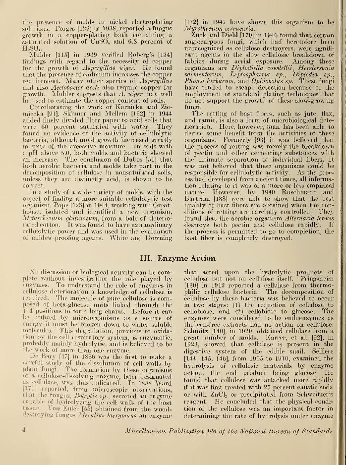
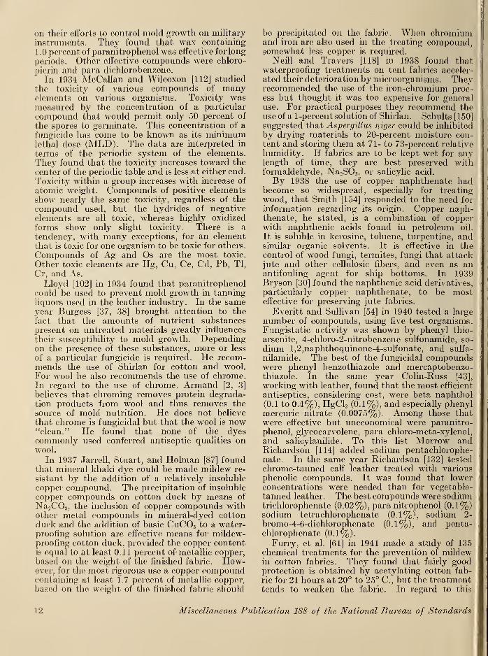
Among the materials which have been reportedas being subjected to microbial degradation,cotton and other cellulosic materials rank highest.Figure 2 shows cotton fabric heavily invaded by
Aspergillus niger. The earliest reports in this
field are, more or less, observations of moldactivity. In 1912 Osborn [125] reported thatmildew of cotton cloth appears to be due to manycommon saprophytic molds which, he believed,fed upon the starchy matter in the sizing. He
Microbiological Deterioration 5
Figure 2. Camouflage fabric with inferior fungicide show-ing the invasion of the weave by the mycelium and spore-heads of Aspergillus niger.
pointed out that the fungi could grow only in thepresence of sufficient moisture. Therefore, if
cotton materials were not permitted to becomedamp, they were not likely to become moldy.In 1913 Kroulik [97] reported that microorgan-isms, which he believed to be bacteria and actino-mycetes, decompose cellulose even at temperaturesas high as 60° to 65° C. In the same year, McBethand Scales [111], unlike the other workers of theperiod, were actually working at the causes ofcellulose degradation by microorganisms. Theyfound that a selective medium like cellulose agarwas superior to Omeliansky's selective culturemethod [100], and the filter-paper method ofVan Iterson [100]. They found that molds andbacteria, capable of destroying cellulose, arepresent in all productive soils. The cellulose-destroying types attack cellulose most rapidlyin the presence of oxygen and often lose theirpower to do so when grown on artificial media.The truth of this fact accounts, in part, for thegreat difficulty in finding suitable test organismsthat will maintain their growth characteristicsfrom generation to generation over long periods
of time. In the same year, Mutterlein [116]found that aerobic organisms were more im-portant than anaerobes in the decomposition of
cellulose in nature. In 1915, Guegen [78] reportedthat the black spots and patches that tend todevelop on tent cloth and sails when exposed todamp air, and that cause weakening of the fibers
as if by sulfuric acid, are due to the growth of
molds. He listed two organisms as being espe-cially destructive
—
Pleospora infectoria and P.herbarum. He believed that the spores of thesemolds were rarely airborne but were in the tissues
of the textile fiber.
In 1916, Otto [127] made as thorough a studyof the cellulolytic activity of microorganisms as
had been made up to that time. He used a widevariety of cellulosic materials as substrates in-
cluding blotting paper (true cellulose), variousnatural celluloses, and modified celluloses suchas oxycellulose, hydrocellulose and hydratedcellulose. He found that the response of molds to
these substrates varied considerably. But hewas able to report that soil molds were capableof "dissolving" cellulose. He believed that "solu-tion" of cellulose was effected by means of en-
zymes which caused hydrolytic cleavage. Thecleavage of various types of cellulose was effected
by the same molds, with hydrolytic cleavagetaking place in those molecular groups whichwere common to all the forms of cellulose tested.
Hutchinson and Clayton [85] in 1919 isolated
a new cellulose-destroying bacteria, Spirochaetacytophaga, which was to be studied later byThom and his coworkers for possible use as a test
organism. Hutchinson and Clayton studied thegrowth habits of this organism and reported that
it was aerobic and had great cellulolytic power.For the next few years various authors were
reporting in the same vein. This was the period
following the First World War, and it is probablethat as a result of the war much practical informa-tion had been gathered. Armstead and Harland
[4, 5], Fleming and Thaysen [58, 59], and Thaysenand Bunker [158] reported on the effects of micro-organisms on textile fibers, especially on the fact
that cottons of different origins had varying re-
sistance to destruction by microorganisms. By1924 Thaysen [159] was able to sum up his ob-
servations with the following: Moist cellulose-
containing materials, when not incrusted withlignin, constitute a very suitable food for manytypes of microorganisms. In the presence of
about 10 percent of moisture the spores develop
a mycelium that may be entirely on the surface
of the fiber, perforate it in places, or may evendevelop within the fiber. Certain bacilli also
develop colonies that gradually penetrate the
fiber. As these spores are normally present onraw cotton, the chances of infection in finishing
processes are great. These organisms produceboth chemical and physical or morphological
6 Miscellaneous Publication 188 of the National Bureau of Standards
changes in the fiber, involving loss of strengthand usually a marked discoloration. The pene-tration of the fiber is aided by the enzyme, cytase,
which converts the cellulose into glucose. Theglucose then is used by the organisms as a sourceof energy and on oxidation yields C02 ,
H, andorganic acids. A primary stage in the productionof glucose is the hydration of the cellulose,
wherein the fiber is more readily stained by dyesand retains the color better. The destructiveaction of the microorganisms on cotton may befollowed, almost from the start, by the swellingaction of a solution containing equal parts of
CS2 and 9 percent of NaOH. This reagent givesthe normal fiber a microscopic appearance similar
to that obtained with Schweitzer's reagent, thatis, swelling with constrictions. Fibers with micro-biological damage swell more quickly and presentan almost uniformly swollen appearance withoutconstrictions, possibly because of the destructionof the cuticle of the fiber. He confirms thefindings of others by stating that acetylated cellu-
lose is resistant to bacterial attack and thatbleached cotton is more resistant than unbleachedcotton. Finally, he agrees that some types ofcotton are more resistant to degradation thanothers. American cotton is most resistant, where-as Indian cotton is least resistant. In 1925 Thay-sen and Bunker [160] reported further on their
work with various cellulose derivatives. Theyfound that cellulose acetate was most resistantto microorganisms, whereas cuprammonium rayonwas least resistant.
In 1927 Serrano [147] showed that the deterio-ration of abaca fibers (manila hemp) was due to theaction of cellulose-digesting organisms of the gen-era Aspergillus, Penicillium, and Chaetomium.The growth of these organisms, he found, wasfavored by dampness, poor ventilation and ele-
vated temperatures. Winogradsky in 1929 [174]
made a careful study of cellulose decompositionand reported that cellulose undergoes rapid oxi-
dation in the soil and the product closely resemblesoxycellulose. The biological oxidation of cellu-
lose, he said, is differentiated from the purelychemical process in that the product does notreduce Fehling's solution. Assimilable nitrogen,preferably inorganic, is essential to this process,the consumption amounting to approximately 2parts of nitrogen for each 100 parts of cellulose
decomposed. The consumed nitrogen is trans-
formed into organic combination, but there is
evidence of some partial reduction to ammoniaeven with unrestricted access to the air. Theoptimum reaction for cellulose decomposition is
pH7. Slight changes of reaction occur during theprocess, depending on the form in which thenitrogen is supplied.
In 1930 Thaysen and Bunker [162] were thefirst to report on controlled field exposures in
which cotton, wool, hemp, and flax fabrics were
exposed to the action of microorganisms at Cey-lon Cyprus, Kenya, Trinidad, the Malay States,
and England. The materials were hung in theair, both in and out of sunlight; buried in thesoil; and submerged in the sea at Ceylon. Inmarine waters disintegration was most rapid,
and was mainly the result of bacterial action. Insoils the deterioration was caused by bacteria,
fungi, and termites. In the air the damage wasproduced by molds and actinomycetes. Theyfound that hemp and flax were most resistant to
degradation.Up to 1937 the most thorough study of cellulose
destruction in the soil was made by Madhok [107].
He found that the most rapid decomposition of
cellulose in the soil took place at an initial pH of
5.5 with NaX03 as a source of nitrogen. Thisindicated that the first rapid attack was by fungi,
followed later by bacteria as the reaction tendedtoward neutrality. Decomposition started mostrapidly at 37° C, but later the rate at 27° and 30°
C approached the 37° C rate. Fertilization of thesoil over a period of 25 years had no appreciableeffect on the microorganisms decomposing cellu-
lose. This latter observation was later verified byBatson, Teunisson, and Porges [16.] In the sameyear Galloway [67, 68] published a general reviewof the microbiology of textiles. He pointed outthat the microbes were useful as well as harmful,as mold enzyme products were used in manytextile procedures.
In 1938 Barker [12] made a study of \-arious bastfibers, including flax, ramie, hemp, pita grass, jute,
and sisal. In such fibers other polysaccharidesare closely associated with cellulose and are con-considered to be an integral part of the cellulose
aggregate. Xylan is the most common of thecarbohydrates retained by the fiber. The molec-ular structures of cellulose and xylan are similar,
and the action of fungi is to hydrolyze the latter,
breaking down its molecular chains into shorterlengths and thus causing loss of strength or rotting.
He emphasizes the accelerating effect of moistureon the deterioration of fibers by microorganisms.In another paper [13] in the same year, Barkerconsiders the use of antiseptics for the protectionof cellulosic fabrics. He finds salicylanilide to beexcellent for this purpose. The effectiveness of theThaysen acetylation process [159] is again affirmed.He believes that it renders a cotton surface unsuit-able as a nutrient source for microorganisms.
Shults [150] in 1938 reported that the develop-ment of Aspergillus niger can be inhibited bydrying cotton fabrics to 20-percent moisture con-tent followed by storage at 71 to 73-percentrelative humidity. This is at variance withThaysen and Bunker [158], who claim that 10percent of moisture is sufficient to cause germina-tion of spores. Shults points out that viscoseproducts are easily degraded by microorganisms.In 1939 Galloway [69] found that the minimum
Microbiological Deterioration 7
moisture regain for the development of micro-organisms on jute fibers was 17 percent.
At about this time the trend in this field wastoward the study of individual organisms thatcould be demonstrated to grow on a cellulose-
containing medium. In 1939 Hooper [84] isolated
from the soil an aerobic micro-organism that wascapable of growing on an inorganic medium, withsuch cellulosic materials as filter paper or cottonas the sole source of carbon. Although theorganism was studied in detail, it was not identi-
fied. By following the disintegration of cottonfibers under the microscope, Hooper found thatthe cuticle is removed from the fiber during theearly stages of attack, and then both the cement-ing material and cellulose are slowly digested.
The particulate nature of the cellulose fibrils of
the cotton-fiber cell wall is clearly visible in thepartially disintegrated fibers.
With the outbreak of war in Europe in 1939, thetempo of research in this field increased. Thaysen,et al. [163] found that in microbiologically active
soil cellulose acetate rayon was completely resist-
ant whereas cellulose, wool, silk, and cellulose
rayon fabrics were disintegrated. Bryson [30]
pointed out the need for preserving jute fabrics for
sandbags. He believed the naphthenic acid
derivatives and particularly copper naphthenateto be most promising.
In 1940 Waksman [170] recognizing the need for
up-to-date information on cellulose deterioration
published a review of the microbiology of cellulose
decomposition. He pointed out that while cellu-
lose was resistant to ordinary chemical reagentsand the digestive juices of higher animals, it wasreadily decomposed by a great variety of micro-organisms.
In 1941 Armstrong [6, 7, 8] and Barker [14]
studied the problems of jute-sandbag preservation.
They found that damp jute bags were subject to
rotting. Molds and bacteria of soils are responsi-
ble for this degradation. Molds attack jute that
contains at least 17 percent of moisture, while at
least 22 percent of moisture is required for attackby some strains of bacteria. Armstrong recom-mends a variety of preservative treatments andpoints out that treated jute bags can be expectedto have a useful life of 18 months to 2 years,
whereas untreated bags usually last a few weeksat best.
In 1945 Larnach and Wyke [98] investigated theproblem of mold growth on books and paper.
They observed that there was no growth of fungi
when the relative humidity was below 75 percent.
Therefore, to control mold growth they recom-mended the use of silica gel in book cases. In the
same year Fargher [57] made a study of thegrowth substances present in cotton. He foundthat raw cottons contain appreciable quantities
of K, Na, Ca, and Mg, all of which are essential
to vigorous growth of molds. The principal trace
metals, Fe, Cu, and Zn, which stimulate thegrowth of certain organisms are also present.
Most of the metals are present as water-solublesalts of organic acids, which are consumed readily
by micro-organisms. Also present are sulfate,
chloride, phosphate, glucose, and more complexcarbohydrates, and nitrogenous substances. Flourused in sizing affords an additional source of
nitrogen as well as of carbon, and supports moreprolific and rapid mold growth than starch.
Modified starches are utilized more readily thanare unmodified starches. The consumption of thenaturally occurring food materials can be followedby measurements of reducing power (coppernumber) and pH of the cotton during exposure to
damp conditions. When the water-soluble con-stituents are removed, cotton becomes moreresistant to micro-organisms. The resistance be-
comes still greater after scouring with alkalies.
For this reason it is believed that linen appears to
be more resistant than cotton to microbiological
degradation.
Bayley and Weatherburn [17, 18] in 1945 drawattention to the fact that while copper preserva-tives appear to be excellent in laboratory tests,
they have been able to isolate copper-tolerant
fungi from copper-treated fabrics.
Goldthwait, et al. [70] in 1946 reported that
cotton fibers, yarns, or even fabrics could bepartially acetylated in such a manner that thecotton retains its outward appearance but ac-
quires several new properties. The resulting
modified cotton is highly resistant to mildewingand rotting and has a lower moisture regain.
2. Proteinaceous Materials
Although the documentation of the micro-biological degradation of proteinaceous materialsis not as extensive as that for cellulosic materials,
the problem is equally serious. The organismsinvolved are, in general, not the same as thosewhich degrade cellulose [60, 153]. Organismsthat attack proteinaceous materials may causerotting of the material, as in the case of wool, orthey may attack important substances in the pro-
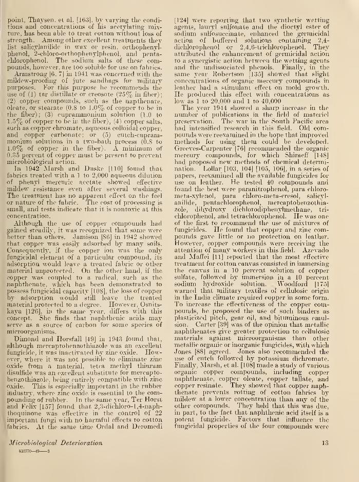
tein material, as the oils in leather. Figure 3
shows a circle of vegetable-tanned leather, heavily
overgrown by molds.
In 1924 Burgess [32] published a study of the
types of molds and bacteria found on damagedwool. He believed that molds caused discolora-
tion, whereas bacteria caused rotting. In 1926Hayes and Holden [82] made the same observa-
tions with regard to silk. In further studies onwool, reported in 1928 and 1929, Burgess [33, 34]
found that mildew on stored woolen goods wasentirely due to molds rather than to bacteria.
Soaps and other conditioners enhance its develop-
ment. The alkalies combine with the wool render-
8 Miscellaneous Publication 188 of the National Bureau of Standards
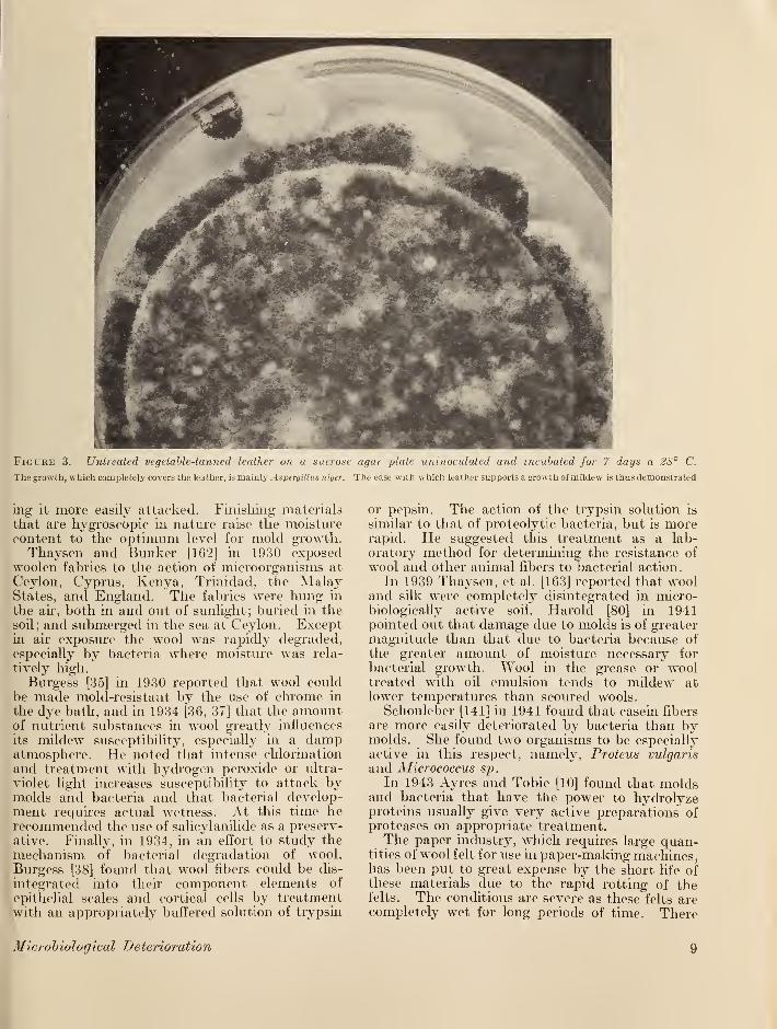
Ficure 3. Untreated vegetable-tanned leather on a sucrose agar plate uninoculated and incubated for 7 days a 28° C.
The growth, which completely covers the leather, is mainly Aspergillus niger. The ease with which leather supports a growth of mildew is thus demonstrated
ing it more easily attacked. Finishing materials
that are hygroscopic in nature raise the moisturecontent to the optimum level for mold growth.Thaysen and Bunker [162] in 1930 exposed
woolen fabrics to the action of microorganisms at
Ceylon, Cyprus, Kenya, Trinidad, the MalayStates, and England. The fabrics were hung in
the air, both in and out of sunlight; buried in the
soil; and submerged in the sea at Ceylon. Exceptin ah exposure the wool was rapidly degraded,especially by bacteria where moisture was rela-
tively high.
Burgess [35] in 1930 reported that wool couldbe made mold-resistant by the use of chrome in
the dye bath, and in 1934 [36, 37] that the amountof nutrient substances hi wool greatly influences
its mildew susceptibility, especially in a dampatmosphere. He noted that intense chlorination
and treatment with hydrogen peroxide or ultra-
violet light increases susceptibility to attack bymolds and bacteria and that bacterial develop-ment requires actual wetness. At this time herecommended the use of salicylanilide as a preserv-
ative. Finally, in 1934, in an effort to study the
mechanism, of bacterial degradation of wool.
Burgess [38] found that wool fibers could be dis-
integrated into their component elements of
epithelial scales and cortical cells by treatmentwith an appropriately buffered solution of trypsin
or pepsin. The action of the trypsin solution is
similar to that of proteolytic bacteria, but is morerapid. He suggested this treatment as a lab-
oratory method for determining the resistance of
wool and other animal fibers to bacterial action.
In 1939 Thaysen, et al. [163] reported that wooland silk were completely disintegrated in micro-biologically active soil. Harold [80] in 1941pointed out that damage due to molds is of greatermagnitude than that due to bacteria because of
the greater amount of moisture necessary for
bacterial growth. Wool in the grease or wooltreated with oil emulsion tends to mildew atlower temperatures than scoured wools.
Schonleber [141] in 1941 found that casein fibers
are more easily deteriorated by bacteria than bymolds. She found two organisms to be especiallyactive in this respect, namely, Proteus vulgaris
and Micrococcus sp.
In 1943 Ayres and Tobie [10] found that moldsand bacteria that have the power to hydrolyzeproteins usually give very active preparations ofproteases on appropriate treatment.The paper industry, which requires large quan-
tities of wool felt for use in paper-making machines,has been put to great expense by the short life ofthese materials due to the rapid rotting of thefelts. The conditions are severe as these felts arecompletely wet for long periods of time. There
Microbiological Deterioration 9
are many papers dealing with this subject but fewworkers have been able to solve the problem.Binns [24] in 1934 measured the extent of bac-
terial degradation of woolen felts by the loss in
strength. He found that attack by bacteria
depends in part on the source of the water used in
paper manufacture. It is generally increased bythe presence of phosphates and is inhibited by acid
conditions. Binns suggested the use of various
antiseptics but expressed the opinion that com-paratively large concentrations are needed for
protection.
3. Leather
Although leather is a protein substance, its
degradation by microorganisms is quite different
from that of other proteinaceous materials.
Although many reports have been received fromtropical battlefields to the effect that leather rots
and falls apart rather rapidly, none of the speci-
mens that were received were observed to be in
this condition. Furthermore, it has not been pos-
sible to rot leather in the laboratory, even in
soil-burial beds and in tropical room exposuretests. To be sure, leather articles such as shoes
and equipment cases have been observed to fall
apart, but on examination it was found that thestitching, usually cellulosic in nature, bad rotted.
No rotting was ever observed when nylon threadwas used for stitching leather. As shall be shown,the primary damage to leather by microorganismsis the stiffening and shrinking caused by the re-
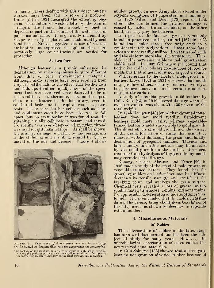
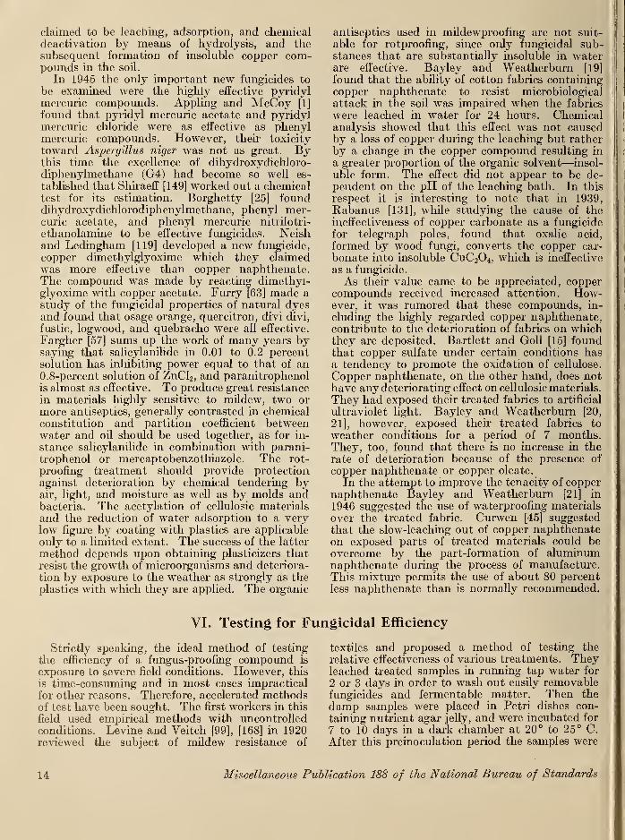
moval of the oils and greases. Figure 4 shows
Figure 4. Two cases of Army shoes received from storage
on the island of Saipan illustrate the importance of 'packaging.
The package on the right was in a badly deteriorated state when received,whereas the package on the left was in excellent condition. On openingthe cases, the shoes in the package on the right were heavily mildewed.
mildew growth on new Army shoes stored underextreme conditions of temperature and humidity.In 1925 Wilson and Daub [173] reported that
after hides are tanned the greatest damage is
caused by molds. Untanned hides, on the otherhand, are easy prey for bacteria.
In regard to the fats and greases commonlyfound in processed leather, Vaubel [167] in 1928found that molds attack free fatty acids to agreater extent than glycerides. Unsaturated fattyacids are more readily utilized than saturated acidsand the cis form more than the trans form. Thus,oleic acid is more susceptible to mold growth thanelaidic acid. In 1932 Grimshaw [77] found thatboth olive and lard oils are good sources of food for
molds but that mineral oil is not as good a source.With reference to the effects of mold growth on
leather, Lloyd [102] in 1934 observed that moldsmay produce stains, destroy tannins, hydrolyzefat, produce spues, and under certain conditionsmay pit the surface.
A study of microbial growth on 55 leathers byColin-Russ [43] in 1940 showed damage when themoisture content was about 30 to 35 percent of thetotal weight.
In 1945 Dempsey [48] found that chrome-tannedleather does not mold readily. Semichromeleathers mold more easily, whereas vegetable-tanned leather is most susceptible to mold growth.The direct effects of mold growth include damageof the grain, formation of stains that cannot beremoved without damaging the grain, and, finally,
destruction of pyrogallol tannins. Threads andfabric linings in leather articles may be affected
by the mold growth on the leather. Free acidresulting from hydrolysis of triglycerides by moldsmay corrode metal fittings.
Kanagy, Charles, Abrams, and Tener [90] in
1946 made a study of the effect of mold growth onvegetable-tanned leather. They found that thegrowth of mildew on leather increases its stiffness,
decreases its tensile strength and stretch at thebreaking point, and weakens the grain surface.
Chemical tests revealed a loss of grease, water-soluble materials, glucose, tannins, andnontannins.No appreciable deterioraton of hide substance wasfound. It was concluded that the molds, in assim-
ilating the grease, bring about decarboxylation of
the fatty acids, as shown by decrease in saponifi-
cation number.
4. Miscellaneous Materials
(a) Rubber
The deterioration of rubber in the latex stage
has been well documented and has been the sub-ject of study for many years. However, the
microbiological deterioration of cured rubber hasnot received equal attention.
In 1914 Sohngen [155] showed that microorgan-isms do not grow on air-dried rubber because of
10 Miscellaneous Publication 188 of the National Bureau of Standards
the low water content, which he found to be ap-proximately 0.5 percent. However, in the pres-ence of large amounts of water, common Indiarubber furnishes a medium for the growth of
bacteria and molds, inasmuch as it contains pro-teins, sugars, and resins.
Scott [142] in 1920 also showed that vulcanizedrubber, especially pure gum compounds, developsgrowths of Stemphyliurn macrosporoideum whenexposed to moist air. This growth was describedas resembling dust. Some threads were seen topenetrate the surface and were probably respon-sible for the loss in strength that occurred.
In 1938 Kalinenko [89] dispersed rubber in a
flask and inoculated it with Aspergillus oryzae.
In 1 month he found that 32 percent of the rubberhad been destroved.
ZoBell and Grant [177] in 1942 studied the deg-radation characteristics of various types of rubber.Their criterion of rubber degradation was in-
creased oxygen consumption and C02 production.They used inocula from sea water, garden soil,
old garden hose, and cracks in tires. All of these
inocula increased the oxygen consumption 5 to
10 times. The rate of oxygen consumption in-
creased exponentially with time.
Dimond and Horsfall [49] in 1943 pointed out
that wherever rubber is to be used either in con-
tact with water or under conditions of high mois-ture, as in the tropics, a mildew-proofing agentshould be used.
(b) Plastics
Plastic materials are of such recent origin that
aside from the experience gained in the recent war,there has been no documentation of microbiolog-ical deterioration. On the contrary, for a timeit was believed that because these materials wereman made or synthetic they could not possibly
be the source of microbiological nutrition. Thisfallacy was disproved early in the recent war,when it was found that in the tropics many typesof plastics supported copious growths of micro-organisms. Brown [29] in 1946 sums up a goodportion of the information gained during the warby reporting the work of several laboratories.
V. Prevention of Deterioration
The early use of mildew-proofing agents washaphazard and was based on empirical ratherthan on experimental information. Fargher [57]
in 1945 stated that prior to 1926, ZnCL, ordinarily
in a concentration of 0.8 percent, was the principal
mildew-proofing agent for fabrics but that it wasnot entirely effective. Other agents used weresalicylic acid, sodium fluoride, sodium silicofluoride,
phenol, cresylic acid, or formaldehyde.Corrigan [44] in 1920 recommended the use of
heat, light, and such antiseptics as copper sulfate,
potassium nitrate, zinc sulfate, zinc chloride, boric-
acid, formaldehyde, and phenols for the destruc-
tion of fungal growths on finished goods.In 1924 Thaysen [159] reported that acetylated
cellulose was resistant to fungal attack. It wasfound that bleached cotton was more resistant to
decay than unbleached cotton.
O'Brien [121, 122], working with rubber, re-
ported in 1926 that a 0.1 percent solution of
paranitrophenol was an effective mildew-proofingagent. In the same year Stevens [156], workingwith crepe rubber, also found paranitrophenol to
be an effective fungicide.
The earlier observations that bright fight tends
to inhibit fungal growth and that ultraviolet light
is actually fungicidal was explained by Bedford[22] in 1927, who found that destruction of micro-organisms by ultraviolet light is due to the pro-
duction of peroxide and is directly proportional to
the quantity generated.In 1928 a group of workers at the Shirley Insti-
tute in Leeds, Great Britain, investigated a large
number of compounds for possible use as mildew-
proofing agents. They found that salicylanilide
was the most efficient of all compounds examined.In honor of the Shirley Institute they named it
Shirlan. Fargher [57] in 1945 describes Shirlanas having all the desired properties:
1. Lack of volatility during fabric processing.2. High degree of solubility to insure uniform distribu-
tion in fabric.
3. It is unaffected by heat, metal surfaces, or by sizingand other finishing materials.
4. It is colorless and odorless.
5. It has no undesirable action on textiles.
6. Xo undesirable action on subsequent bleaching ordyeing.
7. Low cost.
8. Harmless to man.
By 1930 Shirlan had been used rather widely, andHall [79] recommended its use in place of ZnCL.In the same year Burgess [35] pointed out that theuse of chrome in the dye bath imparts a consider-able mold resistance to wool. With 0.5 percentof chrome, either as Cr03 or Cr2 3 . he obtainedgood mold resistance, and with 1.0 percent ofchrome the results were even more satisfactory.In the same year Thaysen and Bunker [162]suggested the use of mixtures of iron and chro-mium salts for both woolen and eellulosic fabricsand recommended copper oleate. Fargher, et al.
[56] summed up their long search for an idealantiseptic for the cotton industry by reiteratingthat Shirlan was the best. Other excellent ma"-terials were stated to be ortho chloromercuri-phenol, para acetoxymercuriacetanilide. thalliumcarbonate, para nitrophenol, and trichlorophenol.Xakazawa and Takeda [117] in 1934 reported
Microbiological Dete rio ratio
n
11
on their efforts to control mold growth on military
instruments. They found that wax containing
1 .0 percent of paranitrophenol was effective for long
periods. Other effective compounds were chloro-
picrin and para dichlorobenzene.
In 1934 McCallan and Wilcoxon [112] studied
the toxicity of various compounds of manyelements on various organisms. Toxicity wasmeasured by the concentration of a particular
compound that would permit only 50 percent of
the spores to germinate. This concentration of afungicide has come to be known as its minimumlethal dose (MLD). The data are interpreted in
terms of the periodic system of the elements.
They found that the toxicity increases toward the
center of the periodic table and is less at either end.
Toxicity within a group increases with increase of
atomic weight. Compounds of positive elementsshow nearly the same toxicity, regardless of the
compound used, but the hydrides of negativeelements are all toxic, whereas highly oxidized
forms show only slight toxicity. There is a
tendency, with many exceptions, for an elementthat is toxic for one organism to be toxic for others.
Compounds of Ag and Os are the most toxic.
Other toxic elements are Hg, Cu, Ce, Cd, Pb, Tl,
Cr, and As.
Lloyd [102] in 1934 found that paranitrophenolcould be used to prevent mold growth in tanningliquors used in the leather industry. In the sameyear Burgess [37, 38] brought attention to thefact that the amounts of nutrient substancespresent on untreated materials greatly influences
their susceptibility to mold growth. Dependingon the presence of these substances, more or less
of a particular fungicide is required. He recom-mends the use of Shirlan for cotton and wool.
For wool he also recommends the use of chrome.In regard to the use of chrome. Armand [2, 3]
believes that chroming removes protein degrada-tion products from wool and thus removes the
source of mold nutrition. He does not believe
that chrome is fungicidal but that the wool is now"clean." He found that none of the dyescommonly used conferred antiseptic qualities onwool.
In 1937 Jarrell, Stuart, and Holman [87] foundthat mineral khaki dye could be made mildew re-
sistant by the addition of a relatively insoluble
copper compound. The precipitation of insoluble
copper compounds on cotton duck by means of
Na2C03 , the inclusion of copper compounds withother metal compounds in mineral-dyed cottonduck and the addition of basic CuC03 to a water-proofing solution are effective means for mildew-proofing cotton duck, provided the copper contentis equal to at least 0.11 percent of-metallic copper,based on the weight of the finished fabric. How-ever, for the most rigorous use a copper compoundcontaining at least 1.7 percent of metallic copper,based on the weight of the finished fabric should
be precipitated on the fabric. When chromiumand iron are also used in the treating compound,somewhat less copper is required.
Neill and Travers [118] in 1938 found thatwaterproofing treatments on tent fabrics acceler-
ated their deterioration by microorganisms. Theyrecommended the use of the iron-chromium proc-ess but thought it was too expensive for generaluse. For practical purposes they recommend theuse of a 1-percent solution of Shirlan. Schults [150]
suggested that Aspergillus niger could be inhibited
by drying materials to 20-percent moisture con-tent and storing them at 71- to 73-percent relative
humidity. If fabrics are to be kept wet for anylength of time, they are best preserved withformaldehyde, Na2S03 , or salicylic acid.
By 1938 the use of copper naphthenate hadbecome so widespread, especially for treatingwood, that Smith [154] responded to the need for
information regarding its origin. Copper naph-thenate, he stated, is a combination of copperwith naphthenic acids found in petroleum oil.
It is soluble in kerosine, toluene, turpentine, andsimilar organic solvents. It is effective in thecontrol of wood fungi, termites, fungi that attackjute and other cellulosic fibers, and even as anantifouling agent for ship bottoms. In 1939Bryson [30] found the naphthenic acid derivatives,
particularly copper naphthenate. to be mosteffective for preserving jute fabrics.
Everitt and Sullivan [54] in 1940 tested a large
number of compounds, using five test organisms.Fungistatic activity was shown by phenyl thio-
arsenite, 4-chloro-2-nitrobenzene sulfonamide, so-
dium l,2,naphthoquinone-4-sulfonate, and sulfa-
nilamide. The best of the fungicidal compoundswere phenyl benzothiazole and mercaptobenzo-thiazole. In the same year Colin-Kuss [43],
working with leather, found that the most efficient
antiseptics, considering cost, were beta naphthol(0.1 to 0.4%), HgCL (0.1%), and especially phenylmercuric nitrate (0.0075%). Among those thatwere effective but uneconomical were paranitro-phenol, glycocarvolene, para ehloro-meta-xylenol,
and salicylanilide. To this list Morrow andRichardson [114] added sodium pentachlorophe-nate. In the same year Richardson [132] tested
chrome-tanned calf leather treated with various
phenolic compounds. It was found that lowerconcentrations were needed than for vegetable-tanned leather. The best compounds were sodiumtrichlorophenate (0.02%), paranitrophenol (0.1%)sodium tetrachlorophenate (0.1%), sodium 2-
bromo-4-6-dichlorophenate (0.1%), and penta-chlorophenate (0.1%).
Furry, et al. [61] in 1941 made a study of 135chemical treatments for the prevention of mildewin cotton fabrics. They found that fairly goodprotection is obtained by acetylating cotton fab-
ric for 21 hours at 20° to 25° C, but the treatmenttends to weaken the fabric. In regard to this
12 Miscellaneous Publication 188 of the National Bureau of Standards
point. Thaysen. et al. [163], by varying the condi-tions and. concentrations of his aeetylating mix-ture, has been able to treat cotton without loss of
strength. Among other excellent treatments theylist salicylanilide in wax or resin, orthophenyl-phenol, 2-chloro-orthophenylphenol, and penta-chlorophenol. The sodium salts of these com-pounds, however, are too soluble for use on fabrics.
Armstrong [6, 7] in 1941 was concerned with themildew-proofing of jute sandbags for militarypurposes. For this purpose he recommends theuse of (1) tar distillate or creosote (25% in fiber);
(2) copper compounds, such as the napthenate,oleate, or stearate (O.S to 1.0% of copper to be in
the fiber); (3) cuprammonium solution (1.0 to
1.5% of copper to be in the fiber), (4) copper salts,
such as copper chromate. aqueous colloidal copper,and copper carbonate; or (5) cutch-cupram-monium solutions in a two-bath process (0.8 to
1.0% of copper in the fiber). A minimum of
0.35 percent of copper must be present to preventmicrobiological action.
In 1942 Marsh and Duske [110] found thatfabrics treated with a 1 to 2,000 aqueous dilution
of phenyl mercuric acetate showed effective
mildew resistance even after several washings.The treatment has no apparent effect on the feel
or nature of the fabric. The cost of processing is
small, and tests indicate that it is nontoxic at this
concentration.
Although the use of copper compounds hadgained steadily, it was recognized that some werebetter than others. Jamison [86] in 1942 showedthat copper was easily adsorbed by many soils.
Consequently, if the copper ion was the onlyfungicidal element of a particular compound, its
adsorption would leave a treated fabric or othermaterial unprotected. On the other hand, if thecopper was coupled to a radical, such as thenaphthenate. which has been demonstrated to
possess fungicidal capacity [108], the loss of copperby adsorption would stiU leave the treatedmaterial protected to a degree. However, Osnits-kaya [126], in the same year, differs with this
concept. She finds that naphthenic acids mayserve as a source of carbon for some species of
microorganisms.
Dimond and Horsfall [49] in 1943 found that,
although mercaptobenzothiazole was an excellent
fungicide, it was inactivated by zinc oxide. How-ever, where it was not possible to eliminate zinc
oxide from a material, tetra methyl thiuramdisulfide was an excellent substitute for mercapto-benzothiazole, being entirely compatible with zinc
oxide. This is especially important in the rubberindustry, where zinc oxide is essential to the com-pounding of rubber. In the same year, Ter Horstand Felix [157] found that 2,3-diehloro-l,4-naph-
thoquinone was effective in the control of 22important fungi with no harmful effects to cottonfabrics. At the same time Ordal and Deromedi
[124] were reporting that two synthetic wettingagents, lauryl sulfonate and the dioctyl ester of
sodium sulfosuccinate, enhanced the germicidalaction of buffered solutions containing 2,4-
dichlorophenol or 2,4,6-trichlorophenol. Theyattributed the enhancement of germicidal action
to a synergistic action between the wetting agentsand the undissociated phenols. Finally, in thesame year Robertson [135] showed that slight
concentrations of organic mercury compounds in
leather had a stimulant effect on mold growth.He produced this effect with concentrations aslow as 1 to 20,000 and 1 to 40,000The year 1944 showed a sharp increase in the
number of publications in the field of materielpreservation. The war in the South Pacific areahad intensified research in this field. Old com-pounds were reexamined in the hope that improvedmethods for using them could be developed.Greeves-Carpenter [76] recommended the organicmercury compounds, for which Shiraeff [148]
had proposed new methods of chemical determi-nation. Lollar [103, 104] [105, 106], in a series of
papers, reexamined all the available fungicides for
use on leather. He tested 40 compounds andfound the best were paranitrophenol, para chloro-
meta-xylenol, para chloro-meta-cresol, salicyl-
anilide, pentachlorophenol, mercaptobenzothia-zole, dihydroxy dichloiodiphenylmethane, tri-
chlorophenol, and tetrachlorophenol. He was oneof the first to recommend the use of mixtures of
fungicides. He found that copper and zinc com-pounds gave little or no protection on leather.
However, copper compounds were receiving theattention of many workers in this field. Azevadoand Maffei [11] reported that the most effective
treatment for cotton canvas consisted in immersingthe canvas in a 10 percent solution of coppersulfate, followed by immersion in a 10 percentsodium hydroxide solution. Woodford [175]
warned that military textiles of cellulosic origin
in the India climate required copper in some form.To increase the effectiveness of the copper com-pounds, he proposed the use of such binders asplasticized pitch, gear oil, and bituminous emul-sion. Carter [39] was of the opinion that metallicnaphthenates give greater protection to cellulosic
materials against microorganisms than othermetallic organic or inorganic fungicides, with whichJones [88] agreed. Jones also recommended theuse of cutch followed by potassium dichromate.Finally, Marsh, et al. [108] made a study of variousorganic copper compounds, including coppernaphthenate, copper oleate, copper tallate, andcopper resinate. They showed that copper naph-thenate prevents rotting of cotton fabrics bymildew at a lower concentration than any of theother compounds. They held that this was due,in part, to the fact that naphthenic acid itself is apotent fungicide. Factors that influence thefungicidal properties of the four compounds were
Microbiological Deterioration
835770—49 3
13
claimed to be leaching, adsorption, and chemicaldeactivation by means of hydrolysis, and thesubsequent formation of insoluble copper com-pounds in the soil.
In 1945 the only important new fungicides to
be examined were the highly effective pyridylmercuric compounds. Appling and McCoy [1]
found that pyridyl mercuric acetate and pyridylmercuric chloride were as effective as phenylmercuric compounds. However, their toxicity
toward Aspergillus niger was not as great. Bythis time the excellence of dihydroxydichloro-diphenylmethane (G4) had become so well es-
tablished that Shiraeff [149] worked out a chemicaltest for its estimation. Borghetty [25] founddihydroxydichlorodiphenylmethane, phenyl mer-curic acetate, and phenyl mercuric nitrilotri-
ethanolamine to be effective fungicides. Neishand Ledingham [119] developed a new fungicide,
copper dimethylglyoxime which they claimedwas more effective than copper naphthenate.The compound was made by reacting dimethyl-glyoxime with copper acetate. Furry [63] made astudy of the fungicidal properties of natural dyesand found that osage orange, quercitron, divi divi,
fustic, logwood, and quebracho were all effective.
Fargher [57] sums up the work of many years bysaying that salicylanilide in 0.01 to 0.2 percentsolution has inhibiting power equal to that of an0.8-percent solution of ZnCl2 , and paranitrophenolis almost as effective. To produce great resistance
in materials highly sensitive to mildew, two or
more antiseptics, generally contrasted in chemicalconstitution and partition coefficient betweenwater and oil should be used together, as for in-
stance salicylanilide in combination with parani-trophenol or mercaptobenzothiazole. The rot-
proofing treatment should provide protectionagainst deterioration by chemical tendering byair, light, and moisture as well as by molds andbacteria. The acetylation of cellulosic materials
and the reduction of water adsorption to a verylow figure by coating with plastics are applicable
only to a limited extent. The success of the latter
method depends upon obtaining plasticizers that
resist the growth of microorganisms and deteriora-
tion by exposure to the weather as strongly as theplastics with which they are applied. The organic
antiseptics used in mildewproofing are not suit-
able for rotproofing, since only fungicidal sub-stances that are substantially insoluble in waterare effective. Bayley and Weatherburn [19]
found that the ability of cotton fabrics containingcopper naphthenate to resist microbiological
attack in the soil was impaired when the fabrics
were leached in water for 24 hours. Chemicalanalysis showed that this effect was not causedby a loss of copper during the leaching but ratherby a change in the copper compound resulting in
a greater proportion of the organic solvent—insol-
uble form. The effect did not appear to be de-
pendent on the pH of the leaching bath. In this
respect it is interesting to note that in 1939,Rabanus [131], while studying the cause of theineffectiveness of copper carbonate as a fungicide
for telegraph poles, found that oxalic acid,
formed by wood fungi, converts the copper car-
bonate into insoluble CuC 2 4 , which is ineffective
as a fungicide.
As their value came to be appreciated, coppercompounds received increased attention. How-ever, it was rumored that these compounds, in-
cluding the highly regarded copper naphthenate,contribute to the deterioration of fabrics on whichthey are deposited. Bartlett and Goll [15] foundthat copper sulfate under certain conditions hasa tendency to promote the oxidation of cellulose.
Copper naphthenate, on the other hand, does nothave any deteriorating effect on cellulosic materials.
They had exposed their treated fabrics to artificial
ultraviolet light. Bayley and Weatherburn [20,
21], however, exposed their treated fabrics to
weather conditions for a period of 7 months.They, too, found that there is no increase in therate of deterioration because of the presence of
copper naphthenate or copper oleate.
In the attempt to improve the tenacity of coppernaphthenate Bayley and Weatherburn [21] in
1946 suggested the use of waterproofing materials
over the treated fabric. Curwen [45] suggested
that the slow-leaching out of copper naphthenateon exposed parts of treated materials could beovercome by the part-formation of aluminumnaphthenate during the process of manufacture.This mixture permits the use of about 80 percent
less naphthenate than is normally recommended.
VI. Testing for Fungicidal Efficiency
Strictly speaking, the ideal method of testing
the efficiency of a fungus-proofing compound is
exposure to severe field conditions. However, this
is time-consuming and in most cases impractical
for other reasons. Therefore, accelerated methodsof test have been sought. The first workers in this
field used empirical methods with uncontrolledconditions. Levine and Veitch [99], [168] in 1920reviewed the subject of mildew resistance of
textiles and proposed a method of testing the
relative effectiveness of various treatments. Theyleached treated samples in running tap water for
2 or 3 days in order to wash out easily removablefungicides and fermentable matter. Then the
damp samples were placed in Petri dishes con-
taining nutrient agar jelly, and were incubated for
7 to 10 days in a dark chamber at 20° to 25° C.
After this preinoculation period the samples were
14 Miscellaneous Publication 188 of the National Bureau of Standards
inoculated with pure cultures "of several species of
molds and reincubated for 3 weeks to a month.They were examined each week for mold growth,and the observed conditions were rated on a scale
of 10. The test was considered to be a severe one.
The only treatment found to survive the full
period of the test was that with copper ammoniumhydroxide, the "cuprammonium process." Corre-lating their test with service conditions, they ob-served that treated canvas which gave a rating of
at least six did not mildew on exposure to Wash-ington, D. C, weather during the summer andfall months. In the same year Fleming and Thay-sen [58] developed a test of mildew damage in
which they treated the cellulosie material withNaOH and CS2 . Under the microscope they wereable to observe and count the damaged fibers.
In 1926 Bright [28] attempted to study theextent of mildew damage by examining the fibers
under the microscope. He used the Congo redtest and the swelling test of Fleming and Thaysen[58]. In the following year Morris [113] tested"fabric antiseptics" for toxicity to at least eightspecies of molds. The efficiency of each antiseptic
was compared to phenol as a standard. He foundthat although one mold may resist 10 times asmuch of one antiseptic as another species, thesituation may be entirely reversed when anotherantiseptic is tested. Therefore, he concluded, aconsiderable number of species must be used in
testing.
In 1929 Searle [143] described a new test
method. To compare the resistance of fabrics to
molds and bacteria, strips were wound aroundfilter candles that had been covered with a thin
layer of soil, and were incubated for as long as 6
weeks in a moist chamber at 30°C. They weretested for strength at periodic intervals. Searle
was the first to observe the interesting phenom-enon that cotton fabric deteriorated by moldshas the same cuprammonium viscosity as normalcellulose.
Thaysen and Bunker [162] in 1930 relied onoutdoor exposures followed by microscopic exa-
minations. They exposed fabrics in various
parts of the world, under a wide variety of con-ditions. In the following year Ellis [52, 53] andalso Hall [79] proposed a new microscopic test
method which involved the use of lactophenoland Cotton Blue. By this method mold hyphaecan be differentiated from cotton fibers.
In 1934 Burgess [38] proposed a laboratorymethod for determining the resistance of wool andother animal fibers to bacterial action. He foundthat wool fibers could be disintegrated into their
component elements of epithelial scales and corti-
cal cells by exposure to an appropriately buffered
solution of trypsin or pepsin. He showed that
the action of the trypsin is similar to that of
proteolytic bacteria but is more rapid.
In 1934 Thorn, et al. [166] examined all of the
available test methods and found that they fell into
two classes: (1) the simulation of the field hazardunder conditions more or less closely observed andrecorded, and (2) accelerated methods in which the
natural agents were given what were assumed to
be special, favorable conditions to attack thefabric and thus to concentrate into a short period
the destructive effects normally extending overlong periods. In both procedures, after incuba-tion, it was necessary that some kind of laboratoryexamination be applied to give comparativevalues to the measurement or estimation of thestage of deterioration. They considered only theeffect of cellulolytic organisms. After investi-
gating the possibilities of a test based on a mixedculture inoculum they came to the conclusionthat only a pure culture inoculum could be ex-
pected to give reproducible results. This decision
necessitated the selection of a suitable test or-
ganism and an appropriate medium. They set
up the following requirements for a suitable test
organism:
1. Capable of rapid destruction of cotton fabrics.
2. Must be easy to cultivate.
3. Must be adaptable to a fairly wide range of condi-tions.
4. Must be easy to identify in order to maintain in pureculture or to free from contaminants.
Thorn, et al. selected a modification of Czapek'sagar medium similar to that used by Dubos [51]
in 1928 but differing in that 3.0 g of NaN03 perliter was used in place of 0.5 g per liter and a pHof 6.8 in place of 7.5. Chaetomium globosum [40]
was found to be the most destructive of themolds tested. It had been reported previouslyas a cellulose destroyer by Galloway [64] in 1930,by Thaysen and Bunker [161] in 1927, and byothers. When tested against other species ofChaetomium, it was found to have the greatestcellulolytic power. This was confirmed byGreathouse and Ames [75] in 1945. In figure 5can be seen C. globosum growing on lacquer-impregnated filter paper.In 1940 Rogers, et al. [136] made an intensive
study of the effects of fungal growth on cottonfabrics. As test organisms they selected Chaeto-mium globosum and Spirochaeta cytophaga. Theyreaffirmed the excellence of the choice of Chaeto-mium globosum as a test organism and observedthat C. globosum is unique in that the pH of themedium is 6.8, both before and after completionof the incubation period.
Everitt and Sullivan [54] in 1940 tested fungi-cides by adding them directly to the growthmedium. They distinguished between fungistaticand fungicidal compounds. In the same yearO'Flaherty and Doherty [123] reported that therewas no standard accelerated test for evaluatingfungicides for leather. Several pages farther inthe same journal, Richardson [132] reported thathe had sprayed treated squares of leather with a
Microbiological Deterioration 1.5
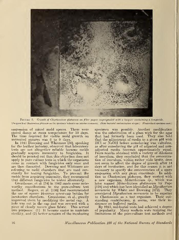
Figuhe 5. Growth of Chaetomium globosum on filter paper impregnated with a lacquer containing a fungicide.
The growth of Chaetomium globosum on the specimen indicates an inferior treatment. (Note bacterial contamination of agar.) (Unsteriiized specimen used.)
suspension of mixed mold spores. These werestored damp at room temperature for 50 days.The time required for visible mold growth onuntreated squares was 6 to 9 days.
In 1941 Downing and Whitmore [50], speakingfor the leather industry, observed that laboratorytests are not altogether reliable because moldsgradually acquire immunity to fungicides. It
should be pointed out that this objection does notapply to pure culture tests in which the organismscome in contact with fungicides only once andare then discarded. Downing and Whitmore are
referring to mold chambers that are used con-stantly for testing fungicides. To prevent themolds from acquiring immunity, they recommendthat different fungicides be tested alternately.
Greathouse, et al. [74] in 1942 made some note-worthy contributions to the pure-culture test
method. Rogers, et al. [136] had recommendedthe use of square 16-ounce screw-cap bottles for
incubation chambers. Greathouse, et al., greatlyimproved them by modifying the metal cap. Ahole was cut in the cap and was covered with apiece of closely woven glass fabric. This servedtwo purposes: (1) It became easier to maintainsterility, and (2) better aeration of the incubating
specimen was possible. Another modification
was the substitution of a glass wick for the agarthat had formerly been used. They also heldthat the adjustment of media to a given pH withHC1 or NaOH before autoclaving was valueless,
as after autoclaving the pH of adjusted and non-adjusted media becomes approximately equal.
From results obtained with a variety of dilutions
of inoculum, they concluded that the concentra-tion of inoculum, within rather wide limits, doesnot seem to affect the degree of growth after 14
days of incubation, and for this reason it is notnecessary to specify the concentration of a spore
suspension with any great exactitude. In addi-
tion to Chaetomium globosum, they worked witha new organism, Metarrhizium sp., which waslater named Metarrhizium glutinosum by Pope[128] and which has been identified as Myrothedumverrucaria by White and Downing [172]. Theyconsidered Myrothedum verrucaria to be superior
to Chaetomium as a test organism. Their out-
standing contribution, it seems, was their in-
sistence on buffered media.By 1943, soil-burial tests had achieved a degree
of popularity. Many workers were realizing the
limitations of the pure-culture test methods and
10 Miscellaneous Publication 188 of the National Bureau of Standards
were seeking a more severe test method. Furryand Zametkin [62] developed the soil-suspensiontest. Test specimens were inoculated by immer-sion in a soil suspension made from compostedsoil. The inoculated specimens were incubatedon agar medium, glass wick-nutrient medium, oreven cotton-batting in nutrient medium, all in
16-ounce square bottles. They claimed excellentdifferentiation of fungicides, approaching the soil-
burial test in potency. Borlaug [26] recom-mended the use of the soil-burial test for syntheticfabrics in preference to pure-culture methods.And Bunker [31] suggested the use of aluminumframes for mounting soil-burial test specimens.These were buried vertically in typical light
garden soil and kept at 30° C and 20-percentwater content. They were tested weekly by at-
tempting to tear them by hand. Finally, Berto-let [23] decided that as a result of extensive workin testing the mildew resistance of jute andosnaburg fabrics used in sandbag construction,the laboratory-accelerated mildew test should beabandoned in favor of the soil-burial test as ameasurement of the service life of this material.
In 1944 Lollar [103, 104] proposed the use of
mixed cultures of molds for testing mildew-proofed leather. ZoBell and Beckwith [178] madea similar recommendation for testing rubber com-pounds. Fabric testing was also showing areturn to less controlled test procedures. Aze-vado [11] described an accelerated rotting test in
which test strips were interlayered with strips of
rotten canvas. The incubation period was 60days under tropical conditions. However, suchworkers as Marsh, Greathouse, and Barker werestill carrying on the fight for standardized, con-trolled test methods. Marsh, et al. [108] in
testing a variety of copper compounds, concludedthat laboratory test methods had not yet de-
veloped to such a stage that then- results alonecould be used as accurate predictions of theresistance to mildewing or rotting of a treated
fabric under all of the varied service conditionsto which it might be subjected. Therefore, theytested treated fabrics by simulating field con-ditions before subjecting them to pure culture
tests. These modifying tests included: leaching,
adsorption by bentonite, and chemical deactiva-
tion by means of hydrolysis. They also made useof a copper-tolerant mold, Aspergillus niger.
They emphasized the futility of attemptingevaluation by the use of mixed cultures.
Klemme, et al. [96] in 1945 examined 43organisms for cellulolytic activity. Of these 29caused more than 50 percent loss in breakingstrength of cotton duck when incubated for 7 dayson a liquid mineral salt medium that containedNH4NO3 as the nitrogen source. Also, theystudied the effect of using different sources of
nitrogen. Whereas most of the organisms grewbest when NH4N03 was the nitrogen source,
Chaetomium globosum was unique in that it grewbest on NaN03 . This was later verified byGreathouse and Ames [75], who studied thecellulolytic acticity of 16 species of Chaetomium.They found that C. globosum was the most active
member of this genus. The ability of this groupto decompose cellulose was found to vary with thesource of nitrogen, the pH, the temperature, andwith the fabric preservative.
Up to 1945 the requirements for leachingspecimens before a mildew test were more or less
haphazard. Thorn, et al. [166] had suggestedthat fabrics be soaked in several changes of wateror in running water for 2 days. Several Govern-ment procurement specifications called for aleaching period of 30 hours in running tap water.Furry and Zametkin [62] suggested a 24 hornleaching period and specified that for each ounceof fabric there should be 1 gallon of water withthree complete changes during the 24 hours.Goodavage [71] described two types of tests-, aspray test for water repellent materials, and aleaching test. For the leaching test he specified
that there be 1 gallon of water at 65° F for eachspecimen 6 inches by 12 inches and suggested thatthere be three changes of water in 24 hours. In1945, Marsh, et al. [109] developed an apparatusthat could deliver water at a temperature of 30° Cto quart Mason jars. The flow was so regulatedthat there were 10 changes of water per hour.Also, they considered the effect of steam steriliza-
tion on organic fungicides and concluded that it
is, perhaps, an unnecessarily severe test for
these fungicides. When a nonsterile technique is
required, as in the case of the steam-distillable
organic fungicides, they found that C. globosumwas more satisfactory than certain other forms as
a test organism as it is less apt to be contaminated.They recommended a filter paper-mat techniquein which the test fabric was planted ou a mat ofmycelium. The mycelium was induced to growon a filter-paper strip supported on a mineralsalts-agar medium. This type of test was foundto be much more severe than the previouslydescribed pipette inoculum procedure. They be-lieved that the choice of the best test or com-bination of tests to determine mildew resistanceof a fabric depends on the service conditionsunder which the fabric is to be used.
In the same year Dean, et al. [46] reported onextensive studies of the soil-burial test method.They used only composted soil and carefully
controlled the temperature and moisture. Toobtain reproducible results they found that it wasnecessary to use fresh soil for each test. The soil-
burial resistance for cotton osnaburg treated withcopper naphthenate was measured for 10 identical
tests and was found to be reproducible with acoefficient of variation of 8.5 percent.
In 1946 Romano 1437] reexamined the role of
pfie rob iological DetenoraHon 17
nitrogen in the growth of Chaetomium globosum.Waksman and Hutchings [169] had shown that aratio exists between the activity of cellulose
decomposing microorganisms and the amount of
available nitrogen present. The higher the cellu-
lose content of soils, the greater is the need for
nitrogen to maintain cellulolytic activity.
Romano used NH4N03 because it maintained amore stabilized pH than NaN03 or KN03 . He
found that the optimum concentration of nitrogenwas 1.4 to 1.75 grams per liter of medium. Whenthe concentration of nitrogen was increased ashigh as 7.35 grams per liter, there were no toxicor inhibitory reactions. Varying the other ele-
ments in the medium had no effect on growth.He concluded that the nitrogen concentration of
the nutrient media in general use was insufficient
for maximum growth.
Part 2. Investigation of Microbiological Tests
VII. Development of Mildew Testing at Bureau
The primary problem of testing for mildewresistance at the National Bureau of Standardshas been the development of tests that are repro-ducible and sufficiently severe to give that marginof safety that is required for tropical use.
Considering the great number and variety of
fungicides, it is obvious that they cannot be equallyeffective, especially as it has been shown that theyact in a variety of ways. In order to determinetheir relative effectiveness, it is necessary to havemethods of bioassay. The term "bioassay" is
stressed because chemical analyses alone are notsufficient. It can easily be ascertained that aparticular fungicide is effective in a particularconcentration. Why, then, is it not sufficient to
make a quantitative analysis of a treated materialto learn whether the desired concentration hasbeen deposited? First, chemical analysis cannotdetermine the degree of penetration of a fungicideor its distribution over the treated material.
Second, as has been pointed out previously,
different materials react in decidedly different
ways to fungicides. For example, fungicides thatare effective for cotton may not be effective for
leather. Furthermore, it is important to test amaterial with regard to its ultimate use. Amaterial that is to be protected only during storageneed not have as permanent a fungicide as amaterial that is to be exposed to the elements andwhich may come in contact with the ground.Many workers have attempted to develop test
methods for the evaluation of fungicide-treatedmaterials. The first to tackle this problem werethose interested in the preservation of cottontextiles. Fabrics to be tested were inoculatedfrom rotted fabrics or from composted soils.
No effort was made to determine the nature of theorganisms used or to accelerate the test by addingnutrients. Within the past decade Thorn andhis coworkers have reexamined the question ofmildew tests and have decided that any test thatmakes use of mixed cultures of organisms doesnot lend itself to scientific accuracy. Thereupon,they studied the growth habits of many cellulose-
destroying organisms. They chose an excep-tionally stable organism Chaetomium globosum
Kunze which, during 5 years of continuous sub-culturing in the Microbiological Laboratory atthe National Bureau of Standards, has not beenobserved to change its growth characteristics.
It grows well on a mineral salts-agar mediumwhen a cellulosic carbon source is provided.When growing the organism for stock cultures,
filter paper is the usual source of carbon. Whentesting a cotton fabric, the test material itself
supplies the carbon. If the specimen does notcontain a fungicide, C. globosum will utilize thecellulose as a carbon source. If, however, thefabric is adequately mildew-proofed, the organismwill either be poisoned or the cellulose will not beavailable as a source of carbon, depending on themildew-proofing agent used.
As originally proposed by Thorn and his co-
workers, the culture medium was a modificationof that proposed by Czapek:
NaN0 3 3.0 g.
K2HP04 1.0 g.
MgS0 4 . 7H 2 0.25 g.
KCl 0.25 g.
FeSO-4 0.01 g.
Agar 10.0 g.
Distilled water 1.0 liter.
Adjust pH to 6.8 with NaOH or HC1 as required.
This medium was originally developed for mycol-ogists in order to study the growth characteristics
of various organisms. For this purpose a rathersparse growth is preferred. Therefore, by usingThorn's medium, which does not give maximumgrowth, C. globosum was not being used to fullest
advantage as a test organism. In brief, the test 1
method was as follows: strips of treated fabric of
a size suitable for determining breaking strengthwere sterilized and laid on a surface of mineralsalts-agar in bottles under aseptic conditions, after
which they were inoculated with an aqueoussuspension of the spores of C. globosum. Un- I
treated fabric was given the same test to serve as
a control. Next the bottles were incubated for|
14 days at a temperature of 28° C, after whichthe specimens were removed and washed, and,
j
when dry, their breaking strength was determined.J
Of course, the untreated fabric was completelyi
18 Miscellaneous Publication 188 of the National Bureau of Standards
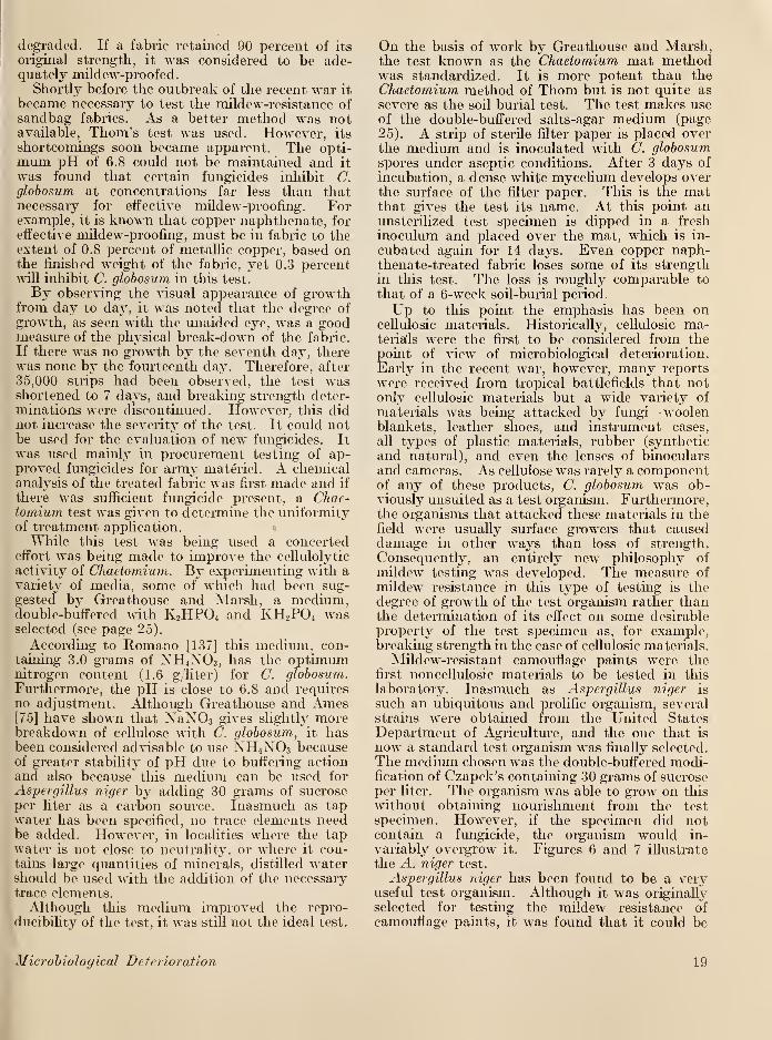
degraded. If a fabric retained 90 percent of its
original strength, it was considered to be ade-quately mildew-proofed.
Shortly before the outbreak of the recent war it
became necessary to test the mildew-resistance ofsandbag fabrics. As a better method was notavailable, Thorn's test was used. However, its
shortcomings soon became apparent. The opti-mum pH of 6.8 could not be maintained and it
was found that certain fungicides inhibit C.globosum at concentrations far less than thatnecessary for effective mildew-proofing. Forexample, it is known that copper naphthenate, for
effective mildew-proofing, must be in fabric to theextent of 0.8 percent of metallic copper, based onthe finished weight of the fabric, yet 0.3 percentwill inhibit C. globosum in this test.
By observing the visual appearance of growthfrom day to day, it was noted that the degree ofgrowth, as seen with the unaided eye, was a goodmeasure of the physical break-down of the fabric.
If there was no growth by the seventh day, therewas none by the fourteenth day. Therefore, after
35,000 strips had been observed, the test wasshortened to 7 days, and breaking strength deter-minations were discontinued. However, this didnot increase the severity of the test. It could notbe used for the evaluation of new fungicides. Itwas used mainly in procurement testing of ap-proved fungicides for army materiel. A chemicalanalysis of the treated fabric was first made and if
there was sufficient fungicide present, a Chae-tornium test was given to determine the uniformityof treatment application.
While this test was being used a concertedeffort was being made to improve the cellulolytic
activity of Chaetomium. By experimenting with avariety of media, some of which had been sug-
gested by Greathouse and Marsh, a medium,double-buffered with K2HP04 and KH2P04 wasselected (see page 25).
According to Romano [137] this medium, con-taining 3.0 grams of NH4N03 , has the optimumnitrogen content (1.6 g y
liter) for C. globosum.Furthermore, the pH is close to 6.8 and requiresno adjustment. Although Greathouse and Ames[75] have shown that NaN03 gives slightly morebreakdown of cellulose with C. globosum, it hasbeen considered advisable to use NH4N03 becauseof greater stability of pH due to buffering actionand also because this medium can be used for
Aspergillus niger by adding 30 grams of sucroseper liter as a carbon source. Inasmuch as tapwater has been specified, no trace elements needbe added. However, in localities where the tapwater is not close to neutrality, or where it con-tains large quantities of minerals, distilled watershould be used with the addition of the necessarytrace elements.Although this medium improved the repro-
ducibility of the test, it was still not the ideal test.
On the basis of work by Greathouse and Marsh,the test known as the Chaetomium mat methodwas standardized. It is more potent than theChaetomium method of Thorn but is not quite as
severe as the soil burial test. The test makes useof the double-buffered salts-agar medium (page
25). A strip of sterile filter paper is placed overthe medium and is inoculated with C. globosumspores under aseptic conditions. After 3 days of
incubation, a dense white mycelium develops overthe surface of the filter paper. This is the matthat gives the test its name. At this point anunsterilized test specimen is dipped in a fresh
inoculum and placed over the mat, which is in-
cubated again for 14 days. Even copper naph-thenate-treated fabric loses some of its strengthin this test. The loss is roughly comparable to
that of a 6-week soil-burial period.
Up to this point the emphasis has been oncellulosic materials. Historically, cellulosic ma-terials were the first to be considered from thepoint of view of microbiological deterioration.
Early in the recent war, however, many reportswere received from tropical battlefields that notoidy cellulosic materials but a wide variety of
materials was being attacked by fungi—woolenblankets, leather shoes, and instrument cases,
all types of plastic materials, rubber (synthetic
and natural), and even the lenses of binocularsand cameras. As cellulose was rarely a componentof any of these products, C. globosum was ob-viously unsuited as a test organism. Furthermore,the organisms that attacked these materials in thefield were usually surface growers that causeddamage in other ways than loss of strength.
Consequently, an entirely new philosophy of
mildew testing was developed. The measure of
mildew resistance in this type of testing is thedegree of growth of the test organism rather thanthe determination of its effect on some desirable
property of the test specimen as, for example,breaking strength in the case of cellulosic materials.
Mildew-resistant camouflage paints were thefirst noncellulosic materials to be tested in this
laboratory. Inasmuch as Aspergillus niger is
such an ubiquitous and prolific organism, several
strains were obtained from the United StatesDepartment of Agriculture, and the one that is
now a standard test organism was finally selected.
The medium chosen was the double-buffered modi-fication of Czapek's containing 30 grams of sucroseper liter. The organism was able to grow on this
without obtaining nourishment from the test
specimen. However, if the specimen did notcontain a fungicide, the organism would in-
variably overgrow it. Figures 6 and 7 illustrate
the A. niger test.
Aspergillus niger has been found to be a veryuseful test organism. Although it was originally
selected for testing the mildew resistance of
camouflage paints, it was found that it could be
Microbiological Deterioration 19
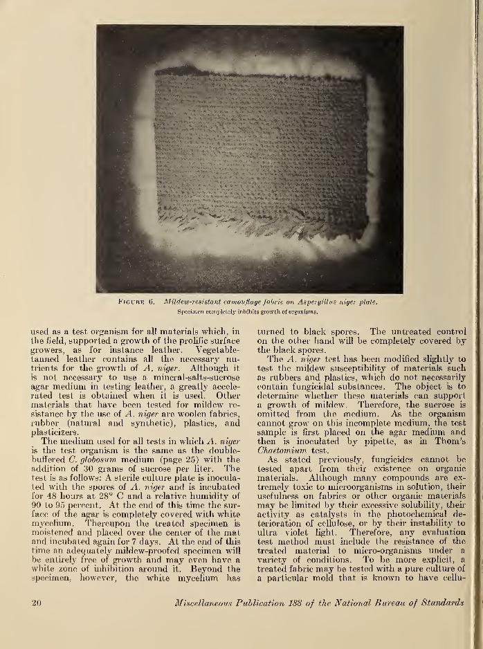
Figure 6. Mildew-resistant camouflage fabric on Aspergillus niger -plate.
Specimen completely inhibits growth of organisms.
used as a test organism for all materials which, in
the field, supported a growth of the prolific surface
growers, as for instance leather. Vegetable-tanned leather contains all the necessary nu-trients for the growth of A. niger. Although it
is not necessary to use a mineral-salts-sucrose
agar medium in testing leather, a greatly accele-
rated test is obtained when it is used. Othermaterials that have been tested for mildew re-
sistance by the use of A. niger are woolen fabrics,
rubber (natural and synthetic), plastics, andplasticizers.
The medium used for all tests in which A. niger
is the test organism is the same as the double-buffered C. globosurn medium (page 25) with theaddition of 30 grams of sucrose per liter. Thetest is as follows: A sterile culture plate is inocula-ted with the spores of A. niger and is incubatedfor 48 hours at 28° C and a relative humidity of
90 to 95 percent. At the end of this time the sur-
face of the agar is completely covered with whitemycelium. Thereupon the treated specimen is
moistened and placed over the center of the matand incubated again for 7 days. At the end of this
time an adequately mildew-proofed specimen will
be entirely free of growth and may even have awhite zone of inhibition around it. Beyond thespecimen, however, the white mycelium has
turned to black spores. The untreated control
on the other hand will be completely covered bythe black spores.
The A. niger test has been modified slightly to
test the mildew susceptibility of materials suchas rubbers and plastics, which do not necessarily
contain fungicidal substances. The object is to
determine whether these materials can supporta growth of mildew. Therefore, the sucrose is
omitted from the medium. As the organismcannot grow on this incomplete medium, the test
sample is first placed on the agar medium andthen is inoculated by pipette, as in Thorn'sChaetomium test.
As stated previously, fungicides cannot betested apart from their existence on organic
materials. Although many compounds are ex-
tremely toxic to microorganisms in solution, their
usefulness on fabrics or other organic materials
may be limited by then excessive solubility, their
activity as catalysts in the photochemical de-
terioration of cellulose, or by then instability to
ultra violet light. Therefore, any evaluation
test method must include the resistance of the
treated material to micro-organisms under a
variety of conditions. To be more explicit, a
treated fabric may be tested with a pure culture of
a particular mold that is known to have cellu-
20 Miscellaneous Publication 188 of the National Bureau of Standards
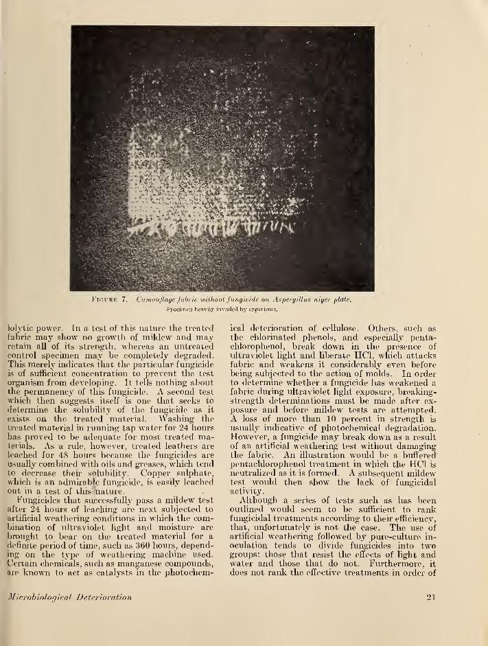
Fioure 7. Camouflage fabric without fungicide on Aspergillus niger plate.
Specimen heavily invaded by organisms.
lolytic power. In a test of this nature the treatedfabric may show no growth of mildew and mayretain all of its strength, whereas an untreatedcontrol specimen may be completely degraded.This merely indicates that the particular fungicideis of sufficient concentration to prevent the test
organism from developing. It tells nothing aboutthe permanency of this fungicide. A second test
which then suggests itself is one that seeks to
determine the solubility of the fungicide as it
exists on the treated material. Washing thetreated material in running tap water for 24 hourshas proved to be adequate for most treated ma-terials. As a rule, however, treated leathers are
leached for 48 hours because the fungicides are
usually combined with oils and greases, which tendto decrease their solubility. Copper sulphate,
which is an admirable fungicide, is easily leachedout in a test of this 'nature.
Fungicides that successfully pass a mildew test
after 24 hours of leaching are next subjected to
artificial weathering conditions in which the com-bination of ultraviolet light and moisture are
brought to bear on the treated material for adefinite period of time, such as 360 hours, depend-ing on the type of weathering machine used.
Certain chemicals, s\ich as manganese compounds,are known to act as catalysts in the photochem-
ical deterioration of cellulose. Others, such as
the chlorinated phenols, and especially penta-chloro phenol, break down in the presence of
ultraviolet light and liberate HC1, which attacksfabric and weakens it considerably even beforebeing subjected to the action of molds. In orderto determine whether a fungicide has weakened afabric during ultraviolet light exposure, breaking-strength determinations must be made after ex-
posure and before mildew tests are attempted.A loss of more than 10 percent in strength is
usually indicative of photochemical degradation.However, a fungicide may break down as a result
of an artificial weathering test without damagingthe fabric. An illustration would be a bufferedpentachlorophenol treatment in which the HC1 is
neutralized as it is formed. A subsequent mildewtest would then show the lack of fungicidalactivity.
Although a series of tests such as has beenoutlined would seem to be sufficient to rankfungicidal treatments according to their efficiency,
that, unfortunately is not the case. The use ofartificial weathering followed by pure-culture in-
oculation tends to divide fungicides into twogroups: those that resist the effects of light andwater and those that do not. Furthermore, it
does not rank the effective treatments in order of
Mierdbiological Deterioration 21
excellence. By this method a fungicide is either
good or bad. But experience has shown that all
good fungicides are not equally good, and someof the bad ones have their uses.
Inasmuch as the military materiel, for whichthese investigations were undertaken, were to beused under the most rigorous conditions, a verysevere test was desired in order to assure a widemargin of safety. Field exposures were time-consuming and otherwise impractical. A prac-tical solution to this problem has been the adoptionof the soil-burial test. This is the most potentmicrobiological test method in use at the presenttime. Top soil or compost is kept in small con-tainers in a cabinet in which the temperature andhumidity are carefully controlled. Originally,
the results obtained with this test were unusuallyvariable, even for a biological test. However, it
has been found that if the temperature andmositure are carefully maintained, the results aremuch more reproducible. As in all other biologi-
cal tests, untreated material of the same type as
the treated test sample is tested at the same timeto show that organisms responsible for deteriora-
tion are active. Inasmuch as this test was sovariable, another control was considered neces-sary. It was decided to choose one fungicidethat was invariably effective and to use it as areference standard for each new fungicide testedin the soil beds. In this way if there were anyvariations in results due to variations in the con-
ditions of the soil bed, they would be reflected in
the standard samples. The unknown could thenbe evaluated in terms of the standard. Thisstandard was, and still is, copper naphthenate in
a concentration of 1 percent of metallic copper,
based on the finished weight of the fabric.
Strangely enough, many of the fungicides thatbroke down during artificial weathering provedto be excellent in the soil-burial test. Among thebest of these were the chlorinated phenols, espe-
cially pentachlorophenol and dihydroxydichlorodi-phenylmethane. Either of these compounds mightbe recommended for materials that were not to beused in direct sunlight, as, for example, in stored
materials. On the other hand, many compoundsthat had successfully passed the accelerated
weathering test plus a pure culture test werequickly degraded in the soil. These were chiefly
mercury compounds and the more soluble chlor-
inated phenols. Such compounds might well beused for materials that would not normally comein contact with the ground as, for example, stored
articles. Again, this is a test that divides treat-
ments into two distinct classes. Although there
is a slight tendency for ranking of the superior
treatments, it is not entirely correlated with theultimate use of the material. The main virtue of
this test is the wide margin of safety it permitswhen one relies on its results, especially for
treated materials that are to receive most severe
exposure as sandbags and tents in tropical regions.
VIII. Representative Problems
The following investigations are representativeof the microbiological research and developmentconducted at the National Bureau of Standardsduring the past 5 years. The problems are
numbered from 1 to 10. This same numberingscheme is followed in the discussion starting onpage 29 and in the summary beginning onpage 38.
1. In developing the Chaetomium test, whichhad been first suggested by Thorn and otherauthors [166] it became necessary to know therate at which cellulosic materials deteriorateunder standard conditions, and whether cellulosic
materials of different origins break down in thesame manner. For experiments to obtain this
data, untreated cotton osnaburg (a coarse, un-bleached cotton fabric), having an average break-ing strength of 73 pounds for 1 inch-wide speci-men, and jute burlap, having a breaking strengthof 121 pounds for a 2-inch-wide specimen, wereused. To reduce the inherent variability of juteburlap, which has only 12 threads per inch, 2-inchraveled specimens were used. Eighty specimensof each type of fabric were sterilized for 1 hour inthe autoclave. Thereupon, 10 specimens of eachsterilized fabric were set aside for breaking-
strength controls. The remaining specimens werelaid on double-buffered mineral salts agar andwere inoculated with 2 milliliters of inoculum pre-
pared in accordance with the method outlined
under Chaetomium test procedures (page 25).
The specimens were then incubated at 28° to30° C and a relative humidity of 90 to 95 percent.
Ten specimens of each fabric were removed after
3, 4, 5, 7, 8, 9, and 10 days. After conditioning
at 70° F and 65-percent relative humidity, theywere broken and were compared with the controls.
In table 1 the results are listed as strength retained
in percentage of the original strength.
2. Inasmuch as cotton fabrics of different
thicknesses, treated with equal concentrations of
the same fungicide, showed varying resistance to
degradation in the soil-burial test, it was decided
to investigate the effect of thickness of fabric onthe rate of deterioration in the soil-burial test.
For this investigation four fabrics were selected.
They were in order of increasing thickness: a 4-
ounce lightweight sheeting with a breaking
strength of 46 pounds per inch width, a cotton
osnaburg having a breaking strength of 84 poundsper inch width, and a 20-ounce duck having a
breaking strength of 219 pounds per inch width.
22 Miscellaneous Publication 188 of the National Bureau of Standards
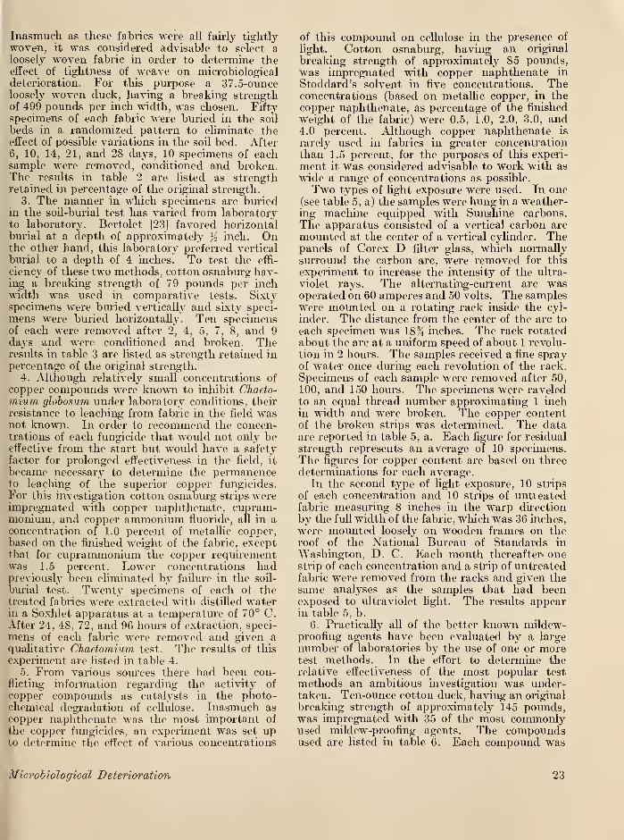
Inasmuch as these fabrics were all fairly tightlywoven, it was considered advisable to select aloosely woven fabric in order to determine theeffect of tightness of weave on microbiologicaldeterioration. For this purpose a 37.5-ounceloosely woven duck, having a breaking strengthof 499 pounds per inch width, was chosen. Fiftyspecimens of each fabric were buried in the soil
beds in a randomized pattern to eliminate theeffect of possible variations in the soil bed. After
6, 10, 14, 21, and 28 days, 10 specimens of eachsample were removed, conditioned and broken.The results in table 2 are listed as strengthretained in percentage of the original strength.
3. The manner in which specimens are buriedin the soil-burial test has varied from laboratoryto laboratory. Bertolet [23] favored horizontalburial at a depth of approximately % inch. Onthe other hand, this laboratory preferred vertical
burial to a depth of 4 inches. To test the effi-
cienev- of these two methods, cotton osnaburg hav-ing a breaking strength of 79 pounds per inchwidth was used in comparative tests. Sixtyspecimens were buried vertically and sixty speci-
mens were buried horizontally. Ten specimensof each were removed after 2, 4, 5, 7, 8, and 9
days and were conditioned and broken. Theresults in table 3 are listed as strength retained inpercentage of the original strength.
4. Although relatively small concentrations of
copper compounds were known to inhibit Chaeto-rnium globosum under laboratory conditions, tbeir
resistance to leaching from fabric in the field wasnot known. In order to recommend the concen-trations of each fungicide that would not only beeffective from the start but would have a safetyfactor for prolonged effectiveness in the field, it
became necessary to determine the permanenceto leaching of the superior copper fungicides.
For this investigation cotton osnaburg strips wereimpregnated with copper naphthenate, cupram-monium, and copper ammonium fluoride, all in aconcentration of 1.0 percent of metallic copper,based on the finished weight of the fabric, exceptthat for cuprammonium the copper requirementwas 1.5 percent. Lower concentrations hadpreviously been eliminated by failure in the soil-
burial test. Twenty specimens of each of thetreated fabrics were extracted with distilled waterin a Soxhlet apparatus at a temperature of 70° C.After 24, 48, 72, and 96 hours of extraction, speci-
mens of each fabric were removed and given aqualitative Chaetomium test. The results of this
experiment are listed in table 4.
5. From various sources there had been con-flicting information regarding the activity of
copper compounds as catalysts in the photo-chemical degradation of cellulose. Inasmuch ascopper napbthenate was the most important of
the copper fungicides, an experiment was set upto determine the effect of various concentrations
Microbiological Deterioration
of this compound on cellulose in the presence of
light. Cotton osnaburg, having an original
breaking strength of approximately 85 pounds,was impregnated with copper naphthenate in
Stoddard's solvent in five concentrations. Theconcentrations (based on metallic copper, in thecopper naphthenate, as percentage of the finished
weight of the fabric) were 0.5, 1.0, 2.0, 3.0, and4.0 percent. Although copper naphthenate is
rarely used in fabrics in greater concentrationthan 1.5 percent, for the purposes of this experi-
ment it was considered advisable to work with as
wide a range of concentrations as possible.
Two types of light exposure were used. In one(see table 5, a) the samples were hung in a weather-ing machine equipped with Sunshine carbons.
The apparatus consisted of a vertical carbon arc
mounted at the center of a vertical cylinder. Thepanels of Corex D filter glass, which normallysurround the carbon arc, were removed for this
experiment to increase the intensity of the ultra-
violet rays. The alternating-current arc wasoperated on 60 amperes and 50 volts. The sampleswere mounted on a rotating rack inside the cyl-
inder. The distance from the center of the axe to
each specimen was 18% inches. The rack rotatedabout the arc at a uniform speed of about 1 revolu-tion in 2 hours. The samples received a fine sprayof water once during each revolution of the rack.
Specimens of each sample were removed after 50,
100, and 150 hours. The specimens were raveledto an equal thread number approximating 1 inchin width and were broken. The copper contentof the broken strips was determined. The dataare reported in table 5, a. Each figure for residual
strength represents an average of 10 specimens.The figures for copper content are based on threedeterminations for each average.
In the second type of light exposure, 10 strips
of each concentration and 10 strips of untreatedfabric measuring 8 inches in the warp direction
by the full width of the fabric, which was 36 inches,
were mounted loosely on wooden frames on theroof of the National Bureau of Standai'ds in
Washington, D. C. Each month thereafter* onestrip of each concentration and a strip of untreatedfabric were removed from the racks and given thesame analyses as the samples that had beenexposed to ultraviolet light. The results appearin table 5, b.
6. Practically all of the better known mildew-proofing agents have been evaluated by a large
number of laboratories by the use of one or moretest methods. In the effort to determine therelative effectiveness of the most popular test
methods an ambitious investigation was under-taken. Ten-ounce cotton duck, having an original
breaking strength of approximately 145 pounds,was impregnated with 35 of the most commonlyused mildew-proofing agents. The compoundsused are listed in table 6. Each compound was
23
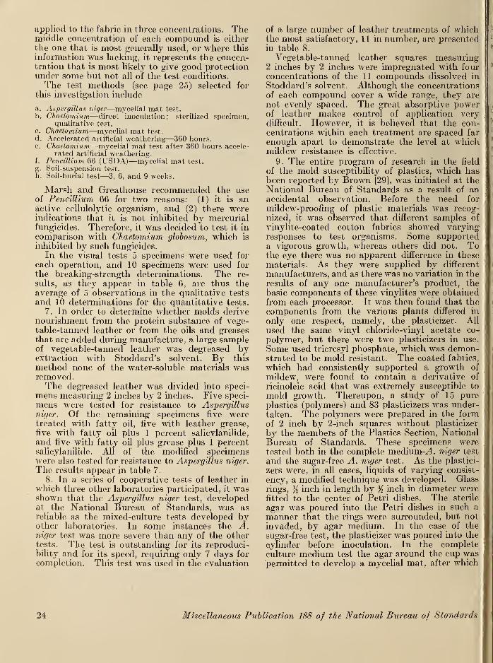
applied to the fabric in three concentrations. Themiddle concentration of each compound is either
the one that is most generally used, or where this
information was lacking, it represents the concen-tration that is most likely to give good protectionunder some but not all of the test conditions.The test methods (see page 25) selected for
this investigation include
a. Aspergillus niger—mycelial mat test.
b. Chaetomium—direct inoculation; sterilized specimen,qualitative test.
c. Chaetomium—mycelial mat test.
d. Accelerated artificial weathering—360 hours.e. Chaetomium—mycelial mat test after 360 hours accele-
rated artificial weathering.f. Pencillium 66 (USDA)—mycelial mat test.
g. Soil-suspension test.
h. Soil-burial test—3, 6, and 9 weeks.
Marsh and Greathouse recommended the useof Pencillium 66 for two reasons: (1) it is anactive cellulolytic organism, and (2) there wereindications that it is not inhibited by mercurialfungicides. Therefore, it was decided to test it in
comparison with Chaetomium globosum, which is
inhibited by such fungicides.
In the visual tests 5 specimens were used for
each operation, and 10 specimens were used for
the breaking-strength determinations. The re-
sults, as they appear in table 6, are thus theaverage of 5 observations in the qualitative tests
and 10 determinations for the quantitative tests.
7. In order to determine whether molds derivenourishment from the protein substance of vege-table-tanned leather or from the oils and greasesthat are added during manufacture, a large sampleof vegetable-tanned leather was degreased byextraction with Stoddard's solvent. By this
method none of the water-soluble materials wasremoved.The degreased leather was divided into speci-
mens measuring 2 inches by 2 inches. Five speci-
mens were tested for resistance to Aspergillusniger. Of the remaining specimens five weretreated with fatty oil, five with leather grease,
five with fatty oil plus 1 percent salicylanilide,
and five with fatty oil plus grease pins 1 percentsalicylanilide. All of the modified specimenswere also tested for resistance to Aspergillus niger.
The results appear in table 7.
8. In a series of cooperative tests of leather in
which three other laboratories participated, it wasshown that the Aspergillus niger test, developedat the National Bureau of Standards, was asreliable as the mixed-culture tests developed byother laboratories. In some instances the A.niger test was more severe than any of the othertests. The test is outstanding for its reproduci-bility and for its speed, requiring only 7 days for
completion. This test was used in the evaluation
of a large number of leather treatments of which 1
the most satisfactory, 1 1 in number, are presented a
in table 8.
Vegetable-tanned leather squares measuring2 inches by 2 inches were impregnated with fourconcentrations of the 11 compounds dissolved in
Stoddard's solvent. Although the concentrationsof each compound cover a wide range, they are
not evenly spaced. The great absorptive power,
of leather makes control of application very 1
difficult. However, it is believed that the con- I
centrations within each treatment are spaced far
enough apart to demonstrate the level at whichmildew resistance is effective.
9. The entire program of research in the field
of the mold susceptibility of plastics, which has1
been reported by Brown [29], was initiated at theNational Bureau of Standards as a result of anaccidental observation. Before the need for
mildew-proofing of plastic materials was recog-
nized, it was observed that different samples of
vinylite-coated cotton fabrics showed varying!
responses to test organisms. Some supporteda vigorous growth, whereas others did not. To i!
|
the eye there was no apparent difference in thesej
materials. As they were supplied by different i
manufacturers, and as there was no variation in the i
results of any one manufacturer's product, the
basic components of these vinylites were obtainedfrom each processor. It was then found that the
components from the various plants differed in I
only one respect, namely, the plasticizer. All t
used the same vinyl chloride-vinyl acetate co-[
i
polymer, but there were two plasticizers in use.\
t
Some used tricresyl phosphate, which was demon- I
strated to be mold resistant. The coated fabrics,[
which had consistently supported a growth of i
mildew, were found to contain a derivative ofJ
ricinoleic acid that was extremely susceptible to 11
mold growth. Thereupon, a study of 15 pure]
plastics (polymers) and 83 plasticizers was under- I
taken. The polymers were prepared in the form 1of 2 inch by 2-inch squares without plasticizer I
by the members of the Plastics Section, NationalBureati of Standards. These specimens were i
tested both in the complete medium-A niger testj
1
and the sugar-free A. niger test. As the plastici- i
zers were, in all cases, liquids of varying consist- i i
ency, a modified technique was developed. Glass j
rings, ji inch in length by % inch in diameter werefitted to the center of Petri dishes. The sterile I I
agar was poured into the Petri dishes in such a 1
manner that the rings were surrounded, but notI i
invaded, by agar medium. In the case of the t
sugar-free test, the plasticizer was poured into the t
cylinder before inoculation. In the complete !
culture medium test the agar around the cup was i
permitted to develop a mycelial mat, after which
24 Miscellaneous Publication 188 of the National Bureau of Standards
the cup was filled with plasticizer. The results
are reported in table 9, a and b, and are averagesof five observations for each test.
10. The study of the susceptibility of plastics
to mold growth stimulated the investigation of the
mold susceptibility of a group of related materials.
These included unvulcanized rubber (natural andsynthetic), nylon fabric, glass fabric, and mineralwool. The results are reported in table 10 andare averages of five observations for each test.
IX. Test Methods
In the foregoing description of investigations,
the test methods are outlined briefly. At this
point it is desirable to set forth in greater detail
the techniques of the methods used. The typesof biological test methods fall into two generalclasses, namely, pure-culture and mixed-culturemethods. In the pure-culture methods, Chaeto-
mium globosum is used to measure the rot resistance
of cellulosic fabrics and Aspergillus niger is usedto test the mildew-resistance of all noncellulosic
materials. The tests are as follows:
1. Pure-Culture Biological Methods
(a) Rot-Resistance Tests with Chaetomium globosum
(1) Direct inoculation—sterilized specimen meth-
od.—As this test can be used for either visual
(qualitative) or breaking strength (quantitative)
determinations, two types of test specimens wereused. For visual examination, squares measuring2 inches by 2 inches were used. Breaking strength
specimens were cut 6 inches long in the warpdirection by 1% inches in the filling direction.
The specimens were raveled to an equal threadcount which was approximately 1 inch in widthin the filling direction before breaking strength wasdetermined. As a control, untreated material,
similar in all other respects to the treated material,
was tested in the same manner as the test sampleto verify the viability of the test organisms.
A double-jacketed autoclave was used at a pressure
of 15 pounds per square inch and an exhaust tem-perature of 121° C for steam sterilization. Forleaching the specimens before the mildew tests,
an apparatus was developed which consisted of a
series of quart Mason jars so arranged that tap
water was delivered to the bottom of each vessel
at a rate of flow to permit five changes per hour.
The water was maintained at 27° to 30° C andthe pH was approximately neutral. Specimenswere leached for 24 hours for most materials and48 hours for treated leather. For incubationchambers, Petri dishes (10 cm) were used for visual
tests. For quantitative tests, 16-ounce squarebottles with modified screw caps were used. Themodified screw caps were prepared by cutting
circles 2 cm in diameter from the centers of the
caps. Circular pieces of glass filter fabric (Owens-Corning Fiberglas—CS. 30A-20) were inserted
on the inside of the caps.
The culture medium was made up as follows:
XH4NO3 3. g.
K 2HP0 4 2. g.
KH,P04 2. 5 g.
MgS0 4 . 7H,0 2. g.
Agar 20. g.
Tap water 1. liter.
The culture medium was melted in the autoclave,and 40 ml was poured into each bottle or sterile
Petri dish. The Petri dishes were then left undis-turbed until the agar had hardened. The bottles
with culture medium were sterilized for 20 minutes,after which they were placed on their sides to
harden
.
In preparing the inoculum, scrapings from a
ripe, fruiting culture of Chaetomium globosumUSDA 1042.4, which completely covered a 10-cmPetri dish, were transferred to an Erlenmeyer flask
containing 100 ml of sterile water by means of a
sterile tungsten loop. The black spore clusters
were squeezed against the sides of the flask witha sterile pipette until the tiny spores could beseen to go into suspension. Specimens that hadbeen leached for 24 hours were sterilized for 1 hourin the autoclave. When the sterilized specimenswere cool, one specimen was placed in contactwith the agar medium in each bottle under aseptic
conditions. One or two milhliters of inoculum,depending on the size of the specimen, was thendistributed evenly over each specimen by meansof a sterile pipette. The inoculated specimenswere incubated for 7 days, for the qualitative test,
and for 14 days, for the quantitative test at atemperature of 28° to 30° C and a relative humid-ity of 90 to 95 percent. In the quantitative test
the breaking-strength specimens were removedfrom the bottles after 14 days of incubation, if notcompletely degraded, and were gently washed toremove any growth of mildew. The specimenswere air-dried and then conditioned at 70° Fand 65 percent relative humidity for 24 hours,after which they were broken. AU averages of
residual strength reported in this paper are basedon 10 specimens for each test.
In the qualitative test the specimens wereexamined for growth of Chaetomium globosum.Any growth which could not be seen with theunaided eye was not considered to be significant.
The average degree of growth was based on five
specimens. The specimens were graded as follows:
No mildew.—No visible growth of mildew.Very slight mildew.—The smallest amount of
mildew growth which can be seen with theunaided eye—usually one or two isolated spots ofgrowth. This is usually indicative of spottytreatment.
Slight mildew.—Small patches of growth, which
Microbiological Deterioration 25

also indicate spotty treatment or perhaps differen-
tial leaching.
Moderate mildew.—Either many small patches of
growth or a thin uniform growth over the entire
specimen.Heavy mildew.—Specimen is completely covered
with mildew, but the underlying fabric structure is
faintly visible.
Very heavy mildew.—Specimen is completelycovered with a heavy growth of mildew, and theunderlying fabric structure is no longer visible.
Aseptic conditions were maintained by carrying
on all work in a dust-free room with filtered air.
As a further precaution, the room was equippedwith Sterilamps.
(2) Mycelial mat method.—Inasmuch as this
test cannot be used for making qualitative visual
observations because a specimen may show little
or no visible growth on the upper surface and yetshow a marked decrease in breaking strength, the
test specimens were prepared for breaking-strengthdeterminations as described in the previous test
method (page 25). It was not necessary to useadditional viability controls for this method, since
each culture bottle contained its own control.
Only culture bottles that had a well-developed3-day mycelium were used. The apparatus, cul-
ture medium, and inoculum for this test were thesame as described for the previous test (page 25),
except that only 16-ounce square screwcap bottles
were used. The culture medium was melted in
the autoclave and was poured into 16-ouncesquare screwcap bottles with modified caps. Thebottles containing agar were sterilized for 20minutes, after which they were placed on their
sides to harden. For the source of carbon, strips
of filter paper measuring 1% by 5K inches weresterilized in bottles for 1 hour. When cool, onestrip of sterile filter paper was transferred to the
surface of the hardened agar medium in each agarbottle under aseptic conditions. Two milliliters
of inoculum were distributed evenly over each strip
of filter paper in the agar bottles by means of a
sterile pipette. The inoculated strips were thenincubated for 3 days to develop mycelial mats.Bottles that failed to develop vigorous mats werediscarded. At this point, unsterilized, leachedtest specimens were inoculated by immersing themin a fresh inoculum in a large beaker. Excessmoisture was drained off by holding each specimenagainst the side of the beaker. The specimenswere then laid out smoothly over the 3-day-oldmycelial mats and Were incubated at 28° to 30°
C and a relative humidity of 90 to 95 percent for
14 days. The data were obtained as described in
the preceding quantitative test method (page25). Figure 8 shows a specimen of wood over-grown by C. globosum in the mycelium mat test.
(b) Mildew-Resistance Tests with Aspergillus niger
(1) Mycelial mat method.—Inasmuch as break-ing strength had no significance in a test in which
the test organisms were surface growers, the test
specimens were cut 2 by 2 inches for visual obser-vation. The apparatus for this test was the sameas described for the preceding tests (page 25),except that only Petri dishes were used as incu-
bation chambers. In order to accommodateliquid plasticizers, Petri dishes were modified byfitting them with Pyrex glass rings y2 inch in
length by }2 inch in diameter. The rings wereground against the central surface of the Petri
dish to assure a tight, leakproof fit. The hotsterile agar was poured around the rings, leavinga hollow cylinder in the center. The culture
medium was the same as described for the pre-
ceding tests (page 25), except that 30 grams of
sucrose per liter was added. The culture mediumwas sterilized in the autoclave for 20 minutes andwas allowed to cool somewhat before pouring.About 40 ml of the hot sterile agar medium waspoured into sterile Petri dishes under aseptic con-ditions. The plates were then covered and left
undisturbed until the agar medium had hardened.The hardened agar medium was inoculated byfirst loading a sterile cameFs-hair brush with spores
from a Petri dish culture of Aspergillus niger
USDA 215-4247 and then brushing the surface
of the sterile agar medium uniformly under aseptic
conditions. The inoculated Petri dishes wereincubated for 42 to 48 hours at 28° to 30° Cand 90- to 95-percent relative humidity until the
white mycelium was evident over the entire sur-
face of the agar medium. Cultures that did notshow a vigorous growth of mycelium were dis-
carded. No other viability controls were required.
At this point leached test specimens were dippedin a fresh aqueous suspension of A. niger and werelaid firmly on the mycelial mats and incubatedat 28° to 30° C and 90- to 95-percent relative
humidity for 14 days. At the end of the incuba-
tion period the test specimens were examined for
growth of A. niger. Any growth which could not
be seen with the unaided eye was not considered
to be significant. The average degree of growthwas based on five specimens. The specimenswere graded as described in the qualitative pro-
cedure of the Chaetomium-direct inoculation meth-od (page 25). Figure 9 shows the conclusion of
a mycelial mat test with A. niger.
(2) Sugar-free-medium method.—The conditions
of this test were similar to those of the Aspergillus—mycelial mat method, except that sucrose was not
added to the medium. For the viability control,
material that is known to support a growth of A.niger was tested in the same manner as the test
sample to verify the viability of the test organisms.
Test specimens were transferred to the sterile agar
plates and were inoculated by dusting them with
the spores of a Petri dish culture of A. niger.
Dusting was effected with a dry, sterile camel's-
hair brush. The inoculated specimens were in-
cubated at 28° to 30° C and 90 to 95-percent
26 Miscellaneous Publication 188 of the National Bureau of Standards
Figure 8. \\'ood treated with inferior fungicide <showiny growth of Chaetomium globosum when tested by the mycelial matmethod.
Figure 9. Aspergillus niger test of mildew-resistant camouflage fabric.
The specimen on the right had an insufficient concentration of the fungicide. The specimen on the left was adequately treated.
; Microbiological Deterioration 27
relative humidity for 14 days. The specimenswere graded as described in the qualitative
procedure of the Chaetomium-direct inoculation
method (page 25).
2. Mixed-Culture Biological Test Methods
(a) Soil-Burial Test
The specimens for the soil-burial test were pre-
pared as described for the quantitative proceduresof the Chaetomium tests (page 25). For this test
two types of control specimens were used. Un-treated cotton duck was exposed in the soil bedsthroughout the periods of testing in order to
verify the microbiological activity of the soil.
The soil was considered to be in a satisfactory con-
dition when the untreated controls deteriorated in
from 7 to 10 days. The reference standard con-
sisted of cotton fabric (similar in construction to
the test fabric) treated with a solution of coppernaphthenate of such concentration that 1.0-percent
plus or minus 0.05-percent of copper was retained
by the fabric. The fabric was impregnated withcopper naphtbenate with the aid of a householdwringer. The desired roll setting was first de-
termined by using Stoddard's solvent alone. Thenstrips of cloth 6 inches long in the warp direction
and the full width of the material were immersedin copper naphthenate solution of the determinedconcentration until thoroughly wet. The strips
were then passed through the rolls of the house-
hold wringer and were dried with constant motionto prevent migration of the fungicide. Before use
the treated fabrics were analyzed for copper con-
tent. The soil used in this test was taken fromthe top 6 inches of a fertile topsoil known as
Chester loam. The pH was 5.7, and the moistureequivalent was 22.6-percent, based on oven-dryweight. The clay content was 16.9-percent, andthere was 2.0-percent of organic matter present.
Stones and roots were removed by sieving before
use. The soil was placed in wooden containers
measuring 30 by 30 by 8 inches in height. Thecontainers were kept in a tight, insulated cabinet
in which the air temperature was maintained at
28° to 30° C, and the relative humidity was 95 to
97 percent. After moisture equilibrium had beenestablished, it was rarely necessary to add waterto the soil to maintain a 23-percent water content.
Test strips were inserted vertically to a depth of
4 inches by means of a metal tool. Care wastaken so that not more than % inch of the lower endof the fabric was bent over to assist burial.
Thirty strips of each test sample were buried so
that 10 specimens could be removed after each of
3 periods of time. The burial periods were 3, 6,
and 9 weeks. The specimens were removed fromthe soil beds at the end of each burial period,
unless they were completely degraded, and weregently washed to remove soil particles. Theywere air-dried and then conditioned for 24 hoursat 70° F and 65-percent relative humidity, after
which they were broken.
(b) Soil- Suspension Test
Breaking-strength specimens were used asdescribed in the quantitative Chaetomium tests
j
(page 25). Untreated material similar in all otherrespects to the treated material was tested in the
,
same manner as the test sample to verify the via-}
bility and cellulolytic activity of the test organisms.The apparatus and culture medium for this test
was the same as described for Chaetomium tests,
except that only 16-ounce screwcap bottles wereused. The inoculum consisted of a thin suspensionof composted soil containing 70 g of soil to 250 mlof distilled water. It was freshly prepared foreach test from a stockpile of soil obtained from
j
Miss Margaret Furry, Bureau of Human Nutritionand Home Economics, U. S. Department of Agri-culture. Agar bottles were prepared as describedin the Chaetomium tests (page 25). The test fabric
j
was inoculated by immersing in the inoculum for
30 minutes with occasional agitation. Each strip
was then removed from the mud and placed on thehardened agar surface in the culture bottle. The
j
bottles were then incubated at 28° to 30° C and 90to 95% relative humidity for 14 days. Data were
j
obtained as described in the quantitative Chaeto-mium tests (page 25).
3. Accelerated Weathering Test
Breaking strength specimens (page 25), in a;
;
sufficient number for subsequent biological tests,|
were exposed in an artificial weathering machine
,
for 360 hours. The apparatus consisted of a ver- I
tical carbon arc mounted at the center of a vertical I
metal cylinder. The arc was enclosed in a clear|
globe of No. 9200 Px Pyrex glass 0.0625 inch thick. I
The globe was cleaned whenever the carbons were u i
changed, which was at least once in every 36 hours :
of operating time. The arc was operated at 17j
amperes, with 140-volt 60-cycle alternating current.[ i
The test specimens were mounted on the inside ofj
the cylinder facing the arc. The distance of the !
face of the specimen from the center of the arc was I
14% inches. The cylinder rotated about the arcjInat a uniform speed of approximately three revolu- 1
tions per hour. A water spray hit each specimenj
in turn for about 1 minute during each revolutionj
of the cylinder. Ten specimens were always broken Iafter a 360-hour exposure to determine whether!there had been any deleterious photochemical effect, jl
If there was no significant loss in strength, the re-jl
maining exposed specimens were then subjected toll
various mildew tests.
28 Miscellaneous Publications 188 of the National Bureau of Standards
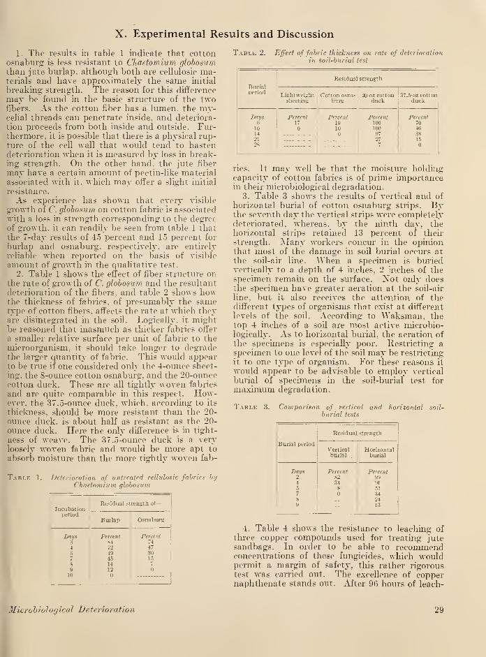
X. Experimental Results and Discussion
1. The results in table 1 indicate that cottonosnaburg is less resistant to Cftaetomium globosumthan jute burlap, although both are cellulosie ma-terials and have approximately the same initial
breaking strength. The reason for this difference
may be found in the basic structure of the twofibers. As the cotton fiber has a lumen, the my-celial threads can penetrate inside, and deteriora-
tion proceeds from both inside and outside. Fur-thermore, it is possible that there is a physical rup-ture of the cell wall that would tend to hastendeterioration when it is measured by loss in break-ing strength. On the other hand, the jute fiber
may have a certain amount of pectin-like materialassociated with it. which may offer a slight initial
resistance.
As experience has shown that every visible
growth of C. globosum on cotton fabric is associated
with a loss in strength corresponding to the degreeof growth, it can readily be seen from table 1 that
the 7-day results of 45 percent and 15 percent for
burlap and osnaburg. respectively, are entirely
reliable when reported on the basis of visible
amount of growth in the qualitative test.
2. Table 1 shows the effect of fiber structure onthe rate of growth of C. globosum and the resultant
deterioration of the fibers, and table 2 shows howthe thickness of fabrics, of presumably the sametype of cotton fibers, affects the rate at which theyare disintegrated in the soil. Logically, it mightbe reasoned that inasmuch as thicker fabrics offer
a smaller relative surface per unit of fabric to the
microorganism, it should take longer to degradethe larger quantity of fabric. This would appearto be true if one considered only the 4-ounce sheet-
ing, the 8-ounce cotton osnaburg. and the 20-ouncecotton duck. These are all tightly woven fabrics
and are quite comparable hi this respect. How-ever, the 37.5-ounce duck, which, according to its
thickness, should be more resistant than the 20-
ounce duck, is about half as resistant as the 20-
ounce duck. Here the only difference is in tight-
ness of weave. The 37.5-ounce duck is a veiyloosely woven fabric and would be more apt to
absorb moisture than the more tightly woven fab-
Table 1. Deterioration of untreated cellulosie fabrics byChaetomium globosum
Incubationperiod
Residual strength of
—
Burlap Osnaburg
Days Percent Percent3 84 744 72 47
5 49 3045 15
8 14 7
9 1210
Table 2. Effect of fabric thickness on rate of deterioration
in soil-burial test
Burialperiod
Residua] strength
Lightweightsheeting
Cotton osna-burg
20-oz cottonduck
37.5-oz cottonduck
Days6101421
28
Percent17
Percent1910
Percent1001009727
Percent70463815
rics. It may well be that the moisture holdingcapacity of cotton fabrics is of prime importancein their microbiological degradation.
3. Table 3 shows the results of vertical and of
horizontal burial of cotton osnaburg strips. Bythe seventh day the vertical strips were completelydeteriorated, whereas, by the ninth day, thehorizontal strips retained 13 percent of then-
strength. Many workers concur in the opinionthat most of the damage in soil burial occurs at
the soil-air line. "When a specimen is buriedvertically to a depth of 4 inches, 2 inches of thespecimen remain on the surface. Xot only doesthe specimen have greater aeration at the soil-air
line, but it also receives the attention of thedifferent types of organisms that exist at different
levels of the sod. According to Waksman, thetop 4 inches of a soil are most active microbio-logically. As to horizontal burial, the aeration of
the specimens is especially poor. Restricting aspecimen to one level of the soil may be restricting
it to one type of organism. For these reasons it
woidd appear to be advisable to employ vertical
burial of specimens in the soil-burial test for
maximum degradation.
Table 3. Comparison of vertical and horizontal soil-
burial tests
Residua] strength
Burial periodVertical Horizontalburial burial
Days Percent Percent2 82 994 34 91
5 8 517 348 249 13
4. Table 4 shows the resistance to leaching of
three copper compounds used for treating jutesandbags. In order to be able to recommendconcentrations of these fungicides, which wouldpermit a margin of safety, this rather rigoroustest was carried out. The excellence of coppernaphthenate stands out. After 96 horns of leach-
Microbiological Deterioration 29
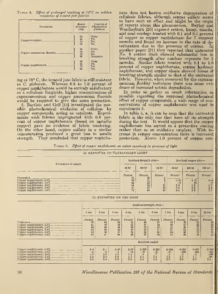
Table 4. Effect of prolonged leaching at 70°C on mildewresistance of treated jute fabrics
Treatment
Cuprammonium _
Copper ammonium fluoride.
Copper naphthenate _
Hoursof extrac-
tion
244872
244872
244872
Growth ofChaetomiumglobosum
Noner>o.Do.Do.
None.Do.Do.Moderate
NoneDo.Do.Do.
ing at 70° C, the treated jute fabric is still resistant
to C. globosum. Whereas 0.8 to 1.0 percent of
copper naphthenate would be entirely satisfactory
as a cellulosic fungicide, higher concentrations of
cuprammonium and copper ammonium fluoride
would be required to give the same protection.
5. Bartlett and Goll [15] investigated the pos-
sible photochemical oxidation of cellulose bycopper compounds, acting as catalysts. Experi-ments with fabrics impregnated with 0.6 per-
cent of copper naphthenate (based on metallic
copper) gave no evidence of fabric tendering.
On the other hand, copper sulfate in a similar
concentration produced a great loss in tensile
strength. They concluded that copper naphthe-
nate does not hasten oxidative degeneration ofcellulosic fabrics, although copper sulfate seemsto have such an effect and might be the originof reports about this phenomenon. Bayley andWeatherburn [20] exposed cotton, hemp, manila,and sisal cordage treated with 0.1 and 0.5 percentof copper as copper naphthenate for 7 summermonths and found no increase in the rate of de-terioration due to the presence of copper. Inanother paper [21] they reported that untreatedNo. 8 cotton duck showed substantial loss in
breaking strength after outdoor exposure for 3months. Similar fabric treated with 0.1 to 1.0
percent of copper naphthenate, copper hydroxynaphthenate, and copper oleate showed losses ofbreaking strength similar to that of the untreatedfabric. However, when measured by the cupram-monium fluidity technique there was some evi-dence of increased actinic degradation.
In order to gather as much information aspossible regarding the supposed photochemicaleffect of copper compounds, a wide range of con-centrations of copper naphthenate was used in
experiment 5.
In table 5, a, it can be seen that the untreatedfabric is the only one that loses all its strengthduring the test. It would appear that the coppernaphthenate has served as a protective coatingrather than as an oxidative catalyst. With in-
crease in copper concentration there is increasedprotection. Above 1.0 percent of copper con-
Table 5. Effect of copper naphthenate on cotton osnaburg in presence of light
(a) EXPOSURE TO ULTRAVIOLET LIGHT
Treatment of sample
Residual strength after
—
Residual copper after
—
50 hr 100 hr 150 hr 50 hr 100 hr 150 hr
Percent Percent Percent Percent Percent PercentUntreated - - - - 31
3810
Copper naphthenate, 0.5%. _ ._ _._ 11 5 0.31 0. 17 0.04Copper naphthenate. 1.0% _ 42 23 7 .74 .53 .41Copper naphthenate, 2.0% -- -- - 44 24 11 2.0 1.9 1.7Copper naphthenate. 3.0% . -.. - 55 37 19 3.0 2.9 2.8Copper naphthenate. 4.0%
.
_-- - --- - 72 45 29 4.0 3.9 3.7
(b) EXPOSURE ON NBS ROOF
UntreatedCopper naphthenate, 0.5%___ _..
Copper naphthenate, 1.0% ,_
Copper naphthenate, 2.0% _
Copper naphthenate, 3.0%Copper naphthenate, 4.0%-_-
Residual strength after—
1 mo 2 mo 3 mo 4 mo 5 mo 6 mo 7 mo 8 mo 9 mo 10 mo
Percent938184828295
Percent644948495061
Percent623834303644
Percent51
3228253236
Percent493729233037
Percent47342521
2632
Percent463021
17
2427
Percent3827
20142026
Percent342217
12
19
25
Percent3316
15
101622
Copper naphthenate, 0.5% _
Copper naphthenate, 1.0%.Copper naphthenate, 2.0%Copper naphthenate, 3.0%Copper naphthenate, 4.0%
Residual copper
0.4.81.93.04.0
0.1.81.92.94.0
0. 06.4
1.52.63.9
0.02.14
1.22. 5
3.6
0.008.01.8
2.03.3
0.005.01.7
2.03.3
0.004.01.3
1.92.9
0. 003.008.3
1.52.8
0.003.008.2
1.1
2.4
0.003.003.098.96
2.4
30 Miscellaneous Publication 188 of the National Bureau of Standards
' centration the loss in copper is negligible, whereas,: at 0.5 and 1.0 percent the loss is considerable,
i
However, if 0.3 percent of copper is considered
|to be the lower limit of fungicidal effectiveness,
then even after 150 hours of exposure the 1.0-
\
^percent sample still retains 0.41 percent of copper.The results of the roof exposures, listed in
. table 5, b, are not as clear-cut as are those for
i
the ultraviolet-light-exposure samples. In the
|
first month of exposure there is a loss in strength
i
for all samples. However, the untreated fabric
Iat 93 percent has not lost as much strength as
I
the treated samples, up to the 3-percent concen-tration. It would seem that copper does have a
|deteriorative effect on cotton fabric. Yet, the
|
4.0-percent sample, which has more copper than. any of the other samples, has a greater residual
. strength (95%) than the untreated fabric.
After 2 months the original fabric has dropped
;
to 64-percent residual strength and the 4-percent
|
copper fabric has dropped to 61 percent. The. residual strength of the remaining copper-treated
i
fabrics has dropped to 48 to 50 percent. After3 months the untreated fabric has lost an addi-
I
tional 2 percent in strength, whereas the 4.0-
(
percent-copper sample has dropped 17 percent,
, and now is not very much better than the other
,treated fabrics. It may be, assuming that there
is a photochemical effect, that it takes longer for
I
the ultraviolet rays to penetrate the 4-percent-
copper sample and thus effect its degradation.In the ensuing months the untreated fabric con-tinues to lose strength slowly until the tenthmonth, when only 33 percent of its original
strength remains. The 4-percent sample also
continues to lose strength slowly and has only22 percent of strength retained at the end of 10
months. Of the remaining concentrations, the
.2-percent-copper sample seems to show thegreatest deterioration during each month after
I
the second month. On the other hand, the 0.5-
i percent-concentration sample loses less strengthas time goes on, and by the fifth month is as
strong as the 4-percent sample. From there onuntil the ninth month it loses less strength thanthe 4-percent sample. Again, the copper loss is
proportionately less for high concentrations thanfor low. The 0.5- and 1.0-percent concentra-tions would probably be ineffective as fungicidesafter the fourth month.From the data in table 5, a, it appears that
copper naphthenate does not increase the tendency
j
to photochemical oxidation of cellulose under theconditions of the experiment. In fact, thereappears to be a protective action with increasing
concentration. However, table 5, b, indicatesthat although untreated fabric loses considerablestrength during outdoor exposure, fabric treated
|
with copper napthenate loses strength to a greaterextent. Whether this increased loss is due to aphotochemical effect in which the copper ion
takes part is difficult to say. It would appearthat the ultraviolet exposure, which completelydeteriorated cotton osnaburg in 150 hours, wasmuch more severe than the 10-month roof
exposure. One possibility presents itself. Thedifference in deteiioration may be due to differ-
ences in moisture. Although the samples weresprayed in the accelerated weathering test, thespray was of short duration, and, furthermore,copper naphthenate is known to render fabrics
slightly water repellent. This combination mightbe sufficient to keep the treated samples relatively
dry, whereas the untreated samples would tendto become wet enough to be affected photo-chemically. In this respect, the high degree of
copper retention, above the 0.5-percent concen-tration, may be considered as circumstantialevidence pointing to the lack of leaching. Onthe other hand, the outdoor samples were subjectedto alternate periods of sunshine and rain andmorning dew. This is reflected in the relatively
greater loss of copper. Furthermore, Bartlettand Goll [15] exposed their treated fabrics toultraviolet light without the addition of moisture.In a 48-hour exposure test they found that coppernaphthenate (0.6% Cu) treated cotton duckretained 75 percent of its strength, whereas theuntreated fabric retained only 69 percent of its
strength. The results of the 50-hour exposuresin table 5, a, are approximately twice as severebut are in the same proportion.
6. Although most of the microbiological labora-tories in the country have standardized on theuse of C. globosum, several laboratories preferother organisms or test methods with manyclaims for superiority. For example, Great-houseand Marsh have extolled the merits of Myrotheciumverrucaria {Metarrhizium glutinosum), althoughthey also find merit in C. globosum as well as agroup of cellulolytic Penicillia. Furry is espe-cially partial to the soil-suspension test, for
which she makes great claims. Bertolet hasdiscarded all pure-culture tests in favor of a soil-
burial test with horizontal burial of specimens.Several laboratories favor the use of mixtures oforganisms either in pure-culture technique or in
mold chambers. Outstanding among these is the"Tropical Room" at the Engineer Board, FortBelvoir, Va.
Table 6 shows that no single test can be usedto determine the usefulness of a fungicide underall service conditions. Although the Aspergillusniger test (column a) is not normally used for
testing fungicidal resistance of cellulosic materials,
it was included in this series of tests to show theeffect of A. niger on copper compounds. Thereis a slight growth of mildew on the 0.5-percentconcentrations of copper napthenate and copperhydroxy naphthenate, but the higher concentra-tions of both compounds show no growth ofmildew, although all specimens originally green
Microbiological Deterioration 31
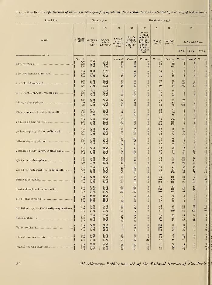
Table 6.
—
Relative effectiveness of various mildew-proofing agents on 10-oz cotton dvck as evaluated by a variety of test methods
Fungicide Growth of— Residual strength
Kind i~ oncGii-
tr&tion
(a)
Aspergil-
. lusmger
(b)
Chneto-mium
d lobOS U771
(0
l nueio-
ffllUW.-
myeelialmat
(d)
Accel-erated
artificial
weather-ing
(e)
Accel-erated
artificial
weather-ing plusL ntie.lv-
mium—mycelialmat
(0
Pen icil
Hum 66
(?)
boil sus-pension
So
3 wk
(h)
il burial
6 kw
for—
y \\ K
Percent Percent Percent Percent Percent Percent Percent Percent Percent.
|1.0 NM NM 14 98 2fi 55
< 2. XT A/TIN XVI XTA/TIN XVL 18 92 39 79
{ 3.0 NM NM 22 91 51 100
[0.
5
HM MM 98 10 54
•J
1.0 OlVl OlVI 9 96 1 1 63
I 1.
5
NM NM 45 90 45 79
| 1.0 NM NM 16 93 70 66 97J 9 o NM NM iy ooy/ 71 GO 1 nnxuu /ft
1 z.o NM NM 26 90 90 94 100 61
1 0.
5
SM NM 100 43 46J
I Q NM \TATIN 1VX 9 100 Au 4/ 51 Q Q A
1 L5 NM NM 19 100 58 64
( 1.0 NM NM 14 90 55 82 76J o o NM NM^ 36 QQOO U 77 Q1 3>, NM NM 96 82 68 86 100
( 0.5 MM NM 96 52 81) 1 (1\ 1. u SM XTA/TIN XVI 98 66 85 Q Q
I 1.5 NM NM 53 100 71 89
1 0.5 NM NM 100 100 29 100
\ 1.0 NM NM 100 93 80 100
I 1.5 NM NM 100 87 100 100
[0.5 NM NM 12 100 22 49 21
\ 1.0 XN XVI NM 33 100 35 78 OO
I 1.5 NM NM 83 100 69 83 39
|0.5 NM NM 33 100 38 69 C
s 1.0 MATIN XVi NM 65 97 67 • /jO i u Q
I 1.5 NM NM 97 96 83 94 39
1 0.5 NM NM 24 97 25 48 15
I 1.0 IN XVL NM 52 100 29 57 e;7 40 u
I 1-5 NM NM 89 100 48 85 91 37
1 1.0 NM NM 30 96 39 87 96 17
\ 2. NM NM 36 94 c 45 98 100 44
[ 3.0 NM NM 45 88 57 98 100 91 12
I 0.5 NM NM 24 100 35 82 55 15
< 1.0 NM NM 29 100 36 csy 40 A
I 1.5 NM NM 33 100 41 100 91 37 41
1 0.5 NM NM 100 96 56 100 36 9
\ 1. NM NM 100 85 100 83 46 38
I 1.5 NM NM 100 78 100 100 96 63 46
|0.5 MM NM 100 100 87 97 71 19
< 1.0 SM NM 100 100 100 100 OO 47
I 1.5 NM NM 100 100 100 100 98 63
I 1.0 HM HM 93 8 58
< 2. HM HM 90 15 64 Q
I 3.0 HM HM 32 84 27 73
1 0.5 NM NM 23 79 53 76 100 55 10
\ 1.0 NM NM 57 78 59 88 100 93 59
1 1.5 NM NM 76 72 61 100 100 100 97
1 0.5 NM NM 48 94 23 72 86 22
\ 1.0 ,vT A/TiN XVI IN XVI 51 95 28 76 86 40
I 1.5 NM NM 69 95 30 85 87 37
I 0.5 NM HM 98 100 63
\ 1.0 NM HM 94 100 74 28
{ 1.5 NM HM 98 100 78 36
| 0.5 NM NM 58 98 50 79 21
\ 1.0 NM NM 68 96 9 59 80 49
I 1.5 NM NM 90 100 24 63 89 97
I 0.5 NM NM 48 100 67 69 8
< 1.0 NM NM 76 100 74 81 44
I 1.5 NM NM 99 100 23 92 96 76
o-Phenylphenol
(/-Phenylphenol. sodium salt,
2. 4, 5-Trichlorophenol
2,4, 5-Trichlorophenol, sodium salt
Chloro-2-phenylphenol
Chloro-2-phenylphenol, sodium salt-. --
2-Chloro-4-phenylphenol
2-Chloro-4-phenylphenol, sodium salt
2-Bromo-4-pheny]phenol
2-Bromo-4-pheiiylphenol, sodium salt
2, 3, 4, 6-Tetraehlorophenol
2, 3, 4, 6-Tetrachlorophenol, sodium salt
Pentachlorophenol
Pentachlorophenol, sodium salt
2, 4, 6-Trichlorophenol
2,2' Dihydroxy, 5,5' Dichloiodiphenylmethane
Salicylanilide
Paranitrophcnol.
Phenyl mercuric acetate.
Phenyl mercuric salicylate.
32 Miscellaneous Publication 188 of the National Bureau of Standards
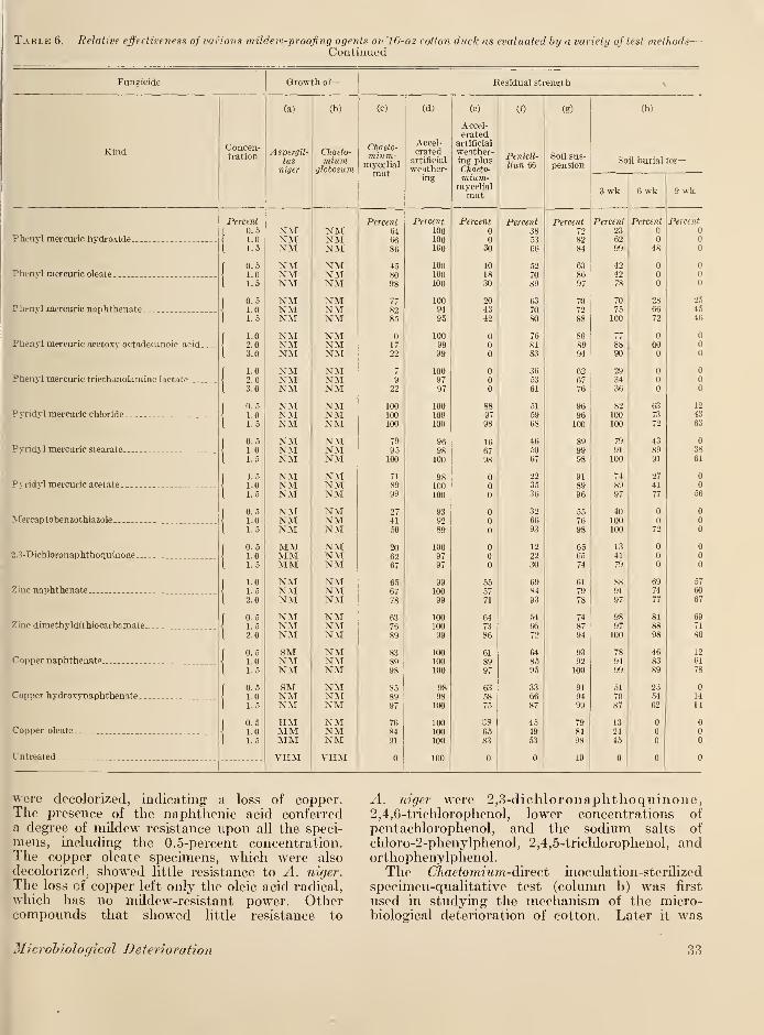
Table 6. Relative effectiveness of various ?nildew-proofing agents on 10-oz cotton duck as evaluated by a variety of test methods—Continued
Fungicide Growth of
—
Residual strength
Kind
Phenyl mercuric hydroxide.
Phcnyl mercuric oleate
.
Phenyl mercuric naphthenate.
Phenyl mercuric acetoxv octadecanoic acid.
Phenyl mercuric triethanolamine lactate.
Pyridyl mercuric chloride.
Pyrid;. 1 mercuric stearate.
Pyridyl mercuric acetate
.
Mercaptobenzothiazole
.
2.3-Dichloronaphthoquinone.
Ziuc naphthenate.
Zine dimethyldithiocarbamate..
Copper naphthenate.
Copper hydroxynaphthenate.
Copper oleate
Untreated
Concen-tration
(a)
Aspergil-lus
Percent0.51.01.5
0.51.01.5
0.51.01.5
1.0
2.03.0
1.02.03.0
n. 5
1.01.5
0. 5
1.01. 5
1.51.01.5
0.51.01.5
0. 5
1.01.5
1.01. 5
2.0
0. 5
1. 5
2.0
0.51.01.5
0.51.01.5
0.51.01.5
NMNMNMNMNMNMNMNMNMNMNMNMNMNMNMNMNMNMNMNMNMNMNMNMNMNMNMMMMMMMNMNMNMNMNMNMSMNMNMSMNMNMHMMMMMVHM
(b)
Chaeto-mium
jlobosum
(0
Chaeto-mium-
mycelialmat
NMNMNMNMNMNMNMNMNMNMNMNMNMNMNMNMNMNMNMNMNMNMNMNMNMNMNMNMNMNMNMNMNMNMNMNMNMNMNMNMNMNMNMNMNMVHM
Percent6466
922
100100100
95100
(d)
Accel-erated
artificial
weather-ing
Percent1001001C0
100100100
1009495
100
100
9797
100100100
100
100
100
100
9797
100
100
100
99
100100
100
100
100100100
100
(e)
Accel-erated
artificial
weather-ing plusChoeto-mium-
mycelialmat
Percent
30
101830
204342
(f)
Penicil-
liun 66
Percent385366
5270
(g)
Soil sus-pension
Percent7282
868994
626776
9696100
98
93
92100
91
9499
Soil burial for-
3 wk 6 wk 9 wk
Percent2362
7075100
8890
293436
82100100
7991
100
40100100
1341
9897100
78
9499
51
70
Percent
were decolorized, indicating a loss of copper.The presence of the naphthenic acid conferreda degree of mildew resistance upon all the speci-mens, including the 0.5-percent concentration.The copper oleate specimens, which were also
decolorized, showed little resistance to A. niger.
The loss of copper left only the oleic acid radical,
which has no mildew-resistant power. Othercompounds that showed little resistance to
A. niger were 2,3-dichloronaphthoquinone,2,4,6-trichlorophenol, lower concentrations of
pentachlorophenol, and the sodium salts of
chloro-2-phenylphenol, 2,4,5-trichlorophenol, andorthophenylphenol.The Chaetomium-dhcct inoculation-sterilized
specimen-qualitative test (column b) was first
used in studying the mechanism of the micro-biological deterioration of cotton. Later it was
Microbiological Deterioration 33
used as a relatively rapid means for determiningthe thoroughness with which acceptable fungicides
had been applied to cellulosic fabrics. With this
method the National Bureau of Standards wasable to test the fabric for 500 million sandbagsduring the first year of the recent war. This test
was also useful in screening unknown fungicides.
If a fabric, treated with an unknown compound,failed to pass this test, no further tests werenecessary. In this investigation it is not surpris-
ing that C. globosum failed to grow on so many of
the compounds. In the first place, the compoundswere selected because of their excellence for someparticular purpose, and, secondly, the concen-trations are close to the upper limit of usefulness.
Treatments that do not resist the growth of
C. globosum in this test are: sodium salts of
orthophenylphenol, 2,4,6-trichlorophenol, andparanitrophenol. Inasmuch as these compoundsare rather unstable at high temperatures, it is
quite likely that the use of steam sterilization is
objectionable.
The Chaetomium-mjcelial mat test representsthe work of several laboratories. It was pioneeredby Greathouse and Marsh and was further
developed in the laboratories at the NationalBureau of Standards. The results in column (c)
show that very few compounds can resist thegrowth of Chaetomium globosum under the con-ditions of this test. A decided advantage of this
test is that treated samples need not be subjectedto steam sterilization—a condition not encounteredin service. Treatments that resist C. globosumin this test are 3.0 percent of chloro-2-phenyl-phenol, 0.5 percent of 2-chloro-4-phenylphenoland its sodium salt, 1.5 percent of 2-bromo-4-phenyl-phenol and its sodium salt, 0.5 percent of
pentachlorophenol and its sodium salt, 1.5 percentof phenyl mercuric acetate, 1.5 percent of phenylmercuric salicylate, 1.5 percent of phenyl mercurichydroxide, 1.0 percent of phenyl mercuric oleate,
1.0 percent of phenyl mercuric naphthenate,0.5 percent of pyridyl mercuric chloride, 0.5
percent of pyridyl mercuric stearate, 1.0 percentof pyridyl mercuric acetate, 2 percent of zincnaphthenate, 1.5 percent of zinc dimethyl-dithio—carbamate, 0.5 percent of copper naphthenate,0.5 percent of copper hydroxy naphthenate, , and0.5 percent of copper oleate. With the exceptionof the copper and mercury compounds, the con-centrations of these compounds are somewhathigher than those customarily used.
Experience has shown that certain compoundspromote deterioration of cellulose. Regardless ofhow they achieve their destructive ends, it is
desirable to know whether a compound will affecta cellulosic fabric adversely even before it has theopportunity to protect the fabric from theravages of the microorganisms. Such informationcould be obtained by lengthy outdoor exposures,but the accelerated weathering test is much
faster. It must be emphasized that no claim is
made as to the correlation of accelerated labora-tory exposure with actual field exposure. Thereare many factors involved in outdoor exposurethat are not yet fully understood. For example,copper naphthenate-treated fabrics actually showno loss of strength in the usual 360-hour exposurein the accelerated weathering test, whereasuntreated fabrics do show a loss in strength. Yetthe reverse is true in outdoor exposure, althoughhere the untreated fabric also shows a loss instrength. At any rate, the accelerated weather-ing test has been useful in eliminating fungicidesthat break down in the presence of light and either
liberate harmful products or are inactivated. Ineither case, the fabric is no longer protected.Column (d) shows that the chlorinated phenols
tend to weaken the fabric as the concentration is
increased. This effect is serious only in the caseof chloro-2-phenylphenol, pentachlorophenol, 2,4,-
6-trichlorophenol and dihydroxydichlorodiphenyl-methane. However, a breaking-strength test
alone does not indicate whether any of thesecompounds has retained its fungicidal potency,even though the residual strength may be 100percent.
In column (e) are listed the results of a Chae-ifommm-mycelial mat test, following 360 hours of
accelerated weathering. The results are quite
striking. Very few compounds are shown to
possess the permanence necessary for the mostrigorous service conditions. These superior com-pounds include pyridyl mercuric chloride, pyridyl
mercuric stearate, zinc dimethyl dithiocarbamate,copper naphthenate, and copper oleate.
As a comparison of the effectiveness of C.
globosum in the mat method another cellulolytic
organism was chosen. Penicillium 66 was recom-mended by Dr. Paul Marsh, United States De-partment of Agriculture, Beltsville, Md. Theresults of tests with this organism are listed in
column (f) Although it is a competent cellulolytic
organism, it does not appear to be as tolerant of
fungicides in general as C. globosum, although there
are exceptions. Some of these are: 2-chloro-2-
phenyl phenol and pentachlorophenol which are
tolerated by Penicillium 66 at 0.5-percent con-centration, whereas C. globosum is completely in-
hibited. Salicylanilide is better tolerated, to aslight extent, by Penicillium 66 than C. globosum.
On the other hand, paranitrophenol completelyinhibits Penicillium 66, whereas C. globosum is notaffected by it at all. Phenyl mercuric acetoxyoctadecanoic acid is vulnerable to C. globosum,
although Penicillium 66 is fairly well inhibited byit. All pyridyl mercuric compounds are fairly
easily attacked by Penicillium 66, although theyare quite resistant to G. globosum.
It is obvious, then, that fungicides are effective
only with respect to the test organisms used.
Unfortunately, one cannot specify the type of or-
34 Miscellaneous Publication 188 of the National Bureau of Standards
ganisms that shall attack a particular treatedfabric in the field. Furthermore, it would be im-possible to foresee all contingencies by attemptingto use a large variety of test organisms. In this
dilemma, the solution has been the soil-burial test,
which is the most potent microbiological test in
use at the present time. In column (h) are listed
the results of the 3-, 6-, and 9-week exposures in
the soil beds. The 6-week results show that greatreliance may be placed on the Chaetomium-mycelial mat test, particularly after acceleratedweathering. Two exceptions to this are pentachlor-ophenol and dihydroxydichlorodiphenylmethane,which are excellent in the soil-burial test.
In an effort to secure the obvious advantages of
the many different types of organisms existing in
composted soil, Furry developed the soil inoculumtest. Unfortunately, the standardization of atest of tins type entails the control of a greatnumber of variables. For example, the soil usedin this test was obtained from Miss Furry and hadbeen stored for several months before use, withoccasional watering to keep it from drying out.
Strictly speaking, the soil should have been keptat standai'd conditions until needed for use in thetest. However, it was not practical to do so.
This lack of standardization in handling of the soil
bank may account for the variation in test results
encountered from laboratory to laboratory. Theresults of the soil-suspension test in this investi-
gation appear in column (g).
Although this investigation was primarily con-cerned with the evaluation of the most widely usedtest methods, the use of such a large number andvariety of fungicides provides an opportunity for
evaluating these fungicides. For want of a betterterm, "fungicide," as used here, refers to all
mildew-resistant compounds, regardless of their
mode of action.
It is quite obvious that, in regard to any par-ticular fungicide, resistance toward any particular
test organism does not necessarily imply resistance,
to all organisms. Furthermore, a compound maybe an effective fungicide under certain conditionsbut may lose its potency when subjected to thephysical agents of weathering. Thus the chlorin-
ated phenols, which are powerful fungicides, arelimited in their use because of their high degree ofsolubility and because of their photochemical in-
stability. With increasing concentration there is
a tendency for these compounds to cause deteriora-tion of the fabric during accelerated weathering.This effect is most pronouuced in the case ofpentachlorophenol, which shows a strength loss of22 percent for the 1.5 percent concentration.
Dihydroxydicliloi'odiphenylmethane, althoughnot a member of the Dowicide series of chlorinatedphenols, is also a chlorinated phenol. Its photo-chemical effect on the fabric is even more severethan that of petachlorophenol. At 1.5-percentconcentration, 28 percent of the fabric strength
Microbiological Deterioration
was lost. However, unlike the other chlorinated
phenols, this compound appears to be exception-
ally resistant to soil burial, particularly at the1.5-percent concentration.
Salicylanilide, which has been widely used underthe trade name of Shirlan, is shown to be readily
inactivated by accelerated weathering. Further-more, its performance in the soil-burial test in-
dicates either excessive solubility or the presence
of organisms that hydrolyze the compound to
salicylic acid and aniline.
Although paranitrophenol appears to be un-usually resistant to PeniciUium 66 at all concentra-
tions, it is seen to be ineffective in all the other
biological tests except the A. niger test. Althoughthis compound appears to be of little value as acotton-fabric fungicide, we shall see later (table 8)
how it is effective as a leather fungicide.
The mercury derivatives have been demon-strated to be rather powerful fungicides. AlthoughPeniciUium 66 appears to tolerate these fungicides
to a slightly greater degree than C. globosum, the
difference does not seem to be significant. Fur-thermore, phenyl mercuric acetoxyoctadecanoicacid and phenyl mercuric triethanolamine lactate
are tolerated to a greater extent by C. globosumthan by PeniciUium 66. On the other hand, theexcellent pyridyl mercuric compounds show onlymoderate resistance to PeniciUium 66, whereas C.
globosum is almost completely inhibited by them.Pyridyl mercuric acetate appears to be the mostsoluble of the pyridyl mercuric group.
Mercaptobenzothiazole and 2,3-dichlonmaptho-quinone are seen to be more or less ineffective
against all organisms used. Inasmuch as these
two compounds are known to be powerful fungi-
cides, their ineffectiveness on fabrics is probablydue to excessive solubility.
The two zinc compounds tested are seen to bemoderately good fungicides. The protection, at
higher concentrations, approaches the ideal of 90-
percent residual strength. A tendency toward sol-
ubility is indicated by the results of the mildewtest after accelerated weathering. These com-pounds, although not as effective as the coppercompounds, can be recommended where the use of
copper is objectionable, as, for instance, in rubber.
Of the three copper compounds tested, coppernaphthenate is by far the superior fungicide. It
is interesting to note that all of these compoundspermitted a growth of A. niger to some extent.
Where there was no growth, as in the higher con-
centrations of copper naphthenate and copper hy-droxy naphthenate, the specimens were decolon-
ized by the growth products in the mycelial mats.This phenomenon offers a clue as to the superiority
of copper naphthenate and copper hydroxy naph-thenate over copper oleate. The loss of color dueto the action of the end products of A. niger maybe simulated by immersing a test specimen in
0.4-percent solution of oxalic acid. Obviously,
35
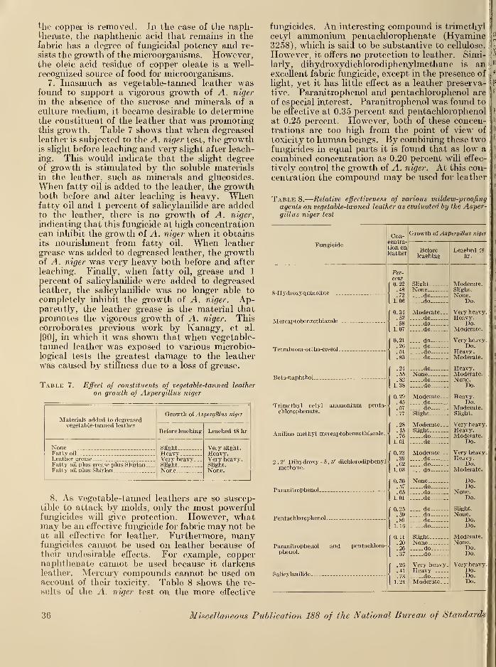
the copper is removed. In the case of the naph-thenate, the naphthenic acid that remains in thefabric has a degree of fungicidal potency and re-
sists the growth of the microorganisms. However,the oleic acid residue of copper oleate is a well-
recognized source of food for microorganisms.7. Inasmuch as vegetable-tanned leather was
found to support a vigorous growth of A. niger
in the absence of the sucrose and minerals of a
culture medium, it became desirable to determinethe constituent of the leather that was promotingthis growth. Table 7 shows that when degreasedleather is subjected to the A. niger test, the growthis slight before leaching and very slight after leach-
ing. This would indicate that the slight degreeof growth is stimulated by the soluble materialsin the leather, such as minerals and glucosides.
When fatty oil is added to the leather, the growthboth before and alter leaching is heavy. Whenfatty oil and 1 percent of salicylanilide are addedto the leather, there is no growth of A. niger,
indicating that this fungicide at high concentrationcan inhibit the growth of A. niger when it obtainsits nourishment from fatty oil. When leather
grease was added to degreased leather, the growthof A. niger was very heavy both before and after
leaching. Finally, when fatty oil, grease and 1
percent of salicylanilide were added to degreasedleather, the salicylanilide was no longer able to
completely inhibit the growth of A. niger. Ap-parently, the leather grease is the material thatpromotes the vigorous growth of A. niger. Thiscorroborates previous work by Kanagy, et al.
[90], in which it was shown that when vegetable-tanned leather was exposed to various microbio-logical tests the greatest damage to the leatherwas caused by stiffness due to a loss of grease.
Table 7. Effect of constituents of vegetable-tanned leather
on growth of Aspergillus niger
Materials added to degreasedvegetable-tanned leather
Growth of Aspergillus niger
Before leaching Leached 48 hr
None. SIight_ Very slight.
Heavy.Very heavy.Slight.
None.
Fatty oil .._ HeavyLeather greaseFatty oil plus grease plus ShirlanFatty oil plus Shirlan
Very heavy. .
.
SlightNone
8. As vegetable-tanned leathers are so suscep-tible to attack by molds, only the most powerfulfungicides will give protection. However, whatmay be an effective fungicide for fabric may not beat all effective for leather. Furthermore, manyfungicides cannot be used on leather because oftheir undesirable effects. For example, coppernaphthenate cannot be used because it darkensleather. Mercury compounds cannot be used onaccount of their toxicity. Table 8 shows the re-
sults of the A. niger test on the more effective
fungicides. An interesting compound is trimethylcetyl ammonium pentachlorophenate (Hyamine3258), which is said to be substantive to cellulose.
However, it offers no protection to leather. Simi-larly, dihydroxydichlorodiphenylmethane is anexcellent fabric fungicide, except in the presence of
light, yet it has little effect as a leather preserva-
tive. Paranitrophenol and pentachlorophenol are
of especial interest. Paranitrophenol was found to
be effective at 0.35 percent and pentachlorophenolat 0.25 percent. However, both of these concen-trations are too high from the point of view of
toxicity to human beings. By combining these twofungicides in equal parts it is found that as low acombined concentration as 0.20 percent will effec-
tively control the growth of A. niger. At this con-centration the compound may be used for leather
Table 8.
—
Relative effectiveness of various mildew-proofingagents on vegetable-tanned leather as evaluated by the Asper-gillus niger test
Fungicide
8-H yd rosyquinoline
Mercaptobenzothiazole
Tetrabrom-ortho-cresol.
Beta-naphthoL
Trimethyl cetyl ammonium penta-
chlorophenate.
Anilino methyl mercaptobenzothiazole.
2 , 2' Dihydroxy -5,5' dichlorodiphenylmethane.
Paranitrophenol.
Pentachlorophenol.
Paranitrophenol and pentachloro-
phenol.
Salicylanilide.
Con-centra-tion onleather
Per-cent0. 22
.48
.721. 06
0.34.52.581.07
0. 21.26.51.83
.24
.55
.821.38
0. 29
.45
.57
. 77
.28
.45
.761.01
0. 23.39.62
1.03
0.30.57.65
1. 01
0. 25.59.89
1. 16
0.11.20.26.37
.26
.41
.731.24
Growth of Aspergillus niger
Beforeleaching
Slight Moderate.None. Slight.
do None.do Do.
Moderate... Very heavy.do Heavy.do Do.do Moderate.
do Very heavy.do Do.do Heavy.do Moderate.
do Heavy.None.. _ ... Moderate.
do. _ None.do Do.
Moderate... Heavy.do Do.do Moderate.
Slight Slight.
Moderate... Very heavy.Slight Heavy.
do Moderate.do Do.
Moderate. __ Very heavy.do Heavy.do Do.do.. Moderate.
None Do.do Do.do None.do Do.
dododo..do
Slight.
Nonedodo
Very heavy.Heavy
doModerate. ..
Leached 48hr.
Slight.
None.Do.Do.
Moderate.None.
Do.Do.
Very heavyDo..Do.Do.
30 Miscellaneous Publication 188 of the National Bureau of Standards
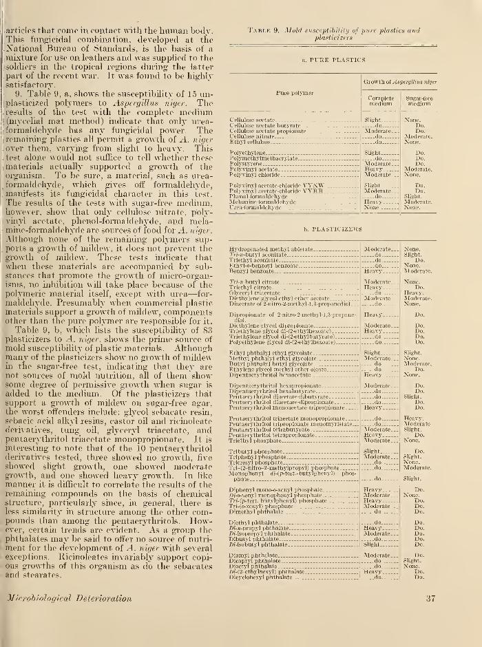
articles that come in contact with the human body.This fungicidal combination, developed at theNational Bureau of Standards, is the basis of amixture for use on leathers and was supplied to thesoldiers in the tropical regions during the latter
part of the recent war. It was found to be highlysatisfactory.
9. Table 9, a, shows the susceptibility of 15 un-plasticized polymers to Aspergillus niger. Theresults of the test with the complete medium(mycelial mat method) indicate that only urea-formaldehyde has any fungicidal power. Theremaining plastics all permit a growth of ^4. nigt r
. over them, varying from slight to heavy. This
. test alone would not suffice to tell whether thesematerials actually supported a growth of theorganism. To be sure, a material, such as urea-formaldehyde, which gives off formaldehyde,
. manifests its fungicidal character in this test.
The results of the tests with sugar-free medium,however, show that only cellulose nitrate, poly-vinyl acetate, phenol-formaldehyde, and mela-mine-formaldehyde are sources of food for A. n iger.
Although none of the remaining polymers sup-
.ports a growth of mildew, it does not prevent thegrowth of mildew. These tests indicate thatwhen these materials are accompanied by sub-stances that promote the growth of micro-organ-isms, no inhibition will take place because of thepolymeric material itself, except with urea—for-
maldehyde. Presumably when commercial plastic
materials support a growth of mildew, componentsother than the pure polymer are responsible for it.
Table 9, b, which lists the susceptibility of 83plasticizers to ^1. niger, shows the prime source of
mold susceptibility of plastic materials. Althoughmany of the plasticizers show no growth of mildewin the sugar-free test, indicating that they are
l not sources of mold nutrition, all of them showsome degree of permissive growth when sugar is
added to the medium. Of the plasticizers thatsupport a growth of mildew on sugar-free agar,the worst offenders include: glycol sebacate resin,
sebacic acid alkyl resins, castor oil and ricinoleate
derivatives, tung oil, glyceryl triacetate, andpentaerythritol triacetate monopropionate. It is
interesting to note that of the 10 pentaerythritolderivatives tested, three showed no growth, five
showed slight growth, one showed moderategrowth, and one showed heavy growth. In like
manner it is difficult to correlate the results of theremaining compounds on the basis of chemicalstructure, particularly since, in general, there is
less similarity in structure among the other com-pounds than among the pentaerythritols. How-ever, certain trends are evident. As a group thephthalates may be said to offer no source of nutri-
ment for the development of A. niger with severalexceptions. Ricinoleates invariably support copi-ous growths of this organism as do the sebacatesand stearates.
Table 9. Mold susceptibility of pure plastics andplasticizers
a. PURE PLASTICS
Pure polymer
Cellulose acetate.Cellulose acetate butyrate.. -
Cellulose acetate propionate.Cellulose nitrateEthyl cellulose
Polyethylene.—Polymethylmethacrylate.PolystyrenePolyvinyl acetatePolyvinyl chloride
Polyvinyl acetate-chloride VYNW.Polyvinyl acetate-chloride VYHH.Phenol-formaldehydeMelamine-formaldehydeUrea-formaldehyde
Growth of Aspergillus niger
Completemedium
Slieht.
do.Moderate.
dodo
Slight....doModerate.HeavyModerate .
SlightModerate.
doHeavyNone
b. PLASTICIZERS
Hydrogenated methyl abietate.7>/-n-butyl aconitateTriethyl aconitateEthyl-o-benzoyl benzoateBenzyl benzoate
Tri-n butyl citrateTriethyl citrate
Glyceryl triacetateDiethylene glycol ethyl ether acetateDiacetate of 2-nitro-2-methyl-l,3-propanediol.
.
Dipropionate of 2-nitro-2-methvl-l,3-propane-diol.
Diethylene glycol dipropionateTriethylene glycol di-(2-ethylhexoate)Triethylene glycol di-(2-ethyIbutyrate)Polyethylene glycol di-(2-ethylhexoate)
Ethyl phthalyl ethyl glycolateMethyl phthalyl ethyl glycolateButyl phthalyl butyl glycolateEthylene slycol methyl ether oleate.Dipentaerythritol hexaacetate
Dipentaerythritol hexapropionateDipentaerythritol hexabutyratePentaerythritol diaeetate-dibutyratePentaerythritol diacetate-dipropionatePentaerythritol monoacetate-tripropionate.
Pentaerythritol triacetate monopropionatePentaerythritol tripropionate monomyristate.Pentaerythritol tetrabutyratePentaerythritol tetrapropionateTriethyl phosphate
Tributyl phosphateTriphenyl phosphate.Tricresyl phosphate .
Tri-(2-nitro-2-methylpropyl) phosphateMonophenyl rf/-(p-tert.-butylphenyl) phos-phate .--
Diphenyl mono-o-xenyl phosphate...-Df'-o-xenyl monophenyl phosphate. ..
Tn'-(p-tert. butylphenyl) phosphate.Tri-(o-xenyl) phosphateDimethyl phthalate
Diethyl phthalateZ>i-n-propyl phthalate. _
-Di-isopropvl phthalate .
Dibutyl phthalate.Di-isobutyl phthalate.
.
Diamyl phthalate.Dicapryl phthalateDioctvl phthalateZ>;'-(2-ethylhexyl) phthalate.Dicyclohexyl phthalate
Moderate.dodo.,
do..
Heavy-
Moderate. .
Heavydo
Moderate.
.
do
Heavy
Moderate.Heavv
do —
_
do.--_
SlightModerate.
do.do-
Heavy. ...
Moderate.dododo
Heavy
dodo
Moderate .
HeavyModerate .
Slight.
Moderate-do.do.
-do-
HeavyModerate.HeavyModerate
.
do
doHeavyModerate .
doSlight.
Moderate.dodo
Heavydo
Sugar-freemedium
None.Do.Do.
Moderate.None.
Do.Do.Do.
Moderate,None.
Do.Do.
Slight.
Moderate.None.
None.Slight.
Do.None.Moderate.
None.Do.
Heavy.Moderate.None.
Do.
Do.Do.Do.Do.
Slight.
None.Moderate.
Do.None.
Do.Do.
Slight.
Do.Do.
Heavy.ModerateSlight.
Do.None.
Do.Slight.
None.Moderate.
Slight.
Do.None.
Do.Do.Do.
Do.Do.Do.Do.Do.
Do.slight.
None.Do.Do.
.1/icrobiological Deterioration 37
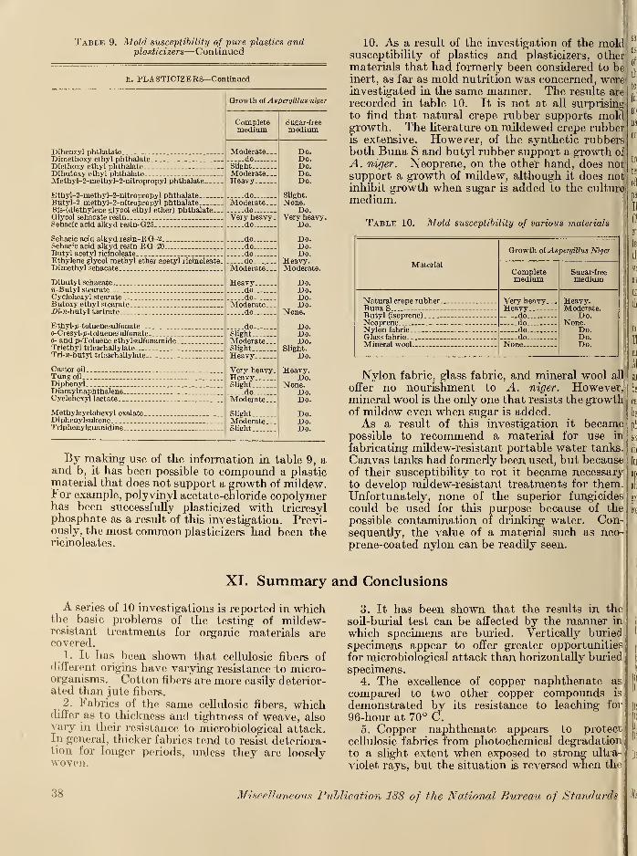
Table 9. Mold susceptibility of pure plastics andplasticizers—Continued
b. PLASTICIZERS—Continued
Dibenzyl phthalateDimetho.xy ethyl phthalateDiethoxy ethyl phthalateDibutoxy ethyl phthalateMethyl-2-methyl-2-nitropropyl phthalate..
Ethyl-2-methyl-2-nitropropyl phthalateButyl-2-methyl-2-nitropropyl phthalateBis-(diethylene glycol ethyl ether) phthalate.Glyool sebacate resinSebacic acid alkyd resin-G25 _.
Sebacic acid alkyd resin-RG-2___Sebacic acid alkyd resin RG-20 ,_.
Butyl acetyl ricinoleate ...
Ethylene glycol methyl ether acetyl ricinoleate.Dimethyl sebacate _
Dibutyl sebacate«-Butyl stearateCyclohexyl stearateButoxy ethyl stearate.Z>!-«-butyl tartrate
Ethyl-p-toluenesulfonateo-Cresyl-p-toluenesulfonate...o- and p-Toluene ethylsulfonamide.Triethyl tricarballylateTri-7i-butyl tricarballylate.-.
Castor oil
Tung oil
DiphenylDiamylnaphthalene.Cyclohexyl lactate. _.
Methylcyclohexyl oxalate.DiphenylsulfoneTriphenylguanidine
Growth of Aspergillus niger
Completemedium
Moderate. _.
doSlightModerate...Heavy
doModerate...
doVery heavy
.
do _.
dodododo....
Moderate .
Heavydodo
Moderate.do
doSlightModerate .
SlightHeavy
Very heavy.HeavySlight
doModerate...
SlightModerate.Slight
Sugar-freemedium
Do.Do.Do.Do.Do.
Slight.
None.Do.
Very heavy.Do.
Do.Do.Do.
Heavy.Moderate.
Do.Do.Do.Do.
None.
Do.Do.Do.
Slight.
Do.
Heavy.Do.
None.Do.Do.
Do.Do.Do.
10. As a result of the investigation of the moldsusceptibility of plastics and plasticizers, othermaterials that had formerly been considered to beinert, as far as mold nutrition was concerned, wereinvestigated in the same manner. The results are
recorded in table 10. It is not at all surprising
to find that natural crepe rubber supports moldgrowth. The literature on mildewed crepe rubberis extensive. However, of the synthetic rubbersboth Buna S and butyl rubber support a growth of
A. niger. Neoprene, on the other hand, does not;
support a growth of mildew, although it does notinhibit growth when sugar is added to the culture!
medium.
Table 10. Mold susceptibility of various materials
Material
Natural crepe rubber.Buna S_ ._.
Butyl (isoprene)Neoprene --
Nylon fabricGlass fabric _
Mineral wool
Growth of Aspergillus Niger
Completemedium
Very heavy-Heavy
do -
do.do.do.
None..
Sugar-freemedium
Heavy.Moderate.
Do.None.
Do.Do.Do.
By making use of the information in table 9, aand b, it has been possible to compound a plasticmaterial that does not support a growth of mildew.For example, polyvinyl acetate-chloride copolymerhas been successfully plasticized with tricresylphosphate as a result of this investigation. Previ-ously, the most common plasticizers had been thericinoleates.
Nylon fabric, glass fabric, and mineral wool all
offer no nourishment to A. niger. However,mineral wool is the only one that resists the growthof mildew even when sugar is added.As a result of this investigation it became
possible to recommend a material for use in
fabricating mildew-resistant portable water tanks.
Canvas tanks had formerly been used, but becauseof their susceptibility to rot it became necessaryto develop mildew-resistant treatments for them.Unfortunately, none of the superior fungicides
could be used for this purpose because of the
possible contamination of drinking water. Con-sequently, the value of a material such as neo-prene-coated nylon can be readily seen.
XI. Summary and Conclusions
A series of 10 investigations is reported in whichthe basic problems of the testing of mildew-resistant treatments for organic materials arecovered.
1.It has been shown that cellulosic fibers of
different origins have varying resistance-to micro-organisms. Cotton fibers are more easily deterior-ated than jute fibers.
2. Fabrics of the same cellulosic fibers, whichdiffer as to thickness and tightness of weave, alsovary in their resistance to microbiological attack.In general, thicker fabrics tend to resist deteriora-tion for longer periods, unless they are looselywoven.
3. It has been shown that the results in the
soil-burial test can be affected by the manner in
which specimens are buried. Vertically buried
specimens appear to offer greater opportunities
for microbiological attack than horizontally buried
specimens.4. The excellence of copper naphthenate as
compared to two other copper compounds is
demonstrated by its resistance to leaching for
96-hour at 70° C.5. Copper naphthenate appears to protect
cellulosic fabrics from photochemical degradationto a slight extent when exposed to strong ultra-
violet rays, but the situation is reversed when the
38 Miscellaneous Publication 188 of the National Bureau of Standards
same samples are exposed outdoors for as longas 10 months. It appears that in the presenceof moisture and light, as in the outdoor exposure,there is a slight tendency for copper naphthenateto reduce the breaking strength of cellulosic
j
fabrics. In the absence of large quantities of
moisture, as in the ultraviolet exposure, coppernaphthenate tends to retard the degradation of
|
cellulosic fabrics.
6. For the purpose of evaluating cotton fabrics
treated with mildew-resistant compounds, eight
test methods have been studied. No single test
offers a complete picture of the performance of aparticular treatment under all service conditions.
The tests that stand out as most useful are
Chaetomium-Mjcelial mat test; Accelerated weath-ering test, followed by a subsequent Chaetomiumtest; and the soil-burial test. Where the partic-
ular conditions of service are known, it may besufficient to select a single test. But wherea mildew-resistant treatment is required to protectunder a wide variety of conditions, all three of
the above tests should be used.
In evaluating these test methods, 35 of themost commonly used fungicides were used.The superiority of the copper compounds, espe-cially copper naphthenate, has been demonstrated.Although not as effective, zinc naphthenate andzinc dimethyldithiocarbamate have been shown to
be acceptable substitutes where copper compoundscannot be used. The chlorinated phenols havebeen shown to be excessively soluble and ratherphotochemically unstable. However, in the ab-sence of light, pentachlorophenol and dihydroxy-dichlorodiphenylmethane would be excellent fabric
fungicides. The pyridyl mercuric compoundsappear to be the best of the mercury fungicidesalthough the acetate is probably too soluble forgeneral use. Finally, it is obvious that no single
compound may be considered the universal fung-
icide. Even copper naphthenate, which with-stands extemely rigorous exposure, is not theideal treatment. However, with a knowledge of
the end use of the treated fabric, it is possible to
select an effective fungicide from these 35compounds.
7. The material that imparts to vegetable-tanned leather its great susceptibility to moldgrowth appears to be the grease commonly usedin leather manufacture.
8. Paranitrophenol and pentachlorophenol havebeen demonstrated to be effective fungicides for
vegetable-tanned leather. Inasmuch as the effec-
tive concentrations of these compounds are toohigh, for practical reasons, a compound containingequal parts of these two fungicides has beendeveloped in which the combination is effective
at as low a concentration as 0.20 percent.
9. Fifteen unplasticized polymers and 83 plas-
ticizers have been investigated for resistance to
Aspergillus niger. Of all the materials tested onlyureaformaldehyde showed a resistance to thegrowth of A. niger on a complete medium. Onsugar-free medium, 4 polymers and 37 plasticizers
were shown to support a growth of A. niger.
The relatively large proportion of mold-susceptibleplasticizers indicates the need for careful selection
of plasticizers in compounding a plastic material.
This data can be used in the selection of resistant
materials for various plastic formulations.
10. A group of seven miscellaneous materialshas been investigated for resistance to A. niger.
Of all the materials tested only mineral woolresisted the growth of A. niger on a completemedium. On sugar-free medium, natural creperubber, Buna S and butyl rubber manifested theability to support a growth of A. niger. Neoprene,nylon fabric, glass fabric, and mineral wool failed
to support the growth of the organism.
XII. References
[1] Appling, J. W., and McCoy, J. F., Paper Trade J.
121, 19 (1945).
[2] Armand, L., Tiba 12, 675 (1934).
[3] — , Tiba 13, 23 (1935).
[4] Armstead, D., and Harland, S. C, Shirley Inst.
Mem. 2, 104 (1923).
[5] — , — , Color Trade J. 13, 72 (1923).
[6] Armstrong, E. F., Dver & Textile Printer 86, 207(1941).
[7] —, Chemistrv & Industry 60, 668 (1941).[8] — , Textile Colorist 64, 196 (1942).
[9] Artari, A., Jahrb. wiss Bot, 46, 443 (1910).[10] Ayres, G. B., and Tobie, W. C, J. Bact. 45, 18 (1943).[11] Azevado, P. da R., and Maffei, F. J., Anais assoc.
quim. Brasil 3, 120 (1944).[12] Barker, S. C, Chemistrv & Industry 57, 1222 (1938).[13] — , Rev. Applied Mvcol. 18, 527 (1938).[14] — , Textile Research 11, 295 (1941).[15] Bartlett, A. E., and Goll, M., Am. Dyestuff Reptr.
34, 225 (1945).
[16] Batson, D. M., Teunisson, D. J., and Porges, N.,Am. Dyestuff Reptr. 33, 449 (1944).
[17] Bayley, C. H., and Weatherburn, M. W., CuaTextile J. 62, 34 (1945).
[18] — , — , Am. Dyestuff Reptr. 34, 247 (1945).
[19] —, — , Am. Dyestuff Reptr. 34, 457 (1945).
[20] — , —, Am. Dyestuff Reptr. 35, 218 (1946).
[21] — , — , Can. J. Research 24F 193 (1946).
[22] Bedford, T. H., Brit. J. Exptl. Path. 8, 437 (1927).
[23] Bertolet, E. C, Am. Dyestuff Reptr. 32, 214 (1943).
[24] Binns, C., Proc. Tech. Sect. Paper Makers' Assn.Gt. Brit. & Ireland 25, 280 (1944).
[25] Borghetty, H. C, Rayon Textile Monthly 26, 479(1945) .
[26] Borlaug, N. E., Ravon Textile Monthly 24, 416(1943).
[27] Bright, T. B., Morris, L. E., and Summers, F., J.
Textile Inst. 15, T547 (1924).
[28] — , J. Textile Inst, 17, T396 (1926).
[29] Brown, A. E., Modern Plastics 23, No. 8, 189, 254(1946) .
[30] Bryson, H. C, J. Textile Inst 30, P346 (1939).
[31] Bunker, H. J., Proc. Soc. Agr. Bact. 9 (1943).
[32] Burgess, R., J. Textile Inst. 15, T573 (1924).
Microbiological Deterioration 39
[33
[34[35'
[36[37"
[38'
[39
[40;
[4i:
[42'
[43;
[44;
[45"
[46;
T47"
[48;
[49;
[50;
[51[52"
[53"
[54;
[55"
[56;
[57;
[58:
[591[60"
[61
[62"
[63'
[64
[65
[66[67'
[68;
[691
[70;
[71
[72;
[73;
[74
[75;[76'
[77;
[78'
[79;
[80"
[81
[82;
[83;
[84;
40
— , J. Textile Inst, 19, T315 (1928).— J.Textile Inst. 20, T333 (1929).— , J. Textile Inst, 21, T441 (1930).— , J. Soc. Dvers Colourists 47, 96 (1931).—, J. Soc. Dvers Colourists 50, 138 (1934).—, J. Textile Inst. 25, T289 (1934).
Carter, W. J., Chem. Age (London) 51, 517 (1944).Chivers, A. H., Mem. Torrev Bot. Club 14, 155
(1915).Clayson, H. F., Chemistry and Industry 20, 516
(1942) .—,Chemistry & Industry 49, (1943).
Colin-Russ, A.. J. Int. Soc. Leather Trades Chem.24, 395 (1940).
Corrigan, J. F., J. Soc. Dvers Colourists 36, 198(1920).
Curwen. M. D., Oil Colour Trades J. 109, 82 (1946).Dean, J. D., Strickland, W. B., and Berard, W. N.,Am. Dyestuff Reptr. 34, 195 (1945).
DeBary, A., Bot, Ztg. 44, 377, 420 (1886).Dempsev, M., J. Int. Soc. Leather Trades' Chem.
29, 133 (1945).Dimond, A. E., and Horsfall, J. G., Science 97, 144
(1943) .
Downing, G. V., and Whitmore, L. M., J. Am.Leather Chem. Assoc. 36, 210 (1941).
Dubos, R. J., J. Bact, 15, 223 (1928).Ellis, H.. Textile Record 49, No. 579, 40 (1931).— , Textile Record 49, No. 580, 25 (1931).Everitt, E. L., and Sullivan, M. X., J. Wash. Acad.
Sci. 30, 125 (1940).von Euler, H., Z. Ang. Chem. 25, 250 (1912).Fargher, R. G., Gallowav, L. D., and Probert, M. E.,
J. Textile Inst, 21, T245 (1930).— J. Soc. Dyers Colourists 61, 118 (1945).Fleming, N., and Thaysen, A. C, Biochem. J. 14,
25 (1920).— ,—
, Biochem, J. 15, 407 (1921).Freedland, V. D., Textile Color 53, 557 (1931).Furry, M. S., Robinson, H. M., and Humfeld, H.,
Ind. Eng. Chem. 33, 538 (1941).—, and Zametkin, M., Am. Dvestuff Reptr. 32, 395(1943).—,Rayon Textile Monthly 26, 603 (1945).
Galloway, L. D., J. Textile Inst. 21, T277 (1930).— , Shirlev Inst. Mem. 10, 75 (1931).—, J. Textile Inst, 26, T123 (1935).—, Textile Record 55, 32 (1937).—,and Burgess R., Applied Mvcologv & Bact,
(London) (1937).—, J. Textile Inst, 30, T127 (1939).
Goldthwait, C. F., McLaren, J., and Voorhies, S. T.,Jr., Textile World 96, (No. 2) 115, 212 (1946).
Goodavage, J. E., Am. Dyestuff Reptr. 32, 265 (1943).Grassmann, W., and Rubenbauer, H., Munch, med.
Wochschr. 43, 1817 (1931).—,Stadler, R., and Bender, R. Liebig's Ann. Chem.
503, 20 (1933).Greathouse, G. A., Klemme, D. E., and Barker, H. D.,
Ind. Eng. Chem., Anal. Ed. 14, 614 (1942).—, and Ames, L. M., Mycologia 37, 138 (1945).
Greeves-Carpenter, C. F., Textile Mfr. 70, 82 (1944).Grimshaw, A. H., Melliand Textile Monthly 4, 253,
300 (1932).Guegen, F., Union pharm. 56, 55 (1916).Hall, A. J., Am. Dyestuff Reptr. 19, 663, 717 (1930).Harold, B. A., Am. Dyestuff Reptr. 30, 274 (1941).Hata, S., Zentr. Bakt. T34, 208 (1904).Hayes, T. F., and Holden, H. S., J. Soc. Chem. Ind.
45, 262 (1926).
Holzinger, F., Zentr. Bakt, Parasitenk., II Abt.21, 449 (1909).
Hooper, F. E., Contrib. Boyce Thompson Inst.10, 267 (1939).
[85] Hutchinson, H. B., and Clavton, J., J. Agr. Sci.
9, 143 (1919).
[86] Jamison, V. G, Soil Sci. 53, 287 (1942).
[87] Jarrell, T. D., Stuart, L. S., and Holman, H. P.,
Am. Dvestuff Reptr. 26, 495 (1937).
[88] Jones, C. S., Indian Textile J. 54, 78 (1944).
[89] Kalinenko, V. O., Microbiology (U. S. S. R.) 7, 119(1938).
[90] Kanagv, R., Charles, A. M., Abrams, E., andTener, R. F., J. Research NBS 36, 441 (1946)RP1713.
[91] Karnicka, H., and Ziemiecka. J., Trans. Third Int.
Congr. Soil Sci. 3, 109 (1935).
[92] Karrer, P., Joos, B., and Staub, M., Helv. Chim.Acta 6, 800 (1923).
[93] Kavser, E., and Delaval, H., Bui. soc. encour. Ind.Nat. 132, 277 (1920).
[94] Kehoe, R, A., J. Lab. Clin. Med. 7, 736 (1922).
[95] Kellerman, E. F., and McBeth, J. G., Zentr. Bakt.Parasitenk., II Abt, 34, 63 (1912).
[96] Klemme, D. E., Greathouse, G. A., Bollenbacher, K.,and Pope, S., U. S. Dept. Agr. Cir. 737 (1945).
[97] Kroulik, A., Zentr. Bakt. Parasitenk., II Abt, 36,339 (1913).
[98] Larnach, S. L., and Wvke, B. D., Lab. J. Australasia3, 92 (1945).
[99] Levine, B. S., and Veitch, F. P., J. Ind. Eng. Chem.12, 139 (1920).
[100] Levine, M., and Schoenlein, H. W., A compilationof culture media for the cultivation of micro-organisms, monographs on systematic bacteriology,vol. 2.
101] Linossier, G., Compt. rend. soc. biol. 80, 429 (1917).
102] Llovd, D. J., J. Am. Leather Chem. Assn. 29, 483(1934).
103] Lollar, R. M., J. Am. Leather Chem. Assn. 39, 12(1944).
104] — , J. Am. Leather Chem. Assn. 39, 179 (1944).
105] — , J. Am. Leather Chem. Assn. 39, 203 (1944).
106] — , J. Am. Leather Chem. Assn. 39, 209 (1944).
107] Madhok, M. R., Soil Sci. 44, 385 (1937).
108] Marsh, P. B., Greathouse, G. A., Bollenbacher, K,and Butler, M. L., Ind. Eng. Chem. 36, 176(1944).
109] — , — . Butler, M. L., and Bollenbacher, K., U. S.
Dept. Agr. Tech. Bui. No. 892 (1945).
110] Marsh, W. S., and Duske, A. E., Textile World 92(No. 8) 58 (1942).
Ill] McBeth, I. G., and Scales, F. M., Bur. Plant Ind.Bui. 266 (1913).
112] McCallan, S. E. A., and Wilcoxon, F., Contrib.Boyce Thompson Inst. 6. 479 (1934).
113] Morris, L. E., J. Textile Inst, 18, 99 (1927).
114] Morrow, F. J., and Richardson, J. F., Hide &Leather 100, (No. 12) 24 (1940).
115] Mulder, E. G., Arch. Mikrobioi. 10, 72 (1939).
116] Mutterlein, C, Univ. Leipzig Diss. (1913).
117] Nakagawa, R., Takeda, Y., and Suematsu, S.,
J. Agr. Chem. Soc. (Japan) 10, 95 (1934).
118] Neill, J. G, and Travers, E., New Zealand J. Sci.
Tech. 19, 646 (1938).
119] Neish, A. G, Ledingham, G. A., and Mackev,A. G., Can. J. Research 23F. 198 (1945).
120] Norman, A. G., and Fuller, W. H., Advances in
enzymologv 2, 239 (1942).
121] O'Brien, T. E. H., Trop. Agric. (Ceylon) 65, 333(1925).
122] —,Trop. Agric. (Cevlon) 66, 283 (1926).
123] O'Flahertv, F., and Dohertv, E. R,, Hide & Leather99, 23 (1940).
124] Ordal, E. J., and Deromedi, F.. J. Bact. 45. 293(1943).
125] Osborn, T. G. B., J. Soc. Dyers Colourists 28, 204(1912).
Miscellaneous Publication 188 of the National Bureau of Standards
[126] Osnitskava, L. K., Microbiology (U. S. S. R.) 11,
83 (1942).
[1271 Otto, H., Zentr. Biochem. Biophvs. 19, 149 (1916).
[128] Pope, S., Mvcologia 36, 343 (1944).
[129] Porges, N.. Metal Ind. 36, 19 (1938).
[130] Pringsheim, H., Z. Physiol. Chem. 78, 266 (1912).
[131] Rabanus, A., Mitt, Deut, Forstver 23, 77 (1939).
[132] Richardson, J. F., Hide & Leather 99, No. 19, 28
(1940) .
[133] Robbins, W. J.. Am. J. Biol. 3, 234 (1916).
[134] Roberg. M., Zentr. Bakt. Parasitenk. II abt. 74, 333(1928).
[135] Robertson, M. E.. Nature 151, 365 (1943).
[136] Rogers, R. E., Wheeler, H. G.. and Humfeld. H.,
U. S. Dept. Agr. Tech. Bui. No. 726 (1940).
[137] Romano, F. R., Am. Dyestuff Reptr. 35, 363 (1946).
[138] Ruschmann, G., and Bartram. H.. Zentr. Bakt.Parasitenk., II Abt. 102, 300, 365 (1940).
[139] Scales, P. M., Botan. Gaz. 60, 149 (1915).
[140] Schmitz, H., J. Gen. Physiol. 3, 795 (l920).
[141] Schonleber, K., Zellwolle, Kunstseide, Seide, 46, 336(1941) .
[142] Scott, J.. India Rubber J. 60, 410 (1920).
[143] Searle, G. O., J. Textile Inst. 20, 162 (1929).
[144] Selhere, G., Compt. rend. Soc. Biol. 58, 409 (1905).
[145] — ,Compt, rend. Soc. Biol. 61, 205 (1906).
[146] — ,Compt, rend. Soc. Biol. 68, 107 (1910).
[147] Serrano, F. B., Philippine J. Sci. 32, 75 (1927).
[148] Shiraeff, D. A., Am. Dvestuff Reptr. 33, 310 (1944).
[149] — , Am. Dvestuff, Reptr. 34, 15 (1945).
[150] Shults, G. 'G., Org. Chem. Ind. (U. S. S. R.) 5, 559(1938).
[151] Siu, R. G. H., and White, W. L., O. Q. M. G. Military
Planning Div. Research and Development Branch,Microbiological Series Rep. 1 (1945).
[152] Skinner, C. E., and Mellem, E. M., Ecology 25, 360(1944).
[153] Smith, A. L., Am. Dvestuff Reptr. 21, 501 (1932).
[154] Smith, P. I., Soap 14, No. 11, 86 (1938).
[155] Sohngen, N. L., and Fol. J. G., Zentr. Bakt, Parasi-
tenk., I abt. 40, 87 (1914).
[156] Stevens, H. P.. Bui. Rubber Growers' Assn. 8, 177
(1926).
[157] Ter Horst, W. P., and Felix, E. L., Ind. Eng. Chem.35, 1255 (1943).
[158] Thavsen, A. C, and Bunker, H. J.. Biochem. J. 18,140 (1924).
159] — , J. Soc. Dvers Colourists 40, 101 (1924).
160] — , and Bunker, H. J., Biochem. J. 19, 1088 (1925).16l] — , — , The microbiology of cellulose, Hemicelluloses,
pectin, and gums, Oxford Univ. Press (London)(1927).
162] — ,— Dept. Sci. Ind. Research 153 (1930). (British
Government Report.)163] — , — . Butlin, K. R., and Williams, L. H., Ann. Ap-
plied Biol. 26, 750 (1939).
164] Thorn, C, and Church, M. B.. The Aspergilli, TheWilliams & Wilkins Co., Baltimore, Md. (1926).
165] — , The Penicillia, The Williams & Wilkins Co., Bal-timore, Md. (1930).
166] — , Humfeld, H., and Holman, H. P., Am. DvestuffReptr. 23, 581 (1934).
167] Vaubel, W., Farben-Ztg. 34, 505 (1928).
168] Veitch, F. P., and Levine, G. S., Science 49, 618(1919).
169] Waksman, S. A., and Hutchings, I. J., Trans. ThirdInt. Congr. Soil Sci. Oxford, 1, 163 (1935).
170] — , Botan. Rev. 6, 637 (1940).
171] Ward, H. M., Ann. Botanv (London) 2, 319 (1888).
172] White, W. L., and Downing, M. H., The identity of
"Metarrhizium glutinosum", Topical Report 20,
Project No. (7B-83-46, Biological Laboratories,
O. Q- M. G.) Military Planning Div., Research andDevelopment Branch (March 1947); to be pub-lished in Mvcologia.
[173] Wilson, J. A.,' and Daub, G., Ind. Eng. Chem. 17,
700 (1925).
[174] Winogradskv, S., Ann. fast. Pasteur 43, 549 (1929).
[175] Woodford, E. K., J. Sci. Ind. Research (India) 2, 182(1944).
[176] Ziese, W., Z. Phvsiol. Chem. 203, 87 (1931).
[177] Zo Bell, C. E., and Grant, C. W., Science 96, 379(1942).
[178] — , and Beckwith, J. D., J. Am. Waterworks Assn.36, 439 (1944).
[179] Zuck, R. K, and Diehl, W. W., Am. J. Botanv 33,
375 (1946).
Washington, July 1, 1947.
o
Microbiological Deterioration 41
I
I