metabolic control analysis of integrated energy metabolism in permeabilized cardiomyocytes
TRANSCRIPT

Regular paper
Metabolic control analysis of integrated energy metabolism in permeabilized cardiomyocytes — experimental study*
Kersti Tepp1*, Natalja Timohhina1, Vladimir Chekulayev1, Igor Shevchuk1, Tuuli Kaambre1 and Valdur Saks1, 2
1Laboratory of Bioenergetics, National Institute of Chemical Physics and Biophysics, Tallinn, Estonia; 2Laboratory of Fundamental and Applied Bioenergetics, INSERM U884, Joseph Fourier University, Grenoble, France
The main focus of this research was to apply Metabolic Control Analysis to quantitative investigation of the reg-ulation of respiration by components of the Mitochon-drial Interactosome (MI, a supercomplex consisting of ATP Synthasome, mitochondrial creatine kinase (MtCK), voltage dependent anion channel (VDAC), and tubulin) in permeabilized cardiomyocytes. Flux control coeffi-cients (FCC) were measured using two protocols: 1) with direct ADP activation, and 2) with MtCK activation by creatine (Cr) in the presence of ATP and pyruvate kinase-phosphoenolpyruvate system. The results show that the metabolic control is much stronger in the latter case: the sum of the measured FCC is 2.7 versus 0.74 (ADP activa-tion). This is consistent with previous data showing recy-cling of ADP and ATP inside the MI due to the functional coupling between MtCK and ANT and limited permeabil-ity of VDAC for these compounds, PCr being the major energy carrier between the mitochondria and ATPases. In physiological conditions, when the MI is activated, the key sites of regulation of respiration in mitochondria are MtCK (FCC = 0.95), adenine nucleotide translocase (FCC = 0.93) and CoQ cytochrome-c oxidoreductase (FCC = 0.4). These results show clearly that under the physiolog-ical conditions the energy transfer from mitochondria to the cytoplasm is regulated by the MI supercomplex and is very sensitive to metabolic signals.
Keywords: mitochondria, respiration, cardiomyocytes, metabolic control analysis, creatine kinase.
Received: 31 August, 2010; revised: 09 December, 2010; accepted: 16 December, 2010; available on-line: 18 December, 2010
INTRODUCTION
Mitochondrial respiration, coupled to production of ATP and fine regulation of energy fluxes to the sites of ATP utilization, is vital for normal cell life. The regulation at different workloads is of main impor-tance in the high energy demanding brain and heart cells. In spite of the fundamental progress in the un-derstanding of mitochondrial bioenergetics (Nicholls & Ferguson, 2002), the nature of respiratory control and, in a more general sense, the mechanisms of regu-lation of energy fluxes during workload changes in the cardiac and other cells in vivo are still highly debated (Vendelin et al., 2000; Beard, 2005; 2006; Saks et al., 2006; 2007a; 2007b; Saks, 2007; Van Beek, 2007; 2008; Wu et al., 2008; Balaban, 2009; Guzun et al., 2009; Wu & Beard, 2009). In explaining respiration regulation,
the cell is often described as a homogenous reaction medium, thus ignoring the impact of the high degree of structural organization of the cell, in particular of cardiomyocytes. In this context, it is especially impor-tant to use Systems Biology approaches to analyze the complex biological systems in situ to discover system-level properties (Saks et al., 2008), which are direct consequences of interactions between cellular compo-nents and are absent for isolated components. Recent research of steady state kinetics of respiration regu-lation in permeabilized cardiomyocytes has revealed striking differences in the behavior of mitochondria in vitro and in situ: the apparent Km for ADP is more than ten times higher in situ than mitochondria in vitro (Gu-zun et al., 2009; Saks et al., 2007c). The apparent dis-sociation constants of MgATP from complexes with mitochondrial creatine kinase (MtCK) were several or-ders of magnitude higher in situ than in vitro, while the apparent dissociation constants of creatine (Cr) were significantly lower in situ than in vitro (Guzun et al., 2009). These results show clearly that the mechanisms of regulation of mitochondrial respiration and energy fluxes in the cardiac cells are system-level properties dependent on the interaction of mitochondria with in-tracellular structures, which are not predictable on the basis of the properties of isolated mitochondria only (Saks, 2007). It has also been shown that the cyto-skeletal component tubulin, which is responsible for the regular arrangement of mitochondria in cardiac cells, also controls the permeability of voltage depen-dent anion channel (VDAC) in mitochondrial outer membrane (MOM) (Rostovtseva & Bezrukov, 2008). Recently, we have demonstrated that the selective permeability of the MOM-VDAC-tubulin complex is crucial for the regulation of energy transfer in cardiac cells (Timohhina et al., 2009): a supercomplex consist-ing of ATP Synthasome, MtCK, VDAC and tubulin,
*e-mail: [email protected]*This work was presented in the poster form at the 16th European Bioenergetics Conference (Warsaw, 2010); abstract in Biochim Bio-phys Acta, 1797 (Suppl): 138–139 (2010).Abbreviations: ANT, adenine nucleotide translocase; BSA, bovine serum albumin; CAT, carboxyatractyloside; CK, creatine kinase; CM, cardiomyocytes; Cr, creatine; DNFB, 2,4-dinitrofluorobenzene; FCC, flux control coefficient; GGG, triglycine; IMS, mitochondrial intermembrane space; IM, isolation medium; MI, Mitochondrial In-teractosome; MtCK, mitochondrial creatine kinase; MCA, Metabolic Control Analysis; MCC, Metabolic Control Coefficient; MOM, mito-chondrial outer membrane; Pi, inorganic phosphate; PIC, inorganic phosphate carrier; PCr, phosphocreatine; PEP, phosphoenolpyru-vate; PK, pyruvate kinase; VDAC, voltage dependent anion channel.
Uncorre
cted P
aper in P
ress
Paper in Press, No. 2051 Vol. 57, 2010
on-line at: www.actabp.pl
2 2010K. Tepp and others
or Mitochondrial Interactosome (MI) (Fig. 1a) in heart mitochondria has been shown to control the regula-tion of respiration (Saks et al., 2010). It is especially interesting to use the method of Metabolic Control Analysis (MCA) to describe quantitatively the distri-bution of control between the complexes of energy transfer in MI. In the present work we use the per-meabilized cardiomyocytes (Fig. 1b) which means that
the complex structural and functional organization of cardiomyocytes and its importance for metabolic regulation are taken into account. To determine the flux control coefficient (FCC), the flux was measured as the rate of oxygen consumption in permeabilized cardiomyocytes when the MI complexes were titrated with their specific inhibitors to stepwise decrease their activity.
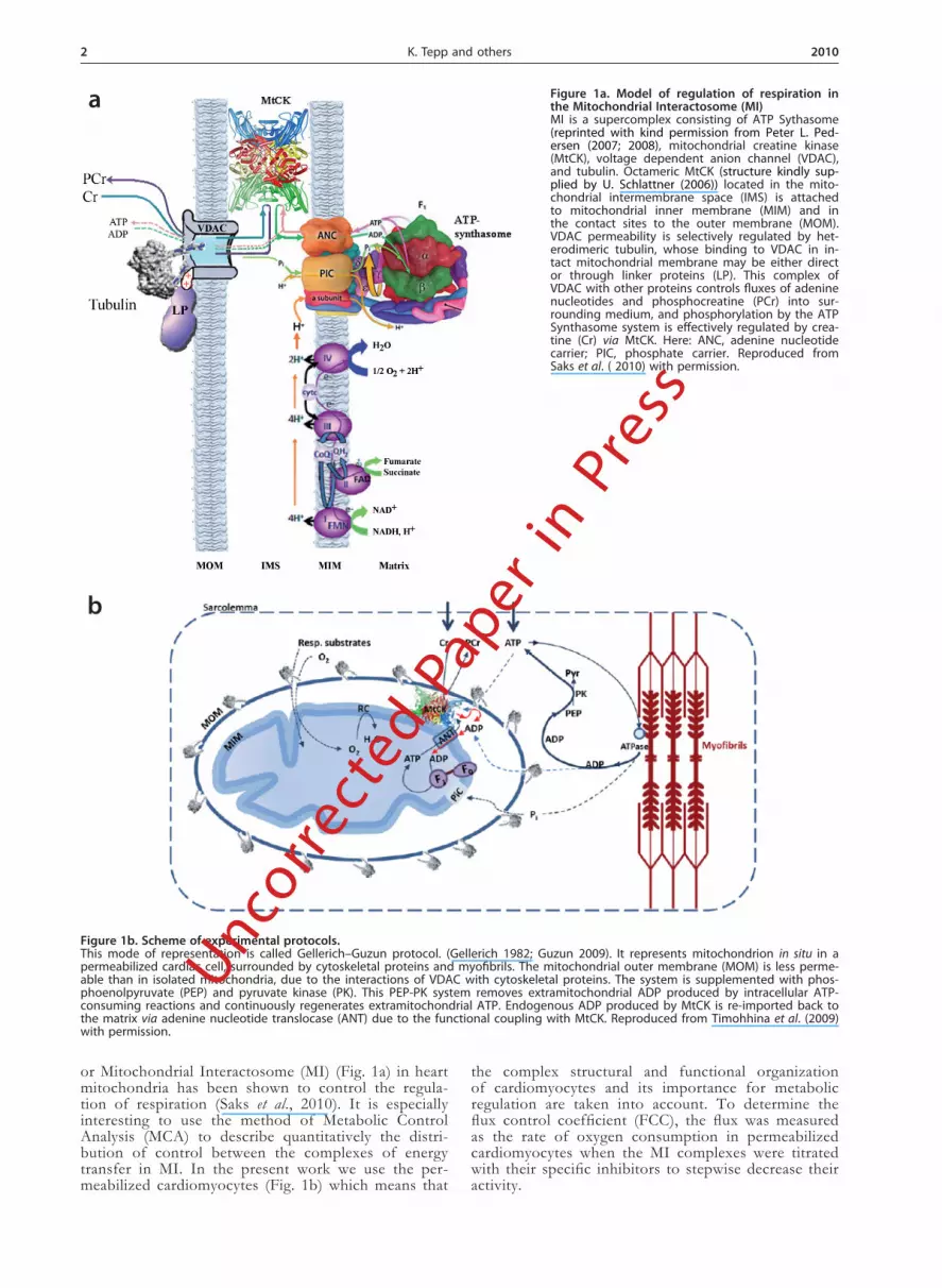
Figure 1a. Model of regulation of respiration in the Mitochondrial Interactosome (MI)MI is a supercomplex consisting of ATP Sythasome (reprinted with kind permission from Peter L. Ped-ersen (2007; 2008), mitochondrial creatine kinase (MtCK), voltage dependent anion channel (VDAC), and tubulin. Octameric MtCK (structure kindly sup-plied by U. Schlattner (2006)) located in the mito-chondrial intermembrane space (IMS) is attached to mitochondrial inner membrane (MIM) and in the contact sites to the outer membrane (MOM). VDAC permeability is selectively regulated by het-erodimeric tubulin, whose binding to VDAC in in-tact mitochondrial membrane may be either direct or through linker proteins (LP). This complex of VDAC with other proteins controls fluxes of adenine nucleotides and phosphocreatine (PCr) into sur-rounding medium, and phosphorylation by the ATP Synthasome system is effectively regulated by crea-tine (Cr) via MtCK. Here: ANC, adenine nucleotide carrier; PIC, phosphate carrier. Reproduced from Saks et al. ( 2010) with permission.
Figure 1b. Scheme of experimental protocols.This mode of representation is called Gellerich–Guzun protocol. (Gellerich 1982; Guzun 2009). It represents mitochondrion in situ in a permeabilized cardiac cell, surrounded by cytoskeletal proteins and myofibrils. The mitochondrial outer membrane (MOM) is less perme-able than in isolated mitochondria, due to the interactions of VDAC with cytoskeletal proteins. The system is supplemented with phos-phoenolpyruvate (PEP) and pyruvate kinase (PK). This PEP-PK system removes extramitochondrial ADP produced by intracellular ATP-consuming reactions and continuously regenerates extramitochondrial ATP. Endogenous ADP produced by MtCK is re-imported back to the matrix via adenine nucleotide translocase (ANT) due to the functional coupling with MtCK. Reproduced from Timohhina et al. (2009) with permission.
a
b
Uncorre
cted P
aper in P
ress

Vol. 57 3Metabolic control analysis of integrated energy metabolism
MATERIALS AND METHODS
Experimental protocols. The principles of our study are illustrated by Figs. 1a and 1b. Figure 1a represents a model of the MI supercomplex (consisting of ATP Syn-thasome, MtCK, VDAC and tubulin in contact sites) in heart mitochondria, which controls the regulation of res-piration. This MI in some cases includes supercomplexes of the respiratory chain (Lenaz & Genova, 2007; Vonck & Schafer, 2009). Along the cristae membranes the MI contains only MtCK and ATP Synthasome.
Figure 1b represents the setup of our experiments; the mitochondrion in situ, in a permeabilized cardiac cell, surrounded by cytoskeleton proteins (tubulin) and my-ofibrils. The respiratory chain complexes, ATP Syntha-some (F1FO) and inorganic phosphate (Pi) carrier (PIC) are integrated within the mitochondrial inner membrane (MIM). MtCK is depicted as an octamer, located in the mitochondrial intermembrane space (IMS) and attached to the inner membrane surface.
In the first part of our experiments (ADP activation) respiration is activated by extramitochondrial ADP. The Cr/phosphocreatine (PCr) transfer network and the MI supercomplex of energy transfer regulation are not acti-vated. In this case, ATP produced in the mitochondria is leaving it through the MOM (Guzun et al., 2009; Timo-hhina et al., 2009).
In the second part of our experiments, MtCK is acti-vated by Cr in the presence of ATP (see Fig. 1b). The permeabilized cardiomyocytes were supplemented with phosphoenolpyruvate (PEP) and pyruvate kinase (PK). This PEP-PK system removes extramitochondrial ADP produced by intracellular ATP-consuming reactions and continuously regenerates extramitochondrial ATP. En-dogenous intramitochondrial ADP produced by MtCK forms microcompartments within the IMS and is re-im-ported into the matrix via adenine nucleotide translocase (ANT) due to its functional coupling with MtCK. The final products of MtCK-forward reaction are PCr and endogenous ADP. Due to the activation of the MI, di-rect interactions of ANT, MtCK, VDAC and cytoskel-eton proteins endogenous ADP and ATP are circulating inside the mitochondria and oxidative phosphorylation is controlled by endogenous ADP.
Animals. Male Wistar rats weighing 300–350 g were used. The animals were housed five per cage at constant temperature (22 ºC) in environmental facilities with a 12 : 12 h light-dark cycle and were given standard labora-tory chow ad libitum. Animal procedures were approved by the Estonian National Committee for Ethics in Ani-mal Experimentation (Estonian Ministry of Agriculture).
Isolation of adult cardiac myocytes. Adult cardio-myocytes were isolated after perfusion of the rat heart with collagenase A (Roche) using an adaptation of the technique described previously (Saks et al., 1991). Rats were anaesthetized with medetomidine and ketamine, decapitated and the heart was quickly excised preserv-ing a part of the aorta and placed into isolation medi-um (IM) of the following composition: 117 mM NaCl, 5.7 mM KCl, 4.4 mM NaHCO3, 1.5 mM KH2PO4, 1.7 mM MgCl2, 11.7 mM glucose, 10 mM Cr, 20 mM tau-rine, 10 mM PCr, 2 mM pyruvate and 21 mM HEPES, pH 7.1. The excised heart was cannulated by the aorta, suspended in the Langendorf system for perfusion and washed for 5 min with a flow rate of 15–20 mL/min. The collagenase treatment was performed at 37 ºC by switching the perfusion to circulating O2-saturated IM supplemented with 1 mg/ml collagenase A and 2 mg/
Vol. 57 3
ml BSA at flow rate of 5 ml/min for 20–30 min. The end of the digestion was determined following the de-crease in perfusion pressure measured by a manometer. After the digestion the heart was washed with IM for 2–3 min and transferred into IM containing 20 μM CaCl2, 10 μM leupeptin, 2 μM soybean trypsin inhibitor and 2 mg/ml fatty acid free BSA. Cardiomyocytes were then gently dissociated using forceps and pipette suction. Cell suspension was filtered through a crude net to re-move tissue remnants and let to settle for 3–4 min at room temperature. After 3–4 min the initial supernatant was discarded, pellet of cardiomyocytes resuspended in 10 ml of IM containing 20 μM CaCl2 and the protease inhibitors. This resuspension-sedimentation cycle with calcium-tolerant cells was performed twice, after that the cardiomyocytes were gradually transferred from IM with 20 μM Ca2+ into calcium-free Mitomed (supplemented with protease inhibitors and BSA) and washed 5 times. Each time, slightly turbid supernatant was removed after 4–5 min of the cells’ sedimentation. Isolated cells were re-suspended in 1–2 ml of Mitomed solution described below for respiration measurements and stored on melt-ing ice. Isolated cardiomyocytes contained 70–90 % of rod-like cells when observed under a light microscope.
Isolation of mitochondria from cardiac muscle. Mitochondria were isolated from adult rat hearts as de-scribed in (Saks et al., 1975).
Permeabilization procedure. In order to study the regulation of mitochondrial respiration in cardiomyo-cytes, the sarcolemma was permeabilized by saponin treatment keeping the mitochondrial membranes intact (Kuznetsov et al., 2008; Saks et al., 1998). The permeabi-lization procedure was carried out at 25 °C with 20 μg/ml saponin for 10 min and then resuspension-sedimen-tation cycle with Mitomed solution was performed twice to remove saponine from solution and Ca2+ from cells.
Measurements of oxygen consumption. The rates of oxygen uptake were determined with a high-resolution respirometer (Oxygraph-2K, from OROBOROS Instru-ments, Austria) in Mitomed solution (Kuznetsov et al., 2008) containing 0.5 mM EGTA, 3 mM MgCl2, 60 mM K-lactobionate, 3 mM KH2PO4, 20 mM taurine, 20 mM Hepes (pH 7.1), 110 mM sucrose, 0.5 mM dithiothreitol, 5 mg/ml fatty acid free BSA, supplemented with 5 mM glutamate and 2 mM malate as respiratory substrates. These measurements were carried out at 25 ºC; solubility of oxygen was taken as 240 nmol/ml (Gnaiger, 2001). In kinetic experiments with MgATP, stock solution of 100 mM MgATP was prepared by mixing equimolar amounts of MgCl2 and ATP, pH was adjusted to 7.2. The respira-tion rates were expressed in pmol of oxygen consumed per second per mg of protein or in nmol of oxygen con-sumed per minute per nmol cytochrome aa3. The con-tent of mitochondrial cytochrome aa3 in the cardiomyo-cytes was measured spectrophotometrically according to the method described before (Monge et al., 2008). Pro-tein concentration was determined using a BCA protein assay kit (Pierce, USA) with BSA as a standard.
Measurements of MtCK activity. For calculation of inhibition curve of MtCK, the creatine kinase (CK) activity was measured at different concentrations of its inhibitor (2,4-dinitrofluorobenzene, DNFB) in a Cary 100 Bio spectrophotometer according to the method de-scribed before (Monge et al., 2009). The MtCK activity was measured at the same conditions as was the oxy-gen consumption change in the Oxygraph. Isolated mi-tochondria were used to measure selectively the activity of MtCK. These measurements were performed at 25 ºC.
Uncorre
cted P
aper in P
ress
4 2010K. Tepp and others
After the addition of DNFB, a 5-min inhibition period of CK was allowed and then the triglycine (GGG) was added to stop the inhibition.
Metabolic Control Coefficient (MCC) determi-nation. MCA helps to understand the mechanisms by which a given enzyme exerts high or low control of met-abolic flux and how the control of the pathway is shared by several pathway enzymes and transporters. By apply-ing MCA it is possible to identify the steps that could be modified to achieve a successful alteration of flux or metabolite concentration in pathways.
The control coefficients are defined as the ratios of the fractional changes in the system variables to the fractional change in the biochemical activity that caused the system change. It allows the identification of system components that are crucial in the control of pathway flux or metabolite concentration and thus also in the regulation of energy transfer and regulatory networks.
FCC quantifies the control that a certain reaction ex-erts on the steady-state flux.
S1 + E1 → S2 + E2 → S3+E3 → P
J = dP/dt = –dS1/dt
Groen et al. (1982) derived a method to determine experimentally the FCC using titration with specific en-zyme inhibitors. As the amount of inhibitor tends to zero the response of the flux to the inhibitor can be ex-pressed in MCA terms.
The FCC is defined according to the equation (Fell, 1997):
in which the expression dJ/dvi describes the variation in flux (J) when an infinitesimal change takes place in the enzyme i concentration or activity. In practice, the infini-tesimal changes in vi are undetectable, and hence measur-able noninfinitesimal changes are undertaken. If a small change in vi promotes a significant variation in J, then this enzyme exerts a high flux control. In contrast, if a rather small or negligible change in the flux is observed when vi is greatly varied, then the enzyme does not exert a significant flux control.
For the case of an irreversible specific inhibitor, an estimation of the value of the FCC coefficient is given by Groen et al. (1982) and Moreno-Sanches et al. (2008):
CE = (ΔJ/ΔI)× (Imax/J0)
where (ΔJ/ΔI) is initial slope of the flux/inhibition graph.
Until now the method of MCA has been used to measure FCC in mitochondria (Moreno-Sanchez et al., 2008). In our work we use the tool to measure the coef-ficient of the complexes in the MI, the model proposed in our previous article (Timohhina et al., 2009).
The inhibitors used were: carboxyatractyloside (CAT) for ATP/ADP transporter, oligomycin for complex V (ATP Synthase), mersalyl for Pi transporter, antimycin for complex III, and DNFB for MtCK (see Table 1). Of the inhibitors used CAT, oligomycin, mersalyl, and antimycin are considered irreversible and noncompetitive in these conditions. For mitochondrial CK was meas-ured also the enzyme activity decrease during inhibition.
The impact of each complex is calculated according the methods described by Fell (1997) and Westerhoff (2008).
Enzymes and other chemicals were obtained from Sigma, Fluka and Roche.
Data analysis. To reduce the possibility of random error the experiments were repeated seven to twenty times and fitting was used to calculate the FCC. All data are presented as mean ± S.E.M. Statistical analysis was performed using Student’s t-test, and P < 0.05 was taken as the level of significance.
RESULTS
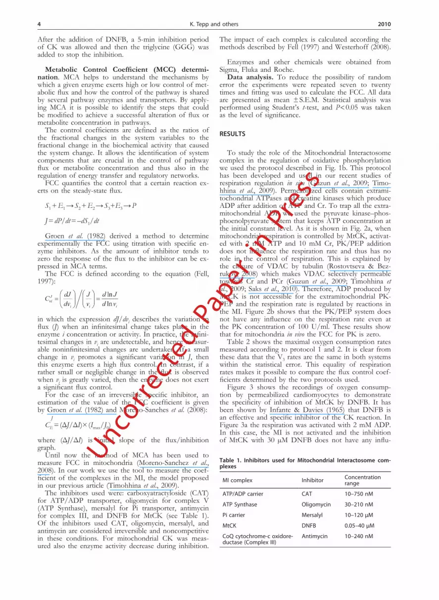
To study the role of the Mitochondrial Interactosome complex in the regulation of oxidative phosphorylation we used the protocol described in Fig. 1b. This protocol has been developed and used in our recent studies of respiration regulation in situ (Guzun et al., 2009; Timo-hhina et al., 2009). Permeabilized cells contain extrami-tochondrial ATPases and creatine kinases which produce ADP after addition of ATP and Cr. To trap all the extra-mitochondrial ADP we used the pyruvate kinase–phos-phoenolpyruvate system that keeps ATP concentration at the initial constant level. As it is shown in Fig. 2a, when mitochondrial respiration is controlled by MtCK, activat-ed with 2 mM ATP and 10 mM Cr, PK/PEP addition does not influence the respiration rate and thus has no role in the control of respiration. This is explained by the closure of VDAC by tubulin (Rostovtseva & Bez-rukov, 2008) which makes VDAC selectively permeable towards Cr and PCr (Guzun et al., 2009; Timohhina et al., 2009; Saks et al., 2010). Therefore, ADP produced by MtCK is not accessible for the extramitochondrial PK-PEP and the respiration rate is regulated by reactions in the MI. Figure 2b shows that the PK/PEP system does not have any influence on the respiration rate even at the PK concentration of 100 U/ml. These results show that for mitochondria in vivo the FCC for PK is zero.
Table 2 shows the maximal oxygen consumption rates measured according to protocol 1 and 2. It is clear from these data that the V3 rates are the same in both systems within the statistical error. This equality of respiration rates makes it possible to compare the flux control coef-ficients determined by the two protocols used.
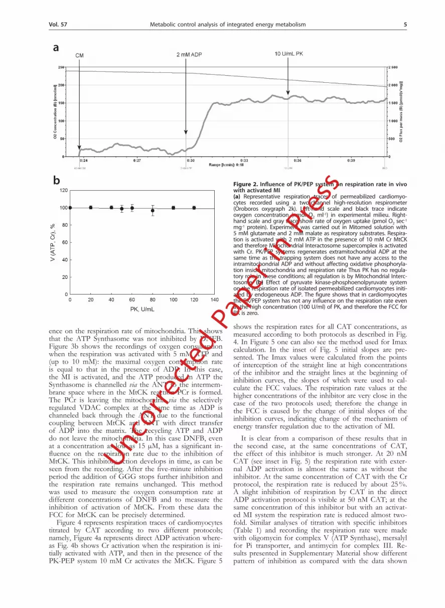
Figure 3 shows the recordings of oxygen consump-tion by permeabilized cardiomyocytes to demonstrate the specificity of inhibition of MtCK by DNFB. It has been shown by Infante & Davies (1965) that DNFB is an effective and specific inhibitor of the CK reaction. In Figure 3a the respiration was activated with 2 mM ADP. In this case, the MI is not activated and the inhibition of MtCK with 30 µM DNFB does not have any influ-
J
Table 1. Inhibitors used for Mitochondrial Interactosome com-plexes
MI complex Inhibitor Concentration range
ATP/ADP carrier CAT 10–750 nM
ATP Synthase Oligomycin 30–210 nM
Pi carrier Mersalyl 10–120 μM
MtCK DNFB 0.05–40 μM
CoQ cytochrome-c oxidore-ductase (Complex III)
Antimycin 10–240 nM
Uncorre
cted P
aper in P
ress
Vol. 57 5Metabolic control analysis of integrated energy metabolism
ence on the respiration rate of mitochondria. This shows that the ATP Synthasome was not inhibited by DNFB. Figure 3b shows the recordings of oxygen consumption when the respiration was activated with 5 mM ATP and (up to 10 mM): the maximal oxygen consumption rate is equal to that in the presence of ADP. In this case, the MI is activated, and the ATP produced in ATP the Synthasome is channelled via the ANT to the intermem-brane space where in the MtCK reaction PCr is formed. The PCr is leaving the mitochondria via the selectively regulated VDAC complex at the same time as ADP is channeled back through the ANT due to the functional coupling between MtCK and ANT with direct transfer of ADP into the matrix. The recycling ATP and ADP do not leave the mitochondria. In this case DNFB, even at a concentration as low as 15 µM, has a significant in-fluence on the respiration rate due to the inhibition of MtCK. This inhibitory action develops in time, as can be seen from the recording. After the five-minute inhibition period the addition of GGG stops further inhibition and the respiration rate remains unchanged. This method was used to measure the oxygen consumption rate at different concentrations of DNFB and to measure the inhibition of activation of MtCK. From these data the FCC for MtCK can be precisely determined.
Figure 4 represents respiration traces of cardiomyocytes titrated by CAT according to two different protocols; namely, Figure 4a represents direct ADP activation where-as Fig. 4b shows Cr activation when the respiration is ini-tially activated with ATP, and then in the presence of the PK-PEP system 10 mM Cr activates the MtCK. Figure 5
shows the respiration rates for all CAT concentrations, as measured according to both protocols as described in Fig. 4. In Figure 5 one can also see the method used for Imax calculation. In the inset of Fig. 5 initial slopes are pre-sented. The Imax values were calculated from the points of interception of the straight line at high concentrations of the inhibitor and the straight lines at the beginning of inhibition curves, the slopes of which were used to cal-culate the FCC values. The respiration rate values at the higher concentrations of the inhibitor are very close in the case of the two protocols used; therefore the change in the FCC is caused by the change of initial slopes of the inhibition curves, indicating change of the mechanism of energy transfer regulation due to the activation of MI.
It is clear from a comparison of these results that in the second case, at the same concentrations of CAT, the effect of this inhibitor is much stronger. At 20 nM CAT (see inset in Fig. 5) the respiration rate with exter-nal ADP activation is almost the same as without the inhibitor. At the same concentration of CAT with the Cr protocol, the respiration rate is reduced by about 25 %. A slight inhibition of respiration by CAT in the direct ADP activation protocol is visible at 50 nM CAT; at the same concentration of this inhibitor but with an activat-ed MI system the respiration rate is reduced almost two-fold. Similar analyses of titration with specific inhibitors (Table 1) and recording the respiration rate were made with oligomycin for complex V (ATP Synthase), mersalyl for Pi transporter, and antimycin for complex III. Re-sults presented in Supplementary Material show different pattern of inhibition as compared with the data shown
Figure 2. Influence of PK/PEP system on respiration rate in vivo with activated MI(a) Representative respiration traces of permeabilized cardiomyo-cytes recorded using a two-channel high-resolution respirometer (Oroboros oxygraph 2k). Left-hand scale and black trace indicate oxygen concentration (nmol O2 ml-1) in experimental milieu. Right-hand scale and gray trace show rate of oxygen uptake (pmol O2 sec-1 mg-1 protein). Experiment was carried out in Mitomed solution with 5 mM glutamate and 2 mM malate as respiratory substrates. Respira-tion is activated with 2 mM ATP in the presence of 10 mM Cr MtCK and therefore Mitochondrial Interactosome supercomplex is activated with Cr. PK/PEP systems regenerates extramitochondrial ADP at the same time as the trapping system does not have any access to the intramitochondrial ADP and without affecting oxidative phosphoryla-tion inside mitochondria and respiration rate Thus PK has no regula-tory role in these conditions; all regulation is by Mitochondrial Interc-tosome. (b) Effect of pyruvate kinase-phosphoenolpyruvate system on the respiration rate of isolated permeabilized cardiomyocytes initi-ated by endogeneous ADP. The figure shows that in cardiomyocytes the PK/PEP system has not any influence on the respiration rate even at the high concentration (100 U/ml) of PK, and therefore the FCC for PK is zero.
a
b
Uncorre
cted P
aper in P
ress
6 2010K. Tepp and others
in Fig. 5. The inhibition of the Pi transporter by mersalyl is shown where no differences between two protocols used can be observed. Thus, the differences between FCC of the same complex according the protocols used are specific with respect to the transporters investigated.
From these data flux control coefficients were calcu-lated and are shown in Fig. 6. It is apparent that the metabolic control is much stronger in the MI when the MI supercomplex is activated with Cr than in the case of respiration activated by exogenous ADP. The sum of the FCC for external ADP activation is less than 1 because not all the complexes of MI are accounted for. The sum of the FCC in the second protocol is more than three times higher: it shows a direct channeling in physiologi-cal conditions (Kholodenko et al., 1993).
The FCC of the ANT increases from 0.21 to 0.95 with Cr activation. The same is seen with oligomycin: the FCC with activated MI complex is ten times higher (0.3 versus 0.03) The FCC for antimycin is the same in
both conditions, which shows that the complex has im-portant regulatory role in energy transfer, but the reg-ulatory impact of the complex III of respiratory chain is the same in both protocols. The FCC for Pi carrier (mersalyl) is minor (0.06) with direct ADP activation as well as with Cr activation. From these results it is clear that the sensitivity increase (with activated MI) is specific for the complexes involved in the ADP-ATP turnover.
Though some researches presume that the Pi carrier have an important regulation role in mitochondrial respi-ration (Balaban 2009, Beard 2006) our results show that the impact of the complex in the regulation is minimal in both protocols.
The results suggest that the key sites of the regulation of respiration in MI are MtCK (FCC = 0.93) and ANT (FCC = 0.95)
DISCUSSION
In previous work with mitochondria in vitro Metabolic Control Analysis was applied to study the regulation of the respiration rate (Groen et al., 1982) in the presence of dif-ferent extramitochondrial ATP-consuming systems (Gel-lerich et al., 1983; Wanders et al., 1984) and in the presence of the PEP/PK system which competes with mitochondria for ADP (Gellerich & Saks, 1982; Gellerich et al., 1982; 1983). However, these studies did not take into account the
Figure 3. Specificity of dinitrofluorobenzene (DNFB) for inhibition of MtCK.(a) Respiration activated with ADP. Without activation of MI complex by creatine (Cr), the respiration rate was not affected by addition of 30 µM DNFB. (b) Respiration activated by Cr. In the second protocol, the respiration of cardiomyocytes (CM) was activated with ATP then PK-PEP system was added to trap extramitochondrial ADP and then 10 mM Cr to activate MtCK. Addition of 15 µM DNFB gives signifi-cant decrease at respiration rate. The inhibitory effect was terminated with triglycine (GGG), resulting in stable level of respiration show-ing steady state of the system. PEP was added to 5 mM and PK to 20 units/ml.
Table 2. Respiration rates
V3, nmolO2min–1mgprot–1
V3, nmolO2 min–1cyt aa3–1
ADP (protocol 1) 77.08 ± 6.62 160.60 ± 13.79
Cr ( protocol 2 ) 80.40 ± 7.34 167.50 ± 15.30
Uncorre
cted P
aper in P
ress

Vol. 57 7Metabolic control analysis of integrated energy metabolism
Figure 4. Respiration inhibition with carboxyatractyloside (CAT).(a) direct ADP activation and (b) the MI complex is activated with addition of 10 mM Cr in the presence of ATP and PK-PEP system. Dot-ted lines mark steady states. In the case of activated MI complex the respiration rate decreases significantly even at lower concentrations of inhibitor.
Figure 5. Titration curves of CAT. Respiration inhibition curves of two experimental conditions: with external ADP activation and Cr activation. In the Fig. 5 and in the insert of the Fig. the respiration rates are presented. From these data initial slope and Imax was calculated, and the FCC for ATP/ADP carrier estimated. The Imax values were calculated from the interception point of the straight line at the high concentrations of inhibitor and the straight line at the beginning of inhibition curve. The inhibition titration points on the higher concentrations are very close; therefore the initial slope of the curve causes the relative change in the FCC.
Figure 6 Flux control coefficients (FCC).The coefficients were determined by two different protocols: the respiration activated with ADP (MtCK and Mitochondrial Interac-tosome (MI) complex are not activated) and activation with Cr, in the presence of ATP and PK-PEP system. The FCC was measured for ATP/ADP carrier (inhibitor, CAT), ATP Synthase (Oligomycin), Pi carrier (Mersalyl), MtCK (DNFB) and CoQ cytochrome-c oxidore-ductase (Antimycin). Also the sum of the measured coefficients is presented. As could be seen from the figure, the sum of the FCC as well as most of the FCC of complexes are multiple times higher upon the Cr activation (respiration is regulated by MI) The results show clearly that direct channeling is taking place in the regula-
Uncorre
cted P
aper in P
ress

8 2010K. Tepp and others
mitochondrial behavior in vivo. In isolated mitochondria the VDAC in the outer mitochondrial membrane is completely open and the PK/PEP system decreases the MtCK con-trolled respiration rate by approximately 50 % (Gellerich & Saks, 1982). As is shown in Fig. 2, in permeabilized car-diomyocytes even at the high concentration of 100 U/ml PK does not have any influence on the respiration rate of mitochondria in situ and therefore the FCC of PK is zero.
According to our best knowledge the MCA has not been used as yet for analysis of the regulation in vivo when the respiration is controlled by the MtCK reaction. Our aim was to investigate this important question be using two protocols. In the first protocol, we activated the respiration with extramitochondrial ADP; in this scheme the coupled MtCK-ANT-VDAC system is not activated and ATP leaves the mitochondria (Saks et al., 2010). In the second protocol, the respiration was acti-vated with ATP (endogenous ADP activation), then the PK-PEP system was added to trap the extramitochon-drial ADP and MtCK and the MI supercomplex were activated by addition of Cr. This protocol represents real physiological conditions when respiration is regulated by coupled reactions at the ATP Synthasome-ANT-MtCK-VDAC-tubulin complex as was shown in our previous works (Saks et al., 2010; Timohhina et al., 2009). In the second experimental conditions, representing in vivo con-ditions, when the PK/PEP system is present and MtCK and therefore the MI supercomplex is activated by Cr, the mitochondrial outer membrane has very low perme-ability for ADP and ATP (Timohhina et al., 2009). The ADP produced by MtCK is directly channeled back into the mitochondrial matrix through the ANT; ATP and ADP do not leave mitochondria and therefore are not accessible for the PK/PEP system. The PK/PEP system traps all the extramitochondrial ADP, but it does not have any access to the intramitochondrial ADP and has no influence on the oxidative phosphorylation inside mi-tochondria and the respiration rate as shown in Fig. 2. This means that the PK does not have any regulative role in these conditions; all the regulation is by the MI.
The results of this study show very clearly that the metabolic control is much stronger in the MI when the MtCK-ANT-VDAC complex is activated by creatine and the ADP–ATP recycling in the coupled reactions of MtCK-ANT-ATP Synthasome: the sum of measured FCC is 2.7 versus 0.74 (ADP activation). This indicates that the responses of mitochondria to metabolic changes in the cell are more extensive with an activated MI.
Our studies confirm the theory of Kholodenko, Wester-hoff and their coworkers, who investigated theoretically the problem of “simple” metabolic pathways versus chan-neled pathways and showed that in channeled pathways the responsiveness to an external signal is enhanced and corresponding coefficients are larger than in non-chan-neled pathways (Kholodenko & Westerhoff, 1993; Peletier et al., 2003). From our results it can be seen that the sum of FCC is increased several-fold when the MI is activated. The exact value of FCC is rather difficult to determine and therefore we used a comparison of two protocols to investigate the relative changes of FCC in the CM with activated and non- activated MI. Even if the absolute val-ues of the coefficients are burdened with an error, the dif-ferences of FCC calculated here are very significant and the sum of the coefficients shows good evidence of ADP recycling between MtCK and ATP Sythasome in the MI.
Our results show that the relative role of most com-plexes in respiration regulation is also significantly changed when the MI is activated (activation with Cr).
Complex III of the respiratory chain has a relatively high FCC in the case of ADP activation, but under physiolog-ical conditions when the MI is activated its relative regu-latory power is much lower. With Cr activation the key sites of energy transfer regulation are ANT and MtCK. Also for ATP Synthase the FCC is significantly differ-ent for ADP and Cr activation, while the Pi carrier has no significant regulatory role in either case. As can be seen from the results, the FCC of MtCK and ANT are very high and of similar value. These results show that MtCK and ANT are the key sites of the regulation of energy fluxes from mitochondria into the cytoplasm and the results suggest the possibility of a direct channelling between these complexes. These results also completely exclude any role of direct transfer of adenine nucleotides between mitochondria and ATPases as recently proposed by other (Kaasik et al., 2001; Piquereau et al., 2010).
We can conclude that under physiological conditions the responsiveness of the energy transfer pathway to metabolic signals is very high and the oxidative phos-phorylation in mitochondria is very effectively regulated by the coupled MI supercomplex.
These results confirm our previous conclusions (Guzun et al., 2009; Timohhina et al., 2009) that the mechanisms of the regulation of mitochondrial respiration and energy fluxes in cardiac cells are system-level properties depend-ent on the interaction of mitochondria with intracellular structures and functional interactions with metabolic sys-tems, which are not predictable on the basis of the prop-erties of isolated mitochondria only. Similar conclusions have been made by several other authors (Kadenbach et al., 2010; Guzun et al., 2010). The strongly decreased permeability of MOM for adenine nucleotides in the IM significantly enhances the functional coupling between MtCK and ANT and the rate of recycling of ADP and ATP inside the mitochondria is very high. However, there is no restriction of diffusion for Cr and PCr, the latter being the major energy carrier between the mitochondria and ATPases (Saks, 2007). These data and the results of this work show clearly that under physiological conditions regulation of energy transfer by the supercomplex of MI takes place in mitochondria and is very sensitive to meta-bolic signals. The novel concept of MI should be taken into account by any intended mathematical modeling.
An Interesting task of further studies is to show the closeness of ANT and MtCK by FRET and other bio-physical methods. Gel electrophoresis shows a the physi-cal association between different proteins and has been useful in several cases, such as determination of the structure of ATP Synthase (Chen et al., 2004), but it is not suitable for experiments in in vivo systems where the intracellular structure has an important regulatory role. It is shown by experiments with mitochondria in vitro ver-sus in vivo that the MOM and its VDAC complex with cytoskeleton proteins (one of which is tubulin) have an important regulatory role in energy transfer regulation in cardiomyocytes (Rostovtseva & Bezrukov 2008; Guzun et al., 2009) and there is direct channeling between MtCK and ANT in mitochondria in vivo. In mitochondria in vitro the regulatory part of MOM is lost and therefore the energy transfer regulation is different from the one in vivo, when the energy transfer is regulated by the MI.
Acknowledgements
The authors thank Maire Peitel from Laboratory of Bioenergetics, National Institute of Chemical Physics and Biophysics, Tallinn for everyday technical support.
Uncorre
cted P
aper in P
ress

Vol. 57 9Metabolic control analysis of integrated energy metabolism
This work was supported by a grant from the Na-tional Agency of Research, France, Project ANR-07-BLAN-0086-01, by grants Nos. 7823 from the Estonian Science Foundation and SF0180114Bs08 from Estonia Ministry of Education and Science as well as by the Doc-toral Studies and Internationalization Programme “DoRa”.
REFERENCES
Balaban RS (2009) Domestication of the cardiac mitochondrion for en-ergy conversion. J Mol Cell Cardiol 46: 832–41.
Beard DA (2005) A biophysical model of the mitochondrial respiratory system and oxidative phosphorylation. PLoS Comput Biol 1: e36.
Beard DA (2006) Modeling of oxygen transport and cellular energet-ics explains observations on in vivo cardiac energy metabolism. PLoS Comput Biol 2: e107.
Chen C, Ko Y, Delannoy M, Ludtke SJ, Chiu W, Pedersen PL (2004) Mitochondrial ATP synthasome: three-dimensional structure by electron microscopy of the ATP synthase in complex formation with carriers for Pi and ADP/ATP. J Biol Chem 279: 31761–31768.
Fell D (1997) Understanding the control of metabolism. London, Miami: Portland Press.
Gellerich F, Saks VA (1982) Control of heart mitochondrial oxygen consumption by creatine kinase: the importance of enzyme localiza-tion. Biochem Biophys Res Commun 105: 1473–1481.
Gellerich FN, Bohnensack R, Kunz W (1983) Control of mitochon-drial respiration. The contribution of the adenine nucleotide translo-cator depends on the ATP- and ADP-consuming enzymes. Biochim Biophys Acta 722: 381–391.
Gnaiger E (2001) Oxygen solubility in experimental media. OROBOROS Bioenergetics Newsletter MiPNet 6.3, Innsbruck, Austria.
Groen AK, Wanders RJ, Westerhoff HV, van der Meer R, Tager JM (1982) Quantification of the contribution of various steps to the control of mitochondrial respiration. J Biol Chem 257: 2754–2757.
Guzun R, Timohhina N, Tepp K, Monge C, Kaambre T, Sikk P, Kuznetsov AV, Pison C, Saks V (2009) Regulation of respiration controlled by mitochondrial creatine kinase in permeabilized cardiac cells in situ. Importance of system level properties. Biochim Biophys Acta 1787: 1089–1105.
Infante AA, Davies RE (1965) The effect of 2,4-dinitrofluorobenzene on the activity of striated muscle. J Biol Chem 240: 3996–4001.
Kaasik A, Veksler V, Boehm E, Novotova M, Minajeva A, Ventura-Clapier R (2001) Energetic crosstalk between organelles: architec-tural integration of energy production and utilization. Circ Res 89: 153–159.
Kadenbach B, Ramzan R, Wen L, Vogt S (2010) New extension of the Mitchell Theory for oxidative phosphorylation in mitochondria of living organisms. Biochim Biophys Acta 1800: 205–212.
Kholodenko BN, Demin OV, Westerhoff HV (1993) ‘Channelled’ pathways can be more sensitive to specific regulatory signals. FEBS Lett 320: 75–78.
Kholodenko BN, Westerhoff HV (1993) Metabolic channelling and control of the flux. FEBS Lett 320: 71–74.
Kuznetsov AV, Veksler V, Gellerich FN, Saks V, Margreiter R, Kunz WS (2008) Analysis of mitochondrial function in situ in permeabi-lized muscle fibers, tissues and cells. Nat Protoc 3: 965–976.
Lenaz G, Genova ML (2007) Kinetics of integrated electron transfer in the mitochondrial respiratory chain: random collisions vs. solid state electron channeling. Am J Physiol Cell Physiol 292: C1221–C1239.
Monge C, Beraud N, Kuznetsov AV, Rostovtseva T, Sackett D, Schlat-tner U, Vendelin M, Saks VA (2008) Regulation of respiration in brain mitochondria and synaptosomes: restrictions of ADP diffu-sion in situ, roles of tubulin, and mitochondrial creatine kinase. Mol Cell Biochem 318: 147–165.
Monge C, Beraud N, Tepp K, Pelloux S, Chahboun S, Kaambre T, Kadaja L, Roosimaa M, Piirsoo A, Tourneur Y, Kuznetsov AV, Saks V, Seppet E (2009) Comparative analysis of the bioenerget-ics of adult cardiomyocytes and nonbeating HL-1 cells: respiratory chain activities, glycolytic enzyme profiles, and metabolic fluxes. Can J Physiol Pharmacol 87: 318–326.
Moreno-Sanchez R, Saavedra E, Rodriguez-Enriquez S, Olin-Sandoval V (2008) Metabolic control analysis: a tool for designing strategies to manipulate metabolic pathways. J Biomed Biotechnol 2008: 597913.
Nicholls DG, Ferguson SJ (2002) Bioenergetics 3 San Diego, Calif.: Aca-demic Press.
Pedersen PL (2007) Warburg, me and Hexokinase 2: Multiple discover-ies of key molecular events underlying one of cancers’ most com-mon phenotypes, the “Warburg Effect”, i.e., elevated glycolysis in the presence of oxygen. J Bioenerg Biomembr 39: 211–222.
Pedersen PL (2008) Voltage dependent anion channels (VDACs): a brief introduction with a focus on the outer mitochondrial compart-
ment’s roles together with hexokinase-2 in the “Warburg effect” in cancer. J Bioenerg Biomembr 40: 123–126.
Peletier MA, Westerhoff HV, Kholodenko BN (2003) Control of spa-tially heterogeneous and time-varying cellular reaction networks: a new summation law. J Theor Biol 225: 477–487.
Piquereau J, Novotova M, Fortin D, Garnier A, Ventura-Clapier R, Veksler V, Joubert F (2010) Postnatal development of mouse heart: formation of energetic microdomains. J Physiol 588: 2443–2454.
Rostovtseva TK, Bezrukov SM (2008) VDAC regulation: role of cy-tosolic proteins and mitochondrial lipids. J Bioenerg Biomembr 40: 163–170.
Saks V, Dzeja P, Schlattner U, Vendelin M, Terzic A, Wallimann T (2006) Cardiac system bioenergetics: metabolic basis of the Frank-Starling law. J Physiol 571: 253–273.
Saks V (2007) Molecular system bioenergetics: energy for life Weinheim: Wiley-VCH.
Saks V, Dzeja PP, Guzun R, Aliev MK, Vendelin M, Terzic A, Wal-limann T (2007a) System analysis of cardiac energetics-excitation-contraction coupling: integration of mitochondrial respiration, phos-photransfer pathways, metabolic pacing, and substrate supply in the heart. In Molecular System Bioenergetics. Saks V ed, pp 367–405, Wiley-VCH.
Saks V, Kaambre T, Guzun R, Anmann T, Sikk P, Schlattner U, Wal-limann T, Aliev M, Vendelin M (2007b) The creatine kinase phos-photransfer network: thermodynamic and kinetic considerations, the impact of the mitochondrial outer membrane and modelling ap-proaches. Subcell Biochem 46: 27–65.
Saks V, Monge C, Anmann T, Dzeja PP (2007c) Integrated and organ-ized cellular energetic systems: theories of cell energetics, compart-mentation, and metabolic channeling. In Molecular System Bioenergetics. Saks V ed, pp 59–109, Wiley-VCH.
Saks V, Beraud N, Wallimann T (2008) Metabolic compartmentation — a system level property of muscle cells: real problems of diffu-sion in living cells. Int J Mol Sci 9: 751–767.
Saks V, Guzun R, Timohhina N, Tepp K, Varikmaa M, Monge C, Beraud N, Kaambre T, Kuznetsov A, Kadaja L, Eimre M, Seppet E (2010) Structure-function relationships in feedback regulation of energy fluxes in vivo in health and disease: Mitochondrial Interacto-some. Biochim Biophys Acta 1797: 678–697.
Saks VA, Chernousova GB, Gukovsky DE, Smirnov VN, Chazov EI (1975) Studies of energy transport in heart cells. Mitochondrial isoenzyme of creatine phosphokinase: kinetic properties and regula-tory action of Mg2+ ions. Eur J Biochem 57: 273–290.
Saks VA, Belikova YO, Kuznetsov AV (1991) In vivo regulation of mitochondrial respiration in cardiomyocytes: specific restrictions for intracellular diffusion of ADP. Biochim Biophys Acta 1074: 302–311.
Saks VA, Veksler VI, Kuznetsov AV, Kay L, Sikk P, Tiivel T, Tranqui L, Olivares J, Winkler K, Wiedemann F, Kunz WS (1998) Permea-bilized cell and skinned fiber techniques in studies of mitochondrial function in vivo. Mol Cell Biochem 184: 81–100.
Schlattner U, Tokarska-Schlattner M, Wallimann T (2006) Mitochon-drial creatine kinase in human health and disease. Biochim Biophys Acta 1762: 164–180.
Timohhina N, Guzun R, Tepp K, Monge C, Varikmaa M, Vija H, Sikk P, Kaambre T, Sackett D, Saks V (2009) Direct measurement of energy fluxes from mitochondria into cytoplasm in permeabilized cardiac cells in situ: some evidence for Mitochondrial Interacto-some. J Bioenerg Biomembr 41: 259–275.
van Beek JH (2007) Adenine nucleotide-creatine-phosphate module in myocardial metabolic system explains fast phase of dynamic regu-lation of oxidative phosphorylation. Am J Physiol Cell Physiol 293: C815–C829.
Van Beek JH (2008) Multiscale and modular analysis of cardiac energy metabolism: repairing the broken interfaces of isolated system com-ponents. Ann N Y Acad Sci 1123: 155–168.
Wanders RJ, Groen AK, Van Roermund CW, Tager JM (1984) Fac-tors determining the relative contribution of the adenine-nucleotide translocator and the ADP-regenerating system to the control of oxidative phosphorylation in isolated rat-liver mitochondria. Eur J Biochem 142: 417–424.
Vendelin M, Kongas O, Saks V (2000) Regulation of mitochondrial respiration in heart cells analyzed by reaction-diffusion model of en-ergy transfer. Am J Physiol Cell Physiol 278: C747–C764.
Westerhoff HV (2008) Signalling control strength. J Theor Biol 252: 555–567.
Vonck J, Schafer E (2009) Supramolecular organization of protein complexes in the mitochondrial inner membrane. Biochim Biophys Acta 1793: 117–124.
Wu F, Zhang EY, Zhang J, Bache RJ, Beard DA (2008) Phosphate metabolite concentrations and ATP hydrolysis potential in normal and ischaemic hearts. J Physiol 586: 4193–4208.
Wu F, Beard DA (2009) Roles of the creatine kinase system and myo-globin in maintaining energetic state in the working heart. BMC Syst Biol 3: 22.
Uncorre
cted P
aper in P
ress