adiponectin is synthesized and secreted by human and murine cardiomyocytes
TRANSCRIPT

FEBS 29925 FEBS Letters 579 (2005) 5163–5169
Adiponectin is synthesized and secreted by humanand murine cardiomyocytes
Roberto Pineiroa, Marıa J. Iglesiasa, Rosalıa Gallegob, Kawtar Raghayb, Sonia Eirasa,Jose Rubioa, Carlos Dieguezc, Oreste Gualillod, Jose R. Gonzalez-Juanateya, Francisca Lagoa,*
a Unidad de Investigacion del Servicio de Cardiologıa, Hospital Clınico Universitario, Santiago de Compostela, Spainb Departamento de Ciencias Morfologicas, Universidad de Santiago de Compostela, Spain
c Departamento de Fisiologıa, Universidad de Santiago de Compostela, Spaind Unidad de Investigacion del Servicio de Reumatologıa, Hospital Clınico Universitario, Santiago de Compostela, Spain
Received 8 July 2005; accepted 19 July 2005
Available online 26 August 2005
Edited by Veli-Pekka Lehto
Abstract Adiponectin is thought to play a decisive role in therelationships among obesity, insulin resistance and cardiovascu-lar risk. This study investigated whether cardiomyocytes synthe-size and secrete adiponectin, and the effects of this hormone oncardiac cells. RT-PCR showed that mouse, rat and humancardiomyocytes produced mRNA for adiponectin and adiponec-tin receptors 1 and 2. Immunohistochemistry confirmed the pres-ence of adiponectin in the cytoplasm of cultured cardiomyocytes,and radioimmunoassay showed that these cells secreted adipo-nectin into the culture medium. Exogenous adiponectin enhancedglucose and fatty acid uptake and induced AMPK phosphoryla-tion in cultured cardiomyocytes. Our results demonstrate thatadiponectin is synthesized and secreted by isolated murine andhuman cardiomyocytes, and suggest that the local productionof this hormone by cardiomyocytes could be involved in theregulation of cardiac metabolism and function.� 2005 Federation of European Biochemical Societies. Publishedby Elsevier B.V. All rights reserved.
Keywords: Cardiomyocyte; Adiponectin; Energy metabolism;AMP-activated protein kinase; Metabolic syndrome
1. Introduction
Recent intensive research on obesity has shown that adipo-
kines mediate many of its cardiovascular and metabolic com-
plications, although the pathogenic relationships among
obesity, metabolic syndrome, and cardiovascular complica-
tions remain poorly understood. Adiponectin is an approxi-
mately 30-kDa protein that is secreted by adipose tissue,
circulates in plasma as multimeric complexes at relatively high
concentration (�2–10 lg/ml), and plays a crucial role in the
association among obesity, type II diabetes, and insulin resis-
tance [1]. In humans, adiponectin is reduced in the serum of
type II diabetic and obese patients, and is further decreased
in patients with cardiovascular disease, being increasingly
recognized as both a potential biomarker for the metabolic
*Corresponding author. Laboratorio de Investigacion 1, Planta Baja,Area de Investigacion y Docencia, Hospital Clınico Universitario deSantiago de Compostela, Travesıa Choupana s/n, 15706 Santiago deCompostela, Spain. Fax: +34 981 951068.E-mail address: [email protected] (F. Lago).
0014-5793/$30.00 � 2005 Federation of European Biochemical Societies. Pu
doi:10.1016/j.febslet.2005.07.098
syndrome and cardiovascular disease, and a possible therapeu-
tic target [2]. Adiponectin has been reported to have antiather-
ogenic and antiinflammatory effects [3], to enhance endothelial
vasodilation [4], and to play a role in myocardial remodelling
after ischaemic injury [5]. On the other hand, plasma adiponec-
tin levels correlate negatively with C-reactive protein levels [6],
high plasma adiponectin concentrations are associated with
lower risk of myocardial infarct in men [7], and hypoadiponec-
tinaemia is a risk factor for hypertension [8]. Recently, an
adiponectin gene mutation has been associated with coronary
artery disease as well as with metabolic syndrome [9].
While adiponectin receptors (Adipo-R1 and Adipo-R2) are
present in most organs [10], adiponectin itself has generally
been written of as it were produced exclusively by adipocytes.
However, adiponectin synthesis has recently been observed in
other tissues [11–14], where it may have autocrine and/or para-
crine functions. In view of this, and given the cardiovascular ef-
fects of adiponectin noted above, we wondered whether
cardiomyocytes synthesize and secrete this hormone. In this
study we found that cardiomyocytes can synthesize and secrete
adiponectin and that exogenous adiponectin increases their
uptake of glucose and free fatty acids and leads to phosphory-
lation of AMPK. The present findings demonstrate that adipo-
nectin is not adipocyte specific but local production of
adiponectin by cardiomyocytes might have important functions
in the regulation of the cardiac function and/or metabolism.
2. Material and methods
All sera and media were from Life Technologies Ltd. (Poole, UK),and all other products from Sigma Chemical Co. (St. Louis, MO,USA), unless otherwise stated.
2.1. CellsHL-1 cells, a line of adult mouse atrial cardiomyocytes, were a
generous gift of Dr. W.C. Claycomb of Louisiana State UniversityMedical Center (New Orleans, LA, USA) and primary cultures ofhuman (with cells obtained from fragments of right atrial appendage,previous informed consent obtained from the patients) and neonatalrat cardiomyocytes were cultured as previously described [15].
2.2. Glucose uptake experiments10 lg/ml full-length recombinant adiponectin (BioCat GmbH, Hei-
delberg, Germany) [16] was used for the treatments. Glucose uptakewas determined as described previously [17]. In some experiments, an
blished by Elsevier B.V. All rights reserved.

5164 R. Pineiro et al. / FEBS Letters 579 (2005) 5163–5169
effective inhibitor of AMPK, 2 mM adenine 9-b-DD-arabinofuranoside(araA) (MP Biomedicals, LLC, Eschwege, Germany) was added20 min before the addition of adiponectin, as previously described [18].
2.3. BODIPY-labeled fatty acid uptakeHL-1 cells (4 · 105) were serum starved for 12 h and then treated
for 0.5, 2, 6 and 12 h with 10 lg/ml adiponectin, and incubated30 s with PBS containing 10 lM 4,4-difluoro-5-methyl-4-bora-3a,4a-diaza-s-indacene-3-dodecanoic acid (BODIPY� 500/510 C1, C12;Molecular Probes, Inc., Eugene, OR, USA), and 20 lM fatty acid-free BSA. Cells were also stained with propidium iodide (1 lM) todetermine dead cells and analyzed by flow cytometry in a FAC-SCALIBUR (Becton & Dickinson, San Jose, CA, USA) using theprogram Cell Quest.
2.4. Western blotsHL-1 cells (5 · 105) were starved 12 h and treated for 5, 30, or
120 min with 10 lg/ml adiponectin. Lysates were analyzed by Westernblot using either rabbit anti-phospho-AMPK antibody (Cell SignallingTechnology, Inc., Beverly, MA, USA) or rabbit anti-AMPK antibody(Cell Signalling Technology, Inc.).
2.5. ImmunohistochemistrySlide-borne cells were fixed in 96% ethanol for 10 min. Antigen re-
trieval was only done for the adiponectin localization: cells were pre-treated for 40 min in 0.1 M sodium citrate buffer (pH 6) in a waterbath at 95–99 �C. Immunohistochemical procedure:1. Primary antibody. Goat anti-adiponectin antibody (ACRP-30 N-
20, Santa Cruz Biotechnology, Santa Cruz, CA, USA), at a dilutionof 1/200, overnight at 4 �C; rabbit anti-Adipo-R1 antibody (41–65)(Phoenix Pharmaceuticals, Belmont, CA, USA) 1/100 for 1 h atroom temperature (RT) or rabbit anti-Adipo-R2 antibody (4–39)(Phoenix Pharmaceuticals) 1/50 overnight at RT.
2. 3% Hydrogen peroxide (Merck, Darmstadt, Germany) 10 min.3. Detection system.
� LSAB� System HRP (Dakocytomation, Carpinteria, CA,USA) (multilink biotinylated anti-mouse, rabbit and goatIgG, 30 min, and streptavidin-conjugated horseradish peroxi-dase, 30 min), for the adiponectin detection, and
� Envision� HRP anti-rabbit and mouse (Dakocytomation) for30 min, in the case of the adiponectin receptors
4. 3,3 0-Diaminobenzidine tetrahydrochloride (Dakocytomation) for10 min.Between steps, sections were washed twice for 5 min with TBS (0.05
M Tris buffer of pH 7.6 containing 0.3 MNaCl), and before step 6 withdistilled water. Counterstaining was done with Harris� haematoxylinfor 1 min.
Table 1Primers used for RT-PCR
Protein Primer sequences
Mouse Adiponectin [19] 5 0-GACGTTACTACAACTGAAGAGC-35 0-CATTCTTTTCCTGATACTGGTC-3
Mouse Adipo-R1 [10] 5 0-ACGTTGGAGAGTCATCCCGTAT-35 0-CTCTGTGTGGATGCGGAAGAT-30
Mouse Adipo-R2 [10] 5 0-TCCCAGGAAGATGAAGGGTTTAT5 0-TTCCATTCGTTCGATAGCATGA-3
Human Adiponectin [20] 5 0-TGGTGAGAAGGGTGAGAA-30
5 0-AGATCTTGGTAAAGCGAATG-3 0
Human Adipo-R1 [10] 5 0-TTCTTCCTCATGGCTGTGATGT-35 0-AAGAAGCGCTCAGGAATTCG-3 0
Human Adipo-R2 [10] 5 0-ATAGGGCAGATAGGCTGGTTGA-35 0-GGATCCGGGCAGCATACA-30
Rat Adiponectin [21] 5 0-ACCCAGGAGATGCTG-30
5 0-ACCTGGAGCCAGACTTGGTC-3 0
Rat Adipo-R1 [16] 5 0-CTTCTACTGCTCCCCACAGC-3 0
5 0-TCCCAGGAACACTCCTGCTC-3 0
Rat Adipo-R2 [16] 5 0-CCACACAACACAAGAATCCG-3 0
5 0-CCCTTCTTCTTGGGAGAATGG-30
Double immunofluorescence. Mouse monoclonal antibodies to car-diac myosin heavy chain (MHC, Abcam, Cambridge, UK, clone 3-48) and the same antibodies to adiponectin, Adipo-R1 or Adipo-R2as above were used. Antigen retrieval using the water bath was onlydone for double immunofluorescence of adiponectin and MHC. Cellswere incubated in:1. Anti-adiponectin antibody, anti-Adipo-R1 antibody or with anti-
Adipo-R2 antibody;2. 1/200 Alexa Fluor� 488 F(ab 0)2 fragment of rabbit anti-goat IgG
(Molecular Probes, Eugene, OR, USA) for adiponectin detection,1 h at RT or 1/200 Alexa Fluor� 488 F(ab 0)2 fragment of goatanti-rabbit IgG (Molecular Probes) for adiponectin receptors detec-tion, 1 h at RT;
3. 1/1000 mouse anti-(cardiac myosin heavy chain) antibody (Abcam;Cambridge, UK); for 1 h at RT and
4. 1/100 Cy3-conjugated F(ab 0)2 fragment of sheep anti-mouse IgG(Sigma, St. Louis, MO, USA) for 1 h.Controls. As positive control tissues, mouse and rat adipose tissue
was used for adiponectin, mouse and rat striated muscle for Adipo-R1, and mouse and rat liver for Adipo-R2. Negative controls includedpreadsorption of the anti-adiponectin antibody with 10 nmol/ml hu-man or mouse adiponectin (BioCat), or with blocking peptide (sc-17044, Santa Cruz).
2.6. RT-PCR1–2 lg of total RNA was back-transcribed and the resulting cDNA
was used as a PCR template in a reaction mixture containing the prim-ers for mouse, human and rat adiponectin and adiponectin receptors[10,16,19–21] listed in Table 1.
2.7. Sequence analysisHuman and mouse adiponectin, and mouse Adipo-R1 and Adipo-
R2 PCR products were sequenced using a BIGDye� Terminator kit(Amersham Biosciences) and an ABI Prism automated DNA sequen-cer (Applied Biosystems, Foster City, CA, USA).
2.8. Adiponectin radioimmunoassayAdiponectin levels in the media in which cultured HL-1 and human
cardiomyocytes had been starved for 24 h were determined using a kitfrom Linco Research, Inc. (St. Charles, MO, USA).
2.9. Statistical analysisResults shown are the means ± S.E.M. of at least three independent
experiments. The significance of differences was estimated by ANOVAfollowed by Student–Newmann–Keuls multiple comparison tests;P < 0.05 was considered significant.
Annealing (�C) Product size (bp)
0 56 53200 60 132
-3 0 60 720
56 221
0 62 70
0 62 75
55 405
60 138
60 117
R. Pineiro et al. / FEBS Letters 579 (2005) 5163–5169 5165
3. Results
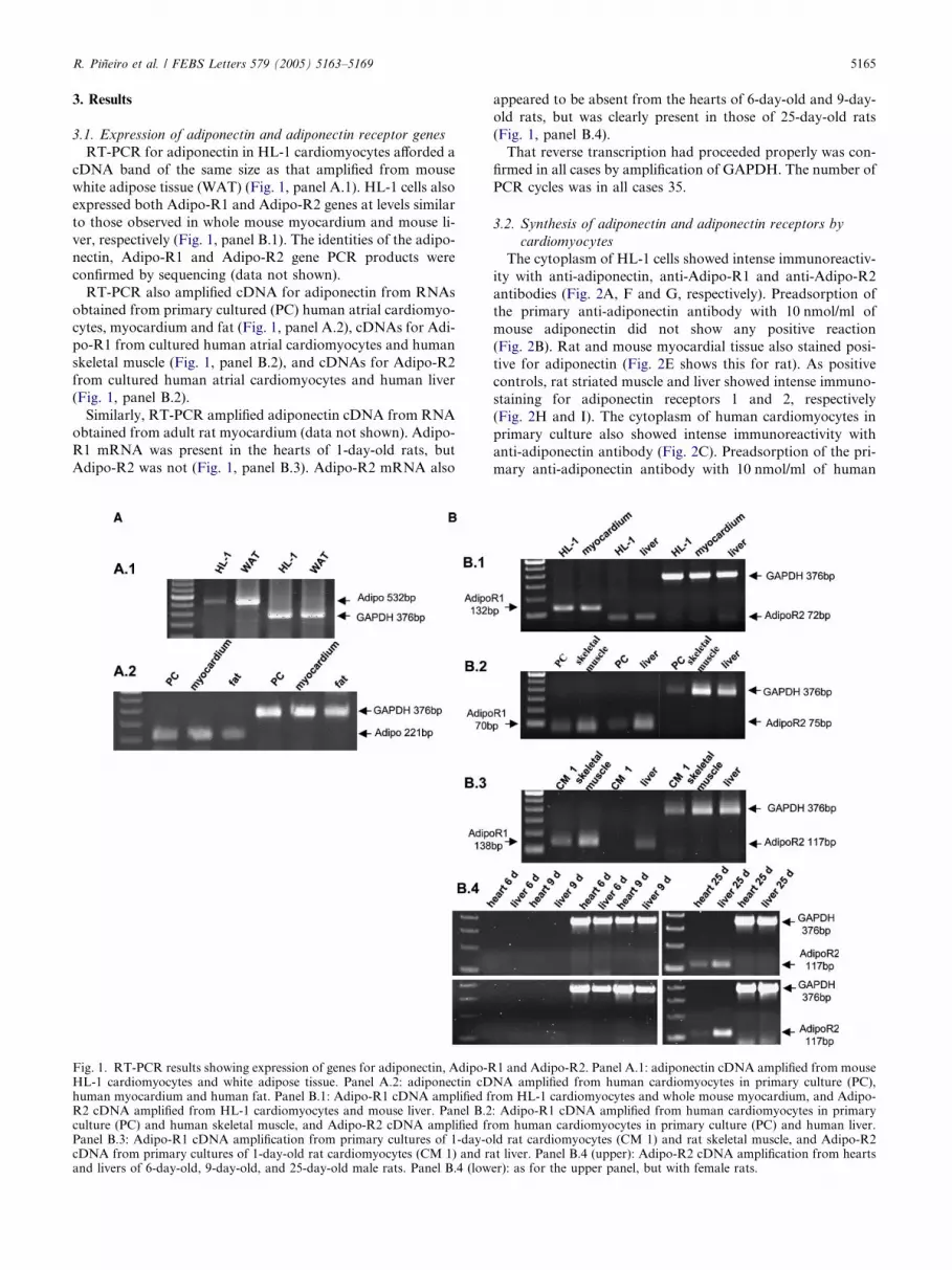
3.1. Expression of adiponectin and adiponectin receptor genes
RT-PCR for adiponectin in HL-1 cardiomyocytes afforded a
cDNA band of the same size as that amplified from mouse
white adipose tissue (WAT) (Fig. 1, panel A.1). HL-1 cells also
expressed both Adipo-R1 and Adipo-R2 genes at levels similar
to those observed in whole mouse myocardium and mouse li-
ver, respectively (Fig. 1, panel B.1). The identities of the adipo-
nectin, Adipo-R1 and Adipo-R2 gene PCR products were
confirmed by sequencing (data not shown).
RT-PCR also amplified cDNA for adiponectin from RNAs
obtained from primary cultured (PC) human atrial cardiomyo-
cytes, myocardium and fat (Fig. 1, panel A.2), cDNAs for Adi-
po-R1 from cultured human atrial cardiomyocytes and human
skeletal muscle (Fig. 1, panel B.2), and cDNAs for Adipo-R2
from cultured human atrial cardiomyocytes and human liver
(Fig. 1, panel B.2).
Similarly, RT-PCR amplified adiponectin cDNA from RNA
obtained from adult rat myocardium (data not shown). Adipo-
R1 mRNA was present in the hearts of 1-day-old rats, but
Adipo-R2 was not (Fig. 1, panel B.3). Adipo-R2 mRNA also
Fig. 1. RT-PCR results showing expression of genes for adiponectin, Adipo-RHL-1 cardiomyocytes and white adipose tissue. Panel A.2: adiponectin cDhuman myocardium and human fat. Panel B.1: Adipo-R1 cDNA amplified fR2 cDNA amplified from HL-1 cardiomyocytes and mouse liver. Panel B.2culture (PC) and human skeletal muscle, and Adipo-R2 cDNA amplified frPanel B.3: Adipo-R1 cDNA amplification from primary cultures of 1-day-ocDNA from primary cultures of 1-day-old rat cardiomyocytes (CM 1) and raand livers of 6-day-old, 9-day-old, and 25-day-old male rats. Panel B.4 (low
appeared to be absent from the hearts of 6-day-old and 9-day-
old rats, but was clearly present in those of 25-day-old rats
(Fig. 1, panel B.4).
That reverse transcription had proceeded properly was con-
firmed in all cases by amplification of GAPDH. The number of
PCR cycles was in all cases 35.
3.2. Synthesis of adiponectin and adiponectin receptors by
cardiomyocytes
The cytoplasm of HL-1 cells showed intense immunoreactiv-
ity with anti-adiponectin, anti-Adipo-R1 and anti-Adipo-R2
antibodies (Fig. 2A, F and G, respectively). Preadsorption of
the primary anti-adiponectin antibody with 10 nmol/ml of
mouse adiponectin did not show any positive reaction
(Fig. 2B). Rat and mouse myocardial tissue also stained posi-
tive for adiponectin (Fig. 2E shows this for rat). As positive
controls, rat striated muscle and liver showed intense immuno-
staining for adiponectin receptors 1 and 2, respectively
(Fig. 2H and I). The cytoplasm of human cardiomyocytes in
primary culture also showed intense immunoreactivity with
anti-adiponectin antibody (Fig. 2C). Preadsorption of the pri-
mary anti-adiponectin antibody with 10 nmol/ml of human
1 and Adipo-R2. Panel A.1: adiponectin cDNA amplified from mouseNA amplified from human cardiomyocytes in primary culture (PC),rom HL-1 cardiomyocytes and whole mouse myocardium, and Adipo-: Adipo-R1 cDNA amplified from human cardiomyocytes in primaryom human cardiomyocytes in primary culture (PC) and human liver.ld rat cardiomyocytes (CM 1) and rat skeletal muscle, and Adipo-R2t liver. Panel B.4 (upper): Adipo-R2 cDNA amplification from heartser): as for the upper panel, but with female rats.

Fig. 2. Immunohistochemical detection of adiponectin, Adipo-R1 and Adipo-R2 in cardiomyocytes and myocardium (objective magnification 40· asotherwise stated). (A) Adiponectin immunostaining was mainly localized in the cytoplasm of HL-1 cells. (B) Preadsorption control with mouserecombinant adiponectin (10 nmol/ml) shows no positive immunostaining. (C) Adiponectin immunostaining was also evident in the cytoplasm ofhuman atrial myocardium cells. Positive reaction was mainly localized in the cytoplasm in the vicinity of the nuclei. (D) Preadsorption control withhuman recombinant adiponectin (10 nmol/ml) resulted in a negative reaction. (E) Rat left ventricle, showingmyocardium positively immunostained foradiponectin (objective magnification 10·). (F–G) HL-1 cells with cytoplasm showing diffuse immunopositivity for Adipo-R1 (F) and Adipo-R2 (G).(H) Rat striated muscle: myocytes, but not connective tissue cells, are positively immunostained for Adipo-R1. (I) Rat liver, positively immunostainedfor Adipo-R2. (J–L) Photomicrographs of human atrial myocardium cells in primary culture showing immunofluorescence of adiponectin (J), cardiacmyosin heavy chain (K), and both these targets (L). (M–O) Photomicrographs of human atrial myocardium cells in primary culture showingimmunofluorescence of Adipo-R1 (M), cardiac myosin heavy chain (N), and both these targets (O). (P–R) Photomicrographs of human atrialmyocardium cells in primary culture showing immunofluorescence of Adipo-R2 (P), cardiac myosin heavy chain (Q), and both these targets (R).
5166 R. Pineiro et al. / FEBS Letters 579 (2005) 5163–5169
adiponectin did not show any positive reaction (Fig. 2D). The
possibility that the human cells were fibroblasts rather than
cardiac muscle fibres was ruled out in colocalization studies
showing positivity for adiponectin, Adipo-R1, and Adipo-
R2, by their immunoreactivity with antibody against cardiac
myosin heavy chain (Fig. 2J–R).
R. Pineiro et al. / FEBS Letters 579 (2005) 5163–5169 5167
3.3. Secretion of adiponectin by cultured HL-1 cells and primary
cultures of human cardiomyocytes
The adiponectin concentrations measured by RIA in the
media in which starving HL-1 cells and human cardiomyocytes
were cultured were 2.7 ± 0.6 ng/ml (n = 5) and 3.1 ± 0.2 ng/ml
(n = 10), respectively.
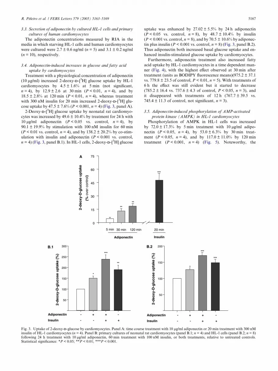
3.4. Adiponectin-induced increases in glucose and fatty acid
uptake by cardiomyocytes
Treatment with a physiological concentration of adiponectin
(10 lg/ml) increased 2-deoxy-DD-[3H] glucose uptake by HL-1
cardiomyocytes by 4.5 ± 1.6% at 5 min (not significant,
n = 4), by 12.9 ± 2.6 at 30 min (P < 0.01, n = 4), and by
18.5 ± 2.8% at 120 min (P < 0.01, n = 4), whereas treatment
with 300 nM insulin for 20 min increased 2-deoxy-DD-[3H] glu-
cose uptake by 47.5 ± 7.6% (P < 0.001, n = 4) (Fig. 3, panel A).
2-Deoxy-DD-[3H] glucose uptake by neonatal rat cardiomyo-
cytes was increased by 49.6 ± 10.4% by treatment for 24 h with
10 lg/ml adiponectin (P < 0.05 vs. control, n = 4), by
90.1 ± 19.9% by stimulation with 100 nM insulin for 60 min
(P < 0.01 vs. control, n = 4), and by 138.2 ± 20.2% by co-stim-
ulation with insulin and adiponectin (P < 0.001 vs. control,
n = 4) (Fig. 3, panel B.1). In HL-1 cells, 2-deoxy-DD-[3H] glucose
50
100
150
200
250
300
*
***
**
2-d
eoxy
-D-g
luco
se u
pta
ke (
%)
Adiponectin - + + -
Insulin - - + +
0
15
30
45
60
75
2-d
eoxy
-D-g
luco
se u
pta
ke
(% o
ver
cont
rol)
5 min 30 min 120 min
Adiponectin
**
**
A
B.1
Fig. 3. Uptake of 2-deoxy-DD-glucose by cardiomyocytes. Panel A: time courseinsulin of HL-1 cardiomyocytes (n = 4). Panel B: primary cultures of neonatalfollowing 24 h treatment with 10 lg/ml adiponectin, 60 min treatment withStatistical significance: *P < 0.05; **P < 0.01; ***P < 0.001.
uptake was enhanced by 27.02 ± 5.5% by 24 h adiponectin
(P < 0.05 vs. control, n = 8), by 48.7 ± 10.4% by insulin
(P < 0.001 vs. control, n = 8), and by 70.5 ± 10.6% by adiponec-
tin plus insulin (P < 0.001 vs. control, n = 8) (Fig. 3, panel B.2).
Thus adiponectin both increased basal glucose uptake and en-
hanced insulin-stimulated glucose uptake by cardiomyocytes.
Furthermore, adiponectin treatment also increased fatty
acid uptake by HL-1 cardiomyocytes in a time dependent man-
ner (Fig. 4), with the highest effect observed at 30 min after
treatment (units as BODIPY fluorescence means)(975.2 ± 37.1
vs. 779.8 ± 23.5 of control, P < 0.01, n = 5). With treatments of
6 h the effect was still evident but it started to decrease
(785.2 ± 16.4 vs. 737.6 ± 4.3 of control, P < 0.05, n = 3), and
it disappeared with treatments of 12 h (767.7 ± 39.5 vs.
745.4 ± 11.3 of control, not significant, n = 3).
3.5. Adiponectin-induced phosphorylation of AMP-activated
protein kinase (AMPK) in HL-1 cardiomyocytes
Phosphorylation of AMPK in HL-1 cells was increased
by 72.0 ± 17.3% by 5 min treatment with 10 lg/ml adipo-
nectin (P < 0.05, n = 4), by 53.0 ± 6.3% by 30 min treat-
ment (P < 0.05, n = 4), and by 117.0 ± 11.0% by 120 min
treatment (P < 0.001, n = 4) (Fig. 5). Noteworthy, the
2-d
eoxy
-D-g
luco
se u
pta
ke (
%)
50
100
150
200
***
***
*
Adiponectin - + + -
Insulin - - + +
Insulin
20 min
***
B.2
treatment with 10 lg/ml adiponectin or 20 min treatment with 300 nMrat cardiomyocytes (panel B.1; n = 4) and HL-1 cells (panel B.2; n = 8)100 nM insulin, or both treatments, relative to untreated controls.

Fig. 5. Panel A: Western blots of HL-1 cell lysates following stainingwith anti-phospho-AMPK antibody (a-pAMPK) or anti-AMPKantibody (a-AMPK); times indicate the duration of adiponectintreatment. Panel B: Amount of phospho-AMPK, relative to controls,in four independently performed experiments (*P < 0.05; ***P <0.001).
600
750
900
1050Control
Adiponectin
0.5Time (h) 2 6 12
****
*
Fat
ty a
cid
up
take
(Mea
ns
flu
ore
scen
ce in
ten
sity
)
Fig. 4. Statistical analysis (of at least three independent experiments)of free fatty acid uptake by HL-1 cardiomyocytes after 10 lg/mladiponectin treatment. *P < 0.05; **P < 0.01.
5168 R. Pineiro et al. / FEBS Letters 579 (2005) 5163–5169
adiponectin-induced increase in glucose uptake by HL-1
cardiomyocytes was diminished in a 72.1 ± 5.3% (P < 0.01,
n = 3) by the addition of the AMPK specific inhibitor araA
20 min before the treatment with adiponectin.
4. Discussion
Until recently it appeared to have been generally accepted
that adiponectin was only synthesized by adipocytes [3].
However, recent studies have found that mouse liver cells
can synthesize adiponectin under appropriate conditions [12];
that adiponectin gene expression can be induced in human
myotubes by exposure to an adiponectin-containing HEK293
cell culture supernatant [11]; that adiponectin synthesis can
be induced both in myotubes and in muscle in response to pro-
inflammatory cytokines [13]; and that adiponectin is expressed
in bone-forming cells [14]. Nevertheless, despite these discover-
ies and the intensive research on the cardiovascular effects of
adiponectin, hitherto no studies have been published that
have investigated the possibility that cardiac cells synthesize
adiponectin.
Our results in this study show in the first place that adipo-
nectin can be synthesized and secreted by cardiomyocytes of
both human and murine origin. As far as we know, this is
the first report of adiponectin synthesis and secretion by
cardiomyocytes. It may be pointed out that although the
adiponectin content of cardiomyocytes seems to be less than
that of adipose tissue, this difference does not suffice to throw
doubts on the efficacy of adiponectin secretion by cardiomyo-
cytes, since cardiomyocyte-secreted adiponectin may well
mediate paracrine and/or autocrine signalling pathways rather
than long-range mechanisms.
We also found that, as expected, HL-1 cells (a cultured line
derived from murine atrial cardiomyocytes that maintains a
heart-specific phenotype and is accordingly used as an
in vitro model in studies of cardiomyocyte biology [15]) and
cultured human cardiomyocytes express genes for the adipo-
nectin receptors Adipo-R1 and Adipo-R2. Interestingly,
1-day-old rat hearts contained Adipo-R1 mRNA but not Adi-
po-R2 mRNA, which only appeared at some time between the
ages of 9 and 25 days. The late appearance of Adipo-R2 may
perhaps be related to the sweeping postnatal changes in the
intermediate metabolism of the heart, which switches from lac-
tate and glucose to fatty acids as its main sources of energy
[22]. Although additional work in this subject is necessary to
elucidate the physiological relevance of this finding, this may
be a physiological mechanism allowing adaptation of cardiac
responsiveness to different physiological and pathological
settings.
Although the processes initiated by the binding of adipo-
nectin to its receptors on cardiomyocytes may be manifold,
two at least were identified in this study, in which adiponectin
significantly enhanced glucose and fatty acid uptake by
cardiomyocytes. These findings are in keeping with recent re-
ports of similar adiponectin effects on glucose uptake by
C2C12 myocytes [23] and adipocytes themselves [18].
We also found that adiponectin treatment induced AMPK
phosphorylation in HL-1 cardiomyocytes. It seems possible
that, as in other cell types [23], the adiponectin-promoted
enhancement of glucose uptake by cardiomyocytes may be
mediated by activation of AMPK, especially since AMPK is
known to mediate glucose uptake and GLUT4 translocation
in heart muscle [24]. In fact, we have observed that, in HL-1
cardiomyocytes, the AMPK specific inhibitor araA blunted
the adiponectin-induced increase on glucose uptake, as it does
in adipocytes [18]. Recently, it has been reported that adiponec-
tin inhibits hypertrophic signaling in the myocardium through
activation of AMPK [25], further supporting the relevance of
adiponectin signaling on cardiomyocyte function.
In conclusion, the main results of this study are that (a)
adiponectin can be synthesized and secreted by human and

R. Pineiro et al. / FEBS Letters 579 (2005) 5163–5169 5169
murine cardiomyocytes in culture, and (b) adiponectin can di-
rectly regulate cardiomyocyte metabolism through AMPK
activation.
The observed expression and secretion of adiponectin by
cardiomyocytes add more complexity to the largely unknown
regulation of cardiomyocyte metabolism/function by adipo-
kines. It may be hypothesized that both fat-derived and local
cardiac-derived adiponectin could be essential for cardiomyo-
cyte metabolism and functionality, and that the failure of
one or both of these mechanisms might be determinant in
the development of several cardiac pathologies.
Acknowledgments: Francisca Lago and Oreste Gualillo are researchersfunded by Spanish Ministry of Health under Health Research Fund(FIS) contracts 99/3040 and 00/3051.
References
[1] Matsuzawa, Y., Funahashi, T., Kihara, S. and Shimomura, I.(2004) Adiponectin and metabolic syndrome. Arterioscler.Thromb. Vasc. Biol. 24, 29–33.
[2] Hug, C. and Lodish, H.F. (2005) The role of the adipocytehormone adiponectin in cardiovascular disease. Curr. Opin.Pharmacol. 5, 129–134.
[3] Goldstein, B.J. and Scalia, R. (2004) Adiponectin: a noveladipokine linking adipocytes and vascular function. J. Clin.Endocrinol. Metab. 89, 2563–2568.
[4] Tan, K.C., Xu, A., Chow, W.S., Lam, M.C., Ai, V.H., Tam, S.C.and Lam, K.S. (2004) Hypoadiponectinemia is associated withimpaired endothelium-dependent vasodilation. J. Clin. Endocri-nol. Metab. 89, 765–769.
[5] Ishikawa, Y., Akasaka, Y., Ishii, T., Yoda-Murakami, M., Choi-Miura, N.H., Tomita, M., Ito, K., Zhang, L., Akishima, Y.,Ishihara, M., Muramatsu, M. and Taniyama, M. (2003) Changesin the distribution pattern of gelatin-binding protein of 28 kDa(adiponectin) in myocardial remodelling after ischaemic injury.Histopathology 42, 43–52.
[6] Ouchi, N., Kihara, S., Funahashi, T., Nakamura, T., Nishida, M.,Kumada, M., Okamoto, Y., Ohashi, K., Nagaretani, H., Kishida,K., Nishizawa, H., Maeda, N., Kobayashi, H., Hiraoka, H. andMatsuzawa, Y. (2003) Reciprocal association of C-reactiveprotein with adiponectin in bloodstream and adipose tissue.Circulation 107, 671–674.
[7] Pischon, T., Girman, C.J., Hotamisligil, G.S., Rifai, N., Hu, F.B.and Rimm, E.B. (2004) Plasma adiponectin levels and risk ofmyocardial infarction in men. JAMA 2291, 1730–1737.
[8] Iwashima, Y., Katsuya, T., Ishikawa, K., Ouchi, N., Ohishi, M.,Sugimoto, K., Fu, Y., Motone, M., Yamamoto, K., Matsuo, A.,Ohashi, K., Kihara, S., Funahashi, T., Rakugi, H., Matsuzawa,Y. and Ogihara, T. (2004) Hypoadiponectinemia is an indepen-dent risk factor for hypertension. Hypertension 43, 1318–1323.
[9] Ohashi, K., Ouchi, N., Kihara, S., Funahashi, T., Nakamura, T.,Sumitsuji, S., Kawamoto, T., Matsumoto, S., Nagaretani, H.,Kumada, M., Okamoto, Y., Nishizawa, H., Kishida, K., Maeda,N., Hiraoka, H., Iwashima, Y., Ishikawa, K., Ohishi, M.,Katsuya, T., Rakugi, H., Ogihara, T. and Matsuzawa, Y.(2004) Adiponectin I164T mutation is associated with themetabolic syndrome and coronary artery disease. J. Am. Coll.Cardiol. 43, 1195–1200.
[10] Yamauchi, T., Kamon, J., Ito, Y., Tsuchida, A., Yokomizo, T.,Kita, S., Sugiyama, T., Miyagishi, M., Hara, K., Tsunoda, M.,Murakami, K., Ohteki, T., Uchida, S., Takekawa, S., Waki, H.,Tsuno, N.H., Shibata, Y., Terauchi, Y., Froguel, P., Tobe, K.,
Koyasu, T., Taira, K., Kitamura, T., Shimizu, T., Nagai, R. andKadowaki, T. (2003) Cloning of adiponectin receptors thatmediate antidiabetic metabolic effects. Nature 423, 762–769.
[11] Staiger, H., Kausch, C., Guirguis, A., Weisser, M., Maerker, E.,Stumvoll, M., Lammers, R., Machicao, F. and Haring, H.U.(2003) Induction of adiponectin gene expression in humanmyotubes by an adiponectin-containing HEK293 cell culturesupernatant. Diabetologia 46, 956–960.
[12] Yoda-Murakami, M., Taniguchi, M., Takahashi, K., Kawamata,S., Saito, K., Choi-Miura, N.H. and Tomita, M. (2001) Change inexpression of GBP28/adiponectin in carbon tetrachloride-admin-istrated mouse liver. Biochem. Biophys. Res. Commun. 285, 372–377.
[13] Delaigle, A.M., Jonas, J.C., Bauche, I.B., Cornu, O. andBrichard, S.M. (2004) Induction of adiponectin in skeletal muscleby inflammatory cytokines: in vivo and in vitro studies. Endocri-nology 145, 5589–5597.
[14] Berner, H.S., Lyngstadaas, S.P., Spahr, A., Monjo, M., Thom-mesen, L., Drevon, C.A., Syversen, U. and Reseland, J.E. (2004)Adiponectin and its receptors are expressed in bone-forming cells.Bone 35, 842–849.
[15] Gonzalez-Juanatey, J.R., Iglesias, M.J., Alcaide, C., Pineiro, R.and Lago, F. (2003) Doxazosin induces apoptosis in cardiomyo-cytes cultured in vitro by a mechanism that is independent ofalpha-adrenergic blockade. Circulation 107, 127–131.
[16] Winzell, M.S., Nogueiras, R., Dieguez, C. and Ahren, B. (2004)Dual action of adiponectin on insulin secretion in insulin-resistantmice. Biochem. Biophys. Res. Commun. 321, 154–160.
[17] Shuralyova, I., Tajmir, P., Bilan, P.J., Sweeney, G. and Coe, I.R.(2004) Inhibition of glucose uptake in the murine cardiomyocytecell line HL-1 by cardioprotective drugs dilazep and dipyridam-ole. Am. J. Physiol. Heart. Circ. Physiol. 286, H627–H632.
[18] Wu, X., Motoshima, H., Mahadev, K., Stalker, T.J., Scalia, R.and Goldstein, B.J. (2003) Involvement of AMP-activated proteinkinase in glucose uptake stimulated by the globular domain ofadiponectin in primary rat adipocytes. Diabetes 52, 1355–1363.
[19] Viengchareun, S., Zennaro, M.C., Le Tallec, L.P. and Lombes,M. (2002) Brown adipocytes are novel sites of expression andregulation of adiponectin and resistin. FEBS Lett. 532, 345–350.
[20] Liu, Y.M., Lacorte, J.M., Viguerie, N., Poitou, C., Pelloux, V.,Guy-Grand, B., Coussieu, C., Langin, D., Basdevant, A. andClement, K. (2003) Adiponectin gene expression in subcutaneousadipose tissue of obese women in response to short term very lowcalorie diet and refeeding. J. Clin. Endocrinol. Metab. 88, 5881–5886.
[21] Altomonte, J., Harbaran, S., Richter, A. and Dong, H. (2003) Fatdepot-specific expression of adiponectin is impaired in Zuckerfatty rats. Metabolism 52, 958–963.
[22] Lopaschuk, G.D., Collins-Nakai, R.L. and Itoi, T. (1992)Developmental changes in energy substrate use by the heart.Cardiovasc. Res. 26, 1172–1180.
[23] Yamauchi, T., Kamon, J., Minokoshi, Y., Ito, Y., Waki, H.,Uchida, S., Yamashita, S., Noda, M., Kita, S., Ueki, K., Eto, K.,Akanuma, Y., Froguel, P., Foufelle, F., Ferre, P., Carling, D.,Kimura, S., Nagai, R., Kahn, B.B. and Kadowaki, T. (2002)Adiponectin stimulates glucose utilization and fatty acid oxida-tion by activating AMP-activated protein kinase. Nat. Med. 8,1288–1295.
[24] Li, J., Hu, X., Selvakumar, P., Russell III, R.R., Cushman, S.W.,Holman, G.D. and Young, L.H. (2004) Role of the nitric oxidepathway in AMPK-mediated glucose uptake and GLUT4 trans-location in heart muscle. Am. J. Physiol. Endocrinol. Metab. 287,E834–E841.
[25] Shibata, R., Ouchi, N., Ito, M., Kihara, S., Shiojima, I., Pimentel,D.R., Kumada, M., Sato, K., Schiekofer, S., Ohashi, K.,Funahashi, T., Colucci, W.S. and Walsh, K. (2004) Adiponec-tin-mediated modulation of hypertrophic signals in the heart. Nat.Med. 10, 1384–1389.