mechanical behaviour of excimer laser irradiated polycrystalline zirconium mechanical behaviour of...
TRANSCRIPT
This content has been downloaded from IOPscience. Please scroll down to see the full text.
Download details:
IP Address: 111.68.103.102
This content was downloaded on 27/01/2014 at 04:23
Please note that terms and conditions apply.
Mechanical behaviour of excimer laser irradiated polycrystalline zirconium
View the table of contents for this issue, or go to the journal homepage for more
2014 Phys. Scr. 89 025703
(http://iopscience.iop.org/1402-4896/89/2/025703)
Home Search Collections Journals About Contact us My IOPscience

Royal Swedish Academy of Sciences Physica Scripta
Phys. Scr. 89 (2014) 025703 (7pp) doi:10.1088/0031-8949/89/02/025703
Mechanical behaviour of excimer laserirradiated polycrystalline zirconium
Mohsan Jelani, Shazia Bashir, Mahreen Akram, Daniel Yousaf,Naveed Afzal and Sajjad Ahmad
Center for Advanced Studies in Physics, Government College University, Lahore 54000, Pakistan
E-mail: [email protected]
Received 30 July 2013Accepted for publication 26 November 2013Published 23 January 2014
AbstractThe effects of laser irradiation on the mechanical response of polycrystalline zirconium (Zr)have been investigated. Zr samples were irradiated with an excimer (KrF) laser (λ ≈ 248 nm,τ ≈ 18 ns and repetition rate ≈ 30 Hz). The irradiation was performed in the ambientenvironment of a gas mixture containing (CO2: N2: He) under a filling pressure of 20 Torr byvarying laser fluences ranging from 3.8 to 5.1 J cm−2. The surface and structural modificationof the irradiated targets were investigated using a scanning electron microscope (SEM) andx-ray diffractometer (XRD). In order to explore the mechanical properties of the irradiated Zr,the tensile testing and Vickers microhardness testing techniques were employed. SEManalysis reveals the grain growth on the irradiated Zr surfaces; however, for increasing fluenceup to 4.7 J cm−2, the appearance of the grains becomes more distinct with an increase in theirnumber density and decrease in size. For the maximum fluence of 5.1 J cm−2, the grainscompletely vanish and the surface becomes diffusive. XRD analysis reveals the appearance ofnew phases of ZrN and ZrO2. The variation in the peak intensity is observed to be anomalous,whereas the decreasing trend in the crystallite size and residual stresses is observed withincreasing fluence. The microhardness analysis reveals the increasing trend in surfacehardness with increasing fluence. The tensile testing demonstrates the anomalous behaviour ofthe yield stress and ultimate tensile strength with increasing fluence.
Keywords: laser irradiation, surface marphology, mechanical properties, crystallite size,microhardness
(Some figures may appear in colour only in the online journal)
1. Introduction
Lasers have been developed as an efficient and versatiletool for material modification and processing. The flexibilityto process a wide range of materials and the advantagesof using lasers over other conventional processes, e.g. areduced heat-affected zone, control over the laser fluence,speed, their non-contact nature, and contamination free andenvironment-friendly ablation processes, make lasers thepreferred and popular choice for many material processingapplications [1]. Lasers have a wide range of industrialapplications, e.g. welding, cutting, drilling, annealing, thinfilm deposition and surface modification [2, 3].
Laser ablation of solid targets in the presence of ambientgas has attained significant importance due to its vast rangeof applications, e.g. pulsed laser deposition, nanoparticle
synthesis and laser-induced breakdown spectroscopy [4, 5].During laser ablation, the presence of the gas as anambient environment enhances the ablation performance dueto the confinement effect [3]. The energy deposited bythe laser-induced plasma ions of gaseous media, chemicalreactivity and diffusion also enhance the density and thevelocity of the ejected particulates [6, 7].
Zirconium (Zr) and its alloys are significantly importantin the engineering industry and in nuclear fusion. In thenuclear industry, they are commonly used due to theirlow thermal neutron absorption cross section, favourablemechanical properties and good corrosion resistance. Itsthermal conductivity, corrosion resistance, formability,strength and minimum creep properties under high servicetemperatures and pressures favour Zr as a highly suitable andappropriate metal for making crucibles, gas turbines, liners
0031-8949/14/025703+07$33.00 1 © 2014 The Royal Swedish Academy of Sciences Printed in the UK
Phys. Scr. 89 (2014) 025703 M Jelani et al
for jet, rocket motor tubes and high resistance and ultra-highfrequency furnaces [8, 9].
The purpose of surface, structural and mechanicalmodification of Zr is to develop and modify its wide rangeof functional properties, including physical, chemical,mechanical, wear resistance and corrosion resistantproperties, and therefore make Zr the most favourable andcommon element for engineering and industrial applications.Zr is significantly important in nuclear reactors for claddingfuel rods due to its low neutron absorption cross-section.
Several techniques such as nitriding, carburizing andcoating have been employed to improve the surface andmechanical properties of Zr and its alloys [8–12]. Ursuet al [10] studied the nitriding and oxidizing of Ti and Zrsamples by the action of microsecond pulsed transverselyexcited atmospheric CO2 laser irradiation in the presenceof a nitrogen and oxygen gas environment. Owing to thesignificant formation of nitride and oxide layers, an increase inhardness was observed. Nishitani et al [11] used a pulsed Nd+
doped glass laser to irradiate pure transition metal targets (Al,Fe, Ni, Nb, Ti, W and Zr) in the ambient environment of airand nitrogen gas to form their oxides and nitrides. It has beenobserved that oxide and nitride formation mainly depends onthe boiling points of the metals. Charles et al [12] studied lasershock processing and its effects on the microstructure andmechanical properties of metal alloys. It has been observedthat laser shock processing can significantly improve thefatigue performance, hardness and mechanical properties ofsome metals and alloys.
The motivation behind this work was to improvethe surface and mechanical properties of Zr after laserirradiation in the presence of a gaseous ambient environment.A pulsed excimer (KrF) laser of (248 nm, 18 ns and30 Hz) was employed to irradiate the Zr targets in thepresence of a gas mixture (CO2: N2: He) environment.There are several ways in which the presence of anambient gas affects laser ablation, material properties andsurface structure formation. The laser ablation of transitionmetals in the ambient environment of gaseous environmentscan produce more chemical reactions at the solid–gasinterface than ordinary evaporation due to the generationof intense pressures and temperatures. Consequently, thematerial is modified with improved mechanical and chemicalproperties [1–3]. The presence of gas as an ambient mediumduring ablation enhances the ablation performance due tothe confinement effect, chemical reactivity, diffusion andthe energy deposited by induced plasma ions [1, 2, 4].Laser ablation of solid targets in the presence of ambientgas has attained significant importance due to its vastranging applications, e.g. pulsed laser deposition, nanoparticlesynthesis and laser-induced breakdown spectroscopy [5,6]. Various diagnostic techniques such as SEM, XRD,microhardness and tensile testing were employed in orderto explore and establish the relation between surface andcompositional changes with the mechanical properties of theirradiated material.
The main objective of this work was to investigatethe laser irradiation effects on the hardness and the tensileproperties of Zr. The novelty of this work is the correlationbetween the surface and compositional changes with thechange in the mechanical properties of the metal.
2. Experimental work
The target material used in this study is polycrystalline Zr withhigh purity of 99.999%. Rectangular-shaped samples with anaverage length of 43 mm, width of 5 mm and thickness of3 mm were selected. The samples were mechanically grindedand polished with silicon carbide papers followed by diamondpaste down to 0.01 µm. The polished specimens were sealedinto silica tubes, evacuated up to a base pressure of 10−6 Torrusing a rotary pump followed by a diffusion pump. To reducedefects and internal stresses, annealing was performed in amuffle furnace at 800 ◦C for 1 h. The annealed specimens wereultrasonically cleaned and were mounted on a sample holderattached to a two-dimensional motor controlled stage. Themounted samples were placed in a UHV chamber made ofstainless steel. The chamber was evacuated to a base pressureof 10−6 Torr and then a gas mixture of CO2: N2: He with theratio 11:6:83 was filled at a pressure of 20 Torr.
An excimer laser beam of wavelength 248 nm, pulseduration of 18 ns and repetition rate of 30 Hz was employed asthe source of irradiation. After passing through a plano convexlens of focal length 50 cm and a quartz transmission windowattached to one port of the chamber, the beam was incidentat an angle of 90◦ with respect to the target surface. Thesamples were exposed for 2100 laser pulses at the very lowscanning speed of 0.6 mm s−1. In order to explore the effect ofthe laser fluence on the surface and structural and mechanicalmodification of the target material, the targets were exposedto four various laser fluences of 3.8, 4.3, 4.7 and 5.1 J cm−2
by keeping all of the other parameters constant.The surface morphology of the unirradiated and laser
irradiated samples was investigated using a scanning electronmicroscope (SEM) (JEOL-JSM-6480). The structural analysisof the pre- and post-irradiated specimens was conductedusing an x-ray diffractometer (X’Pert PRO MPD). TheVickers hardness tester (Zwick/Roell ZHU-5030), equippedwith a pyramid-shaped diamond indenter was employed forthe measurement of surface microhardness. The test wasconducted using a 200 g load for a dentation time of 15 s atroom temperature. The tensile testing of the unirradiated andirradiated samples was performed using an universal tensiletesting machine (AG-1 Shimadzu). All the samples weredeformed under a gauge length of 40 mm with a cross headspeed of 1 mm min−1 at room temperature.
3. Results and discussion
3.1. Surface morphology
The SEM images in figure 1 represent the surface morphologyof (a) unirradiated and excimer laser irradiated Zr for variousfluences (b) 3.8, (c) 4.3, (d) 4.7 and (e) 5.1 J cm−2. Figure 1(b)reveals the growth of diffused grains after being exposedto a laser fluence of 3.8 J cm−2. The grain boundaries arenot distinct and wide. Figure 1(c) predicts that the increasedfluence up to a value of 4.3 J cm−2 enhances grain growth withmore distinct and wider boundaries. Some particulates are alsoseen on the irradiated surface. By increasing the laser fluenceup to 4.7 J cm−2, the large sized grains break into smallerones along with the formation of pores on the specimen
2

Phys. Scr. 89 (2014) 025703 M Jelani et al
Figure 1. SEM images revealing the surface morphology of (a) unirradiated and excimer laser irradiated Zr for various fluences of (b) 3.8,(c) 4.3, (d) 4.7 and (e) 5.1 J cm−2.
Table 1. Variation of grain size with laser fluence.
Laser fluence (J cm−2) Grain size (µm)
3.8 2.5 ± 0.54.3 2.8 ± 0.54.7 1.9 ± 0.5
surface (figure 1(d)). For the highest fluence of 5.1 J cm−2,the specimen surface becomes diffusive with invisible grainboundaries (figure 1(e)).
The quantitative values of average grain size have beenmeasured (from the SEM analysis) and are listed in table 1 forvarious fluences.
Laser-induced rapid heating and cooling, a largetemperature gradient and gas dynamic flows are taken to beresponsible for grain growth [13, 14]. Grain growth occurs asa result of the recrystallization process which arises duringirradiation in order to minimize the absorbed strain andsurface energy. The inter planar atomic motion reorients thelattice at the expense of this energy, which leads to thegrowth of large and smooth grains [15]. When the laserfluence is increased to 4.3 J cm−2, fast melting, cooling andrecrystallization take place due to hydrodynamical effects.
On cooling, the material resolidifies and the backgroundgas atoms diffuse along the grain boundaries. This diffusionmakes the grain boundaries more distinct and wider [14]. Thefurther increase in laser fluence (4.7 J cm−2), breaks the largesized grains into smaller ones which may be attributed torapid heating, cooling, enhanced diffusion and resolidificationprocesses. Moreover, at this fluence the appearance of smallsized cavities or pores is also observed. During irradiation,the temperature of the molten liquid layer beneath thesurface becomes so high that explosive volume boiling wouldoccur. This results in subsurface explosions along with theejection of a large amount of droplets through the generationof pores. The emission of trapped gases and the thermaldecomposition or enhanced absorption of gas atoms alongthe grain boundaries are responsible for the formation ofthese pores [16]. At the maximum fluence of 5.1 J cm−2,surface melting and resolidification of the specimen increasesignificantly. The resolidified material covers all of the grainsby filling the pores and by forming a layer of diffused materialon the specimen’s surface.
Figure 2 represents the fractographs of (a) unirradiatedand laser irradiated Zr for various fluences of (b) 3.8 (c) 4.3(d) 4.7 and (e) 5.1 J cm−2. The fractographs simply representthe appearance of defects and fibre-like structure. The purposeof presenting fractographs is to correlate the fracture and
3

Phys. Scr. 89 (2014) 025703 M Jelani et al
Figure 2. SEM images representing the fractographs of (a) unirradiated and excimer laser irradiated Zr for various fluences of (b) 3.8,(c) 4.3, (d) 4.7 and (e) 5.1 J cm−2.
microstructural modification of the irradiated material withtensile properties. Initially, the compactness of the defects andthe fibrous structures increase with increasing laser fluence upto 4.3 J cm−2 (figures 2(a)–(c)). However, a further increasein the fluence reduces the compression and the compactnessof the defects, as can be clearly seen from figures 2(d) to(e). The initial increase in the compactness is attributableto defect accumulation, microstructural modification andthe grain-growth mechanism which is responsible for theenhancement of the tensile properties (figure 7).
Further increase in laser fluence causes heat treatmentand correspondingly annealing effects which are responsiblefor the reduction in the ultimate tensile strength (UTS) andthe yield stress (YS) (figure 7). Hence, the results of thefractographs and of the tensile properties are well correlated.
The defect density has not been measured quantitatively.However, laser-induced pressure (GPa) and energy depositedper atom (eV atom−1) are evaluated.
The laser-induced pressure (P) is evaluated by using thefollowing formula [10]:
P =(1 − R)E
V. (1)
Here, R is the reflection coefficient with a value of 0.014 for Zrat 248 nm [11]. E is the pulse energy, which varies in the range
of 90120 mJ. V is irradiated volume and is evaluated using thefollowing relation:
Volume = Focused area × Skin depth. (2)
The focused area of the beam and the skin depth of Zrfor 248 nm have the values 0.0235 cm2 (calculated) and 10 nm(calculated), respectively.
In order to evaluate the energy deposited per atom(eV atom−1), the number of atoms per unit volume shouldbe known. The number of atoms per cm3 for Zr is known tobe 1.9 × 1024. The evaluated induced pressure is expressed inthe unit of eV m−3 as (Pa = J m−3). The energy deposited peratom has been evaluated by dividing the energy deposited perunit volume (eV m−3) with the number of atoms per m3.
The evaluated laser-induced pressure and the energydeposited per atom vary from 3.5 × 10³ to 5 × 10³ GPa and 12to 16 eV atom−1, respectively. This implies that the depositedlaser energy is significantly higher than the melting energyper atom and is capable of producing defects in the material.Similarly, the Young’s modulus of Zr is 88 GPa [12] andthe laser-induced pressure (GPa) is significantly higher thanthe Young’s modulus, hence it can generate a permanentlyenhanced number of defects. These defects are thereforeobservable after irradiation.
4
Phys. Scr. 89 (2014) 025703 M Jelani et al
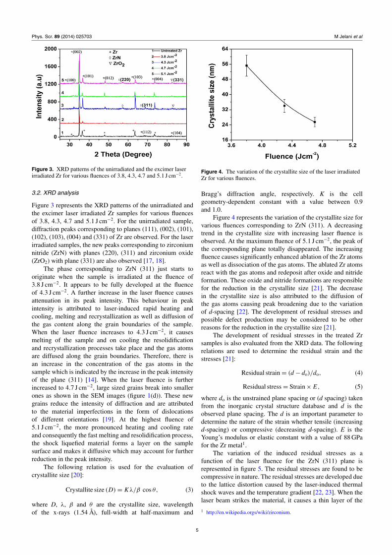
Figure 3. XRD patterns of the unirradiated and the excimer laserirradiated Zr for various fluences of 3.8, 4.3, 4.7 and 5.1 J cm−2.
3.2. XRD analysis
Figure 3 represents the XRD patterns of the unirradiated andthe excimer laser irradiated Zr samples for various fluencesof 3.8, 4.3, 4.7 and 5.1 J cm−2. For the unirradiated sample,diffraction peaks corresponding to planes (111), (002), (101),(102), (103), (004) and (331) of Zr are observed. For the laserirradiated samples, the new peaks corresponding to zirconiumnitride (ZrN) with planes (220), (311) and zirconium oxide(ZrO2) with plane (331) are also observed [17, 18].
The phase corresponding to ZrN (311) just starts tooriginate when the sample is irradiated at the fluence of3.8 J cm−2. It appears to be fully developed at the fluenceof 4.3 J cm−2. A further increase in the laser fluence causesattenuation in its peak intensity. This behaviour in peakintensity is attributed to laser-induced rapid heating andcooling, melting and recrystallization as well as diffusion ofthe gas content along the grain boundaries of the sample.When the laser fluence increases to 4.3 J cm−2, it causesmelting of the sample and on cooling the resolidificationand recrystallization processes take place and the gas atomsare diffused along the grain boundaries. Therefore, there isan increase in the concentration of the gas atoms in thesample which is indicated by the increase in the peak intensityof the plane (311) [14]. When the laser fluence is furtherincreased to 4.7 J cm−2, large sized grains break into smallerones as shown in the SEM images (figure 1(d)). These newgrains reduce the intensity of diffraction and are attributedto the material imperfections in the form of dislocationsof different orientations [19]. At the highest fluence of5.1 J cm−2, the more pronounced heating and cooling rateand consequently the fast melting and resolidification process,the shock liquefied material forms a layer on the samplesurface and makes it diffusive which may account for furtherreduction in the peak intensity.
The following relation is used for the evaluation ofcrystallite size [20]:
Crystallite size (D) = Kλ/β cos θ, (3)
where D, λ, β and θ are the crystallite size, wavelengthof the x-rays (1.54 Å), full-width at half-maximum and
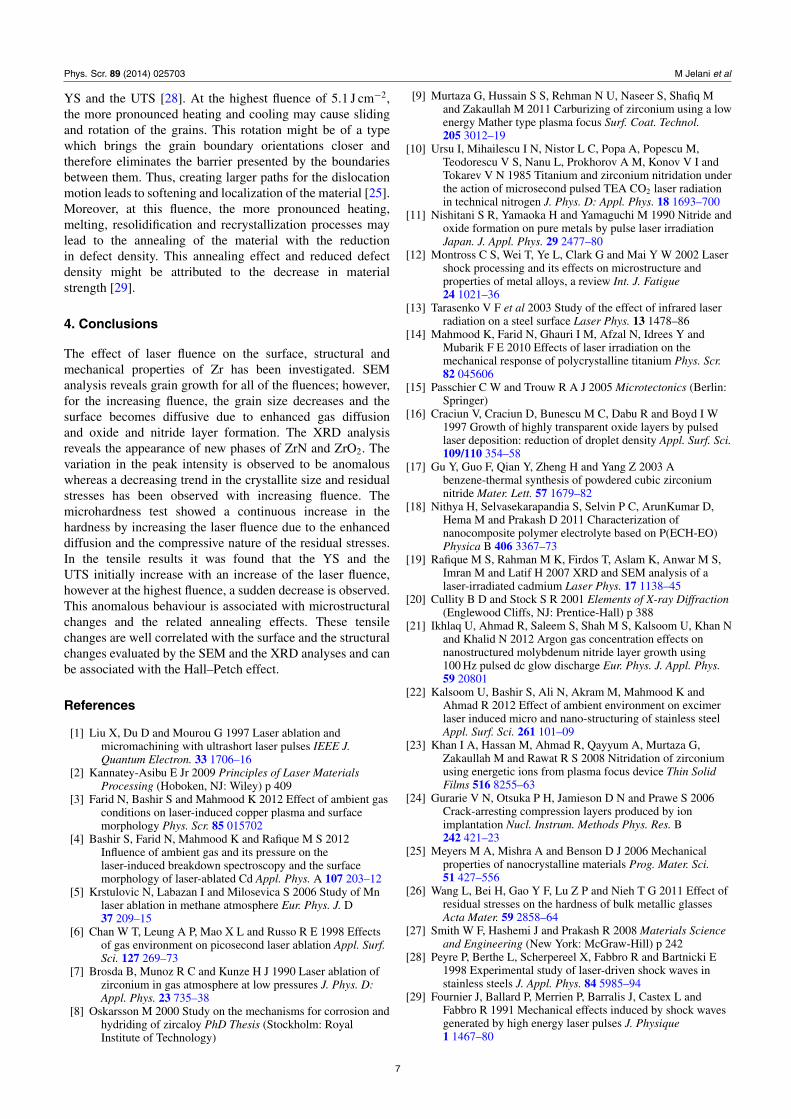
Figure 4. The variation of the crystallite size of the laser irradiatedZr for various fluences.
Bragg’s diffraction angle, respectively. K is the cellgeometry-dependent constant with a value between 0.9and 1.0.
Figure 4 represents the variation of the crystallite size forvarious fluences corresponding to ZrN (311). A decreasingtrend in the crystallite size with increasing laser fluence isobserved. At the maximum fluence of 5.1 J cm−2, the peak ofthe corresponding plane totally disappeared. The increasingfluence causes significantly enhanced ablation of the Zr atomsas well as dissociation of the gas atoms. The ablated Zr atomsreact with the gas atoms and redeposit after oxide and nitrideformation. These oxide and nitride formations are responsiblefor the reduction in the crystallite size [21]. The decreasein the crystallite size is also attributed to the diffusion ofthe gas atoms causing peak broadening due to the variationof d-spacing [22]. The development of residual stresses andpossible defect production may be considered to be otherreasons for the reduction in the crystallite size [21].
The development of residual stresses in the treated Zrsamples is also evaluated from the XRD data. The followingrelations are used to determine the residual strain and thestresses [21]:
Residual strain = (d − do)/do, (4)
Residual stress = Strain × E, (5)
where do is the unstrained plane spacing or (d spacing) takenfrom the inorganic crystal structure database and d is theobserved plane spacing. The d is an important parameter todetermine the nature of the strain whether tensile (increasingd-spacing) or compressive (decreasing d-spacing). E is theYoung’s modulus or elastic constant with a value of 88 GPafor the Zr metal1.
The variation of the induced residual stresses as afunction of the laser fluence for the ZrN (311) plane isrepresented in figure 5. The residual stresses are found to becompressive in nature. The residual stresses are developed dueto the lattice distortion caused by the laser-induced thermalshock waves and the temperature gradient [22, 23]. When thelaser beam strikes the material, it causes a thin layer of the
1 http://en.wikipedia.orgs/wiki/zirconium.
5

Phys. Scr. 89 (2014) 025703 M Jelani et al
Figure 5. The variation of the residual stress of the laser irradiatedZr for various fluences.
Figure 6. The variation of the microhardness of the excimer laserirradiated Zr for various fluences.
material surface to vapourize. The vapourized species reactswith the background gas and rapidly expanding heated gasconfined on the surface generates huge pressure in the GParange. This propagating pressure causes the development ofhigh residual compressive stress. The enhanced diffusion rateof the gas atoms with the increasing fluence is the cause forthe decreasing trend of the residual stresses [24].
3.3. Surface microhardness
The variation of the surface microhardness of the laserirradiated Zr samples for various fluences is represented infigure 6.
An increase in the microhardness is observed with theincreasing laser fluence. The possible explanations for theincrease in the surface hardness of the laser irradiated Zrsamples are smaller crystallite size, related to hardening bythe Hall–Petch relationship [25]
H = Ho + KH(D)−1/2, (6)
where H is the hardness, D is the average crystallite size,whereas Ho and KH are the experimental constants. Thedevelopment of the compressive residual stresses and theformation of the oxide and the nitride layers are alsoresponsible for the increasing surface hardness [14, 26].
Figure 7. The comparison of the stress–strain curves measured bythe tensile testing machine of the unirradiated and the excimer laserirradiated Zr for various fluences of 3.8, 4.3, 4.7 and 5.1 J cm−2.
3.4. Tensile results
Figure 7 represents the tensile stress–strain curves of theunirradiated and excimer laser irradiated Zr for variousfluences of 3.8, 4.3. 4.7 and 5.1 J cm−2. This figure illustratesthe increase in the YS and the UTS by the increasing fluencefrom 3.8 to 4.7 J cm−2. However, at the highest fluenceof 5.1 J cm−2, a decrease in both the YS and the UTS isobserved. Figure 7 predicts the decreasing trend in percentageelongation with the increase of fluence.
These observed changes in the laser irradiated materialare associated with laser-induced microstructural changes.Mechanical behaviour is known to be significantly affectedby variation of the grain size. In this regard, the Hall–Petchrelation gives the dependence of the YS (σy) on the grain size(G) in the conventional polycrystalline range [25]
σy = σo+K (G)−1/2, (7)
where σo and K are the constants for the material concerned.This relationship predicts that the YS increases with theinverse of the square root of the grain size.
Sufficient stress and the absorbed lattice energy producedislocations in the material and the grain boundaries act likea barrier for their movement [27]. The smaller grains havea high surface-area-to-volume ratio, which means a greaterratio of the grain boundaries to the dislocations. Therefore, thegrain boundaries restrict further movement of the dislocations.Owing to their continued motion, there is a dislocation pileup across the grain boundaries. This piled-up dislocationfinally diffuses across the grain boundaries due to increasedrepulsive forces among them. This diffusion leads to initiationof further deformation in the material. In the small sized grainboundaries, the dislocation pile up is small, hence a largeamount of stress is required for their motion [27]. Therefore,the strength of the material increases with an increase inthe grain boundaries and with a decrease in the grain size.Figure 7 shows that maximum YS and UTS is observedfor the fluence of 4.7 J cm−2 which is well correlated withthe minimum grain size (figure 1 SEM images). Moreover,along this Hall–Petch strengthening, the increasing fluenceand the laser-induced shockwaves cause an increase in stressand defect production, which may cause the increase in the
6
Phys. Scr. 89 (2014) 025703 M Jelani et al
YS and the UTS [28]. At the highest fluence of 5.1 J cm−2,the more pronounced heating and cooling may cause slidingand rotation of the grains. This rotation might be of a typewhich brings the grain boundary orientations closer andtherefore eliminates the barrier presented by the boundariesbetween them. Thus, creating larger paths for the dislocationmotion leads to softening and localization of the material [25].Moreover, at this fluence, the more pronounced heating,melting, resolidification and recrystallization processes maylead to the annealing of the material with the reductionin defect density. This annealing effect and reduced defectdensity might be attributed to the decrease in materialstrength [29].
4. Conclusions
The effect of laser fluence on the surface, structural andmechanical properties of Zr has been investigated. SEManalysis reveals grain growth for all of the fluences; however,for the increasing fluence, the grain size decreases and thesurface becomes diffusive due to enhanced gas diffusionand oxide and nitride layer formation. The XRD analysisreveals the appearance of new phases of ZrN and ZrO2. Thevariation in the peak intensity is observed to be anomalouswhereas a decreasing trend in the crystallite size and residualstresses has been observed with increasing fluence. Themicrohardness test showed a continuous increase in thehardness by increasing the laser fluence due to the enhanceddiffusion and the compressive nature of the residual stresses.In the tensile results it was found that the YS and theUTS initially increase with an increase of the laser fluence,however at the highest fluence, a sudden decrease is observed.This anomalous behaviour is associated with microstructuralchanges and the related annealing effects. These tensilechanges are well correlated with the surface and the structuralchanges evaluated by the SEM and the XRD analyses and canbe associated with the Hall–Petch effect.
References
[1] Liu X, Du D and Mourou G 1997 Laser ablation andmicromachining with ultrashort laser pulses IEEE J.Quantum Electron. 33 1706–16
[2] Kannatey-Asibu E Jr 2009 Principles of Laser MaterialsProcessing (Hoboken, NJ: Wiley) p 409
[3] Farid N, Bashir S and Mahmood K 2012 Effect of ambient gasconditions on laser-induced copper plasma and surfacemorphology Phys. Scr. 85 015702
[4] Bashir S, Farid N, Mahmood K and Rafique M S 2012Influence of ambient gas and its pressure on thelaser-induced breakdown spectroscopy and the surfacemorphology of laser-ablated Cd Appl. Phys. A 107 203–12
[5] Krstulovic N, Labazan I and Milosevica S 2006 Study of Mnlaser ablation in methane atmosphere Eur. Phys. J. D37 209–15
[6] Chan W T, Leung A P, Mao X L and Russo R E 1998 Effectsof gas environment on picosecond laser ablation Appl. Surf.Sci. 127 269–73
[7] Brosda B, Munoz R C and Kunze H J 1990 Laser ablation ofzirconium in gas atmosphere at low pressures J. Phys. D:Appl. Phys. 23 735–38
[8] Oskarsson M 2000 Study on the mechanisms for corrosion andhydriding of zircaloy PhD Thesis (Stockholm: RoyalInstitute of Technology)
[9] Murtaza G, Hussain S S, Rehman N U, Naseer S, Shafiq Mand Zakaullah M 2011 Carburizing of zirconium using a lowenergy Mather type plasma focus Surf. Coat. Technol.205 3012–19
[10] Ursu I, Mihailescu I N, Nistor L C, Popa A, Popescu M,Teodorescu V S, Nanu L, Prokhorov A M, Konov V I andTokarev V N 1985 Titanium and zirconium nitridation underthe action of microsecond pulsed TEA CO2 laser radiationin technical nitrogen J. Phys. D: Appl. Phys. 18 1693–700
[11] Nishitani S R, Yamaoka H and Yamaguchi M 1990 Nitride andoxide formation on pure metals by pulse laser irradiationJapan. J. Appl. Phys. 29 2477–80
[12] Montross C S, Wei T, Ye L, Clark G and Mai Y W 2002 Lasershock processing and its effects on microstructure andproperties of metal alloys, a review Int. J. Fatigue24 1021–36
[13] Tarasenko V F et al 2003 Study of the effect of infrared laserradiation on a steel surface Laser Phys. 13 1478–86
[14] Mahmood K, Farid N, Ghauri I M, Afzal N, Idrees Y andMubarik F E 2010 Effects of laser irradiation on themechanical response of polycrystalline titanium Phys. Scr.82 045606
[15] Passchier C W and Trouw R A J 2005 Microtectonics (Berlin:Springer)
[16] Craciun V, Craciun D, Bunescu M C, Dabu R and Boyd I W1997 Growth of highly transparent oxide layers by pulsedlaser deposition: reduction of droplet density Appl. Surf. Sci.109/110 354–58
[17] Gu Y, Guo F, Qian Y, Zheng H and Yang Z 2003 Abenzene-thermal synthesis of powdered cubic zirconiumnitride Mater. Lett. 57 1679–82
[18] Nithya H, Selvasekarapandia S, Selvin P C, ArunKumar D,Hema M and Prakash D 2011 Characterization ofnanocomposite polymer electrolyte based on P(ECH-EO)Physica B 406 3367–73
[19] Rafique M S, Rahman M K, Firdos T, Aslam K, Anwar M S,Imran M and Latif H 2007 XRD and SEM analysis of alaser-irradiated cadmium Laser Phys. 17 1138–45
[20] Cullity B D and Stock S R 2001 Elements of X-ray Diffraction(Englewood Cliffs, NJ: Prentice-Hall) p 388
[21] Ikhlaq U, Ahmad R, Saleem S, Shah M S, Kalsoom U, Khan Nand Khalid N 2012 Argon gas concentration effects onnanostructured molybdenum nitride layer growth using100 Hz pulsed dc glow discharge Eur. Phys. J. Appl. Phys.59 20801
[22] Kalsoom U, Bashir S, Ali N, Akram M, Mahmood K andAhmad R 2012 Effect of ambient environment on excimerlaser induced micro and nano-structuring of stainless steelAppl. Surf. Sci. 261 101–09
[23] Khan I A, Hassan M, Ahmad R, Qayyum A, Murtaza G,Zakaullah M and Rawat R S 2008 Nitridation of zirconiumusing energetic ions from plasma focus device Thin SolidFilms 516 8255–63
[24] Gurarie V N, Otsuka P H, Jamieson D N and Prawe S 2006Crack-arresting compression layers produced by ionimplantation Nucl. Instrum. Methods Phys. Res. B242 421–23
[25] Meyers M A, Mishra A and Benson D J 2006 Mechanicalproperties of nanocrystalline materials Prog. Mater. Sci.51 427–556
[26] Wang L, Bei H, Gao Y F, Lu Z P and Nieh T G 2011 Effect ofresidual stresses on the hardness of bulk metallic glassesActa Mater. 59 2858–64
[27] Smith W F, Hashemi J and Prakash R 2008 Materials Scienceand Engineering (New York: McGraw-Hill) p 242
[28] Peyre P, Berthe L, Scherpereel X, Fabbro R and Bartnicki E1998 Experimental study of laser-driven shock waves instainless steels J. Appl. Phys. 84 5985–94
[29] Fournier J, Ballard P, Merrien P, Barralis J, Castex L andFabbro R 1991 Mechanical effects induced by shock wavesgenerated by high energy laser pulses J. Physique1 1467–80
7