blunt atrial transseptal puncture using excimer laser in swine
TRANSCRIPT
Blunt atrial transseptal puncture using excimer laser in swine
Abdalla A. Elagha, MD, Ann H. Kim, BSc, Ozgur Kocaturk, MSc, and Robert J. Lederman, MD,FSCAIFrom the Cardiovascular Branch, Division of Intramural Research, National Heart Lung and BloodInstitute, National Institutes of Health, Bethesda, MD, USA
AbstractObjectives—We describe a new approach that may enhance safety of atrial transseptal puncture,using a commercially available laser catheter that is capable of perforation only when energized. Wetest this approach in swine.
Background—Despite wide application, conventional needle transseptal puncture continues to riskinadvertent non-target perforation and its consequences.
Methods—We used a commercial excimer laser catheter (0.9mm Clirpath, Spectranetics).Perforation force was compared in vitro with a conventional Brockenbrough needle. Eight swineunderwent laser transseptal puncture under X-ray fluoroscopy steered using a variety of deliverycatheters.
Results—The 0.9mm laser catheter traversed in vitro targets with reduced force compared with aBrockenbrough needle. In vitro, the laser catheter created holes that were 25–30% larger than theBrockenbrough needle.
Laser puncture of the atrial septum was successful and accurate in all animals, evidenced by oximetry,pressure, angiography, and necropsy. The laser catheter was steered effectively using a modifiedMullins introducer sheath and using two different deflectable guiding catheters. The mean proceduretime was 15 ± 6 minutes, with an average 3.0 ± 0.8 seconds of laser activation. There were no adversesequelae after prolonged observation. Necropsy revealed discrete 0.9mm holes in all septae.
Conclusion—Laser puncture of the interatrial septum is feasible and safe in swine, using a bluntlaser catheter that perforates tissues in a controlled fashion.
KeywordsAtrial transseptal puncture; Laser angioplasty; Catheterization
BackgroundAtrial transseptal puncture is the first step in a variety of procedures such as left atrial rhythmmapping and ablation, percutaneous left ventricular assist device implantation, and balloonmitral valvuloplasty. Unfortunately, atrial transseptal puncture continues to confer a discreterisk of serious complications such as aortic and posterior atrial perforation (1–5). Forceaccumulates at the tip of needle during conventional puncture and may lead to suddenuncontrolled advancement, contributing to perforation and pericardial tamponade.
Address for Correspondence: Robert J. Lederman, MD, Cardiovascular Branch, Division of Intramural Research, National Heart Lung,and Blood Institute, National Institutes of Health, Building 10, Room 2c713, MSC1538, Bethesda, MD 20892-1538, USA. Telephone:+1 301-402-6769. [email protected].
NIH Public AccessAuthor ManuscriptCatheter Cardiovasc Interv. Author manuscript; available in PMC 2009 August 13.
Published in final edited form as:Catheter Cardiovasc Interv. 2007 October 1; 70(4): 585–590. doi:10.1002/ccd.21257.
NIH
-PA Author Manuscript
NIH
-PA Author Manuscript
NIH
-PA Author Manuscript
Commercial excimer laser consoles are widely deployed for applications in angioplasty andpacemaker lead extraction (6,7). The controllable activation of laser angioplasty catheters mayhave utility during atrial septal puncture, because the perforation capability can instantly beswitched off, unlike a needle catheter.
We hypothesize that excimer laser transseptal puncture can be performed easily and safely inswine under X-ray fluoroscopy.
MethodsAnimal Protocol
Animal procedures were approved by the National Heart Lung and Blood Institute (NHLBI)Animal Care and Use Committee. Eight healthy Yorkshire swine (mean 57 ± 19 kg) underwentanesthesia induction using tiletamine, zolazepam, and xylazine followed by inhaled isoflurane.Femoral or jugular veins and femoral arteries were accessed percutaneously. Heparin 100 units/kg was given after septal puncture. At the conclusion of experiments, animals were euthanizedwith potassium chloride 4 mEq/kg and necropsy specimens were examined.
DevicesLaser catheters and source—We used off-the-shelf excimer laser catheters (0.9mmClirpath X-80, Spectranetics, Colorado Springs, CO). These fit inside most catheters rated for0.038” guidewires. The laser catheter has an inner lumen diameter of 0.0155” that permitspressure transduction, contrast injection, or 0.014” guidewire delivery. We used an unmodifiedXeCl ultraviolet (308nm) excimer laser source (CVX-300, Spectranetics). All punctures used25 laser pulses per second at a fluence of 45 mJ/ mm2.
Delivery catheters—We used a range of steering devices. We modified a conventional 8FrMullins sheath (Medtronic) to reverse the distal taper of the inner diameter, which ispresumably implemented to smooth the transition to the Brockenbrough needle. We performedthis modification at the bedside using a a 0.040" parylene coated mandrel (New EnglandPrecision Grinding, MA) and a heat gun (Beahm Designs Model 210-A) set at 190° C for 40seconds.
We also tested two off-the-shelf deflectable guiding catheters. The Morph catheter (Biocardia,South San Francisco, CA) is available with a 6Fr outer diameter, 76cm working length, and isavailable with a 3 cm “reach” or lateral deflection. The Naviport catheter (Cardima, Fremont,CA) is available with an 8Fr outer diameter and 90cm working length, but to accommodateshorter dilator catheters we employed the 11Fr version with a 73cm working length and 4 cmreach.
Ex Vivo Perforation Force and Pressure Waveform AnalysisWe measured the forward force required for transseptal puncture using both needle and laser.Both a Spectranetics X80 laser and a Brockenbrough needle (Cook Medical) devices wereattached to a digital force meter (Model ZPH, Imada, IL) and positioned against freshlyexplanted interatrial and interventricular septum from a 90kg pig. The average maximum forcewas recorded while the catheter was advanced across the septum using the force gauge.
Residual holes were compared using fresh atrial septal tissue, examined at rest and undertension sufficient to “flatten” it for measurement using calibrated images.
Pressure waveform damping was compared among three catheter devices: a 0.038” guidewire-compatible 6Fr × 100cm multipurpose catheter (Boston Scientific), a Brockenbrough needle
Elagha et al. Page 2
Catheter Cardiovasc Interv. Author manuscript; available in PMC 2009 August 13.
NIH
-PA Author Manuscript
NIH
-PA Author Manuscript
NIH
-PA Author Manuscript
guidewire port, and the 0.9mm laser catheter, all filled with heparinized normal saline. Invivo arterial and venous waveforms were sampled from the analog output of clinical recordinghardware (Cathcor, Siemens) at 100Hz using an analog-to-digital convertor (Powerlab,ADInstruments). Output spectral analysis was performed (Maclab, ADInstruments) after FastFourier Transformation to obtain the first six harmonics, which were normalized to the catheterwaveform spectrum.
Transseptal Laser CatheterizationBaseline cardiac MRI (breath-held, ECG-gated, segmented, steady state free procession, 1.5TSonata, Siemens using standard torso phased-array coils) verified there was no pre-existingpatent foramen ovale or atrial septal defect.
In the first set of experiments, the modified Mullins sheath and dilator were introducedpercutaneously into the femoral vein and advanced in the superior vena cava over a guidewireand then withdrawn into the fossa ovalis, using a rigid mandril. A transfemoral pigtail catheterwas positioned in the aortic root. The laser catheter was then apposed to the fossa under X-rayguidance. Contrast could be injected, and distal pressure could be measured through theguidewire lumen of the laser catheter. Continuous pressure was applied to the Mullins sheathto maintain apposition between the laser and the target tissue. The laser was activated whileapplying gentle pressure for 2–4 seconds until it advanced across the septum. At this point, theoperator immediately terminated laser activation. Finally, the Mullins dilator and sheath weredelivered across the septum using the laser catheter as a rail. In other experiments, thedeflectable catheters substituted for both the Mullins sheath and dilator.
StatisticsContinuous parameters were reported as mean ± standard deviation, and compared using atwo-tailed Student t test.
ResultsEx vivo findings
Representative force-time curves are shown for the needle and laser catheters (Figure 1). Themaximum force required to perforate fresh interatrial septum ex vivo was 2.2 ± 0.28 N usinga Brockenbrough needle versus 0.21 ± 0.07 N using the X-80 laser catheter, p<0.001. Thecurves show a linear accumulation of forward force using the needle before sudden “give” asthe needle perforates. By contrast the laser force-time curves are less dramatic. The findingswere similar using ventricular myocardium as a test medium (0.27 ± 0.04 N using the laser vs2.7 ± 0.13 N using the needle).
The guidewire lumen of the laser damped pressure recordings more than the guidewire lumenof the needle (Figure 2). The laser created holes that were 25–30% larger than theBrockenbrough needle measured unstretched (laser 1.04 ± 0.08mm; needle 0.82 ± 0.08mm,p=0.02 vs laser; 8Fr sheath 2.63 ± 0.53mm) and under distending tension (laser 0.81 ± 0.08mm;needle 0.63 ± 0.16mm, p=0.04 vs laser; sheath 2.17 ± 0.62mm).
In vivo findingsThe laser transseptal technique is illustrated in Figure 3. Panel A shows the Mullins sheath(arrowhead) positioned using standard technique against the fossa ovalis. For reference, wepositioned a pigtail catheter in the aortic root (white arrow). The laser catheter is advanced tothe distal tip of the Mullins dilator and is identified by its distal radiopaque marker (blackarrow). After a brief application of laser energy, the laser catheter advances easily into the leftatrium (Panel B, black arrow). Contrast injection (panel C), pressure waveforms, and oximetry
Elagha et al. Page 3
Catheter Cardiovasc Interv. Author manuscript; available in PMC 2009 August 13.
NIH
-PA Author Manuscript
NIH
-PA Author Manuscript
NIH
-PA Author Manuscript
confirm position. A guidewire can be introduced through the laser catheter lumen into thedesired left sided chamber. The laser catheter serves as a rail to introduce a dilator into the leftatrium (panel D). When using a Mullins dilator, we found it helpful to apply continuous forwardpressure to maintain apposition to the fossa ovalis before perforation; when using deflectableguiding catheters this proved less important.
Laser transseptal puncture was successful in all eight animals. The total procedure time was15 ± 6 minutes. When using a deflectable guiding catheter, the procedure time was 10 ± 1minutes. The laser was activated for 3.0±0.8 seconds. The force applied to laser catheter duringthe procedure was minimal compared with conventional needle puncture. The laser inducedno arrhythmia.
Correct left atrial position was confirmed by pressure waveforms (right atrial mean 4.0 ± 1.2,left atrial pressure 7.6 ± 2.4mmHg, p<0.05), hemoglobin oximetry (right atrial 74%, left atrial96%, p<0.05), and radiocontrast.
After transseptal puncture, animals were observed for 5.9 ± 2.3 hours without sequelae.Necropsy confirmed the desired position of the puncture at the fossa ovalis (Figure 4). Therewas no hemopericardium in any animal. Holes created in vivo using the laser catheter measured0.9mm. Holes created ex vivo using the laser were the same size but had a smooth contourcompared with holes created using a Brockenbrough needle, which had a “torn” morphology..
DiscussionUsing commercially-available catheters, we have successfully performed excimer lasertransseptal puncture in healthy swine. The laser requires ten-fold less forward force to crosscardiac tissue compared with a conventional needle. Because the laser catheter is blunt anddoes not perforate tissue except when activated, laser puncture may be an attractive alternativeto conventional needle puncture.
Despite widespread application and decades of experience, needle transseptal puncture stillrisks pericardial tamponade and aortic communication. The procedure is more challenging inpatients with abnormal atrial septal morphology, distorted septal contour, thickened atrialseptum especially in elderly patients, atrial baffles, or prior valve surgery (8). Because punctureforce may drive the needle forward across both the target septum and the contralateral freeatrial wall, the incidence of complications is higher in patients with small left atria (9). Repeattransseptal puncture may fail using conventional Brockenbrough needle or radiofrequencyablation system due to thickened atrial septum (10).
Like the needle, the laser contains a central guidewire lumen which can be used for pressuremeasurement, contrast injection, or guidewire delivery. Unlike the needle, the laser catheterrequires minimal forward force during activation, has adjustable energy and pulse rate, and issufficiently rigid to serve as a “rail” to advance larger catheter devices into a suitable left sidedtarget. Though untested, the laser may offer a “rescue” alternative in cases of thickened ordistorted atrial septa.
The tested 0.9mm laser creates slightly larger holes than the Brockenbrough needle, and dampspressure waveforms comparatively more than the Brockenbrough needle. We were still ableto distinguish right and left atrial pressure waveforms in healthy swine.
Nevertheless, the excimer laser remains capable of perforating non-target tissue if improperlydirected. For example, posterior left atrial perforation remains possible if laser energy is notimmediately discontinued after successful traversal of the interatrial septum. The consequences
Elagha et al. Page 4
Catheter Cardiovasc Interv. Author manuscript; available in PMC 2009 August 13.
NIH
-PA Author Manuscript
NIH
-PA Author Manuscript
NIH
-PA Author Manuscript
of inadvertent puncture may be comparably more severe, since perforation with the laser isequivalent to delivering the entire Brockenbrough needle, not just the tip, across a target.
LimitationsThese experiments were conducted in healthy swine which do not model the challenges ofcomplex atrial transseptal puncture in humans with distorted, scarred, calcified, or patchedseptal anatomy. The comparative healing characteristics of laser and needle holes are notcharacterized here.
Transseptal puncture procedures are guided both by visual and tactile cues. Other adjuncts oralternatives are available to enhance safety of septal puncture. Functionally-positionedelectrophysiologic catheters, such as His bundle and coronary sinus recording catheters,provide useful landmarks for identifying the fossa ovalis (11). Extending a guidewire througha conventional needle may reduce the incidence of posterior wall perforation (12), but onlyafter safe septal needle traversal. Right atrial angiography may enhance procedural safety inpatients with distorted anatomy (13). A radiofrequency perforation catheter (14–16) providesan alternative to the laser technique described here.
ConclusionsWe describe percutaneous laser transseptal puncture accurately and safety in swine usingcommercially available laser catheters. The blunt-tip laser catheter does not risk perforationexcept when activated. Since it requires minimal forward force, it may be less likely to “jump”inadvertently across the left atrial free wall. It may provide a valuable alternative to needletransseptal puncture. Clinical testing is warranted, especially in patients with abnormal atrialgeometry.
AcknowledgementsThe authors thank William H. Schenke and Victor J. Wright for their technical assistance, and Katherine Lucas andJoni Taylor for animal care and support. Supported by NIH 1-Z01-HL005062–04 CVB
References1. Adrouny ZA, Sutherland DW, Griswold HE, Ritzmann LW. Complications with transseptal left heart
catheterization. Am Heart J 1963;65:327–333. [PubMed: 14011135]2. Mullins CE. Transseptal left heart catheterization: experience with a new technique in 520 pediatric
and adult patients. Pediatr Cardiol 1983;4(3):239–245. [PubMed: 6647111]3. B. Lundqvist C, Olsson SB, Varnauskas E. Transseptal left heart catheterization: a review of 278
studies. Clin Cardiol 1986;9(1):21–26. [PubMed: 3943231]4. Hung JS. Atrial septal puncture technique in percutaneous transvenous mitral commissurotomy: mitral
valvuloplasty using the Inoue balloon catheter technique. Cathet Cardiovasc Diagn 1992;26(4):275–284. [PubMed: 1394414]
5. Roelke M, Smith AJ, Palacios IF. The technique and safety of transseptal left heart catheterization: theMassachusetts General Hospital experience with 1,279 procedures. Cathet Cardiovasc Diagn 1994;32(4):332–339. [PubMed: 7987913]
6. Scheinert D, Laird JR Jr, Schroder M, Steinkamp H, Balzer JO, Biamino G. Excimer laser-assistedrecanalization of long, chronic superficial femoral artery occlusions. J Endovasc Ther 2001;8(2):156–166. [PubMed: 11357976]
7. Wilkoff BL, Byrd CL, Love CJ, Hayes DL, Sellers TD, Schaerf R, Parsonnet V, Epstein LM, SorrentinoRA, Reiser C. Pacemaker lead extraction with the laser sheath: results of the pacing lead extractionwith the excimer sheath (PLEXES) trial. J Am Coll Cardiol 1999;33(6):1671–1676. [PubMed:10334441]
Elagha et al. Page 5
Catheter Cardiovasc Interv. Author manuscript; available in PMC 2009 August 13.
NIH
-PA Author Manuscript
NIH
-PA Author Manuscript
NIH
-PA Author Manuscript
8. El-Said HG, Ing FF, Grifka RG, Nihill MR, Morris C, Getty-Houswright D, Mullins CE. 18-yearexperience with transseptal procedures through baffles, conduits, and other intra-atrial patches.Catheter Cardiovasc Interv 2000;50(4):434–439. [PubMed: 10931616]discussion 440
9. Braunwald E, Cooperative study on cardiac catheterization. Transseptal left heart catheterization.Circulation 1968;37:III74–III79. [PubMed: 5647084]
10. Marcus GM, Ren X, Tseng ZH, Badhwar N, Lee BK, Lee RJ, Foster E, Olgin JE. Repeat transseptalcatheterization after ablation for atrial fibrillation. J Cardiovasc Electrophysiol 2007;18(1):55–59.[PubMed: 17081207]
11. Cheng A, Calkins H. A Conservative Approach to Performing Transseptal Punctures Without theUse of Intracardiac Echocardiography: Stepwise Approach with Real-Time Video Clips. JCardiovasc Electrophysiol. 2007
12. Hildick-Smith D, McCready J, de Giovanni J. Transseptal puncture: Use of an angioplasty guidewirefor enhanced safety. Catheter Cardiovasc Interv 2007;69(4):519–521. [PubMed: 17286295]
13. Rogers DP, Lambiase PD, Dhinoja M, Lowe MD, Chow AW. Right atrial angiography facilitatestransseptal puncture for complex ablation in patients with unusual anatomy. J Interv CardElectrophysiol 2006;17(1):29–34. [PubMed: 17235679]
14. Veldtman GR, Wilson GJ, Peirone A, Hartley A, Estrada M, Norgard G, Leung RK, Visram N, BensonLN. Radiofrequency perforation and conventional needle percutaneous transseptal left heart access:pathological features. Catheter Cardiovasc Interv 2005;65(4):556–563. [PubMed: 16010688]
15. Sherman W, Lee P, Hartley A, Love B. Transatrial septal catheterization using a new radiofrequencyprobe. Catheter Cardiovasc Interv 2005;66(1):14–17. [PubMed: 15981196]
16. Sakata Y, Feldman T. Transcatheter creation of atrial septal perforation using a radiofrequencytransseptal system: novel approach as an alternative to transseptal needle puncture. CatheterCardiovasc Interv 2005;64(3):327–332. [PubMed: 15736263]
Elagha et al. Page 6
Catheter Cardiovasc Interv. Author manuscript; available in PMC 2009 August 13.
NIH
-PA Author Manuscript
NIH
-PA Author Manuscript
NIH
-PA Author Manuscript
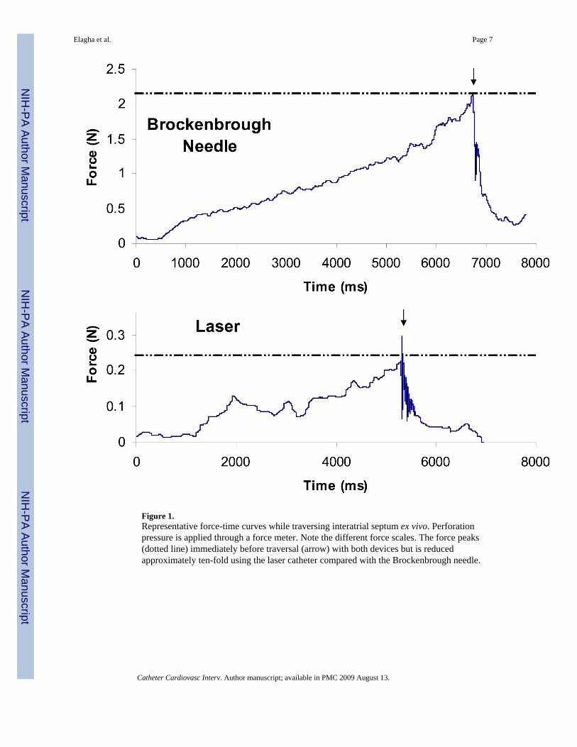
Figure 1.Representative force-time curves while traversing interatrial septum ex vivo. Perforationpressure is applied through a force meter. Note the different force scales. The force peaks(dotted line) immediately before traversal (arrow) with both devices but is reducedapproximately ten-fold using the laser catheter compared with the Brockenbrough needle.
Elagha et al. Page 7
Catheter Cardiovasc Interv. Author manuscript; available in PMC 2009 August 13.
NIH
-PA Author Manuscript
NIH
-PA Author Manuscript
NIH
-PA Author Manuscript
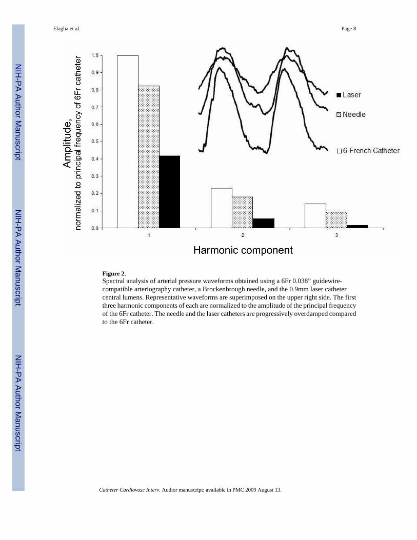
Figure 2.Spectral analysis of arterial pressure waveforms obtained using a 6Fr 0.038” guidewire-compatible arteriography catheter, a Brockenbrough needle, and the 0.9mm laser cathetercentral lumens. Representative waveforms are superimposed on the upper right side. The firstthree harmonic components of each are normalized to the amplitude of the principal frequencyof the 6Fr catheter. The needle and the laser catheters are progressively overdamped comparedto the 6Fr catheter.
Elagha et al. Page 8
Catheter Cardiovasc Interv. Author manuscript; available in PMC 2009 August 13.
NIH
-PA Author Manuscript
NIH
-PA Author Manuscript
NIH
-PA Author Manuscript
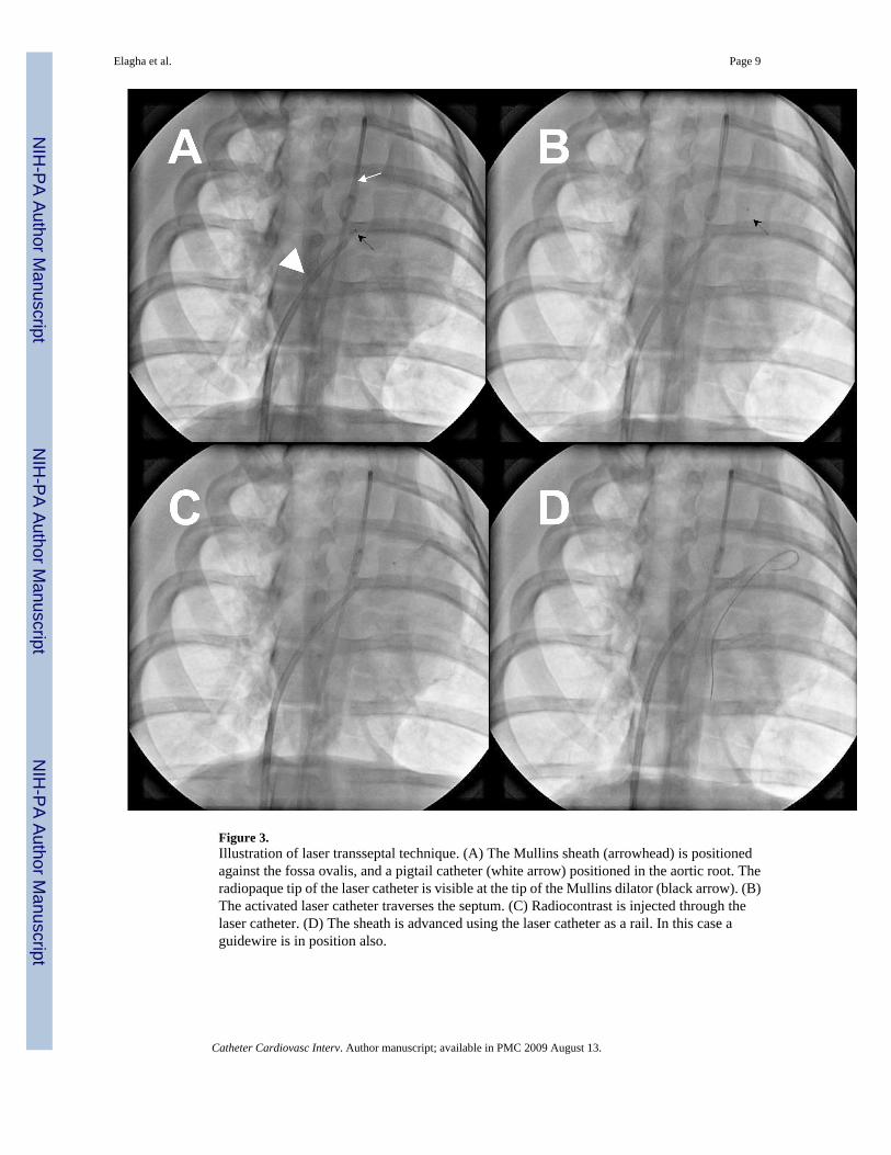
Figure 3.Illustration of laser transseptal technique. (A) The Mullins sheath (arrowhead) is positionedagainst the fossa ovalis, and a pigtail catheter (white arrow) positioned in the aortic root. Theradiopaque tip of the laser catheter is visible at the tip of the Mullins dilator (black arrow). (B)The activated laser catheter traverses the septum. (C) Radiocontrast is injected through thelaser catheter. (D) The sheath is advanced using the laser catheter as a rail. In this case aguidewire is in position also.
Elagha et al. Page 9
Catheter Cardiovasc Interv. Author manuscript; available in PMC 2009 August 13.
NIH
-PA Author Manuscript
NIH
-PA Author Manuscript
NIH
-PA Author Manuscript
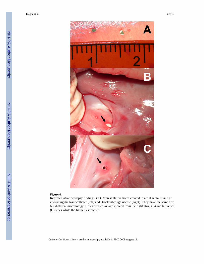
Figure 4.Representative necropsy findings. (A) Representative holes created in atrial septal tissue exvivo using the laser catheter (left) and Brockenbrough needle (right). They have the same sizebut different morphology. Holes created in vivo viewed from the right atrial (B) and left atrial(C) sides while the tissue is stretched.
Elagha et al. Page 10
Catheter Cardiovasc Interv. Author manuscript; available in PMC 2009 August 13.
NIH
-PA Author Manuscript
NIH
-PA Author Manuscript
NIH
-PA Author Manuscript