lrrk2 affects vesicle trafficking, neurotransmitter extracellular level and membrane receptor...
TRANSCRIPT
LRRK2 Affects Vesicle Trafficking, NeurotransmitterExtracellular Level and Membrane Receptor LocalizationRossana Migheli1., Maria Grazia Del Giudice2., Ylenia Spissu1, Giovanna Sanna2, Yulan Xiong3,
Ted M. Dawson3,4,6, Valina L. Dawson3,4,5,6, Manuela Galioto2, Gaia Rocchitta1, Alice Biosa1, Pier
Andrea Serra1, Maria Teresa Carri7,8, Claudia Crosio2,7, Ciro Iaccarino2,7*
1 Department of Clinical and Experimental Medicine, University of Sassari, Sassari, Italy, 2 Department of Biomedical Sciences, University of Sassari, Sassari, Italy,
3 Neuroregeneration and Stem Cell Programs, Institute for Cell Engineering, Johns Hopkins University School of Medicine, Baltimore, Maryland, United States of America,
4 Department of Neurology, Johns Hopkins University School of Medicine, Baltimore, Maryland, United States of America, 5 Department of Physiology, Johns Hopkins
University School of Medicine, Baltimore, Maryland, United States of America, 6 Solomon H. Snyder Department of Neuroscience, Johns Hopkins University School of
Medicine, Baltimore, Maryland, United States of America, 7 Fondazione Santa Lucia, IRCCS, Rome, Italy, 8 Department of Biology, University of Rome ‘‘Tor Vergata’’, Rome,
Italy
Abstract
The leucine-rich repeat kinase 2 (LRRK2) gene was found to play a role in the pathogenesis of both familial and sporadicParkinson’s disease (PD). LRRK2 encodes a large multi-domain protein that is expressed in different tissues. To date, thephysiological and pathological functions of LRRK2 are not clearly defined. In this study we have explored the role of LRRK2in controlling vesicle trafficking in different cellular or animal models and using various readouts. In neuronal cells, thepresence of LRRK2G2019S pathological mutant determines increased extracellular dopamine levels either under basalconditions or upon nicotine stimulation. Moreover, mutant LRRK2 affects the levels of dopamine receptor D1 on themembrane surface in neuronal cells or animal models. Ultrastructural analysis of PC12-derived cells expressing mutantLRRK2G2019S shows an altered intracellular vesicle distribution. Taken together, our results point to the key role of LRRK2 tocontrol vesicle trafficking in neuronal cells.
Citation: Migheli R, Del Giudice MG, Spissu Y, Sanna G, Xiong Y, et al. (2013) LRRK2 Affects Vesicle Trafficking, Neurotransmitter Extracellular Level and MembraneReceptor Localization. PLoS ONE 8(10): e77198. doi:10.1371/journal.pone.0077198
Editor: Patrick Lewis, UCL Institute of Neurology, United Kingdom
Received November 29, 2012; Accepted September 7, 2013; Published October 22, 2013
Copyright: � 2013 Migheli et al. This is an open-access article distributed under the terms of the Creative Commons Attribution License, which permitsunrestricted use, distribution, and reproduction in any medium, provided the original author and source are credited.
Funding: This work was supported by Fondazione Banco di Sardegna, PRIN 2008 (grant nu 20083R593R_002 and 20083R593R_001) and Regione Sardegna (grantnuCRP-17171). Maria Grazia Dal Giudice is supported by a fellowship of Regione Sardegna. This work was supported in part by National Institutes of Health/National Institute of Neurological Disorders and Stroke (NIH/NINDS) P50NS038377. Ted M. Dawson is the Leonard and Madlyn Abramson Professor inNeurodegenerative Diseases. The funders had no role in study design, data collection and analysis, decision to publish, or preparation of the manuscript.
Competing Interests: Ted M. Dawson is a PLOS ONE editorial board member. This does not alter the authors’ adherence to all the PLOS ONE policies on sharingdata and materials.
* E-mail: [email protected]
. These authors contributed equally to this work.
Introduction
Most Parkinson’s disease (PD) cases occur sporadically and
several genes associated with monogenetic forms of the disease
have been identified in patients [1]. Mutations in the leucine-
rich repeat kinase 2 gene (LRRK2, PARK8) cause late-onset,
autosomal dominant PD that is clinically and neurochemically
indistinguishable from idiopathic forms [2,3]. The LRRK2 gene
encodes a large protein of 2527 amino acids belonging to the
ROCO protein family [4]. Similar to other ROCO proteins,
LRRK2 contains a Ras-of-complex (Roc) GTPase domain and
a C-terminal of Roc (COR) domain in conjunction with a
protein kinase domain with close homology to members of the
mixed-lineage and receptor-interacting protein kinase families.
LRRK2 also contains a number of repeat domains (armadillo,
ankyrin, leucine-rich repeats and C-terminal WD40 repeats that
surround the central Roc-COR-kinase catalytic region) of
uncertain function. Interestingly, multiple amino acid substitu-
tions of the same residue R1441 (R1441C, R1441G, and
R1441H) in the highly conserved GTPase domain and multiple
mutations (I2012T, G2019S, and I2020T) in the kinase domain
have been identified in patients [5]. The most common
pathological mutation G2019S increases the kinase activity of
LRRK2 by 2–3 fold [6,7], however some other mutations show
an unchanged or reduced kinase activity [8]. Studies from
several independent groups have evaluated the frequency of
LRRK2 mutations in many different populations, and such
mutations have been found not only in 3–5% of familial PD but
also in approximately 1–3% of idiopathic PD cases [9]. Despite
extensive studies based on both animal and cellular models, the
pathological role of LRRK2 in PD onset and progression is still
largely unclear, and LRRK2 substrate(s) remain fairly elusive.
To date, LRRK2 has been involved in different physiological
functions ranging from miRNA processing [10] to translation
regulation [11], cytoskeleton organization [12,13,14], autopha-
gy-lysosomal pathways [15,16] and immunoregulation [17].
Different experimental approaches seem to suggest a potential
role of LRRK2 in vesicle trafficking [18]. Firstly, LRRK2
appears to be localized in different intracellular compartments
that play a critical role in the control of vesicular trafficking:
endoplasmic reticulum, Golgi apparatus and associated vesicles,
PLOS ONE | www.plosone.org 1 October 2013 | Volume 8 | Issue 10 | e77198
cytoskeleton, lipid rafts and synaptic vesicles [19,20]. Secondly,
alteration in dopamine (DA) release has been described in
different LRRK2 transgenic rodents, ranging from a reduction
in DA extracellular content without [14,21] or with pharma-
cological manipulation [21,22] to an increased DA extracellular
content in transgenic WT LRRK2 mice [22] or in transgenic
rats expressing mutant LRRK2G2019S [23]. In primary neuronal
cells, LRRK2 silencing perturbs vesicle dynamics and distribu-
tion within the recycling pool, leading to a significant decrease
in docked vesicles but an increase in the amount of vesicle
recycling [24]. Moreover, alteration of LRRK2 expression by
knockdown of endogenous LRRK2 in primary neuronal cells
significantly impairs synaptic vesicle endocytosis [24,25], but a
similar effect was observed following LRRK2 overexpression
[25] thus leaving unclear which is the exact role of this protein.
In this work we have analysed the role of LRRK2 in controlling
neurotransmitter extracellular levels as well as the neurotransmit-
ter receptor membrane levels through different experimental
approaches. Taken together, our results point to a key role of
LRRK2 in controlling vesicle trafficking and distribution.
Materials and Methods
AnimalsMice were housed and treated in strict accordance with the
NIH Guide for the Care and Use of Laboratory Animals. All
animal procedures were approved by the Institutional Animal
Care and Use Committees of the Johns Hopkins Medical
Institutions (Animal Welfare Assurance No. A3272-01). Mice
were maintained in a pathogen-free facility and exposed to a 12 h
light/dark cycle with food and water provided ad libitum.
Reagents and SolutionsAntibodies: anti-TH (1:4000 Sigma), anti-Myc (1:5000 Sigma),
anti-DRD1 (1:2000 Sigma), anti-NR1 (1:2000 Sigma), anti-Sec8
(1:4000 BD-Biosciences), anti-clathrin (1:5000 BD-Biosciences).
Reagents: TweenH 20 (Polyethylene glycol sorbitan monolaurate),
Phenylmethanesulfonyl fluoride (PMSF), protease inhibitor cock-
tail and (2)-Nicotine hydrogen tartrate salt were obtained from
Sigma-Aldrich (Milano, Italy). LRRK2 inhibitor GSK2578215A
was from Tocris. The phosphate-buffered saline (PBS) solution was
made using NaCl (137 mM), KCl (2.7 mM), Na2HPO4 (8.1 mM),
KH2PO4 (1.47 mM), CaCl2 (1.19 mM), MgCl2 (0.54 mM), and
glucose (7.5 mM) from Sigma and then adjusted to pH 7.4.
Dulbecco’s modified Eagle’s medium (DMEM)–F12, Streptomy-
cin/Penicillin, Hygromycine B, Geneticin-G418 were purchased
from Invitrogen and doxycycline from BD Biosciences. The
tetracycline-free Fetal Bovine Serum (FBS) was from Lonza Sales
Ltd (Switzerland).
Plasmid ConstructionsThe plasmids for inducible expression or LRRK2 were obtained
by digestions of cDNAs corresponding to human LRRK2 (WT or
R1441C or G2019S) in fusion with 5X myc repeats [26] with
BamHI and XhoI and subcloned in BamHI/EcoRV cloning sites in
pTRE2 vector (Clontech).
cDNA coding for mouse growth hormone (GH, NM_008117.2)
was RT–PCR amplified from mouse pituitary gland mRNA (oligo
forward: ATCAGGATCCTTGGCAATGGCTACAGACTC,
reverse: ATCAGGATCCGAAGGCACAGCTGCTTTCC), di-
gested with BamHI restriction enzyme and cloned in BamHI
cloning site of pCS2-5X-myc-tag containing the tag in C-terminal
position. Plasmid pTL2-DRD1 (kindly provided by E. Borrelli,
University of California, Irvine) was used as template for DRD1
cDNA, the PCR fragment (oligo forward: ATCCTCGAGAA-
GATGGCTCCTAACACTTCTACCA, reverse:
CTCCTCGAGGGTTGAATGCTGTCCGCTGTG) was digest-
ed with XhoI and subcloned into pcDNA3.1-3X-flag-tag contain-
ing the tag in the C-terminal position. p-TK-Hyg (Clontech
Laboratories Inc ) was used to impart hygromycin resistance to
PC12 ON cells.
Cell Lines and PC12 Stable ClonesHuman neuroblastoma SH-SY5Y cells (ATCC number CRL-
2266) were grown in DMEM-F12 (Invitrogen), 10% fetal calf
serum (FCS, Invitrogen) at 37uC. The PC12-TET-ON cell line
(Clontech Laboratories Inc) was cultivated in DMEM-F12
supplemented with 10% Tetracycline-free FCS (Lonza) at 37uC.
The plasmid pTRE2 vectors containing cDNAs coding for
LRRK2 variants (WT or R1441C or G2019S) were co-
transfected with p-TK-Hyg in a 8:1 molar ratio into PC12-
TET-ON cells, using LipofectamineH LTX Reagent (Life
Technologies) according to the manufacturer’s protocol. The
different PC12-TET-ON clones were maintained under selec-
tion by 400 mg/mL of G418 and 200 mg/mL of hygromicin-B.
Individual clones expressing both antibiotic resistances were
picked after 14 days of selection, moved in a 96 well plate, and
maintained in selective medium till confluence growth. Different
individual clones were analyzed for LRRK2 expression upon
induction by doxycycline (0.2 mg/mL).
Analysis of Intracellular and Extracellular Dopamine andMetabolites
Intracellular and extracellular dopamine (DA), 3-methoxytyr-
amine (3-MT), 3,4-Dihydroxyphenylacetic acid (DOPAC), and
homovanillic acid (HVA) were determined by HPLC with
electrochemical detection as previously described [27]. In brief,
cells were lysed in 250 mL 1% metaphosphoric acid containing
1 mM EDTA. After centrifugation (17,500 g for 10 min at 4uC),
the supernatant was filtered, and a 15- mL aliquot was immediately
injected into the HPLC system.
In each experiment, 1006103 cells/cm2 were plated and treated
24 h later (time 0) with doxycycline 0.2 mg/mL. After 48 h, the
medium was aspirated from each well and stored, and the cells
were collected in metaphosphoric acid. Samples were subsequently
analyzed for levels of total DA (DA+3-MT) and its metabolites
DOPAC and HVA in cell lysates and incubation medium. Values
in cell lysate were expressed as nanomoles per milligram of
protein. Total cell extract protein concentration was determined
using the method of Lowry et al. (1951).
Capillary Tube Construction for in vitro MicrodialysisThe capillary tube for microdialysis of PC12 cell lines is an
adaptation of an in vitro device described previously [28,29]. The
microdialysis probe was constructed using two sections of plastic-
coated silica tubing (150 mm o.d., 75 mm i.d., Scientific Glass
Engineering, Milton Keynes, UK), each placed in the centre of a
semipermeable polyacrylonitrile dialysis fiber (AN-69, Hospal
Industrie, Meyzieu, France). Each semipermeable membrane had
an active length of 40 mm. Then each section of plastic-coated
silica tubing was positioned in the centre of polyethylene tubing
(0.58 mm i.d., 35 mm long, Portex). This section of silica tubing
served as the inlet. Dialysates from polyacrylonitrile dialysis fiber
were collected from polyethylene tubing, which served as the
outlet. All parts were coated with quick-drying epoxy glue.
Afterwards the microdialysis probe (the semipermeable polyacry-
lonitrile dialysis fiber plus sealed plastic-coated silica tubing) was
LRRK2 and Vesicular Trafficking
PLOS ONE | www.plosone.org 2 October 2013 | Volume 8 | Issue 10 | e77198
placed in non heparinized microhematocrit capillary tubes
(7.5 mm long, 1.1 mm i.d., Chase Scientific Glass, Rockwood,
IL, USA). The final volume of microdialysis chamber was
approximately 50 mL.
Microdialysis ProceduresMicrodialysis experiments were performed during the expo-
nential phase of cell growth. 56104 cells/cm2 were plated and
treated 24 h later (time 0) with different Doxycycline concentra-
tions. After 48 h cells were washed twice using 5 ml of modified
PBS and 10% DMEM (perfusion medium), harvested and
centrifuged (94 g for 5 min). Cells were resuspended in PBS/
DMEM and the number of cells/ml was assessed in a Burker
chamber. The initial volume of the cell suspension was eventually
adjusted to reach a final concentration of 16106 cells/50 mL.
Nicotine (5 mM) effect on DA secretion from PC12 lines was
evaluated by means of microdialysis in vitro as previously described
[30].
The cellular microdialysis probe was perfused with PBS/
DMEM by means of a peristaltic microinfusion double-channel
pump (P720 peristaltic pump (Instech, Plymouth Meeting, PA,
USA), which pumped PBS/DMEM at a flow rate of 3.0 mL/min.
The pump channels were connected to the inlet by a length of
polythene tubing. The perfusion apparatus was then filled with
50 mL of the PC12 cell suspension by aspiration, which was
performed manually by means of a 1.0 mL syringe connected to
the plastic coated silica tubing sealed outside the polythene tubing.
Thereafter, the perfusion apparatus was kept at 37uC. After 1 h of
stabilization, 3 microdialysis samples (60 mL each) were recovered
at 20 min intervals. Nicotine was added to the perfusion medium
and removed after 60 min. In case of LRRK2 inhibitor
treatments, GSK2578215A (1 mM) was added at the beginning
of stabilization. Samples were recovered during the next two
hours. Subsequently, a 35 mL aliquot of each collected dialysate
was analyzed by HPLC. The concentration of neurochemicals
detected after the first 20 min of perfusion was taken as time 0
concentration. Cell viability was assessed before the start and at
the end of each experiment by trypan blue exclusion. The viability
rate was given as the difference between final and initial
percentage of non-viable cells [29,30].
Chromatographic Analysis of Dialysates from PC12 CellSuspension
DA was quantified in dialysates of selected experiments
(1.06106 cells) by HPLC–EC, as described previously [29] using
an Alltech 426 HPLC pump (Alltech, Sedriano, Italy) equipped
with a Rheodyne injector (model 7725, Rohnert Park, CA, USA),
a column (15 cm, 4.6 mm i.d., ODS80TM C18, Toso Haas,
Stuttgart, Germany), an electrochemical detector ANTEC–
Leyden EC controller (ANTEC, Zoeterwoude, The Netherlands),
and a PC-based ADC system (Varian Star Chromatographic
Workstation, Varian, Walnut Creek, CA, USA). The mobile phase
was citric acid (0.1 M), ethylenediaminetetraacetic acid (EDTA,
1.0 mM), methanol (8.7%) and sodium octylsulfate (48 mg/L),
with a flow rate of 1.2 mL/min and pH 2.9.
Transient Transfections and Analysis of GH SecretionTransient expression of each vector was performed with
Lipofectamine LTX Reagent (Life Technologies) according to
the manufacturer’s instructions. After an incubation of 4–6 h with
transfection reagents, the cells were cultured in normal growth
medium for 24 or 48 h. For GH secretion analysis, SH-SY5Y cells
(1.06105 cells) were seeded in 24 mm plates and co-transfected the
following day either with GH-5Xmyc and pCS2-MTK empty
vector or with GH-5Xmyc and the different pCS2-5Xmyc-
LRRK2 isoforms in a ratio of 1:10. 24 hours after transfection,
the cells were washed twice with fresh medium and normal growth
medium was added for another 16 h. In case of LRRK2 inhibitor
treatments, GSK2578215A was added 1 h before medium change
and then after medium change. The extracellular medium was
then collected and centrifuged at 100006g for 10 min to eliminate
cell debris, while the cells were washed twice with PBS and
immediately lysed by Laemmli buffer 1X. For DRD1 membrane
localization experiments, the cells were co-transfected in 6 cm
plates as described above (in a ratio of 1:5 respectively for DRD1-
3Xflag and 5Xmyc-LRRK2 or empty vector) for 48 hours. The
quantification of either GH-5Xmyc or DRD1-3Xflag in the
different fractions was performed by western blot analysis and
densitometric evaluation of the obtained bands (Quantity-One
Biorad).
Subcellular Fractionation of Cells or Mouse TissuesTissues from 2 months old LRRK2WT or LRRK2G2019S
transgenic mice [16] were quickly dissected and frozen. Subcel-
lular fractionation was conducted as described in [31]. Briefly, SH-
SY5Y or HEK293 cells or striatum were homogenized in ice-cold
homogenization-buffer (320 mM sucrose, 4 mM HEPES, pH 7.4,
protease inhibitor cocktail from Sigma). The homogenates were
centrifuged at 10006g for 10 min to produce the pellet containing
nuclei and large debris fraction (P1). The supernatant (S1) was
further fractionated into pellet (P2 containing the membrane
fraction) and supernatant (S2) by centrifugation at 10,0006g for
20 min. The S2 was ultracentrifuged at 100,0006g to obtain the
pellet (P3 containing the vesicle fraction). Protein content was
determined using the Bradford protein assay. Equal amount of
protein extracts were loaded into the SDS-PAGE.
Western Blot AnalysisWestern blot analysis was performed as previously described
[32]. Briefly, protein content was determined using the Bradford
protein assay. Equal amount of protein extracts were resolved by
standard SDS/PAGE. Samples were electroblotted onto Protan
nitrocellulose (Schleicher & Schuell GmbH). Membranes were
incubated with 3% low-fat milk in 16PBS-Tween 0.05% solution
with the indicated antibody for 16 h at 4uC. Anti-Rabbit IgG
(whole molecule)- and Anti-Mouse IgG (whole molecule)-Perox-
idase antibody were used to reveal immunocomplexes by
enhanced chemioluminescence (Pierce).
Statistical AnalysisConcentrations of neurochemicals in dialysates from PC12 cell
suspension were expressed in mM and given as mean 6 SEM.
Drug effects on neurochemicals were statistically evaluated in
terms of changes in absolute dialysate concentrations. Differences
within or between groups were determined by paired or unpaired
t-tests (ANOVA followed by Student–Newman–Keuls post-hoc
analysis). The null hypothesis was rejected when p,0.05.
Results
Generation and Characterization of PC12 Cells StablyExpressing Doxycycline-inducible WT or PathologicalMutant LRRK2s
Plasmid constructs for wild type LRRK2 (LRRK2wt) or for
pathological mutants LRRK2G2019S and LRRK2R1441C were
transfected in PC12-ON cell lines. After several weeks of selection
LRRK2 and Vesicular Trafficking
PLOS ONE | www.plosone.org 3 October 2013 | Volume 8 | Issue 10 | e77198
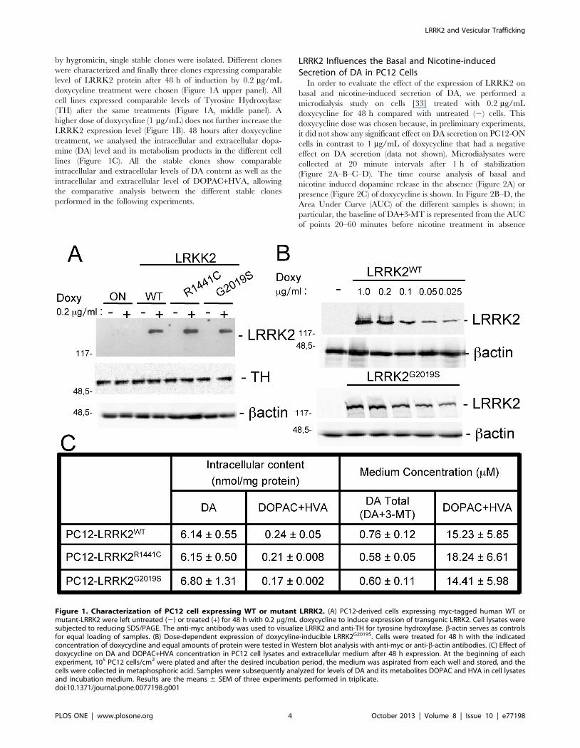
by hygromicin, single stable clones were isolated. Different clones
were characterized and finally three clones expressing comparable
level of LRRK2 protein after 48 h of induction by 0.2 mg/mL
doxycycline treatment were chosen (Figure 1A upper panel). All
cell lines expressed comparable levels of Tyrosine Hydroxylase
(TH) after the same treatments (Figure 1A, middle panel). A
higher dose of doxycycline (1 mg/mL) does not further increase the
LRRK2 expression level (Figure 1B). 48 hours after doxycycline
treatment, we analysed the intracellular and extracellular dopa-
mine (DA) level and its metabolism products in the different cell
lines (Figure 1C). All the stable clones show comparable
intracellular and extracellular levels of DA content as well as the
intracellular and extracellular level of DOPAC+HVA, allowing
the comparative analysis between the different stable clones
performed in the following experiments.
LRRK2 Influences the Basal and Nicotine-inducedSecretion of DA in PC12 Cells
In order to evaluate the effect of the expression of LRRK2 on
basal and nicotine-induced secretion of DA, we performed a
microdialysis study on cells [33] treated with 0.2 mg/mL
doxycycline for 48 h compared with untreated (2) cells. This
doxycycline dose was chosen because, in preliminary experiments,
it did not show any significant effect on DA secretion on PC12-ON
cells in contrast to 1 mg/mL of doxycycline that had a negative
effect on DA secretion (data not shown). Microdialysates were
collected at 20 minute intervals after 1 h of stabilization
(Figure 2A–B–C–D). The time course analysis of basal and
nicotine induced dopamine release in the absence (Figure 2A) or
presence (Figure 2C) of doxycycline is shown. In Figure 2B–D, the
Area Under Curve (AUC) of the different samples is shown; in
particular, the baseline of DA+3-MT is represented from the AUC
of points 20–60 minutes before nicotine treatment in absence
Figure 1. Characterization of PC12 cell expressing WT or mutant LRRK2. (A) PC12-derived cells expressing myc-tagged human WT ormutant-LRRK2 were left untreated (2) or treated (+) for 48 h with 0.2 mg/mL doxycycline to induce expression of transgenic LRRK2. Cell lysates weresubjected to reducing SDS/PAGE. The anti-myc antibody was used to visualize LRRK2 and anti-TH for tyrosine hydroxylase. b-actin serves as controlsfor equal loading of samples. (B) Dose-dependent expression of doxycyline-inducible LRRK2G2019S. Cells were treated for 48 h with the indicatedconcentration of doxycycline and equal amounts of protein were tested in Western blot analysis with anti-myc or anti-b-actin antibodies. (C) Effect ofdoxycycline on DA and DOPAC+HVA concentration in PC12 cell lysates and extracellular medium after 48 h expression. At the beginning of eachexperiment, 105 PC12 cells/cm2 were plated and after the desired incubation period, the medium was aspirated from each well and stored, and thecells were collected in metaphosphoric acid. Samples were subsequently analyzed for levels of DA and its metabolites DOPAC and HVA in cell lysatesand incubation medium. Results are the means 6 SEM of three experiments performed in triplicate.doi:10.1371/journal.pone.0077198.g001
LRRK2 and Vesicular Trafficking
PLOS ONE | www.plosone.org 4 October 2013 | Volume 8 | Issue 10 | e77198
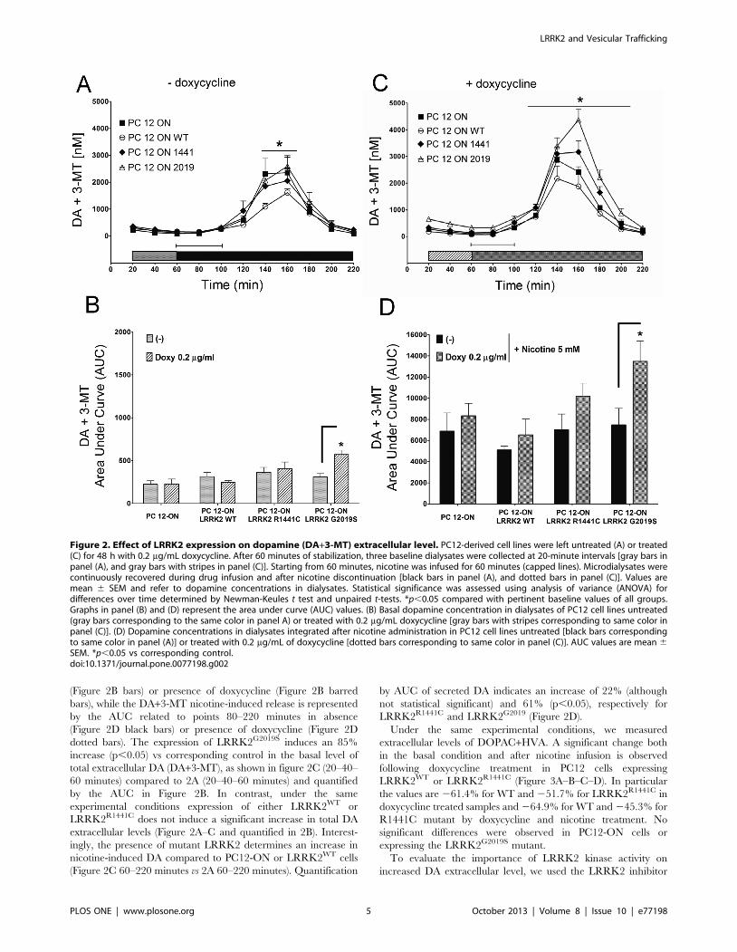
(Figure 2B bars) or presence of doxycycline (Figure 2B barred
bars), while the DA+3-MT nicotine-induced release is represented
by the AUC related to points 80–220 minutes in absence
(Figure 2D black bars) or presence of doxycycline (Figure 2D
dotted bars). The expression of LRRK2G2019S induces an 85%
increase (p,0.05) vs corresponding control in the basal level of
total extracellular DA (DA+3-MT), as shown in figure 2C (20–40–
60 minutes) compared to 2A (20–40–60 minutes) and quantified
by the AUC in Figure 2B. In contrast, under the same
experimental conditions expression of either LRRK2WT or
LRRK2R1441C does not induce a significant increase in total DA
extracellular levels (Figure 2A–C and quantified in 2B). Interest-
ingly, the presence of mutant LRRK2 determines an increase in
nicotine-induced DA compared to PC12-ON or LRRK2WT cells
(Figure 2C 60–220 minutes vs 2A 60–220 minutes). Quantification
by AUC of secreted DA indicates an increase of 22% (although
not statistical significant) and 61% (p,0.05), respectively for
LRRK2R1441C and LRRK2G2019 (Figure 2D).
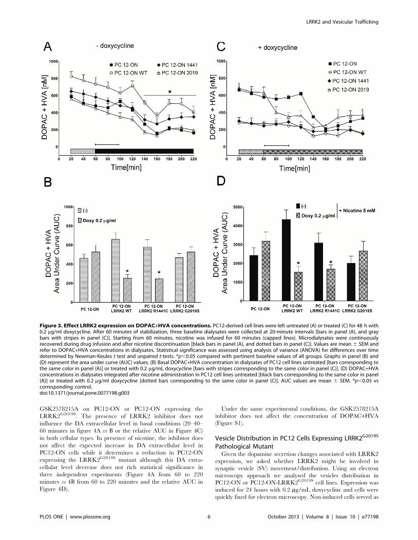
Under the same experimental conditions, we measured
extracellular levels of DOPAC+HVA. A significant change both
in the basal condition and after nicotine infusion is observed
following doxycycline treatment in PC12 cells expressing
LRRK2WT or LRRK2R1441C (Figure 3A–B–C–D). In particular
the values are 261.4% for WT and 251.7% for LRRK2R1441C in
doxycycline treated samples and 264.9% for WT and 245.3% for
R1441C mutant by doxycycline and nicotine treatment. No
significant differences were observed in PC12-ON cells or
expressing the LRRK2G2019S mutant.
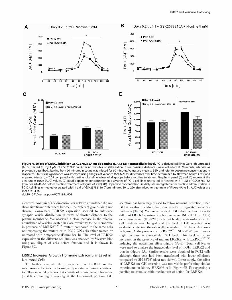
To evaluate the importance of LRRK2 kinase activity on
increased DA extracellular level, we used the LRRK2 inhibitor
Figure 2. Effect of LRRK2 expression on dopamine (DA+3-MT) extracellular level. PC12-derived cell lines were left untreated (A) or treated(C) for 48 h with 0.2 mg/mL doxycycline. After 60 minutes of stabilization, three baseline dialysates were collected at 20-minute intervals [gray bars inpanel (A), and gray bars with stripes in panel (C)]. Starting from 60 minutes, nicotine was infused for 60 minutes (capped lines). Microdialysates werecontinuously recovered during drug infusion and after nicotine discontinuation [black bars in panel (A), and dotted bars in panel (C)]. Values aremean 6 SEM and refer to dopamine concentrations in dialysates. Statistical significance was assessed using analysis of variance (ANOVA) fordifferences over time determined by Newman-Keules t test and unpaired t-tests. *p,0.05 compared with pertinent baseline values of all groups.Graphs in panel (B) and (D) represent the area under curve (AUC) values. (B) Basal dopamine concentration in dialysates of PC12 cell lines untreated(gray bars corresponding to the same color in panel A) or treated with 0.2 mg/mL doxycycline [gray bars with stripes corresponding to same color inpanel (C)]. (D) Dopamine concentrations in dialysates integrated after nicotine administration in PC12 cell lines untreated [black bars correspondingto same color in panel (A)] or treated with 0.2 mg/mL of doxycycline [dotted bars corresponding to same color in panel (C)]. AUC values are mean 6SEM. *p,0.05 vs corresponding control.doi:10.1371/journal.pone.0077198.g002
LRRK2 and Vesicular Trafficking
PLOS ONE | www.plosone.org 5 October 2013 | Volume 8 | Issue 10 | e77198
GSK2578215A on PC12-ON or PC12-ON expressing the
LRRK2G2019S. The presence of LRRK2 inhibitor does not
influence the DA extracellular level in basal conditions (20–40–
60 minutes in figure 4A vs B or the relative AUC in Figure 4C)
in both cellular types. In presence of nicotine, the inhibitor does
not affect the expected increase in DA extracellular level in
PC12-ON cells while it determines a reduction in PC12-ON
expressing the LRRK2G2019S mutant although this DA extra-
cellular level decrease does not rich statistical significance in
three independent experiments (Figure 4A from 60 to 220
minutes vs 4B from 60 to 220 minutes and the relative AUC in
Figure 4D).
Under the same experimental conditions, the GSK2578215A
inhibitor does not affect the concentration of DOPAC+HVA
(Figure S1).
Vesicle Distribution in PC12 Cells Expressing LRRK2G2019S
Pathological MutantGiven the dopamine secretion changes associated with LRRK2
expression, we asked whether LRRK2 might be involved in
synaptic vesicle (SV) movement/distribution. Using an electron
microscopy approach we analysed the vesicles distribution in
PC12-ON or PC12-ON-LRRK2G2019S cell lines. Expression was
induced for 24 hours with 0.2 mg/mL doxycycline and cells were
quickly fixed for electron microscopy. Non-induced cells served as
Figure 3. Effect LRRK2 expression on DOPAC+HVA concentrations. PC12-derived cell lines were left untreated (A) or treated (C) for 48 h with0.2 mg/ml doxycycline. After 60 minutes of stabilization, three baseline dialysates were collected at 20-minute intervals [bars in panel (A), and graybars with stripes in panel (C)]. Starting from 60 minutes, nicotine was infused for 60 minutes (capped lines). Microdialysates were continuouslyrecovered during drug infusion and after nicotine discontinuation [black bars in panel (A), and dotted bars in panel (C)]. Values are mean 6 SEM andrefer to DOPAC+HVA concentrations in dialysates. Statistical significance was assessed using analysis of variance (ANOVA) for differences over timedetermined by Newman-Keules t test and unpaired t-tests. *p,0.05 compared with pertinent baseline values of all groups. Graphs in panel (B) and(D) represent the area under curve (AUC) values. (B) Basal DOPAC+HVA concentration in dialysates of PC12 cell lines untreated [bars corresponding tothe same color in panel (A)] or treated with 0.2 mg/mL doxycycline [bars with stripes corresponding to the same color in panel (C)]. (D) DOPAC+HVAconcentrations in dialysates integrated after nicotine administration in PC12 cell lines untreated [black bars corresponding to the same color in panel(A)] or treated with 0.2 mg/ml doxycycline [dotted bars corresponding to the same color in panel (C)]. AUC values are mean 6 SEM. *p,0.05 vscorresponding control.doi:10.1371/journal.pone.0077198.g003
LRRK2 and Vesicular Trafficking
PLOS ONE | www.plosone.org 6 October 2013 | Volume 8 | Issue 10 | e77198
a control. Analysis of SV dimensions or relative abundance did not
show significant differences between the different groups (data not
shown). Conversely LRRK2 expression seemed to influence
synaptic vesicle distribution in terms of shorter distance to the
plasma membrane. We observed a clear increase in the relative
abundance of vesicles located in close proximity to the membrane
in presence of LRRK2G2019S mutant compared to the same cells
not expressing the mutant or to PC12 ON cells either treated or
untreated with doxycycline (Figure 5A–B). The level of LRRK2
expression in the different cell lines was analysed by Western blot
using an aliquot of cells before fixation and it is shown in
Figure 5C.
LRRK2 Increases Growth Hormone Extracellular Level inNeuronal Cells
To further evaluate the involvement of LRRK2 in the
mechanisms of vesicle trafficking we generated a plasmid construct
to follow secreted proteins that consists of mouse growth hormone
(mGH), containing a myc-tag at the C-terminal position. GH
secretion has been largely used to follow neuronal secretion, since
GH is localized predominantly in vesicles in regulated secretory
pathways [34,35]. We co-transfected mGH alone or together with
different LRRK2 constructs in both neuronal (SH-SY5Y or PC12)
or non-neuronal (HEK293) cells. 24 h after co-transfections the
cell medium was changed and the level of GH secretion was
evaluated collecting the extracellular medium 16 h later. As shown
in figure 6A, the presence of LRRK2WT in SH-SY5Y determines a
slight increase in extracellular GH level. This level is further
increased in the presence of mutant LRRK2, with LRRK2G2019S
inducing the maximum effect (Figure 6A–E). Total cell lysates
were used to analyse the intracellular level of mGH, LRRK2 and
b-actin (Figure 6A). Similar results were obtained in PC12 cells
although these cells had been transfected with lower efficiency
compared to SH-SY5Y (data not shown). Interestingly, the effect
of LRRK2 on GH secretion was not visible performing similar
experiments in kidney HEK293 cells (Figure 6B–E) suggesting a
possible neuronal-specific mechanism of action for LRRK2.
Figure 4. Effect of LRRK2 inhibitor GSK2578215A on dopamine (DA+3-MT) extracellular level. PC12-derived cell lines were left untreated(A) or treated (B) by 1 mM of GSK2578215A. After 60 minutes of stabilization, three baseline dialysates were collected at 20-minute intervals aspreviously described. Starting from 60 minutes, nicotine was infused for 60 minutes. Values are mean 6 SEM and refer to dopamine concentrations indialysates. Statistical significance was assessed using analysis of variance (ANOVA) for differences over time determined by Newman-Keules t test andunpaired t-tests. *p,0.05 compared with pertinent baseline values of all groups before nicotine treatment. Graphs in panel (C) and (D) represent thearea under curve (AUC) values. (C) Basal dopamine concentration in dialysates of PC12 cell lines untreated or treated with 1 mM of GSK2578215A(minutes 20–40–60 before nicotine treatment of Figure 4A vs B). (D) Dopamine concentrations in dialysates integrated after nicotine administration inPC12 cell lines untreated or treated with 1 mM of GSK2578215A (from minutes 80 to 220 after nicotine treatment of Figure 4A vs B). AUC values aremean 6 SEM.doi:10.1371/journal.pone.0077198.g004
LRRK2 and Vesicular Trafficking
PLOS ONE | www.plosone.org 7 October 2013 | Volume 8 | Issue 10 | e77198
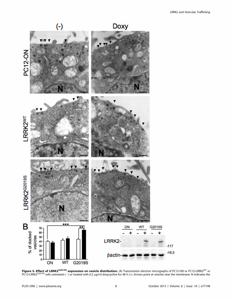
Figure 5. Effect of LRRK2G2019S expression on vesicle distribution. (A) Transmission electron micrographs of PC12-ON or PC12-LRRK2WT orPC12-LRRK2G2019S cells untreated (2) or treated with 0.2 mg/ml doxycycline for 48 h (+). Arrows point at vesicles near the membrane. N indicates the
LRRK2 and Vesicular Trafficking
PLOS ONE | www.plosone.org 8 October 2013 | Volume 8 | Issue 10 | e77198
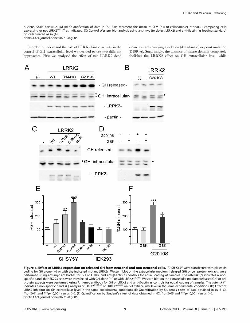
In order to understand the role of LRRK2 kinase activity in the
control of GH extracellular level we decided to use two different
approaches. First we analysed the effect of two LRRK2 dead
kinase mutants carrying a deletion (delta-kinase) or point mutation
(D1994A). Surprisingly, the absence of kinase domain completely
abolishes the LRRK2 effect on GH extracellular level, while
nucleus. Scale bars = 0,5 mM (B) Quantification of data in (A). Bars represent the mean 6 SEM (n = 30 cells/sample). **p,0.01 comparing cellsexpressing or not LRRK2G2019S as indicated. (C) Control Western blot analysis using anti-myc (to detect LRRK2) and anti-bactin (as loading standard)on cells treated as in (A).doi:10.1371/journal.pone.0077198.g005
Figure 6. Effect of LRRK2 expression on released GH from neuronal and non-neuronal cells. (A) SH-SY5Y were transfected with plasmidscoding for GH alone (2) or with the indicated mutant LRRK2s. Western blot on the extracellular medium (released GH) or cell protein extracts wereperformed using anti-myc antibodies for GH or LRRK2 and anti-b-actin as controls for equal loading of samples. The asterisk (*) indicates a non-specific band. (B) HEK293 cells were transfected with GH alone (2) or with LRRK2G2019S. Western blot on the extracellular medium (released GH) or cellprotein extracts were performed using Anti-myc antibody for GH or LRRK2 and anti-b-actin as controls for equal loading of samples. The asterisk (*)indicates a non-specific band. (C) Analysis of LRRK2D1994A or LRRK2Dkinase on GH extracellular level in the same experimental conditions. (D) Effect ofLRRK2 inhibitor on GH extracellular level in the same experimental conditions (E) Quantification by Student’s t test of data obtained in (A–B–C).**p,0,01 and ***p,0,001 versus (2). (F) Quantification by Student’s t test of data obtained in (D). *p,0,05 and ***p,0,001 versus (2).doi:10.1371/journal.pone.0077198.g006
LRRK2 and Vesicular Trafficking
PLOS ONE | www.plosone.org 9 October 2013 | Volume 8 | Issue 10 | e77198
LRRK2D1994A has an effect roughly comparable to LRRKWT
(Figure 6C). To further evaluate the importance of LRRK2 kinase
activity we used the LRRK2 inhibitor GSK2578215A. The
presence of LRRK2 inhibitor significantly reduces the increase in
GH extracellular level due to LRRK2G2019S expression
(Figure 6D–F).
LRRK2 Affects the Localization of Dopamine Receptor D1both in Neuronal Cells and Transgenic Mouse Tissues
Vesicle trafficking is a complex process regulating multiple
different cellular functions, including neurotransmitter or protein
release and localization of membrane receptors. The previous
results prompted us to analyse the possible effect of LRRK2 on
membrane receptor localization using the membrane levels of
dopamine receptor D1 (DRD1) as a read-out. DRD1is highly
expressed in the prefrontal cortex, striatum and nucleus accum-
bens, although it is absent in dopaminergic cells where Dopamine
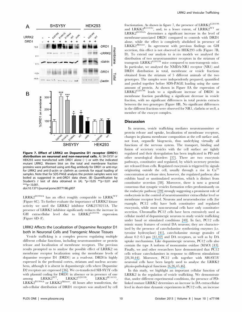
D2 receptors are expressed [36]. We co-transfected SH-SY5Y cells
with plasmid coding for DRD1 in absence or in presence of one
among LRKK2WT, LRKK2G2019S, LRKK2R1441G,
LRKK2D1994A or LRKK2Dkinase. 48 hours after transfection, the
sub-cellular distribution of DRD1 receptors was analysed by cell
fractionation. As shown in figure 7, the presence of LRRK2G2019S
and LRKK2R1441G, and, to a lesser extent, of LRRK2WT or
LRRK2D1994A determines a significant increase in the level of
membrane-associated DRD1 compared to controls with DRD1
alone, while the effect is completely abolished in presence of
LRKK2Dkinase. In agreement with previous findings on GH
secretion, this effect is not observed in HEK293 cells (Figure 7B,
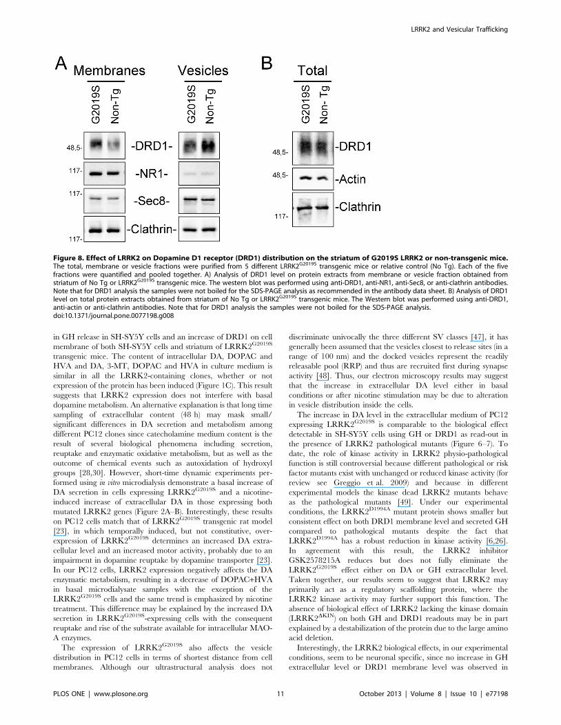
D). To extend our analysis to in vivo models we analysed the
distribution of two neurotransmitter receptors in the striatum of
transgenic LRRK2G2019S mice compared to non-transgenic mice.
In particular, we analysed the NMDA-NR1 receptor (NR1) and
DRD1 distribution in total, membrane or vesicle fractions
obtained from the striatum of 5 different animals of the two
genotypes. The samples were independently prepared, quantified
and pooled together before SDS-PAGE loading using the same
amount of protein. As shown in Figure 8A the expression of
LRRK2G2019S leads to a significant increase of DRD1 in
membrane fraction paralleling a significant decrease in vesicle
fraction, with no significant differences in total protein extracts
between the two genotypes (Figure 8B). No significant differences
in the different fractions were observed for NR1, clathrin or sec8, a
member of the exocyst complex.
Discussion
In neurons, vesicle trafficking mediates neurotransmitter or
protein release and uptake, localization of membrane receptors,
changes in plasma membrane composition at the cell surface and,
not least, organelle biogenesis, thus underlying virtually all
functions of the nervous system. The transport, binding and
fusion of secretory vesicles with the cell surface are tightly
regulated and their dysregulation has been implicated in PD and
other neurological disorders [37]. There are two exocytosis
pathways, constitutive and regulated, by which secretory proteins
are released from cells. Regulated exocytosis is triggered by signals
originating outside the cell, usually through a rise in Ca2+
concentration at release sites; however, the regulated pathway also
exhibits basal or unstimulated secretion, which is distinct from
constitutive secretion [38]. Moreover, there is now a general
consensus that synaptic vesicles formation relies predominantly on
the endocytic pathway [39] strongly suggesting a prominent role of
endocytosis in the control of neurotransmitter extracellular level or
membrane receptor level. Neurons and neuroendocrine cells (for
example, PC12 cells) have both constitutive and regulated
exocytosis, while most non-neuronal cells have only constitutive
secretion. Chromaffin PC12 cells have been extensively used as
cellular model of dopaminergic neurons to study vesicle trafficking
under basal or stimulated conditions [40]. In fact, PC12 cells
mimic many features of central DA neurons; they are character-
ized by the presence of catecholamine synthesizing enzymes (i.e.
tyrosine hydroxylase) [41], catecholamine storage granules of
about 0.2–0.5 mm [41,42] and DA receptors, as well as by DA
uptake mechanisms. Like dopaminergic neurons, PC12 cells also
contain the type A isoform of monoamine oxidase (MAO) [43].
Finally, we and other researchers have demonstrated that PC12
cells release catecholamines in response to different stimulations
[28,30,44]. Moreover, PC12 cells together with SH-SY5Y
neuronal cells have been largely used to analyse the LRRK2
physio-pathological functions [6,26,45,46].
In this study, we highlight an important cellular function of
LRRK2 in the regulation of vesicle trafficking. We demonstrate
that, under different experimental conditions, the presence of PD-
linked mutant LRRK2 determines an increase in DA extracellular
level in short-time dynamic experiments in PC12 cells, an increase
Figure 7. Effect of LRRK2 on Dopamine D1 receptor (DRD1)distribution on neuronal and non-neuronal cells. A) SH-SY5Y orHEK293 were transfected with DRD1 alone (2) or with the indicatedmutant LRRK2. Western blot on the total and membrane fractionproteins were performed using anti-flag antibody for DRD1 or anti-mycfor LRRK2 and anti-b-actin or clathrin as controls for equal loading ofsamples. Note that for SDS-PAGE analysis the protein samples were notboiled as suggested in anti-DRD1 data sheet. (B) Quantification byStudent’s t test of data obtained in (A). *p,0,05 **p,0,01 and***p,0,001.doi:10.1371/journal.pone.0077198.g007
LRRK2 and Vesicular Trafficking
PLOS ONE | www.plosone.org 10 October 2013 | Volume 8 | Issue 10 | e77198
in GH release in SH-SY5Y cells and an increase of DRD1 on cell
membrane of both SH-SY5Y cells and striatum of LRRK2G2019S
transgenic mice. The content of intracellular DA, DOPAC and
HVA and DA, 3-MT, DOPAC and HVA in culture medium is
similar in all the LRRK2-containing clones, whether or not
expression of the protein has been induced (Figure 1C). This result
suggests that LRRK2 expression does not interfere with basal
dopamine metabolism. An alternative explanation is that long time
sampling of extracellular content (48 h) may mask small/
significant differences in DA secretion and metabolism among
different PC12 clones since catecholamine medium content is the
result of several biological phenomena including secretion,
reuptake and enzymatic oxidative metabolism, but as well as the
outcome of chemical events such as autoxidation of hydroxyl
groups [28,30]. However, short-time dynamic experiments per-
formed using in vitro microdialysis demonstrate a basal increase of
DA secretion in cells expressing LRRK2G2019S and a nicotine-
induced increase of extracellular DA in those expressing both
mutated LRRK2 genes (Figure 2A–B). Interestingly, these results
on PC12 cells match that of LRRK2G2019S transgenic rat model
[23], in which temporally induced, but not constitutive, over-
expression of LRRK2G2019S determines an increased DA extra-
cellular level and an increased motor activity, probably due to an
impairment in dopamine reuptake by dopamine transporter [23].
In our PC12 cells, LRRK2 expression negatively affects the DA
enzymatic metabolism, resulting in a decrease of DOPAC+HVA
in basal microdialysate samples with the exception of the
LRRK2G2019S cells and the same trend is emphasized by nicotine
treatment. This difference may be explained by the increased DA
secretion in LRRK2G2019S-expressing cells with the consequent
reuptake and rise of the substrate available for intracellular MAO-
A enzymes.
The expression of LRRK2G2019S also affects the vesicle
distribution in PC12 cells in terms of shortest distance from cell
membranes. Although our ultrastructural analysis does not
discriminate univocally the three different SV classes [47], it has
generally been assumed that the vesicles closest to release sites (in a
range of 100 nm) and the docked vesicles represent the readily
releasable pool (RRP) and thus are recruited first during synapse
activity [48]. Thus, our electron microscopy results may suggest
that the increase in extracellular DA level either in basal
conditions or after nicotine stimulation may be due to alteration
in vesicle distribution inside the cells.
The increase in DA level in the extracellular medium of PC12
expressing LRRK2G2019S is comparable to the biological effect
detectable in SH-SY5Y cells using GH or DRD1 as read-out in
the presence of LRRK2 pathological mutants (Figure 6–7). To
date, the role of kinase activity in LRRK2 physio-pathological
function is still controversial because different pathological or risk
factor mutants exist with unchanged or reduced kinase activity (for
review see Greggio et al. 2009) and because in different
experimental models the kinase dead LRRK2 mutants behave
as the pathological mutants [49]. Under our experimental
conditions, the LRRK2D1994A mutant protein shows smaller but
consistent effect on both DRD1 membrane level and secreted GH
compared to pathological mutants despite the fact that
LRRK2D1994A has a robust reduction in kinase activity [6,26].
In agreement with this result, the LRRK2 inhibitor
GSK2578215A reduces but does not fully eliminate the
LRRK2G2019S effect either on DA or GH extracellular level.
Taken together, our results seem to suggest that LRRK2 may
primarily act as a regulatory scaffolding protein, where the
LRRK2 kinase activity may further support this function. The
absence of biological effect of LRRK2 lacking the kinase domain
(LRRK2DKIN) on both GH and DRD1 readouts may be in part
explained by a destabilization of the protein due to the large amino
acid deletion.
Interestingly, the LRRK2 biological effects, in our experimental
conditions, seem to be neuronal specific, since no increase in GH
extracellular level or DRD1 membrane level was observed in
Figure 8. Effect of LRRK2 on Dopamine D1 receptor (DRD1) distribution on the striatum of G2019S LRRK2 or non-transgenic mice.The total, membrane or vesicle fractions were purified from 5 different LRRK2G2019S transgenic mice or relative control (No Tg). Each of the fivefractions were quantified and pooled together. A) Analysis of DRD1 level on protein extracts from membrane or vesicle fraction obtained fromstriatum of No Tg or LRRK2G2019S transgenic mice. The western blot was performed using anti-DRD1, anti-NR1, anti-Sec8, or anti-clathrin antibodies.Note that for DRD1 analysis the samples were not boiled for the SDS-PAGE analysis as recommended in the antibody data sheet. B) Analysis of DRD1level on total protein extracts obtained from striatum of No Tg or LRRK2G2019S transgenic mice. The Western blot was performed using anti-DRD1,anti-actin or anti-clathrin antibodies. Note that for DRD1 analysis the samples were not boiled for the SDS-PAGE analysis.doi:10.1371/journal.pone.0077198.g008
LRRK2 and Vesicular Trafficking
PLOS ONE | www.plosone.org 11 October 2013 | Volume 8 | Issue 10 | e77198
HEK293 cell when mutant LRRK2 is present. Neuronal-specific
mechanisms of vesicle trafficking are well described [38] and this
aspect could explain at least partially the neuron specific toxicity of
mutant LRRK2. This view is supported by experiments in vivo,
showing that the LRRK2 effects on DRD1 localization in
neuronal cells is observed in the striatum of G2019S transgenic
mice. The transgene expression determines an increase of DRD1
in the plasma membrane fraction that correlates with a decrease in
the vesicle fraction. This effect is possibly specific for this receptor
since it is not detectable for NMDA-NR1 receptor. It is generally
accepted that membrane receptors show different turnover rate
and different endocytic pathways. For instance, NMDARs are
considered to be relatively stable once delivered to the cell surface
of mature neurons, especially when compared with AMPARs that,
even under basal conditions, cycle much more rapidly to and from
the surface [50]. On the other hand, DRD1s internalize rapidly
following agonist-induced activation [51]. Thus, the LRRK2
protein does not seem to exert a general control in secretion but
rather accurately modulates the vesicle turnover.
The amount of secreted neurotransmitter or plasma membrane
protein at steady state level is the sum of the transport reaching the
cell surface from biosynthetic pathways, the leaving of the surface
via the endocytic pathway, and the returning to plasma membrane
from the intracellular endosomal pools; consequently, a change in
any of these parameters could be responsible for the differences
that we observe in DA or GH release and DRD1 membrane
localization. For instance, LRRK2 has been implicated to different
extents in vesicle trafficking either directly throughout endocytosis
regulation [24,25,49] or indirectly throughout interaction with
actin or tubulin [12,52] that in turn may affect vesicle movement
among the cytoskeleton. The dysregulation on vesicle trafficking
may have important consequences on neuronal toxicity. It is
believed that inter-related processes like oxidative stress, excito-
toxicity, inflammation, mitochondrial dysfunction and altered
proteolysis are involved in the cascade of events that will lead to
DA neuron death [53]. In this respect, the increase in
neurotransmitter levels in the inter-synaptic space or the increase
in membrane receptors in neuronal plasma membrane due to the
expression of mutant LRRK2 could alter the normal neuronal
physiology that in turn may lead to neuronal toxicity. In this
respect, the increased neurotransmitter levels in the inter-synaptic
space or the increase in membrane receptors in neuronal plasma
membrane due to the expression of mutant LRRK2 could alter
the normal neuronal physiology that in turn may lead to neuronal
toxicity. For instance, a dysregulation in DA level has been
extensively studied as prominent PD etiological factor for the
neurotoxic properties of DA metabolites [54,55].
Supporting Information
Figure S1 Effect of LRRK2 inhibitor GSK2578215A onDOPAC+HVA concentrations. PC12-derived cell lines were
left untreated (A) or treated (B) with 1 mM of GSK2578215A.
After 60 minutes of stabilization, three baseline dialysates were
collected at 20-minute intervals as previously described. Starting
from 60 minutes, nicotine was infused for 60 minutes. Micro-
dialysates were continuously recovered during drug infusion and
after nicotine discontinuation. Values are mean 6 SEM and refer
to DOPAC+HVA concentrations in dialysates. Statistical signif-
icance was assessed using analysis of variance (ANOVA) for
differences over time determined by Newman-Keules t test and
unpaired t-tests. *p,0.05 compared with pertinent baseline values
of all groups before nicotine treatment. Graphs in panel (C) and
(D) represent the area under curve (AUC) values. (C) Basal
DOPAC+HVA concentrations in dialysates of PC12 cell lines
untreated or treated with 1 mM of GSK2578215A (minutes 20–
40–60 before nicotine treatment of Figure S1A vs B). (D)
DOPAC+HVA in dialysates integrated after nicotine administra-
tion in PC12 cell lines untreated or treated with 1 mM of
GSK2578215A (from minutes 80 to 220 after nicotine treatment
of Figure S1A vs B). AUC values are mean 6 SEM.
(TIF)
Acknowledgments
We would like to acknowledge all the people from the laboratory that
critically read the manuscript. Thanks are due to Salvatore Marceddu for
Electron Microscopy analysis and Giustina Casu for proofreading the
manuscript for English language.
This paper is dedicated to Prof. Giuseppe Rotilio on the occasion of his
72nd birthday.
Author Contributions
Conceived and designed the experiments: CI CC RM PAS. Performed the
experiments: RM MGDG YS GS YX MG GR AB. Analyzed the data: CI
CC RM PAS. Contributed reagents/materials/analysis tools: TMD VLD
MTC. Wrote the paper: CI CC PAS RM MTC.
References
1. Biskup S, Gerlach M, Kupsch A, Reichmann H, Riederer P, et al. (2008) Genes
associated with Parkinson syndrome. J Neurol 255 Suppl 5: 8–17.
2. Paisan-Ruiz C, Jain S, Evans EW, Gilks WP, Simon J, et al. (2004) Cloning of
the gene containing mutations that cause PARK8-linked Parkinson’s disease.
Neuron 44: 595–600.
3. Zimprich A, Biskup S, Leitner P, Lichtner P, Farrer M, et al. (2004) Mutations in
LRRK2 cause autosomal-dominant parkinsonism with pleomorphic pathology.
Neuron 44: 601–607.
4. Marin I, van Egmond WN, van Haastert PJ (2008) The Roco protein family: a
functional perspective. Faseb J 22: 3103–3110.
5. Healy DG, Falchi M, O’Sullivan SS, Bonifati V, Durr A, et al. (2008)
Phenotype, genotype, and worldwide genetic penetrance of LRRK2-associated
Parkinson’s disease: a case-control study. Lancet Neurol 7: 583–590.
6. Smith WW, Pei Z, Jiang H, Dawson VL, Dawson TM, et al. (2006) Kinase
activity of mutant LRRK2 mediates neuronal toxicity. Nat Neurosci 9: 1231–
1233.
7. West AB, Moore DJ, Biskup S, Bugayenko A, Smith WW, et al. (2005)
Parkinson’s disease-associated mutations in leucine-rich repeat kinase 2 augment
kinase activity. Proc Natl Acad Sci U S A 102: 16842–16847.
8. Greggio E, Cookson MR (2009) Leucine-rich repeat kinase 2 mutations and
Parkinson’s disease: three questions. ASN Neuro 1.
9. Bonifati V (2006) The pleomorphic pathology of inherited Parkinson’s disease:
lessons from LRRK2. Curr Neurol Neurosci Rep 6: 355–357.
10. Gehrke S, Imai Y, Sokol N, Lu B (2010) Pathogenic LRRK2 negatively regulates
microRNA-mediated translational repression. Nature 466: 637–641.
11. Imai Y, Gehrke S, Wang HQ, Takahashi R, Hasegawa K, et al. (2008)
Phosphorylation of 4E-BP by LRRK2 affects the maintenance of dopaminergic
neurons in Drosophila. Embo J 27: 2432–2443.
12. Parisiadou L, Xie C, Cho HJ, Lin X, Gu XL, et al. (2009) Phosphorylation of
ezrin/radixin/moesin proteins by LRRK2 promotes the rearrangement of actin
cytoskeleton in neuronal morphogenesis. J Neurosci 29: 13971–13980.
13. Gillardon F (2009) Leucine-rich repeat kinase 2 phosphorylates brain tubulin-
beta isoforms and modulates microtubule stability–a point of convergence in
parkinsonian neurodegeneration? J Neurochem 110: 1514–1522.
14. Melrose HL, Dachsel JC, Behrouz B, Lincoln SJ, Yue M, et al. (2010) Impaired
dopaminergic neurotransmission and microtubule-associated protein tau
alterations in human LRRK2 transgenic mice. Neurobiol Dis 40: 503–517.
15. Tong Y, Giaime E, Yamaguchi H, Ichimura T, Liu Y, et al. (2012) Loss of
leucine-rich repeat kinase 2 causes age-dependent bi-phasic alterations of the
autophagy pathway. Mol Neurodegener 7: 2.
16. Ramonet D, Daher JP, Lin BM, Stafa K, Kim J, et al. (2011) Dopaminergic
neuronal loss, reduced neurite complexity and autophagic abnormalities in
transgenic mice expressing G2019S mutant LRRK2. PLoS One 6: e18568.
17. Liu Z, Lee J, Krummey S, Lu W, Cai H, et al. (2011) The kinase LRRK2 is a
regulator of the transcription factor NFAT that modulates the severity of
inflammatory bowel disease. Nat Immunol 12: 1063–1070.
LRRK2 and Vesicular Trafficking
PLOS ONE | www.plosone.org 12 October 2013 | Volume 8 | Issue 10 | e77198
18. Sanna G, Del Giudice MG, Crosio C, Iaccarino C (2012) LRRK2 and vesicle
trafficking. Biochem Soc Trans 40: 1117–1122.19. Biskup S, Moore DJ, Celsi F, Higashi S, West AB, et al. (2006) Localization of
LRRK2 to membranous and vesicular structures in mammalian brain. Ann
Neurol 60: 557–569.20. Hatano T, Kubo S, Imai S, Maeda M, Ishikawa K, et al. (2007) Leucine-rich
repeat kinase 2 associates with lipid rafts. Hum Mol Genet 16: 678–690.21. Li Y, Liu W, Oo TF, Wang L, Tang Y, et al. (2009) Mutant LRRK2(R1441G)
BAC transgenic mice recapitulate cardinal features of Parkinson’s disease. Nat
Neurosci 12: 826–828.22. Li X, Patel JC, Wang J, Avshalumov MV, Nicholson C, et al. (2010) Enhanced
striatal dopamine transmission and motor performance with LRRK2 overex-pression in mice is eliminated by familial Parkinson’s disease mutation G2019S.
J Neurosci 30: 1788–1797.23. Zhou H, Huang C, Tong J, Hong WC, Liu YJ, et al. (2011) Temporal
expression of mutant LRRK2 in adult rats impairs dopamine reuptake. Int J Biol
Sci 7: 753–761.24. Piccoli G, Condliffe SB, Bauer M, Giesert F, Boldt K, et al. (2011) LRRK2
controls synaptic vesicle storage and mobilization within the recycling pool.J Neurosci 31: 2225–2237.
25. Shin N, Jeong H, Kwon J, Heo HY, Kwon JJ, et al. (2008) LRRK2 regulates
synaptic vesicle endocytosis. Exp Cell Res 314: 2055–2065.26. Iaccarino C, Crosio C, Vitale C, Sanna G, Carri MT, et al. (2007) Apoptotic
mechanisms in mutant LRRK2-mediated cell death. Hum Mol Genet 16: 1319–1326.
27. Migheli R, Godani C, Sciola L, Delogu MR, Serra PA, et al. (1999) Enhancingeffect of manganese on L-DOPA-induced apoptosis in PC12 cells: role of
oxidative stress. J Neurochem 73: 1155–1163.
28. Serra PA, Migheli R, Rocchitta G, Taras MG, Mura MP, et al. (2003) Role ofthe nitric oxide/cyclic GMP pathway and ascorbic acid in 3-morpholinosydno-
nimine (SIN-1)-induced increases in dopamine secretion from PC12 cells. Amicrodialysis in vitro study. Neurosci Lett 353: 5–8.
29. Migheli R, Puggioni G, Dedola S, Rocchitta G, Calia G, et al. (2008) Novel
integrated microdialysis-amperometric system for in vitro detection of dopaminesecreted from PC12 cells: design, construction, and validation. Anal Biochem
380: 323–330.30. Serra PA, Rocchitta G, Delogu MR, Migheli R, Taras MG, et al. (2003) Role of
the nitric oxide/cyclic GMP pathway and extracellular environment in the nitricoxide donor-induced increase in dopamine secretion from PC12 cells: a
microdialysis in vitro study. J Neurochem 86: 1403–1413.
31. Xiong Y, Yuan C, Chen R, Dawson TM, Dawson VL (2012) ArfGAP1 Is aGTPase Activating Protein for LRRK2: Reciprocal Regulation of ArfGAP1 by
LRRK2. J Neurosci 32: 3877–3886.32. Iaccarino C, Mura ME, Esposito S, Carta F, Sanna G, et al. (2011) Bcl2-A1
interacts with pro-caspase-3: implications for amyotrophic lateral sclerosis.
Neurobiol Dis 43: 642–650.33. Bradberry CW, Sprouse JS, Sheldon PW, Aghajanian GK, Roth RH (1991) In
vitro microdialysis: a novel technique for stimulated neurotransmitter releasemeasurements. J Neurosci Methods 36: 85–90.
34. Wick PF, Senter RA, Parsels LA, Uhler MD, Holz RW (1993) Transienttransfection studies of secretion in bovine chromaffin cells and PC12 cells.
Generation of kainate-sensitive chromaffin cells. J Biol Chem 268: 10983–
10989.
35. Lee HW, Park JW, Sandagsuren EU, Kim KB, Yoo JJ, et al. (2008)
Overexpression of APP stimulates basal and constitutive exocytosis in PC12
cells. Neurosci Lett 436: 245–249.
36. Missale C, Nash SR, Robinson SW, Jaber M, Caron MG (1998) Dopamine
receptors: from structure to function. Physiol Rev 78: 189–225.
37. Esposito G, Ana Clara F, Verstreken P (2012) Synaptic vesicle trafficking and
Parkinson’s disease. Dev Neurobiol 72: 134–144.
38. Ghijsen WE, Leenders AG (2005) Differential signaling in presynaptic
neurotransmitter release. Cell Mol Life Sci 62: 937–954.
39. Buckley KM, Melikian HE, Provoda CJ, Waring MT (2000) Regulation of
neuronal function by protein trafficking: a role for the endosomal pathway.
J Physiol 525 Pt 1: 11–19.
40. Martin TF, Grishanin RN (2003) PC12 cells as a model for studies of regulated
secretion in neuronal and endocrine cells. Methods Cell Biol 71: 267–286.
41. Markey KA, Kondo H, Shenkman L, Goldstein M (1980) Purification and
characterization of tyrosine hydroxylase from a clonal pheochromocytoma cell
line. Mol Pharmacol 17: 79–85.
42. Roda LG, Nolan JA, Kim SU, Hogue-Angeletti RA (1980) Isolation and
characterization of chromaffin granules from a pheochromocytoma (PC 12) cell
line. Exp Cell Res 128: 103–109.
43. Finberg JP, Youdim MB (1983) Selective MAO A and B inhibitors: their
mechanism of action and pharmacology. Neuropharmacology 22: 441–446.
44. Fujita K, Lazarovici P, Guroff G (1989) Regulation of the differentiation of
PC12 pheochromocytoma cells. Environ Health Perspect 80: 127–142.
45. Greggio E, Jain S, Kingsbury A, Bandopadhyay R, Lewis P, et al. (2006) Kinase
activity is required for the toxic effects of mutant LRRK2/dardarin. Neurobiol
Dis 23: 329–341.
46. Heo HY, Kim KS, Seol W (2010) Coordinate Regulation of Neurite Outgrowth
by LRRK2 and Its Interactor, Rab5. Exp Neurobiol 19: 97–105.
47. Rizzoli SO, Betz WJ (2005) Synaptic vesicle pools. Nat Rev Neurosci 6: 57–69.
48. Rizzoli SO, Betz WJ (2004) The structural organization of the readily releasable
pool of synaptic vesicles. Science 303: 2037–2039.
49. Matta S, Van Kolen K, da Cunha R, van den Bogaart G, Mandemakers W, et
al. (2012) LRRK2 Controls an EndoA Phosphorylation Cycle in Synaptic
Endocytosis. Neuron 75: 1008–1021.
50. Groc L, Choquet D (2006) AMPA and NMDA glutamate receptor trafficking:
multiple roads for reaching and leaving the synapse. Cell Tissue Res 326: 423–
438.
51. Kotowski SJ, Hopf FW, Seif T, Bonci A, von Zastrow M (2011) Endocytosis
promotes rapid dopaminergic signaling. Neuron 71: 278–290.
52. Kawakami F, Yabata T, Ohta E, Maekawa T, Shimada N, et al. (2012) LRRK2
phosphorylates tubulin-associated tau but not the free molecule: LRRK2-
mediated regulation of the tau-tubulin association and neurite outgrowth. PLoS
One 7: e30834.
53. Schapira AH, Jenner P (2011) Etiology and pathogenesis of Parkinson’s disease.
Mov Disord 26: 1049–1055.
54. Jiang Y, Pei L, Li S, Wang M, Liu F (2008) Extracellular dopamine induces the
oxidative toxicity of SH-SY5Y cells. Synapse 62: 797–803.
55. Stokes AH, Lewis DY, Lash LH, Jerome WG 3rd, Grant KW, et al. (2000)
Dopamine toxicity in neuroblastoma cells: role of glutathione depletion by L-
BSO and apoptosis. Brain Res 858: 1–8.
LRRK2 and Vesicular Trafficking
PLOS ONE | www.plosone.org 13 October 2013 | Volume 8 | Issue 10 | e77198