membrane trafficking and vesicle fusion - indian academy of
TRANSCRIPT
421RESONANCE May 2014
GENERAL ARTICLE
Keywords
Organelle, membrane fusion,
vesicular transport, SNARE, Sec
mutants, neurotransmission.
Membrane Trafficking and Vesicle Fusion
Post-Palade EraResearchers Win theNobel Prize
Riddhi Atul Jani and Subba Rao Gangi Setty
The functions of the eukaryotic cell rely on membrane- bound
compartments called organelles. Each of these possesses dis-
tinct membrane composition and unique function. In the
1970’s, during George Palade’s time, it was unclear how these
organelles communicate with each other and perform their
biological functions. The elegant research work of James E
Rothman, Randy W Schekman and Thomas C Südhof identi-
fied the molecular machinery required for membrane traf-
ficking, vesicle fusion and cargo delivery. Further, they also
showed the importance of these processes for biological func-
tion. Their novel findings helped to explain several biological
phenomena such as insulin secretion, neuron communication
and other cellular activities. In addition, their work provided
clues to cures for several neurological, immunological and
metabolic disorders. This research work laid the foundation
to the field of molecular cell biology and these post-Palade
investigators were awarded the Nobel Prize in Physiology or
Medicine in 2013.
Introduction
Membrane Transport: In the eukaryotic cell, a majority of
proteins are made in the cytosol. But the transmembrane and
secretory proteins are synthesized in an organelle called the rough
endoplasmic reticulum (ER). These proteins undergo certain
chemical modifications such as glycosylation, disulfide formation,
etc., either to attain their proper folding or to modify the protease
cleavage sites. Further, these proteins traverse from the ER to the
Golgi, and then to different cellular organelles (including theplasma
membrane, PM) or are secreted out of the cell. By this process, the
newly synthesized secretory proteins in the ER are transported to their
(left) Riddhi Atul Jani is a
graduate student in Subba
Rao’s Lab at MCB, IISc.
She is interested in
studying the SNARE
dynamics during melano-
some biogenesis.
(right) Subba Rao Gangi
Setty is an Assistant
Professor at the MCB,
IISc, Bangalore. He is
interested in understand-
ing the disease associated
protein trafficking
pathways in mammalian
cells.
422 RESONANCE May 2014
GENERAL ARTICLE
respective target sites for their proper functioning. This movement of
proteins is referred to as ‘protein transport or trafficking’. In addition,
cells also utilize the membranes or vesicles to perform this process,
whichis known as ‘membrane trafficking’or ‘vesicular transport’ [1].
Vesicle Fusion and Neurotransmission: In a cell, the newly
synthesized proteins transport from one place to another in the
form of a transport carrier called a ‘vesicle’. These vesicles are
formed from the donor membrane by a process called ‘vesicle
budding’. They are directed towards the target membrane and
undergo ‘vesicle fusion’ [1]. In addition, vesicle biogenesis and
fusion require coat proteins and a specific fusion machinery
respectively for accurate transport. This process was initially
discovered in neuronal cells, where synaptic vesicles deliver their
neurotransmitters through the PM in a synaptic cleft for the
activation of other neurons. This process is called ‘neurotransmis-
sion’[2].
The cellular components and the mechanism of regulation for the
above processes which is the focus of this article were discovered
in the laboratories of James E Rothman, Randy W Schekman and
Thomas C Südhof.
Cell Biology in George Palade’s Era
In the 1930s and 1940s, biology was more focused on the struc-
ture and function of DNA and RNA using bacteria as a model
system. Very little was known about the structure of the eukary-
otic cell. For the first time, in the early 1930s, Albert Claude at
Rockefeller University described the ultra-structural details of
the majority of the cellular organelles such as mitochondria,
chloroplasts, endoplasmic reticulum, Golgi apparatus and ribo-
somes (except endosomes). In his discoveries, Claude used a
biochemical method called ‘cell fractionation1’ developed in his
laboratory on liver cell lysates, to distinguish the different com-
ponents of the eukaryotic cell [3]. In addition, Claude applied
electron microscopy (EM) techniques2 to analyze the fractions
separated by the above method. He revolutionized the EM
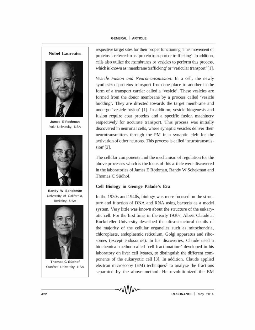
Nobel Laureates
James E Rothman
Yale University, USA
Randy W Schekman
University of California,
Berkeley, USA
Thomas C Südhof
Stanford University, USA
423RESONANCE May 2014
GENERAL ARTICLE
technique by treating the samples with osmium tetroxide, which
preserved the cell membrane and increased the contrast of
electron micrographs. Claude published the detailed structure of
a cell for the first time in 1945 [4] in which he provided an insight
into the structural organization of the eukaryotic cell.
During the course of time, Albert Claude’s friend, Christian de
Duve of Rockefeller University, described two other eukaryotic
organelles. de Duve’s laboratory discovered the lysosomes while
analyzing the activity of enzymes distributed in different frac-
tions obtained through rate-zonal centrifugation3 of liver cell
lysates. In addition, de Duve applied EM technique on these
fractions and described the ultra-structure of the lysosome. Simi-
larly, de Duve’s laboratory identified the peroxisomes while
studying the activity of an enzyme, urate oxidase [5]. Together, the
studies from Claude and de Duve’s laboratories illustrated the
structure and organization of cellular compartments of the eukary-
otic cell and these discoveries initiated the field of cell biology [6,
7]. But it was not clear how these organelles are generated and
how they communicate with each other.
These questions motivated George E Palade while he was work-
ing in Claude’s laboratory as a postdoc. In his own laboratory at
Rockefeller University, Palade carried out integrated morpho-
logical and biochemical analysis of different organelles using
pancreatic exocrine cells of the guinea pig. He also studied the
structure of several pancreatic secretory proteins using EM and
cell fractionation procedures. These studies illustrated the ultra-
structure of the cell and, most significantly, Palade showed for the
first time that the cytoplasm is packed with stacks of ER studded
with ribosomes. This observation prompted Palade to investigate
the role of ribosomes on the ER membrane and other subcellular
organelles. Later studies from his laboratory showed that ribo-
somes synthesize the secretory and transmembrane proteins on
the ER and translocate them into the ER lumen for further
transport [8]. But, it was unknown how these proteins are secreted
out of the cell.
1 Cell fractionation is a method
commonly used to separate the
cell lysate into individual frac-
tions containing different or-
ganelles or membranes. In this
method, cells are lysed in a non-
detergent lysis buffer which will
keep the cell membranes in a
native state and then separated
by density gradient centrifuga-
tion (separation of molecules
based on density).
2 Electron microscopy is a tech-
nique used to reveal the ultra-
structure of cells (both prokary-
otic and eukaryotic), mem-
branes, organelles, materials,
etc. This method uses an elec-
tron beam as a source for illumi-
nating the sample, which will
provide an image with 200 times
higher resolution than light mi-
croscopy. There are two types:
transmission electron micros-
copy, widely used to study the
structure of organelles or mem-
branes and scanning electron
microscopy, used to study sur-
face organization.
3 Rate-zonal centrifugation is a
type of density gradient cen-
trifugation used to separate the
organelles or molecules based
on size and mass instead of
density. This method has the
advantage that the particles with
faster sedimenting rates are not
contaminated with the slower
sedimenting particles as would
occur in the differential (density
gradient) centrifugation method.

424 RESONANCE May 2014
GENERAL ARTICLE
It was a difficult question in those days since the available
techniques were not adequate for studying such processes. So
Palade developed a novel method called ‘pulse-chase analysis’4
and used it to study the protein secretion process in pancreatic
exocrine cells. This technique revolutionized the field and Palade
was able to describe the transport of secretory proteins in pancre-
atic cells [8]. Based on several discoveries from his laboratory,
Palade proposed a model for the cell secretory process in 1974,
involving six successive steps or operations (Figure 1): (1) biosyn-
thesis of new proteins; (2) segregation of proteins on the ER
membrane; (3) intracellular transport between the compartments;
(4) concentration of protein in a new organelle; (5) intracellular
storage in organelles such as lysosomes, secretory granules and
(6) discharge of contents through the PM or to endocytic vacuoles.
This was the first description of the process of protein transport
between cellular organelles. Palade’s laboratory worked exten-
sively on each of thesesix steps, and proposed hypothetical models
for intracellular transport and discharge [8] which will be the focus
of the remaining article.
Intracellular Transport
By using the pulse chase method, Palade’s laboratory showed that
the newly synthesized glycoprotein or secretory protein in the ER
Figure 1.The key steps of the secretory process in
various cell types as described by Palade.
This model was proposed by Palade in his Nobel
Lecture based on the knowledge available at that time
including the research findings from his own labora-
tory on pancreatic exocrine cells. Exceptions to this
model that occur in granulocytes and macrophages
are shown in the figure. He proposed that discharge in
fibroblasts is likely to take place continuously, whereas
it is preferentially through endocytic vacuoles in neu-
trophils, eosinophil granulocytes, and macrophages
[8].
4 Pulse-chase analysis is a
method to study the cellular pro-
cesses over time. Cells are
pulsed with radio-labeled amino
acids, which will incorporate into
the newly synthesized proteins.
The lysates made from these
cells are used to measure the
half-life or turnover rate of any
protein and the data can be cor-
related with cellular processes.
425RESONANCE May 2014
GENERAL ARTICLE
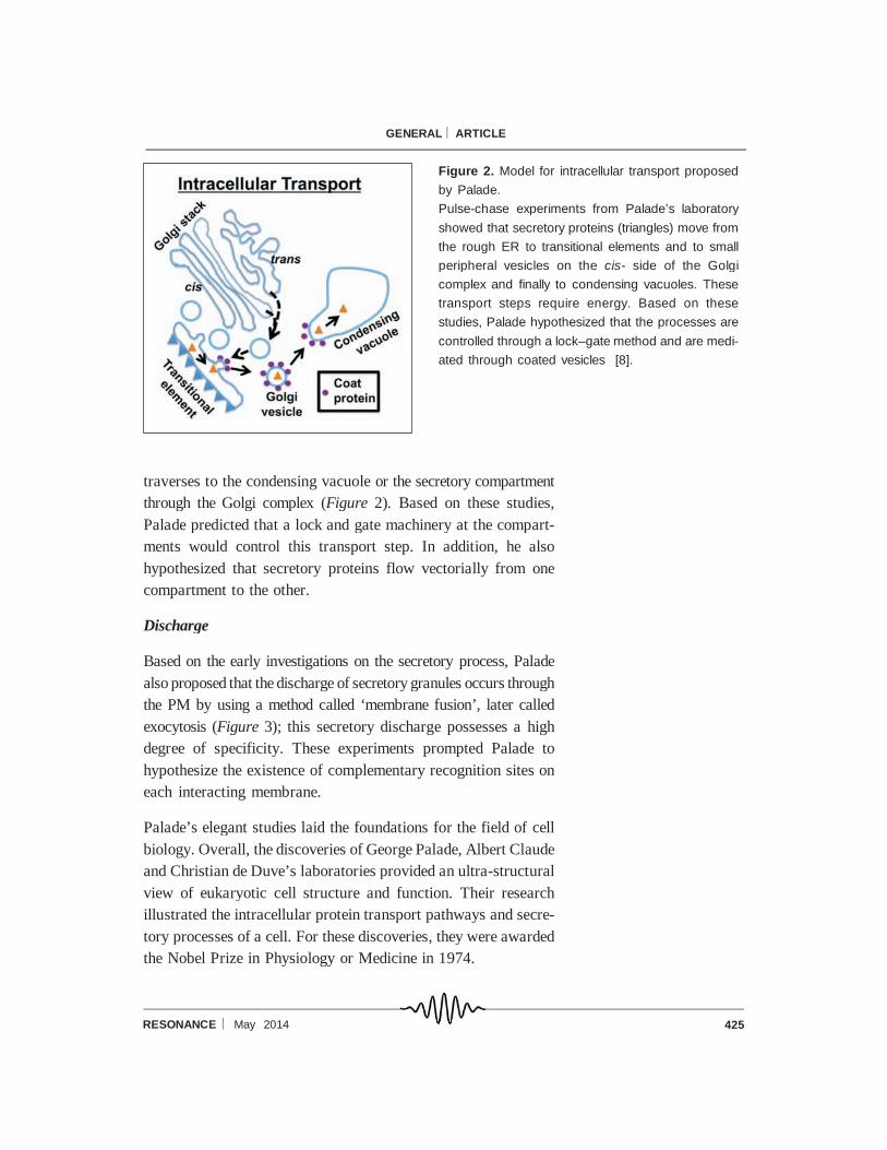
traverses to the condensing vacuole or the secretory compartment
through the Golgi complex (Figure 2). Based on these studies,
Palade predicted that a lock and gate machinery at the compart-
ments would control this transport step. In addition, he also
hypothesized that secretory proteins flow vectorially from one
compartment to the other.
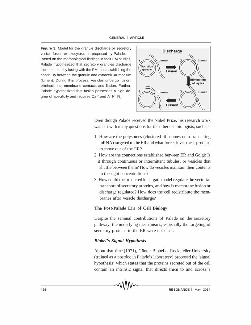
Discharge
Based on the early investigations on the secretory process, Palade
also proposed that the discharge of secretory granules occurs through
the PM by using a method called ‘membrane fusion’, later called
exocytosis (Figure 3); this secretory discharge possesses a high
degree of specificity. These experiments prompted Palade to
hypothesize the existence of complementary recognition sites on
each interacting membrane.
Palade’s elegant studies laid the foundations for the field of cell
biology. Overall, the discoveries of George Palade, Albert Claude
and Christian de Duve’s laboratories provided an ultra-structural
view of eukaryotic cell structure and function. Their research
illustrated the intracellular protein transport pathways and secre-
tory processes of a cell. For these discoveries, they were awarded
the Nobel Prize in Physiology or Medicine in 1974.
Figure 2. Model for intracellular transport proposed
by Palade.
Pulse-chase experiments from Palade’s laboratory
showed that secretory proteins (triangles) move from
the rough ER to transitional elements and to small
peripheral vesicles on the cis- side of the Golgi
complex and finally to condensing vacuoles. These
transport steps require energy. Based on these
studies, Palade hypothesized that the processes are
controlled through a lock–gate method and are medi-
ated through coated vesicles [8].
426 RESONANCE May 2014
GENERAL ARTICLE
Even though Palade received the Nobel Prize, his research work
was left with many questions for the other cell biologists, such as:
1. How are the polysomes (clustered ribosomes on a translating
mRNA) targeted to the ER and what force drives these proteins
to move out of the ER?
2. How are the connections established between ER and Golgi: Is
it through continuous or intermittent tubules, or vesicles that
shuttle between them? How do vesicles maintain their contents
in the right concentrations?
3. How could the predicted lock–gate model regulate the vectorial
transport of secretory proteins, and how is membrane fusion or
discharge regulated? How does the cell redistribute the mem-
branes after vesicle discharge?
The Post-Palade Era of Cell Biology
Despite the seminal contributions of Palade on the secretory
pathway, the underlying mechanisms, especially the targeting of
secretory proteins to the ER were not clear.
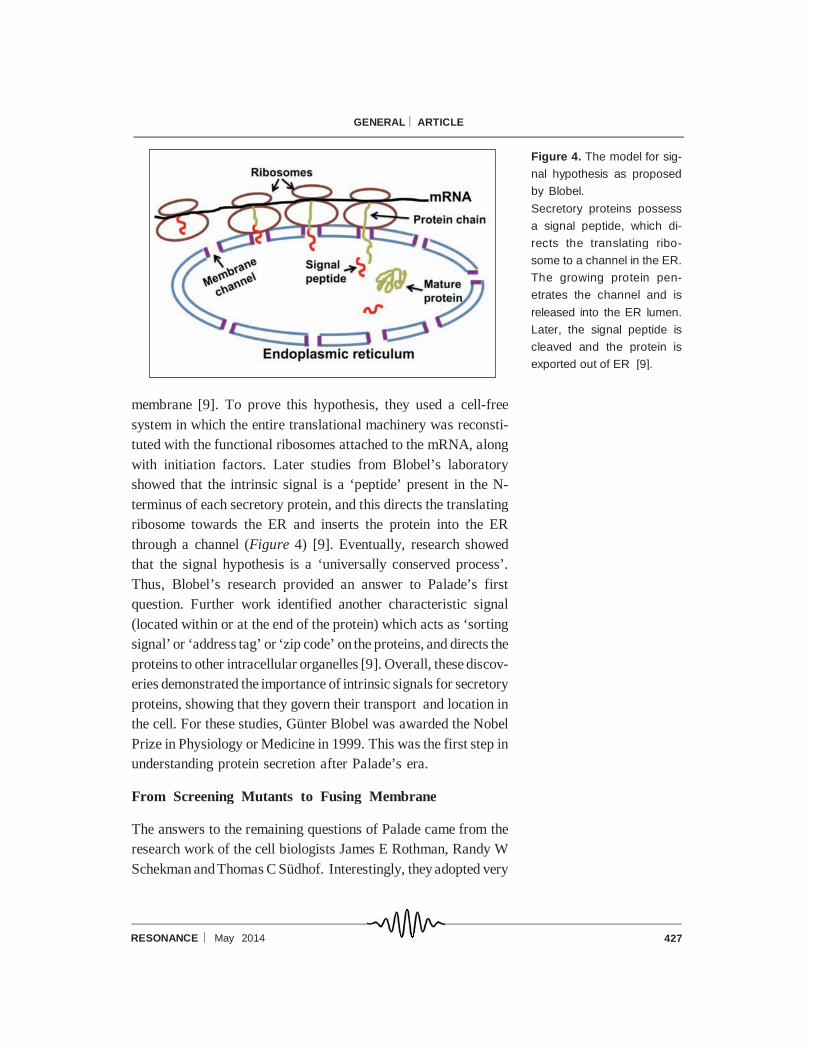
Blobel’s Signal Hypothesis
About that time (1971), Günter Blobel at Rockefeller University
(trained as a postdoc in Palade’s laboratory) proposed the ‘signal
hypothesis’ which states that the proteins secreted out of the cell
contain an intrinsic signal that directs them to and across a
Figure 3. Model for the granule discharge or secretory
vesicle fusion or exocytosis as proposed by Palade.
Based on the morphological findings in their EM studies,
Palade hypothesized that secretory granules discharge
their contents by fusing with the PM thus establishing the
continuity between the granule and extracellular medium
(lumen). During this process, vesicles undergo fusion,
elimination of membrane contacts and fission. Further,
Palade hypothesized that fusion possesses a high de-
gree of specificity and requires Ca2+ and ATP [8].
427RESONANCE May 2014
GENERAL ARTICLE
membrane [9]. To prove this hypothesis, they used a cell-free
system in which the entire translational machinery was reconsti-
tuted with the functional ribosomes attached to the mRNA, along
with initiation factors. Later studies from Blobel’s laboratory
showed that the intrinsic signal is a ‘peptide’ present in the N-
terminus of each secretory protein, and this directs the translating
ribosome towards the ER and inserts the protein into the ER
through a channel (Figure 4) [9]. Eventually, research showed
that the signal hypothesis is a ‘universally conserved process’.
Thus, Blobel’s research provided an answer to Palade’s first
question. Further work identified another characteristic signal
(located within or at the end of the protein) which acts as ‘sorting
signal’ or ‘address tag’ or ‘zip code’ on the proteins, and directs the
proteins to other intracellular organelles [9]. Overall, these discov-
eries demonstrated the importance of intrinsic signals for secretory
proteins, showing that they govern their transport and location in
the cell. For these studies, Günter Blobel was awarded the Nobel
Prize in Physiology or Medicine in 1999. This was the first step in
understanding protein secretion after Palade’s era.
From Screening Mutants to Fusing Membrane
The answers to the remaining questions of Palade came from the
research work of the cell biologists James E Rothman, Randy W
Schekman and Thomas C Südhof. Interestingly, they adopted very
Figure 4. The model for sig-
nal hypothesis as proposed
by Blobel.
Secretory proteins possess
a signal peptide, which di-
rects the translating ribo-
some to a channel in the ER.
The growing protein pen-
etrates the channel and is
released into the ER lumen.
Later, the signal peptide is
cleaved and the protein is
exported out of ER [9].
428 RESONANCE May 2014
GENERAL ARTICLE
different approaches to address Palade’s questions, such as ‘in
vitro biochemistry’ by Rothman, ‘ingenious yeast genetic screen’
by Schekman and ‘curiosity to decipher the electrical activity of
synapse’ by Südhof. Their pioneering approaches launched a
voyage of discovery for our understanding of the cell secretory
transport system which revealed the basic mechanisms that
govern intercellular trafficking, the regulation of membrane fusion
and cell–cell communication.
Intracellular Transport
The synthesis of glycoproteins that are targeted to other intracel-
lular organelles (e.g., lysosomes) or exported out of the cell starts
at the ER. But it was not known how these proteins traverse from
the site of synthesis to its target place. Previous studies from
Palade’s laboratory suggested that the newly synthesized pro-
teins are transported to secretory granules or the vacuole through
the Golgi (Figure 2). But these studies provided no insights into
the nature of protein transport or its mechanism. These questions
engaged James Rothman at Stanford University where he devel-
oped an in vitro transport assay in which he reconstituted the
transport of radiolabeled secretory proteins from one set of
cisternae of the Golgi to other cisternae, isolated from the mam-
malian cell lysates [10]. Rothman hypothesized that this transport
step leads to a modification of the radiolabeled protein, occurring
in the later cisternae of the Golgi. This biochemical transport assay
had several advantages: experiments could be performed any
number of times, it was cost effective and it could be used to
identify the regulators of this reaction step. Using this elegant
technique, Rothman’s laboratory provided the evidence that the
secretory proteins move between the compartments [11]. In
addition, Rothman predicted that a new typeof non-clathrin coated
vesicles might mediate this transport, but the nature of those
vesicles was not clear. Moreover, Rothman’s laboratory identified
two protein factors, NSF [NEM (N-ethylmaleimide) sensitive
factor, AAA ATPase] and SNAP (soluble NSF attachment
protein) that are required for the biochemical transport along with
ATP [11]. This was the first time that the secretory transport step
429RESONANCE May 2014
GENERAL ARTICLE
was shown to require additional components for efficient delivery.
But the precise role of these proteins in the transport was not clear
at that time.
In parallel, Palade’s hypotheses influenced Randy Schekman at
the University of California, Berkeley and he used yeast (Saccha-
romyces cerevisiae) as a model system to study the mechanisms
regulating the transport of secretory proteins to the PM. Previ-
ously, Palade had hypothesized that secretory transport between
organelles could occur through either tubular connections or
vesicles (Figure 2, Palade’s second question) [8], but it was not
clear how this process is mediated. In addition, yeast was not
considered to be a good model to study cell biology in those days
owing to its similarities to bacteria rather than to a mammalian cell,
even when classified as eukaryotic. But, yeast had added advan-
tage that cells were amenable for genetic manipulation. Further,
the secretory pathways of yeast were poorly characterized.
Schekman and his first graduate student, Peter Novick, focused on
characterizing the vesicles observed in the tip of the developing
yeast bud, as seen in their electron micrographs (Figure 5A)
[12].These observations led Schekman to hypothesize that these
vesicles might originate from the secretory pathway and he
proposed that they were delivering the enzymes required for new
cell wall synthesis [13]. He thought that these vesicles would
accumulate in the cell if they mutated the genes of the pathways
involved in their formation. In addition, he predicted that those
mutations are probably lethal due to the essential nature of the
pathways. At that time, Schekman used his experience as a
Figure 5. Schematic dia-
gram representing the elec-
tron micrographs of sec1
mutant yeast grown at per-
missive (A) and restrictive
(B) temperature.
Yeast mutant cells were
grown at the permissive tem-
perature (24 oC) and restric-
tive temperature (ts mutant,
37oC for 3 hr) and analyzed
by EM. Note the presence of
small vesicles at the tip of
yeast bud neck in A (arrow)
and accumulated vesicles
throughout the cytosol in B
(arrow).
Figures were hand drawn simi-
lar to the original electron micro-
graphs published in [12].

430 RESONANCE May 2014
GENERAL ARTICLE
graduate student in Arthur Kornberg’s laboratory, where he had
learnt that the function of essential genes or proteins can be
studied using temperature-sensitive (ts) mutations5.
Using this knowledge, Novick carried out an elegant yeast mu-
tagenesis experiment that had two key assumptions: secretory
mutants would be lethal (ts) and they would accumulate the
secretory proteins inside the cell. The initial mutagenic screen
resulted in about 87 ts yeast mutants, called ‘sec’ mutants, among
which two mutants showed defects in secretion and cell wall
assembly, named as sec1 and sec2 [12]. Later, Novick examined
these mutant cells under the electron microscope and found a large
number of accumulated vesicles in the sec1 cells that had been
grown at the restrictive temperature (Figure 5B) [13]. Subse-
quently, Novick screened a total of 485 ts mutants based on the
above criteria and also on their buoyant properties using Ludox
density gradient6 centrifugation. He predicted that secretion-
defective mutants would be heavier than normal cells and should
sink to the bottom of the tube. This method produced a total of 23
mutants (21 new, plus the original two) involved in the protein
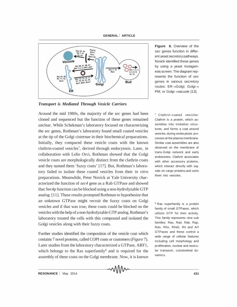
secretion [14]. Studies from Schekman’s laboratory over several
years showed that sec genes function in one of the three different
transport routes: ER to Golgi, Golgi to PM or Golgi to vacuole
(Figure 6) [13, 15]. This was the first time that a genetic approach
had identified a large set of genes required for the intracellular
transport. Further, these studies provided evidence that the essen-
tial genes regulate the secretory pathway and are mediated
majorly through vesicles.
Surprisingly, two of theyeast ts mutants encoding sec18 and sec17
genes discovered by Schekman’s group were homologous to the
NSF and SNAP proteins, identified by Rothman’s group [13, 16].
This suggested that the genes required for the secretory pathway
are universally conserved from yeast to humans. Furthermore, the
discovery of the analogous genes by the two different groups using
two different approaches provided strong evidence that the trans-
port mechanisms are also conserved.
5 Temperature-sensitive muta-
tions (ts): Certain mutations in a
protein are temperature sensi-
tive and these proteins retain
their function at the‘permissive’
temperature but not at the ‘re-
strictive’ temperature. This strat-
egy is used to study the function
of essential proteins (or genes)
and the absence of these pro-
teins causes cell death. These
mutations can be obtained
through a conditional mutagen-
esis method.
6 Ludox density gradient sepa-
ration is a method in which Ludox
particles form a density gradient
during the centrifugation pro-
cess. Ludox (HS-30 or -40) is a
colloidal glass suspension that
contains different sized particles
and is capable of producing self-
generating gradients in a cen-
trifugal field.
431RESONANCE May 2014
GENERAL ARTICLE
Transport is Mediated Through Vesicle Carriers
Around the mid 1980s, the majority of the sec genes had been
cloned and sequenced but the function of these genes remained
unclear. While Schekman’s laboratory focused on characterizing
the sec genes, Rothman’s laboratory found small coated vesicles
at the tip of the Golgi cisternae in their biochemical preparations.
Initially, they compared these vesicle coats with the known
clathrin-coated vesicles7, derived through endocytosis. Later, in
collaboration with Lelio Orci, Rothman showed that the Golgi
vesicle coats are morphologically distinct from the clathrin coats
and they named them ‘fuzzy coats’ [17]. But, Rothman’s labora-
tory failed to isolate these coated vesicles from their in vitro
preparations. Meanwhile, Peter Novick at Yale University char-
acterized the function of sec4 gene as a Rab GTPase and showed
that Sec4p function can be blocked using a non-hydrolyzable GTP
analog [11]. These results prompted Rothman to hypothesize that
an unknown GTPase might recruit the fuzzy coats on Golgi
vesicles and if that was true, these coats could be blocked on the
vesicles with the help ofa non-hydrolyzableGTP analog. Rothman’s
laboratory treated the cells with this compound and isolated the
Golgi vesicles along with their fuzzy coats.
Further studies identified the composition of the vesicle coat which
contains 7 novel proteins, called COPI coats or coatomers (Figure 7).
Later studies from the laboratory characterized a GTPase, ARF1,
which belongs to the Ras superfamily8 and is required for the
assembly of these coats on the Golgi membrane. Now, it is known
Figure 6. Overview of the
sec genes function in differ-
ent yeast secretory pathways.
Novick identified these genes
by using a yeast mutagen-
esis screen. The diagram rep-
resents the function of sec
genes in various secretory
routes: ERGolgi; Golgi
PM; or Golgivacuole [13].
7 Clathrin-coated vesicles:
Clathrin is a protein, which as-
sembles into triskelion struc-
tures, and forms a coat around
vesicles during endocytosis pro-
cesses at the plasma membrane.
Similar coat assemblies are also
observed on the membrane of
trans-Golgi network and early
endosomes. Clathrin associates
with other accessory proteins,
which interact directly with sig-
nals on cargo proteins,and sorts
them into vesicles.
8 Ras superfamily is a protein
family of small GTPases, which
utilizes GTP for their activity.
This family represents nine sub
families: Ras, Rad, Rab, Rap,
Ran, Rho, Rheb, Rit and Arf
GTPases and these control a
wide range of cellular features
including cell morphology and
proliferation, nuclear and vesicu-
lar transport, cytoskeletal dy-
namics.

432 RESONANCE May 2014
GENERAL ARTICLE
that these coatomers help in membrane bending and vesicle
formation, and also in protein sorting [11].
In parallel, Schekman’s laboratory was interested in characteriz-
ing the yeast mutants in which secretion from ER to Golgi is
blocked. Surprisingly, several of their ts mutants showed an
accumulation of ER-derived vesicles with a distinct coat on them.
In collaboration with Lelio Orci, Schekman’s laboratory charac-
terized these ER vesicle coats and found that these coats are
different from the COPI coats and named them as COPII [18].
Later, Schekman’s laboratory identified the COPII coat machin-
ery by isolating the vesicles using an in vitro ER budding reaction.
Interestingly, this set of coat proteins contained several of their
previously described sec proteins [13]. Further studies identified
a GTPase, Sar1p which regulates the recruitment of COPII coat
onto the ER membranes [19]. These coat proteins (COP1 and
COPII) disassemble before the vesicles fuse with the target
membrane (Figure 7) [11, 19]. Thus, the coat proteins play an
essential role in membrane transport. In summary, thestudies from
these two laboratories provided evidence that vesicles mediate the
communication between organelles. The discovery of two differ-
ent coat proteins by two different groups further expanded our
understanding of the secretory protein transport.
Figure 7. Vesicular transport
model by James Rothman.
ARF1 GTPase is proposed
to recruit the COPI subunits
(coatomers) onto the Golgi
membranes (left) thus gener-
ating COPI-coated vesi-cles.
These coats would be disas-
sembled by an unknown
mechanism (?) and utilized
for the next round. Uncoated
vesicles fuse with the target
membrane (right) with the
help of NSF and SNAP [11].
433RESONANCE May 2014
GENERAL ARTICLE
Vesicles Require SNAREs for Membrane Fusion
In the 1980s, Rothman’s and Schekman’s laboratories identified
NSF/Sec18p and SNAP/Sec17p proteins and showed that these
proteins were essential for protein transport between the ER and
Golgi or for intra-Golgi transport. The addition of NSF to
Rothman’s biochemical assay increased the efficiency of mem-
brane transport. But it was not clear how these proteins mechani-
cally regulate protein transport and it was also very difficult to
predict their function from the biochemical assay. Interestingly,
inactivation of NSF using NEM (N-ethylmaleimide) resulted in
the accumulation of uncoated vesicles and few vesicles were
associated with the Golgi stacks [16]. This observation ledRothman
to predict that NSF helps in the membrane fusion process. He
hypothesized that accumulation of uncoated vesicles might be due
to failed membrane fusion events. Later studies from Rothman’s
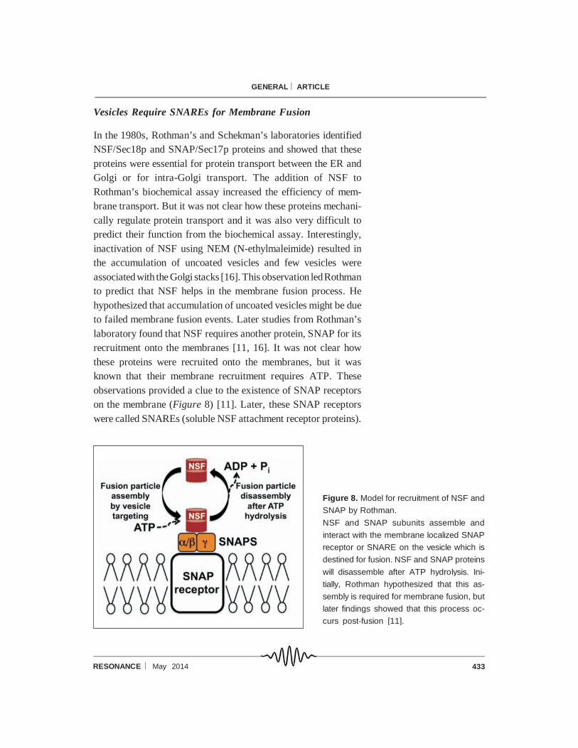
laboratory found that NSF requires another protein, SNAP for its
recruitment onto the membranes [11, 16]. It was not clear how
these proteins were recruited onto the membranes, but it was
known that their membrane recruitment requires ATP. These
observations provided a clue to the existence of SNAP receptors
on the membrane (Figure 8) [11]. Later, these SNAP receptors
were called SNAREs (soluble NSF attachment receptor proteins).
Figure 8. Model for recruitment of NSF and
SNAP by Rothman.
NSF and SNAP subunits assemble and
interact with the membrane localized SNAP
receptor or SNARE on the vesicle which is
destined for fusion. NSF and SNAP proteins
will disassemble after ATP hydrolysis. Ini-
tially, Rothman hypothesized that this as-
sembly is required for membrane fusion, but
later findings showed that this process oc-
curs post-fusion [11].

434 RESONANCE May 2014
GENERAL ARTICLE
In search of the SNAP receptors, Rothman’s laboratory used a
large-scale brain cell lysate to study the NSF/SNAP interaction
with the membranes. These studies showed that SNAP is selec-
tively bound to syntaxin (STX), synaptobrevin (VAMP) and NSF.
In addition, these interactions were sensitive to the addition of
ATP, which causes these proteins to disassociate [20]. Surpris-
ingly, these NSF interacting proteins were identical to the synaptic
vesicle fusion proteins VAMP (on synaptic vesicle), STX (on PM)
and SNAP-25 (on PM) in neurons discovered previously by
Richard Scheller, Thomas Südhof and Michael Wilson (Figure 9).
These research laboratories had shown that this protein machinery
is required for the release of neurotransmitters from synaptic
vesicles in the neuronal synapse [2, 21]. But, it was not clear how
these various proteins mediate fusion of vesicles with the mem-
brane. Later, elegant studies from Cesare Montecucco’s labora-
tory found that two bacterial toxins, tetanus and botulinum toxin,
cleave the SNAP receptors (SNAREs) on the synaptic vesicle by
their protease activity and block neurotransmitter release. These
studies prompted Rothman to propose the ‘SNARE hypothesis’,
wherein a vesicle containing a specific v-SNARE forms a complex
with a specific set of t-SNAREs on the opposing target membrane
and this minimal unit is necessary for membrane fusion [16]. Later,
Rothman’s laboratory provided evidence for this hypothesis by
incorporating the appropriate combination of v- and t-SNAREs on
Figure 9. Schematic diagram representing the
electron micrograph of a nerve ending with numer-
ous neurotransmitters.
Neurotransmitter-filled synaptic vesicles are accu-
mulated at the neuronal synapse. These vesicles
fuse with the PM in a calcium dependent manner.
Forty years later, research of Thomas Südhof and
other laboratories identified the molecular mecha-
nism of neurotransmitter release.
Figure was hand drawn similar to the original electron
micrograph published in [24].
435RESONANCE May 2014
GENERAL ARTICLE
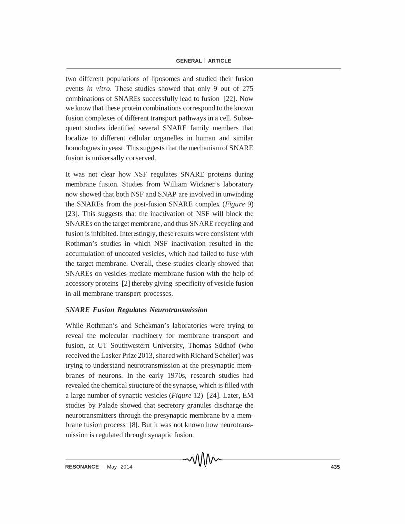
two different populations of liposomes and studied their fusion
events in vitro. These studies showed that only 9 out of 275
combinations of SNAREs successfully lead to fusion [22]. Now
we know that these protein combinations correspond to the known
fusion complexes of different transport pathways in a cell. Subse-
quent studies identified several SNARE family members that
localize to different cellular organelles in human and similar
homologues in yeast. This suggests that the mechanism of SNARE
fusion is universally conserved.
It was not clear how NSF regulates SNARE proteins during
membrane fusion. Studies from William Wickner’s laboratory
now showed that both NSF and SNAP are involved in unwinding
the SNAREs from the post-fusion SNARE complex (Figure 9)
[23]. This suggests that the inactivation of NSF will block the
SNAREs on the target membrane, and thus SNARE recycling and
fusion is inhibited. Interestingly, these results were consistent with
Rothman’s studies in which NSF inactivation resulted in the
accumulation of uncoated vesicles, which had failed to fuse with
the target membrane. Overall, these studies clearly showed that
SNAREs on vesicles mediate membrane fusion with the help of
accessory proteins [2] thereby giving specificity of vesicle fusion
in all membrane transport processes.
SNARE Fusion Regulates Neurotransmission
While Rothman’s and Schekman’s laboratories were trying to
reveal the molecular machinery for membrane transport and
fusion, at UT Southwestern University, Thomas Südhof (who
received the Lasker Prize 2013, shared with Richard Scheller) was
trying to understand neurotransmission at the presynaptic mem-
branes of neurons. In the early 1970s, research studies had
revealed the chemical structure of the synapse, which is filled with
a large number of synaptic vesicles (Figure 12) [24]. Later, EM
studies by Palade showed that secretory granules discharge the
neurotransmitters through the presynaptic membrane by a mem-
brane fusion process [8]. But it was not known how neurotrans-
mission is regulated through synaptic fusion.
436 RESONANCE May 2014
GENERAL ARTICLE
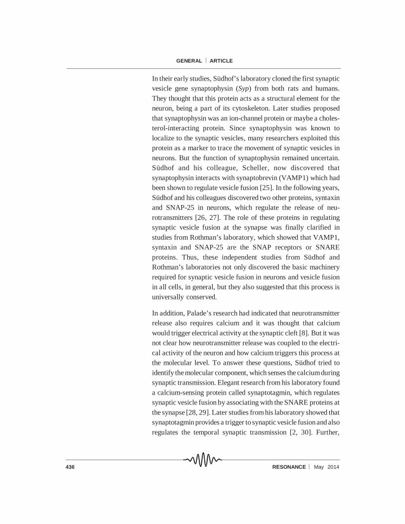
In their early studies, Südhof’s laboratory cloned the first synaptic
vesicle gene synaptophysin (Syp) from both rats and humans.
They thought that this protein acts as a structural element for the
neuron, being a part of its cytoskeleton. Later studies proposed
that synaptophysin was an ion-channel protein or maybe a choles-
terol-interacting protein. Since synaptophysin was known to
localize to the synaptic vesicles, many researchers exploited this
protein as a marker to trace the movement of synaptic vesicles in
neurons. But the function of synaptophysin remained uncertain.
Südhof and his colleague, Scheller, now discovered that
synaptophysin interacts with synaptobrevin (VAMP1) which had
been shown to regulate vesicle fusion [25]. In the following years,
Südhof and his colleagues discovered two other proteins, syntaxin
and SNAP-25 in neurons, which regulate the release of neu-
rotransmitters [26, 27]. The role of these proteins in regulating
synaptic vesicle fusion at the synapse was finally clarified in
studies from Rothman’s laboratory, which showed that VAMP1,
syntaxin and SNAP-25 are the SNAP receptors or SNARE
proteins. Thus, these independent studies from Südhof and
Rothman’s laboratories not only discovered the basic machinery
required for synaptic vesicle fusion in neurons and vesicle fusion
in all cells, in general, but they also suggested that this process is
universally conserved.
In addition, Palade’s research had indicated that neurotransmitter
release also requires calcium and it was thought that calcium
would trigger electrical activity at the synaptic cleft [8]. But it was
not clear how neurotransmitter release was coupled to the electri-
cal activity of the neuron and how calcium triggers this process at
the molecular level. To answer these questions, Südhof tried to
identify themolecular component, which senses the calciumduring
synaptic transmission. Elegant research from his laboratory found
a calcium-sensing protein called synaptotagmin, which regulates
synaptic vesicle fusion by associating with the SNARE proteins at
the synapse [28, 29]. Later studies from his laboratory showed that
synaptotagmin provides a trigger to synaptic vesicle fusion and also
regulates the temporal synaptic transmission [2, 30]. Further,
437RESONANCE May 2014
GENERAL ARTICLE
Südhof and his colleagues identified another important regulator of
synapticvesicle fusion, Munc18, whichassociates with theSNARE
fusion machinery [31, 32]. Interestingly, Scheller’s laboratory
found that Munc18 (also called n-Sec1) is Sec1p, a homologue in
yeast identified by Schekman’s laboratory in their yeast mutagen-
esis screen [12, 32]. In a nutshell, these insights into synaptic
transmission indicated the complexity and tight regulation of
vesicle fusion.
Implications of these Discoveries
These novel findings from the laboratories of Rothman, Schekman
and Südhof not only answered Palade’s key questions, but they
also strengthened the field of ‘molecular cell biology’. In particu-
lar, their studies identified the basic machinery required for
vesicle biogenesis or budding, protein sorting, vesicle transport
and fusion, and organelle identity. Further, these discoveries
illustrated the secretory pathways or protein trafficking routes that
regulate a wide range of eukaryotic cellular activities, including
cell–cell signaling or communication. In addition, these mecha-
nisms are universally conserved in yeast, mouse, humans and
plants. A defect in any of these processes is deleterious to the cells
and can be the basis of neurological (schizophrenia, autism),
immunological (hemophagocytic syndrome) and metabolic (diabe-
tes) disorders [2, 23, 33]. Mutations in the genes of the basic
secretory protein machinery lead to a number of membrane
trafficking diseases such as Charcot–Marie–Tooth disease, Cohen
syndrome, Hermansky–Pudlak syndrome, and Griscelli syndrome
[34, 35].
Current Status – Protein Trafficking, Membrane Fusion,
SNARE Recycling and Regulation
The biosynthesis of proteins occurs both in the cytoplasm and the
ER. The newly made cytosolic proteins either function in the
cytosol or are targeted to organelles such as the nucleus, mitochon-
dria, peroxisomes, ER and plastids (in plants). The laboratories of
Blobel, Schekman and others showed that these proteins possess
438 RESONANCE May 2014
GENERAL ARTICLE
a specific signal sequence, which is intrinsically integrated in the
protein sequence and directs the protein towards the appropriate
organelles. Now we know that the transport of cytosolic proteins
to the different organelles in a cell is mediated by the following
three processes (Figure 10):
1. Transmembrane transport: Fully made cytosolic proteins are
directly transported to the organelles through the protein import-
ers present on their membrane. Several organelles such as ER,
mitochondria, peroxisomes and plastids use this method for their
cytosolic protein import and it is a unidirectional process. In
addition, mitochondria and plastids possess a genome which
codes for certain proteins.
2. Gated transport: Cytosolic proteins enter the organelle through
channels or pores present in their membrane. Protein transport
to the nucleus occurs by this method and is a bidirectional
process. For example, several transcription factors follow this
transport route.
3. Vesicular transport: This is a common method of protein trans-
port beyond the ER and to other organelles including the PM. The
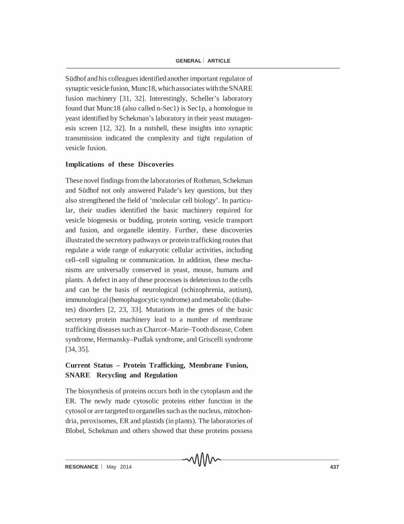
Figure 10. Overview of the secretory transport processes
between organelles.
Newly synthesized proteins in the cytoplasm are trans-
ported to the target places (organelles) by three different
methods. Transport of cytosolic proteins to the ER, mito-
chondria, peroxisomes and plastids utilizes the trans-
membrane transport method (black arrows); whereas
transport to the nucleus uses a gated transport mecha-
nism (red arrow). Protein transport beyond the ER, in-
cluding secretion through the cell surface, degradation in
the lysosome or organelle storage, uses a vesicular
transport process (green arrows) [1].
439RESONANCE May 2014
GENERAL ARTICLE
majority of the transport steps are bi directional, except for
transport fromthe Golgi to lysosomes or to thesecretory granule.
The key machinery mediating these various transport processes is
very well worked out [1]. Among the above transport processes,
protein targeting to theER is different fromthat to other organelles.
It has been shown that biosynthesis of proteins at the ER occurs
through ‘co-translation protein synthesis’9 which is a well-studied
process. Newly synthesized proteins in the ER undergo post-
translational modification such as glycosylation and are targeted
to theGolgi. In addition, non-functional or misfolded proteins in the
ER are exported and degraded by proteasome-mediated degrada-
tion. This process is known as ER-associated protein degradation
(ERAD) [1].
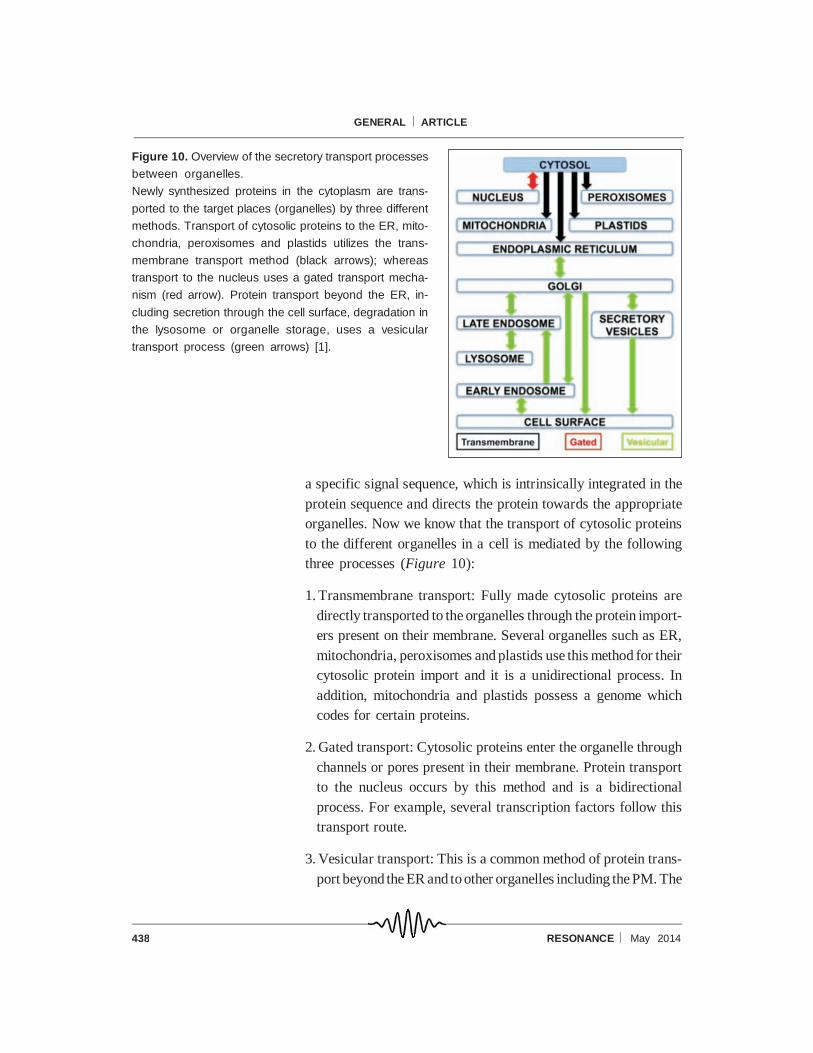
The newly synthesized proteins in the ER are transported to the
Golgi for further modifications including additional glycosylation,
GPI anchoring, metal loading onto metalloproteins. Protein move-
ment between the ER and Golgi is bidirectional and it is mediated
by COPII- and COPI-coated vesicles, as discovered by Rothman’s
and Schekman’s laboratories (Figure 11). From Albert Claude’s
work, we know that the Golgi exists as multiple cisternae (cis-,
medial- and trans- Golgi), called theGolgi complex, and it has been
shown by Rothman’s group that intra-Golgi transport is mediated
through COPI-coated vesicles. From the Golgi, especially the
trans-Golgi, proteins are targetedto multiple sites through vesicular
transport (Figure 11). There are three major transport routes
connecting the Golgi and other organelles, which include: a)
transport to the cell surface through early endosomes (for ex-
ample, proteins such as growth factor receptors, ABC-transport-
ers, G-protein coupled receptors); b) transport to the lysosome
through late endosomes (proteins destined for degradation); c)
transport to the secretory vesicles (for example, neurotransmit-
ters, cytokines). Research from several laboratories has shown
that transport steps from the Golgi utilize different coat proteins
(such as clathrin, adaptor proteins, GGA proteins) for sorting cargo
selectively into these transport pathways. Protein transport be-
yond the Golgi is known to be multidirectional. For the purposes of
9 Co-translational translocation
is a type of protein synthesis
process in which proteins are
integrated into the membrane or
inserted into the lumen of an
organelle while the protein is
translated from mRNA. This is
one of the common processes
followed by all membrane pro-
teins, and occurs on the ER
membranes.
440 RESONANCE May 2014
GENERAL ARTICLE
convention, protein trafficking from one organelle to other or-
ganelles in forward movement is called anterograde transport and
the reverse process is called retrograde transport [1]. It has been
shown that both pathways play important roles in maintaining
organelle identity (by controlling the steady- state localization of
proteins) and in the biogenesis of organelles.
In addition, cell surface localized proteins or extracellular mol-
ecules (for example, nutrients, growth factors, pathogens) are
internalized from the PM by a variety of processes referred to as
‘endocytosis’. This process utilizes different molecular machiner-
ies such as clathrin, caveolin, etc., for the internalization of
different materials from the cell surface, including fluids [1].
Moreover, recent research suggests that special transport routes
exist from the TGN or endosome to specialized organelles includ-
ing the lysosome-related organelles such as melanosomes, dense
granules, weibel-palade bodies in melanocytes, platelets, endothe-
lial cells, etc., respectively [36]. Thus, the cell utilizes multiple
transport routes to target proteins to the right place for their
function.
Figure 11. Overview of the secretory transport
steps in the eukaryotic cell.
Newly synthesized proteins in the ER are
transported to the Golgi and then to the PM or
lysosome or secretory vesicles. In contrast,
cell surface proteins or extracellular molecules
are internalized through endocytosis and then
transported either to the lysosome or to the
Golgi, and sometimes a few molecules will be
recycled back to the cell surface. In the major-
ity of the transport steps, coat proteins are
known to play an important role. These coat
proteins are recruited from the cytosol onto the
specific membrane domain of an organelle to
sort the cargo into distinct vesicles for membrane transport. COPII and COPI coats function at the
endoplasmic reticulum (ER) and Golgi complex respectively. Clathrin functions at multiple sites, but
interacts with different adaptors. Arrows represent the flow of membrane traffic: black arrows – antero-
grade transport; orange arrows – retrograde transport; green arrows – transport from the PM to lysosomes
(L) through early (EE) and late endosomes (LE); and transport of secretory vesicles (SV) towards PM
shown separately. TGN is trans-Golgi network [1].
441RESONANCE May 2014
GENERAL ARTICLE
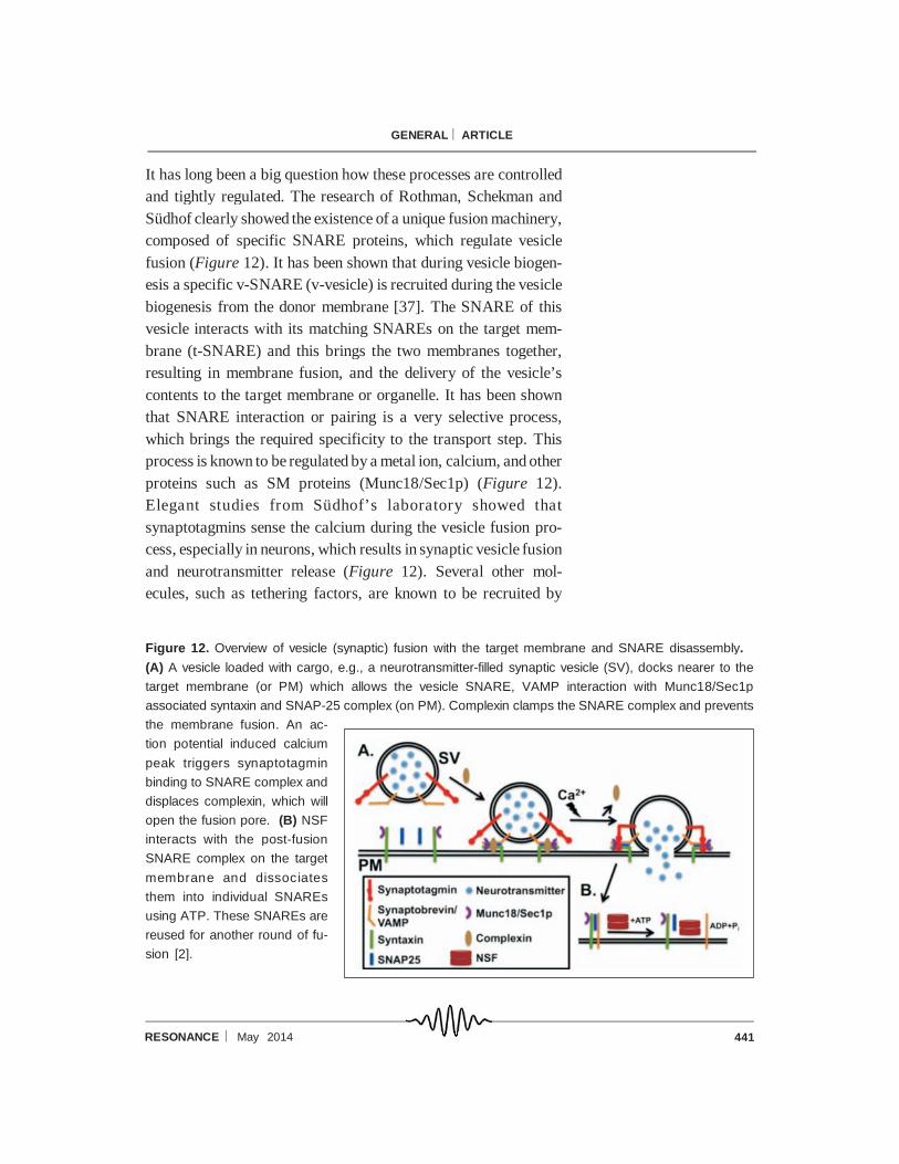
It has long been a big question how these processes are controlled
and tightly regulated. The research of Rothman, Schekman and
Südhof clearly showed the existence of a unique fusion machinery,
composed of specific SNARE proteins, which regulate vesicle
fusion (Figure 12). It has been shown that during vesicle biogen-
esis a specific v-SNARE (v-vesicle) is recruited during the vesicle
biogenesis from the donor membrane [37]. The SNARE of this
vesicle interacts with its matching SNAREs on the target mem-
brane (t-SNARE) and this brings the two membranes together,
resulting in membrane fusion, and the delivery of the vesicle’s
contents to the target membrane or organelle. It has been shown
that SNARE interaction or pairing is a very selective process,
which brings the required specificity to the transport step. This
process is known to be regulated by a metal ion, calcium, and other
proteins such as SM proteins (Munc18/Sec1p) (Figure 12).
Elegant studies from Südhof’s laboratory showed that
synaptotagmins sense the calcium during the vesicle fusion pro-
cess, especially in neurons, which results in synaptic vesicle fusion
and neurotransmitter release (Figure 12). Several other mol-
ecules, such as tethering factors, are known to be recruited by
Figure 12. Overview of vesicle (synaptic) fusion with the target membrane and SNARE disassembly.
(A) A vesicle loaded with cargo, e.g., a neurotransmitter-filled synaptic vesicle (SV), docks nearer to the
target membrane (or PM) which allows the vesicle SNARE, VAMP interaction with Munc18/Sec1p
associated syntaxin and SNAP-25 complex (on PM). Complexin clamps the SNARE complex and prevents
the membrane fusion. An ac-
tion potential induced calcium
peak triggers synaptotagmin
binding to SNARE complex and
displaces complexin, which will
open the fusion pore. (B) NSF
interacts with the post-fusion
SNARE complex on the target
membrane and dissociates
them into individual SNAREs
using ATP. These SNAREs are
reused for another round of fu-
sion [2].

442 RESONANCE May 2014
GENERAL ARTICLE
specific Rab GTPases10 before the SNARE interactions [37]. But,
the identification of additional regulatory molecules for vesicle
fusion is still being pursued in many laboratories. Thus, vesicular
fusion is a highly regulated and controlled process of protein
delivery.
Questions for the Future
The molecular machinery required for vesicle budding, transport
and fusion and their regulation are known. But it is not clear how
cells maintain the appropriate concentration of protein or cargo in
an organelle and how protein transport occurs against a chemical
gradient. Further, how do cells maintain vesicle size and number,
at steady state and after an extracellular stimulus? How do cells
preserve the distinctive membrane composition of the different
organelles (especially lipids)? The fusion machinery is known for
a small set of transport steps but not for all the trafficking
pathways. Finally, how are these fusion events linked to organelle
maturation or biogenesis, especially in the case of lysosome-
related organelles such as melanosomes, dense granules and other
organelles? These are the challenges for the cell biologists of the
post-Rothman–Schekman–Südhof era.
10 Rab GTPases and Rab cycle:
Rab family is a member of the
Ras superfamily, and it contains
70 types of monomeric
GTPases. Rab protein in its in-
active state is bound to GDP
and localizes to the cytosol. This
protein is activated by another
protein,GEF (guanine exchange
factor) that displaces GDP with
GTP and is then recruited onto
the target membrane. Thus, the
Rab proteins cycle between cy-
tosol and the membrane, in the
Rab GTPase cycle, recruiting
downstream factors onto the
membrane in each cycle. These
proteins can be made constitu-
tively active by blocking their
GTP hydrolysis cycle using non-
hydrolyzable GTP analogs such
as GTPs.
Figure 13. Timeline of the
discoveries in the field of
Molecular Cell Biology.
443RESONANCE May 2014
GENERAL ARTICLE
Acknowledgements
We thank Keerthana Ravichandran, Latha Purshotam and Geoff
Hyde for critical reading of the manuscript. This work was
supported by University Grants Commission (to Riddhi Atul Jani),
Wellcome Trust-DBT India Alliance Senior Fellowship (to Subba
Rao Gangi Setty) and Indo–French Centre for the Promotion of
Advanced Research (to Subba Rao Gangi Setty and Graca
Raposo).
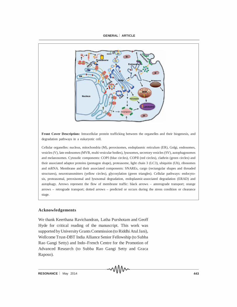
Front Cover Description: Intracellular protein trafficking between the organelles and their biogenesis, and
degradation pathways in a eukaryotic cell.
Cellular organelles: nucleus, mitochondria (M), peroxisomes, endoplasmic reticulum (ER), Golgi, endosomes,
vesicles (V), late endosomes (MVB, multi vesicular bodies), lysosomes, secretory vesicles (SV), autophagosomes
and melanosomes. Cytosolic components: COPI (blue circles), COPII (red circles), clathrin (green circles) and
their associated adaptor proteins (pentagon shape), proteasome, light chain 3 (LC3), ubiquitin (Ub), ribosomes
and mRNA. Membrane and their associated components: SNAREs, cargo (rectangular shapes and threaded
structures), neurotransmitters (yellow circles), glycosylation (green triangles). Cellular pathways: endocyto-
sis, proteasomal, peroxisomal and lysosomal degradation, endoplasmic-associated degradation (ERAD) and
autophagy. Arrows represent the flow of membrane traffic: black arrows – anterograde transport; orange
arrows – retrograde transport; dotted arrows – predicted or occurs during the stress condition or clearance
stage.
444 RESONANCE May 2014
GENERAL ARTICLE
Suggested Reading
[1] B Alberts, A Johnson, J Lewis, M Raff, K Roberts and P Walter, Molecular Biology of the Cell,
Garland Science, Chapters 12–13, p.695–812, 2007.
[2] J H Hurst, Richard Scheller and Thomas Sudhof receive the 2013 Albert Lasker Basic Medical
Research Award, J. Clin. Invest, Vol.123, pp.4095–4101, 2013
[3] A Claude, Fractionation of Mammalian Liver Cells by Differential Centrifugation, J. Expt. Med.,
Vol.84, pp.51–89, 1946.
[4] K R Porter, A Claude, and E F Fullam, A Study of Tissue Culture Cells by Electron Microscopy:
Methods and Preliminary Observations, J. Expt. Med., Vol.81, pp.233–246, 1945.
[5] D D Sabatini and M Adesnik, Christian de Duve: Explorer of the cell who discovered new
organelles by using a centrifuge, PNAS USA, Vol.110, pp.13234–13235, 2013.
[6] A Claude, The Coming Age of the Cell, Nobel Lectures, Physiology or Medicine, 1974.
[7] D de Duve, Exploring Cells with a Centrifuge, Nobel Lectures, Physiology or Medicine, 1974.
[8] G E Palade, Intracellular aspects of the process of protein secretion, Nobel Lectures, Physiology or
Medicine, 1974.
[9] G Blobel, Protein targeting, Nobel Lectures, Physiology or Medicine, 1999.
[10] W E Balch, W G Dunphy, W A Braell, and J E Rothman, Reconstitution of the transport of protein
between successive compartments of the Golgi measured by the coupled incorporation of N-
acetylglucosamine, Cell., Vol.39, pp.405–416, 1984.
[11] J E Rothman and L Orci, Molecular dissection of the secretory pathway, Nature, Vol.355, pp.409–
415, 1992.
[12] P Novick and R Schekman, Secretion and cell-surface growth are blocked in a temperature-
sensitive mutant of Saccharomyces cerevisiae, PNAS USA, Vol.76, pp.1858–1862, 1979.
[13] R Schekman, Lasker Basic Medical Research Award. SEC mutants and the secretory apparatus,
Nat. Med., Vol.8, pp.1055–1058, 2002.
[14] P Novick, C Field and R Schekman, Identification of 23 complementation groups required for
post-translational events in the yeast secretory pathway, Cell, Vol.21, pp.205–215, 1980.
[15] C A Kaiser and R Schekman, Distinct sets of SEC genes govern transport vesicle formation and
fusion early in the secretory pathway, Cell, Vol.61, pp.723–733, 1990.
[16] V Malhotra and S D Emr, Rothman and Schekman SNAREd by Lasker for trafficking, Cell, Vol.111,
pp.1–3, 2002.
[17] L Orci, B S Glick and J E Rothman, A new type of coated vesicular carrier that appears not to
contain clathrin: its possible role in protein transport within the Golgi stack, Cell, Vol.46, pp.171–
184, 1986.
[18] C Barlowe, L Orci, T Yeung, M Hosobuchi, S Hamamoto, N Salama, M F Rexach, M Ravazzola,
M Amherdt and R Schekman, COPII: a membrane coat formed by Sec proteins that drive vesicle
budding from the endoplasmic reticulum, Cell, Vol.77, pp.895–907, 1994.
[19] D Jensen and R Schekman, COPII-mediated vesicle formation at a glance, J. Cell Sci., Vol.124,
pp.1–4, 2011.
[20] T Sollner, S W Whiteheart, M Brunner, H Erdjument-Bromage, S Geromanos, P Tempst and J
E Rothman, SNAP receptors implicated in vesicle targeting and fusion, Nature, Vol.362, pp.318–
324, 1993.
[21] R C Lin and R H Scheller, Structural organization of the synaptic exocytosis core complex,
Neuron, Vol.19, pp.1087–1094, 1997.
445RESONANCE May 2014
GENERAL ARTICLE
Address for Correspondence
Subba Rao Gangi Setty
Department of Microbiology
and Cell Biology
Indian Institute of Science
Bangalore 560 012, India.
Email:
[22] T Weber, B V Zemelman, J A McNew, B Westermann, M Gmachl, F Parlati, T H Sollner and J
E Rothman, SNAREpins: minimal machinery for membrane fusion, Cell, Vol.92, pp.759–772,
1998.
[23] W T Wickner, Profile of Thomas Sudhof, James Rothman, And Randy Schekman, 2013 Nobel
Laureates in Physiology or Medicine, PNAS USA, Vol.110, pp.18349–18350, 2013.
[24] J E Heuser and T S Reese, Evidence for recycling of synaptic vesicle membrane during transmitter
release at the frog neuromuscular junction, J. Cell Biol, Vol.57, pp.315–344, 1973.
[25] W S Trimble, D M Cowan and R H Scheller, VAMP-1: a synaptic vesicle-associated integral
membrane protein, PNAS USA, Vol.85, pp.4538–4542, 1988.
[26] M K Bennett, J E Garcia-Arraras, L A Elferink, K Peterson, A M Fleming, C D Hazuka and R H
Scheller, The syntaxin family of vesicular transport receptors, Cell, Vol.74, pp.863–873, 1993.
[27] G A Oyler, G A Higgins, R A Hart, E Battenberg, M Billingsley, F E Bloom, and M C Wilson, The
identification of a novel synaptosomal-associated protein, SNAP-25, differentially expressed by
neuronal subpopulations, J. Cell Biol., Vol.109, pp.3039–3052, 1989.
[28] M Geppert, Y Goda, R E Hammer, C Li, T W Rosahl, C F Stevens and T C Sudhof, Synaptotagmin
I: a major Ca2+ sensor for transmitter release at a central synapse, Cell, Vol.79, pp.717–727, 1994.
[29] M S Perin, V A Fried, G A Mignery, R Jahn and T C Sudhof, Phospholipid binding by a synaptic
vesicle protein homologous to the regulatory region of protein kinase C. Nature, Vol.345, pp.260–
263, 1990.
[30] A Maximov, J Tang, X Yang, Z P Pang and T C Sudhof, Complexin controls the force transfer
from SNARE complexes to membranes in fusion, Science, Vol.323, pp.516–521, 2009.
[31] Y Hata, C A Slaughter T C Sudhof, Synaptic vesicle fusion complex contains unc-18 homologue
bound to syntaxin, Nature, Vol.366, pp.347–351, 1993.
[32] T C Sudhof and J E Rothman, Membrane fusion: grappling with SNARE and SM proteins, Science,
Vol.323, pp.474–477, 2009.
[33] I Mellman and S D Emr, A Nobel Prize for membrane traffic: vesicles find their journey’s end,
J. Cell Biol., Vol.203, pp.559–561, 2013.
[34] P Gissen and E R Maher, Cargos and genes: insights into vesicular transport from inherited
human disease, J. Med. Genet., Vol.44, pp.545–555, 2007.
[35] V M Olkkonen and E Ikonen, When intracellular logistics fails—genetic defects in membrane
trafficking, J. Cell Sci, Vol.119, pp.5031–5045, 2006.
[36] M S Marks, H F Heijnen and G Raposo, Lysosome-related organelles: unusual compartments
become mainstream, Curr. Opin. in Cell Biol., Vol.25, pp.495–505, 2013.
[37] J S Bonifacino and B S Glick, The mechanisms of vesicle budding and fusion, Cell, Vol.116, pp.153–
166, 2004.