molecular mechanisms in synaptic vesicle recycling
TRANSCRIPT

From the DEPARTMENT OF NEUROSCIENCE Karolinska Institutet, Stockholm, Sweden
MOLECULAR MECHANISMS IN SYNAPTIC VESICLE RECYCYLING
Joel Jakobsson
Stockholm 2010

Back cover image shows a floorball ball.
All previously published papers were reproduced with permission from the publisher. Published by Karolinska Institutet. Printed by Larserics Digital Print AB. © Joel Jakobsson, 2010 ISBN 978-91-7457-035-9

ABSTRACT
Neurons communicate through specialized contacts called synapses, which consist of pre- and postsynaptic compartments separated by a narrow synaptic cleft. The stimulus-dependent rapid influx of calcium ions into the presynaptic compartment induces fusion of neurotransmitter-filled vesicles with the plasma membrane and release of their content into the synaptic cleft. The vesicle membrane then needs to be recycled to avoid disruption of synapse morphology and depletion of synaptic vesicles. Three pathways have been described in the literature: clathrin-mediated endocytosis in which vesicles already the size of synaptic vesicles are retrieved at the periactive zone, bulk endocytosis in which larger pieces of membrane are retrieved, and kiss-and-run in which the vesicle remains intact and forms only a transient fusion pore through which neurotransmitter is released. In this thesis, the molecular mechanisms of synaptic vesicle recycling were studied using acute perturbations in lamprey giant reticulospinal axons. The aim was to clarify three different aspects in the recycling machinery: first – how the clathrin coat assembly is regulated, second – how dynamin-dependent fission is regulated, and third – how the synapse copes with intense activity. Three proteins (epsin, EHD, and syndapin) were cloned in Lampetra fluviatilis using PCR-based methods. Binding partners were identified in pull-down assays and lipid-interacting properties were investigated in liposome sedimentation, tubulation, and budding assays. The cloned proteins were further used in the immunization of rabbits for the generation of domain-specific antibodies. These antibodies were used for Western blot, immunoprecipitations, immunolocalization, and presynaptic microinjections. Microinjection experiments were mainly analyzed with electron microscopy to reveal morphological phenotypes. All three proteins cloned in this thesis showed a high degree of homology in protein and lipid interacting regions with their mammalian orthologs as well as similar biochemical properties. The proteins were all localized to the pre-synaptic compartment where they were shown to accumulate at specific regions during synaptic activity: epsin – to the coat of clathrin-coated pits, EHD – to neck regions of GTP S induced endocytic structures, and syndapin – to the peri-active zone (excluding clathrin-coated pits). Perturbation of epsin, EHD, and syndapin in living synapses impaired synaptic vesicle recycling, observed as a loss of synaptic vesicles. Disruption of epsin’s N-terminal region inhibited coat assembly while perturbation of epsin’s clathrin/AP-2-binding domain resulted in abnormal coat assembly. Perturbation of EHD resulted in an accumulation of clathrin coated pits with long necks indicating an impairment of endocytic fission. The perturbation of syndapin was associated with a massive accumulation of membranous cisternae and invaginations, but not of clathrin-coated pits at the plasma membrane. The findings in this thesis suggest that epsin links membrane deformation to clathrin-coated pit formation with a potential role in regulating the size of synaptic vesicles, that EHD regulates dynamin-mediated fission in an ATP-dependent fashion, and that syndapin is required for bulk endocytosis and for stabilizing the plasma membrane during intense synaptic activity. Thus, this thesis has added new knowledge about the molecular network that mediates synaptic vesicle recycling.

LIST OF PUBLICATIONS
This thesis is based on the following papers, which are referred to in the text according to their Roman numerals: Paper I Joel Jakobsson, Helge Gad, Fredrik Andersson, Peter Löw, Oleg Shupliakov, and Lennart Brodin. “Role of epsin 1 in synaptic vesicle endocytosis.” Proceedings of the National Academy of Sciences (PNAS) U S A. (2008) April 29;105(17):6445-50. Paper II Joel Jakobsson, Frauke Ackermann, Fredrik Andersson, Peter Löw, and Lennart Brodin. “EHD and dynamin interact in budding of synaptic vesicles.” Manuscript.
Paper III Fredrik Andersson, Joel Jakobsson, Peter Löw, Oleg Shupliakov, and Lennart Brodin. “Perturbation of syndapin/PACSIN impairs synaptic vesicle recycling evoked by intense stimulation.” Journal of Neuroscience (2008) April 9;28(15):3925-33.

TABLE OF CONTENTS

LIST OF ABBREVIATIONS
ANTH AP180-N-Terminal-Homology
AP-2 Adaptor protein 2
AP180 Adaptor protein 180 kDa
ATP Adenosine-5'-triphosphate
ATP S Adenosine-5'-O-( -thio triphosphate)
BAR BIN/Amphiphysin/Rvs161/167
DLL Aspartate-leucine-leucine
DPF Aspartate-proline-phenylalanine
DPW Aspartate-proline-tryptophan
EH Eps15 homology
ENTH Epsin-N-Terminal-Homology
Eps15 Epidermal growth factor receptor substrate 15
EPSP Excitatory postsynaptic potential
GED GTPase effector domain
GFP Green fluorescent protein
GST Glutathione-S-transferase
GTP Guanosine-5´-triphosphate
GTP S Guanosine-5´-O-( -thio) triphosphate
Hsc70 Heat shock protein 70
N-WASP Neural Wiskott-Aldrich syndrome protein
NMJ Neuromuscular junction
NPF Asparagine-proline-phenylalanine
PH Pleckstrin homology
PI(4,5)P2 Phosphoinositol-(4,5)-bis-phosphate
PRD Proline rich domain
SDS-PAGE Sodium dodecyl sulfate polyacrylamide gel electrophoresis
SH3 Src homology 3
SNARE Soluble N-ethylmaleimide sensitive fusion protein attachment protein receptors
SNAP-25 Synaptosomal-associated protein 25
VAMP-2 Vesicle-associated membrane protein 2

1
INTRODUCTION
Synapses are specialized junctions between neurons or between neurons and other cells (i.e. muscle cells). They consist of presynaptic compartments with clusters of neurotransmitter-filled vesicles at particular membrane regions called active zones with corresponding postsynaptic compartments containing receptors for the appropriate neurotransmitter. When an action potential reaches the presynaptic terminal, voltage-dependent Ca2+ channels open with a subsequently rapid influx of Ca2+-ions. Ca2+ affects many proteins including synaptotagmin, which is considered to be the Ca2+-sensor for fast, synchronous neurotransmitter release. Upon activation of synaptotagmin, the SNARE complex, consisting of synaptobrevin/VAMP-2 and two plasma membrane proteins SNAP-25 and syntaxin, force the vesicle to fuse with the plasma membrane. The neurotransmitters can be released into the synaptic cleft where they bind to the receptors on the postsynaptic membrane. The vesicle membrane then needs to be recycled to avoid disruption of synapse morphology and depletion of synaptic vesicles. The demands on the recycling system are high since the recycled synaptic vesicle needs to be of the correct size, contain the right protein/lipid composition, and be formed at a sufficient speed. Synaptic vesicle recycling – different pathways
Heuser and Reese first described synaptic vesicle recycling in 1973 (Heuser and Reese, 1973). They used neuromuscular junctions where they observed the uptake of horseradish peroxidase (HRP) at the ultrastructural level. This observation was also the first evidence of the importance of the clathrin-mediated pathway. In a following paper the time scale was described and full endocytosis was estimated to take up to one minute (Miller and Heuser, 1984). This finding by Heuser and Reese gave rise to a model where synaptic vesicles completely collapse into the plasma membrane and the synaptic vesicle membrane diffuses laterally to be retrieved by clathrin-coated invaginations at an area outside the active zone (Figure 1). The importance of clathrin in synaptic vesicle recycling has since then been repeatedly demonstrated in many different types of synapses. Injection of peptides designed to disrupt AP-2/clathrin binding in retinal bipolar cells slows down endocytosis (Jockusch et al., 2005) and knockdown of clathrin or overexpression of a clathrin/AP-2 inhibiting peptide (C-terminus of AP180) in cultured hippocampal neurons also dramatically slows down endocytosis (Granseth et al., 2006). Microinjection of peptides (e.g. the SH3 domain of amphiphysin) in lamprey axons causes an activity-dependent accumulation of clathrin-coated pits (Shupliakov et al., 1997). In addition, purified clathrin-coated vesicles isolated from brain contain synaptic vesicle proteins, highlighting the importance of clathrin-mediated endocytosis in synaptic vesicle recycling (Blondeau et al., 2004). Heuser and Reese also observed cisternae containing endocytic buds. The interpretation at that time was that it was some kind of sorting endosome, from which synaptic vesicles, ready for use, could bud off. It could also be an indication of a second pathway for synaptic vesicle recycling, called bulk endocytosis, which involves uptake of large membrane areas (Figure 1). This type of endocytosis has been mostly associated with high frequency trains of action potentials or membrane depolarization by high extracellular concentrations of potassium (Clayton et al., 2008; De Lange et al., 2003; Heuser and Reese, 1973; Leenders et al., 2002; Teng et al., 2007). The process seems to be independent of clathrin but dependent on dynamin, although a clathrin-dependent mechanism might be important in budding events from the endosome-like structures.

2
In parallel with the Heuser and Reese study, an alternative recycling pathway was proposed, later called kiss-and-run (Ceccarelli et al., 1973). In this pathway a small pore (i.e. fusion pore) opens followed by rapid closure without full dilation and collapse at the site of fusion (He et al., 2006; Lindau and Alvarez de Toledo, 2003; Wu et al., 2005) (Figure 1). This model was based mainly on the electron microscopic observation of uncoated omega membrane profiles with narrow necks connected to the plasma membrane at the active zone of the frog neuromuscular junction (Ceccarelli et al., 1973). It is however not clear if these membrane profiles reflect an ongoing fusion event followed by full collapse or closure of a fusion pore. The transient opening of a fusion pore has been shown repeatedly to occur in secretory and endocrine cells (i.e. mast cells and chromaffin cells), showing that the phenomenon of reversal of fusion exists (Albillos et al., 1997; Ales et al., 1999; Alvarez de Toledo et al., 1993; Fernandez et al., 1984; Neher and Marty, 1982). However, it still remains a matter of debate if kiss-and-run occurs physiologically in neurons.
Figure 1. When an action potential reaches the nerve terminal, voltage-gated Ca2+-channels open, which allows a massive inflow of Ca2+ ions at the active zone. Ca2+-ions stimulate SNARE-dependent exocytosis, which releases the neurotransmitters into the synaptic cleft where they can act on postsynaptic receptors. The vesicle membrane is then recycled via three possible pathways: 1-Clathrin-mediated endocytosis, 2-Bulk endocytosis, and 3-Kiss-and-Run. Pathways 1 and 2 involve full collapse of synaptic vesicles followed by lateral movement to the periactive zone while the third pathway is a reverse form of exocytosis occurring at the active zone.

3
Clathrin-mediated endocytosis
The most extensively studied endocytic pathway is clathrin-mediated endocytosis. Clathrin-coated pits or vesicles can be observed in almost all cell types and this is the major pathway for the uptake of receptors (e.g. transferrin receptors and hormone receptors) from the plasma membrane. The main components of the clathrin coat are the clathrin heavy chain and clathrin light chain (Kirchhausen, 2000). They form triskelions, which are the building blocks of the clathrin cage surrounding the vesicle. Cargo proteins are linked to the growing clathrin-coated pit by adaptor proteins. The main adaptor protein is AP-2, which is a multimeric adaptor protein enriched in plasma membrane associated clathrin-coated pits with multiple binding partners including clathrin, phosphoinositides, membrane proteins, and other endocytic proteins (Traub, 2003). An array of endocytic accessory proteins have been implicated in the assembly of the coat and in the successive shaping of the coated membrane that continues until it has reached the shape of a deeply invaginated coated pit with a narrow neck (Schmid and McMahon, 2007; Ungewickell and Hinrichsen, 2007). The precise roles of these accessory proteins are not yet well defined and it is likely that their roles differ between different endocytic pathways and cell types. At the end of the endocytic process, the clathrin-coated pit is cut off from the membrane, forming a free clathrin-coated vesicle. The fission process depends on the large GTPase dynamin, which is also involved in fission in non-clathrin dependent pathways (Mettlen et al., 2009). After fission, the clathrin coat is shed in an auxillin/Hsc70/ATP dependent reaction (Eisenberg and Greene, 2007). At the synapse the demands on the endocytic machinery are extremely high. New fusion-competent synaptic vesicles of the correct size have to be formed at high speed. The identification and characterization of accessory proteins is of great importance to fully understand the endocytic process at the synapse. In addition to AP-2, there is a set of different adaptor proteins, including stonins, epsins, AP180, and amphiphysin, with the ability to link specific cargo to the clathrin coat. At present, it is not fully clear how components of synaptic vesicles interact with and are sorted by adaptors. AP-2 has been shown to interact with the synaptic vesicle protein synaptotagmin (Zhang et al., 1994) but the incomplete block of endocytosis following knockdown of AP-2 strongly suggests an importance of alternative adaptors (Kim and Ryan, 2009). Stonins bind synaptotagmin and AP-2 (Diril et al., 2006) and AP180 ablation shows mislocalization of synaptic vesicle proteins (Nonet et al., 1999; Zhang et al., 1998), making them candidates for sorting synaptic vesicle proteins. Recent findings by Henne and colleagues suggest that the protein FCHo might work as master nucleator of clathrin-mediated endocytosis but its role in neurons has not yet been clarified (Henne et al., 2010). Despite the possibility that synaptic vesicle proteins may act as internalization signals, PI(4,5)P2 (phosphoinositol-(4,5)-bis-phosphate) at the plasma membrane may work as an additional signal for endocytosis. Many proteins bind PI(4,5)P2 (including AP-2, AP180, epsin, dynamin, syndapin, etc.) and knockdown of the PI(4,5)P2 generating enzyme, PIP5K (phosphatidylinositol-4-phosphate-5-kinase), has severe effects on synaptic transmission (Di Paolo et al., 2004). PI(4,5)P2 levels are further regulated by the phosphoinositol phosphatase synaptojanin (Cremona et al., 1999). Many endocytic proteins have the properties of binding and deforming membranes. For example, ENTH/ANTH and BAR domains have been identified as powerful membrane deformation modules and are found in a range of proteins including epsins, AP180, syndapins, endophilins, and amphiphysins (Itoh and De Camilli, 2006). The fission molecule dynamin binds a range of SH3 domain containing proteins (e.g. syndapin, intersectin, amphiphysin, endophilin) linking fission to clathrin-coated pits. The importance of dynamin in the fission process has been demonstrated in several systems,

4
including the temperature sensitive Shibire mutant in Drosophila melanogaster (Koenig and Ikeda, 1989), treatment with the non-hydrolysable analog of GTP (Takei et al., 1995), perturbation of dynamin-SH3 interactions (Shupliakov et al., 1997), and inhibition by the antagonist dynasore (Macia et al., 2006). The mechanisms underlying dynamin-dependent fission are however unclear. Two hypotheses have been suggested that dynamin either works as a mechanochemical enzyme which forms a spiral around the neck of the coated pit that squeezes or pulls off the vesicle or as a classical GTPase that recruits a down-stream fission protein in its GTP-bound state (Mettlen et al., 2009). Surprisingly, mice without the brain specific isoform dynamin 1 (of a total of three isoforms) survive about two weeks and have only partial defects in synaptic vesicle endocytosis (Ferguson et al., 2007). The defect in endocytosis is mainly seen during high levels of synaptic activity and it is possible that dynamin only works as an accelerator of fission. There are many other accessory proteins such as intersectin and eps15 that bind multiple proteins in the endocytic machinery and may have possible roles as scaffolding molecules.
How to study synaptic vesicle recycling
There is a range of different techniques to study synaptic vesicle recycling and one concern is the choice of synapse model suitable for the appropriate technique. The main synapse model in the peripheral nervous system is the junction of the motorneuron axon terminal with the motor end plate, i.e. the neuromuscular junction (NMJ). For example, the frog NMJ was used for the morphology studies in the pioneering work by Heuser and Reese (1973) and the Drosophila
melanogaster NMJ for the characterization of the Shibire mutant (Koenig and Ikeda, 1989). The use of large synapses as models is an advantage for almost all techniques. The largest synapse in the mammalian central nervous system is the calyx of Held in the mammalian auditory center, which has been successfully used for electrophysiology and capacitance recordings. Another large synapse is the ribbon synapse found in the retina of goldfishes, i.e. retinal bipolar synapses. Large synapses are also found in giant axons in lamprey and squid, which permits intraaxonal microinjections. Primary cultures have the advantage of being accessible for genetic manipulations (e.g. analysis of knockouts and interference RNA) and use of optical tracers. Hippocampal, neocortical, and cerebellar neurons are the most common primary culture models. Capacitance recordings
Presynaptic electrical recordings allow for real-time measurements of action potentials and ion currents but can also be used for measurements of exocytosis and endocytosis. The plasma membrane has the ability of storing charge (i.e. work as a capacitor), which can be measured as capacitance. Increased membrane area due to exocytosis will be seen as an increase in capacitance and the subsequent endocytosis as a reduction of capacitance. Capacitance recordings have primarily been limited to giant synaptic preparations like the retinal bipolar cells, auditory hair cells, and Calyx of Held (Parsons et al., 1994; von Gersdorff and Matthews, 1994; Wu et al., 2005). These recordings made it possible to measure synaptic membrane retrieval in real time following a burst of exocytosis and resulted in the identification of two kinetic components – one fast and one slow with 1 and 10 s time constants, respectively (Beutner et al., 2001; Neves and Lagnado, 1999). The fast component was later shown to be dependent on calcium and independent of clathrin suggesting that it may reflect a kiss-and-run or bulk endocytic pathway (Jockusch et al., 2005; Neves et al., 2001). The physiological relevance of this fast component was however questioned when it was later shown in Calyx of

5
Held to be independent of neurotransmitter release and may not at all be related to synaptic vesicle endocytosis (Yamashita et al., 2005). Optical tracers
The use of the optical probe FM1-43 has made it possible to study the dynamics of the synaptic vesicle cycle (Betz and Bewick, 1992). FM1-43 (and its analogs) is a lipophilic dye that increases in fluorescence 100-fold when it is bound to membranes. The dye cannot penetrate membranes and is therefore applied extracellularly during synaptic activity to load synaptic vesicles. Activity-dependent dye uptake and release can be studied in the NMJ and in neuronal cultures using this technique. This method was used for measuring the time course for exocytosed vesicles to be available for release, which was estimated to be about 60 seconds (Ryan and Smith, 1995). Observations from dye uptake also indicated that synaptic vesicles recycle without passing an endosomal compartment (Murthy and Stevens, 1998). Measuring dye loss at the single-vesicle level has been used to examine a possible kiss-and-run pathway with the idea that the dye will not be able to escape as efficiently from the synaptic vesicle during a kiss-and–run event compared to a full-fusion event. Studies performed in different laboratories have provided evidence favouring both the presence and lack of kiss-and-run events. Problems with extracellular loading and labelling of other compartments might have complicated the quantitative data and with more controlled conditions only a complete loss of dye during synaptic activity could be observed, not supporting the presence of kiss-and-run (Chen et al., 2008). A different type of an optical tracer technique is the use of pHluorins, a modified form of GFP. This pH-sensitive form of GFP can be attached to the luminal part of synaptic vesicle proteins and function as a reporter as it will increase its fluorescence in neutral pH but will be quenched in the acidic environment inside the synaptic vesicle (Sankaranarayanan et al., 2000). VAMP-2 was the first protein used with this probe (synaptopHluorin) and endocytosis could be followed after a single action potential and a time constant for endocytosis of about 10-15 seconds was described (Granseth et al., 2006). Other vesicle proteins such as synaptophysin (Granseth et al., 2006), synaptotagmin (Fernandez-Alfonso et al., 2006), and vGlut (Balaji and Ryan, 2007) have also been fused with pHluorins showing similar kinetics. Granseth and colleagues combined the use of pHluorins with genetic tools (knockdown of clathrin) to show that clathrin-dependent endocytosis is the dominant pathway for synaptic vesicle retrieval (Granseth et al., 2006). Although pHluorins are more specific tracers than FM dyes there are still problems, as a substantial amount of the pHluorins can be found at the plasma membrane and it is not clear whether the uptake reflects endocytosis of synaptic vesicles or reclustering and reinternalization of the pHluorin. A recent study by Opazo and colleagues showed with STED (stimulated emission depletion microscopy) that native vesicle proteins remain clustered while the pHluorins diffuse at the plasma membrane after exocytosis (Opazo et al., 2010). Another optical tracer, the quantum dot, has recently been developed to study the presence of kiss-and-run. Large quantum dot particles can be loaded into synaptic vesicles. The fluorescence of the quantum dot is pH-dependent with a 15% higher fluorescence at neutral pH compared to a vesicle’s luminal pH. Thus, vesicle fusion will result in an increase of the fluorescence signal. The observed increase in fluorescence signal followed by a very fast return (0.5-1 seconds) to baseline was interpreted as kiss-and-run while a loss (or decrease below baseline) of signal as a full-collapse event (Zhang et al., 2009). The authors concluded that kiss-and-run is the predominant pathway at the beginning of stimulus trains. The effect of the relatively large quantum dots on synaptic vesicle release is however unclear.

6
Genetics
Gene knockout is a powerful tool in cell biology to understand molecular mechanisms in a range of biological systems. In synapse research, the technique has been efficiently used for identifying proteins essential for synaptic vesicle fusion (Sudhof, 2004). Synaptic vesicle endocytosis on the other hand has been more difficult to study using gene knockouts and there is no single protein that has been shown to be essential for synaptic vesicle endocytosis using this technique. For example, deletion of amphiphysin in mice causes only small changes in synaptic vesicle endocytosis (Di Paolo et al., 2002). Genetic invertebrate models (e.g. Drosophila melanogaster and Caenorhabditis elegans) may be very powerful systems to find new endocytic proteins in screens but the molecular endocytic machinery may differ from those in vertebrate synapses. For example syndapin and epsin have been shown to be dispensable for synaptic vesicle endocytosis in Drosophila melanogaster (Bao et al., 2008; Kumar et al., 2009). The phenotype of knockouts might be complicated by early lethality, redundancy of other isoforms or proteins, the existence of other pathways, or animal-specific protein functions. The recent development of gene silencing with interference RNA has been used in order to overcome these problems. This knock-down technique, however, causes effects relatively slowly and can be incomplete in its silencing, leaving some active protein left. It has however been successfully used in cultured hippocampal neurons to investigate the role of clathrin in synaptic vesicle endocytosis where knockdown of clathrin almost completely abolished synaptic vesicle recycling (Granseth et al., 2006). Acute perturbations
Another way to study the molecular network in synaptic vesicle recycling is acute perturbation studies where reagents that target specific proteins or protein-protein interactions are injected into the synapse. The two major systems that have been used are the lamprey reticulospinal synapse and the squid giant synapse (Augustine et al., 2006; Brodin and Shupliakov, 2006). The large size of the axons allows for intraaxonal microinjection, which is difficult to perform in other systems. The microinjection is then combined with electrophysiology, immuno-localization, and morphology studies to reveal a phenotype caused by a certain perturbation. The squid synapse has mainly been used for direct clathrin perturbations (Morgan et al., 2000) and the lamprey synapse for perturbing accessory proteins (Andersson et al., 2010; Evergren et al., 2004a; Gad et al., 2000). The main advantage with this method compared to knockout and knockdown studies is that the risk for compensatory mechanisms is very small due to the acute perturbation. Another advantage is that the presynaptic compartment can be studied specifically without interfering with the postsynaptic compartment.

7
AIMS OF THE THESIS
In order to maintain synaptic transmission, the synapse is dependent on the local regeneration of synaptic vesicles at release sites. The general aim of this thesis was to increase the understanding of how proper recycling of synaptic vesicles occurs during synaptic activity. Three different aspects of vesicle recycling were investigated with the following questions: How is clathrin-coat formation regulated in relation to generation of membrane curvature –does the ENTH domain protein epsin contribute? How is dynamin-dependent fission of clathrin-coated vesicles regulated – does the ATPase EHD participate in this process? Are specific molecular components used at high rates of synaptic activity – what is the role of syndapin? To investigate these questions the giant reticulospinal axon model system was used, in which pre-synaptic microinjections can be performed in order to specifically perturb a protein or a protein-protein interaction of interest.

8
METHODOLOGICAL CONSIDERATIONS
The main model system used in this thesis was the lamprey giant reticulospinal axon. Microinjections of antibodies into these axons were used in all three papers to specifically perturb the proteins of interest. A significant challenge was to clone the appropriate lamprey orthologs in order to generate domain-specific polyclonal antibodies. Subsequent in vivo studies were complemented with various in vitro methods to verify the protein’s specificity, functionality, and binding properties. The lamprey reticulospinal axon
Lampetra fluviatilis (European river lamprey) is a jawless vertebrate that survives as a parasite of fish. Its life cycle is similar to that of salmon with a youth stage (larvae) in rivers, an adult stage in the sea, and a final reproduction stage back in the river. Their nervous systems are similar to those of mammals in overall organization, neuronal physiology, ultrastructure, and synaptic pharmacology (Rovainen, 1979). Special characteristics of the lamprey nervous system are the lack of myelin and the existence of large neurons. The largest reticulospinal axons (up to 100 m in diameter) are unbranched and have their soma in the brain stem (Müller cells). Large glutamatergic synapses are located along the axons and connect to motorneurons and interneurons in the spinal cord and are important for locomotion and steering. The synapses are usually separated from each other and have active zones (0.5-2 m) with large dense clusters of about 4000-12000 vesicles per cluster (Gustafsson et al., 2002). There are also gap junctions at many of the synapses, which give rise to an electrical component in the synaptic response. Recycling of synaptic vesicles occurs in an area surrounding the active zone called the peri-active zone (Gad et al., 1998). Transmitter release can be evoked by repetitive electrical stimulation or with high K+ buffers in the presence of a sufficient amount of Ca2+ (Wickelgren et al., 1985). At stimulation rates of about 0.2-5 Hz (e.g. for 30 min at 8°C), the release level does not exceed the capacity of recycling causing no depletion of synaptic vesicles but at higher rates of stimulation (e.g. 20 Hz for 30 min at 8°C) the vesicle cluster becomes partly depleted (Brodin and Shupliakov, 2006). The average activity of the reticulospinal axons during swimming is about 5 Hz but frequencies up to 28 Hz have been recorded (Zelenin, 2005). Intra-axonal microinjection
The large size of lamprey reticulospinal axons makes them uniquely suitable for microinjections of reagents that can perturb specific interactions in the presynaptic compartment (Figure 2). Release sites can be studied with electrophysiology or electron microscopy to reveal phenotypes resulting from different kinds of perturbations. These synapses are also suitable for localization studies with immunofluorescence and immunogold techniques (see below). The animals used in this study were adult lampreys (20-30 cm) caught in rivers during the last stage of their life cycle. They can be kept in an aquarium (at 5-10°C) where they can survive for 7-9 months. Before use, the lampreys were anesthetized with MS-222 (tricaine methanesulfonate) and decapitated. The spinal cord was dissected in chilled and oxygenated physiological buffer (Ringer’s solution) and attached with the ventral side up to a piece of Sylgard in a chamber containing the same buffer and kept at 8°C. The site of injection was marked with a pin (approximately 100 m in diameter) attached at the lateral side of the spinal cord. Extracellular stimulation/recording electrodes were applied to the spinal cord. Sharp

9
glass microelectrodes (resistance 50-90 M ) were used to impale the giant axons. The electrodes were filled with the appropriate reagent, labeled with a fluorophore and dissolved in injection buffer with 250 mM KAc and 10 mM HEPES (pH 7.4), to be injected into the axons using light pressure. In most cases, antibodies were used at a concentration of 5-10 mg/ml labeled at a dye to protein ratio of about 1. GTP S was injected at a concentration of 10 mM. The injections could be visualized with a CCD (charge-coupled device) camera connected to a fluorescence microscope. The absolute concentrations of the injected reagents were not measured, but relative concentrations could be estimated by measuring fluorescence intensity along the gradient of the reagent caused by diffusion from the site of injection.
Electrophysiology
In electrophysiological experiments measuring the excitatory postsynaptic potential (EPSP), a synaptically connected neuron located at a distance of 150-200 μm from the axonal microinjection pipette was impaled with a second microelectrode filled with 3 M KCl (resistance 80-100 M ). After recording the control synaptic response (evoked by positive current pulses in the microinjection pipette) for 5-10 min microinjection was initiated. The fluorescence intensity at the site of the synaptic connection was monitored in parallel with the EPSP amplitude.
Figure 2. Schematic illustration of the microinjection setup. The spinal cord was dissected and placed with the ventral side up in a chamber. The site of injection was marked with a pin and extracellular stimulation/recording electrodes were applied to the spinal cord. Microelectrodes were filled with the fluorescently labeled reagent and injected into axons using a slight amount of pressure. The injections were used for light microscopy, electron microscopy, and electrophysiology analysis (Gad, 2000).

10
Electron microscopy analysis
In order to analyze microinjected axons the spinal cords were prepared for transmission electron microscopy. This includes fixation, embedding in resins, sectioning, and counterstaining. The tissue was chemically fixed with 3% glutaraldehyde, which crosslinks proteins permanently, but leaves tissue sensitive to pH changes and osmolarity. The glutaraldehyde was therefore mixed in a cacodylate buffer at neutral pH. To increase the contrast of proteins, tannic acid was used in the fixative solutions. Post-fixation was carried out with osmium tetroxide to fix lipids and to increase the contrast by staining lipids. Prior to embedding in a resin (Durcupan), water within the tissue had to be replaced by an organic solvent due to Durcupan’s insolubility in water. Dehydration was carried out in a series of steps with increasing concentrations of ethanol to the final concentration of 100%. The ethanol was then replaced with propylene oxide followed by infiltration of the resin in steps with increasing concentrations of Durcupan mixed in propylene oxide. Polymerization was done in 100% Durcupan at 50 °C for 48 hours. Sectioning of the embedded spinal cord was carried out on an ultratome with a diamond knife. The hole made by the pin at the microinjection (see above) was used for orientation of the specimen. Semithin (500-1000 nm) sections were first cut and stained with toluidine blue and examined under a conventional light microscope. By comparing the distribution of the axons with the injection protocol and images taken after the injection, injected axons could be identified. Series of ultrathin sections (200-500 sections, 70 nm each) were cut and placed on formvar coated copper/rubidium grids. To increase the contrast, the thin sections were counterstained with uranyl acetate followed by lead citrate. Sections were examined and photographed in a Tecnai 12 electron microscope. Immunolocalization
The microinjection setup used in this study was coupled to a confocal microscope with an argon and helium laser. This made it possible to do immunolocalization of two different proteins simultaneously. The problems of epitope damage due to fixatives were avoided since the antibodies were injected into living axons. The antibodies were directly labelled with a fluorescent dye using NHS-esters that bind to amine groups on the protein. Typically, antibodies to the protein of interest were labelled with Alexa488 and antibodies to a vesicle marker (i.e. synaptotagmin or VAMP-2) were labelled with Alexa546. Pre-embedding immunogold was used to localize proteins at the ultrastructural level. In this method the spinal cord was initially fixed with a weaker fixative (4% paraformaldehyde, 0.1% glutaraldehyde) than usually used in the preparations for electron microscopy (see above). The spinal cord was either electrically stimulated or kept in high potassium (normal Ca2+ level) or low calcium solution (resting condition) prior to fixation. In some cases microinjection was combined with this method to visualize protein localization after perturbation (Evergren et al., 2004b). In order to make the presynaptic compartment available for antibody recognition the spinal cord was longitudinally sectioned with a vibratome. The primary antibodies were then applied to the opened axons and incubated overnight with agitation in order to increase penetration of the antibodies. Secondary antibodies conjugated to 5 nm gold particles were then applied and spinal cords were prepared for electron microscopy as described above. Quantitative analysis of injected axons and immunogold labeling
Morphological observations reported in the thesis were observed in at least two independent experiments (two different animals) and in several (two to four) microinjected axons. 4-10 synapses were collected and analyzed in each case. Control samples were collected in adjacent

11
non-injected axons or in the same axons but far away from site of injection. The number of synaptic vesicles and coated pits as well as the length of clathrin-covered membrane (in m) were counted/measured in center sections of serially cut synapses, and normalized to one active zone length. To quantify cisternae in synaptic regions, a mesh with squares of 200 x 200 nm was placed over the synapse (center section). The percentage of squares containing membranous structures larger than 100 nm (to exclude synaptic vesicles) was counted in a rectangular area extending laterally one half active zone length (from the edges of the active zone) and into the axon one active zone length. Coated pit length was measured as the length from the base at the plasma membrane to the top of coated pit membrane. The size of coated pits was determined by measuring the length of the invaginated membrane. The size of synaptic vesicles was determined by measuring the diameter. For immunogold labelling in paper I, gold particles were counted in a defined periactive zone area (0.5 x 0.5 m directly lateral to the active zone) and in a control area (0.5 x 0.5 m, 0.5 m laterally from the active zone). For immunogold labelling in paper II, gold particles were counted on coat, neck, or plasma membrane and normalized to coat/membrane length. Differences between variances were tested by F-test and normal distribution by skewness. Student’s t-test or Mann-Whitney test was used where appropriate. The distribution of stages of coated pits was evaluated by Chi-square test. In all statistical tests in this thesis, p-values less than 0.05 were regarded as significant. Bioinformatics
The Lampetra fluviatilis genome has not been sequenced yet and its proteins are frequently not conserved at the amino acid level when compared to mammalian proteins. Since a main part of this thesis regards acute perturbation of proteins with lamprey-specific reagents, achieving this has required an extensive use of different bioinformatic tools in order to clone lamprey orthologs of the proteins of interest. Primarily, sequences from different species were collected from GenBank (National Center for Biotechnology Information, NCBI). Multiple alignments (ClustalW algorithms) of the sequences were used to distinguish conserved regions suitable for primer design. The use of codon usage tables was also helpful in designing primer pairs. Derived sequence data were analyzed with multiple alignments as well as with computer programs like BLASTn and BLASTx (NCBI). The cloning process was recently facilitated by the genome sequencing project of a closely related species, Petromyzon marinus (sea lamprey). The so-called shotgun technique used has generated sequences of many thousands of small DNA fragments (ranging from 35 to 900 nucleotides long). This made it possible to do BLAST searches to find short sequences of Petromyzon marinus orthologs. The evolutionary closeness between Lampetra fluviatilis and Petromyzon marinus made it possible to use the shotgun sequence data for the design of primers. Information about exons/introns in the GenBank was also helpful in finding open reading frames (ORFs) and in building potential mRNA sequences. Polymerase chain reaction (PCR)
Although there are other techniques to find orthologs, the molecular cloning of Lampetra
fluviatilis proteins in this thesis relied on amplifying desired mRNA sequences with PCR based techniques (Mullis et al., 1986). Total brain mRNAs were isolated with commercial kits taking advantage of the polyA-tail. Reverse trancriptase PCR generated cDNA, which was used as templates for amplification with PCR. In some cases commercial cDNA libraries were used as a template. Gel electrophoresis was used to purify and analyze PCR products. Positive products were extracted and transferred to vectors for subsequent sequencing using

12
commercial kits (TA cloning). 3’-RACE and 5’-RACE (rapid amplification of cDNA ends) were used to amplify missing 3’- or 5’-ends of the cDNA. A major problem was that the Lampetra fluviatilis mRNA seems to have a high content of guanine and cytosine nucleotides (~60%, unpublished observations). This so called GC-rich DNA complicates PCRs by forming strong secondary structures, encouraging unspecific primer annealing, and/or by making denaturation of DNA more ineffective. It might also complicate protein expression due to the presence of uncommon codons. These problems were solved by improving the PCR with special running conditions (touchdown PCR), nested PCR, primer optimization, and by careful choice of enzymes, buffers, and additives (e.g. betaine and DMSO). Additives (or enhancers) were used to increase yield and specificity and to overcome problems with GC-rich or long templates. Another key optimization step was the production of a good quality cDNA template (reverse transcriptase PCR). Subcloning
In order to use derived sequence data for further studies, the sequence was moved from one parent vector to another destination vector, using subcloning techniques. For instance, two destination vectors used in this study were pGEX- or pET-vectors, which generated GST-tagged or His-tagged proteins, respectively, upon protein expression. Different restriction enzymes were used to excise the gene of interest followed by gel purification and removal of parent vector. Optimally, the destination vector was excised with the same restriction enzymes at the multiple cloning sites to generate complementary sticky ends. Mixing gene of interest and destination vector with a DNA ligase generated a plasmid that could be transformed to Escherichia coli cells for selection, amplification and harvest. Expression of proteins and antibody purification
The pGEX/pET-vectors with the gene of interest were transformed into Escherichia coli
(BL21 cells) and expression was induced with isopropyl-b-D-thiogalactopyranoside. Cells were lysed with sonication and Triton X-100 treatment in the presence of protease inhibitors. GST-tagged proteins were affinity purified on a glutathione sepharose column and His-tagged proteins on a NiCl coupled chelating column. Although commercial antibodies were used in this thesis, the majority of the antibodies were newly made through immunization of rabbits with lamprey-derived antigens. GST-tagged (with GST tag cleaved) or His-tagged proteins were used as antigens. The antigens were also coupled to a NHS-activated column, which was then used for affinity purification of the generated antibodies. Protein A sepharose purification of the antibodies was also performed to achieve a pure IgG preparation. Fab fragments were produced by cleavage using immobilized papain. The specificity of the antibodies was tested by Western blot using protein extracts from lamprey and rat brain or spinal cord tissues. Specificity was also verified by immunoprecipitation experiments (see below). Pull-down assays and immunoprecipitations
To verify that the cloned lamprey orthologs share the same binding partners as mammalian orthologs, pull-down assays were performed on lamprey tissue extracts. Lamprey brains or spinal cords were homogenized in a buffer containing 20 mM HEPES (pH 7.4), 150-200 mM KCl/NaCL, 1-5 mM MgCl2, 1-10 mM EDTA, and protease inhibitors. The homogenate was incubated with Triton X-100 (0.5-1%) followed by high-speed centrifugation. The supernatant was saved and used as a Triton-extract in pull-down assays and immunoprecipitations. In pull-down assays the supernatant was incubated with glutathione sepharose loaded with the GST fusion protein of interest. In some experiments, domain-specific antibodies were added in this step for analysing interfering properties of the antibodies. In immunoprecipitations, the Triton

13
extract was incubated with antibodies and protein A sepharose. After several washing steps the bound proteins were eluted with SDS-PAGE sample buffer and analyzed with SDS-PAGE and Western blot. Liposome binding/tubulation/budding assays
To investigate protein function in relation to lipids a range of different in vitro methods have recently emerged from many labs. In this thesis, liposomes were prepared in order to investigate lipid binding and tubulating properties of proteins. Liposomes were either prepared from brain extract lipids (Folch fraction) or from pure phospholipid/cholesterol components. In some cases rhodamine labelled phospholipids or lipophilic fluorophores were used for visualization of the liposomes. Liposomes were prepared with a mini-extruder to generate liposomes of an approximate diameter of 0.1-0.4 m. For binding assays, the liposomes were incubated with proteins, followed by centrifugation. The pellet was analyzed with SDS-PAGE and Western blot. For tubulation assays the liposome/protein mix was either put on a grid for EM analysis or on a slide for confocal microscopy analysis. Liposomes were also used in a SUPER template assay to investigate budding properties of proteins (Pucadyil and Schmid, 2008). Here, the fluorescently labelled liposomes were incubated with silica beads to generate a membrane reservoir for studying membrane budding events.

14
MEMBRANE CURVATURE IN CLATHRIN-COAT FORMATION
A major task of the endocytic machinery is to form the plasma membrane into a highly curved synaptic vesicle. There are several considerations that preclude membrane deformation including lipid composition, the membrane proteins present, the surrounding cytoskeleton, scaffolding proteins nearby, and amphipathic helix insertion (McMahon and Gallop, 2005). These processes probably work in parallel and include involvement of many proteins that regulate the timing and spatiality of membrane curvature. An additional unique task for the endocytic machinery in the synapse is the formation of vesicles of uniform size (40-50 nm). In this chapter, one family of endocytic proteins (ENTH/ANTH proteins) that are proposed to be involved in membrane curvature generation will be considered. A major focus will be on the ENTH protein epsin 1, which in Paper I was linked to clathrin-mediated endocytosis at the synapse.
ENTH/ANTH proteins
The epsins, AP180, and CALM (clathrin-assembly lymphoid myeloid leukaemia protein) belong to a family of proteins that share a highly conserved N-terminal region of about 150 amino acids, called the ENTH domain (in the epsins) and the ANTH domain (in AP180 and CALM) (Legendre-Guillemin et al., 2004) (Figure 3). There are four differentially expressed isoforms of epsin in mammals (1-4) where epsin 1 is enriched in brain, epsin 2 and 4 (also called CLINT1, epsinR, or enthoprotin) are ubiquitously expressed, and epsin 3 is expressed in keratinocytes 3 (Chen et al., 1998; Kalthoff et al., 2002; Mills et al., 2003; Rosenthal et al., 1999; Spradling et al., 2001; Wasiak et al., 2002). AP180 and CALM are closely related but AP180 appears to be the brain-specific isoform compared to the ubiquitously expressed CALM. AP180 was found independently by several different labs and been variously named AP-3 (Keen and Black, 1986), NP185 (Kohtz and Puszkin, 1988), and F1-20 (Zhou et al., 1992). The ENTH/ANTH domain has a globular structure containing 8 alpha helices (De Camilli et al., 2002). ENTH/ANTH domains interact preferentially with PI(4,5)P2-containing membranes and the ENTH domain tubulates liposomes in vitro (Ford et al., 2002; Itoh et al., 2001). Upon binding to membranes an additional amphipathic helix (helix O) is formed and inserted in the membrane (only seen with ENTH domains and not with ANTH domains) (Ford et al., 2002). The insertion of an amphipathic helix is thought to promote membrane bending. Further investigation of the ENTH membrane interaction showed that the ENTH domain has the property of self association and is able to form protein complexes that further promote
Figure 3. Domain structure of the ENTH/ANTH family of proteins. The proteins contain a globular ENTH/ANTH domain in the N-terminal and an unstructured C-terminal with various peptide motifs binding to clathrin, AP-2, and EH-domain proteins. A lamprey ortholog of epsin was cloned in Paper I. It showed high sequence similarity to mammalian epsin 1.

15
membrane bending (Yoon et al., 2010). Other protein binding properties have been described in addition to the membrane binding. A two-hybrid screen showed binding to the transcription factor PLZF (promyelocytic leukaemia zinc finger), linking epsin to the nucleus and transcription (Hyman et al., 2000). Tubulin is another protein binding partner of the ENTH/ANTH domain, which potentially could couple the endocytic machinery to microtubuli-mediated transport (Hussain et al., 2003). In addition, studies in yeast have demonstrated that the ENTH domain may be involved in actin dynamics by interacting with GTPase-activating proteins linked to Cdc42 (Aguilar et al., 2006). The c-terminus of epsin in isoforms 1-3 contains repeats of a short peptide motif, DPW (up to eight repeats in epsin 1). These motifs have been shown to preferentially bind to the -ear of AP-2 (Brett et al., 2002). AP180 and CALM also contain -ear binding motifs including DPF and FxDxF motifs (Brett et al., 2002). The epsins bind clathrin via type I and type II clathrin boxes and AP180/CALM binds clathrin via repeats of DLL motifs (Drake and Traub, 2001). Overexpression of the clathrin/AP-2 binding (CLAP) domain of the epsins/AP180 has been shown to be a powerful tool in causing inhibition of clathrin-mediated endocytosis in both non-neuronal and neuronal cell cultures (Chen et al., 1998; Granseth et al., 2006). The epsins and CALM also contain several NPF motifs that interact with EH domain modules found in Eps15 and intersectin (Chen et al., 1998; Yamabhai et al., 1998). In addition the epsins also contain UIMs, linking them to the uptake of ubiquitinated cargo (Polo et al., 2002). In Paper I a Lampetra fluviatilis ortholog of epsin was cloned (Figure 3 and Paper I, Figure 1A). The ENTH domain, including the residues essential for membrane binding, was highly conserved (80% similiarity) (Paper I, Supplementary Figure S1). Its lipid binding and tubulating properties were further confirmed in various in vitro assays (Paper I, Figure 1B and Supplementary Figure S2). Furthermore two or three UIMs (in different splice forms) could be identified followed by the repeated motifs of clathrin boxes, DPW, and NPF. Ubiquitin (unpublished observations), clathrin, and AP-2 binding (Paper I, Figure 1C and 1H) were conserved in lamprey epsin but no binding to EH-domain proteins could be detected (unpublished observations). The lamprey epsin clone was further used to generate domain-specific reagents (i.e. polyclonal antibodies) to investigate the function of epsin in synaptic vesicle endocytosis (see below). The epsins – key components of clathrin-mediated endocytosis?
The structure of ENTH/ANTH proteins suggests a model where the ENTH/ANTH domain is anchored to the membrane leaving its C-terminus to recruit coat components and perhaps to stimulate clathrin assembly. Both AP180 and epsin can efficiently recruit clathrin to PI(4,5)P2-enriched phospholipid monolayers and polymerize clathrin into lattices (Ford et al., 2002). In the case of clathrin-epsin the lattices were invaginated while for clathrin-AP180 the lattices were flat. Thus, the epsins seem to be more potent in deforming membranes than AP180. These findings support a model where epsin function is a key component in clathrin-mediated endocytosis driving membrane curvature. The presence of at least one kind of epsin in almost all cells furthermore supports a key role of the epsins in clathrin-mediated endocytosis. When expressed as GFP-epsin in COS7 cells, epsin colocalizes with clathrin (Rappoport et al., 2006; Zoncu et al., 2007) and knockdown studies and overexpression of dominant negative mutants have linked epsin 1 with transferrin uptake and EGF receptor uptake, respectively (Deinhardt et al., 2006; Ford et al., 2002; Vanden Broeck and De Wolf, 2006). However, epsin 1 appears not to be an obligatory component in endocytosis, as it is not involved in clathrin-dependent uptake of Tetanus toxin into motor neurons (Deinhardt et al., 2006). Drosophila melanogaster
epsin (i.e. liquid facets) has been specifically linked to uptake of ubiquitinated cargo and
16
regulation of the Notch signalling pathway (Cadavid et al., 2000; Chen et al., 2002; Overstreet et al., 2004; Tian et al., 2004). Knockouts of epsin 1 and 2 in mice result in embryonic lethality with a Notch signalling defect phenotype consistent with the Drosophila studies (Chen et al., 2009). In yeast, epsin and AP180 seem to have a redundant role where only complete knockout of all isoforms affects endocytosis (Maldonado-Baez et al., 2008). Epsin in synaptic vesicle endocytosis
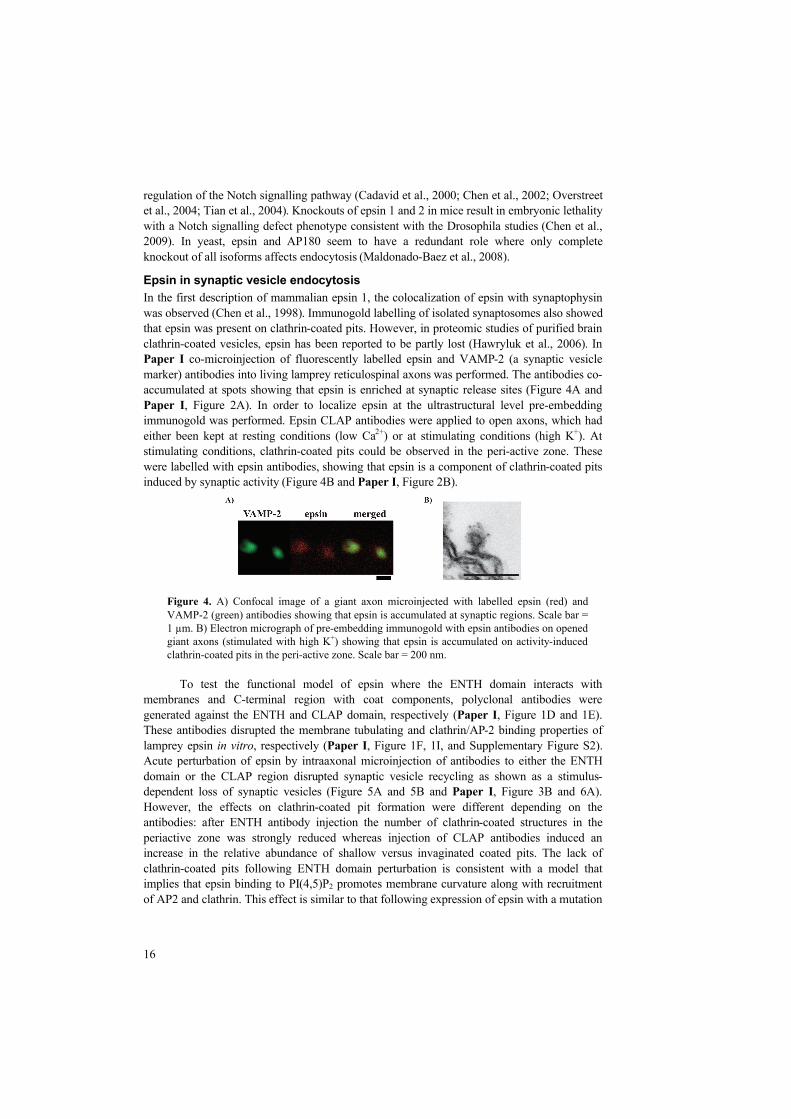
In the first description of mammalian epsin 1, the colocalization of epsin with synaptophysin was observed (Chen et al., 1998). Immunogold labelling of isolated synaptosomes also showed that epsin was present on clathrin-coated pits. However, in proteomic studies of purified brain clathrin-coated vesicles, epsin has been reported to be partly lost (Hawryluk et al., 2006). In Paper I co-microinjection of fluorescently labelled epsin and VAMP-2 (a synaptic vesicle marker) antibodies into living lamprey reticulospinal axons was performed. The antibodies co-accumulated at spots showing that epsin is enriched at synaptic release sites (Figure 4A and Paper I, Figure 2A). In order to localize epsin at the ultrastructural level pre-embedding immunogold was performed. Epsin CLAP antibodies were applied to open axons, which had either been kept at resting conditions (low Ca2+) or at stimulating conditions (high K+). At stimulating conditions, clathrin-coated pits could be observed in the peri-active zone. These were labelled with epsin antibodies, showing that epsin is a component of clathrin-coated pits induced by synaptic activity (Figure 4B and Paper I, Figure 2B).
To test the functional model of epsin where the ENTH domain interacts with membranes and C-terminal region with coat components, polyclonal antibodies were generated against the ENTH and CLAP domain, respectively (Paper I, Figure 1D and 1E). These antibodies disrupted the membrane tubulating and clathrin/AP-2 binding properties of lamprey epsin in vitro, respectively (Paper I, Figure 1F, 1I, and Supplementary Figure S2). Acute perturbation of epsin by intraaxonal microinjection of antibodies to either the ENTH domain or the CLAP region disrupted synaptic vesicle recycling as shown as a stimulus-dependent loss of synaptic vesicles (Figure 5A and 5B and Paper I, Figure 3B and 6A). However, the effects on clathrin-coated pit formation were different depending on the antibodies: after ENTH antibody injection the number of clathrin-coated structures in the periactive zone was strongly reduced whereas injection of CLAP antibodies induced an increase in the relative abundance of shallow versus invaginated coated pits. The lack of clathrin-coated pits following ENTH domain perturbation is consistent with a model that implies that epsin binding to PI(4,5)P2 promotes membrane curvature along with recruitment of AP2 and clathrin. This effect is similar to that following expression of epsin with a mutation
Figure 4. A) Confocal image of a giant axon microinjected with labelled epsin (red) and VAMP-2 (green) antibodies showing that epsin is accumulated at synaptic regions. Scale bar = 1 m. B) Electron micrograph of pre-embedding immunogold with epsin antibodies on opened giant axons (stimulated with high K+) showing that epsin is accumulated on activity-induced clathrin-coated pits in the peri-active zone. Scale bar = 200 nm.

17
in the ENTH domain (disrupting lipid binding) in HeLa cells where clathrin-coated endocytosis was inhibited (Ford et al., 2002).
ENTH antibody injections were also combined with pre-embedding immunogold labelling for the coat marker amphiphysin. Amphiphysin labelling was decreased in the peri-active zone consistent with a mislocalization of coat components (Paper I, Supplementary Figure S4). Surprisingly, disrupting epsin’s clathrin/AP2 binding domains does not affect the recruitment of coat proteins to sites of endocytosis (Figure 5B), indicating that other proteins are probably responsible for this recruitment (e.g. AP180 and amphiphysin). However, the coated pits formed were flatter and larger than those in unperturbed synapses (Figure 6A-B and Paper I, Figure 4A-G). Synaptic vesicle size was not changed showing that the enlarged coated pits did not proceed to vesiculation. Another abnormality observed was large patches of clathrin and large clathrin-covered membrane invaginations (Figure 6C-D and Paper I, Figure 4E-F and 5A-D). A possible explanation for these effects is that steric hindrance by the epsin-bound antibody prevents the formation of highly curved coated pits. Similar effects have been seen when amphiphysin’s CLAP domain has been perturbed with antibodies (Evergren et al.,
Figure 5. Electron micrographs of stimulated synapses where epsin function has been perturbed with either ENTH (A) or CLAP (B) antibodies. Note the depletion of synaptic vesicle in both cases and the increase of clathrin-coated structures on cisternae (arrows) and plasma membrane (open arrows) in the CLAP perturbed synapse. Scale bar = 1 m. a, axoplasmic matrix; d, dendrite; sv, synaptic vesicles.
Figure 6. Examples of clathrin-coated pits that have been perturbed with epsin-CLAP antibodies. Shallow (A) and non-constricted (B) clathrin-coated pits at the plasma membrane were larger than control clathrin-coated pits. C) Example of a flat clathrin-coated patch at the plasma membrane. D) Example of a large abnormal clathrin-coated structure with vacuolar and tubular regions. Scale bars = 100 nm (A-C) and 200 nm (D).

18
2004a). Another possible explanation is that epsin controls synaptic vesicle size by linking curvature formation with coat assembly thereby restricting assembly to curved membrane regions. According to this model, epsin could still contribute to curvature formation after perturbation of clathrin-AP2 interactions, but stabilization of curvature by the emerging coat is disturbed. Under these conditions epsin may retain indirect connections with clathrin and AP2 via NPF–EH domain interactions (e.g. intersectin or eps15). Forming the perfect synaptic vesicle
The control of synaptic vesicle size probably involves several proteins. For example, clathrin itself has been shown to be essential for curvature formation (Hinrichsen et al., 2006). AP180 was identified as an abundant component of clathrin-coated pits purified from brain extracts and has been shown in vitro to stimulate clathrin polymerization (Ahle and Ungewickell, 1986). Only one isoform of CALM/AP180 exists in Drosophila melanogaster and Caenorhabditis elegans and interestingly, knockouts in both organisms have effects on synaptic vesicle size, strongly suggesting a direct role of AP180 in vesicle size regulation (Nonet et al., 1999; Zhang et al., 1998). However the inability of the ANTH domain to deform membranes in vitro indicates that AP180 may not be able to work on its own. A possible hypothesis is that the ENTH domain of epsin performs the membrane bending while the C-terminus of AP180 with dispersed clathrin-binding sites stabilizes the growing clathrin coat to the correct size. It is also possible that epsins only act at the beginning of endocytosis in forming the initial clathrin-coated bud (by inducing membrane curvature) and that other membrane bending proteins (e.g. BAR domain proteins) are important for later stages. The strong homology and possible redundancy seen in knockout studies between the ANTH and ENTH domains indicate probable similar functions for the two domains. An alternative and tempting hypothesis is that the ENTH/ANTH domain proteins are important in protein sorting. Although no direct binding to synaptic vesicle proteins have been observed, AP180 mutants showed that AP180 is required for either recycling of vesicle proteins and/or maintaining the distribution of both vesicle and synaptic proteins in the nerve terminal (Nonet et al., 1999; Zhang et al., 1998). In addition, a systemic gene analysis of mutants responsible for snc1 (VAMP/synaptobrevin homologue) endocytosis in yeast, identified AP180 as a cargo-specific adaptor (Burston et al., 2009). Furthermore, epsin 4 has been shown to bind the SNARE homolog Vti1B, suggesting a potential role for epsins in SNARE sorting (Hirst et al., 2004). Together the ENTH/ANTH domain containing proteins, stonins, and AP-2 (adaptors for synaptotagmin) may assemble the synaptic vesicle proteins into the growing clathrin coat to the correct size in order to form a new fusion-competent synaptic vesicle.

19
FISSION OF CLATHRIN-COATED VESICLES – ROLE OF EHD
The final step of endocytosis involves cutting the clathrin-coated pit from the plasma membrane. The dynamin protein is generally considered to be responsible for fission but its exact mechanism is not clear and the characterization of other interacting proteins is important for a full understanding of dynamin-mediated fission. For example, the SH3 domain containing proteins (e.g. syndapin, amphiphysin, endophilin, intersectin) may help in recruiting dynamin to sites of endocytosis and in forming tubular membrane structures via their BAR domains (discussed in next chapter). Here, a completely different type of possible fission regulator is discussed, the ATPase protein EHD, which was linked to dynamin-mediated fission of clathrin-coated vesicles in the synapse in Paper II.
EHD – a membrane deforming ATPase
EHDs are conserved membrane binding ATPases implicated in intracellular membrane trafficking (Grant and Caplan, 2008). The mammalian genome contains four genes (EHD1-4), which are broadly expressed in different tissues, including brain (EHD1, 3, and 4) (Blume et al., 2007). A single EHD gene is present in Caenorhabditis elegans (termed RME-1) and Drosophila melanogaster (termed Past-1) (Pohl et al., 2000). EHDs consist of an N-terminal G-domain, which binds ATP, a coiled coil domain involved in dimerization, and a C-terminal EH domain (Figure 7). The EH domain interacts with NPF motif-containing proteins like Rabenosyn-5, Rab11-FIP2, and syndapin (Braun et al., 2005; Naslavsky et al., 2004; Naslavsky et al., 2006; Xu et al., 2004). The previously solved crystal structure of EHD2 revealed that its nucleotide-binding G-domain is similar to the GTP-binding domain of dynamin (Daumke et al., 2007). Its ATPase activity is stimulated upon lipid binding and oligomerization, similar to the assembly-stimulated activity of dynamin (i.e. GTPase activity for dynamin) (Daumke et al., 2007). Recent studies in Caenorhabditis elegans have shown that RME-1 interacts and colocalizes with the BAR domain containing protein AMPH-1 (the only Caenorhabditis elegans member of the Amphiphysin/BIN1 family of BAR-domain proteins); AMPH-1 was proposed to regulate EHD spiral assembly (Pant et al., 2009). In Paper II, a lamprey ortholog of EHD was cloned. Lamprey EHD is most similar to mammalian EHD1 and EHD3 and it is broadly expressed in different tissues, including the brain (Paper II, Figure 2B and Supplementray Figure 1). The domain structure as well as the lipid binding and tubulating properties of EHD were found to be conserved (Paper II, Figure
Figure 7. Domain structure of human dynamin 1, EHD 1-4, and the lamprey EHD ortholog cloned in this study. Dynamin consists of five domains: a GTPase domain that binds and hydrolyzes GTP, a PH domain that binds membranes, a GED domain that stimulates self-assembly and GTPase activity, a middle domain important for self-assembly, and a proline-rich domain (PRD) domain that binds SH3 domains in other proteins. EHDs consists of an N-terminal ATPase domain that binds and hydrolyzes ATP, a coiled-coil (CC) domain involved in dimerization, and a C-terminal EH domain that binds NPF motifs.

20
1A and 1B). A functional budding assay was set up to measure a possible EHD budding mechanism. In this assay (SUPER template methodology), silica beads are covered with an excess fluorescently labeled membrane (Pucadyil and Schmid, 2008). Upon addition of dynamin in the presence of GTP vesicle budding occurs which can be monitored in real-time (Figure 8B, Paper II, Figure 1E). EHD was shown to efficiently bind the beads although no budding events could be detected (Figure 8A, Paper II, Figure 1C and 1D). However, the possibility that EHD could enhance dynamin-induced budding still remained, which was tested by adding both EHD and dynamin to the SUPER templates in the presence of ATP and GTP. Surprisingly, the presence of EHD strongly suppressed budding induced by dynamin (Figure 8C, Paper II, Figure 1F). The inhibition of budding was strictly dependent on ATP hydrolysis since no inhibition was observed when the non-hydrolysable analog of ATP (ATP S) was used (Figure 8D, Paper II, Figure 1G). These results indicate that EHD and dynamin can interact in a nucleotide-dependent manner in a reduced system, but the physiological relevance of this observation is not clear.
EHD and endocytic trafficking
A role of EHD proteins in endocytic trafficking was first described in the Caenorhabditis
elegans mutant RME-1 (Grant et al., 2001). It was shown to be important for yolk receptor recycling and is expressed in all kind of cells where it localizes to recycling endosomes and the plasma membrane. In mammalian cells, EHDs have been linked to the recycling of many transmembrane cargo proteins like transferrin receptors, AMPA receptors, and 1 integrins (Jovic et al., 2007; Park et al., 2004a; Rapaport et al., 2006) as well as to the internalization of
Figure 8. Superimposed images (150 images in total) of recorded membrane dynamics in the SUPER template assay system for 150 seconds. (A) No budding was observed when EHD was added to the beads in the constant presence of ATP and GTP. (B) Addition of purified rat dynamin led to massive budding of small membrane vesicles from the SUPER-templates (constant presence of GTP and ATP). (C) No budding events were detected when Dynamin and EHD were mixed (constant presence of GTP and ATP). (D) By replacing ATP with ATP S the EHD-mediated inhibition of dynamin function could be reversed. Membrane budding thus occurs in the constant present of ATP S and GTP.

21
the transferrin receptor, IGF1-receptor, Trk-receptor, GLUT4, and L1NgCAM (Guilherme et al., 2004; Park et al., 2004b; Rotem-Yehudar et al., 2001; Shao et al., 2002; Yap et al., 2010). Although the four mammalian isoforms have very high sequence identity they seem to work in different pathways (Grant and Caplan, 2008). Most of the identified pathways with known EHD involvement are clathrin-independent but EHD1 has been shown to form complexes with clathrin and AP-2 indicating a role of EHDs in clathrin-dependent endocytosis as well (Rotem-Yehudar et al., 2001). EHD in synaptic vesicle recycling
The subcellular localization of the EHD family of proteins in neurons had not been yet fully described. Therefore, the localization of EHD in neurons was investigated in Paper II by microinjecting antibodies in living lamprey reticulospinal axons. The antibodies accumulated in spots and colocalized with the synaptic vesicle marker synaptotagmin, showing that EHD is accumulated at synaptic release sites (Figure 9A and Paper II, Figure 3A). Endocytic structures enriched with dynamin and its interaction partners like endophilin, amphiphysin, and intersectin can be trapped with GTP S (Sundborger, 2010). In Paper II, axons were injected with GTP S followed by pre-embedding immunogold labeling for EHD. Gold particles could be detected on the neck region of GTP S trapped clathrin-coated pits but not on the coat (Figure 9B and Paper II, Figure 3B and 3C).
The immunogold labeling data suggest a role of EHD in the clathrin-dependent synaptic vesicle recycling pathway. However, EHDs have been shown in other systems to have a preference for tubular structures in the cell, which in this case was induced artificially with GTP S injections (Blume et al., 2007; Galperin et al., 2002; Jovic et al., 2009). To test whether EHD indeed has a role in the clathrin-dependent synaptic vesicle endocytosis pathway EHD function was perturbed by intraaxonal microinjection of EHD antibodies. Perturbation of EHD in living reticulospinal axons resulted in a stimulus-dependent partial depletion of synaptic vesicles (Paper II, Figure 4A and 4C). The loss of vesicles was accompanied with an increase in the number of clathrin-coated pits strongly suggesting the importance of EHD in clathrin-dependent recycling of synaptic vesicles (Paper II, Figure 4B). A large proportion of the clathrin-coated pits had long necks, which were considerably longer than those of clathrin-
Figure 9. A) Confocal image of a giant axon microinjected with labelled EHD (green) and synaptotagmin (red) antibodies showing that EHD is accumulated at synaptic regions. B) Electron micrograph of pre-embedding immunogold labelling with EHD antibodies on opened giant axons (stimulated with 5 Hz) showing that EHD is accumulated on GTP S-induced clathrin-coated pits in the peri-active zone. Scale bar = 100 nm.

22
coated pits in control axons (Figure 10A and 10B, Paper II, Figure 4C and 4D). In EHD antibody-injected axons the length from the base at the plasma membrane to the top of the coated pit membrane reached up to 200 nm, while in control axons the length never exceeded 100 nm (Paper II, Figure 4E). This phenotype further suggests that EHD is important at a late stage of clathrin-coated vesicle formation. Similar effects have been seen following microinjection of GTP S (Tomilin et al., 2007) or the endophilin SH3 domain (Gad et al., 2000). These reagents are expected to affect dynamin supporting the idea that the present phenotype is due to defects in dynamin function. In a separate experiment, dynamin pre-embedding immunogold labeling was performed on the EHD antibody-induced long necks. Dynamin was detected on the necks, thus indicating that EHD functions in a dynamin-dependent pathway but also that dynamin recruitment to neck regions seems unaffected (Paper II, Figure 5A and 5B). It is possible that EHD has a role in other pathways, such as the bulk endocytic pathway. Some endosome-like structures with clathrin-coated pits were seen in EHD antibody-injected axons although it is difficult to judge if these reflect a direct defect in bulk endocytosis or are a result of membrane expansion due to defects in the direct clathrin-dependent pathway (Paper II, Supplementary Figure 2). The in vitro budding assays, synaptic localization, and acute perturbation of EHD in living synapses indicate that EHD participates in recycling of synaptic vesicles. The data further suggest that EHD is involved in dynamin-mediated fission.
Role of EHD in regulating GTPase proteins
The mechanism of dynamin-mediated fission is unclear although extensive research has been done during the last few decades. Studies in in vitro systems have shown that dynamin alone is capable of binding, tubulating, and cutting liposomes into small vesicles (Pucadyil and Schmid, 2008; Roux et al., 2006). Surprisingly, studies of dynamin 1 knockout mice have shown that fission can still go on without dynamin although it seems to occur at a slower speed (Ferguson et al., 2007). In yeast, fission seems to be independent of dynamin and instead dependent on amphiphysins and actin-related mechanisms (Toret and Drubin, 2006). The phenotype observed in EHD perturbation experiments in Paper II is morphologically similar
Figure 10. In vivo perturbation of EHD. (A) Electron micrograph of a synapse in an axon microinjected with L-EHD antibodies and stimulated at 5 Hz for 30 min. Note the clathrin-coated pit with long neck (arrow). Scale bar = 500 nm. a=axoplasmic matrix, d=dendrite, sv=synaptic vesicles. (B) Examples of clathrin-coated pits with long necks. Thin ring-like structures are visible at the necks (arrows). Scale bar = 200 nm.

23
to the clathrin-coated pit phenotype induced by GTP S injection. Thus, it is possible that interfering with EHD results in a defect in the GTPase activity of dynamin. The role of GTP binding and hydrolysis in regulating dynamin-dependent fission is not fully understood. Dynamin can deform liposomes into tubes and subsequent addition of GTP contracts the tubes resulting in tube fragmentation (Roux et al., 2006). In other studies GTP hydrolysis was shown to cause dynamin oligomers to disassemble suggesting a dynamic GTP-dependent binding and release to membranes, thereby promoting fission (Bashkirov et al., 2008; Pucadyil and Schmid, 2008; Pucadyil and Schmid, 2009). It was also shown that short dynamin spirals were more efficient in fission than longer spirals (Pucadyil and Schmid, 2008). An interesting parallel is that EHD has also been linked previously to other GTP-binding proteins belonging to the Rab family. For example, both EHD1 and Rab11 have been shown to participate in the recycling pathway from the perinuclear endoplasmatic reticulum compartment to the plasma membrane (Caplan et al., 2002; Lin et al., 2001; Ullrich et al., 1996). In addition, both EHD4 and Rab4 localize to early endosomes where they have been suggested to control the recycling of receptors from this organelle (Sharma et al., 2008; Van der Sluijs et al., 1992). As in the case of EHD and dynamin in Paper II, no direct interaction between Rabs and EHDs has been described. But interestingly, direct interactions have been described between EHDs and Rab effectors, i.e. Rabenosyn and Rab11-Fab2 (Naslavsky et al., 2004; Naslavsky et al., 2006). Another interesting finding is the possibility of EHD in the regulation of Rabs. Depletion of EHD4 leads to enhanced levels of GTP-bound active Rab5 associated with endosomal membranes (Sharma et al., 2008). Thus, it seems that EHD and small GTPases cooperate in endocytic processes making the possible dynamin-EHD cooperativity found in Paper II appealing. However, it is also possible that the phenotype observed in EHD perturbation experiments in Paper II can be coupled to regulation of Rabs. Equally, GTP S injections may potentially affect all GTPase proteins in the synapse, including Rab proteins. Although Rabs are more functionally connected to membrane transport and fusion rather than with clathrin-mediated endocytosis and fission from the plasma membrane, the interesting finding that many of the Rabs (including Rab3, Rab5, Rab7, and Rab11) are specifically localized to synaptic vesicles suggests that there might be uncovered endocytic functions of Rabs (Takamori et al., 2006). Future studies incorporating other components of the protein machinery that coordinates late stages of synaptic vesicle endocytosis will be required to elucidate the precise role of EHD in this process. It will also be interesting to investigate if EHD’s role in fission is a general mechanism in other types of endocytic pathways.

24
SYNAPTIC VESICLE ENDOCYTOSIS AT HIGH RATES OF STIMULATION
During intense synaptic activity synaptic membrane is added to the plasma membrane at a high rate. Under such conditions clathrin-mediated endocytosis might be too slow to keep up with recycling new synaptic vesicles and to preserving synapse morphology. However, another way to cope with a large load of synaptic vesicle membrane would be that larger pieces of membrane (endosome-like) are retrieved, from which vesicles can bud at a later stage. This form of endocytosis, often called bulk endocytosis, has indeed been observed as a result of intense stimulation by many investigators (Clayton et al., 2008; De Lange et al., 2003; Heuser and Reese, 1973; Leenders et al., 2002; Teng et al., 2007). The process can occur rapidly although the subsequent budding from the endosome-like structures can be slow (Clayton et al., 2008; Wu and Wu, 2007). The molecular components of bulk endocytosis are unknown although both actin and dynamin have been linked to this process (Ferguson et al., 2007; Holt et al., 2003; Richards et al., 2004). Here the role of the F-BAR protein, syndapin, in synaptic vesicle endocytosis is discussed (Paper III). Syndapin is able to bind and tubulate membranes and to interact with dynamin and actin-regulating proteins, making it a possible key candidate molecular component in bulk endocytosis (Ahuja et al., 2007; Kessels and Qualmann, 2004; Qualmann et al., 1999).
Syndapin - an F-BAR domain protein
The BAR (Bin/Amphiphysin/RVS) domain is a protein module which is conserved from yeast to mammals (Gallop and McMahon, 2005). The domain can be found in many proteins involved in membrane trafficking (Figure 11). They form dimers that are banana shaped and bind to membranes with their concave faces. The binding of BAR domains to liposomes is affected by the curvature of the membrane and it is possible that BAR domains function as both curvature sensors and generators (Gallop and McMahon, 2005). Some of the BAR domains have an N-terminal amphipathic helix that can be inserted into membranes and induce membrane curvature (Gallop et al., 2006). The amphiphysins and endophilins contain such N-BAR domains. Other BAR domains contain an FCH (FER-CIP4 homology) domain at their N-terminus and are consequently called F-BAR domains (Aspenstrom, 2009; Itoh et al., 2005). Pacsins/syndapins and formin binding protein 17 (FBP-17) are examples of proteins belonging to this subfamily of BAR proteins. Crystallization of the F-BAR domain of FBP-17 showed that the banana-shaped dimer has a larger curvature diameter when compared to other BAR domains (Shimada et al., 2007). The F-BAR domain was later shown to be capable of forming
Figure 11. Domain structure of some of the BAR domain containing proteins. The proteins contain an F-BAR/N-BAR domain in the N-terminal and a SH3 domain in the C-terminal. Syndapin and amphiphysin also contain various peptide motifs that bind to clathrin, AP2 and EH-domain proteins. A lamprey ortholog of syndapin was cloned in Paper III. It showed high sequence similarity to mammalian syndapin 1.

25
oligomers via lateral interactions, which was suggested as the underlying mechanism for driving membrane curvature formation (Frost et al., 2008). The subsequent analysis of syndapin´s F-BAR domain confirmed that it has a similar structure to that of FBP-17 (Rao et al., 2010; Wang et al., 2009). In addition, BAR domain proteins may contain other domains that link them to other functions. For example lipid binding domains like the PH and PX domain can be found in oligophrenin and sorting nexins, respectively (Ellson et al., 2002; Lemmon, 2007). The brain enriched BAR proteins endophilin 1 (N-BAR), amphiphysin 1 (N-BAR), and syndapin 1 (F-BAR) all have a SH3 domain in the C-terminal, which is a protein module that binds proline rich domains (PRDs) in other proteins such as dynamin. The SH3 domain of syndapin in addition binds to synaptojanin, synapsin, N-WASP, and Cordon-Blue (Ahuja et al., 2007; Qualmann et al., 1999). Syndapin also contains two repeats of NPF motifs, which mediate binding to EH domain proteins as discussed in the previous chapter. In Paper
III, a syndapin ortholog in Lampetra fluviatilis was cloned (Figure 11 and Paper III, Figure 1A-B). The F-BAR domain, NPF motifs, and SH3 domains were conserved as well as the SH3 binding partners dynamin and N-WASP (Paper III, Figure 1C). The lamprey syndapin clone was further used to generate domain-specific reagents (i.e. polyclonal antibodies and Fab fragments) to investigate the function of syndapin in synaptic vesicle endocytosis (Paper III, Figure 2A). Cellular functions of syndapin
Overexpression of syndapin in cultured cells leads to the formation of filopodia, which is an actin-dependent process. As this induction was rescued by overexpression of the C-terminal region of syndapin’s binding partner N-WASP, this finding strongly suggests a role of syndapin in regulating actin polymerization (Qualmann and Kelly, 2000). In cultured neurons, colocalization of syndapin with dynamin, N-WASP, and synaptophysin was observed indicating a synaptic localization of syndapin (Kessels and Qualmann, 2006; Qualmann et al., 1999). Syndapin’s role in clathrin-mediated endocytosis is somewhat unclear. It co-localizes poorly with clathrin (Modregger et al., 2002) and no direct binding to endocytic coat proteins have been reported. Overexpression of the SH3 domain in HeLa cells does inhibit transferrin receptor uptake, although this can be explained by a general inhibition of dynamin function (Qualmann and Kelly, 2000; Simpson et al., 1999). Overexpression of syndapin II in primary rabbit lacrimal acini led to an accumulation of clathrin-coated pits, and postsynaptic endocytosis of a subset of NMDA (N-methyl-D-aspartic acid) receptors appears to be dependent on syndapin thus indicating a possible role of syndapin in clathrin-mediated endocytosis at least in some cell types (Da Costa et al., 2003; Perez-Otano et al., 2006). Role of syndapin in synaptic vesicle endocytosis
Although syndapin has been localized to synapses in previous studies (Qualmann et al., 1999), it remains unclear whether this occurs in the presynaptic compartment. Co-microinjection of labelled syndapin and VAMP-2 antibodies in Paper III showed a specific presynaptic localization of syndapin although it cannot be excluded that syndapin is also present in the post-synaptic compartment (Figure 12A, Paper III, Figure 2B). Gold particles accumulated in the peri-active zone area during pre-embedding immunogold labelling on opened axons but no labelling of clathrin-coated pits could be detected (unpublished observations). Syndapin antibodies (raised against the full-length protein) were used in microinjection experiments in lamprey giant reticulospinal axons to examine the functional role of syndapin. Recordings in postsynaptic target neurons showed that the amplitude of the EPSPs was not affected by syndapin antibody injection at a low rate of stimulation (0.2 Hz) (Figure 12B,

26
Paper III, Figure 3A). Furthermore, the morphology of synapses in syndapin antibody injected axons stimulated at this rate for 30 minutes appeared unaffected: the number of synaptic vesicles and clathrin-coated pits when compared to control synapses was not significantly changed (Paper III, Figure 3B-C). This rate of stimulation is sufficient for detecting clathrin-related phenotypes since a reduction of synaptic vesicles and accumulation of clathrin-coated pits was observed at this condition after microinjection of the SH3 domain of amphiphysin (Shupliakov et al., 1997). Thus, syndapin does not appear to be important for synaptic vesicle recycling at low levels of stimulation.
To test the function of syndapin at higher rates of stimulation, reticulospinal EPSPs were recorded before and after a period of very intense stimulation (50 Hz for 10 minutes). When comparing control and syndapin antibody injected axons, the recovery of the EPSPs was delayed in syndapin antibody injected axons (Paper III, Figure 6E). In morphological experiments the effects of syndapin antibodies could be detected even with a less extreme (i.e. physiological) stimulation paradigm. A stimulus-dependent loss of synaptic vesicles accompanied with an accumulation of membrane structures (cisternae) were observed when axons were stimulated at a rate of 5 Hz for 30 minutes (Figure 13A and Paper III, Figure 5A and 6A). Thus, during more intense stimulation (i.e. 5-50 Hz), syndapin participates in synaptic vesicle endocytosis. It is notable that after perturbation of several endocytic proteins, the number of coated pits increases at similar stimulation conditions (Evergren et al., 2004a; Shupliakov et al., 1997). After perturbation of syndapin, however, the number of clathrin-coated pits on the plasma membrane induced by stimulation (5 Hz for 30 minutes) did not differ from that in control axons (Paper III, Figure 6D). To further investigate the role of syndapin in clathrin-mediated budding, the ultrastructural changes were examined after a 15 minute recovery period. The depletion of synaptic vesicles as well as accumulated cisternae remained while the number of clathrin-coated pits on the plasma membrane was decreased to control values (Paper III, Figure 6D). Thus, syndapin perturbations appear to have little direct effect on clathrin-mediated endocytosis at the plasma membrane. The decrease in the number of synaptic vesicles corresponded with an accumulation of cisternae (Figure 13A-B and Paper III Figure 5A-E and 6B). The absence of detectable effects on clathrin-mediated endocytosis suggests that their occurrence cannot merely be
Figure 12. (A) Confocal image of a giant axon microinjected with labelled syndapin (green) and VAMP-2 (red) antibodies, showing that syndapin is accumulated at synaptic release sites. (B) Amplitude of a reticulospinal EPSP (dots and line) recorded during stimulation at 0.2 Hz. Labelled syndapin antibodies were injected after 5 minutes of recording. The bars show the fluorescence intensity at the release sites. Sample traces show average at 1-2 and 17-18 min, respectively.

27
explained by a compensatory expansion of the plasma membrane. It might instead reflect a specific perturbation of the bulk endocytic pathway. To verify that the cisternae contained synaptic vesicle membrane, microinjection experiments were combined with pre-embedding immunogold for the synaptic vesicle protein VAMP-2. The cisternae were highly positive for VAMP-2 indicating that the accumulated cisternae contained synaptic vesicle membrane (Figure 13A-B and Paper III, Figure 4 and 5A-E). This hypothesis was further supported by the finding of narrow connections of the cisternae to the plasma membrane (arrows in Figure 13B). A separate type of experiment was performed in which endocytosis was investigated in the absence of ongoing exocytosis. Axons were stimulated at 20 Hz for 30 min to deplete synaptic vesicle clusters followed by a rapid cooling to 1°C to inhibit endocytosis. Antibodies were then injected and the temperature was subsequently raised to 8°C to allow endocytosis and recovery of the synaptic vesicle pool. A recovery was observed in control antibody injected synapses but depletion of synaptic vesicles and occurrence of cisternae could be observed in syndapin-perturbed synapses (Paper III, Figure 7). This indicates that the accumulated cisternae, rather than resulting from a gradual invagination during sustained stimulation, reflected an endocytic process such as bulk retrieval.
Syndapin interacts with dynamin and N-WASP via its SH3 domain, linking it to both fission and actin regulation. Analysis of dynamin and actin levels after syndapin stimulation did not reveal any change in the synaptic distribution of dynamin (at least at the light microscopic level) while a stimulation-dependent decrease in actin levels, as monitored with Alexa-labeled actin, was observed (Paper III, Supplementary Figures 4 and 5). To further investigate the role of syndapin’s SH3 interaction partners, Fab fragments were purified against the SH3 domain. Injection of these Fab fragments produced a similar effect as injections of antibodies made against the full-length syndapin (Paper III, Figure 8). Thus, antibody perturbation of syndapin in a living synapse showed an involvement of syndapin in synaptic vesicle recycling evoked by intense stimulation. The data further suggest that syndapin is important for bulk endocytosis and that it may take part in stabilizing the plasma membrane in the periactive zone. The activation of the bulk endocytic pathway during intense synaptic activity suggests that there is a molecular sensor for such a change in intensity. Calcium influx is a plausible trigger since an increase in the presynaptic calcium concentration is directly related to the level of synaptic activity. Electrophysiological studies have indeed shown that this pathway (as well as other types of endocytosis) is dependent on calcium and the calcium binding protein
Figure 13. Electron micrograph of a synapse from an axon injected with syndapin antibodies and stimulated at 5 Hz for 30 minutes. (A) The number of synaptic vesicles was decreased but no accumulation of clathrin-coated pits on the plasma membrane was observed. Instead an accumulation of cisternae with clathrin-coated pits was observed. (B) The cisternae were connected to the plasma membrane (arrow) and positive for the synaptic vesicle marker VAMP-2 (gold particles).

28
calmodulin (Wu et al., 2009). Calmodulin activates the protein phosphatase calcineurin, which has been shown to dephosphorylate a range of synaptic proteins (i.e. the dephosphins) (Marks and McMahon, 1998; Robinson et al., 1993). Interestingly, the dephosphorylation of one of the key dephosphins, dynamin, occurs at the same stimulation conditions that activate bulk endocytosis (Clayton et al., 2009). This finding is further supported by the accumulation of endosome-like structures at high rates of stimulation in dynamin 1 knockouts (Ferguson et al., 2007). Although dynamin interacts with many SH3 domain-containing proteins via its PRD, syndapin is its only known binding partner that requires dynamin dephosphorylation to bind (Anggono et al., 2006). Thus, it is possible that syndapin is the key recruiter and regulator of dynamin in the bulk endocytic pathway where the observed accumulation of cisternae in paper III could be an effect of the mislocalization or dysfunction of dynamin as a result of perturbing syndapin. How syndapin specifically recognizes bulk endocytic membrane, rather than membrane for single-synaptic vesicle endocytosis, is unclear. However, it is possible that the F-BAR domain has a preference for a more shallow curvature, which could recruit syndapin to large invaginations. Syndapin also interacts with N-WASP, which may activate the arp2/3 complex to nucleate actin polymerization. The F-BAR domain can form dimers and oligomers and it is thus possible that syndapin can interact with both dynamin and N-WASP at the same time. The reduction in actin labelling in syndapin-perturbed synapses indicated that the accumulated cisternae could be an effect of impaired actin dynamics. Recent studies in Drosophila melanogaster identified the lipase Rolling blackout as another possible key component of bulk endocytosis, indicating a role of phospholipid modifcations in this pathway (Vijayakrishnan et al., 2009). Future studies incorporating other molecular components of the protein machinery that coordinates bulk endocytosis will be required to elucidate the mechanisms underlying this process.

29
CONCLUDING REMARKS
The molecular machinery responsible for proper recycling of synaptic vesicles is far from completely understood. In this thesis, three proteins were linked to this process for the first time. Morphological phenotypes of antibody perturbations also linked the proteins to distinct and specific functions within the synapse. In paper I, epsin was shown to be a component of clathrin-coated pits induced by synaptic activity. Perturbation of epsin with two different antibodies impaired synaptic vesicle recycling, measured as a loss of synaptic vesicles. Perturbation of the N-terminus inhibited coat assembly while perturbation of the clathrin/AP-2-binding domain resulted in abnormal coat assembly. These findings suggest a model where epsin links membrane deformation to clathrin-coated pit formation within the periactive zone and thus may regulate synaptic vesicle size. In paper II, the ATPase EHD was shown in vitro to be capable of inhibiting dynamin-dependent budding in a manner that was strictly dependent on ATP hydrolysis. EHD was localized to the neck region of endocytic structures induced by GTP S in the periactive zone of synapses. Perturbation of EHD resulted in a depletion of synaptic vesicles and an accumulation of clathrin coated pits with very long necks indicating an impairment of endocytic fission. These findings suggest a new regulatory mechanism of dynamin-mediated fission involving EHD. In paper III, an accumulation of syndapin at pre-synaptic release sites was shown. Presynaptic microinjection of syndapin antibodies inhibited synaptic vesicle recycling evoked by intense stimulation but not by light stimulation. The perturbation was further associated with massive accumulation of membranous cisternae and invaginations, but not of clathrin-coated pits at the plasma membrane. The findings suggest a model where syndapin is required for bulk endocytosis and to stabilize the plasma membrane at intense synaptic activity. Thus, the results in this thesis from immunolocalization studies together with acute perturbations of endocytic proteins in the lamprey giant reticulospinal axons have added new knowledge about the molecular network that mediates synaptic vesicle recycling.

30
ACKNOWLEDGEMENTS
My supervisor Lennart Brodin for your encouragement and guidance in the field of neuroscience. You have taught me a lot how to read and evaluate the literature in relation to designing projects. Writing papers with you has been a true pleasure and I really admire your genuine interest in writing the perfect sentence. My co-supervisor Peter Löw for your expertise and guidance in biochemistry and molecular biology. You have taught me a lot although I still do not know how a “rullindunstare” works... Thanks also for being the spirit in the lab making it into a friendly and nice place to do science in. I will definitely miss the music computer, beer club, Christmas decorations, and magic Fridays! My co-supervisor Oleg Shupliakov for letting me use the EM lab and for believing in my ideas. My long-time fellow PhD student Fredrik Andersson for teaching me most of the techniques used in this thesis, from purification of heatshock antibodies to cutting the perfect series of sections with the ultratome (i.e. “a touch-down”). Thanks also for being such a good friend and trying to be interested in football, fishing, and Bob Dylan. All former and present members in the Brodin and Shupliakov labs. My co-authors Helge Gad for starting up my first project and Frauke Ackermann for your contribution to my last project and for being a good “substitute” for Fredrik. Cynthia, for all laughs and great time in the lab, for all proofreadings, and for always being “online”. Wei and Anna, for being my constant lunch mates with a lot of interesting and funny discussions. Emma, for being helpful in the beginning and for introducing me to the EM techniques. Arndt, Elena, Felicia, Fransisco, Kristin, Malin, Nikolay, Olga, Sevi, Simina, and Ylva. You have all contributed with your knowledge, kindness, and willingness to help. All the people at the Neuroscience department, CMB, and members of CEDB/DBRM. You have all contributed to an inspiring scientific atmosphere. A big thank you also to all my friends outside Karolinska Institutet! My old study mates from the Medical Biology programme in Linköping, long-time friends from Knivsta and Uppsala, and the best floorball team in the world, Stockholm IBK. Mina föräldrar, svärföräldrar, bröder och övriga släktingar. Även om ni kanske aldrig förstått något av vad jag gjort de senaste åren så har ni alltid funnits där och stöttat mig - ”är det okej om vi säger att du forskar om hjärnan?” Min älskade fru Hedvig. Du har lyft min tillvaro med din närvaro, skönhet och kärlek. Vi har under den här tiden gjort så otroligt mycket roligt tillsammans men framförallt så har vi frambringat våra två underbara barn, Samuel och Liv. Ni betyder allt!
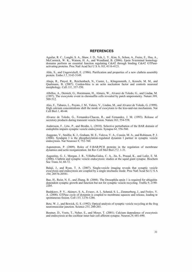
31
REFERENCES
Aguilar, R. C., Longhi, S. A., Shaw, J. D., Yeh, L. Y., Kim, S., Schon, A., Freire, E., Hsu, A., McCormick, W. K., Watson, H. A., and Wendland, B. (2006). Epsin N-terminal homology domains perform an essential function regulating Cdc42 through binding Cdc42 GTPase-activating proteins. Proc Natl Acad Sci U S A 103, 4116-4121.
Ahle, S., and Ungewickell, E. (1986). Purification and properties of a new clathrin assembly protein. Embo J 5, 3143-3149.
Ahuja, R., Pinyol, R., Reichenbach, N., Custer, L., Klingensmith, J., Kessels, M. M., and Qualmann, B. (2007). Cordon-bleu is an actin nucleation factor and controls neuronal morphology. Cell 131, 337-350.
Albillos, A., Dernick, G., Horstmann, H., Almers, W., Alvarez de Toledo, G., and Lindau, M. (1997). The exocytotic event in chromaffin cells revealed by patch amperometry. Nature 389, 509-512.
Ales, E., Tabares, L., Poyato, J. M., Valero, V., Lindau, M., and Alvarez de Toledo, G. (1999). High calcium concentrations shift the mode of exocytosis to the kiss-and-run mechanism. Nat Cell Biol 1, 40-44.
Alvarez de Toledo, G., Fernandez-Chacon, R., and Fernandez, J. M. (1993). Release of secretory products during transient vesicle fusion. Nature 363, 554-558.
Andersson, F., Löw, P., and Brodin, L. (2010). Selective perturbation of the BAR domain of endophilin impairs synaptic vesicle endocytosis. Synapse 64, 556-560.
Anggono, V., Smillie, K. J., Graham, M. E., Valova, V. A., Cousin, M. A., and Robinson, P. J. (2006). Syndapin I is the phosphorylation-regulated dynamin I partner in synaptic vesicle endocytosis. Nat Neurosci 9, 752-760.
Aspenstrom, P. (2009). Roles of F-BAR/PCH proteins in the regulation of membrane dynamics and actin reorganization. Int Rev Cell Mol Biol 272, 1-31.
Augustine, G. J., Morgan, J. R., Villalba-Galea, C. A., Jin, S., Prasad, K., and Lafer, E. M. (2006). Clathrin and synaptic vesicle endocytosis: studies at the squid giant synapse. Biochem Soc Trans 34, 68-72.
Balaji, J., and Ryan, T. A. (2007). Single-vesicle imaging reveals that synaptic vesicle exocytosis and endocytosis are coupled by a single stochastic mode. Proc Natl Acad Sci U S A 104, 20576-20581.
Bao, H., Reist, N. E., and Zhang, B. (2008). The Drosophila epsin 1 is required for ubiquitin-dependent synaptic growth and function but not for synaptic vesicle recycling. Traffic 9, 2190-2205.
Bashkirov, P. V., Akimov, S. A., Evseev, A. I., Schmid, S. L., Zimmerberg, J., and Frolov, V. A. (2008). GTPase cycle of dynamin is coupled to membrane squeeze and release, leading to spontaneous fission. Cell 135, 1276-1286.
Betz, W. J., and Bewick, G. S. (1992). Optical analysis of synaptic vesicle recycling at the frog neuromuscular junction. Science 255, 200-203.
Beutner, D., Voets, T., Neher, E., and Moser, T. (2001). Calcium dependence of exocytosis and endocytosis at the cochlear inner hair cell afferent synapse. Neuron 29, 681-690.

32
Blondeau, F., Ritter, B., Allaire, P. D., Wasiak, S., Girard, M., Hussain, N. K., Angers, A., Legendre-Guillemin, V., Roy, L., Boismenu, D., et al. (2004). Tandem MS analysis of brain clathrin-coated vesicles reveals their critical involvement in synaptic vesicle recycling. Proc Natl Acad Sci U S A 101, 3833-3838.
Blume, J. J., Halbach, A., Behrendt, D., Paulsson, M., and Plomann, M. (2007). EHD proteins are associated with tubular and vesicular compartments and interact with specific phospholipids. Exp Cell Res 313, 219-231.
Braun, A., Pinyol, R., Dahlhaus, R., Koch, D., Fonarev, P., Grant, B. D., Kessels, M. M., and Qualmann, B. (2005). EHD proteins associate with syndapin I and II and such interactions play a crucial role in endosomal recycling. Mol Biol Cell 16, 3642-3658.
Brett, T. J., Traub, L. M., and Fremont, D. H. (2002). Accessory protein recruitment motifs in clathrin-mediated endocytosis. Structure (Camb) 10, 797-809.
Brodin, L., and Shupliakov, O. (2006). Giant reticulospinal synapse in lamprey: molecular links between active and periactive zones. Cell Tissue Res 326, 301-310.
Burston, H. E., Maldonado-Baez, L., Davey, M., Montpetit, B., Schluter, C., Wendland, B., and Conibear, E. (2009). Regulators of yeast endocytosis identified by systematic quantitative analysis. J Cell Biol 185, 1097-1110.
Cadavid, A. L., Ginzel, A., and Fischer, J. A. (2000). The function of the Drosophila fat facets deubiquitinating enzyme in limiting photoreceptor cell number is intimately associated with endocytosis. Development 127, 1727-1736.
Caplan, S., Naslavsky, N., Hartnell, L. M., Lodge, R., Polishchuk, R. S., Donaldson, J. G., and Bonifacino, J. S. (2002). A tubular EHD1-containing compartment involved in the recycling of major histocompatibility complex class I molecules to the plasma membrane. Embo J 21, 2557-2567.
Ceccarelli, B., Hurlbut, W. P., and Mauro, A. (1973). Turnover of transmitter and synaptic vesicles at the frog neuromuscular junction. J Cell Biol 57, 499-524.
Chen, H., Fre, S., Slepnev, V. I., Capua, M. R., Takei, K., Butler, M. H., Di Fiore, P. P., and De Camilli, P. (1998). Epsin is an EH-domain-binding protein implicated in clathrin-mediated endocytosis. Nature 394, 793-797.
Chen, H., Ko, G., Zatti, A., Di Giacomo, G., Liu, L., Raiteri, E., Perucco, E., Collesi, C., Min, W., Zeiss, C., et al. (2009). Embryonic arrest at midgestation and disruption of Notch signaling produced by the absence of both epsin 1 and epsin 2 in mice. Proc Natl Acad Sci U S A 106, 13838-13843.
Chen, X., Barg, S., and Almers, W. (2008). Release of the styryl dyes from single synaptic vesicles in hippocampal neurons. J Neurosci 28, 1894-1903.
Chen, X., Zhang, B., and Fischer, J. A. (2002). A specific protein substrate for a deubiquitinating enzyme: Liquid facets is the substrate of Fat facets. Genes Dev 16, 289-294.
Clayton, E. L., Anggono, V., Smillie, K. J., Chau, N., Robinson, P. J., and Cousin, M. A. (2009). The phospho-dependent dynamin-syndapin interaction triggers activity-dependent bulk endocytosis of synaptic vesicles. J Neurosci 29, 7706-7717.
Clayton, E. L., Evans, G. J., and Cousin, M. A. (2008). Bulk synaptic vesicle endocytosis is rapidly triggered during strong stimulation. J Neurosci 28, 6627-6632.
Cremona, O., Di Paolo, G., Wenk, M. R., Luthi, A., Kim, W. T., Takei, K., Daniell, L., Nemoto, Y., Shears, S. B., Flavell, R. A., et al. (1999). Essential role of phosphoinositide metabolism in synaptic vesicle recycling. Cell 99, 179-188.

33
Da Costa, S. R., Sou, E., Xie, J., Yarber, F. A., Okamoto, C. T., Pidgeon, M., Kessels, M. M., Mircheff, A. K., Schechter, J. E., Qualmann, B., and Hamm-Alvarez, S. F. (2003). Impairing actin filament or syndapin functions promotes accumulation of clathrin-coated vesicles at the apical plasma membrane of acinar epithelial cells. Mol Biol Cell 14, 4397-4413.
Daumke, O., Lundmark, R., Vallis, Y., Martens, S., Butler, P. J., and McMahon, H. T. (2007). Architectural and mechanistic insights into an EHD ATPase involved in membrane remodelling. Nature 449, 923-927.
De Camilli, P., Chen, H., Hyman, J., Panepucci, E., Bateman, A., and Brunger, A. T. (2002). The ENTH domain. FEBS Lett 513, 11-18.
De Lange, R. P., de Roos, A. D., and Borst, J. G. (2003). Two modes of vesicle recycling in the rat calyx of Held. J Neurosci 23, 10164-10173.
Deinhardt, K., Berninghausen, O., Willison, H. J., Hopkins, C. R., and Schiavo, G. (2006). Tetanus toxin is internalized by a sequential clathrin-dependent mechanism initiated within lipid microdomains and independent of epsin1. J Cell Biol 174, 459-471.
Di Paolo, G., Moskowitz, H. S., Gipson, K., Wenk, M. R., Voronov, S., Obayashi, M., Flavell, R., Fitzsimonds, R. M., Ryan, T. A., and De Camilli, P. (2004). Impaired PtdIns(4,5)P2 synthesis in nerve terminals produces defects in synaptic vesicle trafficking. Nature 431, 415-422.
Di Paolo, G., Sankaranarayanan, S., Wenk, M. R., Daniell, L., Perucco, E., Caldarone, B. J., Flavell, R., Picciotto, M. R., Ryan, T. A., Cremona, O., and De Camilli, P. (2002). Decreased synaptic vesicle recycling efficiency and cognitive deficits in amphiphysin 1 knockout mice. Neuron 33, 789-804.
Diril, M. K., Wienisch, M., Jung, N., Klingauf, J., and Haucke, V. (2006). Stonin 2 is an AP-2-dependent endocytic sorting adaptor for synaptotagmin internalization and recycling. Dev Cell 10, 233-244.
Drake, M. T., and Traub, L. M. (2001). Interaction of two structurally distinct sequence types with the clathrin terminal domain beta-propeller. J Biol Chem 276, 28700-28709.
Eisenberg, E., and Greene, L. E. (2007). Multiple roles of auxilin and hsc70 in clathrin-mediated endocytosis. Traffic 8, 640-646.
Ellson, C. D., Andrews, S., Stephens, L. R., and Hawkins, P. T. (2002). The PX domain: a new phosphoinositide-binding module. J Cell Sci 115, 1099-1105.
Evergren, E., Marcucci, M., Tomilin, N., Löw, P., Slepnev, V., Andersson, F., Gad, H., Brodin, L., De Camilli, P., and Shupliakov, O. (2004a). Amphiphysin is a component of clathrin coats formed during synaptic vesicle recycling at the lamprey giant synapse. Traffic 5, 514-528.
Evergren, E., Tomilin, N., Vasylieva, E., Sergeeva, V., Bloom, O., Gad, H., Capani, F., and Shupliakov, O. (2004b). A pre-embedding immunogold approach for detection of synaptic endocytic proteins in situ. J Neurosci Methods 135, 169-174.
Ferguson, S. M., Brasnjo, G., Hayashi, M., Wolfel, M., Collesi, C., Giovedi, S., Raimondi, A., Gong, L. W., Ariel, P., Paradise, S., et al. (2007). A selective activity-dependent requirement for dynamin 1 in synaptic vesicle endocytosis. Science 316, 570-574.
Fernandez, J. M., Neher, E., and Gomperts, B. D. (1984). Capacitance measurements reveal stepwise fusion events in degranulating mast cells. Nature 312, 453-455.
Fernandez-Alfonso, T., Kwan, R., and Ryan, T. A. (2006). Synaptic vesicles interchange their membrane proteins with a large surface reservoir during recycling. Neuron 51, 179-186.
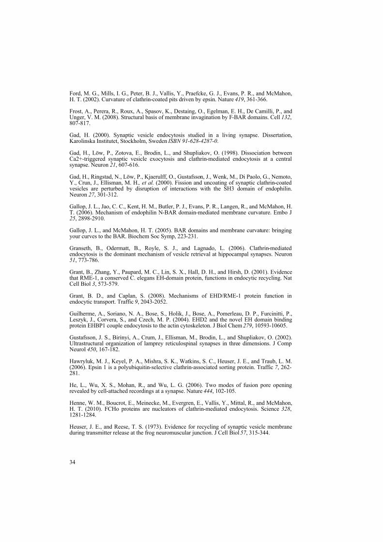
34
Ford, M. G., Mills, I. G., Peter, B. J., Vallis, Y., Praefcke, G. J., Evans, P. R., and McMahon, H. T. (2002). Curvature of clathrin-coated pits driven by epsin. Nature 419, 361-366.
Frost, A., Perera, R., Roux, A., Spasov, K., Destaing, O., Egelman, E. H., De Camilli, P., and Unger, V. M. (2008). Structural basis of membrane invagination by F-BAR domains. Cell 132, 807-817.
Gad, H. (2000). Synaptic vesicle endocytosis studied in a living synapse. Dissertation, Karolinska Institutet, Stockholm, Sweden ISBN 91-628-4287-0.
Gad, H., Löw, P., Zotova, E., Brodin, L., and Shupliakov, O. (1998). Dissociation between Ca2+-triggered synaptic vesicle exocytosis and clathrin-mediated endocytosis at a central synapse. Neuron 21, 607-616.
Gad, H., Ringstad, N., Löw, P., Kjaerulff, O., Gustafsson, J., Wenk, M., Di Paolo, G., Nemoto, Y., Crun, J., Ellisman, M. H., et al. (2000). Fission and uncoating of synaptic clathrin-coated vesicles are perturbed by disruption of interactions with the SH3 domain of endophilin. Neuron 27, 301-312.
Gallop, J. L., Jao, C. C., Kent, H. M., Butler, P. J., Evans, P. R., Langen, R., and McMahon, H. T. (2006). Mechanism of endophilin N-BAR domain-mediated membrane curvature. Embo J 25, 2898-2910.
Gallop, J. L., and McMahon, H. T. (2005). BAR domains and membrane curvature: bringing your curves to the BAR. Biochem Soc Symp, 223-231.
Granseth, B., Odermatt, B., Royle, S. J., and Lagnado, L. (2006). Clathrin-mediated endocytosis is the dominant mechanism of vesicle retrieval at hippocampal synapses. Neuron 51, 773-786.
Grant, B., Zhang, Y., Paupard, M. C., Lin, S. X., Hall, D. H., and Hirsh, D. (2001). Evidence that RME-1, a conserved C. elegans EH-domain protein, functions in endocytic recycling. Nat Cell Biol 3, 573-579.
Grant, B. D., and Caplan, S. (2008). Mechanisms of EHD/RME-1 protein function in endocytic transport. Traffic 9, 2043-2052.
Guilherme, A., Soriano, N. A., Bose, S., Holik, J., Bose, A., Pomerleau, D. P., Furcinitti, P., Leszyk, J., Corvera, S., and Czech, M. P. (2004). EHD2 and the novel EH domain binding protein EHBP1 couple endocytosis to the actin cytoskeleton. J Biol Chem 279, 10593-10605.
Gustafsson, J. S., Birinyi, A., Crum, J., Ellisman, M., Brodin, L., and Shupliakov, O. (2002). Ultrastructural organization of lamprey reticulospinal synapses in three dimensions. J Comp Neurol 450, 167-182.
Hawryluk, M. J., Keyel, P. A., Mishra, S. K., Watkins, S. C., Heuser, J. E., and Traub, L. M. (2006). Epsin 1 is a polyubiquitin-selective clathrin-associated sorting protein. Traffic 7, 262-281.
He, L., Wu, X. S., Mohan, R., and Wu, L. G. (2006). Two modes of fusion pore opening revealed by cell-attached recordings at a synapse. Nature 444, 102-105.
Henne, W. M., Boucrot, E., Meinecke, M., Evergren, E., Vallis, Y., Mittal, R., and McMahon, H. T. (2010). FCHo proteins are nucleators of clathrin-mediated endocytosis. Science 328, 1281-1284.
Heuser, J. E., and Reese, T. S. (1973). Evidence for recycling of synaptic vesicle membrane during transmitter release at the frog neuromuscular junction. J Cell Biol 57, 315-344.

35
Hinrichsen, L., Meyerholz, A., Groos, S., and Ungewickell, E. J. (2006). Bending a membrane: how clathrin affects budding. Proc Natl Acad Sci U S A 103, 8715-8720.
Hirst, J., Miller, S. E., Taylor, M. J., von Mollard, G. F., and Robinson, M. S. (2004). EpsinR is an adaptor for the SNARE protein Vti1b. Mol Biol Cell 15, 5593-5602.
Holt, M., Cooke, A., Wu, M. M., and Lagnado, L. (2003). Bulk membrane retrieval in the synaptic terminal of retinal bipolar cells. J Neurosci 23, 1329-1339.
Hussain, N. K., Yamabhai, M., Bhakar, A. L., Metzler, M., Ferguson, S. S., Hayden, M. R., McPherson, P. S., and Kay, B. K. (2003). A role for epsin N-terminal homology/AP180 N-terminal homology (ENTH/ANTH) domains in tubulin binding. J Biol Chem 278, 28823-28830.
Hyman, J., Chen, H., Di Fiore, P. P., De Camilli, P., and Brunger, A. T. (2000). Epsin 1 undergoes nucleocytosolic shuttling and its eps15 interactor NH(2)-terminal homology (ENTH) domain, structurally similar to Armadillo and HEAT repeats, interacts with the transcription factor promyelocytic leukemia Zn(2)+ finger protein (PLZF). J Cell Biol 149, 537-546.
Itoh, T., and De Camilli, P. (2006). BAR, F-BAR (EFC) and ENTH/ANTH domains in the regulation of membrane-cytosol interfaces and membrane curvature. Biochim Biophys Acta 1761, 897-912.
Itoh, T., Erdmann, K. S., Roux, A., Habermann, B., Werner, H., and De Camilli, P. (2005). Dynamin and the actin cytoskeleton cooperatively regulate plasma membrane invagination by BAR and F-BAR proteins. Dev Cell 9, 791-804.
Itoh, T., Koshiba, S., Kigawa, T., Kikuchi, A., Yokoyama, S., and Takenawa, T. (2001). Role of the ENTH domain in phosphatidylinositol-4,5-bisphosphate binding and endocytosis. Science 291, 1047-1051.
Jockusch, W. J., Praefcke, G. J., McMahon, H. T., and Lagnado, L. (2005). Clathrin-dependent and clathrin-independent retrieval of synaptic vesicles in retinal bipolar cells. Neuron 46, 869-878.
Jovic, M., Naslavsky, N., Rapaport, D., Horowitz, M., and Caplan, S. (2007). EHD1 regulates beta1 integrin endosomal transport: effects on focal adhesions, cell spreading and migration. J Cell Sci 120, 802-814.
Kalthoff, C., Groos, S., Kohl, R., Mahrhold, S., and Ungewickell, E. J. (2002). Clint: a novel clathrin-binding ENTH-domain protein at the Golgi. Mol Biol Cell 13, 4060-4073.
Keen, J. H., and Black, M. M. (1986). The phosphorylation of coated membrane proteins in intact neurons. J Cell Biol 102, 1325-1333.
Kessels, M. M., and Qualmann, B. (2004). The syndapin protein family: linking membrane trafficking with the cytoskeleton. J Cell Sci 117, 3077-3086.
Kessels, M. M., and Qualmann, B. (2006). Syndapin oligomers interconnect the machineries for endocytic vesicle formation and actin polymerization. J Biol Chem 281, 13285-13299.
Kim, S. H., and Ryan, T. A. (2009). Synaptic vesicle recycling at CNS snapses without AP-2. J Neurosci 29, 3865-3874.
Kirchhausen, T. (2000). Clathrin. Annu Rev Biochem 69, 699-727.
Koenig, J. H., and Ikeda, K. (1989). Disappearance and reformation of synaptic vesicle membrane upon transmitter release observed under reversible blockage of membrane retrieval. J Neurosci 9, 3844-3860.

36
Kohtz, D. S., and Puszkin, S. (1988). A neuronal protein (NP185) associated with clathrin-coated vesicles. Characterization of NP185 with monoclonal antibodies. J Biol Chem 263, 7418-7425.
Kumar, V., Alla, S. R., Krishnan, K. S., and Ramaswami, M. (2009). Syndapin is dispensable for synaptic vesicle endocytosis at the Drosophila larval neuromuscular junction. Mol Cell Neurosci 40, 234-241.
Leenders, A. G., Scholten, G., de Lange, R. P., Lopes da Silva, F. H., and Ghijsen, W. E. (2002). Sequential changes in synaptic vesicle pools and endosome-like organelles during depolarization near the active zone of central nerve terminals. Neuroscience 109, 195-206.
Legendre-Guillemin, V., Wasiak, S., Hussain, N. K., Angers, A., and McPherson, P. S. (2004). ENTH/ANTH proteins and clathrin-mediated membrane budding. J Cell Sci 117, 9-18.
Lemmon, M. A. (2007). Pleckstrin homology (PH) domains and phosphoinositides. Biochem Soc Symp, 81-93.
Lin, S. X., Grant, B., Hirsh, D., and Maxfield, F. R. (2001). Rme-1 regulates the distribution and function of the endocytic recycling compartment in mammalian cells. Nat Cell Biol 3, 567-572.
Lindau, M., and Alvarez de Toledo, G. (2003). The fusion pore. Biochim Biophys Acta 1641, 167-173.
Macia, E., Ehrlich, M., Massol, R., Boucrot, E., Brunner, C., and Kirchhausen, T. (2006). Dynasore, a cell-permeable inhibitor of dynamin. Dev Cell 10, 839-850.
Maldonado-Baez, L., Dores, M. R., Perkins, E. M., Drivas, T. G., Hicke, L., and Wendland, B. (2008). Interaction between Epsin/Yap180 adaptors and the scaffolds Ede1/Pan1 is required for endocytosis. Mol Biol Cell 19, 2936-2948.
Marks, B., and McMahon, H. T. (1998). Calcium triggers calcineurin-dependent synaptic vesicle recycling in mammalian nerve terminals. Curr Biol 8, 740-749.
McMahon, H. T., and Gallop, J. L. (2005). Membrane curvature and mechanisms of dynamic cell membrane remodelling. Nature 438, 590-596.
Mettlen, M., Pucadyil, T., Ramachandran, R., and Schmid, S. L. (2009). Dissecting dynamin's role in clathrin-mediated endocytosis. Biochem Soc Trans 37, 1022-1026.
Miller, T. M., and Heuser, J. E. (1984). Endocytosis of synaptic vesicle membrane at the frog neuromuscular junction. J Cell Biol 98, 685-698.
Mills, I. G., Praefcke, G. J., Vallis, Y., Peter, B. J., Olesen, L. E., Gallop, J. L., Butler, P. J., Evans, P. R., and McMahon, H. T. (2003). EpsinR: an AP1/clathrin interacting protein involved in vesicle trafficking. J Cell Biol 160, 213-222.
Modregger, J., DiProspero, N. A., Charles, V., Tagle, D. A., and Plomann, M. (2002). PACSIN 1 interacts with huntingtin and is absent from synaptic varicosities in presymptomatic Huntington's disease brains. Hum Mol Genet 11, 2547-2558.
Morgan, J. R., Prasad, K., Hao, W., Augustine, G. J., and Lafer, E. M. (2000). A conserved clathrin assembly motif essential for synaptic vesicle endocytosis. J Neurosci 20, 8667-8676.
Mullis, K., Faloona, F., Scharf, S., Saiki, R., Horn, G., and Erlich, H. (1986). Specific enzymatic amplification of DNA in vitro: the polymerase chain reaction. Cold Spring Harb Symp Quant Biol 51 Pt 1, 263-273.

37
Murthy, V. N., and Stevens, C. F. (1998). Synaptic vesicles retain their identity through the endocytic cycle. Nature 392, 497-501.
Naslavsky, N., Boehm, M., Backlund, P. S., Jr., and Caplan, S. (2004). Rabenosyn-5 and EHD1 interact and sequentially regulate protein recycling to the plasma membrane. Mol Biol Cell 15, 2410-2422.
Naslavsky, N., Rahajeng, J., Sharma, M., Jovic, M., and Caplan, S. (2006). Interactions between EHD proteins and Rab11-FIP2: a role for EHD3 in early endosomal transport. Mol Biol Cell 17, 163-177.
Neher, E., and Marty, A. (1982). Discrete changes of cell membrane capacitance observed under conditions of enhanced secretion in bovine adrenal chromaffin cells. Proc Natl Acad Sci U S A 79, 6712-6716.
Neves, G., Gomis, A., and Lagnado, L. (2001). Calcium influx selects the fast mode of endocytosis in the synaptic terminal of retinal bipolar cells. Proc Natl Acad Sci U S A 98, 15282-15287.
Neves, G., and Lagnado, L. (1999). The kinetics of exocytosis and endocytosis in the synaptic terminal of goldfish retinal bipolar cells. J Physiol 515 ( Pt 1), 181-202.
Nonet, M. L., Holgado, A. M., Brewer, F., Serpe, C. J., Norbeck, B. A., Holleran, J., Wei, L., Hartwieg, E., Jorgensen, E. M., and Alfonso, A. (1999). UNC-11, a Caenorhabditis elegans AP180 homologue, regulates the size and protein composition of synaptic vesicles. Mol Biol Cell 10, 2343-2360.
Opazo, F., Punge, A., Buckers, J., Hoopmann, P., Kastrup, L., Hell, S. W., and Rizzoli, S. O. (2010). Limited intermixing of synaptic vesicle components upon vesicle recycling. Traffic 11, 800-812.
Overstreet, E., Fitch, E., and Fischer, J. A. (2004). Fat facets and Liquid facets promote Delta endocytosis and Delta signaling in the signaling cells. Development 131, 5355-5366.
Pant, S., Sharma, M., Patel, K., Caplan, S., Carr, C. M., and Grant, B. D. (2009). AMPH-1/Amphiphysin/Bin1 functions with RME-1/Ehd1 in endocytic recycling. Nat Cell Biol 11, 1399-1410.
Park, M., Penick, E. C., Edwards, J. G., Kauer, J. A., and Ehlers, M. D. (2004a). Recycling endosomes supply AMPA receptors for LTP. Science 305, 1972-1975.
Park, S. Y., Ha, B. G., Choi, G. H., Ryu, J., Kim, B., Jung, C. Y., and Lee, W. (2004b). EHD2 interacts with the insulin-responsive glucose transporter (GLUT4) in rat adipocytes and may participate in insulin-induced GLUT4 recruitment. Biochemistry 43, 7552-7562.
Parsons, T. D., Lenzi, D., Almers, W., and Roberts, W. M. (1994). Calcium-triggered exocytosis and endocytosis in an isolated presynaptic cell: capacitance measurements in saccular hair cells. Neuron 13, 875-883.
Perez-Otano, I., Lujan, R., Tavalin, S. J., Plomann, M., Modregger, J., Liu, X. B., Jones, E. G., Heinemann, S. F., Lo, D. C., and Ehlers, M. D. (2006). Endocytosis and synaptic removal of NR3A-containing NMDA receptors by PACSIN1/syndapin1. Nat Neurosci 9, 611-621.
Pohl, U., Smith, J. S., Tachibana, I., Ueki, K., Lee, H. K., Ramaswamy, S., Wu, Q., Mohrenweiser, H. W., Jenkins, R. B., and Louis, D. N. (2000). EHD2, EHD3, and EHD4 encode novel members of a highly conserved family of EH domain-containing proteins. Genomics 63, 255-262.

38
Polo, S., Sigismund, S., Faretta, M., Guidi, M., Capua, M. R., Bossi, G., Chen, H., De Camilli, P., and Di Fiore, P. P. (2002). A single motif responsible for ubiquitin recognition and monoubiquitination in endocytic proteins. Nature 416, 451-455.
Pucadyil, T. J., and Schmid, S. L. (2008). Real-time visualization of dynamin-catalyzed membrane fission and vesicle release. Cell 135, 1263-1275.
Pucadyil, T. J., and Schmid, S. L. (2009). Conserved functions of membrane active GTPases in coated vesicle formation. Science 325, 1217-1220.
Qualmann, B., and Kelly, R. B. (2000). Syndapin isoforms participate in receptor-mediated endocytosis and actin organization. J Cell Biol 148, 1047-1062.
Qualmann, B., Roos, J., DiGregorio, P. J., and Kelly, R. B. (1999). Syndapin I, a synaptic dynamin-binding protein that associates with the neural Wiskott-Aldrich syndrome protein. Mol Biol Cell 10, 501-513.
Rao, Y., Ma, Q., Vahedi-Faridi, A., Sundborger, A., Pechstein, A., Puchkov, D., Luo, L., Shupliakov, O., Saenger, W., and Haucke, V. (2010). Molecular basis for SH3 domain regulation of F-BAR-mediated membrane deformation. Proc Natl Acad Sci U S A 107, 8213-8218.
Rapaport, D., Auerbach, W., Naslavsky, N., Pasmanik-Chor, M., Galperin, E., Fein, A., Caplan, S., Joyner, A. L., and Horowitz, M. (2006). Recycling to the plasma membrane is delayed in EHD1 knockout mice. Traffic 7, 52-60.
Rappoport, J. Z., Kemal, S., Benmerah, A., and Simon, S. M. (2006). Dynamics of clathrin and adaptor proteins during endocytosis. Am J Physiol Cell Physiol 291, C1072-1081.
Richards, D. A., Rizzoli, S. O., and Betz, W. J. (2004). Effects of wortmannin and latrunculin A on slow endocytosis at the frog neuromuscular junction. J Physiol 557, 77-91.
Robinson, P. J., Sontag, J. M., Liu, J. P., Fykse, E. M., Slaughter, C., McMahon, H., and Sudhof, T. C. (1993). Dynamin GTPase regulated by protein kinase C phosphorylation in nerve terminals. Nature 365, 163-166.
Rosenthal, J. A., Chen, H., Slepnev, V. I., Pellegrini, L., Salcini, A. E., Di Fiore, P. P., and De Camilli, P. (1999). The epsins define a family of proteins that interact with components of the clathrin coat and contain a new protein module. J Biol Chem 274, 33959-33965.
Rotem-Yehudar, R., Galperin, E., and Horowitz, M. (2001). Association of insulin-like growth factor 1 receptor with EHD1 and SNAP29. J Biol Chem 276, 33054-33060.
Roux, A., Uyhazi, K., Frost, A., and De Camilli, P. (2006). GTP-dependent twisting of dynamin implicates constriction and tension in membrane fission. Nature 441, 528-531.
Rovainen, C. M. (1979). Neurobiology of lampreys. Physiol Rev 59, 1007-1077.
Ryan, T. A., and Smith, S. J. (1995). Vesicle pool mobilization during action potential firing at hippocampal synapses. Neuron 14, 983-989.
Sankaranarayanan, S., De Angelis, D., Rothman, J. E., and Ryan, T. A. (2000). The use of pHluorins for optical measurements of presynaptic activity. Biophys J 79, 2199-2208.
Schmid, E. M., and McMahon, H. T. (2007). Integrating molecular and network biology to decode endocytosis. Nature 448, 883-888.
Shao, Y., Akmentin, W., Toledo-Aral, J. J., Rosenbaum, J., Valdez, G., Cabot, J. B., Hilbush, B. S., and Halegoua, S. (2002). Pincher, a pinocytic chaperone for nerve growth factor/TrkA signaling endosomes. J Cell Biol 157, 679-691.

39
Sharma, M., Naslavsky, N., and Caplan, S. (2008). A role for EHD4 in the regulation of early endosomal transport. Traffic 9, 995-1018.
Shimada, A., Niwa, H., Tsujita, K., Suetsugu, S., Nitta, K., Hanawa-Suetsugu, K., Akasaka, R., Nishino, Y., Toyama, M., Chen, L., et al. (2007). Curved EFC/F-BAR-domain dimers are joined end to end into a filament for membrane invagination in endocytosis. Cell 129, 761-772.
Shupliakov, O., Löw, P., Grabs, D., Gad, H., Chen, H., David, C., Takei, K., De Camilli, P., and Brodin, L. (1997). Synaptic vesicle endocytosis impaired by disruption of dynamin-SH3 domain interactions. Science 276, 259-263.
Simpson, F., Hussain, N. K., Qualmann, B., Kelly, R. B., Kay, B. K., McPherson, P. S., and Schmid, S. L. (1999). SH3-domain-containing proteins function at distinct steps in clathrin-coated vesicle formation. Nat Cell Biol 1, 119-124.
Spradling, K. D., McDaniel, A. E., Lohi, J., and Pilcher, B. K. (2001). Epsin 3 is a novel extracellular matrix-induced transcript specific to wounded epithelia. J Biol Chem 276, 29257-29267.
Sudhof, T. C. (2004). The synaptic vesicle cycle. Annu Rev Neurosci 27, 509-547.
Sundborger, A. (2010). Molecular mechanisms controlling dynamin recruitment to sites of endocytosis by SH3-domain containing proteins. Dissertation, Karolinska Institutet, Stockholm, Sweden ISBN 978-91-7409-96.
Takamori, S., Holt, M., Stenius, K., Lemke, E. A., Gronborg, M., Riedel, D., Urlaub, H., Schenck, S., Brugger, B., Ringler, P., et al. (2006). Molecular anatomy of a trafficking organelle. Cell 127, 831-846.
Takei, K., McPherson, P. S., Schmid, S. L., and De Camilli, P. (1995). Tubular membrane invaginations coated by dynamin rings are induced by GTP-gamma S in nerve terminals. Nature 374, 186-190.
Teng, H., Lin, M. Y., and Wilkinson, R. S. (2007). Macroendocytosis and endosome processing in snake motor boutons. J Physiol 582, 243-262.
Tian, X., Hansen, D., Schedl, T., and Skeath, J. B. (2004). Epsin potentiates Notch pathway activity in Drosophila and C. elegans. Development 131, 5807-5815.
Tomilin, N. V., Vasil'eva, E. V., Giliarov, A. V., and Cherniak, T. F. (2007). [Perturbations of the vesicle cycle in reticulospinal synapses of axons in lamprey after presynaptic microinjections of GTPgammaS]. Tsitologiia 49, 671-679.
Toret, C. P., and Drubin, D. G. (2006). The budding yeast endocytic pathway. J Cell Sci 119, 4585-4587.
Traub, L. M. (2003). Sorting it out: AP-2 and alternate clathrin adaptors in endocytic cargo selection. J Cell Biol 163, 203-208.
Ullrich, O., Reinsch, S., Urbe, S., Zerial, M., and Parton, R. G. (1996). Rab11 regulates recycling through the pericentriolar recycling endosome. J Cell Biol 135, 913-924.
Ungewickell, E. J., and Hinrichsen, L. (2007). Endocytosis: clathrin-mediated membrane budding. Curr Opin Cell Biol 19, 417-425.
Van der Sluijs, P., Hull, M., Webster, P., Male, P., Goud, B., and Mellman, I. (1992). The small GTP-binding protein rab4 controls an early sorting event on the endocytic pathway. Cell 70, 729-740.

40
Vanden Broeck, D., and De Wolf, M. J. (2006). Selective blocking of clathrin-mediated endocytosis by RNA interference: epsin as target protein. Biotechniques 41, 475-484.
Wang, Q., Navarro, M. V., Peng, G., Molinelli, E., Goh, S. L., Judson, B. L., Rajashankar, K. R., and Sondermann, H. (2009). Molecular mechanism of membrane constriction and tubulation mediated by the F-BAR protein Pacsin/Syndapin. Proc Natl Acad Sci U S A 106, 12700-12705.
Wasiak, S., Legendre-Guillemin, V., Puertollano, R., Blondeau, F., Girard, M., de Heuvel, E., Boismenu, D., Bell, A. W., Bonifacino, J. S., and McPherson, P. S. (2002). Enthoprotin: a novel clathrin-associated protein identified through subcellular proteomics. J Cell Biol 158, 855-862.
Wickelgren, W. O., Leonard, J. P., Grimes, M. J., and Clark, R. D. (1985). Ultrastructural correlates of transmitter release in presynaptic areas of lamprey reticulospinal axons. J Neurosci 5, 1188-1201.
Vijayakrishnan, N., Woodruff, E. A., 3rd, and Broadie, K. (2009). Rolling blackout is required for bulk endocytosis in non-neuronal cells and neuronal synapses. J Cell Sci 122, 114-125.
Von Gersdorff, H., and Matthews, G. (1994). Dynamics of synaptic vesicle fusion and membrane retrieval in synaptic terminals. Nature 367, 735-739.
Wu, W., and Wu, L. G. (2007). Rapid bulk endocytosis and its kinetics of fission pore closure at a central synapse. Proc Natl Acad Sci U S A 104, 10234-10239.
Wu, W., Xu, J., Wu, X. S., and Wu, L. G. (2005). Activity-dependent acceleration of endocytosis at a central synapse. J Neurosci 25, 11676-11683.
Wu, X. S., McNeil, B. D., Xu, J., Fan, J., Xue, L., Melicoff, E., Adachi, R., Bai, L., and Wu, L. G. (2009). Ca(2+) and calmodulin initiate all forms of endocytosis during depolarization at a nerve terminal. Nat Neurosci 12, 1003-1010.
Xu, Y., Shi, H., Wei, S., Wong, S. H., and Hong, W. (2004). Mutually exclusive interactions of EHD1 with GS32 and syndapin II. Mol Membr Biol 21, 269-277.
Yamabhai, M., Hoffman, N. G., Hardison, N. L., McPherson, P. S., Castagnoli, L., Cesareni, G., and Kay, B. K. (1998). Intersectin, a novel adaptor protein with two Eps15 homology and five Src homology 3 domains. J Biol Chem 273, 31401-31407.
Yamashita, T., Hige, T., and Takahashi, T. (2005). Vesicle endocytosis requires dynamin-dependent GTP hydrolysis at a fast CNS synapse. Science 307, 124-127.
Yap, C. C., Lasiecka, Z. M., Caplan, S., and Winckler, B. (2010). Alterations of EHD1/EHD4 protein levels interfere with L1/NgCAM endocytosis in neurons and disrupt axonal targeting. J Neurosci 30, 6646-6657.
Yoon, Y., Tong, J., Lee, P. J., Albanese, A., Bhardwaj, N., Kallberg, M., Digman, M. A., Lu, H., Gratton, E., Shin, Y. K., and Cho, W. (2010). Molecular basis of the potent membrane-remodeling activity of the epsin 1 N-terminal homology domain. J Biol Chem 285, 531-540.
Zelenin, P. V. (2005). Activity of individual reticulospinal neurons during different forms of locomotion in the lamprey. Eur J Neurosci 22, 2271-2282.
Zhang, B., Koh, Y. H., Beckstead, R. B., Budnik, V., Ganetzky, B., and Bellen, H. J. (1998). Synaptic vesicle size and number are regulated by a clathrin adaptor protein required for endocytosis. Neuron 21, 1465-1475.

41
Zhang, J. Z., Davletov, B. A., Sudhof, T. C., and Anderson, R. G. (1994). Synaptotagmin I is a high affinity receptor for clathrin AP-2: implications for membrane recycling. Cell 78, 751-760.
Zhang, Q., Li, Y., and Tsien, R. W. (2009). The dynamic control of kiss-and-run and vesicular reuse probed with single nanoparticles. Science 323, 1448-1453.
Zhou, S., Sousa, R., Tannery, N. H., and Lafer, E. M. (1992). Characterization of a novel synapse-specific protein. II. cDNA cloning and sequence analysis of the F1-20 protein. J Neurosci 12, 2144-2155.
Zoncu, R., Perera, R. M., Sebastian, R., Nakatsu, F., Chen, H., Balla, T., Ayala, G., Toomre, D., and De Camilli, P. V. (2007). Loss of endocytic clathrin-coated pits upon acute depletion of phosphatidylinositol 4,5-bisphosphate. Proc Natl Acad Sci U S A 104, 3793-3798.