extracellular vesicle-based therapy for covid-19 - mdpi
TRANSCRIPT
biomedicines
Review
Extracellular Vesicle-Based Therapy for COVID-19:Promises, Challenges and Future Prospects
Vamika Karn 1,†, Shaista Ahmed 2,†, Lung-Wen Tsai 3,4,†, Rajni Dubey 3, Shreesh Ojha 5 ,Himanshu Naryan Singh 6, Mukesh Kumar 7, Piyush Kumar Gupta 8 , Soumi Sadhu 8, Niraj Kumar Jha 9 ,Ashutosh Kumar 10, Soumya Pandit 8 and Sanjay Kumar 8,*
�����������������
Citation: Karn, V.; Ahmed, S.; Tsai,
L.-W.; Dubey, R.; Ojha, S.; Singh, H.N.;
Kumar, M.; Gupta, P.K.; Sadhu, S.;
Jha, N.K.; et al. Extracellular
Vesicle-Based Therapy for COVID-19:
Promises, Challenges and Future
Prospects. Biomedicines 2021, 9, 1373.
https://doi.org/10.3390/
biomedicines9101373
Academic Editor:
Pasquale Ambrosino
Received: 29 July 2021
Accepted: 25 September 2021
Published: 1 October 2021
Publisher’s Note: MDPI stays neutral
with regard to jurisdictional claims in
published maps and institutional affil-
iations.
Copyright: © 2021 by the authors.
Licensee MDPI, Basel, Switzerland.
This article is an open access article
distributed under the terms and
conditions of the Creative Commons
Attribution (CC BY) license (https://
creativecommons.org/licenses/by/
4.0/).
1 Department of Biotechnology, Amity University, Mumbai 410221, India; [email protected] Faculty of Medical and Paramedical Sciences, Aix-Marseille University, 13005 Marseille, France;
[email protected] Department of Medicine Research, Taipei Medical University Hospital, Taipei 11031, Taiwan;
[email protected] (L.-W.T.); [email protected] (R.D.)4 Department of Information Technology Office, Taipei Medical University Hospital, Taipei 11031, Taiwan5 Department of Pharmacology and Therapeutics, College of Medicine and Health Sciences, UAE University,
Al Ain, Abu Dhabi P.O. Box 17666, United Arab Emirates; [email protected] Department of System Biology, Columbia University Irving Medical Center, New York, NY 10032, USA;
[email protected] Department of Biophysics, All India Institute of Medical Sciences, New Delhi 110029, India;
[email protected] Department of Life Sciences, School of Basic Sciences and Research, Sharda University,
Greater Noida 201310, India; [email protected] (P.K.G.); [email protected] (S.S.);[email protected] (S.P.)
9 Department of Biotechnology, School of Engineering & Technology (SET), Sharda University,Greater Noida 201310, India; [email protected]
10 Department of Anatomy, All India Institute of Medical Sciences, Patna 801507, India;[email protected]
* Correspondence: [email protected] or [email protected]; Tel.: +91-120-4570-000† These authors contributed equally.
Abstract: The COVID-19 pandemic has become a serious concern and has negatively impacted publichealth and the economy. It primarily targets the lungs, causing acute respiratory distress syndrome(ARDS); however, it may also lead to multiple organ failure (MOF) and enhanced mortality rates.Hence, there is an urgent need to develop potential effective therapeutic strategies for COVID-19patients. Extracellular vesicles (EVs) are released from various types of cells that participate inintercellular communication to maintain physiological and pathological processes. EVs derived fromvarious cellular origins have revealed suppressive effects on the cytokine storm during systemichyper-inflammatory states of severe COVID-19, leading to enhanced alveolar fluid clearance, pro-moted epithelial and endothelial recovery, and cell proliferation. Being the smallest subclass of EVs,exosomes offer striking characteristics such as cell targeting, being nano-carriers for drug delivery,high biocompatibility, safety, and low-immunogenicity, thus rendering them a potential cell-free ther-apeutic candidate against the pathogeneses of various diseases. Due to these properties, numerousstudies and clinical trials have been performed to assess their safety and therapeutic efficacy againstCOVID-19. Hence, in this review, we have comprehensively described current updates on progressand challenges for EVs as a potential therapeutic agent for the management of COVID-19.
Keywords: COVID-19; extracellular vesicles; SARS-CoV2; therapeutic agents
1. Introduction
In December 2019, an outbreak of pneumonia fever was reported in Wuhan, HubeiProvince, China with an unknown cause of infection. Later, in January 2020, a novelcoronavirus was isolated from infected patients, which was termed SARS-CoV-2 (severe
Biomedicines 2021, 9, 1373. https://doi.org/10.3390/biomedicines9101373 https://www.mdpi.com/journal/biomedicines
Biomedicines 2021, 9, 1373 2 of 21
acute respiratory syndrome coronavirus 2) [1–5]. Owing to its occurrence in 2019, theWorld Health Organization (WHO) designated this infection as coronavirus disease 2019(COVID-19), which has created an alarming situation through higher global mortality rates.Before COVID-19, the world had witnessed two similar strains of coronaviruses in theearly 21st century, which were the severe acute respiratory distress syndrome coronavirus(SARS-CoV) and the Middle East respiratory syndrome (MERS-CoV) [6].
The SARS-CoV-2 virus comes under the family Coronaviridae and order of Nidovi-rales. The genome of SARS-CoV-2 is single-stranded, positive sense 26–32 kb large RNAwith nucleocapsid (N) protein, which is packed within an envelope and responsible forviral replication in host cells. The virus envelope is made up of three types of structuralproteins i.e., membrane (M), spike (S), and envelope (E) proteins. Among these, trimericspike (S) glycoproteins protrude from the envelope (Figure 1) and play a key role in virusentry into host cells [7].
Biomedicines 2021, 9, x FOR PEER REVIEW 3 of 21
[19–22]. Apoptotic bodies are the largest (greater than 1000 nm) and are synthesized dur-ing the apoptosis process.
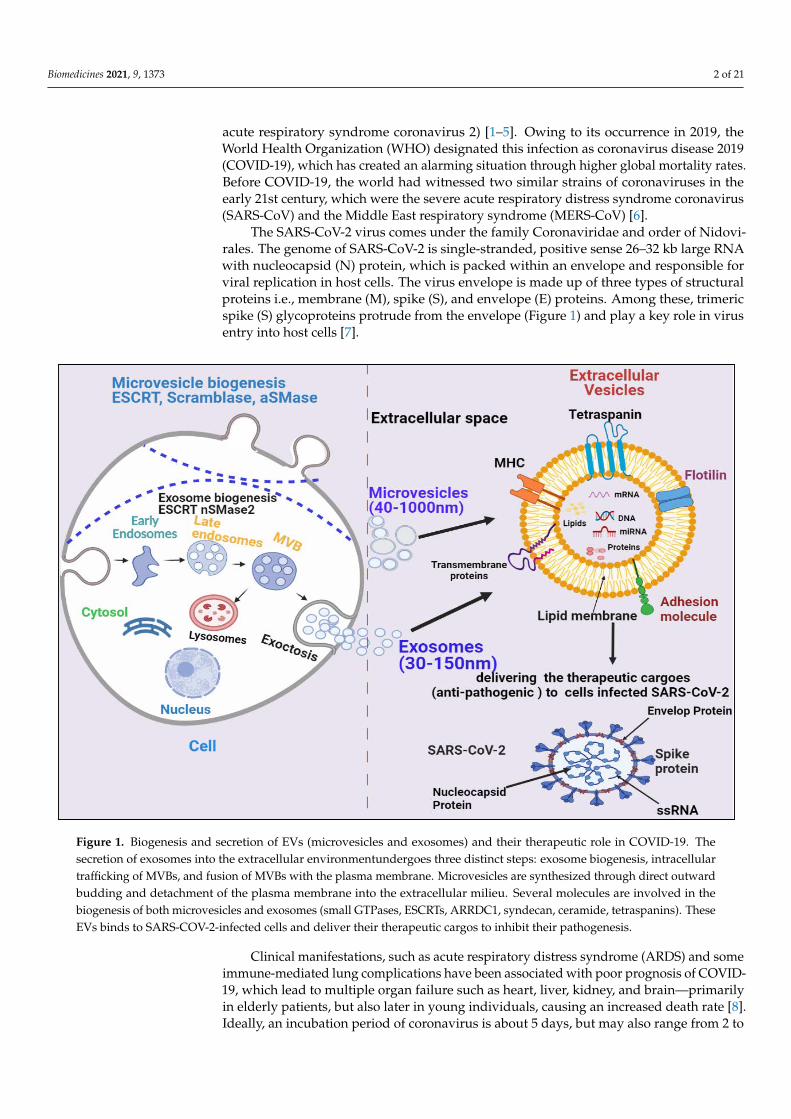
Figure 1. Biogenesis and secretion of EVs (microvesicles and exosomes) and their therapeutic role in COVID-19. The se-cretion of exosomes into the extracellular environmentundergoes three distinct steps: exosome biogenesis, intracellular trafficking of MVBs, and fusion of MVBs with the plasma membrane. Microvesicles are synthesized through direct out-ward budding and detachment of the plasma membrane into the extracellular milieu. Several molecules are involved in the biogenesis of both microvesicles and exosomes (small GTPases, ESCRTs, ARRDC1,syndecan, ceramide, tetraspanins.). These EVs binds to SARS-COV-2-infected cells and deliver their therapeutic cargos to inhibit their pathogenesis.
EVs have been demonstrated to enhance lung immunity, and have been implicated in the pathogenesis of many types of lung diseases that include viral infection. This might be attributed to the structural similarity between SARS-CoV-2 and EVs (Figure 1) [11]. Recent studies have also shown that viruses employ EVs to exit from cells, while EVs use a virus penetration mechanism for cargo delivery [23]. Hence, EV-virus interactions could be utilized for the development of antiviral vaccines and drugs to terminate viral patho-genesis. Despite EVs’ role in viral pathogenesis, their therapeutic potential has also been explored in many studies, which are discussed in the following section.
2. COVID-19-Associated Multiple Organ Failure and EV-Mediated Recovery Lungs are the first organ where SARS-CoV-2 virus enters and causes infection (Fig-
ure 2). In addition to the lungs, other vital organs such as the heart, kidney, liver, brain, and blood vessels are also infected in severely ill COVID-19 patients, which renders it a systemic disease (Figure 3). Although the mechanistic insight underlying multiple organ infection in COVID-19 is yet to be described, it might be mediated in two possible ways.
Figure 1. Biogenesis and secretion of EVs (microvesicles and exosomes) and their therapeutic role in COVID-19. Thesecretion of exosomes into the extracellular environmentundergoes three distinct steps: exosome biogenesis, intracellulartrafficking of MVBs, and fusion of MVBs with the plasma membrane. Microvesicles are synthesized through direct outwardbudding and detachment of the plasma membrane into the extracellular milieu. Several molecules are involved in thebiogenesis of both microvesicles and exosomes (small GTPases, ESCRTs, ARRDC1, syndecan, ceramide, tetraspanins). TheseEVs binds to SARS-COV-2-infected cells and deliver their therapeutic cargos to inhibit their pathogenesis.
Clinical manifestations, such as acute respiratory distress syndrome (ARDS) and someimmune-mediated lung complications have been associated with poor prognosis of COVID-19, which lead to multiple organ failure such as heart, liver, kidney, and brain—primarilyin elderly patients, but also later in young individuals, causing an increased death rate [8].Ideally, an incubation period of coronavirus is about 5 days, but may also range from 2 to
Biomedicines 2021, 9, 1373 3 of 21
14 days [9]. Following the initial symptoms of COVID-19, hypoxemia and pneumonia feverprogress, leading to the requirement of a ventilator support system [7–9]. The possibleroot cause of the higher mortality rate of COVID-19 patients is hypoxemia and respiratoryfailure, that lead to lung injury with several other complications like edema, intra-alveolarfibrin deposition, and hemorrhage resulting in ARDS [9]. It has been observed also thatindividuals with a history of cardiovascular disease, lung disorder, hypertension, anddiabetes are at higher risk of COVID-19 infection [10].
Given the present circumstances around the world, to date, various potential drugsare being clinically tested against COVID-19; however, their adequate efficacy remains tobe achieved. Therefore, it is important to develop an alternative therapeutic strategy forinfected patients and to stop the chain of SARS-CoV-2 transmission. In recent years, EVshave shown promising anti-inflammatory properties against viral infection [11]. EVs arelipid bilayer membrane-bound structures, which are released from various kinds of cells,and contain many bioactive compounds (cargo) such as mRNAs, microRNAs (miRNAs),DNA, lipids, and various proteins. Due to their intact structure, EVs can circulate throughbody fluids freely and can deliver their cargo to neighboring or remote cells to help maintaintheir physiological condition [12].
Based on their size and biogenesis, EVs have been categorized into three types, whichinclude exosomes, microvesicles, and apoptotic bodies. Exosomes are smaller in size(30–120 nm) and synthesized by the endosomal pathway, involving the formation ofintraluminal vesicles (ILVs) inside multivesicular bodies (MVBs) in the cytoplasmic com-partment of cells. MVBs bind to the inner plasma membrane, releasing their ILVs in theextracellular environment in the form of exosomes. There are several proteins involved inexosome biogenesis such as endosomal sorting complex required for transport (ESCRT),vacuolar ATPase, and Vps4, which segregate and sort ubiquitylated proteins into ILVs(Figure 1) [13–18]. Microvesicles are a little larger (40–1000 nm) in size and are releasedthrough pinching-off the plasma membrane via a direct budding process. Similar toexosomes, multiple protein factors also participate in microvesicle generation, such as Ca2+-dependent aminophospholipid translocases (flippases and floppases), sphingomyelinase 2(nSMase2), scramblases, and calpain, which carry out the rearrangement of phospholipids,curving of the membrane, and reconstitution of the actin cytoskeleton, leading to pinchingof the membrane in the form of microvesicles in extracellular milieu (Figure 1), [19–22].Apoptotic bodies are the largest (greater than 1000 nm) and are synthesized during theapoptosis process.
EVs have been demonstrated to enhance lung immunity, and have been implicated inthe pathogenesis of many types of lung diseases that include viral infection. This mightbe attributed to the structural similarity between SARS-CoV-2 and EVs (Figure 1) [11].Recent studies have also shown that viruses employ EVs to exit from cells, while EVsuse a virus penetration mechanism for cargo delivery [23]. Hence, EV-virus interactionscould be utilized for the development of antiviral vaccines and drugs to terminate viralpathogenesis. Despite EVs’ role in viral pathogenesis, their therapeutic potential has alsobeen explored in many studies, which are discussed in the following section.
2. COVID-19-Associated Multiple Organ Failure and EV-Mediated Recovery
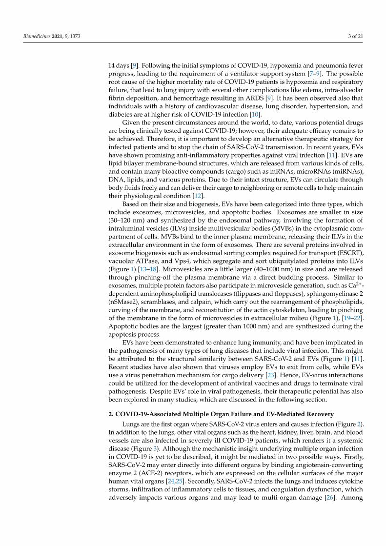
Lungs are the first organ where SARS-CoV-2 virus enters and causes infection (Figure 2).In addition to the lungs, other vital organs such as the heart, kidney, liver, brain, and bloodvessels are also infected in severely ill COVID-19 patients, which renders it a systemicdisease (Figure 3). Although the mechanistic insight underlying multiple organ infectionin COVID-19 is yet to be described, it might be mediated in two possible ways. Firstly,SARS-CoV-2 may enter directly into different organs by binding angiotensin-convertingenzyme 2 (ACE-2) receptors, which are expressed on the cellular surfaces of the majorhuman vital organs [24,25]. Secondly, SARS-CoV-2 infects the lungs and induces cytokinestorms, infiltration of inflammatory cells to tissues, and coagulation dysfunction, whichadversely impacts various organs and may lead to multi-organ damage [26]. Among
Biomedicines 2021, 9, 1373 4 of 21
multiple possible ways to tackle the COVID-19 pandemic, EVs have emerged as a potentialcell-free therapy. Based on this, EV-mediated reparation and restoration of COVID-19-affected multiple organs is being investigated and has been highlighted in this section.
Biomedicines 2021, 9, x FOR PEER REVIEW 4 of 21
Firstly, SARS-CoV-2 may enter directly into different organs by binding angiotensin-con-verting enzyme 2 (ACE-2) receptors, which are expressed on the cellular surfaces of the major human vital organs [24,25]. Secondly, SARS-CoV-2 infects the lungs and induces cytokine storms, infiltration of inflammatory cells to tissues, and coagulation dysfunction, which adversely impacts various organs and may lead to multi-organ damage [26]. Among multiple possible ways to tackle the COVID-19 pandemic, EVs have emerged as a potential cell-free therapy. Based on this, EV-mediated reparation and restoration of COVID-19-affected multiple organs is being investigated and has been highlighted in this section.
Figure 2. The pathogenesis of COVID-19 and its EV-mediated therapeutic recovery. (A)SARS-CoV-2 binds to ACE2 recep-tors on alveolar type 2 (AT2) cells in the lung and induces cytokine storms leading to lung damage. (B,C)Therapeutic effects; ACE2 receptors expressed on MSC-derived EVs competitively bind to SARS-CoV-2 and inhibit the binding of the virus to AT2 cells, and consequently inhibit the viral infection. MSC-derived EVs transfer mitochondria, proteins (KGF, and AgoI), mRNA and miRNAs via binding to CD44 receptors on macrophages, suppress the cytokine storm (IL-8, TNF-α, MIP2) and enhance anti-inflammatory cytokines (IL-10), ATP production, and oxidative phosphorylation—which helps in recovery from lung injury. MSC-derived EVs also bind to monocytes via CD44 receptors, repress the cytokine storm, and enhance anti-inflammatory cytokines IL-10, leading to recovery of the injury.
Figure 2. The pathogenesis of COVID-19 and its EV-mediated therapeutic recovery. (A) SARS-CoV-2 binds to ACE2receptors on alveolar type 2 (AT2) cells in the lung and induces cytokine storms leading to lung damage. (B,C) Therapeuticeffects; ACE2 receptors expressed on MSC-derived EVs competitively bind to SARS-CoV-2 and inhibit the binding of thevirus to AT2 cells, and consequently inhibit the viral infection. MSC-derived EVs transfer mitochondria, proteins (KGF,and AgoI), mRNA and miRNAs via binding to CD44 receptors on macrophages, suppress the cytokine storm (IL-8, TNF-α,MIP2) and enhance anti-inflammatory cytokines (IL-10), ATP production, and oxidative phosphorylation—which helps inrecovery from lung injury. MSC-derived EVs also bind to monocytes via CD44 receptors, repress the cytokine storm, andenhance anti-inflammatory cytokines IL-10, leading to recovery of the injury.
2.1. COVID-19-Associated Lung Damage and Its Recovery by EVs
The epithelium lining of lung alveoli comprises a single layer of alveolar type I (AT1)and type II (AT2) cells. AT1 and AT2 cells are firmly linked by tight junctions through whichions and fluids pass across the epithelium, whereas AT2 cells secrete a surfactant on epithe-lium linings to facilitate alveolar expansion. The expression of ACE2 receptors has beenshown on lung surfaces, mainly on AT2 cells, along with resident alveolar macrophages [27].SARS-CoV-2 binds ACE2 receptors expressed on target AT2 cells for their entry into thelung. Transmembrane serine protease 2 (TMPRSS2) expressed on alveolar cells is involvedin priming of S (spike) protein of SARS-CoV-2 that enhances the infection of other alve-olar cells [28]. This results in an elevated production of pro-inflammatory cytokines andchemokines that recruit more and more inflammatory macrophages and circulatory im-mune cells into the infected alveoli, which leads to a systemic over-inflammatory statecalled ‘cytokine storm’ [7,28]. Additionally, cytokine storms affect AT1 and AT2 cells that
Biomedicines 2021, 9, 1373 5 of 21
reduce the production of surfactants; this causes an increase in alveolar surface tensionand collapse, as well as a decrease in gaseous exchange and refractory hypoxemia, and,ultimately, leading to ARDS [23,29].
Biomedicines 2021, 9, x FOR PEER REVIEW 5 of 21
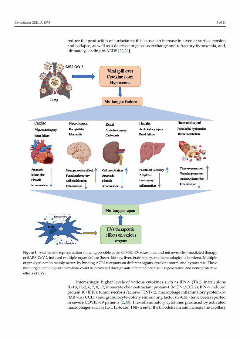
Figure 3. A schematic representation showing possible paths of MSC-EV (exosomes and microvesicles)-mediated therapy of SARS-CoV-2-induced multiple organ failure (heart, kidney, liver, brain injury, and hematological disorders). Multiple organ dysfunction mainly occurs by binding ACE2 receptors on different organs, cytokine storm, and hypoxemia. These multiorgan pathological aberrations could be recovered through anti-inflammatory, tissue regenerative, and neuroprotec-tive effects of EVs.
2.1. COVID-19-Associated Lung Damage and Its Recovery by EVs The epithelium lining of lung alveoli comprises a single layer of alveolar type I (AT1)
and type II (AT2) cells. AT1 and AT2 cells are firmly linked by tight junctions through which ions and fluids pass across the epithelium, whereas AT2 cells secrete a surfactant on epithelium linings to facilitate alveolar expansion. The expression of ACE2 receptors has been shown on lung surfaces, mainly on AT2 cells, along with resident alveolar mac-rophages [27]. SARS-CoV-2 binds ACE2 receptors expressed on target AT2 cells for their entry into the lung. Transmembrane serine protease 2 (TMPRSS2) expressed on alveolar cells is involved in priming of S (spike) protein of SARS-CoV-2 that enhances the infection of other alveolar cells [28]. This results in an elevated production of pro-inflammatory cytokines and chemokines that recruit more and more inflammatory macrophages and
Figure 3. A schematic representation showing possible paths of MSC-EV (exosomes and microvesicles)-mediated therapyof SARS-CoV-2-induced multiple organ failure (heart, kidney, liver, brain injury, and hematological disorders). Multipleorgan dysfunction mainly occurs by binding ACE2 receptors on different organs, cytokine storm, and hypoxemia. Thesemultiorgan pathological aberrations could be recovered through anti-inflammatory, tissue regenerative, and neuroprotectiveeffects of EVs.
Interestingly, higher levels of various cytokines such as IFN-γ (Th1), interleukinsIL-1β, IL-2, 6, 7, 8, 17, monocyte chemoattractant protein-1 (MCP-1/CCL2), IFN-γ inducedprotein 10 (IP10), tumor necrosis factor-α (TNF-α), macrophage inflammatory protein-1α(MIP-1α/CCL3) and granulocyte-colony stimulating factor (G-CSF) have been reportedin severe COVID-19 patients [2,30]. Pro-inflammatory cytokines produced by activatedmacrophages such as IL-1, IL-6, and TNF-α enter the bloodstream and increase the capillary
Biomedicines 2021, 9, 1373 6 of 21
permeability by dilating smooth muscle and contracting endothelial cells. Consequently,blood vessel plasma leaks into the interstitial spaces and causes alveolar edema [30].
It has been shown that EVs possess immunomodulatory effects that regulate macroph-ages by inhibiting TNFα secretion and enhancing anti-inflammatory IL-10 secretion [31].Furthermore, MSC-derived EVs have been demonstrated to boost energy productionby increasing mitochondrial performance in alveolar cells and increasing their repairingcapability in the injured lung. Data has also suggested that human-derived EVs downregulate macrophage inflammatory protein-2 (MIP-2) levels and reduce lung inflammationby lowering the recruitment of neutrophils and preventing macrophage polarization intopro-inflammatory M1 macrophages [32,33]. Apart from MSC-EVs, neutrophil-derived EVspossess an anti-inflammatory role in lung epithelium via PARP-1 inhibition by miR-223 andenhance the recovery of the injured lung [34]. Recently, EVs have been shown to reducelung edema and permeability of epithelial–endothelial barriers through binding with CD44expressed on alveolar target cells [33]. Based on these findings, human MSC-exosomesmight be a useful treatment approach in combating cytokine storms in COVID-19 patients.
2.2. COVID-19-Associated Cardiovascular Disease and Its Recovery by EVs
The knowledge of COVID-19 impact on the heart is very crucial for healthcareproviders to prescribe the appropriate treatment for patients. In a recent study, RNA-seq analysis revealed that ACE2 was expressed in over 7.5 percent of myocardial cells [35],suggesting that the heart might be at high risk of SARS-CoV-2 in case of viremia. In additionto myocardial cells, Gheblawi et al. also reported ACE2 expression in various other partsof the heart, mainly in cardiac fibroblasts, pericytes, epicardial adipose, and endothelialcells [36], thereby increasing the risk of direct infection by SARS-CoV-2 in heart tissue. Anelectron microscopy-based study showed particles consistent with COVID-19 virus presentwithin a cardiac endothelial cell and also in CD4 and CD8-positive cells around the vas-cular endothelium, which suggests that immune cells can infiltrate to cardiac tissues [37].Furthermore, histopathological examination of patients with COVID-19 reported a higherprevalence of fibrosis and myocyte hypertrophy in cardiac tissues [38], and hypothesizedthat cardiac tissue injuries may be caused indirectly by cytokine storms [39]. Additionally,coronary microvasculature dysfunction due to elevated cytokines levels can lead to myocar-dial injury [40]. In COVID-19 patients, Huang et al. demonstrated a high concentration ofpro-inflammatory mediators such as IL-1β, IL-6, IL-12, monocyte chemoattractant protein-1(MCP-1), IFNγ, and IFN-inducible protein, leading to coagulation activation [2,40]. Inautopsy studies, megakaryocytes were detected in cardiac microvasculature and bonemarrow, which suggested their having a role in diffusing microvascular thrombosis inCOVID-19 patients [41]. These data suggest that COVID-19-related cardiovascular disease(CVD) may be induced either directly by SARS-CoV-2 infection in the cardiac system orindirectly via the virus’s cytokine storm, endothelial dysregulation, infiltrating immunecells, and microvascular thrombosis.
A significant proportion of COVID-19-infected individuals develop cardiac-relatedcomplications such as acute myocardial injury (AMI), arrhythmia, or heart failure [42],which necessitates the development of novel treatment strategies. Recently, EVs attractedgreat attention from researchers over the world because of their potential role in anti-inflammation, immunomodulation, and pro-angiogenesis [43]. Lai et al. (2010) firstdemonstrated the therapeutic potential of EVs—especially MSC-EVs—in the recovery ofmyocardial ischemia or reperfusion injury in a mouse model [44]. Various other in vivostudies also reported the protective role of MSCs-EVs in AMI [45,46]. Arslan et al. showedthat a single dose of intravenous injection of MSC-derived exosomes resulted in reducedinfarct size and oxidative stress, and enhanced NADH and ATP levels, which are a sign ofrecovery of reperfusion injury in the mouse AMI model [45]. Later, Bian et al. reporteda potential pathway involving MSC-EVs in repairing ischemic myocardial injury by in-ducing neovascularization [47]. Several other notable effects of EV-mediated ischemicmyocardial repair have been achieved by reducing fibrosis and apoptosis of myocardial
Biomedicines 2021, 9, 1373 7 of 21
cells [48,49]. One of these works in rats has shown that MSC-EVs from human umbilicalcord participate in reducing cardiac fibrosis by preventing apoptosis of cardiomyocytes andenhancing cell proliferation [50]. These promising findings support our hypothesis regard-ing the therapeutic potential of MSC derived-EVs against COVID-19-related cardiovascularcomplications.
2.3. COVID-19 Associated Kidney Diseases and Their Recovery by EVs
The kidney is one of the critical organs most severely impacted by COVID-19, whichmay manifest as damage in renal resident cells [41]. Recent reports have also confirmed thatkidney disease is associated with the death of severely ill COVID-19 patients [51]. Infectioncaused due to virus–host cell interactions through ACE2 or the cytokine storm is assumedto be the underlying mechanism for renal injury [52]. Owing to ACE2 receptor-basedvirus–host cell crosstalk, ACE2 expression has been detected in various renal cells suchas proximal tubule epithelial cells, glomerular endothelial cells, podocytes, and kidneyvasculature [36]. A post mortem study of kidney biopsies from six COVID-19 patients withacute kidney injury (AKI) revealed macrophage and lymphocyte infiltration, as well assignificant acute tubular necrosis. COVID-19 nucleocapsid protein (NP) antigen has alsobeen found in kidney tubules and virus-like structures in the cytoplasm of renal tissue,tubular epithelium, and podocytes, suggesting that SARS-CoV-2 may infiltrate kidney cellsdirectly [53]. Thus, a better understanding of the biology of kidney injury in associationwith COVID-19 is highly needed.
It has recently been shown that EVs may play a role in the repair and regenerationof kidney tissue injuries by relaying signals between nephrons [54]. These signals may bedelivered by EVs, which bind receptors and transfer cargo such as proteins, mRNAs, andmiRNAs to their target cells [55]. Thus, the potential use of EVs as a therapeutic vectorhas gained significant attention in management of acute kidney injury [54,55]. Growingevidence has shown that MSC-derived EVs could reconstitute kidney structure and functionin various in vivo models of acute kidney injury (AKI). Studies also suggest that MSC-EVshave been involved in immunomodulation and anti-apoptotic activities, thus enhancingcellular proliferation and protecting against renal damage [56,57]. In various animalmodels, MSC-EVs have been demonstrated to reduce pro-inflammatory cytokines andrepair renal injuries [58]. Other studies also reported that activated macrophages infiltraterenal tissues and cause the progression of AKI. Thus, restricting infiltrating macrophagesby EVs could be an important mechanism for recovery in AKI [59,60]. Later, Shen et al.discovered higher CCR2 expression on MSC-EVs, which could lower circulating CCL2levels and reduce its ability to recruit or activate macrophages in renal tissues, and thatCCR2 knockdown reduced the protective function of MSC-exosomes for renal I/R injuriesin an in vivo model [61], indicating that receptor expression on EVs could play a key rolein their therapeutic utility. Hence, more basic and clinical research is needed to have abetter understanding of these pathways so that EVs can be used to treat COVID-19-relatedkidney damage and AKI.
2.4. COVID-19-Associated Liver Disease and Its Recovery by EVs
COVID-19 has been associated with acute liver injury (ALI), which is manifestedby elevated levels of liver enzymes i.e., alanine aminotransferase (ALT) and aspartateaminotransferase (AST) [62]. Xu et al. demonstrated the pathological results of a COVID-19-related liver biopsy, which revealed moderate microvesicular steatosis and lobular activity,as well as portal inflammation [63]. Though the mechanism underlying this pathology isnot fully understood, multiple theories have been proposed, which include direct ACE2-mediated injury in liver; specifically, the expression of ACE2 is very low in hepatocytes(2.6%), but cholangiocytes express 59.7% of total ACE2 in the liver, which is equivalent totheir expression in AT2 cells, implying that the liver is another vulnerable target organ forSARS-CoV-2 [64]. Alternately, cytokine storm-mediated dysregulation of inflammatoryand immune processes also contributes to hepatic fibrosis [65]. Besides pneumonia-related
Biomedicines 2021, 9, 1373 8 of 21
hypoxia, hypotension may also lead to liver damage or even failure in critically ill COVID-19 patients [66]. Reports have indicated that a variety of COVID-19 medications mayalso participate in hepatotoxicity that could contribute to liver damage [65,67]. However,these pathological outcomes have partially been treated by currently available therapeuticalternatives. Along with combating the virus, it is also essential to maintain the health oforgans with suitable and targeted therapy.
Recent advancements in pre-clinical studies have shown that MSC-derived EVs couldexert positive impacts on liver diseases, such as liver fibrosis, inflammation, drug-inducedliver injuries (DILI), and ALI in in vivo models [68]. Li et al., have shown that humanumbilical cord MSC-derived-EVs could alleviate carbon tetrachloride-induced liver fibrosisin mice by inhibiting the epithelial–mesenchymal transition of hepatocytes and collagensynthesis [69]. Recently, engineered human umbilical cord perivascular cell (HUCPVC)-derived EVs have been shown to produce insulin-like growth factor-I (IGF-I) upon theiradministration, reducing hepatic fibrosis in mice [70]. Amnion MSC (AMSC)-derivedEVs could reduce inflammation and fibrosis by downregulating the production of pro-inflammatory cytokines such as TNF-α, IL-1β, and IL-6,as well as inhibiting the expressionof kuffer cells—particularly M1 macrophages in mice liver [71]. Additionally, embryonicMSC-derived EVs facilitate regeneration of hepatocytes in carbon tetrachloride-inducedliver injury by activating the IL¬6/STAT3 pathway [68]. Furthermore, Lou et al. foundthat adipose tissue MSC-exosomes could reduce elevated serum ALT and AST levels aswell as the production of pro-inflammatory cytokines in concanavalin A (Con A)-inducedhepatitis in C57BL/6 mice [72]. Interestingly, liver stem cell-derived EVs have been shownto accelerate liver structural integrity and function in 70% of hepatectomized rats bypromoting hepatocyte proliferation [73]. Based on these pieces of evidence, it could beinferred that EVs from different sources may prevent various types of liver disease byreducing inflammation and collagen production and enhancing hepatocyte proliferation.Since COVID-19-induced liver pathologies such as fibrosis, DILI, and ALI have alreadybeen reported, it is likely that MSC-derived EVs could be potential therapeutic candidatesfor such complications.
2.5. COVID-19 Associated Neurological Diseases and Their EV-Mediated Recovery
Numerous studies have reported that COVID-19 is associated with several life-threatening neuropathologic manifestations such as encephalopathy, meningitis, andGuillain–Barre Syndrome [25,71–74]. In addition, the COVID-19 virus has been detectedin human brain tissues and cerebrospinal spinal fluids (CSF) [75–77]. In the brain, ACE2is expressed in neurons, astrocytes, and oligodendrocytes with higher prevalence in themotor cortex, posterior cingulate cortex, ventricles, circumventricular organs, thalamus,and olfactory bulb [76,78]. Furthermore, COVID-19 viral-like particles have also beendetected in brain endothelial cells of autopsied patient tissues presenting at least one cellmembrane bleb [75,79]. Although COVID-19 virus was not detected in primary humanendothelial cells from brain tissues lacking ACE2 expression in vitro, endothelial cellsover expressed with ACE2 were shown to promote infection in vivo [80], implying thatCOVID-19 infection in endothelial cells depends on the expression of ACE2. Currently,COVID-19 infected neurons have been associated with neurodegeneration and neurovas-cular alterations [81]. Elevated levels of inflammatory cytokines such as IL-6 and TNF-αhave also been shown in the CSF of COVID-19 patients with neurological presentation,indicating an ongoing inflammatory process in the brain [82,83]. Cytokines such as IL-6,TNF-α, IL-1β, and IFN-γ, along with chemokines and acute phase C-reactive protein candisrupt and modulate the functions of the blood–brain barrier (BBB), which can influenceadsorptive transcytosis [84–87]. Coagulation abnormalities due to high inflammatoryresponses leading to stroke were confirmed in COVID-19 patients [88]. Taken together, theabove-mentioned evidence implies that COVID-19 infection may participate in damage,apoptosis, and dysfunction of brain microvascular endothelial cells and neurons, which
Biomedicines 2021, 9, 1373 9 of 21
may lead to neurological dysfunction [81,88,89]. Therefore, it is much needed to find abetter therapeutic approach to facilitate positive clinical outcomes in COVID-19 patients.
Recent progress in EV research has demonstrated MSC-derived EVs as potentialtherapeutic tools for neurological disorders [90]. Specifically, exosomes may facilitatethe functional restoration of neurological abnormalities by promoting neurogenesis andBBB integrity, suppressing inflammation and apoptosis, and leading to mitigated diseaseprogression [91,92]. Bone marrow MSC-derived exosomes have been reported to suppressneuronal apoptosis and foster the functional recovery of the spinal cord after CNS injuryby stimulating Wnt/β-catenin signaling [93,94]. Human umbilical cord MSC-derivedexosomes can inhibit the activation of A1 astrocytes and act as anti-inflammatory mediatorsby regulating Nrf2/NF-κB signaling [95]. This study indicates that these exosomes may bea potential therapy for the treatment of inflammation-associated neurological dysfunction.In a preclinical cerebral hemorrhage stroke model, MSC-derived exosomes have beenshown to support functional restoration and to remodel neurovascular defects [96,97]. Insum, these data show that EVs can traverse blood–tissue barriers to repair injured neuronsduring the development of COVID-19-related neurological disorders [90].
2.6. EV-Mediated Recovery of COVID-19-Associated Hematological Disorders
Several hematological abnormalities like lymphopenia, thrombocytopenia, and co-agulation defects have been associated with COVID-19 patients. Of these, lymphopeniahas been the most commonly observed disease in COVID-19 [98]. Therefore, it is specu-lated that the virus might directly infect lymphocytes, which express ACE2 receptors [99].COVID-19 patients with lymphopenia also seem to have elevated levels of different pro-inflammatory cytokines [100]. Lungs, the primary site for platelet biogenesis, also exhibita substantial hematopoietic potential [101]. This could be proven in terms of hamperedplatelet production in the damaged lungs of COVID-19 patients, resulting in thrombocy-topenia [102]. Later on, COVID-19-related coagulation abnormalities are often associatedwith the combination of inflammation, activation of platelets, and endothelial dysfunc-tion [103]. In COVID-19 patients, the higher levels of Factor VIII and von Wille brandfactor were reported, which could promote endothelial injury—possibly mediated viaACE2 receptor binding [79,104]. Therefore, the aggravated endothelial injury observedin COVID-19 may lead to a pro-coagulatory state resulting in both macro and microvas-cular thrombotic episodes. Thus, therapies targeting the restoration and prevention ofhematological changes such as endothelial dysfunction and coagulation abnormalities mayimprove COVID-19 patient outcomes.
The use of EVs for therapeutic and diagnostic purposes in hematological disorders isa emerging field of research. In hematological findings, circulating EVs, particularly thoseproduced by leukocytes, neutrophils, and endothelial cells, have been shown to activatenumerous other cells in the blood arteries, including endothelial cells. The intrinsic im-munomodulatory characteristics of EVs may also enhance tissue regeneration and vascularrepair. Neutrophil-derived EVs autocrinally reduce immune activation and significantlydampen pro-inflammatory cytokine secretion from monocytes [105]. In COVID-19, themajor etiologies of ARDS include pneumonia, sepsis, and the invading pathogens [106].The recruitment of neutrophils to inflamed tissue is required to eliminate pathogens; theseneutrophils may secrete EVs at the site of inflammation [55,107] and contribute to reducingcytokine storms caused in COVID-19 [105]. Many studies have shown that endothelial-derived EVs manifest anticoagulant and vasculo-protective potential [108,109] and can aidin plasmin synthesis by plasminogen, which in turn facilitates clot dissolution throughamplified fibrinolysis [110]. As a result, we may infer that EVs generated from neutrophilsand endothelial cells could be used to treat COVID-19-related coagulation and hematologi-cal problems. However, further research is needed to understand the varying roles of EVsproduced from various sources in the blood in order to use them as a cell-free treatment forCOVID-19 patients with hematologic diseases.
Biomedicines 2021, 9, 1373 10 of 21
3. Translational Potential of EVs in COVID-19 Management3.1. MSC-Derived EVs as Promising Medications
MSCs have been extensively investigated for their therapeutic usefulness in treatingvarious disorders due to their strong regenerative and immunomodulatory capabilities.There are several available sources of MSCs, for instance, bone marrow, adipose tissue,dental pulp, umbilical cord tissue, and amniotic tissue; however, their therapeutic poten-tial may vary depending on their source of origin and the activation of various Toll-likereceptors [111,112]. MSCs secrete various cytokines and growth factors such as IL-10,vascular endothelial growth factor (VEGF), hepatocyte growth factor, and keratinocytegrowth factor (KGF), which resist fibrosis, mitigate ARDS, and are involved in regenerationand repair of lung damage [113,114]. MSCs could not only restrict aberrant T cell andmacrophage production but also enhance their differentiation into functional T cells andanti-inflammatory macrophages, respectively. Additionally, MSCs regulate B cells anddendritic cells, which may be useful in tackling the cytokine storm observed in COVID-19patients [115–118]. A plethora of studies have shown that MSC-derived EVs perform simi-lar functions to their parental cells i.e., MSCs, which suggests that the therapeutic efficiencyof MSCs in different diseases has been mainly contributed by their secreted EVs [119–122].Studies have revealed comparable therapeutic effects of EVs and MSCs in suppressinginflammatory process and edema development in the lungs [123]. Therefore, MSC-derivedEVs have gained more attention for exploitation as a cell-free therapy (Figure 2).
MSC-derived EVs are thought to play a therapeutic function in COVID-19 by deliveringprotective and anti-inflammatory RNAs and proteins to damaged or activated cells in lungtissues [124–126]. Reportedly, MSC-EVs are enriched with various types of microRNAs—for instance, let-7, miR-124-3p, miR-21-5p, miR-146a and miR-145 [124,125,127]. Of these,miR-124-3p has been involved in suppressing oxidative stress and inflammatory cytokinesby binding to its receptor P2X ligand-gated ion channel 7 (P2X7) [124]. Another miR-21-5phas been associated with reducing lung cell apoptosis through inhibition of PTEN andPDCD4, whilst miR-146a participates in transforming macrophages from pro-inflammatoryto anti-inflammatory states by suppressing the NF-κbsignalling pathway [124]. Lastly,miR-145 increases the phagocytic property of macrophages for fast clearance of pathogensat the site of infection [127]. However, our understanding of these EVs is limited, and morestudies are required to ensure their robustness and dependability as a viable therapy forcombating COVID-19.
3.2. Platelet-Derived EV-Based Therapy
Immunomodulatory properties of convalescent blood products such as whole blood,plasma, and serum aid in wound healing of damaged lungs [128]. In particular, plasmahas been successfully used for treatment of COVID-19 patients. During apheresis, manygrowth factors, neutralizing antibodies, and EVs found in plasma are delivered into pa-tients. EVs in blood circulation are mainly contributed by platelets, which is more thanhalf of the total EVs in the peripheral blood [128,129]. Many studies have demonstratedthat plasma-derived EVs express abundant growth factors and participate in the activationof various signaling mechanisms and changes in vascular reactivity, as well as inducing an-giogenesis for tissue repair [128–130]. Additionally, platelet-derived EVs promote woundhealing in several organs by inducing cell proliferation and migration via various signal-ing pathways [130,131], which suggests that convalescent plasma therapy for COVID-19patients is mainly contributed to by their circulating EVs.
Engineered platelet-derived EVs packed with anti-inflammatory molecule TPCA-1have been shown to be very promising in the curing of pneumonia by inhibiting the inflam-matory process and reducing the cytokinestorm in a mouse model [132]. A report has alsoshown that SARS-CoV-2 binds to ACE2 expressed on endothelial cells and causes damageto endothelial integrity, leading to abnormal angiogenesis [133]. Additionally, it has beenproven that platelet-derived EVs enhance the angiogenesis process to repair endothelialintegrity after vascular injury [129–134]. Another study has also indicated that platelet-EVs
Biomedicines 2021, 9, 1373 11 of 21
carry a variety of growth factors associated with the Akt and Erk pathways, and play keyroles in angiogenesis and neurogenesis [135–137]. Additionally, the combination of bonemarrow stromal cells (BMSCs) and platelet-EVs carrying proteins and non-coding RNAsenhances cell proliferation, migration, and osteogenesis [129,130]. Based on these piecesof evidence, platelet-derived EVs could be deployed as an alternate potential therapeuticoption for COVID-19 patients.
3.3. EV-Based Vaccines for COVID-19 Prevention
EVs have been characterized as highly stable, less toxic, and low-immunogenic, mak-ing them a potential candidate for developing vaccines against COVID-19 [138]. Besidestherapeutics, vaccines are very important for preventing SARS-CoV-2 infection in humans.Currently, multiple vaccines are being used worldwide to boost immunity against SARS-CoV-2 over a large population [139]. Several other clinical trials on different vaccines areunderway to assess their efficacy and safety against COVID-19. Lipid nanoparticles havebeen utilized as a vehicle for vaccine development against COVID-19. Vaccines usingnanoparticles encapsulated in mRNAs-1273 (BNT162b1, CVnCoV) and saRNAs (LNPn-CoVsaRNA) have been employed to prevent COVID-19 virus infection in many countriessuch as Germany, Belgium, and the United States [140]. Being natural lipid bilayer mem-brane nano-vesicles, EVs could be an alternate novel avenue in development of vaccines todeal with this pandemic [141,142]. EV-based vaccines carrying SARS-S spike proteins wereassessed and compared with adenoviral vector vaccine. Both EV-vaccines and adenoviralvectors have shown encouraging outcomes in neutralizing antibody titers at the samelevel. After combination with both adenoviral vector and EV-vaccine-carrying S protein,the highest level of neutralization of antibody titer was achieved, which was greater thanthe convalescent serum of SARS patients [143]. EVs have also been shown to interactwith immune cells and activate immune responses to recognize and neutralize specifictypes of cells [144]. Additionally, EVs have been found to have a higher efficiency thanthat of soluble proteins utilized in vaccines. This might be attributed to the production ofmultiple copies of the same viral protein exposed to EVs, which facilitates the cross-linkingof EVs and B-cell receptors [145]. These findings imply that EVs containing SARS-CoV-2components might be used as a COVID-19 vaccine.
3.4. Engineered EVs as Delivery Vehicles for COVID-19 Therapy
Exosome therapy can promote endogenous repair and reduce the cytokine stormstimulated by the immune system. It also offer several advantages such as easy storage, lowimmunogenicity, high stability, and the capability to pass through the BBB [146]. Along withthese advantages, their biocompatibility, potentiality for off-shelf availability, and stablemembrane composition make them the perfect choice for a drug delivery vehicle [147].Apart from being endogenous in nature, exosomes can also be engineered and utilizedas carriers for delivering specific payloads or drugs. Therefore, the antiviral drugs orimmune modulator-loaded exosomes can be delivered directly and internally to targetedsites such as the nasal mucosa and lungs to stimulate antigen-specific immune responses.This strategy of encapsulating drugs into exosomes enhances delivery to targeted organsand minimizes toxicities caused by native drugs. Numerous research works have shownpromising outcomes such as successful delivery of therapeutic molecules through EVs.There are two types of advantages observed with engineered EVs: firstly, they cannot berecognized by the host immune system. Secondly, they enhance tissue or cell-specificityfor targeted delivery. In order to enhance the targeted delivery and biodistribution oftherapeutic components to particular sites in the human body, engineered EVs can beanchored with specific peptides that recognize specific cell surfaces in target tissues [148].Along with their natural anti-inflammatory effects, these engineered EVs suppress viralreplication in host cells, and reduce the cytokine storm and ARDS associated with COVID-19 patients [149].
Biomedicines 2021, 9, 1373 12 of 21
EVs have been utilized to deliver a variety of therapeutic molecules to treat variouslung disorders, including lung inflammation. Small molecules transported by EVs, suchas MyD88 siRNA or miR-223/142, have been shown to block the NF-kb signaling path-way or the activation of the Nlrp3 inflammasome in alveolar macrophages, leading to areduction in lung inflammation [150,151]. While, by using modified surface molecules,EVs may be utilized to target SARS-CoV-2-infected specific cells or tissues for therapeuticpurposes [152]. Other compounds, such as nano/antibodies, DNA aptamers, and peptideswith caveolin-1 or Ly-6G specificity, were loaded into EVs, allowing anti-inflammatorydrugs to be delivered to particular lung epithelial cells and macrophages;this might be a keyapproach in COVID-19 management to overcome the cytokine storm [153,154]. Anotherapproach using SARS-CoV-2 model cell line (Vero CCL-81 or Vero E6)-derived EVs carryingsurface proteins could be utilized for delivering encapsulated drugs to specific alveolarmacrophages produced due to SARS-CoV2 infection and reduce the cytokine storm [154].Hence, these strategies can be very useful in repurposing drugs for treating COVID-19 viaEV-based drug delivery.
4. Clinical Trials on EVs for COVID-19 Treatments
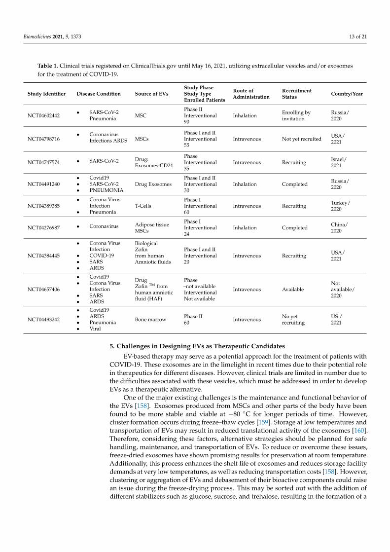
EVs derived from various sources of MSCs including bone marrow, adipose tissue,peripheral blood, placenta, umbilical cord, amniotic fluid, and gingival tissues are beinginvestigated for development as therapeutics targeting several diseases [155]. We identifiednine clinical trial-based studies on EVs for COVID-19 therapy on the clinicaltrials.govwebsite. Some of these studies are under trial, while two others have been completed andhave shown encouraging data in terms of their effectiveness and safety against COVID-19(Table 1).
In a completed clinical trial (NCT04276987), the efficacy and safety of exosomesderived from adipose tissue-MSCs were assessed in 24 COVID-19 pneumonia patients.In this study, 2 × 10⁸ EVs were administered to the patients, though data have not beenpublished yet. The Direct Biologics company has launched ExoFlo™, an exosome-baseddrug derived from bone marrow MSCs, and its efficacy has been checked in 24 severelyill COVID-19 patients. A single dose of ExoFlo™ has been found very safe without anysevere side effects in those patients, with significant rises in oxygen levels and reducedARDS symptoms;as additionally, declined levels of acute-phase reactant markers such asC-reactive proteins, ferritin and D-dimers were shown after 14 days of drug administration—although some patients had died during the trial due to other complications not related toExoFloTM [156]. The Direct Biologics company has also started a multicentric clinical trial(EXIT-COVID-19) with sixty COVID-19 patients with ARDS and pneumonia to analyzethe therapeutic potential of exosomes (NCT04493242). However, recruitment of patientshas not been initiated as per the clinicaltrial.gov website. Despite the fact that ExoFloTMhas demonstrated a number of therapeutic benefits, several questions are to be answeredabout its production, including how it was derived from bone marrow-MSCs, its biologicalactivities, infusion dose concentrations, and long-term (72-h) effects after administration topatients [157]. These issues need to addressed prior to its therapeutic use. Additionally,there is a need for more EV-based clinical trials on large numbers of severe COVID-19patients in order to evaluate their therapeutic relevance in combating this pandemic.
Biomedicines 2021, 9, 1373 13 of 21
Table 1. Clinical trials registered on ClinicalTrials.gov until May 16, 2021, utilizing extracellular vesicles and/or exosomesfor the treatment of COVID-19.
Study Identifier Disease Condition Source of EVsStudy PhaseStudy TypeEnrolled Patients
Route ofAdministration
RecruitmentStatus Country/Year
NCT04602442• SARS-CoV-2
Pneumonia MSCPhase IIInterventional90
Inhalation Enrolling byinvitation
Russia/2020
NCT04798716• Coronavirus
Infections ARDS MSCsPhase I and IIInterventional55
Intravenous Not yet recruited USA/2021
NCT04747574 • SARS-CoV-2 Drug:Exosomes-CD24
PhaseInterventional35
Intravenous Recruiting Israel/2021
NCT04491240• Covid19• SARS-CoV-2• PNEUMONIA
Drug ExosomesPhase I and IIInterventional30
Inhalation Completed Russia/2020
NCT04389385• Corona Virus
Infection• Pneumonia
T-CellsPhase IInterventional60
Intravenous Recruiting Turkey/2020
NCT04276987 • Coronavirus Adipose tissueMSCs
Phase IInterventional24
Inhalation Completed China/2020
NCT04384445
• Corona VirusInfection
• COVID-19• SARS• ARDS
BiologicalZofinfrom humanAmniotic fluids
Phase I and IIInterventional20
Intravenous Recruiting USA/2021
NCT04657406
• Covid19• Corona Virus
Infection• SARS• ARDS
DrugZofin TM fromhuman amnioticfluid (HAF)
Phase–not availableInterventionalNot available
Intravenous AvailableNotavailable/2020
NCT04493242
• Covid19• ARDS• Pneumonia• Viral
Bone marrow Phase II60 Intravenous No yet
recruitingUS /2021
5. Challenges in Designing EVs as Therapeutic Candidates
EV-based therapy may serve as a potential approach for the treatment of patients withCOVID-19. These exosomes are in the limelight in recent times due to their potential rolein therapeutics for different diseases. However, clinical trials are limited in number due tothe difficulties associated with these vesicles, which must be addressed in order to developEVs as a therapeutic alternative.
One of the major existing challenges is the maintenance and functional behavior ofthe EVs [158]. Exosomes produced from MSCs and other parts of the body have beenfound to be more stable and viable at −80 ◦C for longer periods of time. However,cluster formation occurs during freeze–thaw cycles [159]. Storage at low temperatures andtransportation of EVs may result in reduced translational activity of the exosomes [160].Therefore, considering these factors, alternative strategies should be planned for safehandling, maintenance, and transportation of EVs. To reduce or overcome these issues,freeze-dried exosomes have shown promising results for preservation at room temperature.Additionally, this process enhances the shelf life of exosomes and reduces storage facilitydemands at very low temperatures, as well as reducing transportation costs [158]. However,clustering or aggregation of EVs and debasement of their bioactive components could raisean issue during the freeze-drying process. This may be sorted out with the addition ofdifferent stabilizers such as glucose, sucrose, and trehalose, resulting in the formation of a
Biomedicines 2021, 9, 1373 14 of 21
hydration circle surrounding EVs throughout the freeze-drying process, preventing theiraggregation and maintaining their membrane integrity.
Another difficulty with modified EVs as drug delivery vehicles is that their depositionin specific cells, tissues, or organs might have a variety of negative consequences, affectingtheir efficacy and safety in curing illness. To overcome this, coating of EVs with syntheticmaterials such as polyethylene glycol or streptavidin have been shown to increase vesiclebioavailability and extravasation capacity, and thus could increase their accumulationin lung tissue affected by COVID-19. Engineered EVs with certain specific proteins orpeptides have exhibited the ability to increase the tissue specificity of EVs, which mighthelp us better comprehend such customized treatment approaches in the future [148].
One of the most prominent issues with EVs is their source of origin, since the majorityof EVs derived from cancer cell cultures include tumorigenic miRNAs, which have beenproven to dramatically increase the process of carcinogenesis [161,162]. Hence, they arenot appropriate to be utilized as therapeutic agents. Thus, the source of EVs should beconsidered in order to better fulfil the needs of EV-based therapies.
Another limitation with EVs is the lack of standardized functional assays to determinethe activity of EV preparations [163]. The techniques used in the isolation and purificationof EVs for large-scale production have not been well standardized up to the gold standard,making it challenging to employ EVs as a therapeutic alternative [164]. Gel filtrationchromatography and ultracentrifugation are being used for the isolation and purificationof EVs. Ultracentrifugation, on the other hand, has a number of limitations, includinghigh equipment costs, long run durations, labor-intensiveness, and restricted portability,whereas gel filtration chromatography has low yields and is time-consuming [148,165].Overall, perfection of these methods to achieve highly purified EV yields at large scalesis very challenging. Therefore, genetic engineering can play a crucial role in developingstrategies for improved therapeutic function of EVs. However, there are still many issuesremaining for debate, such as transformation and differentiation of EV properties.
6. Concluding Remarks and Future Directions
To combat the COVID-19 pandemic, a multidirectional approach must be establishedto reduce its pervasiveness. For the treatment of severe cases and prevention of aggrava-tion, MSCs and exosome therapy could be potential therapeutic options. MSCs have thepotential to stimulate endothelium and epithelial healing by transferring EV componentsacross cells via intercellular communication and secretion of soluble factors— resulting inincreased alveolar fluid clearance, making them a viable therapeutic option for COVID-19treatment. Following this, several clinical trials are being conducted to determine theeffectiveness and safety of EVs; however, only a few have been accomplished. The symp-toms of pneumonia, ARDS, inflammation, and sepsis, which are important contributors toCOVID-19 pathogenesis, have demonstrated steady improvement with EV-based treatment.However, the most effective and safest method of EV distribution has yet to be identified.The immunomodulatory, regenerative, and antibacterial properties of EVs have been as-cribed as their contribution in COVID-19 therapy. Exosomes also offer multiple benefits,including the capacity to transport drugs, high biocompatibility, minimal immunogenicity,and cell targeting in host cells, making them an attractive choice for off-shelf therapies.Based on the numerous clinical findings stated above, EVs can be established as a cell-freetherapy and as drug delivery vehicles in COVID-19 management. However, procedures forisolating EVs, as well as effectiveness and safety measures, and appropriate ethical normsshould all be well standardized.
Author Contributions: Conceptualization, writing—original draft preparation, writing—reviewand editing, supervision, artwork and schemes, project administration S.K.; writing—original draftpreparation, M.K. writing—review V.K. and S.A.; review and editing—S.O., H.N.S., M.K., R.D.,L.-W.T., A.K., P.K.G., S.S., S.P. and N.K.J. All authors have read and agreed to the published versionof the manuscript.
Biomedicines 2021, 9, 1373 15 of 21
Funding: This work required no external funding.
Institutional Review Board Statement: This study did not involve humans or animals.
Informed Consent Statement: This study did not involve humans.
Data Availability Statement: This study did not report any data.
Acknowledgments: All the authors are grateful to the Department of Life Sciences, School of BasicSciences and Research, Sharda University, Greater Noida, for providing the infrastructure andfacilities for this research. Also, biorender software is highly acknowledged for artwork and schemes.
Conflicts of Interest: All authors declare no competing interests with the work presented in themanuscript.
References1. Du, J.; Dong, L.; Wang, T.; Yuan, C.; Fu, R.; Zhang, L.; Liu, B.; Zhang, M.; Yin, Y.; Qin, J.; et al. Psychological symptoms among
frontline healthcare workers during COVID-19 outbreak in Wuhan. Gen. Hosp. Psychiatry 2020, 67, 144–145. [CrossRef] [PubMed]2. Huang, C.; Wang, Y.; Li, X.; Ren, L.; Zhao, J.; Hu, Y.; Zhang, L.; Fan, G.; Xu, J.; Gu, X.; et al. Clinical Features of Patients Infected
with 2019 Novel Coronavirus in Wuhan, China. Lancet 2020, 395, 497–506. [CrossRef]3. Nagoor Meeran, M.F.; Javed, H.; Sharma, C.; Goyal, S.N.; Kumar, S.; Jha, N.K.; Ojha, S. Can Echinacea Be a Potential Candidate
to Target Immunity, Inflammation, and Infection-The Trinity of Coronavirus Disease 2019. Heliyon 2021, 7, e05990. [CrossRef][PubMed]
4. Zhou, F.; Yu, T.; Du, R.; Fan, G.; Liu, Y.; Liu, Z.; Xiang, J.; Wang, Y.; Song, B.; Gu, X.; et al. Clinical Course and Risk Factors forMortality of Adult Inpatients with COVID-19 in Wuhan, China: A Retrospective Cohort Study. Lancet 2020, 395, 1054–1062.[CrossRef]
5. Meeran, M.F.N.; Sharma, C.; Goyal, S.N.; Kumar, S.; Ojha, S. CB2 Receptor-Selective Agonists as Candidates for TargetingInfection, Inflammation, and Immunity in SARS-CoV-2 Infections. Drug Dev. Res. 2021, 82, 7–11. [CrossRef] [PubMed]
6. Sahin, A.R.; Erdogan, A.; Mutlu Agaoglu, P.; Dineri, Y.; Cakirci, A.Y.; Senel, M.E.; Okyay, R.A.; Tasdogan, A.M. 2019 NovelCoronavirus (COVID-19) Outbreak: A Review of the Current Literature. EURASIAN J. Med. Oncol. 2020, 4, 1–7. [CrossRef]
7. Kumar, A.; Prasoon, P.; Sekhawat, P.S.; Pareek, V.; Faiq, M.A.; Kumari, C.; Narayan, R.K.; Kulandhasamy, M.; Kant, K. Pathogenesisguided therapeutic management of COVID-19: An immunological perspective. Int. Rev. Immunol. 2021, 40, 54–71. [CrossRef][PubMed]
8. Tsuchiya, A.; Takeuchi, S.; Iwasawa, T.; Kumagai, M.; Sato, T.; Motegi, S.; Ishii, Y.; Koseki, Y.; Tomiyoshi, K.; Natsui, K.; et al.Therapeutic potential of mesenchymal stem cells and their exosomes in severe novel coronavirus disease 2019 (COVID-19) cases.Inflamm. Regen. 2020, 40, 14. [CrossRef]
9. Al-Khawaga, S.; Abdelalim, E.M. Potential application of mesenchymal stem cells and their exosomes in lung injury: An emergingtherapeutic option for COVID-19 patients. Stem Cell Res. Ther. 2020, 11, 437. [CrossRef]
10. Sanyaolu, A.; Okorie, C.; Marinkovic, A.; Patidar, R.; Younis, K.; Desai, P.; Hosein, Z.; Padda, I.; Mangat, J.; Altaf, M. Comorbidityand Its Impact on Patients with COVID-19. SN Compr. Clin. Med. 2020, 1–8. [CrossRef]
11. Pocsfalvi, G.; Mammadova, R.; Ramos Juarez, A.P.; Bokka, R.; Trepiccione, F.; Capasso, G. COVID-19 and Extracellular Vesicles:An Intriguing Interplay. Kidney Blood Press. Res. 2020, 45, 661–670. [CrossRef]
12. Fujita, Y.; Hoshina, T.; Matsuzaki, J.; Kadota, T.; Fujimoto, S.; Kawamoto, H.; Watanabe, N.; Sawaki, K.; Sakamoto, Y.; Miyajima,M.; et al. Early Prediction of COVID-19 Severity Using Extracellular Vesicles and Extracellular RNAs. medRxiv 2020. [CrossRef]
13. Jabbari, N.; Karimipour, M.; Khaksar, M.; Akbariazar, E.; Heidarzadeh, M.; Mojarad, B.; Aftab, H.; Rahbarghazi, R.; Rezaie, J.Tumor-derived extracellular vesicles: Insights into bystander effects of exosomes after irradiation. Lasers Med. Sci. 2020, 35,531–545. [CrossRef]
14. Kowal, J.; Tkach, M.; Théry, C. Biogenesis and Secretion of Exosomes. Curr. Opin. Cell Biol. 2014, 29, 116–125. [CrossRef]15. Jeppesen, D.K.; Fenix, A.M.; Franklin, J.L.; Higginbotham, J.N.; Zhang, Q.; Zimmerman, L.J.; Liebler, D.C.; Ping, J.; Liu, Q.; Evans,
R.; et al. Reassessment of Exosome Composition. Cell 2019, 177, 428–445.e18. [CrossRef]16. Hurley, J.H. ESCRTs Are Everywhere. EMBO J. 2015, 34, 2398–2407. [CrossRef]17. Zhang, J.; Kumar, S.; Jayachandran, M.; Herrera Hernandez, L.P.; Wang, S.; Wilson, E.M.; Lieske, J.C. Excretion of Urine
Extracellular Vesicles Bearing Markers of Activated Immune Cells and Calcium/Phosphorus Physiology Differ between CalciumKidney Stone Formers and Non-Stone Formers. BMC Nephrol. 2021, 22, 204. [CrossRef] [PubMed]
18. Jayachandran, M.; Yuzhakov, S.V.; Kumar, S.; Larson, N.B.; Enders, F.T.; Milliner, D.S.; Rule, A.D.; Lieske, J.C. Specific Popu-lations of Urinary Extracellular Vesicles and Proteins Differentiate Type 1 Primary Hyperoxaluria Patients without and withNephrocalcinosis or Kidney Stones. Orphanet J. Rare Dis. 2020, 15, 319. [CrossRef]
19. Nabhan, J.F.; Hu, R.; Oh, R.S.; Cohen, S.N.; Lu, Q. Formation and release of arrestin domain-containing protein 1-mediatedmicrovesicles (ARMMs) at plasma membrane by recruitment of TSG101 protein. Proc. Natl. Acad. Sci. USA 2012, 109, 4146–4151.[CrossRef] [PubMed]
Biomedicines 2021, 9, 1373 16 of 21
20. Wang, Q.; Lu, Q. Plasma Membrane-Derived Extracellular Microvesicles Mediate Non-Canonical Intercellular NOTCH Signaling.Nat. Commun. 2017, 8, 709. [CrossRef]
21. Li, B.; Antonyak, M.A.; Zhang, J.; Cerione, R.A. RhoA Triggers a Specific Signaling Pathway That Generates TransformingMicrovesicles in Cancer Cells. Oncogene 2012, 31, 4740–4749. [CrossRef] [PubMed]
22. Yang, J.-M.; Gould, S.J. The Cis-Acting Signals That Target Proteins to Exosomes and Microvesicles. Biochem. Soc. Trans. 2013, 41,277–282. [CrossRef] [PubMed]
23. Xia, X.; Yuan, P.; Liu, Y.; Wang, Y.; Cao, W.; Zheng, J.C. Emerging roles of extracellular vesicles in COVID-19, a double-edgedsword? Immunology 2021, 163, 416–430. [CrossRef] [PubMed]
24. Dong, M.; Zhang, J.; Ma, X.; Tan, J.; Chen, L.; Liu, S.; Xin, Y.; Zhuang, L. ACE2, TMPRSS2 distribution and extrapulmonary organinjury in patients with COVID-19. Biomed. Pharmacother. 2020, 131, 110678. [CrossRef]
25. Kumar, A.; Prasoon, P.; Kumari, C.; Pareek, V.; Faiq, M.A.; Narayan, R.K.; Kulandhasamy, M.; Kant, K. SARS-CoV-2-specificvirulence factors in COVID-19. J. Med. Virol. 2021, 93, 1343–1350. [CrossRef] [PubMed]
26. Bhaskar, S.; Sinha, A.; Banach, M.; Mittoo, S.; Weissert, R.; Kass, J.S.; Rajagopal, S.; Pai, A.R.; Kutty, S. Cytokine Storm in COVID-19—Immunopathological Mechanisms, Clinical Considerations, and Therapeutic Approaches: The REPROGRAM ConsortiumPosition Paper. Front. Immunol. 2020, 11. [CrossRef] [PubMed]
27. Li, Y.; Zhou, W.; Yang, L.; You, R. Physiological and pathological regulation of ACE2, the SARS-CoV-2 receptor. Pharmacol. Res.2020, 157, 104833. [CrossRef]
28. Chatterjee, S. Understanding the Nature of Variations in Structural Sequences Coding for Coronavirus Spike, Envelope, Membrane andNucleocapsid Proteins of SARS-CoV-2; Social Science Research Network: Rochester, NY, USA, 2020.
29. Hussain, A.; Kaler, J.; Tabrez, E.; Tabrez, S.; Tabrez, S.S.M. Novel COVID-19: A Comprehensive Review of Transmission,Manifestation, and Pathogenesis. Cureus 2020, 12. [CrossRef]
30. Amawi, H.; Abu Deiab, G.I.; Aljabali, A.A.A.; Dua, K.; Tambuwala, M.M. COVID-19 pandemic: An overview of epidemiology,pathogenesis, diagnostics and potential vaccines and therapeutics. Ther. Deliv. 2020, 11, 245–268. [CrossRef]
31. Moll, G.; Rasmusson-Duprez, I.; von Bahr, L.; Connolly-Andersen, A.-M.; Elgue, G.; Funke, L.; Hamad, O.A.; Lönnies, H.;Magnusson, P.U.; Sanchez, J.; et al. Are Therapeutic Human Mesenchymal Stromal Cells Compatible with Human Blood? StemCells 2012, 30, 1565–1574. [CrossRef]
32. Pacienza, N.; Lee, R.H.; Bae, E.-H.; Kim, D.-K.; Liu, Q.; Prockop, D.J.; Yannarelli, G. In Vitro Macrophage Assay Predicts the InVivo Anti-inflammatory Potential of Exosomes from Human Mesenchymal Stromal Cells. Mol. Ther. Methods Clin. Dev. 2019, 13,67–76. [CrossRef]
33. Campagnoli, C.; Roberts, I.A.; Kumar, S.; Bennett, P.R.; Bellantuono, I.; Fisk, N.M. Identification of mesenchymal stem/progenitorcells in human first-trimester fetal blood, liver, and bone marrow. Blood 2001, 98, 2396–2402. [CrossRef]
34. Neudecker, V.; Brodsky, K.S.; Clambey, E.T.; Schmidt, E.P.; Packard, T.A.; Davenport, B.; Standiford, T.J.; Weng, T.; Fletcher, A.A.;Barthel, L.; et al. Neutrophil transfer of miR-223 to lung epithelial cells dampens acute lung injury in mice. Sci. Transl. Med. 2017,9, eaah5360. [CrossRef] [PubMed]
35. Zou, X.; Chen, K.; Zou, J.; Han, P.; Hao, J.; Han, Z. Single-cell RNA-seq data analysis on the receptor ACE2 expression reveals thepotential risk of different human organs vulnerable to 2019-nCoV infection. Front. Med. 2020, 1–8. [CrossRef]
36. Gheblawi, M.; Wang, K.; Viveiros, A.; Nguyen, Q.; Zhong, J.C.; Turner, A.J.; Raizada, M.K.; Grant, M.B.; Oudit, G.Y. Angiotensin-Converting Enzyme 2: SARS-CoV-2 Receptor and Regulator of the Renin-Angiotensin System. Circ. Res. 2020, 126, 1456–1474.[CrossRef]
37. Fox, S.E.; Li, G.; Akmatbekov, A.; Harbert, J.L.; Lameira, F.S.; Brown, J.Q.; Vander Heide, R.S. Unexpected Features of CardiacPathology in COVID-19 Infection. Circulation 2020, 142, 1123–1125. [CrossRef]
38. Bradley, B.T.; Maioli, H.; Johnston, R.; Chaudhry, I.; Fink, S.L.; Xu, H.; Najafian, B.; Deutsch, G.; Lacy, J.M.; Williams, T.; et al.Histopathology and ultrastructural findings of fatal COVID-19 infections in Washington State: A case series. Lancet 2020, 396,320–332. [CrossRef]
39. Wu, C.; Hu, X.; Song, J.; Du, C.; Xu, J.; Yang, D.; Chen, D.; Zhong, M.; Jiang, J.; Xiong, W.; et al. Heart injury signs are associatedwith higher and earlier mortality in coronavirus disease 2019 (COVID-19). medRxiv 2020. [CrossRef]
40. Libby, P. The Heart in COVID-19: Primary Target or Secondary Bystander? JACC Basic Transl. Sci. 2020, 5, 537–542. [CrossRef][PubMed]
41. Rapkiewicz, A.V.; Mai, X.; Carsons, S.E.; Pittaluga, S.; Kleiner, D.E.; Berger, J.S.; Thomas, S.; Adler, N.M.; Charytan, D.M.; Gasmi,B.; et al. Megakaryocytes and platelet-fibrin thrombi characterize multi-organ thrombosis at autopsy in COVID-19: A case series.EClinicalMedicine 2020, 24, 100434. [CrossRef]
42. Long, B.; Brady, W.J.; Koyfman, A.; Gottlieb, M. Cardiovascular complications in COVID-19. Am. J. Emerg. Med. 2020, 38,1504–1507. [CrossRef]
43. Wiklander, O.P.B.; Brennan, M.Á.; Lötvall, J.; Breakefield, X.O.; Andaloussi, S.E. Advances in therapeutic applications ofextracellular vesicles. Sci. Transl. Med. 2019, 11, 11. [CrossRef] [PubMed]
44. Lai, R.C.; Arslan, F.; Lee, M.M.; Sze, N.S.K.; Choo, A.; Chen, T.S.; Salto-Tellez, M.; Timmers, L.; Lee, C.N.; El Oakley, R.M.; et al.Exosome secreted by MSC reduces myocardial ischemia/reperfusion injury. Stem Cell Res. 2010, 4, 214–222. [CrossRef] [PubMed]
45. Arslan, F.; Lai, R.C.; Smeets, M.B.; Akeroyd, L.; Choo, A.; Aguor, E.N.E.; Timmers, L.; van Rijen, H.V.; Doevendans, P.A.;Pasterkamp, G.; et al. Mesenchymal stem cell-derived exosomes increase ATP levels, decrease oxidative stress and activate
Biomedicines 2021, 9, 1373 17 of 21
PI3K/Akt pathway to enhance myocardial viability and prevent adverse remodeling after myocardial ischemia/reperfusioninjury. Stem Cell Res. 2013, 10, 301–312. [CrossRef] [PubMed]
46. Teng, X.; Chen, L.; Chen, W.; Yang, J.; Yang, Z.; Shen, Z. Mesenchymal Stem Cell-Derived Exosomes Improve the Microenviron-ment of Infarcted Myocardium Contributing to Angiogenesis and Anti-Inflammation. Cell. Physiol. Biochem. 2015, 37, 2415–2424.[CrossRef]
47. Bian, S.; Zhang, L.; Duan, L.; Wang, X.; Min, Y.; Yu, H. Extracellular vesicles derived from human bone marrow mesenchymalstem cells promote angiogenesis in a rat myocardial infarction model. J. Mol. Med. 2014, 92, 387–397. [CrossRef]
48. Feng, Y.; Huang, W.; Wani, M.; Yu, X.; Ashraf, M. Ischemic Preconditioning Potentiates the Protective Effect of Stem Cells throughSecretion of Exosomes by Targeting Mecp2 via MiR-22. PLoS ONE 2014, 9. [CrossRef]
49. Yu, B.; Kim, H.W.; Gong, M.; Wang, J.; Millard, R.W.; Wang, Y.; Ashraf, M.; Xu, M. Exosomes Secreted from GATA-4 OverexpressingMesenchymal Stem Cells Serve as a Reservoir of Anti-Apoptotic microRNAs for Cardioprotection. Int. J. Cardiol. 2015, 182,349–360. [CrossRef]
50. Zhao, Y.; Sun, X.; Cao, W.; Ma, J.; Sun, L.; Qian, H.; Zhu, W.; Xu, W. Exosomes Derived from Human Umbilical Cord MesenchymalStem Cells Relieve Acute Myocardial Ischemic Injury. Stem Cells Int. 2015, 2015, 761643. [CrossRef]
51. Cheng, Y.; Luo, R.; Wang, K.; Zhang, M.; Wang, Z.; Dong, L.; Li, J.; Yao, Y.; Ge, S.; Xu, G. Kidney disease is associated within-hospital death of patients with COVID-19. Kidney Int. 2020, 97, 829–838. [CrossRef]
52. Naicker, S.; Yang, C.-W.; Hwang, S.-J.; Liu, B.-C.; Chen, J.-H.; Jha, V. The Novel Coronavirus 2019 epidemic and kidneys. KidneyInt. 2020, 97, 824–828. [CrossRef]
53. Diao, B.; Wang, C.; Wang, R.; Feng, Z.; Tan, Y.; Wang, H.; Wang, C.; Liu, L.; Liu, Y.; Liu, Y.; et al. Human Kidney Is a Target forNovel Severe Acute Respiratory Syndrome Coronavirus 2 Infection. Nat. Commun. 2021, 12, 2506. [CrossRef]
54. Pitt, J.M.; Kroemer, G.; Zitvogel, L. Extracellular Vesicles: Masters of Intercellular Communication and Potential ClinicalInterventions. J. Clin. Investig. 2021, 126, 1139–1143. [CrossRef]
55. Camussi, G.; Deregibus, M.C.; Bruno, S.; Cantaluppi, V.; Biancone, L. Exosomes/microvesicles as a mechanism of cell-to-cellcommunication. Kidney Int. 2010, 78, 838–848. [CrossRef]
56. Aghajani Nargesi, A.; Lerman, L.O.; Eirin, A. Mesenchymal Stem Cell-Derived Extracellular Vesicles for Kidney Repair: CurrentStatus and Looming Challenges. Stem Cell Res. Ther. 2017, 8. [CrossRef]
57. Tsuji, K.; Kitamura, S.; Wada, J. Immunomodulatory and Regenerative Effects of Mesenchymal Stem Cell-Derived ExtracellularVesicles in Renal Diseases. Int. J. Mol. Sci. 2020, 21, 756. [CrossRef]
58. Lv, L.; Wu, W.; Feng, Y.; Li, Z.; Tang, T.; Liu, B. Therapeutic application of extracellular vesicles in kidney disease: Promises andchallenges. J. Cell. Mol. Med. 2018, 22, 728–737. [CrossRef] [PubMed]
59. Lv, L.L.; Tang, P.M.-K.; Li, C.J.; You, Y.K.; Li, J.; Huang, X.-R.; Ni, J.; Feng, M.; Liu, B.C.; Lan, H.-Y. The pattern recognition receptor,Mincle, is essential for maintaining the M1 macrophage phenotype in acute renal inflammation. Kidney Int. 2017, 91, 587–602.[CrossRef] [PubMed]
60. Meng, X.-M.; Tang, P.M.-K.; Li, J.; Lan, H.Y. Macrophage Phenotype in Kidney Injury and Repair. Kidney Dis. 2015, 1, 138–146.[CrossRef] [PubMed]
61. Shen, B.; Liu, J.; Zhang, F.; Wang, Y.; Qin, Y.; Zhou, Z.; Qiu, J.; Fan, Y. CCR2 Positive Exosome Released by Mesenchymal StemCells Suppresses Macrophage Functions and Alleviates Ischemia/Reperfusion-Induced Renal Injury. Stem Cells Int. 2016, 2016,1–9. [CrossRef] [PubMed]
62. Phipps, M.M.; Barraza, L.H.; LaSota, E.D.; Sobieszczyk, M.E.; Pereira, M.R.; Zheng, E.X.; Fox, A.N.; Zucker, J.; Verna, E.C. AcuteLiver Injury in COVID-19: Prevalence and Association with Clinical Outcomes in a Large U.S. Cohort. Hepatology 2020, 72,807–817. [CrossRef]
63. Xu, Z.; Shi, L.; Wang, Y.; Zhang, J.; Huang, L.; Zhang, C.; Liu, S.; Zhao, P.; Liu, H.; Zhu, L.; et al. Pathological findings of COVID-19associated with acute respiratory distress syndrome. Lancet Respir. Med. 2020, 8, 420–422. [CrossRef]
64. Chai, X.; Hu, L.; Zhang, Y.; Han, W.; Lu, Z.; Ke, A.; Zhou, J.; Shi, G.; Fang, N.; Fan, J.; et al. Specific ACE2 Expression inCholangiocytes May Cause Liver Damage After 2019-nCoV Infection. biorxiv 2020. [CrossRef]
65. Robinson, M.W.; Harmon, C.; O’Farrelly, C. Liver Immunology and Its Role in Inflammation and Homeostasis. Cell Mol. Immunol.2016, 13, 267–276. [CrossRef] [PubMed]
66. Chand, N.; Sanyal, A.J. Sepsis-induced cholestasis. Hepatology 2007, 45, 230–241. [CrossRef] [PubMed]67. Fix, O.K.; Hameed, B.; Fontana, R.J.; Kwok, R.M.; McGuire, B.M.; Mulligan, D.C.; Pratt, D.S.; Russo, M.W.; Schilsky, M.L.; Verna,
E.C.; et al. Clinical Best Practice Advice for Hepatology and Liver Transplant Providers During the COVID-19 Pandemic: AASLDExpert Panel Consensus Statement. Hepatology 2020, 72, 287–304. [CrossRef] [PubMed]
68. Tan, C.Y.; Lai, R.C.; Wong, W.; Dan, Y.Y.; Lim, S.-K.; Ho, H.K. Mesenchymal stem cell-derived exosomes promote hepaticregeneration in drug-induced liver injury models. Stem Cell Res. Ther. 2014, 5, 76. [CrossRef]
69. Li, T.; Yan, Y.; Wang, B.; Qian, H.; Zhang, X.; Shen, L.; Wang, M.; Zhou, Y.; Zhu, W.; Li, W.; et al. Exosomes Derived from HumanUmbilical Cord Mesenchymal Stem Cells Alleviate Liver Fibrosis. Stem Cells Dev. 2013, 22, 845–854. [CrossRef] [PubMed]
70. Fiore, E.; Domínguez, L.M.; Bayo, J.; Malvicini, M.; Atorrasagasti, C.; Rodriguez, M.; Cantero, M.J.; García, M.; Yannarelli, G.;Mazzolini, G. Human umbilical cord perivascular cells-derived extracellular vesicles mediate the transfer of IGF-I to the liver andameliorate hepatic fibrogenesis in mice. Gene Ther. 2020, 27, 62–73. [CrossRef] [PubMed]
Biomedicines 2021, 9, 1373 18 of 21
71. Ohara, M.; Ohnishi, S.; Hosono, H.; Yamamoto, K.; Yuyama, K.; Nakamura, H.; Fu, Q.; Maehara, O.; Suda, G.; Sakamoto, N.Extracellular Vesicles from Amnion-Derived Mesenchymal Stem Cells Ameliorate Hepatic Inflammation and Fibrosis in Rats.Stem Cells Int. 2018, 2018, 1–15. [CrossRef]
72. Lou, G.; Chen, Z.; Zheng, M.; Liu, Y. Mesenchymal stem cell-derived exosomes as a new therapeutic strategy for liver diseases.Exp. Mol. Med. 2017, 49, e346. [CrossRef]
73. Herrera, M.B.; Fonsato, V.; Gatti, S.; Deregibus, M.C.; Sordi, A.; Cantarella, D.; Calogero, R.; Bussolati, B.; Tetta, C.; Camussi, G.Human liver stem cell-derived microvesicles accelerate hepatic regeneration in hepatectomized rats. J. Cell. Mol. Med. 2010, 14,1605–1618. [CrossRef]
74. Caress, J.B.; Castoro, R.J.; Simmons, Z.; Scelsa, S.N.; Lewis, R.A.; Ahlawat, A.; Narayanaswami, P. COVID-19-associatedGuillain-Barré syndrome: The early pandemic experience. Muscle Nerve 2020, 62, 485–491. [CrossRef]
75. Paniz-Mondolfi, A.; Bryce, C.; Grimes, Z.; Gordon, R.E.; Reidy, J.; Lednicky, J.; Sordillo, E.M.; Fowkes, M. Central nervous systeminvolvement by severe acute respiratory syndrome coronavirus-2 (SARS-CoV-2). J. Med. Virol. 2020, 92, 699–702. [CrossRef]
76. Lersy, F.; Benotmane, I.; Helms, J.; Collange, O.; Schenck, M.; Brisset, J.-C.; Chammas, A.; Willaume, T.; Lefebvre, N.; Solis, M.;et al. Cerebrospinal Fluid Features in Patients With Coronavirus Disease 2019 and Neurological Manifestations: Correlation withBrain Magnetic Resonance Imaging Findings in 58 Patients. J. Infect. Dis. 2021, 223, 600–609. [CrossRef]
77. Matschke, J.; Lütgehetmann, M.; Hagel, C.; Sperhake, J.P.; Schröder, A.S.; Edler, C.; Mushumba, H.; Fitzek, A.; Allweiss, L.;Dandri, M.; et al. Neuropathology of patients with COVID-19 in Germany: A post-mortem case series. Lancet Neurol. 2020, 19,919–929. [CrossRef]
78. Zubair, A.S.; McAlpine, L.S.; Gardin, T.; Farhadian, S.; Kuruvilla, D.E.; Spudich, S. Neuropathogenesis and Neurologic Manifes-tations of the Coronaviruses in the Age of Coronavirus Disease 2019 A Review. JAMA Neurol. 2020, 77, 1018–1027. [CrossRef][PubMed]
79. Varga, Z.; Flammer, A.J.; Steiger, P.; Haberecker, M.; Andermatt, R.; Zinkernagel, A.S.; Mehra, M.R.; Schuepbach, R.A.; Ruschitzka,F.; Moch, H. Endothelial cell infection and endotheliitis in COVID-19. Lancet 2020, 395, 1417–1418. [CrossRef]
80. Nascimento Conde, J.; Schutt, W.R.; Gorbunova, E.E.; Mackow, E.R. Recombinant ACE2 Expression Is Required for SARS-CoV-2To Infect Primary Human Endothelial Cells and Induce Inflammatory and Procoagulative Responses. mBio 2020, 11, e03185-20.[CrossRef] [PubMed]
81. Song, E.; Zhang, C.; Israelow, B.; Lu-Culligan, A.; Prado, A.V.; Skriabine, S.; Lu, P.; Weizman, O.-E.; Liu, F.; Dai, Y.; et al.Neuroinvasion of SARS-CoV-2 in human and mouse brain. bioRxiv 2020. [CrossRef]
82. Perrin, P.; Collongues, N.; Baloglu, S.; Bedo, D.; Bassand, X.; Lavaux, T.; Gautier-Vargas, G.; Keller, N.; Kremer, S.; Fafi-Kremer,S.; et al. Cytokine release syndrome-associated encephalopathy in patients with COVID-19. Eur. J. Neurol. 2021, 28, 248–258.[CrossRef]
83. Pilotto, A.; Masciocchi, S.; Volonghi, I.; De Giuli, V.; Caprioli, F.; Mariotto, S.; Ferrari, S.; Bozzetti, S.; Imarisio, A.; Risi, B.; et al.SARS-CoV-2 encephalitis is a cytokine release syndrome: Evidences from cerebrospinal fluid analyses. Clin. Infect. Dis 2021.[CrossRef] [PubMed]
84. Erickson, M.A.; Wilson, M.L.; Banks, W.A. In vitro modeling of blood–brain barrier and interface functions in neuroimmunecommunication. Fluids Barriers CNS 2020, 17, 26. [CrossRef] [PubMed]
85. Hsuchou, H.; Kastin, A.J.; Mishra, P.K.; Pan, W. C-reactive protein increases BBB permeability: Implications for obesity andneuroinflammation. Cell. Physiol. Biochem. 2012, 30, 1109–1119. [CrossRef] [PubMed]
86. Erickson, M.A.; Banks, W.A. Neuroimmune Axes of the Blood-Brain Barriers and Blood-Brain Interfaces: Bases for PhysiologicalRegulation, Disease States, and Pharmacological Interventions. Pharmacol. Rev. 2018, 70, 278–314. [CrossRef]
87. Banks, W.A.; Freed, E.O.; Wolf, K.M.; Robinson, S.M.; Franko, M.; Kumar, V.B. Transport of human immunodeficiency virustype 1 pseudoviruses across the blood-brain barrier: Role of envelope proteins and adsorptive endocytosis. J. Virol. 2001, 75,4681–4691. [CrossRef]
88. Li, Y.; Li, M.; Wang, M.; Zhou, Y.; Chang, J.; Xian, Y.; Wang, D.; Mao, L.; Jin, H.; Hu, B. Acute cerebrovascular disease followingCOVID-19: A single center, retrospective, observational study. Stroke Vasc. Neurol. 2020, 5, 279–284. [CrossRef]
89. Jin, Y.; Ji, W.; Yang, H.; Chen, S.; Zhang, W.; Duan, G. Endothelial activation and dysfunction in COVID-19: From basicmechanisms to potential therapeutic approaches. Signal. Transduct. Target. Ther. 2020, 5, 1–13. [CrossRef]
90. Andjus, P.; Kosanovic, M.; Milicevic, K.; Gautam, M.; Vainio, S.J.; Jagecic, D.; Kozlova, E.N.; Pivoriunas, A.; Chachques, J.-C.;Sakaj, M.; et al. Extracellular Vesicles as Innovative Tool for Diagnosis, Regeneration and Protection against Neurological Damage.Int. J. Mol. Sci. 2020, 21, 6859. [CrossRef]
91. Jin, Q.; Wu, P.; Zhou, X.; Qian, H.; Xu, W. Extracellular Vesicles: Novel Roles in Neurological Disorders. Stem Cells Int. 2021, 2021,e6640836. [CrossRef]
92. Huang, J.-H.; Yin, X.-M.; Xu, Y.; Xu, C.-C.; Lin, X.; Ye, F.-B.; Cao, Y.; Lin, F.-Y. Systemic Administration of Exosomes Released fromMesenchymal Stromal Cells Attenuates Apoptosis, Inflammation, and Promotes Angiogenesis after Spinal Cord Injury in Rats. J.Neurotrauma 2017, 34, 3388–3396. [CrossRef]
93. Li, C.; Jiao, G.; Wu, W.; Wang, H.; Ren, S.; Zhang, L.; Zhou, H.; Liu, H.; Chen, Y. Exosomes from Bone Marrow MesenchymalStem Cells Inhibit Neuronal Apoptosis and Promote Motor Function Recovery via the Wnt/β-catenin Signaling Pathway. CellTransplant. 2019, 28, 1373–1383. [CrossRef] [PubMed]
Biomedicines 2021, 9, 1373 19 of 21
94. Lambert, C.; Cisternas, P.; Inestrosa, N.C. Role of Wnt Signaling in Central Nervous System Injury. Mol. Neurobiol. 2016, 53,2297–2311. [CrossRef] [PubMed]
95. Xian, P.; Hei, Y.; Wang, R.; Wang, T.; Yang, J.; Li, J.; Di, Z.; Liu, Z.; Baskys, A.; Liu, W.; et al. Mesenchymal stem cell-derivedexosomes as a nanotherapeutic agent for amelioration of inflammation-induced astrocyte alterations in mice. Theranostics 2019, 9,5956–5975. [CrossRef] [PubMed]
96. Hong, S.-B.; Yang, H.; Manaenko, A.; Lu, J.; Mei, Q.; Hu, Q. Potential of Exosomes for the Treatment of Stroke. Cell Transplant.2019, 28, 662–670. [CrossRef]
97. Li, Y.; Cheng, Q.; Hu, G.; Deng, T.; Wang, Q.; Zhou, J.; Su, X. Extracellular vesicles in mesenchymal stromal cells: A noveltherapeutic strategy for stroke. Exp. Ther. Med. 2018, 15, 4067–4079. [CrossRef]
98. Araya, S.; Wordofa, M.; Mamo, M.A.; Tsegay, Y.G.; Hordofa, A.; Negesso, A.E.; Fasil, T.; Berhanu, B.; Begashaw, H.; Atlaw, A.;et al. The Magnitude of Hematological Abnormalities Among COVID-19 Patients in Addis Ababa, Ethiopia. JMDH 2021, 14,545–554. [CrossRef]
99. Xu, H.; Zhong, L.; Deng, J.; Peng, J.; Dan, H.; Zeng, X.; Li, T.; Chen, Q. High expression of ACE2 receptor of 2019-nCoV on theepithelial cells of oral mucosa. Int. J. Oral Sci. 2020, 12, 8. [CrossRef]
100. Unsinger, J.; McDonough, J.S.; Shultz, L.D.; Ferguson, T.A.; Hotchkiss, R.S. Sepsis-induced human lymphocyte apoptosis andcytokine production in “humanized” mice. J. Leukoc. Biol. 2009, 86, 219–227. [CrossRef]
101. Lefrançais, E.; Ortiz-Muñoz, G.; Caudrillier, A.; Mallavia, B.; Liu, F.; Sayah, D.M.; Thornton, E.E.; Headley, M.B.; David, T.;Coughlin, S.R.; et al. The lung is a site of platelet biogenesis and a reservoir for hematopoietic progenitors. Nature 2017, 544,105–109. [CrossRef]
102. Liu, X.; Zhang, R.; He, G. Hematological findings in coronavirus disease 2019: Indications of progression of disease. Ann. Hematol.2020, 99, 1421–1428. [CrossRef]
103. The Lancet Haematology. COVID-19 coagulopathy: An evolving story. Lancet Haematol. 2020, 7, e425.104. Escher, R.; Breakey, N.; Lämmle, B. Severe COVID-19 infection associated with endothelial activation. Thromb. Res. 2020, 190, 62.
[CrossRef]105. Gasser, O.; Schifferli, J.A. Activated polymorphonuclear neutrophils disseminate anti-inflammatory microparticles by ectocytosis.
Blood 2004, 104, 2543–2548. [CrossRef]106. Ware, L.B.; Matthay, M.A. The acute respiratory distress syndrome. N. Engl. J. Med. 2000, 342, 1334–1349. [CrossRef] [PubMed]107. Phillipson, M.; Kubes, P. The neutrophil in vascular inflammation. Nat. Med. 2011, 17, 1381–1390. [CrossRef] [PubMed]108. Mostefai, H.A.; Meziani, F.; Mastronardi, M.L.; Agouni, A.; Heymes, C.; Sargentini, C.; Asfar, P.; Martinez, M.C.; Andriantsito-
haina, R. Circulating microparticles from patients with septic shock exert protective role in vascular function. Am. J. Respir. Crit.Care Med. 2008, 178, 1148–1155. [CrossRef]
109. Morel, O.; Toti, F.; Morel, N.; Freyssinet, J.-M. Microparticles in endothelial cell and vascular homeostasis: Are they really noxious?Haematologica 2009, 94, 313–317. [CrossRef] [PubMed]
110. Lacroix, R.; Sabatier, F.; Mialhe, A.; Basire, A.; Pannell, R.; Borghi, H.; Robert, S.; Lamy, E.; Plawinski, L.; Camoin-Jau, L.; et al.Activation of plasminogen into plasmin at the surface of endothelial microparticles: A mechanism that modulates angiogenicproperties of endothelial progenitor cells in vitro. Blood 2007, 110, 2432–2439. [CrossRef] [PubMed]
111. Gholizadeh-Ghaleh Aziz, S.; Alipour, S.; Ranjbarvan, P.; Azari, A.; Babaei, G.; Golchin, A. Critical Roles of TLRs on the Polarizationof Mesenchymal Stem Cells for Cell Therapy of Viral Infections: A Notice for COVID-19 Treatment. Comp. Clin. Pathol. 2021, 1–10.[CrossRef]
112. Raicevic, G.; Najar, M.; Stamatopoulos, B.; De Bruyn, C.; Meuleman, N.; Bron, D.; Toungouz, M.; Lagneaux, L. The source ofhuman mesenchymal stromal cells influences their TLR profile as well as their functional properties. Cell. Immunol. 2011, 270,207–216. [CrossRef]
113. Cruz, T.; Rojas, M. Preclinical Evidence for the Role of Stem/Stromal Cells in Targeting ARDS. In Stem Cell-Based Therapy forLung Disease; Burgess, J.K., Heijink, I.H., Eds.; Springer International Publishing: Cham, Switzerland, 2019; pp. 199–217, ISBN978-3-030-29403-8.
114. Zanoni, M.; Cortesi, M.; Zamagni, A.; Tesei, A. The Role of Mesenchymal Stem Cells in Radiation-Induced Lung Fibrosis. Int. J.Mol. Sci. 2019, 20, 3876. [CrossRef]
115. Watanabe, Y.; Tsuchiya, A.; Seino, S.; Kawata, Y.; Kojima, Y.; Ikarashi, S.; Starkey Lewis, P.J.; Lu, W.-Y.; Kikuta, J.; Kawai, H.; et al.Mesenchymal Stem Cells and Induced Bone Marrow-Derived Macrophages Synergistically Improve Liver Fibrosis in Mice. StemCells Transl. Med. 2019, 8, 271–284. [CrossRef] [PubMed]
116. Kojima, Y.; Tsuchiya, A.; Ogawa, M.; Nojiri, S.; Takeuchi, S.; Watanabe, T.; Nakajima, K.; Hara, Y.; Yamashita, J.; Kikuta, J.; et al.Mesenchymal stem cells cultured under hypoxic conditions had a greater therapeutic effect on mice with liver cirrhosis comparedto those cultured under normal oxygen conditions. Regen. Ther. 2019, 11, 269–281. [CrossRef]
117. Ikarashi, S.; Tsuchiya, A.; Kawata, Y.; Kojima, Y.; Watanabe, T.; Takeuchi, S.; Igarashi, K.; Ideta-Otsuka, M.; Oki, K.; Takamura,M.; et al. Effects of Human Adipose Tissue-Derived and Umbilical Cord Tissue-Derived Mesenchymal Stem Cells in a DextranSulfate Sodium-Induced Mouse Model. BioRes. Open Access 2019, 8, 185–199. [CrossRef] [PubMed]
118. Kawata, Y.; Tsuchiya, A.; Seino, S.; Watanabe, Y.; Kojima, Y.; Ikarashi, S.; Tominaga, K.; Yokoyama, J.; Yamagiwa, S.; Terai, S. Earlyinjection of human adipose tissue-derived mesenchymal stem cell after inflammation ameliorates dextran sulfate sodium-inducedcolitis in mice through the induction of M2 macrophages and regulatory T cells. Cell Tissue Res. 2019, 376, 257–271. [CrossRef]
Biomedicines 2021, 9, 1373 20 of 21
119. Yáñez-Mó, M.; Siljander, P.R.-M.; Andreu, Z.; Zavec, A.B.; Borràs, F.E.; Buzas, E.I.; Buzas, K.; Casal, E.; Cappello, F.; Carvalho, J.;et al. Biological properties of extracellular vesicles and their physiological functions. J. Extracell. Vesicles 2015, 4, 27066. [CrossRef]
120. Kim, D.; Nishida, H.; An, S.Y.; Shetty, A.K.; Bartosh, T.J.; Prockop, D.J. Chromatographically isolated CD63+CD81+ extracellularvesicles from mesenchymal stromal cells rescue cognitive impairments after TBI. Proc. Natl. Acad. Sci. USA 2016, 113, 170–175.[CrossRef]
121. Rani, S.; Ryan, A.E.; Griffin, M.D.; Ritter, T. Mesenchymal Stem Cell-derived Extracellular Vesicles: Toward Cell-free TherapeuticApplications. Mol. Ther. 2015, 23, 812–823. [CrossRef]
122. Keshtkar, S.; Azarpira, N.; Ghahremani, M.H. Mesenchymal stem cell-derived extracellular vesicles: Novel frontiers in regenera-tive medicine. Stem Cell Res. Ther. 2018, 9, 63. [CrossRef] [PubMed]
123. Zhu, Y.; Feng, X.; Abbott, J.; Fang, X.; Hao, Q.; Monsel, A.; Qu, J.; Matthay, M.A.; Lee, J.W. Human Mesenchymal Stem CellMicrovesicles for Treatment of E.coli Endotoxin-Induced Acute Lung Injury in Mice. Stem Cells 2014, 32, 116–125. [CrossRef]
124. Xia, X.; Wang, Y.; Huang, Y.; Zhang, H.; Lu, H.; Zheng, J.C. Exosomal miRNAs in central nervous system diseases: Biomarkers,pathological mediators, protective factors and therapeutic agents. Prog. Neurobiol. 2019, 183, 101694. [CrossRef] [PubMed]
125. Ti, D.; Hao, H.; Tong, C.; Liu, J.; Dong, L.; Zheng, J.; Zhao, Y.; Liu, H.; Fu, X.; Han, W. LPS-preconditioned mesenchymal stromalcells modify macrophage polarization for resolution of chronic inflammation via exosome-shuttled let-7b. J. Transl. Med. 2015, 13,1–14. [CrossRef]
126. Li, J.W.; Wei, L.; Han, Z.; Chen, Z. Mesenchymal Stromal Cells-Derived Exosomes Alleviate Ischemia/Reperfusion Injury inMouse Lung by Transporting Anti-Apoptotic MiR-21-5p. Eur. J. Pharmacol. 2019, 852, 68–76. [CrossRef]
127. Hao, Q.; Gudapati, V.; Monsel, A.; Park, J.H.; Hu, S.; Kato, H.; Lee, J.H.; Zhou, L.; He, H.; Lee, J.W. Mesenchymal StemCell-Derived Extracellular Vesicles Decrease Lung Injury in Mice. J. Immunol. 2019, 203, 1961–1972. [CrossRef]
128. Iqbal Yatoo, M.; Hamid, Z.; Rather, I.; Nazir, Q.U.A.; Bhat, R.A.; Ul Haq, A.; Magray, S.N.; Haq, Z.; Sah, R.; Tiwari, R.; et al.Immunotherapies and Immunomodulatory Approaches in Clinical Trials—A Mini Review. Hum. Vaccin. Immunother. 2020, 17,1897–1909. [CrossRef]
129. Tao, S.-C.; Guo, S.-C.; Zhang, C.-Q. Platelet-derived Extracellular Vesicles: An Emerging Therapeutic Approach. Int. J. Biol. Sci.2017, 13, 828–834. [CrossRef] [PubMed]
130. Torreggiani, E.; Perut, F.; Roncuzzi, L.; Zini, N.; Baglìo, S.R.; Baldini, N. Exosomes: Novel effectors of human platelet lysateactivity. Eur. Cell. Mater. 2014, 28, 137–151; discussion 151. [CrossRef] [PubMed]
131. Guo, S.-C.; Tao, S.-C.; Yin, W.-J.; Qi, X.; Yuan, T.; Zhang, C.-Q. Exosomes derived from platelet-rich plasma promote the re-epithelization of chronic cutaneous wounds via activation of YAP in a diabetic rat model. Theranostics 2017, 7, 81–96. [CrossRef][PubMed]
132. Ma, Q.; Fan, Q.; Xu, J.; Bai, J.; Han, X.; Dong, Z.; Zhou, X.; Liu, Z.; Gu, Z.; Wang, C. Calming Cytokine Storm in Pneumonia byTargeted Delivery of TPCA-1 Using Platelet-Derived Extracellular Vesicles. Matter 2020, 3, 287–301. [CrossRef] [PubMed]
133. Yan, Y.; Zhou, W.; Wang, Y.; Guo, Q.; Zhao, F.; Zhu, Z.; Xing, Y.; Zhang, H.; Aljofan, M.; Jarrahi, A.M.; et al. The Potential Role ofExtracellular Vesicles in COVID-19 Treatment: Opportunity and Challenge. Front Mol. Biosci. 2021, 8, 699929. [CrossRef]
134. Varon, D.; Shai, E. Role of Platelet-Derived Microparticles in Angiogenesis and Tumor Progression. Discov. Med. 2009, 8, 237–241.[PubMed]
135. Hayon, Y.; Dashevsky, O.; Shai, E.; Brill, A.; Varon, D.; Leker, R.R. Platelet Microparticles Induce Angiogenesis and Neurogenesisafter Cerebral Ischemia. Curr. Neurovasc. Res. 2012, 9, 185–192. [CrossRef] [PubMed]
136. Hayon, Y.; Dashevsky, O.; Shai, E.; Varon, D.; Leker, R.R. Platelet Microparticles Promote Neural Stem Cell Proliferation, Survivaland Differentiation. J. Mol. Neurosci. 2012, 47, 659–665. [CrossRef] [PubMed]
137. Hayon, Y.; Shai, E.; Varon, D.; Leker, R.R. The Role of Platelets and Their Microparticles in Rehabilitation of Ischemic Brain Tissue.CNS Neurol. Disord. Drug Targets 2012, 11, 921–925. [CrossRef]
138. Sabanovic, B.; Piva, F.; Cecati, M.; Giulietti, M. Promising Extracellular Vesicle-Based Vaccines against Viruses, IncludingSARS-CoV-2. Biology 2021, 10, 94. [CrossRef]
139. Kaur, S.P.; Gupta, V. COVID-19 Vaccine: A Comprehensive Status Report. Virus Res. 2020, 288, 198114. [CrossRef]140. Jeyanathan, M.; Afkhami, S.; Smaill, F.; Miller, M.S.; Lichty, B.D.; Xing, Z. Immunological considerations for COVID-19 vaccine
strategies. Nat. Rev. Immunol. 2020, 20, 615–632. [CrossRef] [PubMed]141. E Pecq, J.-B. Dexosomes as a Therapeutic Cancer Vaccine: From Bench to Bedside. Blood Cells Mol. Dis. 2005, 35, 129–135.
[CrossRef] [PubMed]142. Tan, A.; De La Peña, H.; Seifalian, A.M. The Application of Exosomes as a Nanoscale Cancer Vaccine. Int. J. Nanomed. 2010, 5,
889–900. [CrossRef]143. Kuate, S.; Cinatl, J.; Doerr, H.W.; Überla, K. Exosomal vaccines containing the S protein of the SARS coronavirus induce high
levels of neutralizing antibodies. Virology 2007, 362, 26. [CrossRef]144. Théry, C.; Ostrowski, M.; Segura, E. Membrane vesicles as conveyors of immune responses. Nat. Rev. Immunol. 2009, 9, 581–593.
[CrossRef]145. Polak, K.; Greze, N.; Lachat, M.; Merle, D.; Chiumento, S.; Bertrand-Gaday, C.; Trentin, B.; Mamoun, R.Z. Extracellular Vesicle-
Based Vaccine Platform Displaying Native Viral Envelope Proteins Elicits a Robust Anti-SARS-CoV-2 Response in Mice. bioRxiv2020. [CrossRef]
Biomedicines 2021, 9, 1373 21 of 21
146. Kowalewski, M.; Fina, D.; Słomka, A.; Raffa, G.M.; Martucci, G.; Lo Coco, V.; De Piero, M.E.; Ranucci, M.; Suwalski, P.; Lorusso,R. COVID-19 and ECMO: The Interplay between Coagulation and Inflammation—A Narrative Review. Crit. Care 2020, 24.[CrossRef]
147. Millar, J.E.; von Bahr, V.; Malfertheiner, M.V.; Ki, K.K.; Redd, M.A.; Bartnikowski, N.; Suen, J.Y.; McAuley, D.F.; Fraser, J.F.Administration of mesenchymal stem cells during ECMO results in a rapid decline in oxygenator performance. Thorax 2019, 74,194–196. [CrossRef]
148. De Jong, B.; Barros, E.R.; Hoenderop, J.G.J.; Rigalli, J.P. Recent Advances in Extracellular Vesicles as Drug Delivery Systems andTheir Potential in Precision Medicine. Pharmaceutics 2020, 12, 1006. [CrossRef]
149. Duong, N.; Curley, K.; Brown, A.; Campanelli, A.; Do, M.A.; Levy, D.; Tantry, A.; Marriott, G.; Lu, B. Decoy exosomes as a novelbiologic reagent to antagonize inflammation. Int. J. Nanomed. 2019, 14, 3413–3425. [CrossRef] [PubMed]
150. Zhang, D.; Lee, H.; Wang, X.; Groot, M.; Sharma, L.; Cruz, C.S.D.; Jin, Y. A potential role of microvesicle-containing miR-223/142in lung inflammation. Thorax 2019, 74, 865. [CrossRef] [PubMed]
151. Zhang, D.; Lee, H.; Wang, X.; Rai, A.; Groot, M.; Jin, Y. Exosome-Mediated Small RNA Delivery: A Novel Therapeutic Approachfor Inflammatory Lung Responses. Mol. Ther. 2018, 26, 2119–2130. [CrossRef] [PubMed]
152. Fu, S.; Wang, Y.; Xia, X.; Zheng, J.C. Exosome Engineering: Current Progress in Cargo Loading and Targeted Delivery. NanoImpact2020, 20, 100261. [CrossRef]
153. Lee, H.; Zhang, D.; Laskin, D.L.; Jin, Y. Functional Evidence of Pulmonary Extracellular Vesicles in Infectious and NoninfectiousLung Inflammation. J. Immunol. 2018, 201, 1500–1509. [CrossRef] [PubMed]
154. Harcourt, J.; Tamin, A.; Lu, X.; Kamili, S.; Sakthivel, S.K.; Wang, L.; Murray, J.; Queen, K.; Lynch, B.; Whitaker, B.; et al. Isolationand characterization of SARS-CoV-2 from the first US COVID-19 patient. bioRxiv 2020. [CrossRef]
155. Sharma, D.; Zhao, F. Updates on clinical trials evaluating the regenerative potential of allogenic mesenchymal stem cells inCOVID-19. Npj Regen. Med. 2021, 6, 1–11. [CrossRef]
156. Sengupta, V.; Sengupta, S.; Lazo, A.; Woods, P.; Nolan, A.; Bremer, N. Exosomes Derived from Bone Marrow Mesenchymal StemCells as Treatment for Severe COVID-19. Stem Cells Dev. 2020, 29, 747–754. [CrossRef]
157. Lim, S.K.; Giebe, B.; Weiss, D.J.; Witwer, K.W.; Rohde, E. Re: Exosomes Derived from Bone Marrow Mesenchymal Stem Cells asTreatment for Severe COVID-19” by Sengupta et al. Stem Cells Dev. 2020, 29, 877–878. [CrossRef]
158. Scarfe, L.; Taylor, A.; Sharkey, J.; Harwood, R.; Barrow, M.; Comenge, J.; Beeken, L.; Astley, C.; Santeramo, I.; Hutchinson, C.; et al.Non-Invasive Imaging Reveals Conditions That Impact Distribution and Persistence of Cells after in Vivo Administration. StemCell Res. Ther. 2018, 9. [CrossRef] [PubMed]
159. Pinky; Gupta, S.; Krishnakumar, V.; Sharma, Y.; Dinda, A.K.; Mohanty, S. Mesenchymal Stem Cell Derived Exosomes: A NanoPlatform for Therapeutics and Drug Delivery in Combating COVID-19. Stem Cell Rev. Rep. 2021, 17, 33–43. [CrossRef]
160. Plava, J.; Cihova, M.; Burikova, M.; Matuskova, M.; Kucerova, L.; Miklikova, S. Recent Advances in Understanding TumorStroma-Mediated Chemoresistance in Breast Cancer. Mol. Cancer 2019, 18. [CrossRef]
161. Li, L.; Li, C.; Wang, S.; Wang, Z.; Jiang, J.; Wang, W.; Li, X.; Chen, J.; Liu, K.; Li, C.; et al. Exosomes Derived from Hypoxic OralSquamous Cell Carcinoma Cells Deliver miR-21 to Normoxic Cells to Elicit a Prometastatic Phenotype. Cancer Res. 2016, 76,1770–1780. [CrossRef]
162. Meng, W.; He, C.; Hao, Y.; Wang, L.; Li, L.; Zhu, G. Prospects and Challenges of Extracellular Vesicle-Based Drug Delivery System:Considering Cell Source. Drug Deliv. 2020, 27, 585–598. [CrossRef] [PubMed]
163. Gimona, M.; Brizzi, M.F.; Choo, A.B.H.; Dominici, M.; Davidson, S.M.; Grillari, J.; Hermann, D.M.; Hill, A.F.; de Kleijn, D.; Lai,R.C.; et al. Critical considerations for the development of potency tests for therapeutic applications of mesenchymal stromalcell-derived small extracellular vesicles. Cytotherapy 2021, 23, 373–380. [CrossRef] [PubMed]
164. Bustos, M.L.; Huleihel, L.; Kapetanaki, M.G.; Lino-Cardenas, C.L.; Mroz, L.; Ellis, B.M.; McVerry, B.J.; Richards, T.J.; Kaminski,N.; Cerdenes, N.; et al. Aging Mesenchymal Stem Cells Fail to Protect Because of Impaired Migration and AntiinflammatoryResponse. Am. J. Respir. Crit. Care Med. 2014, 189, 787–798. [CrossRef] [PubMed]
165. Kurian, T.K.; Banik, S.; Gopal, D.; Chakrabarti, S.; Mazumder, N. Elucidating Methods for Isolation and Quantification ofExosomes: A Review. Mol. Biotechnol. 2021, 63, 249–266. [CrossRef] [PubMed]