neurotransmitter release regulated by a mals-liprin- presynaptic complex
TRANSCRIPT

Seediscussions,stats,andauthorprofilesforthispublicationat:https://www.researchgate.net/publication/7578666
NeurotransmitterreleaseregulatedbyaMALS-liprin-presynapticcomplex
ARTICLEinTHEJOURNALOFCELLBIOLOGY·OCTOBER2005
ImpactFactor:9.83·DOI:10.1083/jcb.200503011·Source:PubMed
CITATIONS
74
READS
29
11AUTHORS,INCLUDING:
OlavOlsen
TheRockefellerUniversity
21PUBLICATIONS637CITATIONS
SEEPROFILE
AlmaLBurlingame
UniversityofCalifornia,SanFrancisco
622PUBLICATIONS26,697CITATIONS
SEEPROFILE
DavidBredt
JanssenPharmaceuticalsInc
231PUBLICATIONS46,950CITATIONS
SEEPROFILE
Allin-textreferencesunderlinedinbluearelinkedtopublicationsonResearchGate,
lettingyouaccessandreadthemimmediately.
Availablefrom:AlmaLBurlingame
Retrievedon:03February2016
TH
EJ
OU
RN
AL
OF
CE
LL
BIO
LO
GY
©
The Rockefeller University Press $8.00The Journal of Cell Biology, Vol. 170, No. 7, September 26, 2005 1127–1134http://www.jcb.org/cgi/doi/10.1083/jcb.200503011
JCB: ARTICLE
JCB 1127
Neurotransmitter release regulated by a MALS–liprin-
�
presynaptic complex
Olav Olsen,
1
Kimberly A. Moore,
2
Masaki Fukata,
1
Toshinari Kazuta,
1
Jonathan C. Trinidad,
3
Fred W. Kauer,
1
Michel Streuli,
4
Hidemi Misawa,
5
Alma L. Burlingame,
3
Roger A. Nicoll,
1,2
and David S. Bredt
1
1
Department of Physiology,
2
Department of Cellular and Molecular Pharmacology, and
3
Department of Pharmaceutical Chemistry, University of California, San Francisco, San Francisco, CA 94143
4
ImmunoGen, Inc., Cambridge, MA 02139
5
Department of Neurology, Metropolitan Institute for Neuroscience, Tokyo 183-8526, Japan
ynapses are highly specialized intercellular junc-tions organized by adhesive and scaffolding mole-cules that align presynaptic vesicular release with
postsynaptic neurotransmitter receptors. The MALS/Veli–CASK–Mint-1 complex of PDZ proteins occurs on bothsides of the synapse and has the potential to link trans-synaptic adhesion molecules to the cytoskeleton. In thisstudy, we purified the MALS protein complex from brainand found liprin-
�
as a major component. Liprin proteinsorganize the presynaptic active zone and regulate neu-
S
rotransmitter release. Fittingly, mutant mice lacking allthree MALS isoforms died perinatally with difficultybreathing and impaired excitatory synaptic transmission.Excitatory postsynaptic currents were dramatically re-duced in autaptic cultures from MALS triple knockoutmice due to a presynaptic deficit in vesicle cycling.These findings are consistent with a model whereby theMALS–CASK–liprin-
�
complex recruits components ofthe synaptic release machinery to adhesive proteins ofthe active zone.
Introduction
Synaptic transmission requires precise alignment of pre- andpostsynaptic specializations. On the presynaptic side, synapticvesicles containing neurotransmitters must be aligned anddocked at active zones, where vesicles fuse with the presynapticmembrane for secretion (Südhof, 2004). On the postsynapticside, neurotransmitter receptors must be clustered together withrelevant signal transduction machinery to respond to releasedtransmitters. Recent studies have begun to elucidate the molec-ular machinery responsible for the organization of synapticjunctions. Adhesion molecules that span the synaptic cleftfunction in both stabilization and definition of the presynapticactive zone and postsynaptic specialization (Ichtchenko et al.,1995; Fannon and Colman, 1996; Flanagan and Vanderhaeghen,1998). Cytosolic molecules associated with these adhesivefactors help position synaptic vesicles and neurotransmitter re-ceptors on their respective sides of the synapse (Hata et al.,1996; Torres et al., 1998; Perego et al., 2000).
One such set of modular scaffolding proteins comprises aternary complex of MALS/Veli (mammalian LIN-7/vertebratehomologue of LIN-7), CASK (peripheral plasma membraneprotein), and Mint-1 (munc-18 interacting protein 1), which arevertebrate homologues of a complex first identified in
Cae-norhabditis elegans
that mediates vulval development (Kaechet al., 1998). In mammalian brain, the MALS–CASK–Mint-1complex occurs on both sides of synaptic junctions and isthought to serve distinct roles in these two locations. Presynap-tically, this complex links to neurexin (Hata et al., 1996), anadhesion molecule that binds across the synapse to postsynap-tic neuroligin (Ichtchenko et al., 1995). Furthermore, Mint-1associates with Munc18-1, an essential component of the syn-aptic vesicle fusion machinery (Okamoto and Südhof, 1997).Postsynaptically, MALS binds to the
N
-methyl-
D
-aspartate(NMDA)–type of glutamate receptors (Jo et al., 1999) and isreported to transport NMDA receptor vesicles along microtu-bules (Setou et al., 2000).
Genetic studies have failed to establish the essential rolesof the MALS–CASK–Mint-1 complex in brain. Three MALSgenes exist in mammals (Borg et al., 1998; Butz et al., 1998; Joet al., 1999), and targeted disruption of MALS-1 and MALS-2leads to compensatory up-regulation of MALS-3 in the CNS(Misawa et al., 2001). Mint-1 mutant mice show no defects in
Correspondence to David S. Bredt: [email protected] used in this paper: AMPA,
�
-amino-3-hydroxy-5-methyl-4-isox-azolepropionate; CaMK, CAM kinase; DIV, days in vitro; EPSC, excitatorypostsynaptic current; MS, mass spectrometry; NMDA,
N
-methyl-
D
-aspartate; PSD,postsynaptic density; SAM, sterile
�
motif; TKO, triple knockout; WT, wild type.The online version of this article contains supplemental material.

JCB • VOLUME 170 • NUMBER 7 • 20051128
excitatory synaptic transmission and only a subtle defect in in-hibitory synaptic transmission (Ho et al., 2003). Also, no syn-aptic analysis has been reported for CASK knockouts that dieat birth due to midline defects (Laverty and Wilson, 1998).
Several molecules that mediate synapse developmenthave been identified through invertebrate genetic studies.For example, mutation of
C. elegans syd-2
disperses presyn-aptic active zones (Zhen and Jin, 1999). A similar structuraldefect occurs in flies lacking the
Drosophila melanogastersyd
orthologue liprin-
�
, which exhibits a concomitant de-crease in synaptic transmission (Kaufmann et al., 2002). Lip-rin-
�
binds to a receptor protein tyrosine phosphatase, Dlar(Serra-Pages et al., 1998), suggesting a model whereby lip-rin-
�
and Dlar cooperate to organize presynaptic activezones. How liprin-
�
links to the synaptic vesicle machineryremains uncertain.
To define the essential roles for the MALS complex inmammals, we purified the MALS complex from brain. Isola-tion of the MALS complex revealed an association with afamily of cytoskeletal and presynaptic adhesion molecules.Importantly, we found liprin-
�
1, -
�
2, -
�
3, and -
�
4 in theMALS complex. Association with this complex is mediatedthrough the SAM domains in liprin-
�
and an NH
2
-terminalregion in CASK. Using the sterile
�
motif (SAM) domains ofliprin-
�
as a dominant negative, we disrupted the MALS–lip-rin complex in dissociated neurons. To understand the func-tion of the MALS complex, we generated mutant mice lack-ing all three MALS genes. Mice lacking any single gene wereviable and fertile. However, mice lacking all three MALSgenes died within one hour of birth. This perinatal lethality isassociated with impaired presynaptic function, reflecting thepresynaptic deficits of invertebrates lacking liprin-
�
ortho-logues. These studies establish a crucial role for the MALScomplex in synaptic vesicle exocytosis and implicate liprin-
�
in this process.
Results
Proteomic characterization of the MALS complex in brain
To identify molecular roles for MALS, we assessed the compo-sition of the MALS protein complex. We performed prepara-tive immunoprecipitation of MALS-3 from brain homogenatesand used MALS-3 knockout mice (Fig. S1, available at http://www.jcb.org/cgi/content/full/jcb.200503011/DC1) as a power-ful control. A series of protein bands were present in theMALS-3 immunoprecipitation that were absent in precipita-tions from MALS-3 knockouts. Several known components ofthe MALS-3 complex were identified, including neurexin,CASKIN, NMDA receptor 2B, Mint-1, and PALS-1, which isa protein associated with lin-7 (Fig. 1 A). Silver staining of im-munoprecipitates showed specific bands at 140, 120, and 105kD (Fig. 1 A). Mass spectrometry indicated that the 105-kDband corresponds to CASK, the 120-kD band corresponds toSAP-97, and the 140-kD band contained Mint-1, as well as lip-rin-
�
2, -
�
3, and -
�
4 (Fig. 1 A). Western blotting confirmed theefficient coimmunoprecipitation of CASK, Mint-1, and liprin-
�
1 and -
�
2 (Fig. 1 B).
Interaction of liprin-
�
with the MALS complex
Liprin-
�
mutants in
D. melanogaster
(and
syd
mutants in
C.elegans
) display impaired synaptic vesicle exocytosis. Our dis-covery that liprin-
�
binds to the MALS–CASK complex isnovel. Consistent with this, MALS, CASK, and liprin-
�
2 wereenriched in synaptic biochemical fractionations of brain ex-tracts (Fig. 1 C). Furthermore, MALS partially colocalizedwith liprin-
�
2 and the presynaptic marker synaptophysin incultured hippocampal neurons (Fig. 1, D and E).
Liprin-
�
proteins contain conserved coiled-coil regions,three SAM domains, and a COOH-terminal region that binds to
Figure 1. Identification of a neuronal protein complex containing MALS and liprin-�. (A) Immunoprecipitation of MALS-3 from brain extracts showeda series of bands in heterozygote (H) that were absent from MALS-3 knockout (K). Bands were identified by MS/MS obtained using a micro-ionspraysource attached to a mass spectrometer (red) and confirmed by Western blotting (black). Molecular weights are presented in blue and Mint-1 degradationproducts are shown with asterisks. (B) Western blotting of heterozygote and knockout brain extracts immunoprecipitated for MALS-3 shows specific asso-ciation of CASK, Mint-1, liprin-�1, and -�2 with MALS-3. (C) Western blotting shows that MALS-3, CASK, and liprin-�2 are highly enriched with synapto-physin (Synphy) in the synaptosome (Syn) fraction and PSD-95 in PSD fractions. (D and E) Hippocampal cultures (28 DIV) were stained for MALS, liprin-�2,and synaptophysin. Immunostaining reveals that both liprin-�2 (D) and synaptophysin (E) partially colocalize with MALS (arrowheads). Bar, 20 �m.

MALS–LIPRIN-
�
COMPLEX REGULATES VESICLE RELEASE • OLSEN ET AL.
1129
certain PDZ domains (Fig. 2 B). Using immunoprecipitationanalysis and the yeast two-hybrid system, we found thatMALS-1 does not directly bind to liprin-
�
2 (Fig. 2, A [top left]and C). We therefore asked whether other core components ofthe MALS complex might directly associate with liprin-
�
2.Indeed, CASK, but not Mint-1, bound to liprin-
�
2 directly(Fig. 2 A, bottom left and top right). Furthermore, we foundthat CASK can link liprin-
�
2 to a MALS-1 complex (Fig. 2 A,bottom right). These biochemical associations also redirectprotein distribution in transfected cells, and all three MALSisoforms can associate with CASK to form MALS–CASK–liprin-
�
complexes (Figs. 1 and 2; unpublished data).To define the site for interaction between liprin-
�
2 andCASK, we used the yeast two-hybrid system. We found thatfull-length CASK readily bound to full-length liprin-
�
2 (Fig.2 C). This binding was mediated specifically by the first SAMdomain in liprin-
�
2. Deletion analysis of CASK showed thatthe CAM kinase (CaMK) and first L27 domain of CASK werenecessary for binding. We were unable to map this interactionfurther, suggesting that the binding domain may require largesequences for proper folding.
Targeted disruption of MALS-3 and breeding of MALS-deficient mice
To examine the essential roles for MALS in this complex, wetargeted disruption of MALS-3. Our targeting vector replacedexons 3, 4, and 5 of MALS-3 with a neomycin cassette (Fig. S1,A–C). After targeted disruption in embryonic stem cells, wegenerated MALS-3–deficient mice. MALS-3 mutant mice wereborn at the expected Mendelian ratios and displayed no overtbehavioral abnormalities. Western blotting showed a completeabsence of MALS-3 protein in the knockout (Fig. S1 D). Histo-logical inspection of brain showed no gross anatomical abnor-malities. As previously reported (Misawa et al., 2001), MALS-3occurs diffusely in numerous neuronal populations in the brain(Fig. S1 E). Furthermore, expression of MALS-3 is up-regu-
lated, especially in the dentate gyrus region of the hippocampus,in MALS-1/2 double knockout mice (Fig. S1 E).
We interbred MALS-3 knockout mice with the previouslygenerated MALS-1/2 mutants, which yielded 27 possible geno-types. These compound genotypes are presented in Fig. 3 A. Wefound that most mice lacking both MALS-1 and -3 died shortlyafter birth, whereas mice lacking MALS-1 and -2 were viableand fertile. Mice lacking MALS-2 and -3 and heterozygous forMALS-1 died during the second postnatal week. Finally, micelacking all three MALS isoforms exhibited irregular, laboredbreathing and died within one hour of birth. The complete ab-sence of MALS is not associated with embryonic lethality, as thepredicted Mendelian ratio of fetuses was found when a caesariansection was performed at embryonic day 18 (Fig. 3 B).
Figure 2. Domain mapping of the MALS–liprin-� interaction.(A) CASK, but not MALS-1 or Mint-1, coimmunoprecipitatedliprin-�2 in transfected COS cells. In the presence of CASK,MALS coimmunoprecipitated liprin-�2. (B) Schematics rep-resenting the structural domains of MALS-3 (blue), CASK(orange), and liprin-�2 (yellow). (C) By yeast two-hybridanalysis, CASK, but not MALS-3, interacted with liprin-�2.For CASK–liprin-� binding, the SAM1 domain of liprin-�2 issufficient for interaction with CASK. Both the CaMK-like do-main and first L27 domain of CASK were necessary forbinding to liprin-�2.
Figure 3. Generation of mice lacking all three MALS isoforms. (A) PCRgenotyping of MALS mice. Single asterisk indicates mice that died withinhours of birth and double asterisk indicates a line that died in their secondpostnatal week. (B) Statistics obtained from crossing MALS-1/2 K andMALS-3 H mice. Genotyping 2-wk-old mice showed no TKO mice. However,embryonic mice (E18) showed the predicted ratio of W, H, and K mice.
JCB • VOLUME 170 • NUMBER 7 • 20051130
Disruption of the MALS–CASK–liprin-
�
complex
MALS triple knockout (TKO) mice appear anatomically normalat birth; however, their perinatal death and difficulty breathingsuggest neurological deficits. To assess whether components ofthe MALS complex or other synaptic proteins show quantitative
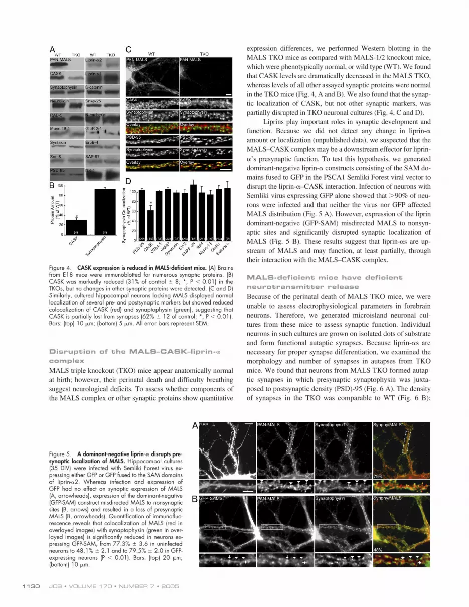
expression differences, we performed Western blotting in theMALS TKO mice as compared with MALS-1/2 knockout mice,which were phenotypically normal, or wild type (WT). We foundthat CASK levels are dramatically decreased in the MALS TKO,whereas levels of all other assayed synaptic proteins were normalin the TKO mice (Fig. 4, A and B). We also found that the synap-tic localization of CASK, but not other synaptic markers, waspartially disrupted in TKO neuronal cultures (Fig. 4, C and D).
Liprins play important roles in synaptic development andfunction. Because we did not detect any change in liprin-
�
amount or localization (unpublished data), we suspected that theMALS–CASK complex may be a downstream effector for liprin-
�
’s presynaptic function. To test this hypothesis, we generateddominant-negative liprin-
�
constructs consisting of the SAM do-mains fused to GFP in the PSCA1 Semliki Forest viral vector todisrupt the liprin-
�
–CASK interaction. Infection of neurons withSemliki virus expressing GFP alone showed that
�
90% of neu-rons were infected and that neither the virus nor GFP affectedMALS distribution (Fig. 5 A). However, expression of the liprindominant-negative (GFP-SAM) misdirected MALS to nonsyn-aptic sites and significantly disrupted synaptic localization ofMALS (Fig. 5 B). These results suggest that liprin-
�
s are up-stream of MALS and may function, at least partially, throughtheir interaction with the MALS–CASK complex.
MALS-deficient mice have deficient neurotransmitter release
Because of the perinatal death of MALS TKO mice, we wereunable to assess electrophysiological parameters in forebrainneurons. Therefore, we generated microisland neuronal cul-tures from these mice to assess synaptic function. Individualneurons in such cultures are grown on isolated dots of substrateand form functional autaptic synapses. Because liprin-
�
s arenecessary for proper synapse differentiation, we examined themorphology and number of synapses in autapses from TKOmice. We found that neurons from MALS TKO formed autap-tic synapses in which presynaptic synaptophysin was juxta-posed to postsynaptic density (PSD)-95 (Fig. 6 A). The densityof synapses in the TKO was comparable to WT (Fig. 6 B);
Figure 4. CASK expression is reduced in MALS-deficient mice. (A) Brainsfrom E18 mice were immunoblotted for numerous synaptic proteins. (B)CASK was markedly reduced (31% of control � 8; *, P � 0.01) in theTKOs, but no changes in other synaptic proteins were detected. (C and D)Similarly, cultured hippocampal neurons lacking MALS displayed normallocalization of several pre- and postsynaptic markers but showed reducedcolocalization of CASK (red) and synaptophysin (green), suggesting thatCASK is partially lost from synapses (62% � 12 of control; *, P � 0.01).Bars: (top) 10 �m; (bottom) 5 �m. All error bars represent SEM.
Figure 5. A dominant-negative liprin-� disrupts pre-synaptic localization of MALS. Hippocampal cultures(35 DIV) were infected with Semliki Forest virus ex-pressing either GFP or GFP fused to the SAM domainsof liprin-�2. Whereas infection and expression ofGFP had no effect on synaptic expression of MALS(A, arrowheads), expression of the dominant-negative(GFP-SAM) construct misdirected MALS to nonsynapticsites (B, arrows) and resulted in a loss of presynapticMALS (B, arrowheads). Quantification of immunofluo-rescence reveals that colocalization of MALS (red inoverlayed images) with synaptophysin (green in over-layed images) is significantly reduced in neurons ex-pressing GFP-SAM, from 77.3% � 3.6 in uninfectedneurons to 48.1% � 2.1 and to 79.5% � 2.0 in GFP-expressing neurons (P � 0.01). Bars: (top) 20 �m;(bottom) 10 �m.

MALS–LIPRIN-� COMPLEX REGULATES VESICLE RELEASE • OLSEN ET AL. 1131
however, the distribution of synaptic areas in the TKO wasshifted to slightly larger sizes (Fig. 6 C).
Electrophysiological experiments demonstrated that excita-tory postsynaptic currents (EPSCs) in the MALS TKO cultureswere profoundly reduced relative to WT (Fig. 7 A). Furthermore,the rate and degree of EPSC depression during high frequencystimulation (10 Hz) was enhanced in the MALS TKO autapses(Fig. 7 B), suggesting that the MALS–liprin-� complex plays arole in presynaptic vesicle cycling. Because MALS mutant micedie around birth, structural analyses of mature synapses are notfeasible. However, the magnitude and distribution of miniatureEPSCs was comparable in MALS TKO and WT cultures (Fig. 7C), arguing against a postsynaptic defect. Indeed, the ratio of the�-amino-3-hydroxy-5-methyl-4-isoxazolepropionate (AMPA)/NMDA components of the EPSC was normal (Fig. 7 D), suggest-ing that trafficking and clustering of these receptors are intact.
DiscussionMALS proteins are essentialThis study establishes MALS as an essential protein familyinvolved in neurotransmitter release. MALS are components
of a large presynaptic protein complex that includes scaffold-ing proteins, adaptor proteins, and adhesion molecules (Butzet al., 1998). Importantly, we find that liprin-� isoforms arecomponents of this complex. Because liprin-� orthologuesdefine the dimensions of the active zone in invertebrate syn-apses (Zhen and Jin, 1999; Kaufmann et al., 2002), the MALScomplex has the potential to link adhesion molecules to theexocytotic machinery.
Although each strain of individual MALS knockouts isviable and fertile, certain combinations are synthetically lethal.Mice lacking all three MALS isoforms die within one hour ofbirth and have a severe breathing defect. This defect resemblesthe phenotype of mice lacking all three neurexin isoforms(Missler et al., 2003), which are also part of the MALS com-plex (Hata et al., 1996). Previous studies showed that MALScan bind to the COOH termini of proteins typical for morpho-logical development, including �-catenin and epidermal growthfactor receptors (Garcia et al., 2000; Perego et al., 2000; Shellyet al., 2003). That MALS TKO mice show no gross externalabnormalities suggests that MALS does not play general
Figure 6. Synapse size, but not number, is altered in neurons lackingMALS. Autaptic cultures (14 DIV) from control and TKO mice were stainedwith antibodies to PSD-95 and synaptophysin. (A) Representative autapticneurons from WT and TKO mice stained with synaptophysin. Bars: (top) 20�m; (bottom) 5 �m. (B) PSD-95/synaptophysin staining revealed that thenumber of synapses was unaltered in TKO cultures (0.65 � 0.15 and 0.67 �0.18 synapses/�m for WT and TKO, respectively). (C) As determined bysynaptophysin staining, the distribution of presynaptic terminal size wasshifted to the right in the TKO (P � 0.01). All error bars represent SEM.
Figure 7. Neurons from MALS-deficient mice display abnormal synaptictransmission. (A) EPSCs recorded in autaptic cultures prepared from E18MALS TKO mice were profoundly reduced relative to EPSCs recorded inWT cultures (*, P � 0.01). Typical EPSCs recorded in WT and MALSTKO pyramidal cell autapses are shown above. (B) Normalized synapticresponses during a 10-Hz (4 s) stimulus train. Decay of EPSCs during thehigh frequency train was greater in the MALS TKO autapses than in WTautapses (*, P � 0.05). EPSCs returned to baseline values within 5 s ofthe end of the stimulus train in both populations (n � 18 and 7 for WTand MALS TKO, respectively, for the recovery). (C) The distribution andamplitude of miniature EPSCs was the same in autapses prepared fromWT and MALS TKO mice (P � 0.1). (D) The ratio of AMPA EPSCs toNMDA EPSCs was also similar in WT and MALS TKO autapses (P � 0.1).AMPA EPSCs were measured at the peak (�8–10 ms after the action po-tential), whereas NMDA EPSCs were measured at 40 ms after the actionpotential, a time point when the AMPA EPSC has decayed to baseline.Typical AMPA/NMDA EPSCs from the two populations are shown. Allerror bars represent SEM.
JCB • VOLUME 170 • NUMBER 7 • 20051132
roles in tissue morphogenesis. Detailed histological evaluationmay, however, reveal specific tissues whose development re-quires the MALS complex.
Presynaptic defect in MALS mutantsThe perinatal lethality and labored breathing of MALS TKOare phenotypes often seen in mice with impaired synaptictransmission. Consistent with this, we found a profound pre-synaptic defect in these mutants. No change in the mEPSC am-plitude distribution and no change in the ratio of AMPA/NMDA receptor–mediated currents were observed. In contrastto normal postsynaptic function, high-frequency stimulationproduced an accelerated and more pronounced synaptic depres-sion, suggesting greater depletion of vesicles in the readily re-leasable pool of MALS TKO mice. These results also implythat the MALS–CASK–liprin-� complex helps determine thesize of the releasable pool and is important for replenishing thispool from the reserve pool.
Assembly of the MALS protein complexThe presynaptic defects in MALS TKO mice are paralleled bythe association of MALS with a large presynaptic complex.Numerous modular protein interaction interfaces assemblethis complex. The membrane-associated guanylate kinaseCASK directly binds to many components and therefore con-stitutes the core. MALS uses its coiled-coil L27 domain tobind to the second of two L27 domains in CASK (Lee et al.,2002). The first L27 domain in CASK associates with theNH2-terminal L27 domain of SAP-97 (Lee et al., 2002). Neur-exin binds to the PDZ domain of CASK (Hata et al., 1996) andMint-1 binds to the CaMK domain of CASK (Butz et al.,1998; Borg et al., 1999). Furthermore, we find that liprin-�proteins associate with the NH2-terminal region of CASK,which includes the CaMK domain and the first L27 region.Previous studies showed that liprin-� proteins also bind toLAR-family receptor protein tyrosine phosphatase (Serra-Pages et al., 1998) and to the Rab3A binding protein RIM1�
(Schoch et al., 2002). Our failure to detect LAR or RIM pro-teins in the MALS complex may suggest that interaction ofCASK with the SAM domains of liprin-� occludes associationwith these other presynaptic molecules.
The liprin protein family comprises seven liprin isoformsthat are subdivided into the �- and �-type. We identified onlyliprin-� family members as part of the MALS complex. TheSAM1 domain of liprin-�s mediates their interaction withCASK. These domains are highly conserved between liprin-�proteins, sharing �90% identity. In contrast, liprin-� SAM1domains share �45% identity with the most homologous lip-rin-� member. The SAM1 domain of liprin-� is evolutionarilyconserved; mammalian liprin-�s share �90% identity withorthologues in D. melanogaster and C. elegans, which suggestthat the liprin-�–CASK interaction is likely conserved.
Potential mechanisms for MALS complex regulating transmitter releaseOur discovery that the MALS complex contains liprin-� pro-teins can explain how this complex participates in synaptic
vesicle exocytosis. Genetic analysis of invertebrates shows thatliprin-� in D. melanogaster and its homologue syd in C. eleganscontrol presynaptic function and morphology (Zhen and Jin,1999; Kaufmann et al., 2002). In addition to these developmen-tal defects, these mutants show decreased synaptic transmis-sion resembling that seen in the MALS TKO. Because themammalian genome contains four liprin-� isoforms (Serra-Pages et al., 1998), functional analyses are difficult. Some bio-chemical work suggests that mammalian liprin-� proteins maybe postsynaptic and regulate AMPA receptors through theglutamate receptor interacting protein GRIP (Wyszynski et al.,2002). lin-10 also has been shown to regulate GLR-1 in C. ele-gans (Rongo et al., 1998). MALS and liprin-�s are also en-riched in PSD fractions from rat brain (Fig. 1 C) and partiallycolocalize with PSD-95 in cultured hippocampal neurons(Fig. S2, available at http://www.jcb.org/cgi/content/full/jcb.200503011/DC1). We cannot rule out the possibility thatpostsynaptic MALS may contribute to the synaptic defect in theMALS TKOs, but our failure to detect GRIP or AMPA recep-tors in the MALS complex suggests no role for postsynapticliprin-� in our analysis. Because MALS mutant mice die aroundbirth, conditional mutants of the MALS may be required toevaluate roles for this complex in synaptic morphogenesis.
This study provides the first evidence that the MALScomplex plays an important role in controlling transmitter re-lease at excitatory synapses in brain. The myriad of interactionsfor the core MALS–CASK complex has suggested diverseroles; however, decisive genetic evidence has been lacking.Our work is consistent with a model whereby this complex re-cruits components of the synaptic release machinery to adhe-sive proteins of the active zone. According to this proposal, theMALS–CASK complex would couple extracellular synapticinteractions to the intercellular organization of the presynapticsecretory machinery.
Materials and methodsAntibodiesIsoform-specific and pan antibodies against MALS-1, -2, and -3 were gen-erated in rabbits as described previously (Misawa et al., 2001). Mouseanti–liprin-�1 (C3-77) and -�2 (245:2:1) antibodies have also been de-scribed previously (Serra-Pages et al., 1998). Mouse anti-CASK, MINT,ZO-2, Sec8, �-catenin, and N-cadherin were all purchased from Trans-duction Labs. Munc-18, Rab5, and neuroligin antibodies were purchasedfrom Synaptic Systems GmbH. Rabbit anti-CASK and antisynaptophysinwere obtained from Zymed Laboratories. The mouse anti–Erb-B4 antibodywas purchased from Lab Vision/Neomarkers and anti–PSD-95 antibodywas obtained from Affinity BioReagents, Inc. The mouse anti-GluR 2/4 an-tibody was purchased from Chemicon International and rabbit synapto-physin was obtained from Sigma-Aldrich.
ImmunoprecipitationsUsing a Potter homogenizer (Braun), adult mouse brains from either MALS-3heterozygote or knockout mice were homogenized in three volumes ofSTE buffer (320 mM sucrose, 20 mM Tris, pH 8.0, and 2 mM EDTA) con-taining 10 �g/ml leupeptin and 200 �g/ml PMSF. Homogenates werespun at 20,000 g for 1 h and pellets were resuspended in TET buffer (20mM Tris, pH 8.0, 1 mM EDTA, and 1.3% Triton X-100) containing 10�g/ml leupeptin and 50 �g/ml PMSF. After rehomogenization with a Pot-ter homogenizer and a 1-h incubation at 4�C, the lysates were spun at100,000 g for 1 h. The supernatant was collected and precleared withprotein A–Sepharose (GE Healthcare) for 1 h at 4�C. Precleared lysateswere immunoprecipitated with 10 �g MALS-3 antibody or control rabbitIgG for 2 h at 4�C. To collect immunoprecipitated protein complexes,
MALS–LIPRIN-� COMPLEX REGULATES VESICLE RELEASE • OLSEN ET AL. 1133
80 �l of a 50% protein A–Sepharose slurry was added to the lysates andincubated for 1 h at 4�C. Immunoprecipitates were washed extensivelyand loaded onto SDS-PAGE to separate the proteins. Gels were either sil-ver stained or transferred to nitrocellulose for Western blotting. Immuno-precipitations from COS cells were performed similarly, except that thecells were directly lysed in TET buffer and spun at 20,000 g for 10 min,and supernatants were collected.
ImmunocytochemistryHippocampal cultures grown on poly-D-lysine–treated coverslips werefixed with 4% PFA on ice for 10 min followed by methanol at 20�C.After thorough washing with PBS containing 0.1% Triton X-100 (PBS-X),cells were blocked with PBS-X containing 3% normal goat serum (blockingsolution). Primary antibodies were incubated for 2 h at RT or 4�C over-night in blocking solution. Cells were washed three times with PBS-X andincubated with secondary antibodies in blocking solution for 1 h at RT.Cells were washed with PBS-X and mounted onto slides with Fluoromount-G(Southern Biotechnology Associates, Inc.).
Colocalization experiments in neurons were performed using im-ages acquired from a confocal microscope (model LSM510; Carl ZeissMicroImaging, Inc.) and analyzed using image analysis software (Meta-Morph). Neurons were chosen randomly for quantification. For all im-ages, thresholds were set at a predetermined level and colocalizationvalues were obtained using MetaMorph software. Statistical significancewas determined by an unpaired t test.
Nano-LC-ESI-Qq-TOF tandem mass spectrometry (MS) analysisIndividual gel bands were reduced with 10 mM dithiothreitol at 56�C for1 h followed by alkylation with 55 mM iodoacetamide for 45 min at RT.The proteins were digested overnight with 12 ng/�L trypsin at 37�C. Pep-tides were extracted with a 50% acetonitrile/5% formic acid solution. Thepeptides were dried down and resuspended in 0.1% formic acid, andthen separated via HPLC using a 75 �M 15 cm reverse phase C-18 col-umn (LC Packings) at a flow rate of 350 nl/min running a 3–32% acetoni-trile gradient in 0.1% formic acid on an 1100 series HPLC (Agilent Tech-nologies). The LC eluent was coupled to a micro-ionspray source attachedto a mass spectrometer (model QSTAR Pulsar; MDS Sciex). Peptides wereanalyzed in positive ion mode. MS spectra were acquired for 1 s, fol-lowed by 3-s MS/MS on the most intense multiply charged peak.
Subcellular fractionationSubcellular fractions of rat brain were prepared by differential centrifuga-tion. Brains were homogenized in buffer containing 320 mM sucrose and10 mM Hepes-NaOH, pH 7.4. Homogenate (C, crude lysate) was centri-fuged for 10 min at 1,000 g to produce a pellet. The supernatant wascentrifuged at 13,800 g for 10 min to produce a pellet (P13.8) and super-natant. The pellet was resuspended in the original volume of homogeniza-tion buffer and centrifuged at 100,000 g to yield a pellet (P100, crudesynaptosomal vesicle pellet) and supernatant (S100, crude cytosolic syn-aptosomal supernatant). Synaptosome and PSD fractions were isolated bya discontinuous sucrose gradient centrifugation using P13.8. The pelletwas extracted twice with ice cold 0.5% Triton X-100 (synaptosome) andthen centrifuged to obtain the PSD pellet.
Cell culture and transfectionsHippocampal cultures were prepared as described previously (Tomita etal., 2003). In brief, hippocampi were dissected from 17- and 18-d-old em-bryos, digested with papain solution and plated at a density of 5 104
neurons/well in 24-well dishes. Neurons were cultured in Neurobasalmedia (GIBCO BRL) supplemented with B27, penicillin, streptomycin,and L-glutamine according to the manufacturer’s protocol (GIBCO BRL).COS cells were grown in DME supplemented with 10% FBS, 100 U/mlpenicillin, and 100 �g/ml streptomycin. Transfections were performed us-ing LipofectAMINE Plus (Invitrogen) according to the manufacturer’s proto-cols. Transfected cells were used for immunocytochemistry or immunopre-cipitations 48 h after the transfection of plasmids.
ConstructspcDNA3 MALS-1, -2, and -3 and HA-liprin-�2 constructs have been de-scribed previously (Serra-Pages et al., 1998; Misawa et al., 2001). Forconstruction of EGFP-MALS and CASK, full-length clones were amplifiedby PCR and cloned downstream of EGFP. For construction of yeast two-hybrid clones, liprin-�2 constructs were amplified by PCR and cloned intothe pGBKT7 vector. Liprin-�2 constructs were as follows: full-length (aa2–1257), SAMS 1/2/3 (aa 705–1257), SAM 1 (aa 876–984), SAM 2 (aa997–1105), and SAM 3 (aa 1086–1197). MALS and CASK clones were
amplified and cloned into the pGADT7 vector as follows: full-length MALS-1(aa 2–233), full-length CASK (aa 2–909), CASK STK/L27AB (aa 2–476),CASK L27/PDZ (aa 276–590), CASK PDZ/SH3GK (aa 476–909), CASKSTK/L27A (aa 2–405), and CASK STK (aa 2–346). Viral constructs weresubcloned from pGBKT7 into a modified pBK-CMV plasmid to produce anNH2-terminal GFP-fusion and then subcloned into PSCA1 plasmid.
Yeast two-hybridYeast cotransformation was performed according to the manufacturer’sprotocol (CLONTECH Laboratories, Inc.) with the yeast strain AH109.Binding was assessed by quantification of colonies transformed with theindicated plasmids on –LWH plates. Interactions were scored as positive(�) if many (�50) individual colonies were observed or negative () if nocolonies were present on the –LWH plate. Control plates (–LW) were usedto verify transformation efficiency.
Isolation of MALS-3 genomic DNA and construction of targeting vectorA mouse MALS-3 cDNA probe was used to isolate bacterial artificial chro-mosome clones from a 129Sv/J mouse genomic library (Genome Sys-tems, Inc.). The targeting vector was constructed using the pPNT replace-ment vector. A 1-kb region downstream from the targeted exons was PCR-amplified, digested with XhoI and NotI, and subcloned into the XhoI–NotIsites of pPNT. Similarly, a 5-kb genomic region upstream from the tar-geted exons was PCR-amplified, digested with BamHI and XbaI, and in-serted into the BamHI–XbaI sites of the pPNT vector. In the targeting vec-tor, the third, fourth, and fifth exons (as well as the third and fourth intronsof MALS-3) were replaced with a neocassette.
Generation of MALS-3 null miceThe targeting vector was linearized with BamHI and electroporated into R-1embryonic stem cells. Clones resistant to G418 and gancyclovir were ana-lyzed for recombination by PCR. To ensure proper homologous recombi-nation, PCR-positive clones were further analyzed by Southern blotting us-ing probes containing genomic sequences outside of the targeting vectorand with a neoprobe. Properly targeted clones were injected into blasto-cysts from C57BL6 (The Jackson Laboratory) mice and transferred to surro-gate mothers. Male chimeras were mated with C57BL6 females for trans-mission of the mutated allele through the germ line. Heterozygous micewere interbred to generate MALS-3 null mice. The genotypes from theseand subsequent matings were determined by Southern blotting or PCR us-ing allele-specific primers as follows: GGAAGAAATGGAGTCCCGCTTTGand ACAGCAGGACAGAACTGTCC for the WT allele; GCTAAAGCG-CATGCTCCAGACTG and ACAGCAGGACAGAACTGTCC for the tar-geted allele. The null phenotype was confirmed by Western blotting ofbrain homogenates with antibodies to MALS-3.
Generation of MALS TKO miceFemale MALS-1/2 null mice (Misawa et al., 2001) and male MALS-3 nullmice were mated, generating MALS-1/2/3 triple heterozygous mice. Tri-ple heterozygous mice were bred back to MALS-1/2 null mice, and MALS-1/2 double null and 3 heterozygous mice were selected. These mice wereinterbred to generate MALS-1/2/3 triple null mice. Genotyping for theMALS-1 and -2 targeted loci has been described previously (Misawa etal., 2001).
ElectrophysiologyMicrodot cultures were prepared as described previously (Bekkers andStevens, 1991; Augustin et al., 1999). For whole cell voltage-clamp re-cordings in autaptic culture (10–18 days in vitro [DIV]), patch pipette solu-tions contained the following: 135 mM potassium gluconate, 10 mMHepes, 1 mM EGTA, 4.6 mM MgCl2, 4 mM NaATP, 15 mM creatinephosphate, 50 U/ml phosphocreatine kinase, pH 7.3, and 300 mMmOsm. The extracellular solution contained 140 mM NaCl, 2.4 mM KCl,10 mM Hepes, 10 mM glucose, 4 mM CaCl2, 4 mM MgCl2, pH 7.3, and300 mM mOsm. To determine the ratio of AMPA/NMDA EPSCs, MgCl2was omitted from the extracellular solution; the patch pipette and extracel-lular solutions were prepared as previously described (Tovar and West-brook, 1999). Cells were held at 70 mV and stimulated at 0.1 Hz witha 1–4 ms 80 mV depolarizing current pulse. Pyramidal cells were distin-guished based on the decay kinetics of the evoked current and by applica-tion of 10 �M CNQX at the end of the experiment.
ImmunohistochemistryAdult mice were anesthetized with pentobarbital and perfused with 4% PFAin 0.1 M phosphate buffer. The brain was removed and immersed in thesame fixative for 4 h at 4�C and then cryoprotected in 20% sucrose in PBS
JCB • VOLUME 170 • NUMBER 7 • 20051134
overnight at 4�C. 35-�m free-floating sections were cut on a sliding micro-tome. Endogenous peroxidase activity was inactivated by incubating brainsections in 0.5% H2O2 for 10 min. Sections were blocked for 1 h in PBScontaining 3% normal goat serum and then incubated in the same buffercontaining diluted 0.1 �g/ml MALS-3 antibody for 2 d at 4�C. Immunohis-tochemical staining was performed with an avidin/biotin/peroxidase sys-tem (ABC Elite; Vector Laboratories) and DAB (Vector Laboratories).
Online supplemental materialFig. S1 shows the targeted disruption of the MALS-3 gene. Fig. S2 showsthe colocalization of MALS and liprin-� with PSD-95. Online supple-mental material is available at http://www.jcb.org/cgi/content/full/jcb.200503011/DC1.
The authors wish to thank David R.C. House for his technical help.This work was supported by grants (to D.S. Bredt, R.A. Nicoll, O.
Olsen, and K.A. Moore) from the National Institutes of Health and from theChristopher Reeves Paralysis Foundation (to D.S. Bredt).
The authors declare there is no financial conflict of interest related tothis work.
Submitted: 3 March 2005Accepted: 22 August 2005
ReferencesAugustin, I., C. Rosenmund, T.C. Südhof, and N. Brose. 1999. Munc13-1 is es-
sential for fusion competence of glutamatergic synaptic vesicles. Nature.400:457–461.
Bekkers, J.M., and C.F. Stevens. 1991. Excitatory and inhibitory autaptic cur-rents in isolated hippocampal neurons maintained in cell culture. Proc.Natl. Acad. Sci. USA. 88:7834–7838.
Borg, J.-P., Y. Yang, M. De Taddéo-Borg, B. Margolis, and R.S. Turner.1998. The X11alpha protein slows cellular amyloid precursor proteinprocessing and reduces Abeta40 and Abeta42 secretion. J. Biol. Chem.273:14761–14766.
Borg, J.-P., M.O. Lõpez-Figueroa, M. de Taddèo-Borg, D.E. Kroon, R.S.Turner, S.J. Watson, and B. Ben Margolis. 1999. Molecular analysis ofthe X11-mLin-2/CASK complex in brain. J. Neurosci. 19:1307–1316.
Butz, S., M. Okamoto, and T.C. Südhof. 1998. A tripartite protein complex withthe potential to couple synaptic vesicle exocytosis to cell adhesion inbrain. Cell. 94:773–782.
Fannon, A.M., and D.R. Colman. 1996. A model for central synaptic junctionalcomplex formation based on the differential adhesive specificities of thecadherins. Neuron. 17:423–434.
Flanagan, J.G., and P. Vanderhaeghen. 1998. The ephrins and Eph receptors inneural development. Annu. Rev. Neurosci. 21:309–345.
Garcia, R.A., K. Vasudevan, and A. Buonanno. 2000. The neuregulin receptorErbB-4 interacts with PDZ-containing proteins at neuronal synapses.Proc. Natl. Acad. Sci. USA. 97:3596–3601.
Hata, Y., S. Butz, and T.C. Südhof. 1996. CASK: a novel dlg/PSD95 homologwith an N-terminal calmodulin-dependent protein kinase domain identi-fied by interaction with neurexins. J. Neurosci. 16:2488–2494.
Ho, A., W. Morishita, R.E. Hammer, R.C. Malenka, and T.C. Südhof. 2003. Arole for Mints in transmitter release: Mint 1 knockout mice exhibit im-paired GABAergic synaptic transmission. Proc. Natl. Acad. Sci. USA.100:1409–1414.
Ichtchenko, K., Y. Hata, T. Nguyen, B. Ullrich, M. Missler, C. Moomaw, andT.C. Südhof. 1995. Neuroligin 1: a splice site-specific ligand for beta-neurexins. Cell. 81:435–443.
Jo, K., R. Derin, M. Li, and D.S. Bredt. 1999. Characterization of MALS/Velis-1,-2, and -3: a family of mammalian LIN-7 homologs enriched at brainsynapses in association with the postsynaptic density-95/NMDA recep-tor postsynaptic complex. J. Neurosci. 19:4189–4199.
Kaech, S.M., C.W. Whitfield, and S.K. Kim. 1998. The LIN-2/LIN-7/LIN-10complex mediates basolateral membrane localization of the C. elegansEGF receptor LET-23 in vulval epithelial cells. Cell. 94:761–771.
Kaufmann, N., J. DeProto, R. Ranjan, H. Wan, and D. Van Vactor. 2002.Drosophila liprin-alpha and the receptor phosphatase Dlar control syn-apse morphogenesis. Neuron. 34:27–38.
Laverty, H.G., and J.B. Wilson. 1998. Murine CASK is disrupted in a sex-linked cleft palate mouse mutant. Genomics. 53:29–41.
Lee, S., S. Fan, O. Makarova, S. Straight, and B. Margolis. 2002. A novel andconserved protein-protein interaction domain of mammalian Lin-2/CASK binds and recruits SAP97 to the lateral surface of epithelia. Mol.
Cell. Biol. 22:1778–1791.
Misawa, H., Y. Kawasaki, J. Mellor, N. Sweeney, K. Jo, R.A. Nicoll, and D.S.Bredt. 2001. Contrasting localizations of MALS/LIN-7 PDZ proteins inbrain and molecular compensation in knockout mice. J. Biol. Chem.276:9264–9272.
Missler, M., W. Zhang, A. Rohlmann, G. Kattenstroth, R.E. Hammer, K. Gott-mann, and T.C. Südhof. 2003. Alpha-neurexins couple Ca2� channels tosynaptic vesicle exocytosis. Nature. 423:939–948.
Okamoto, M., and T.C. Südhof. 1997. Mints, Munc18-interacting proteins insynaptic vesicle exocytosis. J. Biol. Chem. 272:31459–31464.
Perego, C., C. Vanoni, S. Massari, R. Longhi, and G. Pietrini. 2000. MammalianLIN-7 PDZ proteins associate with beta-catenin at the cell-cell junctionsof epithelia and neurons. EMBO J. 19:3978–3989.
Rongo, C., C.W. Whitfield, A. Rodal, S.K. Kim, and J.M. Kaplan. 1998. LIN-10 is a shared component of the polarized protein localization pathwaysin neurons and epithelia. Cell. 94:751–759.
Schoch, S., P.E. Castillo, T. Jo, K. Mukherjee, M. Geppert, Y. Wang, F.Schmitz, R.C. Malenka, and T.C. Südhof. 2002. RIM1alpha forms a pro-tein scaffold for regulating neurotransmitter release at the active zone.Nature. 415:321–326.
Serra-Pages, C., Q.G. Medley, M. Tang, A. Hart, and M. Streuli. 1998. Liprins,a family of LAR transmembrane protein-tyrosine phosphatase-interact-ing proteins. J. Biol. Chem. 273:15611–15620.
Setou, M., T. Nakagawa, D.-H. Seog, and N. Hirokawa. 2000. Kinesin super-family motor protein KIF17 and mLin-10 in NMDA receptor-containingvesicle transport. Science. 288:1796–1802.
Shelly, M., Y. Mosesson, A. Citri, S. Lavi, Y. Zwang, N. Melamed-Book, B.Aroeti, and Y. Yarden. 2003. Polar expression of ErbB-2/HER2 in epi-thelia. Bimodal regulation by Lin-7. Dev. Cell. 5:475–486.
Südhof, T.C. 2004. The synaptic vesicle cycle. Annu. Rev. Neurosci. 27:509–547.
Tomita, S., L. Chen, Y. Kawasaki, R.S. Petralia, R.J. Wenthold, R.A. Nicoll,and D.S. Bredt. 2003. Functional studies and distribution define a fam-ily of transmembrane AMPA receptor regulatory proteins. J. Cell Biol.161:805–816.
Torres, R., B.L. Firestein, J. Staudinger, H. Dong, E.N. Olson, R.L. Huganir,D.S. Bredt, N.W. Gale, and G.D. Yancopoulos. 1998. PDZ proteins bind,cluster and synaptically co-localize with Eph receptors and their ligands,the ephrins. Neuron. 21:1453–1463.
Tovar, K.R., and G.L. Westbrook. 1999. The incorporation of NMDA receptorswith a distinct subunit composition at nascent hippocampal synapses invitro. J. Neurosci. 19:4180–4188.
Wyszynski, M., E. Kim, A.W. Dunah, M. Passafaro, J.G. Valtschanoff, C.Serra-Pages, M. Streuli, R.J. Weinberg, and M. Sheng. 2002. Interactionbetween GRIP and liprin-alpha/SYD2 is required for AMPA receptortargeting. Neuron. 34:39–52.
Zhen, M., and Y. Jin. 1999. The liprin protein SYD-2 regulates the differentia-tion of presynaptic termini in C. elegans. Nature. 401:371–375.