role of the wnt receptor frizzled-1 in presynaptic differentiation and function
TRANSCRIPT

BioMed CentralNeural Development
ss
Open AcceResearch articleRole of the Wnt receptor Frizzled-1 in presynaptic differentiation and functionLorena Varela-Nallar1,2, Catalina P Grabowski1, Iván E Alfaro1, Alejandra R Alvarez2 and Nibaldo C Inestrosa*1Address: 1Centro de Envejecimiento y Regeneración (CARE), Centro de Regulación Celular y Patología "Joaquín V Luco" (CRCP) and MIFAB, Chile and 2Laboratorio de Señalización Celular, Departamento de Biología Celular y Molecular, Facultad de Ciencias Biológicas, Pontificia Universidad Católica de Chile, Santiago, Chile
Email: Lorena Varela-Nallar - [email protected]; Catalina P Grabowski - [email protected]; Iván E Alfaro - [email protected]; Alejandra R Alvarez - [email protected]; Nibaldo C Inestrosa* - [email protected]
* Corresponding author
AbstractBackground: The Wnt signaling pathway regulates several fundamental developmental processesand recently has been shown to be involved in different aspects of synaptic differentiation andplasticity. Some Wnt signaling components are localized at central synapses, and it is thus possiblethat this pathway could be activated at the synapse.
Results: We examined the distribution of the Wnt receptor Frizzled-1 in cultured hippocampalneurons and determined that this receptor is located at synaptic contacts co-localizing withpresynaptic proteins. Frizzled-1 was found in functional synapses detected with FM1-43 staining andin synaptic terminals from adult rat brain. Interestingly, overexpression of Frizzled-1 increased thenumber of clusters of Bassoon, a component of the active zone, while treatment with theextracellular cysteine-rich domain (CRD) of Frizzled-1 decreased Bassoon clustering, suggesting arole for this receptor in presynaptic differentiation. Consistent with this, treatment with theFrizzled-1 ligand Wnt-3a induced presynaptic protein clustering and increased functionalpresynaptic recycling sites, and these effects were prevented by co-treatment with the CRD ofFrizzled-1. Moreover, in synaptically mature neurons Wnt-3a was able to modulate the kinetics ofneurotransmitter release.
Conclusion: Our results indicate that the activation of the Wnt pathway through Frizzled-1 occursat the presynaptic level, and suggest that the synaptic effects of the Wnt signaling pathway could bemodulated by local activation through synaptic Frizzled receptors.
BackgroundThe Wnt signaling pathway plays a crucial role duringdevelopment, regulating specification of cell fate, cell pro-liferation, migration and morphogenesis [1]. Wnt signal-ing is activated by the interaction of Wnt ligands withmembers of the Frizzled (Fz) family of seven-transmem-
brane cell surface receptors. Three different Wnt pathwayshave been described downstream of Fz receptors: thecanonical Wnt/β-catenin pathway; and the non-canonicalpathways involving intracellular signaling by Ca2+ or thec-Jun-N-terminal kinase (JNK) cascade [1,2]. In thecanonical Wnt/β-catenin signaling pathway, Wnt ligands
Published: 2 November 2009
Neural Development 2009, 4:41 doi:10.1186/1749-8104-4-41
Received: 9 June 2009Accepted: 2 November 2009
This article is available from: http://www.neuraldevelopment.com/content/4/1/41
© 2009 Varela-Nallar et al; licensee BioMed Central Ltd. This is an Open Access article distributed under the terms of the Creative Commons Attribution License (http://creativecommons.org/licenses/by/2.0), which permits unrestricted use, distribution, and reproduction in any medium, provided the original work is properly cited.
Page 1 of 15(page number not for citation purposes)
Neural Development 2009, 4:41 http://www.neuraldevelopment.com/content/4/1/41
interact with Fz receptors and their co-receptor LRP5/6and signal through Dishevelled to inhibit the kinase activ-ity of glycogen synthase kinase-3β in a protein degrada-tion complex containing Axin and adenomatouspolyposis coli (APC) protein. When Wnt signaling is inac-tive, β-catenin is phosphorylated by glycogen synthasekinase-3β and thus rapidly degraded via the proteasomepathway. When cells receive Wnt signals, the degradationpathway is inhibited, and β-catenin consequently accu-mulates in the cytoplasm and is translocated to thenucleus where it binds the TCF/LEF family of transcrip-tion factors to regulate the expression of Wnt target genes[1].
Fz receptors have an extracellular amnio-terminal regionthat contains a cysteine-rich domain (CRD) consisting of120 to 125 residues with 10 conserved cysteines that isnecessary for the binding of Wnt molecules [3,4]. In mam-mals, 19 different Wnts are known, and 10 Fz proteinshave been identified as Wnt receptors. In addition to Fz,other Wnt receptors have been described more recently[2,5], and it has been shown that a single Wnt ligand cansignal through different pathways depending on receptorcontext [6], increasing the complexity of the Wnt signalingcascade.
In the past decade, it has been well established that Wntsignaling plays a key role in diverse aspects of neuronalconnectivity by regulating axon guidance and remodeling[7,8], dendritic development [9], and synapse formation[8,10,11]. Additionally, intracellular modulators of theWnt pathway enhanced excitatory transmission in adulthippocampal preparations, acting predominantly via apresynaptic mechanism to increase neurotransmitterrelease [12], and Wnt-7a was shown to induce recyclingand exocytosis of synaptic vesicles in cultured hippocam-pal neurons and enhance synaptic transmission in adulthippocampal slices [13]. Furthermore, Wnt-3a is releasedfrom synapses in an activity-dependent manner, and thesecreted Wnt and the consequent activation of Wnt signal-ing facilitates long-term potentiation, suggesting that Wntsignaling plays a role in regulating synaptic plasticity [14].Wnt-3a was also shown to induce the recycling of synapticvesicles in cultured hippocampal neurons [13].
In the present work, we have studied the distribution ofthe canonical Wnt receptor Fz1 in neurons, and the poten-tial role of this receptor in synapse structure and function.We determined that, in cultured hippocampal neurons,Fz1 clusters are localized in synapses co-localizing withpresynaptic markers in close apposition to the postsynap-tic protein PSD-95. In addition, Fz1 was observed in func-tional synapses detected with FM1-43 staining and insynaptic terminals from adult rat brain. Overexpression ofFz1 increased the number of Bassoon clusters, and treat-
ment with the Fz1 ligand Wnt-3a induced presynapticprotein clustering and modulated the kinetics of synapticvesicle release, suggesting that the activation of Wnt sign-aling through Fz1 modulates presynaptic differentiationand function.
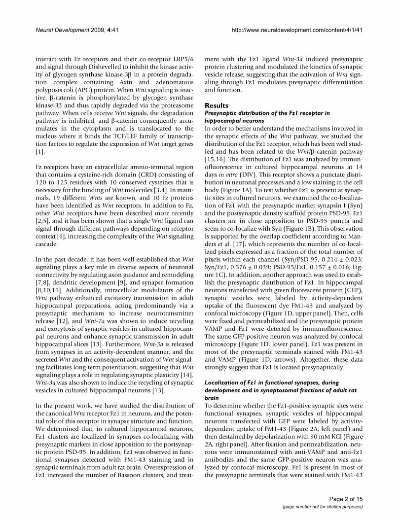
ResultsPresynaptic distribution of the Fz1 receptor in hippocampal neuronsIn order to better understand the mechanisms involved inthe synaptic effects of the Wnt pathway, we studied thedistribution of the Fz1 receptor, which has been well stud-ied and has been related to the Wnt/β-catenin pathway[15,16]. The distribution of Fz1 was analyzed by immun-ofluorescence in cultured hippocampal neurons at 14days in vitro (DIV). This receptor shows a punctate distri-bution in neuronal processes and a low staining in the cellbody (Figure 1A). To test whether Fz1 is present at synap-tic sites in cultured neurons, we examined the co-localiza-tion of Fz1 with the presynaptic marker synapsin I (Syn)and the postsynaptic density scaffold protein PSD-95. Fz1clusters are in close apposition to PSD-95 puncta andseem to co-localize with Syn (Figure 1B). This observationis supported by the overlap coefficient according to Man-ders et al. [17], which represents the number of co-local-ized pixels expressed as a fraction of the total number ofpixels within each channel (Syn/PSD-95, 0.214 ± 0.023;Syn/Fz1, 0.376 ± 0.039; PSD-95/Fz1, 0.157 ± 0.016; Fig-ure 1C). In addition, another approach was used to estab-lish the presynaptic distribution of Fz1. In hippocampalneurons transfected with green fluorescent protein (GFP),synaptic vesicles were labeled by activity-dependentuptake of the fluorescent dye FM1-43 and analyzed byconfocal microscopy (Figure 1D, upper panel). Then, cellswere fixed and permeabilized and the presynaptic proteinVAMP and Fz1 were detected by immunofluorescence.The same GFP-positive neuron was analyzed by confocalmicroscopy (Figure 1D, lower panel). Fz1 was present inmost of the presynaptic terminals stained with FM1-43and VAMP (Figure 1D, arrows). Altogether, these datastrongly suggest that Fz1 is located presynaptically.
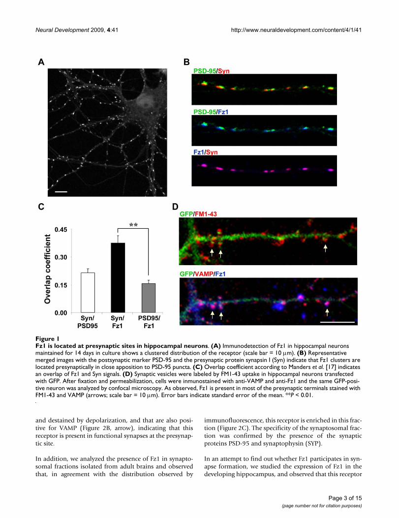
Localization of Fz1 in functional synapses, during development and in synaptosomal fractions of adult rat brainTo determine whether the Fz1-positive synaptic sites werefunctional synapses, synaptic vesicles of hippocampalneurons transfected with GFP were labeled by activity-dependent uptake of FM1-43 (Figure 2A, left panel) andthen destained by depolarization with 90 mM KCl (Figure2A, right panel). After fixation and permeabilization, neu-rons were inmunostained with anti-VAMP and anti-Fz1antibodies and the same GFP-positive neuron was ana-lyzed by confocal microscopy. Fz1 is present in most ofthe presynaptic terminals that were stained with FM1-43
Page 2 of 15(page number not for citation purposes)
Neural Development 2009, 4:41 http://www.neuraldevelopment.com/content/4/1/41
and destained by depolarization, and that are also posi-tive for VAMP (Figure 2B, arrow), indicating that thisreceptor is present in functional synapses at the presynap-tic site.
In addition, we analyzed the presence of Fz1 in synapto-somal fractions isolated from adult brains and observedthat, in agreement with the distribution observed by
immunofluorescence, this receptor is enriched in this frac-tion (Figure 2C). The specificity of the synaptosomal frac-tion was confirmed by the presence of the synapticproteins PSD-95 and synaptophysin (SYP).
In an attempt to find out whether Fz1 participates in syn-apse formation, we studied the expression of Fz1 in thedeveloping hippocampus, and observed that this receptor
Fz1 is located at presynaptic sites in hippocampal neuronsFigure 1Fz1 is located at presynaptic sites in hippocampal neurons. (A) Immunodetection of Fz1 in hippocampal neurons maintained for 14 days in culture shows a clustered distribution of the receptor (scale bar = 10 μm). (B) Representative merged images with the postsynaptic marker PSD-95 and the presynaptic protein synapsin I (Syn) indicate that Fz1 clusters are located presynaptically in close apposition to PSD-95 puncta. (C) Overlap coefficient according to Manders et al. [17] indicates an overlap of Fz1 and Syn signals. (D) Synaptic vesicles were labeled by FM1-43 uptake in hippocampal neurons transfected with GFP. After fixation and permeabilization, cells were inmunostained with anti-VAMP and anti-Fz1 and the same GFP-posi-tive neuron was analyzed by confocal microscopy. As observed, Fz1 is present in most of the presynaptic terminals stained with FM1-43 and VAMP (arrows; scale bar = 10 μm). Error bars indicate standard error of the mean. **P < 0.01.
Page 3 of 15(page number not for citation purposes)
Neural Development 2009, 4:41 http://www.neuraldevelopment.com/content/4/1/41
Figure 2 (see legend on next page)
Page 4 of 15(page number not for citation purposes)
Neural Development 2009, 4:41 http://www.neuraldevelopment.com/content/4/1/41
Expression of Fz1 in functional synapses, synaptosomal fractions from adult brain and during hippocampal developmentFigure 2 (see previous page)Expression of Fz1 in functional synapses, synaptosomal fractions from adult brain and during hippocampal development. (A) In hippocampal neurons transfected with GFP, synaptic vesicles were labeled by activity-dependent uptake of FM1-43 and subsequently destained by depolarization with 90 mM KCl to identify functional synapses (arrow, scale bar = 5 μm). (B) After fixation and permeabilization, neurons were inmunostained with anti-VAMP and anti-Fz1 antibodies and the same GFP-positive neuron was analyzed by confocal microscopy. Fz-1 is present in most of the synaptic terminals that were destained by depolarization and display immunoreactivity for VAMP (arrow). (C) Analysis of the total homogenized (H) and synaptosomal fractions (S) obtained from adult rat brains shows that Fz1 is enriched in the synaptosomal fraction. PSD-95 and synatophysin (SYP) were used as synaptic markers. (D) Immunoblots of proteins in the developing hippocampus from embry-onic day 18 (E18) and postnatal days 2 (P2) to P50. The same amount of protein was applied to all lanes. Molecular weight standards are indicated at the right (kDa). β-Tub, β-tubulin; N-Cad, N-cadherin; VGlut1, vesicular glutamate transporter 1.
Page 5 of 15(page number not for citation purposes)
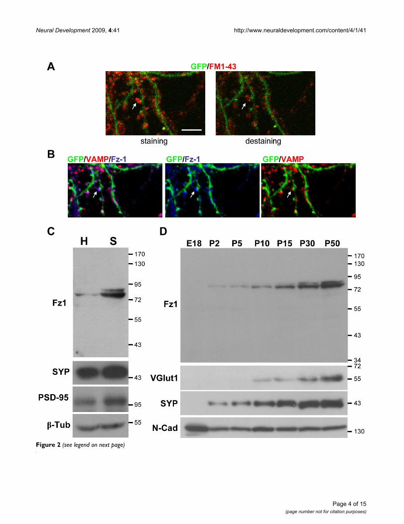
Expression of Fz1 in hippocampal neurons during developmentFigure 3Expression of Fz1 in hippocampal neurons during development. (A) Immunoblots of protein extracts from cultured hippocampal neurons during differentiation from 4 to 14 DIV. The same amount of protein was applied to all lanes. Molecular weight standards are indicated at the right (kDa). N-Cad, N-cadherin. (B) Immunodetection of Fz1 and phosphorylated MAP1B in cultured hippocampal neurons at 4 DIV. (C-E) Immunodetection of Fz1 and Bassoon (scale bar = 10 μm). (C) On DIV 7, Fz1 is observed in isolated axons as fine puncta. (D) On DIV 10, larger clusters of Fz1 co-localize with Bassoon puncta in axons contacting other neurons (arrows), but not in isolated axons (E).
Neural Development 2009, 4:41 http://www.neuraldevelopment.com/content/4/1/41
is undetectable before birth, and increases from a smallamount at P2 to high levels in adult brain, as was alsoobserved for the synaptic proteins vesicular glutamatetransporter 1 (VGlut1) and SYP (Figure 2D) [18]. N-cad-herin was used as a loading control since it is known to bepresent at all developmental stages [19].
We also analyzed the temporal appearance of Fz1 in dif-ferentiating hippocampal neurons, and observed anincrease of Fz1 during development concomitant with theincrease of VGlut1 and SYP levels (Figure 3A). The spatialappearance of Fz1 was also analyzed by immunofluores-cence. At early stages of differentiation (4 DIV), Fz1 dis-tributes mostly at the axon as determined by doublelabeling with the axonal marker phosphorylated MAP1B[20] (Figure 3B), supporting the presynaptic distributionof this receptor. By 7 DIV, Fz1 is observed in isolatedaxons as fine puncta that mostly do not co-localize withBassoon puncta (Figure 3C), suggesting that Fz1 is notassociated with presynaptic proteins during the initialstages of synaptic assembly. Bassoon is a presynapticcytomatrix protein recruited early during synapse forma-tion [21]. By 10 DIV, larger clusters of Fz1 are observedand it is possible to find co-localization of Fz1 with Bas-soon mainly in axons contacting other neurons (Figure3D, arrows), but not in isolated axons (Figure 3E). Thenumber of Fz1 clusters increases steadily during neuronalmaturation, suggesting an association of Fz1 with synapseformation.
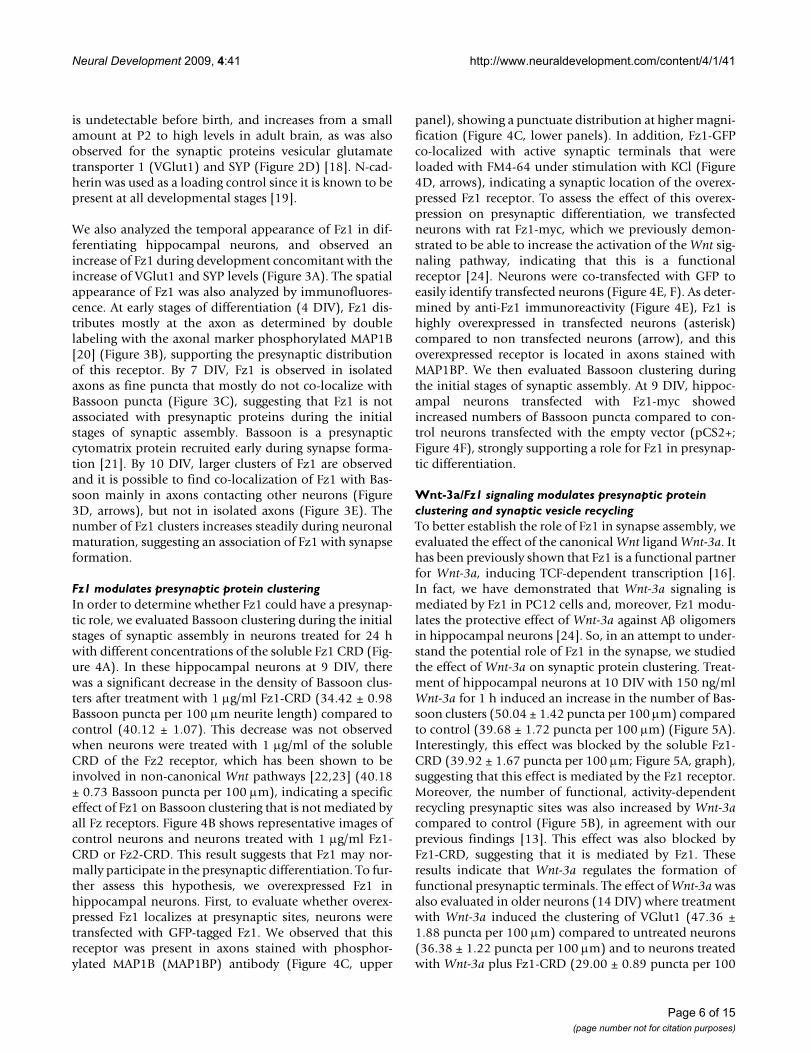
Fz1 modulates presynaptic protein clusteringIn order to determine whether Fz1 could have a presynap-tic role, we evaluated Bassoon clustering during the initialstages of synaptic assembly in neurons treated for 24 hwith different concentrations of the soluble Fz1 CRD (Fig-ure 4A). In these hippocampal neurons at 9 DIV, therewas a significant decrease in the density of Bassoon clus-ters after treatment with 1 μg/ml Fz1-CRD (34.42 ± 0.98Bassoon puncta per 100 μm neurite length) compared tocontrol (40.12 ± 1.07). This decrease was not observedwhen neurons were treated with 1 μg/ml of the solubleCRD of the Fz2 receptor, which has been shown to beinvolved in non-canonical Wnt pathways [22,23] (40.18± 0.73 Bassoon puncta per 100 μm), indicating a specificeffect of Fz1 on Bassoon clustering that is not mediated byall Fz receptors. Figure 4B shows representative images ofcontrol neurons and neurons treated with 1 μg/ml Fz1-CRD or Fz2-CRD. This result suggests that Fz1 may nor-mally participate in the presynaptic differentiation. To fur-ther assess this hypothesis, we overexpressed Fz1 inhippocampal neurons. First, to evaluate whether overex-pressed Fz1 localizes at presynaptic sites, neurons weretransfected with GFP-tagged Fz1. We observed that thisreceptor was present in axons stained with phosphor-ylated MAP1B (MAP1BP) antibody (Figure 4C, upper
panel), showing a punctuate distribution at higher magni-fication (Figure 4C, lower panels). In addition, Fz1-GFPco-localized with active synaptic terminals that wereloaded with FM4-64 under stimulation with KCl (Figure4D, arrows), indicating a synaptic location of the overex-pressed Fz1 receptor. To assess the effect of this overex-pression on presynaptic differentiation, we transfectedneurons with rat Fz1-myc, which we previously demon-strated to be able to increase the activation of the Wnt sig-naling pathway, indicating that this is a functionalreceptor [24]. Neurons were co-transfected with GFP toeasily identify transfected neurons (Figure 4E, F). As deter-mined by anti-Fz1 immunoreactivity (Figure 4E), Fz1 ishighly overexpressed in transfected neurons (asterisk)compared to non transfected neurons (arrow), and thisoverexpressed receptor is located in axons stained withMAP1BP. We then evaluated Bassoon clustering duringthe initial stages of synaptic assembly. At 9 DIV, hippoc-ampal neurons transfected with Fz1-myc showedincreased numbers of Bassoon puncta compared to con-trol neurons transfected with the empty vector (pCS2+;Figure 4F), strongly supporting a role for Fz1 in presynap-tic differentiation.
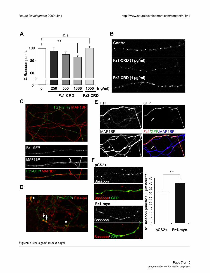
Wnt-3a/Fz1 signaling modulates presynaptic protein clustering and synaptic vesicle recyclingTo better establish the role of Fz1 in synapse assembly, weevaluated the effect of the canonical Wnt ligand Wnt-3a. Ithas been previously shown that Fz1 is a functional partnerfor Wnt-3a, inducing TCF-dependent transcription [16].In fact, we have demonstrated that Wnt-3a signaling ismediated by Fz1 in PC12 cells and, moreover, Fz1 modu-lates the protective effect of Wnt-3a against Aβ oligomersin hippocampal neurons [24]. So, in an attempt to under-stand the potential role of Fz1 in the synapse, we studiedthe effect of Wnt-3a on synaptic protein clustering. Treat-ment of hippocampal neurons at 10 DIV with 150 ng/mlWnt-3a for 1 h induced an increase in the number of Bas-soon clusters (50.04 ± 1.42 puncta per 100 μm) comparedto control (39.68 ± 1.72 puncta per 100 μm) (Figure 5A).Interestingly, this effect was blocked by the soluble Fz1-CRD (39.92 ± 1.67 puncta per 100 μm; Figure 5A, graph),suggesting that this effect is mediated by the Fz1 receptor.Moreover, the number of functional, activity-dependentrecycling presynaptic sites was also increased by Wnt-3acompared to control (Figure 5B), in agreement with ourprevious findings [13]. This effect was also blocked byFz1-CRD, suggesting that it is mediated by Fz1. Theseresults indicate that Wnt-3a regulates the formation offunctional presynaptic terminals. The effect of Wnt-3a wasalso evaluated in older neurons (14 DIV) where treatmentwith Wnt-3a induced the clustering of VGlut1 (47.36 ±1.88 puncta per 100 μm) compared to untreated neurons(36.38 ± 1.22 puncta per 100 μm) and to neurons treatedwith Wnt-3a plus Fz1-CRD (29.00 ± 0.89 puncta per 100
Page 6 of 15(page number not for citation purposes)
Neural Development 2009, 4:41 http://www.neuraldevelopment.com/content/4/1/41
Figure 4 (see legend on next page)
Page 7 of 15(page number not for citation purposes)
Neural Development 2009, 4:41 http://www.neuraldevelopment.com/content/4/1/41
μm) (Figure 5C). At 14 DIV, Wnt-3a treatment alsoinduced the clustering of SYP (control, 38.56 ± 2.04; Wnt-3a, 50.70 ± 2.71 puncta per 100 μm; P < 0.01). Consider-ing the increase observed in presynaptic protein puncta byWnt-3a treatment, we evaluated whether synaptic contactnumbers, identified by PSD-95/Syn or PSD-95/SV2 co-clusters, were also affected in 14 DIV neurons. After a 1-h
treatment, Wnt-3a did not increase the number of syn-apses per 100 μm neurite length (PSD-95/Syncontrol, 19.94± 2.37; PSD-95/SynWnt-3a, 19.32 ± 2.33; PSD-95/SV2control,19.69 ± 3.23; PSD-95/SV2Wnt-3a, 20.96 ± 3.04). Theseresults suggest that Wnt-3a/Fz1 signaling induces a fastincrease in pre-synaptic protein clustering, which mayprecede the increase in synaptic contact number. Indeed,
Fz1 overexpression induces presynaptic protein clusteringFigure 4 (see previous page)Fz1 overexpression induces presynaptic protein clustering. (A) The number of Bassoon clusters per neurite length was evaluated in hippocampal neurons (9 DIV) treated for 24 h with different concentrations of the soluble extracellular Fz1 CRD or 1,000 ng/ml Fz2-CRD. (B) Representative images of control neurons and neurons treated with 1 μg/ml Fz1-CRD or Fz2-CRD for 24 h. (C) Hippocampal neurons were transfected with Fz1-GFP, which was detected in axons stained with phos-phorylated MAP1B (MAP1BP). (D) Fz1-GFP clusters co-localize with pre-synaptic terminals loaded with FM4-64 under stimu-lation with 90 mM KCl (arrows). (E) Hippocampal neurons were transfected with Fz1-myc plus GFP to easily identify transfected neurons. Fz1 staining (red) is highly increased in transfected neurons (asterisk) compared to non-transfected neu-rons (arrow). (F) Bassoon puncta were analyzed in neurons transfected with Fz1-myc or with empty vector (pCS2+) using an anti-Bassoon antibody and quantifying the clusters per neurite length in GFP-positive axons. There was an increased number of Bassoon puncta in neurons transfected with Fz1-myc compared to control. *P < 0.05; **P < 0.01; NS, not significant. Error bars indicate standard error of the mean.
Wnt-3a induces presynaptic differentiation in cultured hippocampal neurons mediated by the Fz1 receptorFigure 5Wnt-3a induces presynaptic differentiation in cultured hippocampal neurons mediated by the Fz1 receptor. (A) Neurons at 9 DIV exposed to Wnt-3a for 1 h show elevated numbers of Bassoon clusters detected by immunofluorescence. Addition of the soluble Fz1-CRD prevents this induction. (B) Synaptic vesicle recycling is visualized by FM4-64 uptake (11 DIV). One-hour treatment with Wnt-3a induced a significant increase in the number of FM4-64-positive sites, which was pre-vented by co-treatment with the Fz1-CRD. (C) Wnt-3a induces the number of VGlut1 clusters in neurons at 14 DIV, which was prevented by co-treatment with Fz1-CRD. Error bars indicate standard error of the mean. * P < 0.05; ** P < 0.01.
Page 8 of 15(page number not for citation purposes)
Neural Development 2009, 4:41 http://www.neuraldevelopment.com/content/4/1/41
after 24 h of treatment, Wnt-3a induced an almost 20%increase in synaptic contact number, which was notobserved in neurons treated with Wnt-3a plus Fz1-CRD(Wnt-3a versus control, 1.19 ± 0.05, P < 0.05; Wnt-3a+CRD versus control, 0.95 ± 0.04).
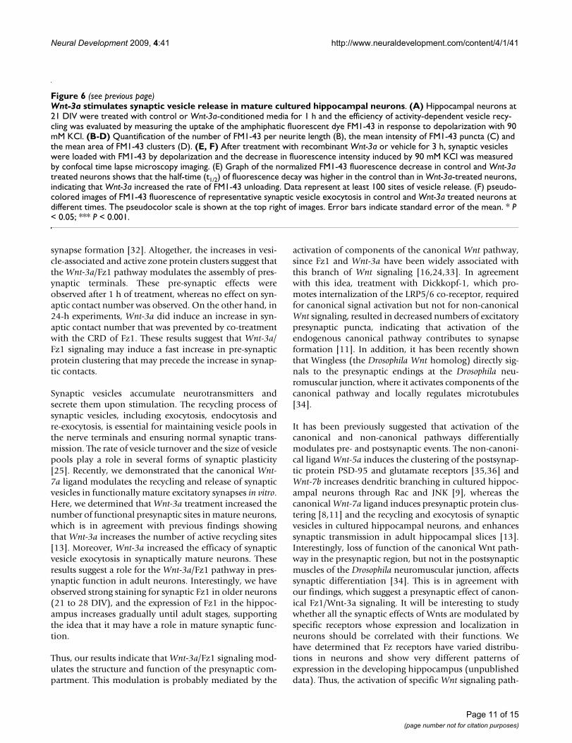
The rate and efficacy of synaptic vesicle recycling at pre-synaptic nerve terminals determine normal synaptic trans-mission and play a role in several forms of synaptic plas-ticity [25]. Recently, we demonstrated that Wnt-7amodulates the synaptic vesicle cycle in presynaptic nerveterminals of hippocampal neurons and that this canonicalWnt ligand is able to increase the synaptic transmission inCA3-CA1 synapses of hippocampal slices by a presynapticmechanism [13]. To evaluate if the Fz1 ligand Wnt-3a isable to modulate the pre-synaptic function, mature hip-pocampal neurons at 21 DIV were treated with control orWnt-3a conditioned media for 1 h and the efficiency ofthe activity-dependent vesicle recycling was evaluated bymeasuring the uptake of the amphiphatic fluorescent dyeFM1-43 in response to depolarization with 90 mM KCl(Figure 6A). In contrast to the effect observed in youngneurons, no differences were found in the density of FM1-43 clusters in response to Wnt-3a in mature neurons at 21DIV (Figure 6B), although the mean intensity and area ofFM1-43 puncta were increased in Wnt-3a-treated neuronscompared to control cells (Mean intensitycontrol, 193.580± 1.55; Mean intensityWnt-3a, 200.939 ± 1.24; Areacontrol,0.39 ± 0.02; AreaWnt-3a, 0.44 ± 0.02; Figure 6C and 6D).The fluorescence intensity of buttons depends on thenumber of labeled vesicles retrieved during endocytosis,and is indicative of the total functional recycling pool sizeof synaptic vesicles [26].
We also analyzed the probability of synaptic vesiclerelease. After treatment with recombinant Wnt-3a or vehi-cle for 3 h, synaptic vesicles were loaded with FM1-43 bydepolarization with 60 mM KCl and the decrease in fluo-rescence intensity induced by the activity dependent exo-cytosis at 90 mM KCl was measured by confocal timelapse microscopy imaging. As indicated in Figure 6E, F,pre-incubation of neurons with Wnt-3a increased the rateof release of the FM1-43 fluorescence trapped in vesicles.The half-time (t1/2) of fluorescence decay in Wnt-3a-treated neurons was 43.97 ± 1.20 s versus 65.48 ± 2.86 sfor control neurons, indicating that Wnt-3a increased therate of FM1-43 unloading. The effect of Wnt-3a was ana-lyzed in a total of more than 100 sites of vesicle release.These results indicate that a Wnt-3a-dependent signalingmodulates the activity of the presynaptic compartment,increasing the rate of release of synaptic vesicles.
DiscussionThe Wnt signaling pathway regulates several fundamentaldevelopmental processes and modulates synaptic struc-
ture and function. Although roles for Wnt ligands in regu-lating synaptic assembly and plasticity have been shown[10,13,14], little is known about the receptors that medi-ate these effects. Previously, we and others determinedthat Fz1 is highly expressed in the hippocampus[24,27,28]. In the present work, we determined the synap-tic distribution of the canonical Wnt receptor Fz1. Wefound that this receptor is located at synaptic sites in hip-pocampal neurons, co-localizing with presynaptic pro-teins and with active synaptic vesicle recycling sitesstained under KCl stimulation with a fluorescent dye,indicating that this receptor is distributed presynaptically.In addition, Fz1 was detected in a synaptosome prepara-tion from adult rat brains, supporting the synaptic distri-bution of this receptor.
Specifically, the presynaptic distribution of Fz1 deter-mined in this work suggests that the activation of the Wntsignaling pathway through Fz1 could be operating at thepresynaptic level, where it could modulate presynapticstructure and function. Consistent with this view, weobserved that overexpression of Fz1 induced the cluster-ing of Bassoon, which is a component of the presynapticcytoskeletal matrix involved in the structural organizationof neurotransmitter release sites and is delivered to nas-cent synapses via vesicles that are detectable early duringthe formation of synaptic junctions [21,29]. Furthermore,treatment with the CRD of Fz1, the extracellular region ofthe receptor that binds Wnt molecules with high affinity[3], decreased the number of Bassoon puncta per neuritelength, indicating that activation of endogenous Fz1-mediated signaling contributes to synapse formation. Thiseffect was not observed when neurons were treated withthe CRD of the Fz2 receptor, which has been shown toactivate non-canonical Wnt pathways [22,23]. The specif-icity of the Wnt-Fz interaction remains largely unknown,particularly in vertebrates, because of the large number ofWnts and Fzs; however, it is well known that Wnt-3a andFz1 are functional partners [16,24,30]. We observed thatWnt-3a induced the clustering of Bassoon during the ini-tial stages of synaptic assembly, increased synaptic vesiclerecycling and increased the clustering of VGlut1. All theseeffects were prevented by co-treatment with the CRD ofFz1, suggesting the involvement of Wnt-3a/Fz1 signaling.VGluts are vesicular glutamate transporters that mediatethe transport of glutamate from the cytoplasm into synap-tic vesicles; they are therefore used as specific markers ofthe glutamatergic phenotype [31]. VGlut1 is the majorisoform in cortex, hippocampus and cerebellar cortex. Theincrease in the number of VGlut1 clusters indicates thatWnt-3a/Fz1 signaling increased the number of excitatorypresynaptic puncta. In addition, we observed an increasein the number of SYP clusters, a synaptic vesicle mem-brane protein that has been associated with synaptic vesi-cle cycling and was shown to regulate activity-dependent
Page 9 of 15(page number not for citation purposes)

Neural Development 2009, 4:41 http://www.neuraldevelopment.com/content/4/1/41
Figure 6 (see legend on next page)
Page 10 of 15(page number not for citation purposes)
Neural Development 2009, 4:41 http://www.neuraldevelopment.com/content/4/1/41
synapse formation [32]. Altogether, the increases in vesi-cle-associated and active zone protein clusters suggest thatthe Wnt-3a/Fz1 pathway modulates the assembly of pres-ynaptic terminals. These pre-synaptic effects wereobserved after 1 h of treatment, whereas no effect on syn-aptic contact number was observed. On the other hand, in24-h experiments, Wnt-3a did induce an increase in syn-aptic contact number that was prevented by co-treatmentwith the CRD of Fz1. These results suggest that Wnt-3a/Fz1 signaling may induce a fast increase in pre-synapticprotein clustering that may precede the increase in synap-tic contacts.
Synaptic vesicles accumulate neurotransmitters andsecrete them upon stimulation. The recycling process ofsynaptic vesicles, including exocytosis, endocytosis andre-exocytosis, is essential for maintaining vesicle pools inthe nerve terminals and ensuring normal synaptic trans-mission. The rate of vesicle turnover and the size of vesiclepools play a role in several forms of synaptic plasticity[25]. Recently, we demonstrated that the canonical Wnt-7a ligand modulates the recycling and release of synapticvesicles in functionally mature excitatory synapses in vitro.Here, we determined that Wnt-3a treatment increased thenumber of functional presynaptic sites in mature neurons,which is in agreement with previous findings showingthat Wnt-3a increases the number of active recycling sites[13]. Moreover, Wnt-3a increased the efficacy of synapticvesicle exocytosis in synaptically mature neurons. Theseresults suggest a role for the Wnt-3a/Fz1 pathway in pres-ynaptic function in adult neurons. Interestingly, we haveobserved strong staining for synaptic Fz1 in older neurons(21 to 28 DIV), and the expression of Fz1 in the hippoc-ampus increases gradually until adult stages, supportingthe idea that it may have a role in mature synaptic func-tion.
Thus, our results indicate that Wnt-3a/Fz1 signaling mod-ulates the structure and function of the presynaptic com-partment. This modulation is probably mediated by the
activation of components of the canonical Wnt pathway,since Fz1 and Wnt-3a have been widely associated withthis branch of Wnt signaling [16,24,33]. In agreementwith this idea, treatment with Dickkopf-1, which pro-motes internalization of the LRP5/6 co-receptor, requiredfor canonical signal activation but not for non-canonicalWnt signaling, resulted in decreased numbers of excitatorypresynaptic puncta, indicating that activation of theendogenous canonical pathway contributes to synapseformation [11]. In addition, it has been recently shownthat Wingless (the Drosophila Wnt homolog) directly sig-nals to the presynaptic endings at the Drosophila neu-romuscular junction, where it activates components of thecanonical pathway and locally regulates microtubules[34].
It has been previously suggested that activation of thecanonical and non-canonical pathways differentiallymodulates pre- and postsynaptic events. The non-canoni-cal ligand Wnt-5a induces the clustering of the postsynap-tic protein PSD-95 and glutamate receptors [35,36] andWnt-7b increases dendritic branching in cultured hippoc-ampal neurons through Rac and JNK [9], whereas thecanonical Wnt-7a ligand induces presynaptic protein clus-tering [8,11] and the recycling and exocytosis of synapticvesicles in cultured hippocampal neurons, and enhancessynaptic transmission in adult hippocampal slices [13].Interestingly, loss of function of the canonical Wnt path-way in the presynaptic region, but not in the postsynapticmuscles of the Drosophila neuromuscular junction, affectssynaptic differentiation [34]. This is in agreement withour findings, which suggest a presynaptic effect of canon-ical Fz1/Wnt-3a signaling. It will be interesting to studywhether all the synaptic effects of Wnts are modulated byspecific receptors whose expression and localization inneurons should be correlated with their functions. Wehave determined that Fz receptors have varied distribu-tions in neurons and show very different patterns ofexpression in the developing hippocampus (unpublisheddata). Thus, the activation of specific Wnt signaling path-
Wnt-3a stimulates synaptic vesicle release in mature cultured hippocampal neuronsFigure 6 (see previous page)Wnt-3a stimulates synaptic vesicle release in mature cultured hippocampal neurons. (A) Hippocampal neurons at 21 DIV were treated with control or Wnt-3a-conditioned media for 1 h and the efficiency of activity-dependent vesicle recy-cling was evaluated by measuring the uptake of the amphiphatic fluorescent dye FM1-43 in response to depolarization with 90 mM KCl. (B-D) Quantification of the number of FM1-43 per neurite length (B), the mean intensity of FM1-43 puncta (C) and the mean area of FM1-43 clusters (D). (E, F) After treatment with recombinant Wnt-3a or vehicle for 3 h, synaptic vesicles were loaded with FM1-43 by depolarization and the decrease in fluorescence intensity induced by 90 mM KCl was measured by confocal time lapse microscopy imaging. (E) Graph of the normalized FM1-43 fluorescence decrease in control and Wnt-3a treated neurons shows that the half-time (t1/2) of fluorescence decay was higher in the control than in Wnt-3a-treated neurons, indicating that Wnt-3a increased the rate of FM1-43 unloading. Data represent at least 100 sites of vesicle release. (F) pseudo-colored images of FM1-43 fluorescence of representative synaptic vesicle exocytosis in control and Wnt-3a treated neurons at different times. The pseudocolor scale is shown at the top right of images. Error bars indicate standard error of the mean. * P < 0.05; *** P < 0.001.
Page 11 of 15(page number not for citation purposes)
Neural Development 2009, 4:41 http://www.neuraldevelopment.com/content/4/1/41
ways would be controlled temporally and spatially duringthe development of neuronal circuits. In addition, thereare alternative Wnt receptors [37] that could spatiallymodulate the activation of the Wnt pathway. This is thecase for the axonal localization of Ryk receptors in mam-mals and Drosophila [38], and the Ror2 receptor [39],which is highly concentrated in the growth cones ofimmature neurons and are present throughout the soma-todendritic compartment of mature hippocampal cells[40].
In summary, we show for the first time the presynapticdistribution of a Fz receptor in mammalian neurons,which could mediate the synaptic effects of the Wnt sign-aling pathway activation. This synaptic localization sug-gests that there could be a local activation of the Wntpathway at the synapse. In addition, the synaptic expres-sion of some downstream components of the Wnt path-way have been described [10,11,14,34,41], indicating thatthe machinery required for the local activation of thepathway is present at central synapses. Altogether, theseresults suggest that Wnts binding to synaptic Fz-LRP5/6could activate the canonical Wnt signaling at the synapseand, as a consequence, increase presynaptic inputs andthe recycling and release of synaptic vesicles. Our findingsgive new insight into the mechanisms by which the Wntsignaling pathway could modulate the synapse.
Materials and methodsPrimary culture of rat hippocampal neuronsRat hippocampal cultures were prepared as described pre-viously [42,43]. Hippocampi from Sprague-Dawley rats atembryonic day 18 were removed, dissected free of menin-ges in Ca2+/Mg2+-free Hanks' balanced salt solution(HBSS), and rinsed twice with HBSS by allowing the tissueto settle to the bottom of the tube. After the second wash,the tissue was resuspended in HBSS containing 0.25% (w/v) trypsin and incubated for 15 minutes at 37°C. Afterthree rinses with HBSS, the tissue was mechanically disso-ciated in plating medium (Dulbecco's modified Eagle'smedium (GIBCO, Rockville, MD, USA)), supplementedwith 10% horse serum (GIBCO), 100 U/ml penicillin,and 100 μg/ml streptomycin by gentle passage throughPasteur pipettes. Dissociated hippocampal cells wereseeded onto poly-L-lysine-coated six-well culture plates ata density of 7 × 10⁵ cells per well in plating medium. Cul-tures were maintained at 37°C in 5% CO2 for 2 h beforethe plating medium was replaced with neurobasal growthmedium (GIBCO) supplemented with B27 (GIBCO), 2mM L-glutamine, 100 U/ml penicillin, and 100 μg/mlstreptomycin. On day 2, cultured neurons were treatedwith 2 μM cytosine arabinoside (AraC) for 24 h; thismethod resulted in cultures highly enriched for neurons(approximately 5% glia). For Wnt-3a treatments, neuronswere treated with 150 ng/ml recombinant Wnt-3a (R&D
Systems, Minneapolis, MN, USA) and with 300 ng/mlrecombinant Fz-1-CRD/Fc Chimera (R&D Systems).
ImmunofluorescenceHippocampal neurons were seeded onto poly-L-lysine-coated coverslips in 24-well culture plates at a density of2.5 × 104 cells per well. Cells were rinsed twice in ice-coldphosphate-buffered saline (PBS) and fixed with a freshlyprepared solution of 4% paraformaldehyde in PBS for 20minutes and permeabilized for 5 minutes with 0.2% Tri-ton X-100 in PBS. After several rinses in ice-cold PBS, cellswere incubated in 0.2% gelatin in PBS (blocking solution)for 30 minutes at room temperature, followed by an over-night incubation at 4°C with primary antibodies. Cellswere extensively washed with PBS and then incubatedwith Alexa-conjugated secondary antibodies (MolecularProbes, Carlsbad, CA, USA) for 30 minutes at 37°C. Cov-erslips were mounted in mounting medium and analyzedon a Zeiss LSM 5 Pascal confocal microscope. Primaryantibodies used were goat anti-Fz1 (R&D Systems), rabbitanti-Synapsin I (Santa Cruz Biotechnology Inc., SantaCruz, CA, USA), rabbit anti-VAMP (Santa Cruz Biotech-nology Inc.), goat anti-Synapsin I (Santa Cruz Biotechnol-ogy Inc.), goat anti-SYP (Santa Cruz Biotechnology Inc.),monoclonal anti-Bassoon antibody (Assay designs, AnnArbor, MI, USA), and monoclonal anti-MAP1BP antibody(Sternberger Monoclonals, Baltimore, MD, USABalti-more, MDBaltimore). The monoclonal antibodies anti-PSD-95 and anti-VGlut1 were developed by and obtainedfrom the UC Davis/NIH NeuroMab Facility, supported byNIH grant U24NS050606 and maintained by the Depart-ment of Neurobiology, Physiology and Behavior, Collegeof Biological Sciences, University of California, Davis, CA,USA.
Images were captured with a Zeiss LSM 5 Pascal confocalmicroscope. Images were analyzed using NIH ImageJ soft-ware. Co-localization analysis and quantification of thenumber of puncta were carried out under threshold con-ditions to identify independent clusters. Co-localizationanalysis was performed on randomly selected images,using the NIH ImageJ software with the co-localizationanalysis plug-in. Mander's coefficients represent thenumber of co-localized pixels [17]; they range from 0 to 1,indicating no co-localization to complete co-localization,and are independent of the pixel intensities within eachrespective channel.
FM uptakeFM4-64 FX or FM1-43 (Molecular Probes) were added ata concentration of 15 μM to Tyrode saline solution (119mM NaCl, 2.5 mM KCl, 2 mM CaCl2, 2 mM, MgCl2, 25mM HEPES, 30 mM glucose buffered to pH 7.4). Neuronswere incubated with this dye solution for 1 minute andthen the FM dye was loaded using 90 mM KCl stimulation
Page 12 of 15(page number not for citation purposes)
Neural Development 2009, 4:41 http://www.neuraldevelopment.com/content/4/1/41
for 30 s. Neurons were washed five times for 10 minutesin dye-free Tyrode solution to decrease background stain-ing of the membrane. For some of the experiments, neu-rons were fixed in 4% paraformaldehyde in PBS for 20minutes and FM4-64 puncta were analyzed by confocalmicroscopy. For retrospective immunofluorescence theneurons on coverslips were mounted in a microscope per-fusion chamber and stained with FM1-43 as describedabove. Images were taken before and after neurons weredestained in high KCl solution for 30 s. Cells were thenfixed and permeabilized with 0.2% Triton X-100 for 10minutes to wash out the FM4-64 fluorescence and proc-essed for immunofluorescence.
Synaptosome preparationSynaptosomes were isolated from adult rat brain using thePercoll gradient method [44]. In brief, adult mouse brainwas homogenized in buffer A (0.32 M sucrose, 5 mMHepes, 0.1 mM EDTA, pH 7.4, plus protease and phos-phatase inhibitors) at 800 rpm ten times at 4°C, and thencentrifuged at 1,000 × g for 10 minutes. The supernatantwas centrifuged at 10,000 × g for 25 minutes, and the pel-let was resuspended in buffer B (0.25 M sucrose, 5 mMHepes, 0.1 mM EDTA, pH 7.5) 8.5% Percoll, and layeredon top of a Percoll discontinuous gradient. Synaptosomeswere taken from a 10 to 16% interface and washed inbuffer B. Proteins were quantified using the BCA proteinassay kit (Pierce, Rockford, IL, USA) and analyzed byimmunoblotting. The relative purity of the synaptosomepreparations was established by electron microscopy.
Immunoblot analysisNeurons growing on six-well culture plates or hippocam-pus obtained from rat brains at different ages were lysedin ice-cold lysis buffer (10 mM Tris-HCl, pH 7.8, 100 mMNaCl, 10 mM EDTA, 0.5% Nonidet P-40, and 0.5%sodium deoxycholate) supplemented with proteaseinhibitors. The homogenates were maintained in ice for30 minutes and then neuronal culture homogenates werecentrifuged at 1,000 × g for 5 minutes (4°C) and hippoc-ampus homogenates were centrifuged at 15,000 × g for 10minutes (4°C) to remove nuclei and large debris. Thesupernatant was recovered and protein concentration wasdetermined by BCA protein assay kit (Pierce). Proteinswere resolved in SDS-PAGE (10% polyacrylamide), trans-ferred to PVDF membrane and reacted with primary anti-bodies. The reactions were followed by incubation withperoxidase-labeled secondary antibodies (Pierce) anddeveloped using the ECL technique (PerkinElmer,Waltham, MA, USA). Primary antibodies were the sameused for immunofluorescence in addition to rabbit anti-β-tubulin (Santa Cruz Biotechnology Inc.) and rabbit anti-N-cadherin (Santa Cruz Biotechnology Inc.).
Neuronal transfectionNeurons were transfected using LipofectAMINE 2000(Invitrogen, Carlsbad, CA, USA) 2 days after seeded oncover slips in 24-well culture plates at a density of 4 × 10⁴
cells per well. Briefly, 0.25 μg of Fz1-GFP plasmid or GFPvector and 0.75 μl of LipofectAMINE 2000 were mixed in100 μl of OptiMEM (GIBCO) according to the manufac-turer's instructions. For the co-transfection of Fz1-Mycwith GFP the amounts were 0.35 μg and 0.15 μg, respec-tively. After 20 minutes the DNA-LipofectAMINE 2000Reagent complex was added to the cells. Neurons wereincubated for 2 h at 37°C and then the media wasreplaced with Neurobasal growth medium (GIBCO) sup-plemented with B27 (GIBCO), 2 mM L-glutamine, 100 U/ml penicillin, and 100 μg/ml streptomycin.
Imaging of FM1-43 destaining in presynaptic terminals of cultured hippocampal neuronsHippocampal neurons at 21 DIV were incubated for 3 hwith Wnt-3a (150 ng/ml) or vehicle at 37°C. Neurons oncoverslips were then washed with Tyrode modified solu-tion, mounted in a microscope perfusion chamber, andincubated for 30 s with 10 μM FM1-43 (MolecularProbes) followed by 1 minute of loading by mild depo-larization with 30 mM KCl. Nonspecific and non-synapticFM1-43 staining was diminished by washing with 10 min-utes of continued perfusion of Tyrode solution at 1 to 2ml/minute controlled with a peristaltic pump (ColePalmer, Vernon Hills, IL, USA). The chamber was adaptedat the stage of a Zeiss Axiovert 200 M microscope coupledto Pascal LSM5 confocal laser scanning system. Neuronswere imaged with a 63 × 1.4 NA oil objective at 512 × 512full-frame resolution using a 488-nm argon laser to excitethe FM1-43 probe, and the fluorescence signals were col-lected over 505 nm. Then, after a period of 50 s of basalfluorescence acquisition, neurons were depolarized with90 mM KCl and imaged for 300 s at 1-s intervals. Imagesfrom presynaptic loaded puncta were selected for measur-ing fluorescence intensities using areas of the region ofinterest of 1.5 × 1.5 μm. Images of Wnt-3a-treated neuronsand control neurons were obtained using identical set-tings for laser power, confocal thickness, and detector sen-sitivity. All measurements were taken at roomtemperature (25°C).
Statistical analysisStatistical analysis was performed using statistical soft-ware Prism 5 (GraphPad Software Inc., San Diego, CA,USA). Values are expressed as mean ± standard error of themean. Statistical significance of differences was assessedwith the non-paired Student's t-test or ANOVA, and non-normally distributed data were analyzed using the Mann-Whitney test or Kruskal Wallis (P < 0.05 was consideredsignificant).
Page 13 of 15(page number not for citation purposes)
Neural Development 2009, 4:41 http://www.neuraldevelopment.com/content/4/1/41
AbbreviationsCRD: cysteine-rich domain; DIV: days in vitro; Fz: Frizzled;GFP: green fluorescent protein; HBSS: Hanks' balancedsalt solution; MAP1BP: phosphorylated MAP1B; PBS:phosphate-buffered saline; Syn: synapsin I; SYP: synapto-physin; VGlut1: vesicular glutamate transporter 1.
Competing interestsThe authors declare that they have no competing interests.
Authors' contributionsLV-N performed the experiments on the expression anddistribution of Fz1 in neurons and the effects of Wnt-3aand Fz1 on presynaptic differentiation, and drafted themanuscript. CPG carried out the co-localization analysisof Fz1 in functional synapses and the preparation of syn-aptosomal fractions. IEA carried out the imaging of FM1-43. ARA participated in the design of experiments. NCIparticipated in the design of the experiments and writingof the manuscript. All authors read and approved the finalmanuscript.
AcknowledgementsWe would like to thank to Dr Randall Moon (University of Washington, Seattle, WA) for generously providing the Fz1-GFP and Fz1-myc con-structs. This work was supported by FONDAP-Biomedicine N° 13980001, the Millennium Institute for Fundamental and Applied Biology (MIFAB), Basal Center of Excellence in Aging and Regeneration (CONICYT-PFB12/2007) to NCI, FONDECYT N°1080221, a FONDECYT Postdoctoral Fel-lowship to LV-N (N° 3070017) and a Predoctoral Fellowship form CONI-CYT to IEA.
References1. Logan CY, Nusse R: The Wnt signaling pathway in develop-
ment and disease. Annu Rev Cell Dev Biol 2004, 20:781-810.2. Gordon MD, Nusse R: Wnt signaling: multiple pathways, mul-
tiple receptors, and multiple transcription factors. J Biol Chem2006, 281:22429-22433.
3. Dann CE, Hsieh JC, Rattner A, Sharma D, Nathans J, Leahy DJ:Insights into Wnt binding and signalling from the structuresof two Frizzled cysteine-rich domains. Nature 2001, 412:86-90.
4. Bhanot P, Brink M, Samos CH, Hsieh JC, Wang Y, Macke JP, AndrewD, Nathans J, Nusse R: A new member of the frizzled familyfrom Drosophila functions as a Wingless receptor. Nature1996, 382:225-230.
5. Toledo EM, Colombres M, Inestrosa NC: Wnt signaling in neuro-protection and stem cell differentiation. Prog Neurobiol 2008,86:281-296.
6. Mikels AJ, Nusse R: Purified Wnt5a protein activates or inhibitsbeta-catenin-TCF signaling depending on receptor context.PLoS Biol 2006, 4:e115.
7. Lucas FR, Salinas PC: WNT-7a induces axonal remodeling andincreases synapsin I levels in cerebellar neurons. Dev Biol 1997,192:31-44.
8. Hall AC, Lucas FR, Salinas PC: Axonal remodeling and synapticdifferentiation in the cerebellum is regulated by WNT-7asignaling. Cell 2000, 100:525-535.
9. Rosso SB, Sussman D, Wynshaw-Boris A, Salinas PC: Wnt signalingthrough Dishevelled, Rac and JNK regulates dendritic devel-opment. Nat Neurosci 2005, 8:34-42.
10. Ahmad-Annuar A, Ciani L, Simeonidis I, Herreros J, Fredj NB, RossoSB, Hall A, Brickley S, Salinas PC: Signaling across the synapse: arole for Wnt and Dishevelled in presynaptic assembly andneurotransmitter release. J Cell Biol 2006, 174:127-139.
11. Davis EK, Zou Y, Ghosh A: Wnts acting through canonical andnoncanonical signaling pathways exert opposite effects onhippocampal synapse formation. Neural Dev 2008, 3:32.
12. Beaumont V, Thompson SA, Choudhry F, Nuthall H, Glantschnig H,Lipfert L, David GR, Swain CJ, McAllister G, Munoz-Sanjuan I: Evi-dence for an enhancement of excitatory transmission inadult CNS by Wnt signaling pathway modulation. Mol CellNeurosci 2007, 35:513-524.
13. Cerpa W, Godoy JA, Alfaro I, Farias GG, Metcalfe MJ, Fuentealba R,Bonansco C, Inestrosa NC: Wnt-7a modulates the synaptic ves-icle cycle and synaptic transmission in hippocampal neurons.J Biol Chem 2008, 283:5918-5927.
14. Chen J, Park CS, Tang SJ: Activity-dependent synaptic Wntrelease regulates hippocampal long term potentiation. J BiolChem 2006, 281:11910-11916.
15. Sancho E, Batlle E, Clevers H: Signaling pathways in intestinaldevelopment and cancer. Annu Rev Cell Dev Biol 2004,20:695-723.
16. Gazit A, Yaniv A, Bafico A, Pramila T, Igarashi M, Kitajewski J, Aaron-son SA: Human frizzled 1 interacts with transforming Wntsto transduce a TCF dependent transcriptional response.Oncogene 1999, 18:5959-5966.
17. Manders EMM, Verbeek FJ, Aten JA: Measurement of co-localiza-tion of objects in dual color confocal images. J Microsc 1993,169:375-382.
18. Boulland JL, Qureshi T, Seal RP, Rafiki A, Gundersen V, Bergersen LH,Fremeau RT Jr, Edwards RH, Storm-Mathisen J, Chaudhry FA:Expression of the vesicular glutamate transporters duringdevelopment indicates the widespread corelease of multipleneurotransmitters. J Comp Neurol 2004, 480:264-280.
19. Petralia RS, Sans N, Wang YX, Wenthold RJ: Ontogeny of postsy-naptic density proteins at glutamatergic synapses. Mol CellNeurosci 2005, 29:436-452.
20. Boyne LJ, Martin K, Hockfield S, Fischer I: Expression and distribu-tion of phosphorylated MAP1B in growing axons of culturedhippocampal neurons. J Neurosci Res 1995, 40:439-450.
21. Zhai R, Olias G, Chung WJ, Lester RA, tom Dieck S, Langnaese K,Kreutz MR, Kindler S, Gundelfinger ED, Garner CC: Temporalappearance of the presynaptic cytomatrix protein bassoonduring synaptogenesis. Mol Cell Neurosci 2000, 15:417-428.
22. Slusarski DC, Corces VG, Moon RT: Interaction of Wnt and aFrizzled homologue triggers G-protein-linked phosphatidyli-nositol signalling. Nature 1997, 390:410-413.
23. Sheldahl LC, Park M, Malbon CC, Moon RT: Protein kinase C is dif-ferentially stimulated by Wnt and Frizzled homologs in a G-protein-dependent manner. Curr Biol 1999, 9:695-698.
24. Chacon MA, Varela-Nallar L, Inestrosa NC: Frizzled-1 is involvedin the neuroprotective effect of Wnt3a against Abeta oli-gomers. J Cell Physiol 2008, 217:215-227.
25. Sudhof TC: The synaptic vesicle cycle. Annu Rev Neurosci 2004,27:509-547.
26. Ryan TA, Reuter H, Wendland B, Schweizer FE, Tsien RW, Smith SJ:The kinetics of synaptic vesicle recycling measured at singlepresynaptic boutons. Neuron 1993, 11:713-724.
27. Lein ES, Hawrylycz MJ, Ao N, Ayres M, Bensinger A, Bernard A, BoeAF, Boguski MS, Brockway KS, Byrnes EJ, Chen L, Chen L, Chen TM,Chin MC, Chong J, Crook BE, Czaplinska A, Dang CN, Datta S, DeeNR, Desaki AL, Desta T, Diep E, Dolbeare TA, Donelan MJ, DongHW, Dougherty JG, Duncan BJ, Ebbert AJ, Eichele G, et al.: Genome-wide atlas of gene expression in the adult mouse brain.Nature 2007, 445:168-176.
28. Shimogori T, VanSant J, Paik E, Grove EA: Members of the Wnt,Fz, and Frp gene families expressed in postnatal mouse cer-ebral cortex. J Comp Neurol 2004, 473:496-510.
29. Zhai RG, Vardinon-Friedman H, Cases-Langhoff C, Becker B, Gun-delfinger ED, Ziv NE, Garner CC: Assembling the presynapticactive zone: a characterization of an active one precursorvesicle. Neuron 2001, 29:131-143.
30. Zilberberg A, Yaniv A, Gazit A: The low density lipoproteinreceptor-1, LRP1, interacts with the human frizzled-1(HFz1) and down-regulates the canonical Wnt signalingpathway. J Biol Chem 2004, 279:17535-17542.
31. Santos MS, Li H, Voglmaier SM: Synaptic vesicle protein traffick-ing at the glutamate synapse. Neuroscience 2009, 158:189-203.
Page 14 of 15(page number not for citation purposes)

Neural Development 2009, 4:41 http://www.neuraldevelopment.com/content/4/1/41
Publish with BioMed Central and every scientist can read your work free of charge
"BioMed Central will be the most significant development for disseminating the results of biomedical research in our lifetime."
Sir Paul Nurse, Cancer Research UK
Your research papers will be:
available free of charge to the entire biomedical community
peer reviewed and published immediately upon acceptance
cited in PubMed and archived on PubMed Central
yours — you keep the copyright
Submit your manuscript here:http://www.biomedcentral.com/info/publishing_adv.asp
BioMedcentral
32. Tarsa L, Goda Y: Synaptophysin regulates activity-dependentsynapse formation in cultured hippocampal neurons. ProcNatl Acad Sci USA 2002, 99:1012-1016.
33. Galceran J, Farinas I, Depew MJ, Clevers H, Grosschedl R: Wnt3a-/--like phenotype and limb deficiency in Lef1(-/-)Tcf1(-/-) mice.Genes Dev 1999, 13:709-717.
34. Miech C, Pauer HU, He X, Schwarz TL: Presynaptic local signalingby a canonical wingless pathway regulates development ofthe Drosophila neuromuscular junction. J Neurosci 2008,28:10875-10884.
35. Inestrosa NC, Varela-Nallar L, Grabowski CP, Colombres M: Synap-totoxicity in Alzheimer's disease: the Wnt signaling pathwayas a molecular target. IUBMB Life 2007, 59:316-321.
36. Farias GG, Alfaro IE, Cerpa W, Grabowski CP, Godoy JA, BonanscoC, Inestrosa NC: WNT-5a/JNK signaling promotes the cluster-ing of PSD-95 in hippocampal neurons. J Biol Chem 2009,284:15857-15866.
37. Kikuchi A, Yamamoto H, Kishida S: Multiplicity of the interac-tions of Wnt proteins and their receptors. Cell Signal 2007,19:659-671.
38. Bovolenta P, Rodriguez J, Esteve P: Frizzled/RYK mediated sig-nalling in axon guidance. Development 2006, 133:4399-4408.
39. Oishi I, Suzuki H, Onishi N, Takada R, Kani S, Ohkawara B, KoshidaI, Suzuki K, Yamada G, Schwabe GC, Mundlos S, Shibuya H, Takada S,Minami Y: The receptor tyrosine kinase Ror2 is involved innon-canonical Wnt5a/JNK signalling pathway. Genes Cells2003, 8:645-654.
40. Paganoni S, Ferreira A: Expression and subcellular localizationof Ror tyrosine kinase receptors are developmentally regu-lated in cultured hippocampal neurons. J Neurosci Res 2003,73:429-440.
41. Franco B, Bogdanik L, Bobinnec Y, Debec A, Bockaert J, ParmentierML, Grau Y: Shaggy, the homolog of glycogen synthase kinase3, controls neuromuscular junction growth in Drosophila. JNeurosci 2004, 24:6573-6577.
42. Alvarez AR, Godoy JA, Mullendorff K, Olivares GH, Bronfman M,Inestrosa NC: Wnt-3a overcomes beta-amyloid toxicity in rathippocampal neurons. Exp Cell Res 2004, 297:186-196.
43. Caceres A, Banker G, Steward O, Binder L, Payne M: MAP2 is local-ized to the dendrites of hippocampal neurons which developin culture. Brain Res 1984, 315:314-318.
44. Nagy A, Delgado-Escueta AV: Rapid preparation of synapto-somes from mammalian brain using nontoxic isoosmoticgradient material (Percoll). J Neurochem 1984, 43:1114-1123.
Page 15 of 15(page number not for citation purposes)