ligand-induced conformational changes in the lactose permease of escherichia coli : evidence for two...
TRANSCRIPT
Protein Science (1994), 3:2294-2301. Cambridge University Press. Printed in the USA. Copyright 0 1994 The Protein Society
Ligand-induced conformational changes in the lactose permease of Escherichia coli: Evidence for two binding sites
JIANHUA WU, STATHIS FRILLINGOS, JOHN VOSS, AND H. RONALD KABACK Department of Physiology and Department of Microbiology and Molecular Genetics, Howard Hughes Medical Institute, Molecular Biology Institute, University of California at Los Angeles, Los Angeles, California 90024-1662
(RECEIVED August 15, 1994; ACCEPTED October 7, 1994)
Abstract
By using a lactose permease mutant containing a single Cys residue in place of Val 331 (helix X), conformational changes induced by ligand binding were studied. With right-side-out membrane vesicles containing Val 331 +
Cys permease, lactose transport is inactivated by either N-ethylmaleimide (NEM) or 7-diethylamino-3-(4'- maleimidylphenyl)-4-methylcoumarin (CPM). Remarkably, 0,D-galactopyranosyl I-thio-0,D-galactopyranoside (TDG) enhances the rate of inactivation by CPM, a hydrophobic sulfhydryl reagent, whereas NEM inactivation is attenuated by the ligand. Val 33 1 + Cys permease was then purified and studied in dodecyl-0,D-maltoside by site-directed fluorescence spectroscopy. The reactivity of Val 331 "+ Cys permease with 2-(4'-maleimidylanilino)- naphthalene-6-sulfonic acid (MIANS) is not changed over a low range of TDG concentrations (<0.8 mM), but the fluorescence of the MIANS-labeled protein is quenched in a saturable manner (apparent Kd 0.12 mM) with- out a change in emission maximum. In contrast, over a higher range of TDG concentrations (1-10 mM), the re- activity of Val 331 + Cys permease with MIANS is enhanced and the emission maximum of MIANS-labeled permease is blue shifted by 3-7 nm. Furthermore, the fluorescence of MIANS-labeled Val 331 --t Cys permease is quenched by both acrylamide and iodide, but the former is considerably more effective. A low concentration of TDG (0.2 mM) does not alter quenching by either compound, whereas a higher concentration of ligand (10mM) decreases the quenching constant for iodide by about 50% and for acrylamide by about 20%. Finally, the EPR spectrum of nitroxide spin-labeled Val 331 + Cys permease exhibits 2 components with different mobilities, and TDG causes the immobilized component to increase. The results provide evidence for the argument that lac per- mease has more than a single binding site. TDG binding to a higher affinity site quenches the fluorescence of MIANS-labeled Val 33 1 + Cys permease, and occupation of a second lower affinity site causes position 33 1 to become more accessible from a hydrophobic environment.
Keywords: C-less permease; ligand-protein interaction; MIANS; site-directed fluorescence; site-directed spin labeling
Lactose permease of Escherichia coli is a hydrophobic, poly- topic, plasma membrane protein that catalyzes the coupled stoi- chiometric translocation of 0-galactosides and H + (reviewed in Kaback, 1983, 1989, 1992). The permease is encoded by the lacy
Reprint requests to: H. Ronald Kaback, HHMI/UCLA, 6-720 Mac- Donald Building, 10833 Le Conte Avenue, Los Angeles, California 90024-1662; e-mail: [email protected].
Abbreviurions: lac, lactose; C-less permease, functional lac per- mease devoid of Cys residues; NEM, N-ethylmaleimide; CPM, 7-diethylamino-3-(4"maleimidylphenyl)-4-methylcoumarin; MIANS, 2-(4'-maleimidylanilino)-naphthalene-6-sulfonic acid; A H + , the H + electrochemical gradient across the membrane; dodecylmaltoside, dodecyl-0,D-maltoside; KPi, potassium phosphate; TDG, 6,D-galcto- pyranosyl 1-thio-0,D-galactopyranoside; IPTG, isopropyl I-thio-69- galactopyranoside; PBS, 100 rnM Napi (pH 7.4)/150 mM NaCI; EPR, electron paramagnetic resonance.
gene, which has been cloned (Teather et al., 1980) and sequenced (Biichel et al., 1980). Moreover, the lacy gene product has been solubilized from the membrane, purified to homogeneity, recon- stituted into proteoliposomes, and shown to be solely respon- sible for P-galactoside transport (Newman et al., 1981; Viitanen et al., 1986) as a monomer (see Sahin-Toth et al., 1994). Based on CD and the hydropathy analysis of the primary amino acid sequence, a secondary-structure model was proposed (Foster et al., 1983) in which the permease is composed of 12 hydropho- bic segments in a-helical conformation that traverse the mem- brane in zig-zag fashion connected by hydrophilic domains (loops) with both the N- and C-termini on the cytoplasmic face. Evidence favoring general aspects of the model and showing that the C-terminus, as well as the second and third cytoplasmic loops, are on the cytoplasmic face of the membrane has been
2294
Two binding sites in lac permease 2295
obtained from a variety of experimental approaches (see Ka- back, 1983, 1989, 1992). Moreover, analysis of a large number of lac permease-alkaline phosphatase (lacy-phoA) fusions has provided unequivocal support for the topological predictions of the 12-helices model (Calamia & Manoil, 1990). Most recently, a model has been proposed describing helix packing in the C-terminal half of the permease based on site-directed excimer fluorescence (Jung et al., 1993; Kaback et al., 1993).
Site-directed mutagenesis has allowed delineation of certain amino acid residues in lac permease that are essential for the transport activity and/or substrate binding (Kaback, 1989, 1992; Kaback et al., 1993). However, it has become clear that struc- tural and dynamic information at high resolution is required to understand the role of these residues in the transport mechanism (Kaback et al., 1993). In this respect, a functional permease mol- ecule has been engineered in which all of the native Cys residues have been replaced (C-less permease; van Iwaarden et al., 1991), and single Cys residues have been placed at more than 300 different positions (Cys-scanning mutagenesis). In addition to providing evidence that very few amino acid residues play a mandatory role in the transport mechanism, the Cys-replace- ment mutants represent unique molecules that can be used to study static and dynamic aspects of permease structure and func- tion (Jung et al., 1993, 1994a, 1994b; Wu & Kaback, 1994).
Single- or double-Cys replacement mutants containing a biotin acceptor domain to facilitate rapid purification and sulfhydryl-specific fluorophores have been utilized to study in- teractions of the permease with ligand either in detergent (Wu & Kaback, 1994) or after reconstitution into proteoliposomes (Jung et al., 1994a, 1994b). Thus, excimer fluorescence in pro- teoliposomes containing pyrene-labeled E269CIH322C perme- ase is quenched by thalium, and the effect is strongly attenuated by permease substrates (Jung et al., 1994b). It has also been shown (Sahin-Toth & Kaback, 1993) that permease with a sin- gle Cys residue in place of Val 3 15 is inactivated by NEM much more rapidly in the presence of TDG or ApH+ (interior nega- tive and/or alkaline). Recently (Jung et al., 1994a), V315C per- mease has been purified, and the reactivity of the Cys residue has been studied in the presence and absence of TDG or ApH+. The studies confirm the observations described with right-side- out membrane vesicles and provide an exciting preliminary suggestion that ligand binding or A p H + may cause the perme- ase to assume the same conformation. In addition to V315C permease, permease molecules with single-Cys replacements for His 322 (Jung et al., 1994b) or Glu 325 in helix X (K. Jung & H.R. Kaback, unpubl. information), Glu 269 in helix VI11 (Jung et al., 1994b), as well as Gly 147 in helix V (Wu & Kaback, 1994), Phe 162 in helix V, or Pro 31 and Pro 28 in helix I (J. Wu & H.R. Kaback, unpubl. information) all exhibit increased re- activity in the presence of TDG. The results, albeit preliminary, suggest that the permease undergoes widespread conformational changes.
This report describes site-directed spectroscopic studies on purified lac permease containing a single Cys residue in place of Val 331 (helix X). Several lines of evidence are presented sup- porting the postulate (Lolkema & Walz, 1990; Lolkema et al., 1991; van Iwaarden et al., 1993) that there are at least 2 bind- ing sites in the permease, a high-affinity site for TDG with a Kd of about 0.1 mM, and a low-affinity site with a Kd of 3-5 mM. Evidence is also presented suggesting that occupation of the low-affinity site induces a conformational change that causes
position 331 to become more accessible from a hydrophobic environment.
Results
Effect of TDG on the inactivation of V331C permease by sulfhydryl reagents
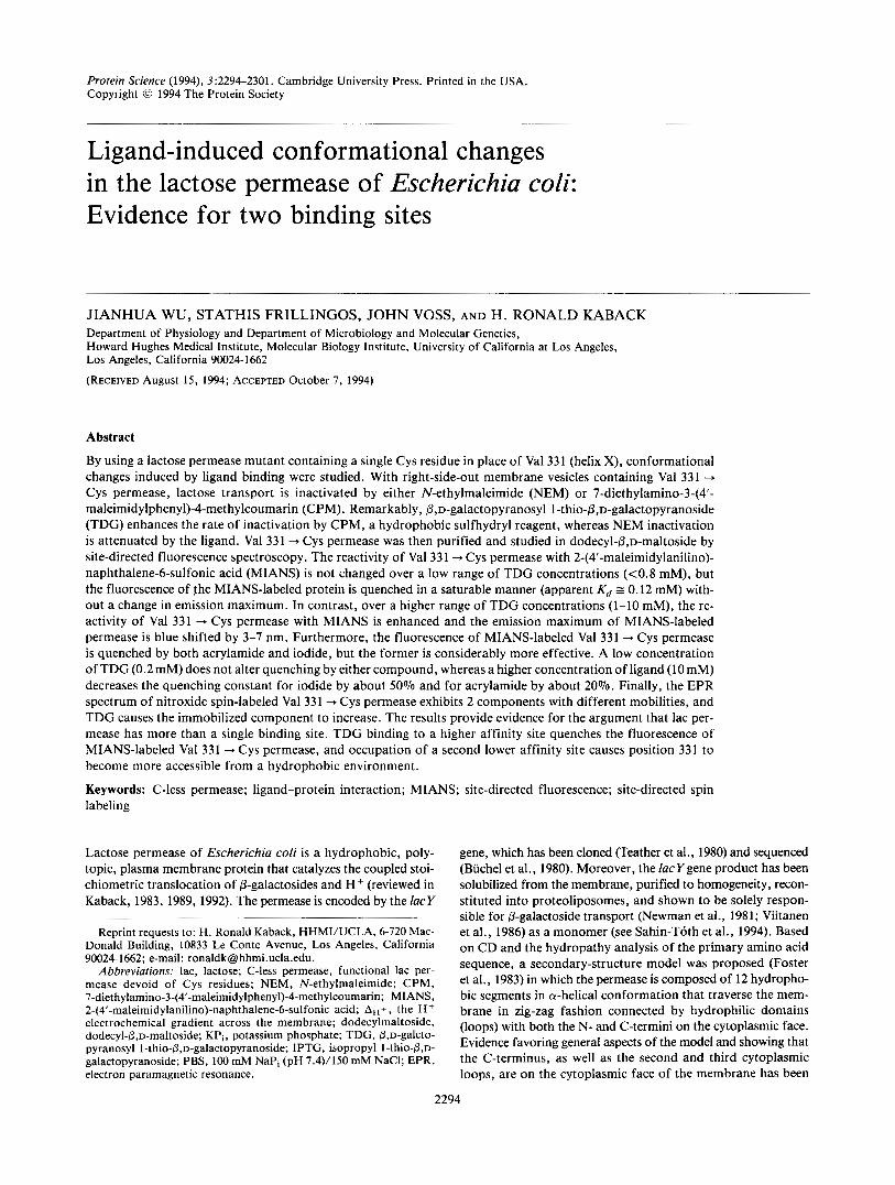
As part of a Cys-scanning mutagenesis study of helices IX and X in lac permease (Sahin-Toth & Kaback, 1993), it was found that the activity of V331C is highly sensitive to inactivation by NEM. Moreover, with the sole exception of V331C, the NEM- sensitive Cys-replacement mutants in helix X fall on the same face of the helix (Fig. 1). In right-side-out membrane vesicles containing V33 1C permease, the hydrophobic sulfhydryl reagent CPM at a final concentration of 50 mM inactivates lactose trans- port by about 70% within 10 min in the absence of ligand
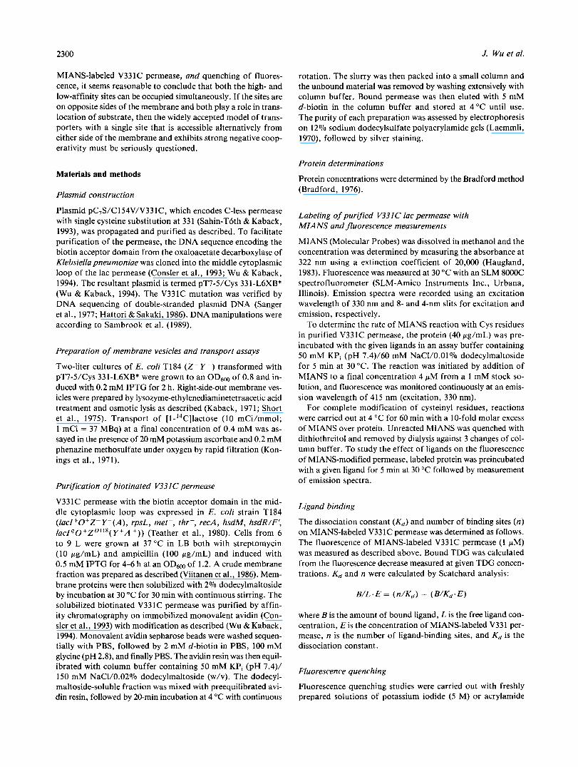
= 2.5 min; Fig. 2A). Importantly, inactivation is not due to an effect on ApH+ generation because the activity of vesicles containing C-less permease is not altered under the same con- ditions (data not shown). Interestingly, inactivation by CPM is faster in the presence of 10 mM TDG (tl,z I 0.5 min). In con- trast, similar rates of inactivation are observed with NEM in the absence of ligand, but TDG markedly attenuates the effect of the alkylating agent (Fig. 2B; t1,2 > 10 min).
MIANS labeling of Cys 331 and the effect of ligand
MIANS is a sulfhydryl reagent that reacts relatively specifically and covalently with thiol groups (Gupte & Lane, 1979), and it is not fluorescent until the maleimide group reacts (Haugland, 1983). As shown previously (Wu & Kaback, 1994), reaction of MIANS with purifed permease molecules containing single Cys residues can be followed readily fluorimetrically. Reaction of MIANS with purified V331C permease is rapid for 1.5 min and
T720 ~
F324 / ""
V m H322
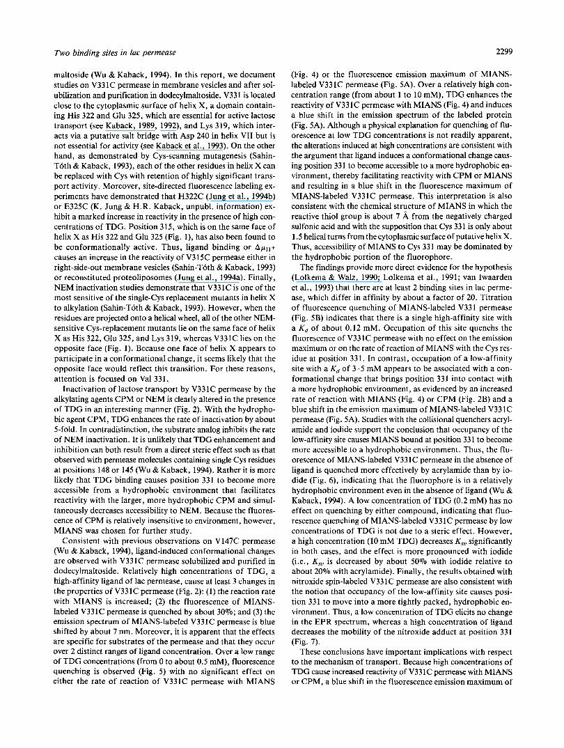
Fig. 1. Helical wheel plot of putative helix X. Eighteen residues (posi- tions 315-332) of putative helix X are shown on a helical wheel plot viewed from the periplasmic surface of the membrane. The NEM- sensitive Cys-replacement mutants are underlined, and Val 315, Lys 319, His 322, Glu 325, and Val 331 are highlighted (see text). The smaller, italicized numbers represent the positions in the putative transmembrane helix starting at the periplasmic surface.
2296 J. Wu et al.
A
0 ' I I I I I I 0 2 4 6 8 1 0 1 2
I I I I I I
0 2 4 6
Time (min)
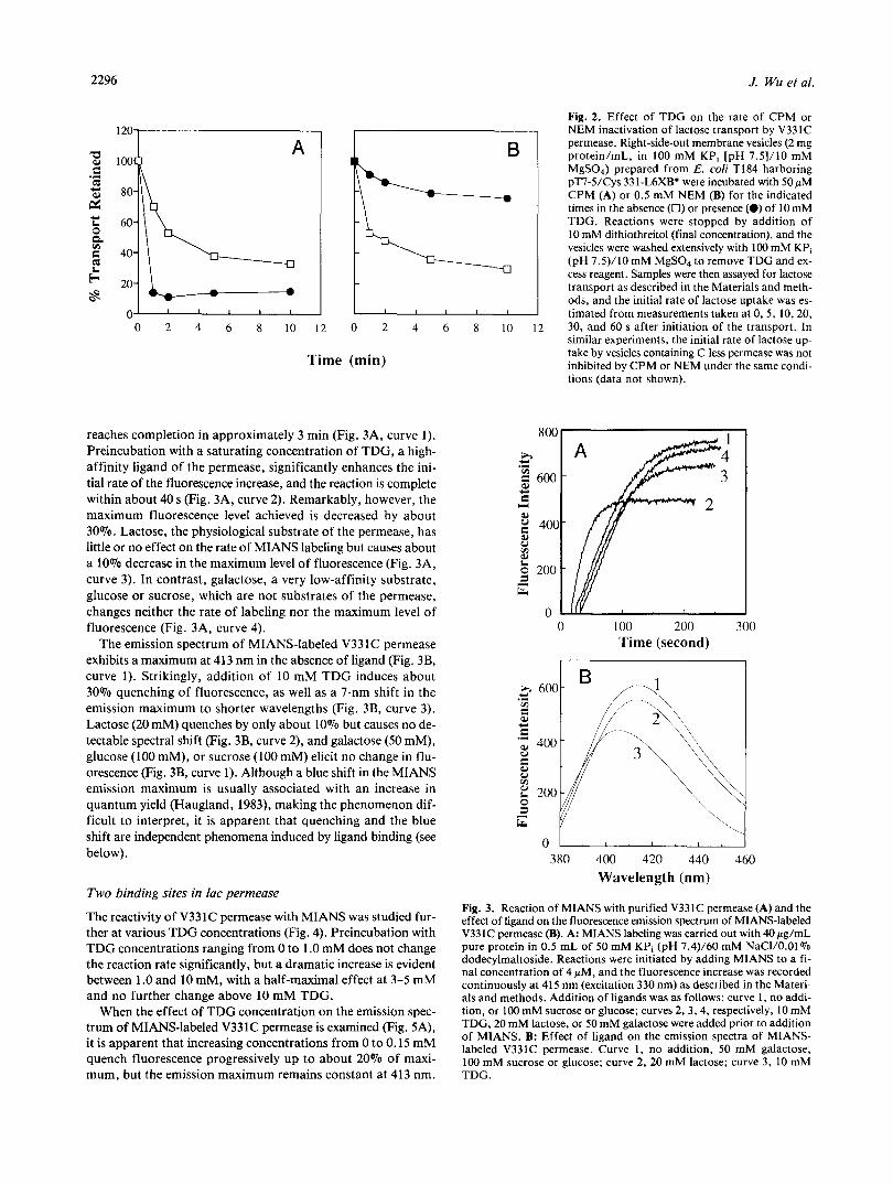
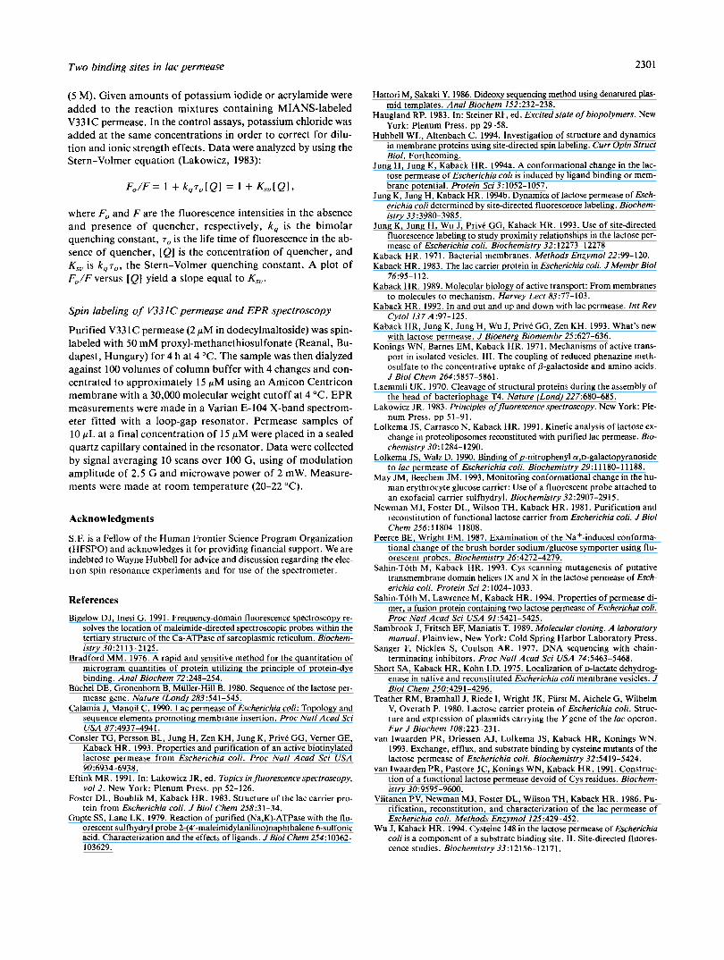
reaches completion in approximately 3 min (Fig. 3A, curve 1). Preincubation with a saturating concentration of TDG, a high- affinity ligand of the permease, significantly enhances the ini- tial rate of the fluorescence increase, and the reaction is complete within about 40 s (Fig. 3A, curve 2). Remarkably, however, the maximum fluorescence level achieved is decreased by about 30%. Lactose, the physiological substrate of the permease, has little or no effect on the rate of MIANS labeling but causes about a 10% decrease in the maximum level of fluorescence (Fig. 3A, curve 3). In contrast, galactose, a very low-affinity substrate, glucose or sucrose, which are not substrates of the permease, changes neither the rate of labeling nor the maximum level of fluorescence (Fig. 3A, curve 4).
The emission spectrum of MIANS-labeled V33 1C permease exhibits a maximum at 413 nm in the absence of ligand (Fig. 3B, curve 1). Strikingly, addition of 10 mM TDG induces about 30% quenching of fluorescence, as well as a 7-nm shift in the emission maximum to shorter wavelengths (Fig. 3B, curve 3). Lactose (20 mM) quenches by only about 10% but causes no de- tectable spectral shift (Fig. 3B, curve 2), and galactose (50 mM), glucose (100 mM), or sucrose (100 mM) elicit no change in flu- orescence (Fig. 3B, curve 1). Although a blue shift in the MIANS emission maximum is usually associated with an increase in quantum yield (Haugland, 1983), making the phenomenon dif- ficult to interpret, it is apparent that quenching and the blue shift are independent phenomena induced by ligand binding (see below).
Two binding sites in lac permease
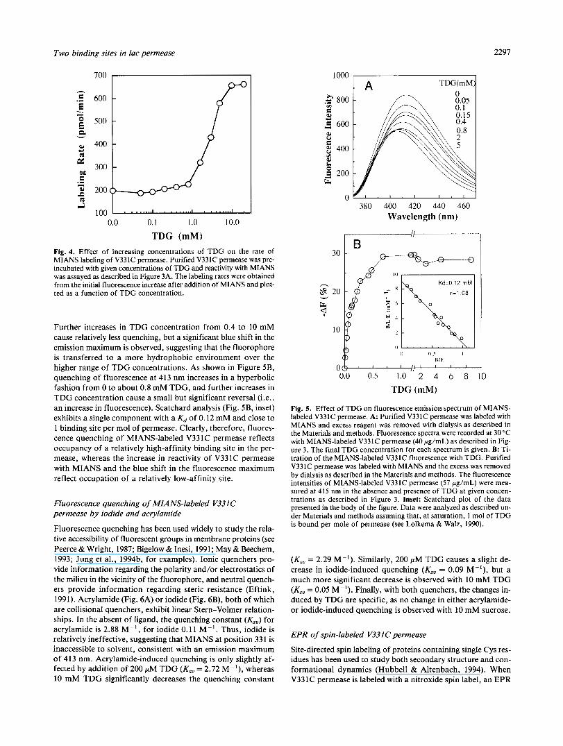
The reactivity of V331C permease with MIANS was studied fur- ther at various TDG concentrations (Fig. 4). Preincubation with TDG concentrations ranging from 0 to 1 .O mM does not change the reaction rate significantly, but a dramatic increase is evident between 1 .O and 10 mM, with a half-maximal effect at 3-5 mM and no further change above 10 mM TDG.
When the effect of TDG concentration on the emission spec- trum of MIANS-labeled V331C permease is examined (Fig. 5A), it is apparent that increasing concentrations from 0 to 0.15 mM quench fluorescence progressively up to about 20% of maxi- mum. but the emission maximum remains constant at 413 nm.
8 I O 12
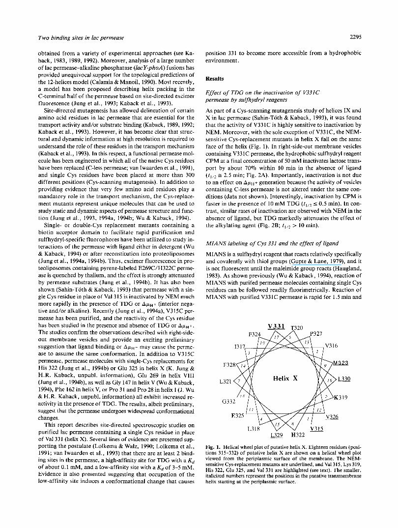
Fig. 2. Effect of TDG on the rate of CPM or NEM inactivation of lactose transport by V331C permease. Right-side-out membrane vesicles (2 mg protein/mL, in 100 mM KPi [pH 7.5]/10 mM MgSO,) prepared from E. coli T184 harboring pT7-5/Cys 33 1-L6XBt were incubated with 50 pM CPM (A) or 0.5 mM NEM (B) for the indicated times in the absence (0) or presence (0) of 10 mM TDG. Reactions were stopped by addition of 10 mM dithiothreitol (final concentration), and the vesicles were washed extensively with 100 mM KPi (pH 7.5)/10 mM MgSO, to remove TDG and ex- cess reagent. Samples were then assayed for lactose transport as described in the Materials and meth- ods, and the initial rate of lactose uptake was es- timated from measurements taken at 0.5, 10,20, 30, and 60 s after initiation of the transport. In similar experiments, the initial rate of lactose up- take by vesicles containing C-less permease was not inhibited by CPM or NEM under the same condi- tions (data not shown).
0 100 200 300 Time (second)
380 400 420 440 460 Wavelength (nm)
Fig. 3. Reaction of MIANS with purified V331C permease (A) and the effect of ligand on the fluorescence emission spectrum of MIANS-labeled V331C permease (B). A: MIANS labeling was carried out with 4Opg/mL pure protein in 0.5 mL of 50 mM KPi (pH 7.4)/60 mM NaCI/O.Ol% dodecylmaltoside. Reactions were initiated by adding MIANS to a fi- nal concentration of 4 pM, and the fluorescence increase was recorded continuously at 415 nm (excitation 330 nm) as described in the Materi- als and methods. Addition of ligands was as follows: curve 1, no addi- tion, or 100 mM sucrose or glucose; curves 2,3,4, respectively, 10 mM TDG, 20 mM lactose, or 50 mM galactose were added prior to addition of MIANS. B: Effect of ligand on the emission spectra of MIANS- labeled V331C permease. Curve 1, no addition, 50 mM galactose,
TDG. 100 mM sucrose or glucose; curve 2, 20 mM lactose; curve 3, 10 mM
Two binding sites in lac permease 2297
1000 TDG(mM
600
500
400
300
200
100 0.0 0.1 1 .O 10.0
TDG (mM) Fig. 4. Effect of increasing concentrations of TDG on the rate of MIANS labeling of V331C permease. Purified V331C permease was pre- incubated with given concentrations of TDG and reactivity with MIANS was assayed as described in Figure 3A. The labeling rates were obtained from the initial fluorescence increase after addition of MIANS and plot- ted as a function of TDG concentration.
Further increases in TDG concentration from 0.4 to 10 mM cause relatively less quenching, but a significant blue shift in the emission maximum is observed, suggesting that the fluorophore is transferred to a more hydrophobic environment over the higher range of TDG concentrations. As shown in Figure 5B, quenching of fluorescence at 413 nm increases in a hyperbolic fashion from 0 to about 0.8 mM TDG, and further increases in TDG concentration cause a small but significant reversal (i.e., an increase in fluorescence). Scatchard analysis (Fig. 5B, inset) exhibits a single component with a Kd of 0.12 mM and close to 1 binding site per mol of permease. Clearly, therefore, fluores- cence quenching of MIANS-labeled V33 1C permease reflects occupancy of a relatively high-affinity binding site in the per- mease, whereas the increase in reactivity of V331C permease with MIANS and the blue shift in the fluorescence maximum reflect occupation of a relatively low-affinity site.
Fluorescence quenching of MIANS-labeled V331 C permease by iodide and acrylamide
Fluorescence quenching has been used widely to study the rela- tive accessibility of fluorescent groups in membrane proteins (see Peerce & Wright, 1987; Bigelow & Inesi, 1991; May & Beechem, 1993; Jung et al., 1994b, for examples). Ionic quenchers pro- vide information regarding the polarity and/or electrostatics of the milieu in the vicinity of the fluorophore, and neutral quench- ers provide information regarding steric resistance (Eftink, 1991). Acrylamide (Fig. 6A) or iodide (Fig. 6B), both of which are collisional quenchers, exhibit linear Stern-Volmer relation- ships. In the absent of ligand, the quenching constant (&) for acrylamide is 2.88 M-I, for iodide 0.11 "I. Thus, iodide is relatively ineffective, suggesting that MIANS at position 331 is inaccessible to solvent, consistent with an emission maximum of 413 nm. Acrylamide-induced quenching is only slightly af- fected by addition of 200 pM TDG (Ksu = 2.72 "I), whereas 10 mM TDG significantly decreases the quenching constant
.g 800 v)
9 c) A 600 W V e 8 400 r.i s 200 E;
L
0
h
8
Q v
k
30
20
10
380 400 420 440 460 Wavelength (nm)
0.0 0.5 1.0 2 4 6 8 10 TDG (mM)
Fig. 5. Effect of TDG on fluorescence emission spectrum of MIANS- labeled V331C permease. A: Purified V331C permease was labeled with MIANS and excess reagent was removed with dialysis as described in the Materials and methods. Fluorescence spectra were recorded at 30 "C with MIANS-labeled V331C permease (40 pg/mL) as described in Fig- ure 3. The final TDG concentration for each spectrum is given. B: Ti- tration of the MIANS-labeled V331C fluorescence with TDG. Purified V331C permease was labeled with MIANS and the excess was removed by dialysis as described in the Materials and methods. The fluorescence intensities of MIANS-labeled V331C permease (57 pg/mL) were mea- sured at 41 5 nm in the absence and presence of TDG at given concen- trations as described in Figure 3. Inset: Scatchard plot of the data presented in the body of the figure. Data were analyzed as described un- der Materials and methods assuming that, at saturation, 1 mol of TDG is bound per mole of permease (see Lolkema & Walz, 1990).
(Kso = 2.29 M"). Similarly, 200 pM TDG causes a slight de- crease in iodide-induced quenching (Ksu = 0.09 "'), but a much more significant decrease is observed with 10 mM TDG (Kso = 0.05 M"). Finally, with both quenchers, the changes in- duced by TDG are specific, as no change in either acrylamide- or iodide-induced quenching is observed with 10 mM sucrose.
EPR of spin-labeled V33ICpermease
Site-directed spin labeling of proteins containing single Cys res- idues has been used to study both secondary structure and con- formational dynamics (Hubbell & Altenbach, 1994). When V331C permease is labeled with a nitroxide spin label, an EPR

2298 J. Wu et al.
A
2*5 2 i \ k
k 0
1.5 -
1 I I
0 0.2 0.4 0.6 Acrylamide (M)
1.02 I 0 0.2 0.4 0.6
Potassium Iodide (M)
Fig. 6. Quenching of MIANS-labeled V331C fluorescence by acrylamide (A) or iodide (B) and the effect of TDG. Purified V331C permease was labeled with MIANS and the excess reagent was removed by dialysis. The fluorescence emission spectra in the absence or presence of given concentrations of acrylamide or iodide were recorded with an excitation wavelength at 330 nm. The data were fit to a linear regression plot according to the Stern-Volmer equation as described in the Materials and methods. 0, MIANS-labeled V331C permease in the absence of ligand; A, MIANS-labeled V331C permease in the presence of 0.2 mM TDG; 0, MIANS-labeled V331C permease in the presence of 10 mM TDG.
spectrum typical of a nitroxide with intermediate mobility is ob- served (Fig. 7). Addition of 0.2 mM TDG induces no significant change in the spectrum (not shown), but 10 mM TDG causes sig- nificant broadening of the nitroxide line shape, implying that ligand binding causes the labeled side chain to become more restricted motionally. When compared to the control, the de- creased motional averaging in the presence of 10 mM TDG is especially apparent in the relative amplitudes of the weakly (w) and strongly (s) immobilized components at the low field posi- tions. These results are consistent with the observations obtained from site-directed fluorescent labeling and indicate that residue
33 1 is transferred into a more tightly packed, hydrophobic en- vironment upon occupation of a low-affinity binding site in the permease.
Discussion
Previous studies from this laboratory demonstrate that ligand- induced conformational changes in the lac permease can be stud- ied by site-directed fluorescence labeling of purified permease containing single Cys residues either reconstituted into proteo- liposomes (Jung et al., 1994a, 1994b) or solubilized in dodecyl-
Fig. 7. Effect of TDG on the EPR spec- trum of nitroxide-labeled V331C permease. Spectra were recorded with 15 pM spin- labeled permease in the absence (solid line) and presence (dotted line) of 10 mM TDG as described in the Materials and methods. s and w, strongly and weakly immobilized spectral components, respectively. Each spectrum represents the average of 10 scans, and the scan range is 100 G.
Two binding sites in lac permease 2299
maltoside (Wu & Kaback, 1994). In this report, we document studies on V331C permease in membrane vesicles and after sol- ubilization and purification in dodecylmaltoside. V331 is located close to the cytoplasmic surface of helix X, a domain contain- ing His 322 and Glu 325, which are essential for active lactose transport (see Kaback, 1989, 1992), and Lys 319, which inter- acts via a putative salt bridge with Asp 240 in helix VI1 but is not essential for activity (see Kaback et al., 1993). On the other hand, as demonstrated by Cys-scanning mutagenesis (Sahin- Toth & Kaback, 1993), each of the other residues in helix X can be replaced with Cys with retention of highly significant trans- port activity. Moreover, site-directed fluorescence labeling ex- periments have demonstrated that H322C (Jung et al., 1994b) or E325C (K. Jung & H.R. Kaback, unpubl. information) ex- hibit a marked increase in reactivity in the presence of high con- centrations of TDG. Position 315, which is on the same face of helix X as His 322 and Glu 325 (Fig. l), has also been found to be conformationally active. Thus, ligand binding or ApH+ causes an increase in the reactivity of V315C permease either in right-side-out membrane vesicles (Sahin-Toth & Kaback, 1993) or reconstituted proteoliposomes (Jung et al., 1994a). Finally, NEM inactivation studies demonstrate that V331C is one of the most sensitive of the single-Cys replacement mutants in helix X to alkylation (Sahin-Toth & Kaback, 1993). However, when the residues are projected onto a helical wheel, all of the other NEM- sensitive Cys-replacement mutants lie on the same face of helix X as His 322, Glu 325, and Lys 319, whereas V331C lies on the opposite face (Fig. 1). Because one face of helix X appears to participate in a conformational change, it seems likely that the opposite face would reflect this transition. For these reasons, attention is focused on Val 331.
Inactivation of lactose transport by V331C permease by the alkylating agents CPM or NEM is clearly altered in the presence of TDG in an interesting manner (Fig. 2). With the hydropho- bic agent CPM, TDG enhances the rate of inactivation by about 5-fold. In contradistinction, the substrate analog inhibits the rate of NEM inactivation. It is unlikely that TDG enhancement and inhibition can both result from a direct steric effect such as that observed with permease molecules containing single Cys residues at positions 148 or 145 (Wu & Kaback, 1994). Rather it is more likely that TDG binding causes position 331 to become more accessible from a hydrophobic environment that facilitates reactivity with the larger, more hydrophobic CPM and simul- taneously decreases accessibility to NEM. Because the fluores- cence of CPM is relatively insensitive to environment, however, MIANS was chosen for further study.
Consistent with previous observations on V147C permease (Wu & Kaback, 1994), ligand-induced conformational changes are observed with V33 1C permease solubilized and purified in dodecylmaltoside. Relatively high concentrations of TDG, a high-affinity ligand of lac permease, cause at least 3 changes in the properties of V331C permease (Fig. 2): (1) the reaction rate with MIANS is increased; (2) the fluorescence of MIANS- labeled V331C permease is quenched by about 30%; and (3) the emission spectrum of MIANS-labeled V331C permease is blue shifted by about 7 nm. Moreover, it is apparent that the effects are specific for substrates of the permease and that they occur over 2 distinct ranges of ligand concentration. Over a low range of TDG concentrations (from 0 to about 0.5 mM), fluorescence quenching is observed (Fig. 5) with no significant effect on either the rate of reaction of V331C permease with MIANS
(Fig. 4) or the fluorescence emission maximum of MIANS- labeled V331C permease (Fig. SA). Over a relatively high con- centration range (from about 1 to 10 mM), TDG enhances the reactivity of V331C permease with MIANS (Fig. 4) and induces a blue shift in the emission spectrum of the labeled protein (Fig. SA). Although a physical explanation for quenching of flu- orescence at low TDG concentrations is not readily apparent, the alterations induced at high concentrations are consistent with the argument that ligand induces a conformational change caus- ing position 331 to become accessible to a more hydrophobic en- vironment, thereby facilitating reactivity with CPM or MIANS and resulting in a blue shift in the fluorescence maximum of MIANS-labeled V33 1C permease. This interpretation is also consistent with the chemical structure of MIANS in which the reactive thiol group is about 7 A from the negatively charged sulfonic acid and with the supposition that Cys 33 1 is only about 1.5 helical turns from the cytoplasmic surface of putative helix X. Thus, accessibility of MIANS to Cys 33 1 may be dominated by the hydrophobic portion of the fluorophore.
The findings provide more direct evidence for the hypothesis (Lolkema & Walz, 1990; Lolkema et al., 1991; van Iwaarden et al., 1993) that there are at least 2 binding sites in lac perme- ase, which differ in affinity by about a factor of 20. Titration of fluorescence quenching of MIANS-labeled V33 1 permease (Fig. 5B) indicates that there is a single high-affinity site with a Kd of about 0.12 mM. Occupation of this site quenchs the fluorescence of V331C permease with no effect on the emission maximum or on the rate of reaction of MIANS with the Cys res- idue at position 331. In contrast, occupation of a low-affinity site with a Kd of 3-5 mM appears to be associated with a con- formational change that brings position 331 into contact with a more hydrophobic environment, as evidenced by an increased rate of reaction with MIANS (Fig. 4) or CPM (Fig. 2B) and a blue shift in the emission maximum of MIANS-labeled V331C permease (Fig. SA). Studies with the collisional quenchers acryl- amide and iodide support the conclusion that occupancy of the low-affinity site causes MIANS bound at position 331 to become more accessible to a hydrophobic environment. Thus, the flu- orescence of MIANS-labeled V331C permease in the absence of ligand is quenched more effectively by acrylamide than by io- dide (Fig. 6), indicating that the fluorophore is in a relatively hydrophobic environment even in the absence of ligand (Wu & Kaback, 1994). A low concentration of TDG (0.2 mM) has no effect on quenching by either compound, indicating that fluo- rescence quenching of MIANS-labeled V331C permease by low concentrations of TDG is not due to a steric effect. However, a high concentration (10 mM TDG) decreases K,, significantly in both cases, and the effect is more pronounced with iodide (i.e., K,, is decreased by about 50% with iodide relative to about 20% with acrylamide). Finally, the results obtained with nitroxide spin-labeled V331C permease are also consistent with the notion that occupancy of the low-affinity site causes posi- tion 33 1 to move into a more tightly packed, hydrophobic en- vironment. Thus, a low concentration of TDG elicits no change in the EPR spectrum, whereas a high concentration of ligand decreases the mobility of the nitroxide adduct at position 331 (Fig. 7).
These conclusions have important implications with respect to the mechanism of transport. Because high concentrations of TDG cause increased reactivity of V331C permease with MIANS or CPM, a blue shift in the fluorescence emission maximum of
2300 J. Wu et al.
MIANS-labeled V331C permease, and quenching of fluores- cence, it seems reasonable to conclude that both the high- and low-affinity sites can be occupied simultaneously. If the sites are on opposite sides of the membrane and both play a role in trans- location of substrate, then the widely accepted model of trans- porters with a single site that is accessible alternatively from either side of the membrane and exhibits strong negative coop- erativity must be seriously questioned.
Materials and methods
Plasmid construction
Plasmid pC7S/C154V/V331C, which encodes C-less permease with single cysteine substitution at 331 (Sahin-Toth & Kaback, 1993), was propagated and purified as described. To facilitate purification of the permease, the DNA sequence encoding the biotin acceptor domain from the oxaloacetate decarboxylase of Klebsiella pneumoniae was cloned into the middle cytoplasmic loop of the lac permease (Consler et al., 1993; Wu & Kaback, 1994). The resultant plasmid is termed pT7-5ICys 331-L6XB* (Wu & Kaback, 1994). The V331C mutation was verified by DNA sequencing of double-stranded plasmid DNA (Sanger et al., 1977; Hattori & Sakaki, 1986). DNA manipulations were according to Sambrook et al. (1989).
Preparation of membrane vesicles and transport assays
Two-liter cultures of E. coli T184 ( Z - Y - ) transformed with pT7-5/Cys 331-L6XB* were grown to an OD,, of 0.8 and in- duced with 0.2 mM IPTG for 2 h. Right-side-out membrane ves- icles were prepared by lysozyme-ethylenediaminetetraacetic acid treatment and osmotic lysis as described (Kaback, 1971; Short et al., 1975). Transport of [l-'4C]lactose (10 mCi/mmol; 1 mCi = 37 MBq) at a final concentration of 0.4 mM was as- sayed in the presence of 20 mM potassium ascorbate and 0.2 mM phenazine methosulfate under oxygen by rapid filtration (Kon- ings et al., 1971).
Purification of biotinated V331C permease
V331C permease with the biotin acceptor domain in the mid- dle cytoplasmic loop was expressed in E. coli strain T184 (lacI+O+Z-Y-(A), rpsL, met-, thr-, recA, hsdM, hsdR/F', l a c 1 9 0 + ~ ~ " ~ ( Y + A +)) (Teather et al., 1980). Cells from 6 to 9 L were grown at 37 "C in LB both with streptomycin (10 pg/mL) and ampicillin (100 pg/mL) and induced with 0.5 mM IPTG for 4-6 h at an ODm of 1.2. A crude membrane fraction was prepared as described (Viitanen et al., 1986). Mem- brane proteins were then solubilized with 2% dodecylmaltoside by incubation at 30 "C for 30 min with continuous stirring. The solubilized biotinated V331C permease was purified by affin- ity chromatography on immobilized monovalent avidin (Con- sler et al., 1993) with modification as described (Wu & Kaback, 1994). Monovalent avidin sepharose beads were washed sequen- tially with PBS, followed by 2 mM d-biotin in PBS, 100 mM glycine (pH 2.8), and finally PBS. The avidin resin was then equil- ibrated with column buffer containing 50 mM KP, (pH 7.4)/ 150 mM NaC1/0.02% dodecylmaltoside (w/v). The dodecyl- maltoside-soluble fraction was mixed with preequilibrated avi- din resin, followed by 20-min incubation at 4 "C with continuous
rotation. The slurry was then packed into a small column and the unbound material was removed by washing extensively with column buffer. Bound permease was then eluted with 5 mM d-biotin in the column buffer and stored at 4 "C until use. The purity of each preparation was assessed by electrophoresis on 12% sodium dodecylsulfate polyacrylamide gels (Laemmli, 1970), followed by silver staining.
Protein determinations
Protein concentrations were determined by the Bradford method (Bradford, 1976).
Labeling of purified V331C lac permease with MIANS and fluorescence measurements
MIANS (Molecular Probes) was dissolved in methanol and the concentration was determined by measuring the absorbance at 322 nm using a extinction coefficient of 20,000 (Haugland, 1983). Fluorescence was measured at 30 "C with an SLM 8000C spectrofluorometer (SLM-Amico Instruments Inc., Urbana, Illinois). Emission spectra were recorded using an excitation wavelength of 330 nm and 8- and 4-nm slits for excitation and emission, respectively.
To determine the rate of MIANS reaction with Cys residues in purified V331C permease, the protein (40 pg/mL) was pre- incubated with the given ligands in an assay buffer containing 50 mM KP, (pH 7.4)/60 mM NaCl/O.Ol% dodecylmaltoside for 5 min at 30 "C. The reaction was initiated by addition of MIANS to a final concentration 4 pM from a 1 mM stock so- lution, and fluorescence was monitored continuously at an emis- sion wavelength of 415 nm (excitation, 330 nm).
For complete modification of cysteinyl residues, reactions were carried out at 4 "C for 60 min with a 10-fold molar excess of MIANS over protein. Unreacted MIANS was quenched with dithiothreitol and removed by dialysis against 3 changes of col- umn buffer. To study the effect of ligands on the fluorescence of MIANS-modified permease, labeled protein was preincubated with a given ligand for 5 min at 30 "C followed by measurement of emission spectra.
Ligand binding
The dissociation constant (Kd) and number of binding sites ( n ) on MIANS-labeled V331C permease was determined as follows. The fluorescence of MIANS-labeled V331C permease (1 pM) was measured as described above. Bound TDG was calculated from the fluorescence decrease measured at given TDG concen- trations. Kd and n were calculated by Scatchard analysis:
where B is the amount of bound ligand, L is the free ligand con- centration, E is the concentration of MIANS-labeled V331 per- mease, n is the number of ligand-binding sites, and Kd is the dissociation constant.
Fluorescence quenching
Fluorescence quenching studies were carried out with freshly prepared solutions of potassium iodide ( 5 M) or acrylamide
Two binding sites in lac permease 2301
(5 M). Given amounts of potassium iodide or acrylamide were added to the reaction mixtures containing MIANS-labeled V331C permease. In the control assays, potassium chloride was added at the same concentrations in order to correct for dilu- tion and ionic strength effects. Data were analyzed by using the Stern-Volmer equation (Lakowicz, 1983):
where F, and F a r e the fluorescence intensities in the absence and presence of quencher, respectively, k, is the bimolar quenching constant, 7, is the life time of fluorescence in the ab- sence of quencher, [Q] is the concentration of quencher, and K,, is kqro, the Stern-Volmer quenching constant. A plot of F,/F versus [Q] yield a slope equal to K,,.
Spin labeling of V331Cpermease and EPR spectroscopy
Purified V33 IC permease (2 p M in dodecylmaltoside) was spin- labeled with 50 mM proxyl-methanethiosulfonate (Reanal, Bu- dapest, Hungary) for 4 h at 4 "C. The sample was then dialyzed against 100 volumes of column buffer with 4 changes and con- centrated to approximately 15 pM using an Amicon Centricon membrane with a 30,000 molecular weight cutoff at 4 "C. EPR measurements were made in a Varian E-104 X-band spectrom- eter fitted with a loop-gap resonator. Permease samples of 10 pL at a final concentration of 15 pM were placed in a sealed quartz capillary contained in the resonator. Data were collected by signal averaging 10 scans over 100 G, using of modulation amplitude of 2.5 G and microwave power of 2 mW. Measure- ments were made at room temperature (20-22 "C).
Acknowledgments
S.F. is a Fellow of the Human Frontier Science Program Organization (HFSPO) and acknowledges it for providing financial support. We are indebted to Wayne Hubbell for advice and discussion regarding the elec- tron spin resonance experiments and for use of the spectrometer.
References Bigelow DJ, Inesi G. 1991. Frequency-domain fluorescence spectroscopy re-
solves the location of maleimide-directed spectroscopic probes within the tertiary structure of the Ca-ATPase of sarcoplasmic reticulum. Biochem- istry 30:2113-2125.
Bradford MM. 1976. A rapid and sensitive method for the quantitation of microgram quantities of protein utilizing the principle of protein-dye binding. Anal Biochem 72:248-254.
Buchel DE, Gronenborn B, Muller-Hill B. 1980. Sequence of the lactose per- mease gene. Nature (Lond) 283:541-545.
Calamia J, Manoil C. 1990. Lac permease of Escherichia coli: Topology and sequence elements promoting membrane insertion. Proc Nut1 Acad Sci USA 87:4937-4941.
Consler TG, Persson BL, Jung H, Zen KH, Jung K, Prive GG, Verner GE, Kaback HR. 1993. Properties and purification of an active biotinylated lactose permease from Escherichia coli. Proc Nut1 Acad Sci USA 90:6934-6938.
Eftink MR. 1991. In: Lakowicz JR, ed. Topics influorescencespectroscopy,
Foster DL, Boublik M, Kaback HR. 1983. Structure of the lac carrier pro- vol2. New York: Plenum Press. pp 52-126.
tein from Escherichia coli. JBiol Chem 258:31-34. Gupte SS, Lane LK. 1979. Reaction of purified (Na,K)-ATPase with the flu-
orescent sulfhydryl probe 2-(4"maleimidylanilino)naphthalene 6-sulfonic acid. Characterization and the effects of ligands. JBiol Chem 254:10362- 103629.
Hattori M, Sakaki Y. 1986. Dideoxy sequencing method using denatured plas- mid templates. Anal Biochem 152:232-238.
Haugland RP. 1983. In: Steiner RF, ed. Excitedstate of biopolymers. New York: Plenum Press. pp 29-58.
Hubbell WL, Altenbach C. 1994. Investigation of structure and dynamics in membrane proteins using site-directed spin labeling. Curr Opin Struct Biol. Forthcoming.
Jung H, Jung K, Kaback HR. 1994a. A conformational change in the lac- tose permease of Escherichia coli is induced by ligand binding or mem- brane potential. Protein Sci3:1052-1057.
Jung K, Jung H, Kaback HR. 1994b. Dynamics of lactose permease of Esch- erichia coli determined bv site-directed fluorescence labeling. Biochem- istry 33:3980-3985.
Jung K. Jung H, Wu J, Prive GG, Kaback HR. 1993. Use of site-directed - fluorescence labeling to study proximity relationships in the lactose per- mease of Escherichia coli. Biochemistry 32:12273-12278
Kaback HR. 1971. Bacterial membranes. Methods Enzymol22:99-120. Kaback HR. 1983. The lac carrier protein in Escherichia coli. JMembr Biol
Kaback HR. 1989. Molecular biology of active transport: From membranes
Kaback HR. 1992. In and out and up and down with lac permease. Inf Rev
Kaback HR, Jung K, Jung H, Wu J, Prive GG, Zen KH. 1993. What's new with lactose permease. J Bioenerg Biomembr 25:627-636.
Konings WN, Barnes EM, Kaback HR. 1971. Mechanisms of active trans- port in isolated vesicles. 111. The coupling of reduced phenazine meth- osulfate to the concentrative uptake of &galactoside and amino acids. J Biol Chem 264:5857-5861.
Laemmli UK. 1970. Cleavage of structural proteins during the assembly of the head of bacteriophage T4. Nature (Lond) 227:680-685.
Lakowicz JR. 1983. Principles ofjluorescencespectroscopy. New York: Ple-
Lolkema JS, Carrasco N, Kaback HR. 1991. Kinetic analysis of lactose ex- num Press. pp 51-91.
change in proteoliposomes reconstituted with purified lac permease. Bio- chemistry 30:1284-1290.
Lolkema JS, Walz D. 1990. Binding ofp-nitrophenyl a,D-galactopyranoside to lac permease of Escherichia coli. Biochemistry 29: 11 180-1 1188.
May JM, Beechem JM. 1993. Monitoring conformational change in the hu- man erythrocyte glucose carrier: Use of a fluorescent probe attached to
Newman MJ, Foster DL, Wilson TH, Kaback HR. 1981. Purification and an exofacial carrier sulfhydryl. Biochemistry 32:2907-2915.
reconstitution of functional lactose carrier from Escherichia coli. J Biol Chem 256: 1 1804- 1 1808.
Peerce BE, Wright EM. 1987. Examination of the Na+induced conforma- tional change of the brush border sodium/glucose symporter using flu- orescent probes. Biochemistry 26:4272-4279.
Sahin-T6th M, Kaback HR. 1993. Cys scanning mutagenesis of putative transmembrane domain helices IX and X in the lactose permease of Esch- erichia coli. Protein Sci 2:1024-1033.
Sahin-Toth M, Lawrence M, Kaback HR. 1994. Properties of permease di-
Proc Natl Acad sci USA 91:5421-5425. mer, a fusion protein containing two lactose permease of Escherichia coli.
Sambrook J, Fritsch EF, Maniatis T. 1989. Molecularcloning. A laboratory
Sanger F, Nicklen S, Coulson AR. 1977. DNA sequencing with chain- manual. Plainview, New York: Cold Spring Harbor Laboratory Press.
terminating inhibitors. Proc Nut1 Acad Sci USA 74:5463-5468. Short SA, Kaback HR, Kohn LD. 1975. Localization of D-lactate dehydrog-
enase in native and reconstituted Escherichia coli membrane vesicles. J Biol Chem 250:4291-4296.
Teather RM, Bramhall J, Riede 1, Wright JK, Fiirst M, Aichele G, Wilhelm V, Overath P. 1980. Lactose carrier protein of Escherichia coli. Struc- ture and expression of plasmids carrying the Y gene of the lac operon. Eur J Biochem 108:223-23 1.
van Iwaarden PR, Driessen AJ, Lolkema JS, Kaback HR, Konings WN. 1993. Exchange, efflux, and substrate binding by cysteine mutants of the lactose permease of Escherichia coli. Biochemistry 32:5419-5424.
van Iwaarden PR, Pastore JC, Konings WN, Kaback HR. 1991. Construc- tion of a functional lactose permease devoid of Cys residues. Biochem- istry 30:9595-9600.
Viitanen PV, Newman MJ, Foster DL, Wilson TH, Kaback HR. 1986. Pu- rification, reconstitution, and characterization of the lac permease of Escherichia coli. Methods Enzymol125:429-452.
Wu J, Kaback HR. 1994. Cysteine 148 in the lactose permease of Escherichia coli is a component of a substrate binding site. 11. Site-directed fluores- cence studies. Biochemistry33:12156-12171.
76:95-112.
to molecules to mechanism. Harvey Lect 83:77-103.
cytol 137 A:97-125.