alginate/lactose-modified chitosan hydrogels: a bioactive biomaterial for chondrocyte encapsulation
TRANSCRIPT
Alginate/lactose-modified chitosan hydrogels:A bioactive biomaterial for chondrocyte encapsulation
Eleonora Marsich,1 Massimiliano Borgogna,1 Ivan Donati,1 Pamela Mozetic,1 Berit L. Strand,2
Santiago Gomez Salvador,3 Franco Vittur,1 Sergio Paoletti11Department of Biochemistry, Biophysics and Macromolecular Chemistry, University of Trieste,Via Licio Giorgieri 1, I-34127 Trieste, Italy2Institute of Biotechnology, Norwegian University of Science and Technology (NTNU), Sem Sælands v 6/8,N-7491 Trondheim, Norway3Department of Pathological Anatomy, Faculty of Medicine, University of Cadiz, Plaza del Falla 9,SP-11003 Cadiz, Spain
Received 5 October 2006; revised 22 December 2006; accepted 19 January 2007Published online 6 July 2007 in Wiley InterScience (www.interscience.wiley.com). DOI: 10.1002/jbm.a.31307
Abstract: A new bioactive scaffold was prepared from abinary polysaccharide mixture composed of a polyanion(alginate) and a polycation (a lactose-modified chitosan,chitlac). Its potential use for articular chondrocytes encap-sulation and cartilage reconstructive surgery applicationshas been studied. The hydrogel combines the ability ofalginate to act as a 3D supporting structure with the capa-bility of the second component (chitlac) to provide interac-tions with porcine articular chondrocytes. Physico-chemi-cal characterization of the scaffold was accomplished bygel kinetics and compression measurements and demon-strated that alginate-chitlac mixture (AC-mixture) hydro-gels exhibit better mechanical properties when compared
with sole alginate hydrogels. Furthermore, biochemicaland biological studies showed that these 3D scaffoldsare able to maintain chondrocyte phenotype and particu-larly to significantly stimulate and promote chondrocytegrowth and proliferation. In conclusion, the presentstudy can be considered as a first step towards an engi-neered, biologically active scaffold for chondrocyte invitro cultivation, expansion, and cell delivery. � 2007Wiley Periodicals, Inc. J Biomed Mater Res 84A: 364–376,2008
Key words: alginate; chitosan; lactose; chitlac; chondro-cytes; cell encapsulation; polyanion-polycation mixture
INTRODUCTION
Cartilage degeneration represents a serious healthproblem, causing progressive debilitation and mark-ed decrease of quality of life. Its damages occur as aconsequence of aging, congenital abnormalities, dis-eases and traumas, and are related to the limitedintrinsic healing potential of this tissue. As a conse-quence of the lack of blood supply and subsequentinsufficient inflammatory response, cartilage lesionsresult in an incomplete repairing attempt by localchondrocytes.1,2 Recently, an approach based on theso-called A.C.I. (Autologous Chondrocyte Implanta-tion) has been presented as the ultimate technique in
the field of cartilage regeneration. Delivery of autolo-gous chondrocytes to the sites of wound requirescell isolation, in vitro cultivation, and expansion.However, chondrocytes undergo a process of pheno-typic and functional de-differentiation when culturedin monolayer systems, due to the lack of the crucialinfluence of physiological cell–cell and cell–extracel-lular matrix (ECM) interactions. For this reasonchondrocytes are generally seeded in biomimeticporous scaffolds that recreate the cellular microen-vironment, providing a defined 3D structure thatguides the tissue development.2
The tissue engineering approach (also known asregenerative strategy) has been regarded as an ex-tremely promising and appealing technique for thepotential to manipulate and combine cells and sup-portive biocompatible matrices aiming at restoringthe original architecture and function of the tissueand hence providing cartilaginous constructs to betransplanted. Until now, a wide variety of biocom-patible polymer-based materials (fibrin, hyaluronan
Correspondence to: E. Marsich; e-mail: [email protected] grant sponsor: Italian Ministry MIUR; contract
grant number: RBAU01LETEContract grant sponsors: University of Trieste, Bracco
Imaging SpA
' 2007 Wiley Periodicals, Inc.
(HA), collagen, and polyesters of a-hydroxyacids toname a few) have been proposed for 3D-scaffolddesign.3–6
Algal polysaccharide hydrogels represent verygood candidates for 3D-scaffold design, having theadvantage of being biocompatible and allowing auniform distribution of seeded cells throughout thematrix.7 Among these versatile biomaterials, alginatehas been widely employed for cell immobilizationprocedures. Alginate is a family of linear copolymers(produced by brown algae and bacteria), containing1-4-linked b-D-mannuronic acid (M) and a-L-gulur-onic acid (G) arranged in a blockwise pattern alongthe chain with homopolymeric regions of M (Mblocks) and G (G blocks) residues interspersed withregions of alternating structure (MG blocks). Algi-nate has the ability to form stable gels in presence ofmillimolar concentrations of calcium or other diva-lent cations.8 Cell encapsulation in calcium alginatebeads represents a well established method for cellprotection from host immune system,9–11 but thebiological inertness of alginate has largely hamperedits use in all those applications where cell adhesionis mandatory for survival and proliferation.12 As anexample, although alginate beads have been used toencapsulate chondrocytes, allowing them to preservetheir original phenotype,13–17 a reduced cell prolifer-ation was reported.18
Biopolymer engineering is an appealing approachto overcome the limit of nonbioadhesivity displayedby alginate.18 Introduction of biologically activesignals within the gel material might enable its inter-action with the cells, hence enhancing their embed-ding.19,20 We have recently reported on the biologicalproperties of a lactose-modified chitosan (chitlac) inthe aggregation of porcine articular chondrocytesand stimulation of chondro-specific glycosaminogly-cans (GAGs) and collagen production.21 It has beenshown that the interaction between the engineeredmaterial and the cells is mediated by galectin-1 thatspecifically recognizes the galactose inserted as sidechain into the chitosan backbone.22 However, thebiological significance of chitlac is counterbalancedby the complete lack of gel forming properties undercell-friendly conditions.
Prompted by these considerations, we decided tomerge the properties displayed singularly by the twopolysaccharides presented above, i.e. alginate andchitlac, thus designing a new bioactive biomaterialbased on their soluble binary mixtures.23,24 In suchway, the final construct benefits both the characteris-tics of the two polysaccharides, i.e. good gel formingproperties due to the presence of alginate and abilityto interact with cells induced by the presence of chi-tlac. Along this line, the present contribution exploresthe developed system for chondrocyte in vitro culti-vation, expansion, and cell delivery based on cell
encapsulation in alginate/chitlac blended matrices.In particular, the mechanical properties of thecalcium hydrogels produced with alginate/chitlacmixture have been characterized by gel kinetics andgel strength measurements. Moreover, the solublemixture of the two polysaccharides was used toencapsulate porcine articular chondrocytes and thestimulation of chondro-specific markers expressionand cellular growth has been evaluated.
MATERIALS AND METHODS
Materials
Sodium alginate samples isolated from Laminaria hyper-borea stipe were purchased/ provided by FMC Biopolymer(Norway) (Mw ¼ 1.3�105, FG ¼ 0.69; FGG ¼ 0.56). Chitlac(lactose modified chitosan, Mw * 1.5 3 106 CAS registrynumber 85941-43-1) sample was prepared according to theprocedure reported elsewhere.21,25 Dulbecco’s modifiedEagle’s medium (DMEM), fetal bovine serum (FCS), peni-cillin, streptomycin, trypsin/EDTA solutions, phosphate-buffered saline (PBS), and glutamine were purchased fromBiochrom KG Seromed (Germany). Hyaluronidase, fluores-ceine isothiocyanate (FITC), N-hydroxysuccinimide (NHS),2-[N-morpholino]ethanesulfonic acid (MES), Tween 20,and Taq polymerase were from Sigma (USA). Moloneymurine leukemia virus reverse transcriptase (MLV-RT)was from Life technologies (USA). Collagenase type IIwas from Worthington Biochemical Corp. (USA). Chitosan,1-ethyl-3-[3-(dimethylamino)-propyl]carbodiimide hydro-chloride (EDC) and sodium cyanoborohydride were pur-chased from Aldrich Chemical Co. (USA). Live/dead1
Reduced Biohazard viability/cytotoxicity kit and Rhoda-mine were from Molecular Probes (Leiden, NL). All otherchemicals were of analytical grade.
Sodium alginate/chitlac binary mixtureformation (AC-mixture)
The sodium alginate/chitlac binary mixture was ob-tained by dropwise addition of a chitlac solution (1% w/v,0.15M NaCl, 0.01M Hepes, pH 7.4) to an equal volume ofa sodium alginate solution (3% w/v, 0.15M NaCl, 0.01MHepes, pH 7.4) under vigorous stirring. The final totalpolymer concentration was 2% with an alginate weightfraction of 0.75%.
Gelling kinetics and rheological characterization
Gelling kinetics and dynamic viscoelastic characteriza-tion were carried out on a Stress-Tech general-purposerheometer (Reologica instruments AB, 22363 Lund, Swe-den). Briefly, to an AC-mixture sample (final concentra-tions: 1.5% w/v alginate, 0.5% w/v chitlac, 0.15M NaCl,0.01M Hepes, pH 7.4), CaCO3 (20 mM) and GDL (40 mM)were added and the mixture was stirred for 30 s priorto the measurements. A L. hyperborea solution (1.5% w/v
A BIOACTIVE BIOMATERIAL FOR CHONDROCYTES ENCAPSULATION 365
Journal of Biomedical Materials Research Part A DOI 10.1002/jbm.a
alginate, 0.15M NaCl, 0.01M Hepes, pH 7.4) was used as acontrol. A serrated plate–plate (d ¼40 mm) measuringgeometry with T ¼ 258C and gap ¼ 1.00 mm was used.The kinetics of gelation was followed by repeated determi-nation of G0 and G@ (x ¼ 6.28 rad s�1) at intervals of 3 minfor *18 h. The dynamic viscoelastic characterization wascarried out 24 h after induction of gelation by determiningthe frequency dependence of the storage (G0) and lossmoduli (G@). Frequency sweeps were performed at a con-stant strain (0.001) in the frequency range 0.01–50 Hz. Thesamples were sealed with a low-density silicon oil to avoidadverse effects associated with evaporation of the solventthroughout the gelation experiments.
Preparation of gels cylinders
Two approaches for the preparation of gel cylinderswere followed. In the first case (‘‘in situ calcium release’’cylinders), the AC-mixture was blended with an inacti-vated form of Ca2þ (20 mM CaCO3) followed by the addi-tion of the slowly hydrolyzing d-glucono-d-lactone (GDL)(GDL/Ca2þ ¼ 2). Aliquots of this gelling solution werecured in 24-well tissue culture plates (h ¼ 18 mm, Ø ¼16 mm, Costar, Cambridge, MA) for 24 h prior to measure-ment. In the second case (‘‘calcium excess’’ cylinders), theAC-mixture was dialyzed against a solution containingCaCl2 (50 mM) and NaCl (0.2M) for 48 h at 48C prior toanalysis.26 In both cases, a 1.5% alginate solution in thesame buffer solutions was used as control.
The Young’s modulus (E) was calculated from the initialslope of the force/deformation curve27 as measured with aStable Micro Systems TA-XT2 texture analyzer at 208C. Forall gels exhibiting syneresis, the final polymer concentra-tion was determined and E was corrected by adaptation ofE ! c2.28
Measurement of the creep compliance response
The creep compliance of the AC-mixture and from the1.5% alginate solution was measured in uniaxial compres-sion on gel cylinders (prepared following the abovereported ‘‘in situ calcium release’’ procedure) using a StableMicro System TA-XT2 Texture analyzer at 228C. For eachspecimen, a stress (r) was applied in order to obtain an‘‘instantaneous’’ strain (e) equal to 0.08. The developingstrain was monitored for 2000 s. The compliance at a giveninstant t, J(t), was calculated from the time dependentstrain (J(t) ¼ e(t)/r).
Bead formation
Calcium beads from AC-mixture (final concentrations:1.5% w/v alginate, 0.5%w/v chitlac, 0.15M NaCl, 0.01MHepes, pH 7.4) were obtained by dripping the polymerblend into the gelling solution (0.05M CaCl2, 0.15M manni-tol, 0.01M Hepes, pH 7.4). The droplet size was controlledby use of a high-voltage electrostatic bead generator29
(5 kV, 10 mL/h, steel needle with 0.4 mm outer diameter,1.7 cm distance from the needle to the gelling solution).
The gel beads obtained were stirred for 30 min in thegelling solution prior to use.
1H NMR spectroscopy
The 1H NMR spectra were recorded in D2O at 908Cwith Bruker WM 300. The chemical shifts are expressed inparts per million (ppm) downfield from the signal for 3-(trimethylsilyl)propanesulfonate. Beads from AC-mixturewere treated with HCl (0.1M) to remove the calcium ionsprior to analysis.
Bead stability in saline solution (0.9% NaCl)
The dimensional stability of calcium beads obtainedfrom AC-mixture was measured with an inverted lightmicroscope (Zeiss) adding 0.5 mL of gel beads to 3 mL ofsaline solution (0.9% NaCl). The sample was stirred for1 h. The saline solution was replaced several times and thediameter of the beads was determined (n ¼ 15) beforeeach change.
Visualization of the beads by confocallaser scan microscopy
Beads from labeled AC-mixture, obtained by couplingalginate and chitlac with rhodamine 123 and FITC respec-tively,23 have been prepared and visualized in the CLSM.Gelling solution composition: 0.05M CaCl2, 0.003M BaCl2,0.15M mannitol, 0.01M Hepes (pH 7.4), and 0.1% (v/v)Tween 20. High-voltage electrostatic bead generatorparameters: 5 kV, 10 mL/h, steel needle with 0.7 mm outerdiameter, 2.5 cm distance from the needle to the gellingsolution.
The capsules were examined in a Zeiss LSM 510Confocal Microscope with a C-Apochromat 103/45 Wobjective and by software LSM 510, release 2.02 (CarlZeiss, Oberkochen, Germany). All the settings for theconfocal microscope and the imaging of the beads werecomputer-controlled. The settings used for imaging of theAC beads were as follows: for visualization of alginates(Rhodamin 123 labeled) a 543 nm HeNe-laser was usedand the fluorescence was detected with a 560 nm longpass filter; for visualization of chitlac (FITC labeled) a488 nm Argon-laser was used and the fluorescence wasdetected with a 505–530 nm bypass filter. To obtain animage of the capsules’ core centre, all beads were exam-ined by scanning through an equatorial slice of the capsu-les. Eight scans were performed and the mean used toreduce the noise.
Isolation and encapsulation of porcinearticular chondrocytes
Thin slices of articular cartilage were asepticallyremoved from the humeral proximal head of mature pigswithin 2 h from the sacrifice. Cells were then isolated byenzymatic digestion of the tissue as described.21 Sterile
366 MARSICH ET AL.
Journal of Biomedical Materials Research Part A DOI 10.1002/jbm.a
polymer solutions (AC-mixture and control alginate solu-tion) were poured on chondrocyte pellets previouslywashed with PBS to prepare a mixture containing 5 3 105
cells/mL. Chondrocytes have been encapsulated in gelbeads of different size, by extruding the cell suspensionthrough a 23-gauche needle into a gelling solution (0.05MCaCl2, 0.15M mannitol, 0.01M Hepes, pH 7.4; resultingbead diameter ¼ 2 6 0.5 mm) or using the high-voltageelectrostatic bead generator (bead diameter ¼ 0.5 60.1 mm; bead generator parameters: 5 kV, 10 mL/h, steelneedle with 0.7 mm outer diameter, 2.5 cm distance fromthe needle to the gelling solution; gelling solution composi-tion: 0.05M CaCl2, 0.003M BaCl2, 0.15M mannitol, 0.01MHepes, pH 7.4). The gel beads obtained were stirred for 10min in the gelling solution and then rinsed with saline so-lution (0.9% NaCl) and DMEM medium prior to use.
Viability assays
Viability was evaluated by means of a Live/dead assaykit (Molecular Probes). Cells encapsulated using AC-mix-ture and sodium alginate as control were recovered frombeads by chelating the Ca2þ ions with citrate (50 mMcitrate, 100 mM NaCl, 10 mm glucose, pH 7.4), washedtwice with PBS, and stained with a mixture of two fluores-cent probes according manufacturer’s protocol. Thisstaining procedure allowed for a clear distinction betweenlive cells (in green) and dead cells (in red).
DNA content determination
Samples were assayed for DNA content using the Fluo-rescence Assay DNA Quantization Kit (Sigma).
Cell proliferation assay
Cell proliferation of chondrocytes grown in calciumbeads produced using AC-mixture was assessed by exam-ining the incorporation of labeled thymidine into nucleicacids. A defined number of beads have been incubatedwith 1 lCi/mL [3H]-thymidine (specific activity 25 Ci/mmol) in tubes containing complete DMEM at 378C for24 h. Gel beads have been dissolved using a sodium citratesolution (0.05M sodium citrate, 0.1M NaCl, 0.01M glucose,pH 7.4). The tubes were centrifuged to separate cells fromthe polymers. Cell pellets were washed twice with ice-coldPBS, once with 5% trichloroacetic acid and disrupted byadding 0.5 mL 0.5N NaOH/0.5% SDS. The suspensionswere transferred into tubes containing 4 mL of scintillationcocktail and radioactivity incorporated in DNA wasmeasured in a scintillation counter (Betamatic V, KontronInstruments). Chondrocytes grown in calcium alginatebeads have been used as control. The values obtained havebeen normalized on the total DNA content.
GAGs content and collagen synthesis
Sulphated GAGs content was measured using a modi-fied dimethylmetylene blue (DMMB) assay as previously
described.30 Briefly, AC-mixture or alginate beads weredissolved and digested for 24 h at 658C using a sodiumcitrate-papaine solution (0.055M sodium citrate, 0.005MEDTA, 0.005M cysteine hydrochloride, 0.5 U/mL papain,pH 7.4). To avoid any electrostatic interference with thecolorimetric assay, alginate has been removed from thesample by means of acid precipitation, adjusting pH to 1.5by adding HCl (6M). Forty microliters of papain-digestedsample was then added to 250 lL of DMMB solution (pH1.5) and absorbance measured at 525 nm. Chondrotinsulphate has been used to construct a standard curve andGAGs content was normalized on the DNA content.
Collagen synthesis by encapsulated chondrocytes wasdetermined by pulse-labeling using [3H] proline. Alginatebeads were incubated for 12 h in culture medium supple-mented with 1.5 lCi/mL [3H] proline and washed in PBScontaining 0.001M CaCl2. Beads were then digested for12 h at 608C in an EDTA-papain buffered solution (PBSsupplemented with 0.01M EDTA and 2 U/mL papain),each digested sample added to 4 mL of scintillation cock-tail and radioactivity was measured with a scintillationcounter (Betamatic V, Kontron Instruments). Chondrocytesgrown in calcium alginate beads were used as control.Aliquots of each sample were mixed with PBS and used todetermine the DNA content.
RNA extraction and RT-PCR analysis
Total RNA from encapsulated chondrocytes wasobtained, after gel beads dissolution, with the TRIZOL1
based Gibco RNA isolation system as indicated by themanufacturer. For cDNA synthesis, 1 lg total RNA wasdenatured at 708C for 5 min and quickly chilled on ice,added to the reverse transcriptase (RT) mixture containing13 M-MLV-RT buffer, 30 U Rnase out, 10 mM DTT,25 pmol random hexanucleotides, 0.5 mM dNTPs, and200 U of M-MLV-RT to a total volume of 50 lL, and incu-bated for 1 h at 378C. Reaction was blocked by heatingmixture at 958C for 5 min. Polymerase chain reaction(PCR) was carried out in 100 lL reaction volume using5 lL of the cDNA reaction product (corresponding to 100ng of RNA equivalent) as template mixed with PCR mix(13 Taq buffer, 50 pmol of each primer, 0.1 mM dNTPs,0.5 U Taq polymerase, 2% DMSO, 0.0015M MgCl2) usingthe following conditions for each couple of primers: 948Cfor 30 s, 558C for 1 min, 748C for 1 min for 35 PCR cycles.PCR primers sequences have been reported in Ref. 19.TaqMan GAPDH Control Reagents (Applied Biosystems)were used to evaluate the transcription of the glyceralde-hyde-3-phosphate-dehydrogenase (GAPDH) housekeepinggene as an endogenous reference.
Optical microscopy
The beads were incubated at room temperature for30 min in a buffer 10 mM HEPES pH 7.4 containing10 mM CaCl2, 100 mM NaCl, 5 mM glucose, and 5% BSA.They were then fixated with 10% neutral formalin for 4 hat 48C and dehydrated in acetone. A final embedding inImmuno-bed (Electron Microscopy Sciences, WA) was
A BIOACTIVE BIOMATERIAL FOR CHONDROCYTES ENCAPSULATION 367
Journal of Biomedical Materials Research Part A DOI 10.1002/jbm.a
done according to the manufacturer instructions. Toluidineblue and silver impregnation staining for GAGs and colla-gen identification was performed on 5 lm thick sections.
Statistical analysis
The significance was determined using the two-tailedStudent’s test. Differences are accepted as significant atp < 0.05.
RESULTS
Gel formation and properties
The presence of 1.5% alginate in the binary poly-mer mixture allowed obtaining, upon treatment withcalcium ions, hydrogels from the AC-solutions.Hence, the overall mechanical properties of the cal-cium gels obtained from the system composed of thetwo oppositely charged polysaccharides were asses-sed. Since the gel formation process is due to the che-lation of calcium ions by the alginate G-residues,31 a1.5% alginate solution has been used for comparison.An ‘‘internal gelation’’ process, that comprises theuse of inactivated form of calcium ions (CaCO3), andthe slow hydrolyzing lactone GDL, was used toevaluate the kinetics of gel formation. The slow hy-drolysis of GDL releases protons that convert the in-soluble CaCO3 into HCO3
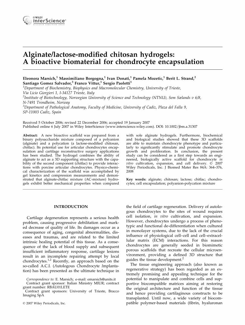
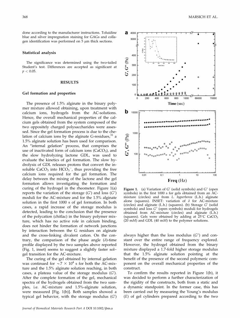
�, thus providing the freecalcium ions required for the gel formation. Thedelay between the mixing of the lactone and the gelformation allows investigating the formation andcuring of the hydrogel in the rheometer. Figure 1(a)reports the variation of the storage (G0) and loss (G@)moduli for the AC-mixture and for the 1.5% alginatesolution in the first 1000 s of gel formation. In bothcases, a rapid increase of the storage modulus isdetected, leading to the conclusion that the presenceof the polycation (chitlac) in the binary polymer mix-ture, which has no active role in calcium binding,does not hinder the formation of network junctionsby interaction between the G residues on alginateand the cross-linking divalent cation. On the con-trary, the comparison of the phase angle (d)-timeprofile displayed by the two samples above reported(Fig. 1, inset) seems to suggest a slightly faster sol–gel transition for the AC-mixture.
The curing of the gel obtained by internal gelationwas continued for *7 3 104 s for both the AC-mix-ture and the 1.5% alginate solution reaching, in bothcases, a plateau value of the storage modulus (G0).After the complete formation of the gel, mechanicalspectra of the hydrogels obtained from the two sam-ples, i.e. AC-mixture and 1.5%-alginate solution,were measured [Fig. 1(b)]. Both samples showed atypical gel behavior, with the storage modulus (G0)
always higher than the loss modulus (G@) and con-stant over the entire range of frequency explored.However, the hydrogel obtained from the binarymixture displayed a 1.7-fold higher storage modulusthat the 1.5% alginate solution pointing at thebenefit of the presence of the second polymeric com-ponent on the overall mechanical properties of theconstruct.
To confirm the results reported in Figure 1(b), itwas decided to perform a further characterization ofthe rigidity of the constructs, both from a static anda dynamic standpoint. In the former case, this hasbeen carried out by measuring the Young’s modulus(E) of gel cylinders prepared according to the two
Figure 1. (a) Variation of G0 (solid symbols) and G@ (opensymbols) in the first 1000 s for gels obtained from an AC-mixture (circles) and from L. hyperborea (L.h.) alginatealone (squares). INSET: variation of d for AC-mixture(circles) and alginate (L.h.) (squares). (b) Storage G0 (solidsymbols) and loss G@ (open symbols) moduli for hydrogelsobtained from AC-mixture (circles) and alginate (L.h.)(squares). Gels were obtained by adding at 258C CaCO3
(20 mM) and GDL (40 mM) to the polymer solutions.
368 MARSICH ET AL.
Journal of Biomedical Materials Research Part A DOI 10.1002/jbm.a
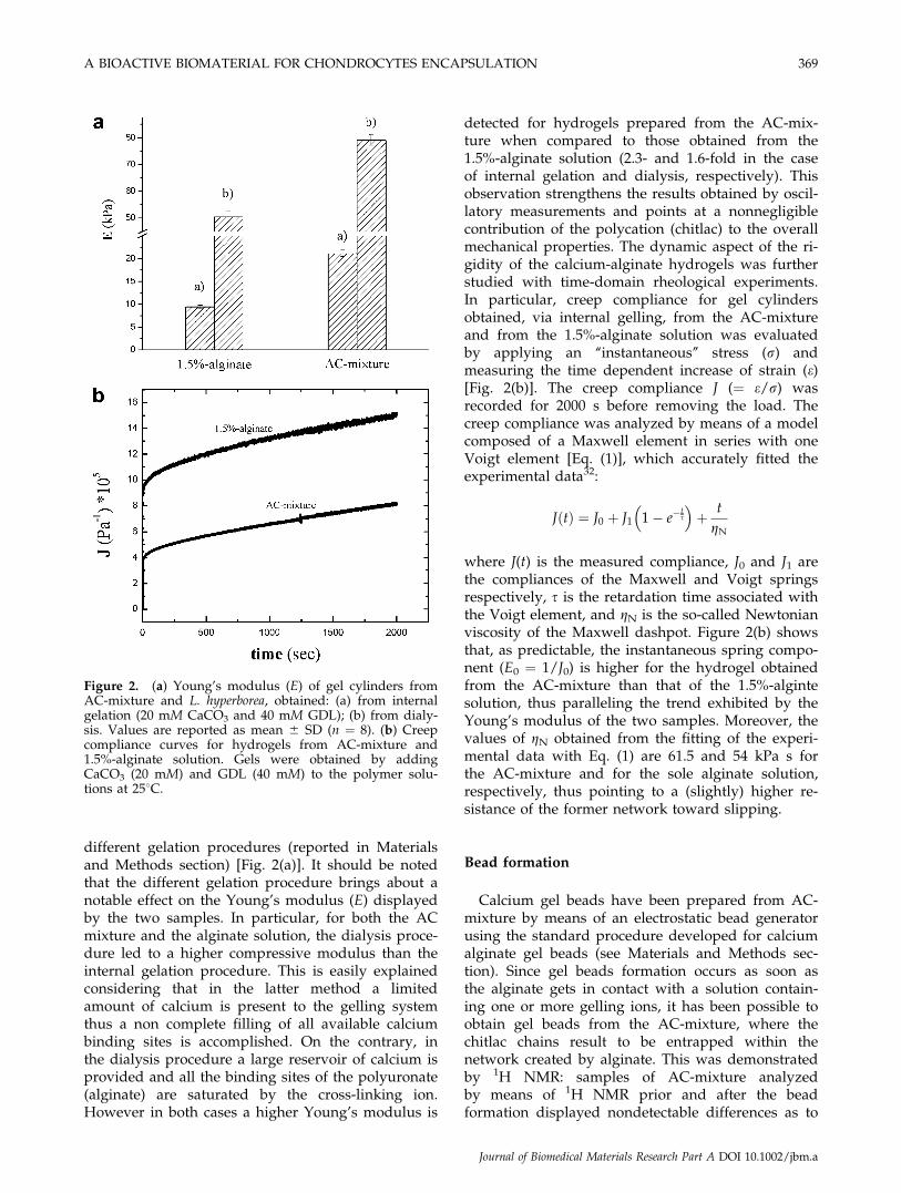
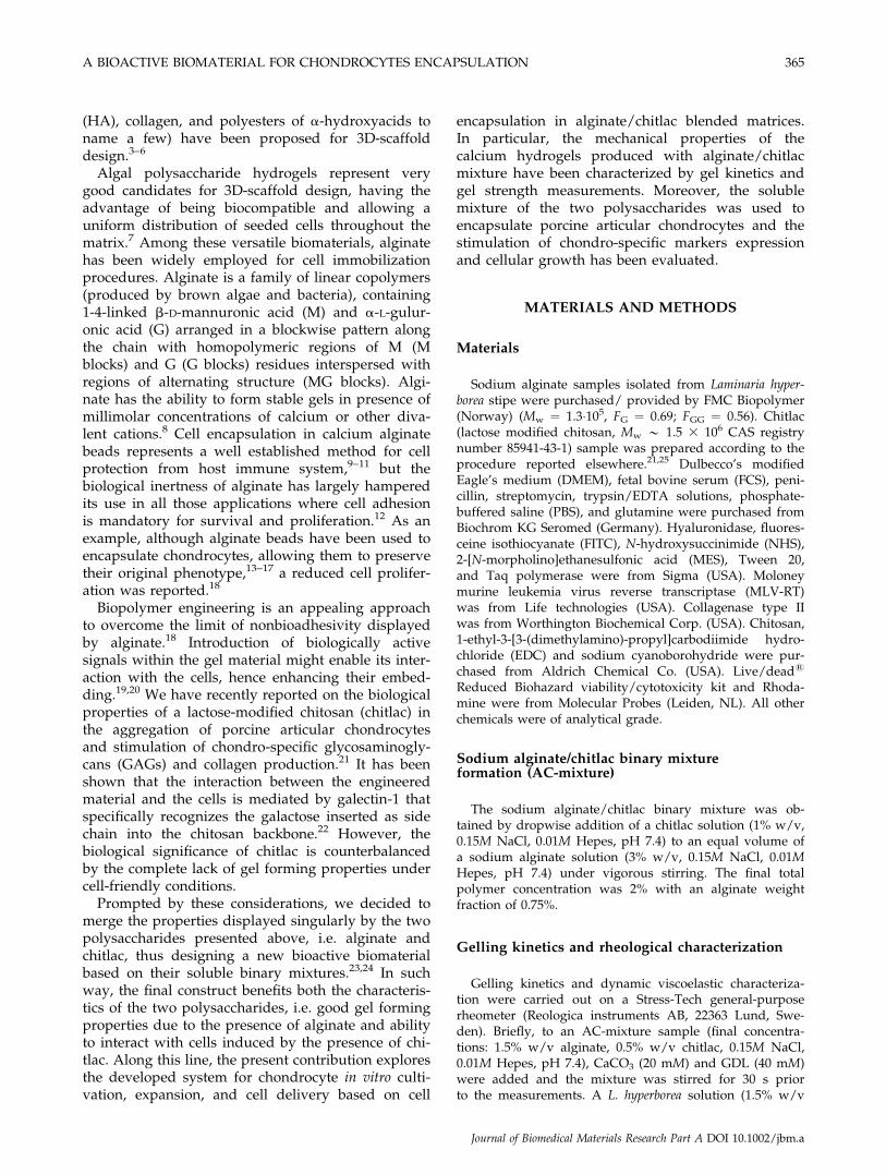
different gelation procedures (reported in Materialsand Methods section) [Fig. 2(a)]. It should be notedthat the different gelation procedure brings about anotable effect on the Young’s modulus (E) displayedby the two samples. In particular, for both the ACmixture and the alginate solution, the dialysis proce-dure led to a higher compressive modulus than theinternal gelation procedure. This is easily explainedconsidering that in the latter method a limitedamount of calcium is present to the gelling systemthus a non complete filling of all available calciumbinding sites is accomplished. On the contrary, inthe dialysis procedure a large reservoir of calcium isprovided and all the binding sites of the polyuronate(alginate) are saturated by the cross-linking ion.However in both cases a higher Young’s modulus is
detected for hydrogels prepared from the AC-mix-ture when compared to those obtained from the1.5%-alginate solution (2.3- and 1.6-fold in the caseof internal gelation and dialysis, respectively). Thisobservation strengthens the results obtained by oscil-latory measurements and points at a nonnegligiblecontribution of the polycation (chitlac) to the overallmechanical properties. The dynamic aspect of the ri-gidity of the calcium-alginate hydrogels was furtherstudied with time-domain rheological experiments.In particular, creep compliance for gel cylindersobtained, via internal gelling, from the AC-mixtureand from the 1.5%-alginate solution was evaluatedby applying an ‘‘instantaneous’’ stress (r) andmeasuring the time dependent increase of strain (e)[Fig. 2(b)]. The creep compliance J (¼ e/r) wasrecorded for 2000 s before removing the load. Thecreep compliance was analyzed by means of a modelcomposed of a Maxwell element in series with oneVoigt element [Eq. (1)], which accurately fitted theexperimental data32:
JðtÞ ¼ J0 þ J1 1� e�ts
� �þ t
gN
where J(t) is the measured compliance, J0 and J1 arethe compliances of the Maxwell and Voigt springsrespectively, s is the retardation time associated withthe Voigt element, and gN is the so-called Newtonianviscosity of the Maxwell dashpot. Figure 2(b) showsthat, as predictable, the instantaneous spring compo-nent (E0 ¼ 1/J0) is higher for the hydrogel obtainedfrom the AC-mixture than that of the 1.5%-algintesolution, thus paralleling the trend exhibited by theYoung’s modulus of the two samples. Moreover, thevalues of gN obtained from the fitting of the experi-mental data with Eq. (1) are 61.5 and 54 kPa s forthe AC-mixture and for the sole alginate solution,respectively, thus pointing to a (slightly) higher re-sistance of the former network toward slipping.
Bead formation
Calcium gel beads have been prepared from AC-mixture by means of an electrostatic bead generatorusing the standard procedure developed for calciumalginate gel beads (see Materials and Methods sec-tion). Since gel beads formation occurs as soon asthe alginate gets in contact with a solution contain-ing one or more gelling ions, it has been possible toobtain gel beads from the AC-mixture, where thechitlac chains result to be entrapped within thenetwork created by alginate. This was demonstratedby 1H NMR: samples of AC-mixture analyzedby means of 1H NMR prior and after the beadformation displayed nondetectable differences as to
Figure 2. (a) Young’s modulus (E) of gel cylinders fromAC-mixture and L. hyperborea, obtained: (a) from internalgelation (20 mM CaCO3 and 40 mM GDL); (b) from dialy-sis. Values are reported as mean 6 SD (n ¼ 8). (b) Creepcompliance curves for hydrogels from AC-mixture and1.5%-alginate solution. Gels were obtained by addingCaCO3 (20 mM) and GDL (40 mM) to the polymer solu-tions at 258C.
A BIOACTIVE BIOMATERIAL FOR CHONDROCYTES ENCAPSULATION 369
Journal of Biomedical Materials Research Part A DOI 10.1002/jbm.a
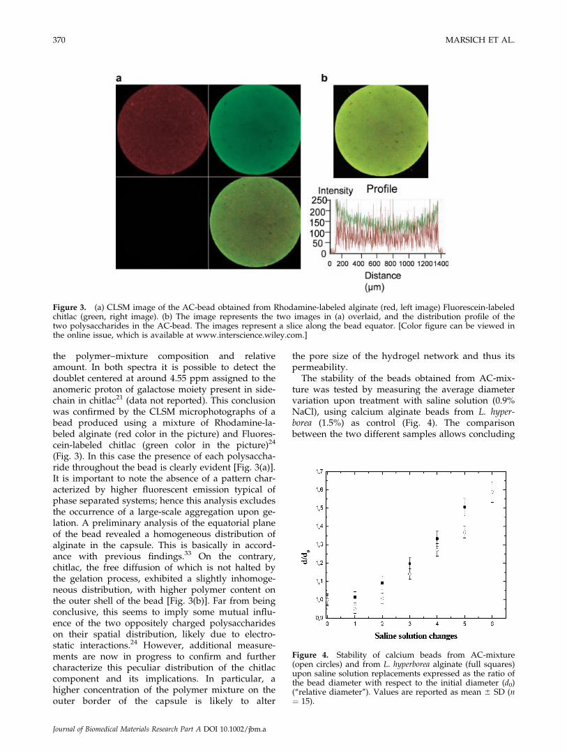
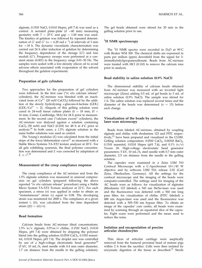
the polymer–mixture composition and relativeamount. In both spectra it is possible to detect thedoublet centered at around 4.55 ppm assigned to theanomeric proton of galactose moiety present in side-chain in chitlac21 (data not reported). This conclusionwas confirmed by the CLSM microphotographs of abead produced using a mixture of Rhodamine-la-beled alginate (red color in the picture) and Fluores-cein-labeled chitlac (green color in the picture)24
(Fig. 3). In this case the presence of each polysaccha-ride throughout the bead is clearly evident [Fig. 3(a)].It is important to note the absence of a pattern char-acterized by higher fluorescent emission typical ofphase separated systems; hence this analysis excludesthe occurrence of a large-scale aggregation upon ge-lation. A preliminary analysis of the equatorial planeof the bead revealed a homogeneous distribution ofalginate in the capsule. This is basically in accord-ance with previous findings.33 On the contrary,chitlac, the free diffusion of which is not halted bythe gelation process, exhibited a slightly inhomoge-neous distribution, with higher polymer content onthe outer shell of the bead [Fig. 3(b)]. Far from beingconclusive, this seems to imply some mutual influ-ence of the two oppositely charged polysaccharideson their spatial distribution, likely due to electro-static interactions.24 However, additional measure-ments are now in progress to confirm and furthercharacterize this peculiar distribution of the chitlaccomponent and its implications. In particular, ahigher concentration of the polymer mixture on theouter border of the capsule is likely to alter
the pore size of the hydrogel network and thus itspermeability.
The stability of the beads obtained from AC-mix-ture was tested by measuring the average diametervariation upon treatment with saline solution (0.9%NaCl), using calcium alginate beads from L. hyper-borea (1.5%) as control (Fig. 4). The comparisonbetween the two different samples allows concluding
Figure 3. (a) CLSM image of the AC-bead obtained from Rhodamine-labeled alginate (red, left image) Fluorescein-labeledchitlac (green, right image). (b) The image represents the two images in (a) overlaid, and the distribution profile of thetwo polysaccharides in the AC-bead. The images represent a slice along the bead equator. [Color figure can be viewed inthe online issue, which is available at www.interscience.wiley.com.]
Figure 4. Stability of calcium beads from AC-mixture(open circles) and from L. hyperborea alginate (full squares)upon saline solution replacements expressed as the ratio ofthe bead diameter with respect to the initial diameter (d0)(‘‘relative diameter’’). Values are reported as mean 6 SD (n¼ 15).
370 MARSICH ET AL.
Journal of Biomedical Materials Research Part A DOI 10.1002/jbm.a
that the swelling behavior of AC-mixture beadsclosely parallels the trend of those composed exclu-sively of alginate, showing in addition a slightincrease of stability (less than 1.4-fold diameterincrease after five saline solution replacements)when compared to alginate alone (more than 1.5-foldincrease). It is remarkable that after six saline solu-tion replacements, it is no more possible to visualizeand measure intact alginate beads, while there arestill intact and measurable AC-mixture beads, pre-senting a 1.6-fold mean diameter increase. Since the
swelling of the beads is likely to be accompanied byan increase in the diameter of the pores of the gel,the presence of the polycation (chitlac) after the six-saline shifts was assessed. Samples of AC-mixturebeads were collected prior and after the swellingstability tests and treated with HCl to remove cal-cium ions and thus dissolve the beads. The 1H NMRspectra of the solution proves the presence of a con-siderable amount of chitlac at the end of the test.Indeed, by comparing the peaks corresponding tothe alginate component and that of the galactose
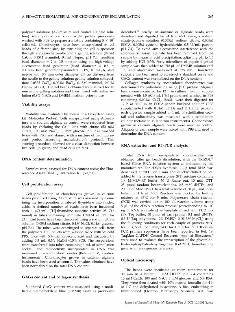
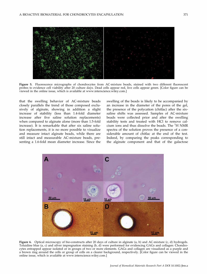
Figure 5. Fluorescence micrographs of chondrocytes from AC-mixture beads, stained with two different fluorescentprobes to evidence cell viability after 20 culture days. Dead cells appear red, live cells appear green. [Color figure can beviewed in the online issue, which is available at www.interscience.wiley.com.]
Figure 6. Optical microscopy of bio-constructs after 20 days of culture in alginate (a, b) and AC-mixture (c, d) hydrogels.Toluidine blue (a, c) and silver impregnation staining (b, d) were performed for evidencing GAGs and collagen: Chondro-cytes entrapped appear isolated or in groups of two or more elements. GAGs and collagen are visualized as a purple anda brown ring around the cells or group of cells on a clearer background, respectively. [Color figure can be viewed in theonline issue, which is available at www.interscience.wiley.com.]
A BIOACTIVE BIOMATERIAL FOR CHONDROCYTES ENCAPSULATION 371
Journal of Biomedical Materials Research Part A DOI 10.1002/jbm.a
group in chitlac, there seem to be an increase of thechitlac/alginate ratio after six saline shifts for AC-mixture. This is actually due to the leakage ofalginate chains from the bead structure, as a conse-quence of the calcium displacement and the cor-responding gel weakening. However, the networkcreated by the alginate chains still remains denseenough to trap the chitlac chains, not allowingtheir diffusion from the gel structure after six salineshifts.
Biological properties
The influence of bead composition on chondro-cytes biology and metabolism was evaluated bymeans of biochemical analysis: cell viability, cell pro-liferation, GAGs and collagen synthesis at differenttimes. Cell viability in AC-mixture scaffold wasassessed using a live/dead kit. The basis of this via-bility test is the differential permeability of live anddead cells to a pair of fluorescent stains (SYTO1 10and DEAD RedTM), respectively. SYTO 10 is a greenfluorescent nucleic acid stain, highly membrane-per-meable, which labels all cells, including those withintact plasma membranes. DEAD Red is a cell-impermeable red fluorescent nucleic acid stain whichlabels only cells with compromised membranes. Livecells appear fluorescent green and dead cells fluores-cent red. As reported in Figure 5, it is evident thatafter 20 culture days more than 95% of chondrocytesfrom AC-mixture beads are fully viable as well aschondrocytes grown in alginate scaffolds.
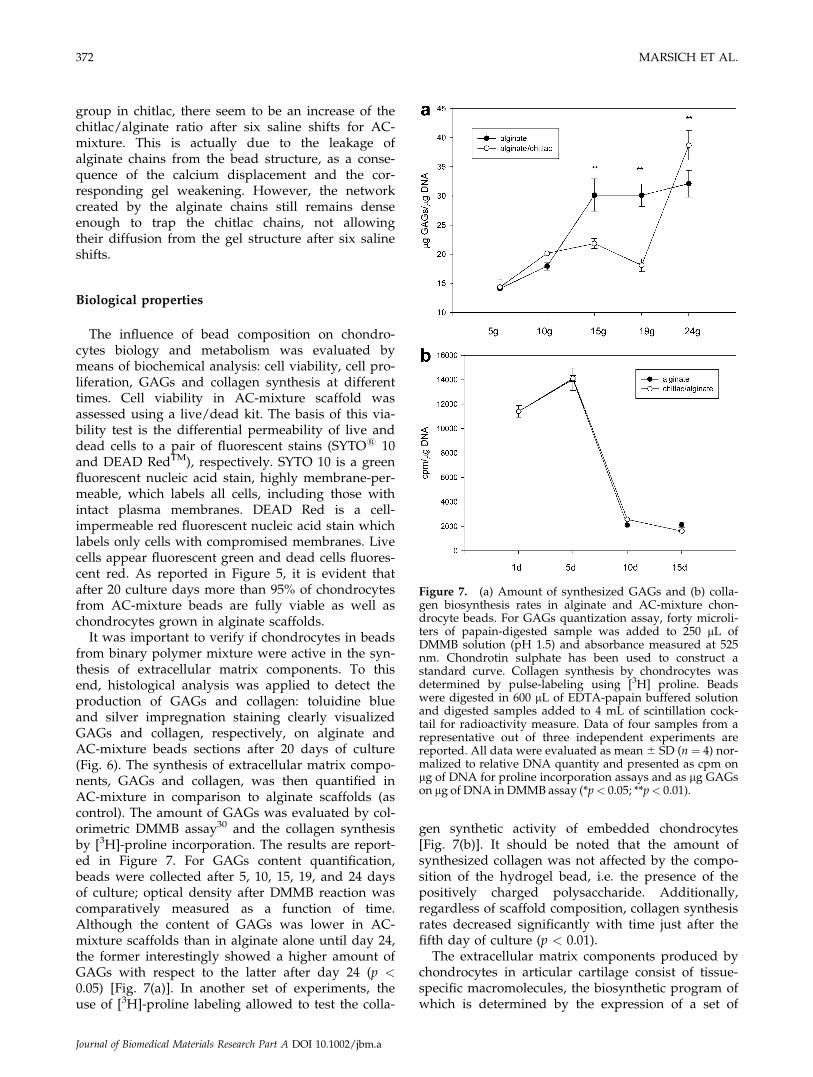
It was important to verify if chondrocytes in beadsfrom binary polymer mixture were active in the syn-thesis of extracellular matrix components. To thisend, histological analysis was applied to detect theproduction of GAGs and collagen: toluidine blueand silver impregnation staining clearly visualizedGAGs and collagen, respectively, on alginate andAC-mixture beads sections after 20 days of culture(Fig. 6). The synthesis of extracellular matrix compo-nents, GAGs and collagen, was then quantified inAC-mixture in comparison to alginate scaffolds (ascontrol). The amount of GAGs was evaluated by col-orimetric DMMB assay30 and the collagen synthesisby [3H]-proline incorporation. The results are report-ed in Figure 7. For GAGs content quantification,beads were collected after 5, 10, 15, 19, and 24 daysof culture; optical density after DMMB reaction wascomparatively measured as a function of time.Although the content of GAGs was lower in AC-mixture scaffolds than in alginate alone until day 24,the former interestingly showed a higher amount ofGAGs with respect to the latter after day 24 (p <0.05) [Fig. 7(a)]. In another set of experiments, theuse of [3H]-proline labeling allowed to test the colla-
gen synthetic activity of embedded chondrocytes[Fig. 7(b)]. It should be noted that the amount ofsynthesized collagen was not affected by the compo-sition of the hydrogel bead, i.e. the presence of thepositively charged polysaccharide. Additionally,regardless of scaffold composition, collagen synthesisrates decreased significantly with time just after thefifth day of culture (p < 0.01).
The extracellular matrix components produced bychondrocytes in articular cartilage consist of tissue-specific macromolecules, the biosynthetic program ofwhich is determined by the expression of a set of
Figure 7. (a) Amount of synthesized GAGs and (b) colla-gen biosynthesis rates in alginate and AC-mixture chon-drocyte beads. For GAGs quantization assay, forty microli-ters of papain-digested sample was added to 250 lL ofDMMB solution (pH 1.5) and absorbance measured at 525nm. Chondrotin sulphate has been used to construct astandard curve. Collagen synthesis by chondrocytes wasdetermined by pulse-labeling using [3H] proline. Beadswere digested in 600 lL of EDTA-papain buffered solutionand digested samples added to 4 mL of scintillation cock-tail for radioactivity measure. Data of four samples from arepresentative out of three independent experiments arereported. All data were evaluated as mean 6 SD (n ¼ 4) nor-malized to relative DNA quantity and presented as cpm onlg of DNA for proline incorporation assays and as lg GAGson lg of DNA in DMMB assay (*p< 0.05; **p< 0.01).
372 MARSICH ET AL.
Journal of Biomedical Materials Research Part A DOI 10.1002/jbm.a
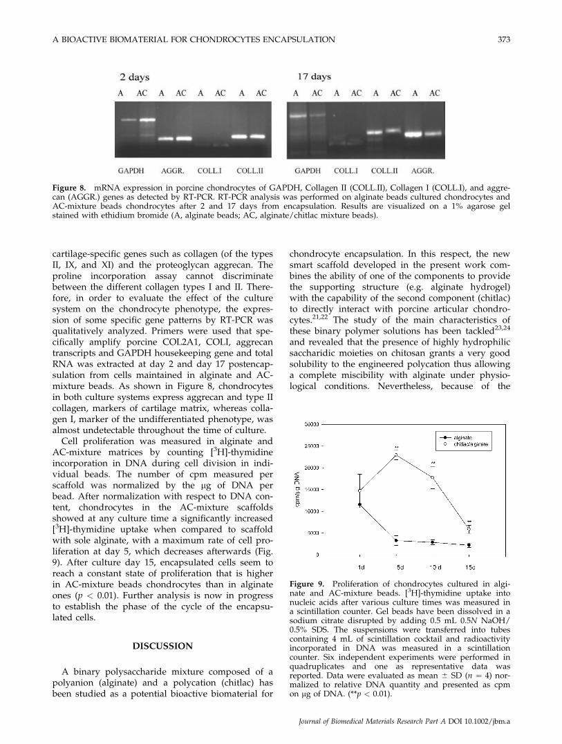
cartilage-specific genes such as collagen (of the typesII, IX, and XI) and the proteoglycan aggrecan. Theproline incorporation assay cannot discriminatebetween the different collagen types I and II. There-fore, in order to evaluate the effect of the culturesystem on the chondrocyte phenotype, the expres-sion of some specific gene patterns by RT-PCR wasqualitatively analyzed. Primers were used that spe-cifically amplify porcine COL2A1, COLI, aggrecantranscripts and GAPDH housekeeping gene and totalRNA was extracted at day 2 and day 17 postencap-sulation from cells maintained in alginate and AC-mixture beads. As shown in Figure 8, chondrocytesin both culture systems express aggrecan and type IIcollagen, markers of cartilage matrix, whereas colla-gen I, marker of the undifferentiated phenotype, wasalmost undetectable throughout the time of culture.
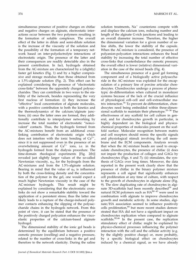
Cell proliferation was measured in alginate andAC-mixture matrices by counting [3H]-thymidineincorporation in DNA during cell division in indi-vidual beads. The number of cpm measured perscaffold was normalized by the lg of DNA perbead. After normalization with respect to DNA con-tent, chondrocytes in the AC-mixture scaffoldsshowed at any culture time a significantly increased[3H]-thymidine uptake when compared to scaffoldwith sole alginate, with a maximum rate of cell pro-liferation at day 5, which decreases afterwards (Fig.9). After culture day 15, encapsulated cells seem toreach a constant state of proliferation that is higherin AC-mixture beads chondrocytes than in alginateones (p < 0.01). Further analysis is now in progressto establish the phase of the cycle of the encapsu-lated cells.
DISCUSSION
A binary polysaccharide mixture composed of apolyanion (alginate) and a polycation (chitlac) hasbeen studied as a potential bioactive biomaterial for
chondrocyte encapsulation. In this respect, the newsmart scaffold developed in the present work com-bines the ability of one of the components to providethe supporting structure (e.g. alginate hydrogel)with the capability of the second component (chitlac)to directly interact with porcine articular chondro-cytes.21,22 The study of the main characteristics ofthese binary polymer solutions has been tackled23,24
and revealed that the presence of highly hydrophilicsaccharidic moieties on chitosan grants a very goodsolubility to the engineered polycation thus allowinga complete miscibility with alginate under physio-logical conditions. Nevertheless, because of the
Figure 8. mRNA expression in porcine chondrocytes of GAPDH, Collagen II (COLL.II), Collagen I (COLL.I), and aggre-can (AGGR.) genes as detected by RT-PCR. RT-PCR analysis was performed on alginate beads cultured chondrocytes andAC-mixture beads chondrocytes after 2 and 17 days from encapsulation. Results are visualized on a 1% agarose gelstained with ethidium bromide (A, alginate beads; AC, alginate/chitlac mixture beads).
Figure 9. Proliferation of chondrocytes cultured in algi-nate and AC-mixture beads. [3H]-thymidine uptake intonucleic acids after various culture times was measured ina scintillation counter. Gel beads have been dissolved in asodium citrate disrupted by adding 0.5 mL 0.5N NaOH/0.5% SDS. The suspensions were transferred into tubescontaining 4 mL of scintillation cocktail and radioactivityincorporated in DNA was measured in a scintillationcounter. Six independent experiments were performed inquadruplicates and one as representative data wasreported. Data were evaluated as mean 6 SD (n ¼ 4) nor-malized to relative DNA quantity and presented as cpmon lg of DNA. (**p < 0.01).
A BIOACTIVE BIOMATERIAL FOR CHONDROCYTES ENCAPSULATION 373
Journal of Biomedical Materials Research Part A DOI 10.1002/jbm.a
simultaneous presence of positive charges on chitlacand negative charges on alginate, electrostatic inter-actions occur between the two polymers resulting inthe formation of soluble complexes. The overalleffect of the latter in dilute and semi-dilute solutionis the increase of the viscosity of the solution andthe possibility of the formation of a temporary net-work based on inter-polyelectrolyte interactions ofelectrostatic origin. The presence of the latter andtheir consequences are readily detectable also in thepresent contribution. In fact, hydrogels obtainedfrom the AC-mixture are characterized by a (slightly)faster gel kinetics (Fig. 1) and by a higher compres-sive and storage modulus than those obtained froma 1.5%-alginate solution (Fig. 2). This effect can beexplained considering the presence of ‘‘electrostaticcross-links’’ between the oppositely charged polysac-charides. They can contribute in two ways to the sta-bility of the network, besides representing the veryfirst nucleus of formation: (i) they increase the‘‘effective’’ local concentration of alginate molecules,with a positive contribution to both the kinetics andthe thermodynamics of the calcium/alginate junc-tions; (ii) once the latter ones are formed, they addi-tionally contribute to interpolymer networking byincrease the total number of electrostatic ‘‘nonco-valent’’ contacts. Thus, the hydrogels formed fromthe AC-mixtures benefit from an additional cross-linking contribution of electrostatic origin whichdoes not interfere with the ionic bonding of Ca2þ,since it is not suppressed even in the presence of anoverwhelming amount of Ca2þ ions, i.e. in thehydrogels formed from the dialysis procedure. Thetime-domain rheological experiments [Fig. 2(b)]revealed just slightly larger values of the so-calledNewtonian viscosity, gN, for the hydrogels from theAC-mixtures and from the 1.5%-alginate solution.Bearing in mind that the value of gN is influencedby both the cross-linking density and the concentra-tion of the polymer in the gel, one would expect amuch higher Newtonian viscosity in the case of theAC-mixture hydrogels. This result might beexplained by considering that the electrostatic cross-links do not show a remarkable stability toward theapplication of a persistent stress; thus the latter onelikely leads to a rupture of the charge-induced poly-mer contacts enhancing the slipping of the polysac-charide chains in the hydrogels network. From thispoint of view, it can be stated that the presence ofthe positively charged polycation enhances the visco-elastic properties of the calcium-based alginatehydrogel.
The dimensional stability of the ionic gel beads isdetermined by the equilibrium between a positiveosmotic pressure (swelling) and a negative pressure,related to the number of cross-links in the gel andtherefore to the network elasticity. During the saline
solution treatments, Naþ counterions compete withand displace the calcium ions, reducing number andlength of the alginate G-rich junctions and leading toan overall diameter increase. Therefore, the higherthe dimensional variation for a given number of sa-line shifts, the lower the stability of the capsule.When the AC-mixture is considered, the presence ofpolyanion-polycation interactions enhances the beadstability by increasing the total number of effectivecross-links that counterbalance the osmotic pressure;the overall effect is lower (relative) dimensional vari-ation in the case of the mixed beads (Fig. 4).
The simultaneous presence of a good gel formingcomponent and of a biologically active polysaccha-ride in the AC-mixture was exploited for the encap-sulation of a primary line of porcine articular chon-drocytes. Chondrocytes undergo a process of pheno-typic de-differentiation when cultured in monolayersystems because of the lack of the crucial influenceof physiological cell–cell and cell–extracellular ma-trix interaction.34 To prevent de-differentiation, chon-drocytes need being embedded within three-dimen-sional scaffolds to maintain their phenotype.35–38 Theeffectiveness of any scaffold for cell culture in gen-eral, and for chondrocytes growth in particular, ishighly dependent on the biological and physico-chemical properties of molecules present on the scaf-fold surface. Molecular recognition between matrixand cell receptors should mimic the specific signalsand physiological stimuli involving the cell mem-brane and the ECM components. Analysis revealsthat when the AC-mixture beads are used to encap-sulate chondrocytes, the presence of chitlac (i) doesnot impair the production of collagen and GAGs bychondrocytes (Figs. 6 and 7); (ii) stimulates, the syn-thesis of GAGs over long times. Moreover, the datareported in the present work clearly show that thepresence of chitlac in the binary polymer mixturerepresents a cell signal that significantly enhancescell proliferation at any time of culture, with respectto the growth of chondrocytes in alginate alone (Fig.9). The slow duplicating rate of chondrocytes in algi-nate 3D-scaffolds had been recently described39 andnatural ECM polymers such as HA40–43 were used incombination with alginate to improve chondrocytesgrowth and metabolic activity. In some studies, algi-nate/HA association seemed to influence positivelycells proliferation,44 but more recent studies demon-strated that HA did not have a significant impact onchondrocytes replication when compared to alginatescaffolds.45,46 In the present case, the replicationstimulatory effect of chitlac might be explained byphysico-chemical processes influencing the polymerinteraction with the cell and the cellular activity (e.g.by the slightly positive charge) or, more probably,by a specific biological effect on chondrocytesinduced by a chemical signal, as we have already
374 MARSICH ET AL.
Journal of Biomedical Materials Research Part A DOI 10.1002/jbm.a

described.21,22 In fact, this chitosan-derived glycopoly-mer exhibits the ability to induce chondrocyte aggre-gation and to stimulate cartilage matrix synthesis.21
The specificity of the effect of chitlac stems fromthe presence of the galactose moiety; the bridgingrole between cellular membrane and polymer is car-ried out by a protein, a member of the S-typegalactoside-binding animal lectins, Galectin-1.22
Therefore, it can be very reasonably suggested thatsuch a Galectin-mediated specific interaction be-tween the galactose-engineered chitosan and chon-drocyte might be responsible for the stimulation ofthe cell proliferation. Further work is being carriedout to investigate the molecular mechanisms under-lining the chondrocytes–glycopolymer specific inter-actions.
CONCLUSIONS
In this study we report experimental results aboutpreparation of biocompatible three dimensionalhydrogels for tissue engineering applications, usingternary aqueous solutions of alginate and a cationicchitosan derivate, chitlac (AC-mixture). The systemcombines the gelling properties of alginate with thedemonstrated bioactive properties of chitlac.
The experimental data show that AC-mixturehydrogels exhibit better physical and mechanicalproperties when compared to sole alginate hydro-gels, as can be evinced from the physico-chemicalcharacterization. This can be explained consideringthe formation of soluble complexes between the twooppositely charged polysaccharides, which repre-sents an ionic extra-contribution to the hydrogel for-mation and results in a structural stabilization. Thisphenomenon leads to the possibility to produce AC-mixture calcium beads characterized by a slightlyhigher stability than standard alginate beads. More-over, these 3D-scaffolds represent a supportive ma-trix bearing, additionally, bioactive molecules able tomaintain chondrocyte phenotype and particularly tostimulate and promote cell growth and proliferation.In view of the above reported properties, the AC-mix-ture beads could be successfully employed as aneffective system for the culture of chondrocytesand as efficient support for their delivery in tissueregeneration.
In conclusion, the reported data represent thestarting point for the development of a new multi-component scaffold based on natural polymers, withimproved stability, mechanical, and biological prop-erties than sole alginate hydrogels.
Ivan Donati thanks Prof. Gudmund Skjak-Bræk (Insti-tute of Biotechnology, University of Trondheim (NTNU),Norway) for hospitality and helpful discussions.
References
1. Tuli R, Li W, Tuan RS. Current state of cartilage tissue engi-neering. Arthritis Res Ther 2003;5:235–238.
2. Risbud MV, Sittinger M. Tissue engineering: Advances in invitro cartilage generation. Trends Biotechnol 2002;20:351–356.
3. Temenoff JS, Mikos AG. Tissue engineering for regenerationof articular cartilage. Biomaterials 2000;21:431–440.
4. Buckwalter JA, Mankin HJ. Articular cartilage. II. Degenera-tion and osteoarthrosis, repair, regeneration and transplanta-tion. J Bone J Surg 1997;79:612–632.
5. Madihally SV, Matthew HWT. Porous chitosan scaffolds fortissue engineering. Biomaterials 1999;20:1133–1142.
6. Barbucci R, Lamponi S, Borzacchiello A, Ambrosio L, Fini M,Torricelli P, Giardino R. Hyaluronic acid hydrogel in thetreatment of osteoarthritis. Biomaterials 2002;23:4503–4513.
7. Frenkel SR, Di Cesare PE. Scaffolds for articular cartilagerepair. Ann Biomed Eng 2004;32:26–34.
8. Haug A, Larsen B, Smidsrod O. A study of the constitutionof alginic acid by partial hydrolysis. Acta Chem Scand1966;20:183–190.
9. Chang TMS. Semipermeable microcapsules. Science 1964;146:524–525.
10. Sun Y, Ma X, Zhou D, Vacek I, Sun AM. Normalization of di-abetes in spontaneously diabetic cynomologus monkeys byxenografts of microencapsulated porcine islets withoutimmunosuppression. J Clin Invest 1996;98:1417–1422.
11. Hortelano G, Al-Hendy A, Ofosu FA, Chang PL Delivery ofhuman factor IX in mice by encapsulated recombinant myo-blasts: A novel approach towards allogeneic gene therapy ofhemophilia B. Blood 1996;87:5095–5103.
12. Yang J, Goto M, Ise H, Cho CS, Akaike T. Galactosylatedalginate as a scaffold for hepatocytes entrapment. Biomateri-als 2002;23:471–479.
13. De Ceuninck F, Lesur C, Pastoureau P, Caliez A, Sabatini M.Culture of chondrocytes in alginate beads. Methods Mol Med2004;100:15–22.
14. Masuda K, Sah RL, Hejna MJ, Thonar EJ. A novel two-stepmethod for the formation of tissue-engineered cartilageby mature bovine chondrocytes: The alginate-recovered-chon-drocyte (ARC) method. J Orthop Res 2003;21:139–148.
15. Stevens MM, Qanadilo HF, Langer R, Prasad Shastri V, Arapid-curing alginate gel system: Utility in periosteum-deriv-ed cartilage tissue engineering. Biomaterials 2004;25:887–894.
16. Mierisch CM, Wilson HA, Turner MA, Milbrandt TA,Berthoux L, Hammarskjold ML, Rekosh D, Balian G, DiduchDR. Chondrocyte transplantation into articular cartilagedefects with use of calcium alginate: The fate of the cells.J Bone Joint Surg Am 2003;85:1757–1767.
17. Lee DA, Reisler T, Bader DL. Expansion of chondrocytes fortissue engineering in alginate beads enhances chondrocyticphenotype compared to conventional monolayer techniques.Acta Orthop Scand 2003;74:6–15.
18. Yamaoka H, Asato H, Ogasawara T, Nishizawa S, TakahashiT, Nakatsuka T, Koshima I, Nakamura K, Kawaguchi H,Chung UI, Takato T, Hoshi K. Cartilage tissue engineeringusing human auricular chondrocytes embedded in differenthydrogel materials J Biomed Mater Res A 2006;78:1–11.
19. Pound JC, Green DW, Chaudhuri JB, Mann S, Roach HI,Oreffo RO. Strategies to promote chondrogenesis and osteo-genesis from human bone marrow cells and articular chon-drocytes encapsulated in polysaccharide templates. TissueEng 2006. [Epub ahead of print]
20. Iwasaki N, Yamane ST, Majima T, Kasahara Y, Minami A,Harada K, Nonaka S, Maekawa N, Tamura H, Tokura S,Shiono M, Monde K, Nishimura S. Feasibility of polysaccha-ride hybrid materials for scaffolds in cartilage tissueengineering: Evaluation of chondrocyte adhesion to polyion
A BIOACTIVE BIOMATERIAL FOR CHONDROCYTES ENCAPSULATION 375
Journal of Biomedical Materials Research Part A DOI 10.1002/jbm.a
complex fibers prepared from alginate and chitosan. Biomacro-molecules 2004;5:828–833.
21. Donati I, Stredanska S, Silvestrini G, Vetere A, Marcon P,Marsich E, Mozetic P, Gamini A, Paoletti S, Vittur F. Theaggregation of pig articular chondrocyte and synthesis ofextracellular matrix by a lactose-modified chitosan. Biomate-rials 2005;26:987–998.
22. Marcon P, Marsich E, Vetere A, Mozetic P, Campa C, DonatiI, Vittur F, Gamini A, Paoletti S. The role of Galectin-1 in theinteraction between chondrocytes and a lactose-modified chi-tosan. Biomaterials 2005;26:4975–4984.
23. Donati I, Haug IJ, Scarpa T, Borgogna M, Draget KI, Skjak-Bræk G, Paoletti S. Synergistic effects in semi-dilute mixedsolutions of alginate and lactose-modified chitosan (chitlac).Biomacromolecules 2007;8:957–962.
24. Donati I, Borgogna M, Turello E, Cesaro A, Paoletti S. Tuningsupramolecular structuring at the nanoscale level. Non-stoi-chiometric soluble complexes in dilute mixed solutions of al-ginate and lactose-modified chitosan (chitlac). Biomacromole-cules 2007. In Press.
25. Yalpani M, Hall LD. Some chemical and analytical aspects ofpolysaccharide modifications. 3: Formation of branched-chain, soluble chitosan derivatives. Macromolecules 1984;17:272–281.
26. Draget KI, Skjak Bræk G, Smidsrød O. Alginic acid gels: Theeffect of alginate chemical composition and molecular weight.Carbohydr Polym 1994;25:31–38.
27. Smidsrød O, Haug A. Properties of poly(1,4-hexuronates) ingel state. II. Comparison of gels of different chemical compo-sition. Acta Chem Scand 1972;26:79–88.
28. Martinsen A, Skjak-Bræk G, Smidsrød O. Alginate as immo-bilization material. I. Correlation between chemical and phys-ical properties of alginate gel beads. Biotechnol Bioeng 1989;33:79–89.
29. Strand BL, Gaserød O, Kulseng B, Espevik T, Skjak-Bræk G.Alginate-polylysine-alginate microcapsules: Effect of sizereduction on capsule properties J Microencapsul 2002;19:615.
30. Enobakhare BO, Bader DL, Lee DA. Quantification of sul-fated glycosaminoglycans in chondrocyte/alginate cultures,by use of 1,9-dimethylmethylene blue. Anal Biochem1996;243:189–191.
31. Grant GT, Morris ER, Rees DA, Smith PJC, Thom D. Biologi-cal interactions between polysaccharides and divalent cations:The egg-box model. FEBS Lett 1973;32:195–198.
32. Donati I, Holtan S, Mørch Y, Borgogna M, Dentini M,Skjak-Bræk G. New hypothesis on the role of alternatingsequences in calcium-alginate gels. Biomacromolecules 2005;6:1031–1040.
33. Strand BL, Morch YA, Espevik T, Skjak-Braek G. Visualiza-tion of alginate-poly-L-lysine-alginate microcapsules by confo-
cal laser scanning microscopy. Biotechnol Bioeng 2003;82:386–394.
34. Knudson W, Loeser RF. CD44 and integrin matrix receptorsparticipate in cartilage homeostasis. Cell Mol Life Sci2002;59:36–44.
35. Chubinskaya S, Huch K, Schulze M, Otten L, Aydelotte MB,Cole AA. Gene expression by human articular chondrocytescultured in alginate beads. Histochem Cytochem 2001;49:1211–1220.
36. Benya PD, Shaffer JD. Dedifferentiated chondrocytes re-express the differentiated collagen phenotype when culturedin agarose gels. Cell 1982;30:215–224.
37. Van Susante JL, Buma P, van Osch GJ, Versleyen D, van derKraan PM, van der Berg WB, Homminga GN. Culture ofchondrocytes in alginate and collagen carrier gels. ActaOrthop Scand 1995;66:549–556.
38. Mauck RL, Soltz MA, Wang CC, Wong DD, Chao PH,Valhmu WB, Hung CT, Ateshian GA. Functional tissue engi-neering of articular cartilage through dynamic loading ofchondrocyte-seeded agarose gels. J Biomech Eng 122:252–260.
39. Liu H, Lee YW, Dean MF. Re-expression of differentiatedproteoglycan phenotype by dedifferentiated human chondro-cytes during culture in alginate beads. Biochim Biophys Acta1998;1425:505–515.
40. Tampieri A, Sandri M, Landi E, Celotti G, Roveri N, Mattioli-Belmonte M, Virgili L, Gabbanelli F, Biagini G. HA/alginatehybrid composites prepared through bio-inspired nucleation.Acta Biomater 2005;1:343–351.
41. Kikuchi T, Yamada H, Fujikawa K. Effects of high molecularweight hyaluronan on the distribution and movement of pro-teoglycan around chondrocytes cultured in alginate beads.Osteoarthritis Cartilage 2001;9:351–356.
42. Li Z, Zhang M. Chitosan-alginate as scaffolding material forcartilage tissue engineering. J Biomed Mater Res A75:485–493.
43. Hsu SH, Whu SW, Hsieh SC, Tsai CL, Chen DC, Tan TS.Evaluation of chitosan-alginate-hyaluronate complexes modi-fied by an RGD-containing protein as tissue-engineering scaf-folds for cartilage regeneration. Artif Organs 2004;28:693–703.
44. Homandberg GA, Hui F, Wen C, Kuettner KE, Williams JM.Hyaluronic acid suppresses fibronectin fragment mediatedcartilage chondrolysis in vitro. Osteoarthritis Cartilage 1997;5:309–319.
45. Gerard C, Catuogno C, Amargier-Huin C, Grossin L, HubertP, Gilet P, Netter P, Dellacherie E, Payan E. The effect of algi-nate, hyaluronate and hyaluronate derivates biomaterials onsynthesis of a non-articular chondrocyte extracellular matrix.J Mater Sci Mater Med 2005;16:541–551.
46. Stove J, Gerlach C, Huch K, Gunther KP, Puhl W, Scharf HP.Effects of hyaluronan on proteoglycan content of osteoar-thritic chondrocytes in vitro. J Orthop Res 2002;20:551–555.
376 MARSICH ET AL.
Journal of Biomedical Materials Research Part A DOI 10.1002/jbm.a