what other biomaterial has so many uses: flowables - modern
TRANSCRIPT
46 INTERNATIONAL DENTISTRY – AFRICAN EDITION VOL. 7, NO. 2
What other biomaterial has so many uses:Flowables
Douglas A. Terry1
Introduction1996 was an exciting year around the world. The Dow Jones reached a record highof 6,000; the Nobel Prize in Chemistry was awarded to Curl, Kroto and Smalley fortheir discovery of fullerenes, a molecule composed entirely of carbon; General Motorslaunched the first electric car of the modern era; e-bay opened its door for business;DVD�s hit the market in Japan; Will Smith made his electrifying performance in“Independence Day”; David Bowie was inducted into the Rock and Roll Hall of Fame;and flowable composite resins were developed and introduced to the world as arevolutionary restorative biomaterial.1 The average individual would probably rank thisdiscovery the least significant of events, yet this milestone dramatically effected thepractice of adhesive dentistry.The evolution of adhesive dentistry with filled adhesives and sealants was the
catalyst that sparked this development and discovery of flowable composite resins.Although, it was not until 19961 that these biomaterials had their own identity and
became known as flowables. These “first generation” formulations were designed tosimplify the placement technique and expand the range of clinical applications forcomposite resins.1,2 These early flowable formulations were configured by utilizing theidentical filler particle sizes of conventional hybrid composite, while reducing the fillerload and/or increasing the diluent monomers.3,4 Thus, a multitude of variations inviscosity, consistency, and handling characteristics were available to the discriminatingclinician for many of the restorative and aesthetic challenges presented to them each day.These biomaterials were marketed by manufacturers for a wide range of
applications which included all classifications of anterior and posterior compositerestorations, amalgam margin repair, block out materials, composite repair, core build-up, crown margin repair, cavity liners, pit and fissure sealants, porcelain repair,anterior incisal edge repair, preventative resin restorations, provisional repair,porcelain veneer cementation, composite veneer fabrication, tunnel preparationrestorations, adhesive cementation, restoring enamel defects, air abrasion cavitypreparations and void repairs in conventional composite resin restorations.1,5
Unfortunately, these earlier flowable formulations demonstrated poor clinicalperformance with inferior mechanical properties such as flexural strength and wearresistance compared to the conventional hybrid composites.1,2
In fact, the mechanical and physical properties of composite materials improve inproportion to the volume of filler added6 and the filler content of these earlier flowableformulations were reported to be 20 to 25% less than that of the universal compositematerials.1 Numerous mechanical properties depend on this filler phase, including
C L I N I C A L
1 Douglas A. Terry, DDSClinical Assistant Professor,Department of RestorativeDentistry and Biomaterials,University of Texas Health Science Center Dental Branch,Houston, Texas, USA. PrivatePractice, Houston, Texas, USA.
E-mail: [email protected] [email protected]
VOL. 7, NO. 2 INTERNATIONAL DENTISTRY – AFRICAN EDITION 47
C L I N I C A L
previously suggested. Clinicians realized that theapplications for these first generation flowable compositeswere not the same nor were they adequate substitutes forthe highly filled conventional composites.Since the past provides information to improve the future,
the lack of evidenced based research and clinical trial dataon these flowable biomaterials requires clinicians toevaluate their individual mechanical properties fordetermining whether a new material’s properties are equalor superior to those of existing materials. Although there isnot a direct correlation of a material’s mechanical andphysical properties to clinical performance it might suggestthe potential success of a restorative biomaterial for aspecific clinical situation.1 However, clinical longevity forrestorations developed with these biomaterials remain to bedetermined through clinical studies and trial data for eachspecific clinical application.
compression strength and/or hardness, flexural strength, theelastic modulus, coefficient of thermal expansion, waterabsorption, and wear resistance.6 Thus, a reduction in thefiller content of these first generation flowables substantiatesthe reports by Bayne et al, that the mechanical properties ofthese low viscosity materials were approximately 60 to 80%of those of conventional hybrid composites.1 One scientificstudy reports that a comparison of flowable light-curedcomposite resins and conventional composite resins of thesame brand name had very different characteristics andmechanical properties.7 Early attempts to utilize theseflowable formulations in a wide variety of applicationsresulted in shortcomings that led to confusion anduncertainty for clinical predictability and performance whenusing these biomaterials. Thus, the unfavorable marketingmentality of the manufacturer proved unsuccessful andrequired limitations on the expanded applications
1a
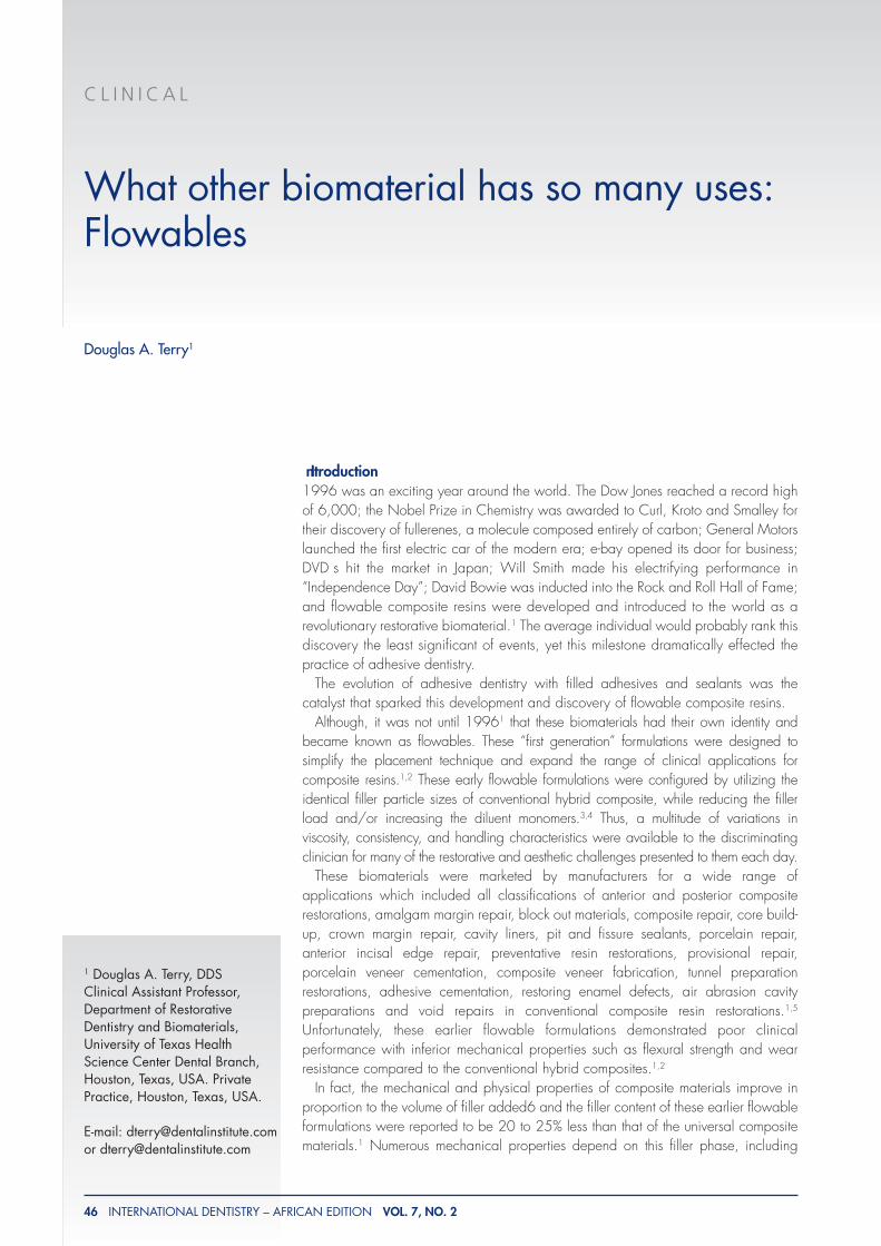
Figure 2a: Preoperative view of saucer-shaped noncariouscervical lesions on the mandibular left cuspid and first premolar.
1b
2a 2b
Figure 1a: Preoperative occlusal view of a defective compositerestoration with recurrent decay on the mandibular left firstmolar.
Figure 1b: The completed posterior restoration reveals theharmonious Integration of flowable composite resin (G-aenialUniversal Flo, GC America) and tooth structure.
Figures 2b: The post-treatment view of the finished and polishedrestorations utilizing a nano particle hybrid flowable compositeresin (G-aenial Universal Flo, GC America).

T E R R Y
48 INTERNATIONAL DENTISTRY – AFRICAN EDITION VOL. 7, NO. 2
and seal, while improving the reliability and longevity of theadhesive restoration.8 At present, there is no restorativematerial which fulfills all these prerequisites. However,nanotechnology used in dental applications may providesome of these solutions.
Restorative material selectionFor selection of the proper material for any particular clinicalsituation, the anticipated utilization requires twoconsideration factors- the mechanical and/or aestheticrequirements. Although, there are other compoundingvariables that should be considered prior to restorativetreatment because they also have the potential to influencethe clinical behavior and material performance. These include the placement technique, cavity
configuration, anticipated margin placement, curing lightintensity, tooth anatomy and position, occlusion, patients’oral habits and ability to isolate the operative field.10-15 Inview of the previous considerations, it is understandable thatclinicians have uncertainties about selection of biomaterialsand techniques to optimize the materials’ properties and
Next generation flowable composite resinsPresently, there are a multitude of flowables that areundergoing evaluation and continuous improvement throughscientific research and development. These next generationflowable composites are being reengineered as alternativesto conventional hybrid composites. As with every profession,the continual development of new technology improves theability of the scientist, manufacturer and clinician to measuremore effectively, thereby possessing the ability to create amore ideal composite. However, the search continues for anideal restorative material which is similar to tooth structure,resistant to masticatory forces, has similar physical andmechanical properties to that of the natural tooth and possessan appearance akin to natural dentin and enamel. As themechanical properties of a restorative material approximatethe enamel and dentin, the restoration’s longevity increases.8
An ideal restorative material should fulfill the three basicrequirements of function, esthetics and biocompatability.9 Inaddition, optimizing the adhesion of restorative biomaterialsto the mineralized hard tissues of the tooth is a decisive factorfor enhancing the mechanical strength, marginal adaptation
3a
Figure 3b: Completed transitional veneers using a next generation flowable composite resin (G-aenial Universal Flo, GC America). (b)
3b
Figure 3a: Preoperative view of maxillary centrals with incisal fractures.
T E R R Y
50 INTERNATIONAL DENTISTRY – AFRICAN EDITION VOL. 7, NO. 2
achieve predictable, long term results. A review of theaforementioned consideration factors (i.e., mechanical andaesthetic requirements) for choosing a composite resinsystem for a specific clinical situation may provide insightinto future selection and application.
Mechanical and aesthetic requirementsIn composite resin technology, particle size and the amountof particles represent crucial information in determining howbest to utilize the composite materials. Alteration of the fillercomponent remains the most significant development in theevolution of composite resins,16 because the filler particlesize, distribution, and the quantity incorporated dramaticallyaffects the mechanical properties and clinical success ofcomposite resins,17 In general, mechanical and physicalproperties of composites improve in relationship to theamount of filler added. Many of the mechanical propertiesdepend on this filler phase, including compression strengthand/or hardness, flexural strength, the elastic modulus,coefficient of thermal expansion, water absorption, and wearresistance.
The aesthetic appearance of the surface of a compositeresin restoration is also a direct reflection of the particlesize. Aesthetic restorations require biomaterials to haveoptical properties similar to tooth structure. Since compositeresin does not have hydroxyapatite crystals, enamel rods,and dentinal tubules, the composite restoration requiresdeveloping an illusion of the way light is reflected,refracted, transmitted, and absorbed by dentin and enamelmicrostructures. Re-creating a natural anatomical surfacerequires a similar orientation of enamel and dentin. Newerformulations of composite resins possess optical propertiesthat render the tooth polychromatic. In addition, the fillerparticle sizes and distribution can influence the color andaesthetics of a restoration through a phenomenon called”double-layer effect” or also known as the “chameleoneffect”, or “blending effect.”18-20 This mechanism applies tothe relationship between natural tooth structure andaesthetic materials. It occurs when a composite material isplaced as a restoration and diffused light enters from thesurrounding hard dental tissues, when emitted from therestoration the shade is altered by absorbing color from the
4a
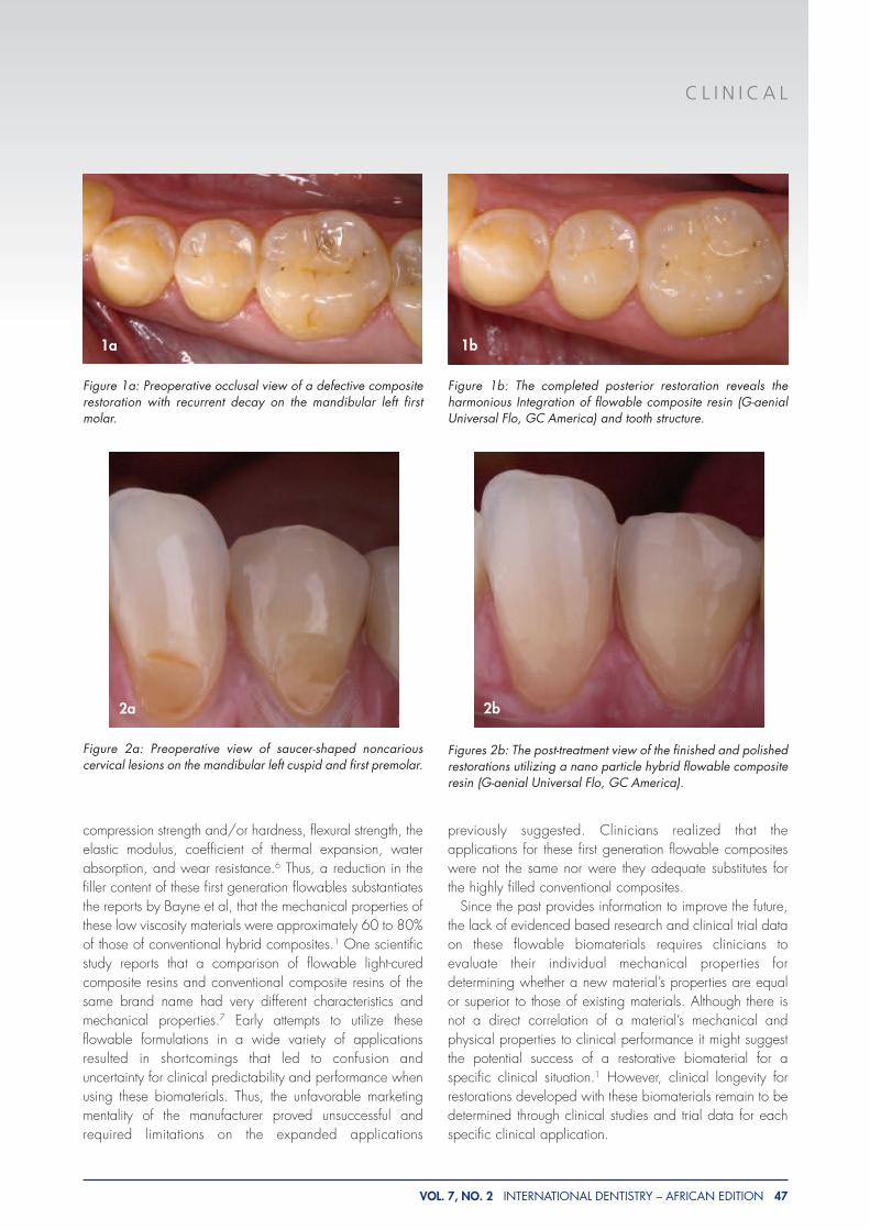
Figures 4 a-d: The composite mock-up is the key to success in function, aesthetics and phonetics.This composite prototype was developed from the indirect/direct technique utilizing a diagnosticwax-up, clear vinyl polysiloxane matrix and a next generation flowable composite resin.
4b
4c
4d
T E R R Y
52 INTERNATIONAL DENTISTRY – AFRICAN EDITION VOL. 7, NO. 2
tooth and adjacent teeth. This color alteration depends onthe scattering and absorption coefficients of the surroundinghard dental tissues and restorative material, which canproduce an undetectable color match by blending withtooth color.21 Furthermore, the surface quality of thecomposite restoration is influenced by the composition andthe filler characteristics of the composite.22,23 Newerformulations of nanocomposites have altered fillercomponents with finer filler size, shape, orientation andconcentration, improving not only their physical andmechanical properties but also their optical characteristics.These nanosized particle hybrid composite resin systemsallow the resin to be polished to a higher degree which caninfluence color integration between the material and toothstructure.
Nanotechnology with composite resinsNanotechnology or molecular manufacturing24 may providecomposite resin with filler particle size that is dramaticallysmaller in size, can be dissolved in higher concentrationsand polymerized into the resin system with molecules thatcan be designed to be compatible when coupled with apolymer, and provide unique characteristics (physical,mechanical, and optical). In addition, optimizing theadhesion of restorative biomaterials to the mineralized hard
tissues of the tooth is a decisive factor for enhancing themechanical strength, marginal adaptation and seal, whileimproving the reliability and longevity of the adhesiverestoration. Currently, the particle size of many of theconventional composites are so dissimilar to the structuralsizes of the hydroxyapatite crystal, dental tubule and enamelrod, there is a potential for compromises in adhesionbetween the macroscopic (40 nm to 0.7 um) restorativematerial and the nanoscopic (1 to 10 nanometers in size)tooth structure.25 However, nanotechnology has the potentialto improve this continuity between the tooth structure and thenanosized filler particle and provide a more stable andnatural interface between the mineralized hard tissues of thetooth and these advanced restorative biomaterials.A recently developed nano-hybrid flowable composite
resin system (G-aenial Universal Flo, GC America) maypossess the physical, mechanical and optical properties toprovide these mechanical and physical requisites. Theseproperties and the clinical behavior of this biomaterialformulation is contingent upon its structure. This new resinfiller technology allows a higher filler loading because ofthe fine filler size, uniform shape and distribution ofparticles. This unique resin filler chemistry allows theparticles to be situated very closely to each other and thisreduced interparticle spacing and homogeneous dispersionof the particles in the resin matrix increases thereinforcement and protects the matrix.26-28 In addition, theproprietary chemical treatment of the filler particles allowsproper wettability of the filler surface by the monomer andthus an improved dispersion and a stable and strongerbond between the filler and resin. Research studies clearlyindicate the significance that filler content and couplingagents represent in determining characteristics such asstrength and wear resistance.28-32 Recent studies report thatflowable composites have comparable shrinkage stress toconventional composites.4,33,34 According to themanufacturers, this next generation flowable resinformulation is purported to offer mechanical, physical andaesthetic properties similar or greater than manyconventional hybrid composites.35 The material’s clinicalattributes include easier insertion and manipulation,improved adaptation to the internal cavity wall, increasedwear resistance, greater elasticity, color stability, enhancedpolishability and retention of polish, and radiopacity similarto enamel. With the competent mechanical propertiesreported, this evolutionary designed formulation is indicatedfor use in anterior and posterior restorative applications.
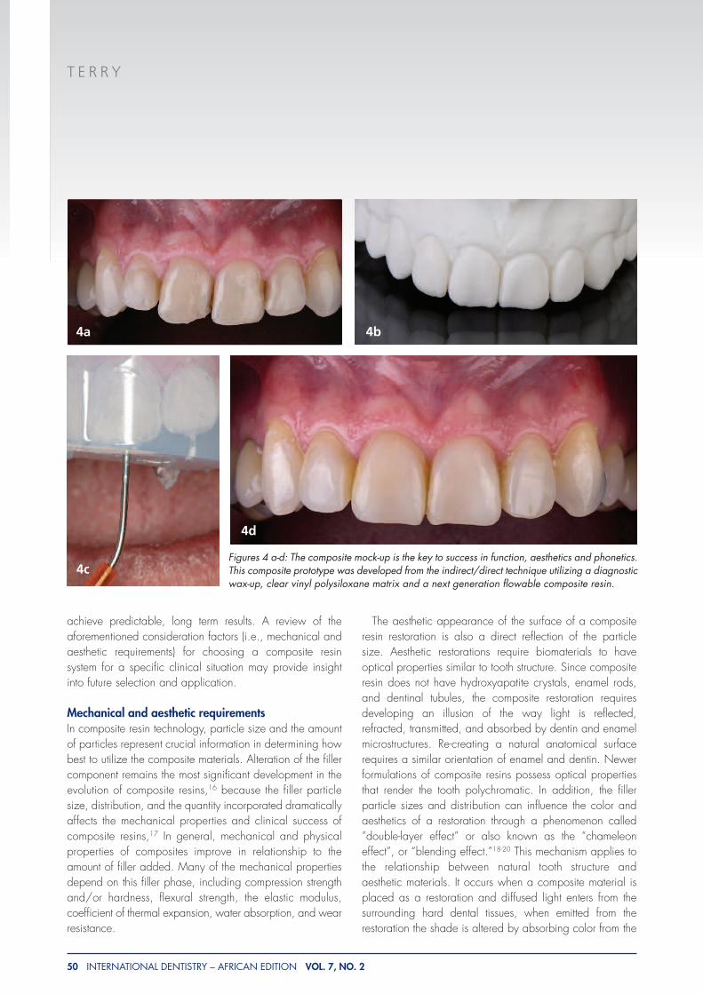
Figure 5: These advanced formulations of flowables have thepotential to increase wear resistance and can be utilized assealants and preventative resin restorations.
T E R R Y
54 INTERNATIONAL DENTISTRY – AFRICAN EDITION VOL. 7, NO. 2
Restorative applications for next generation flowablecomposite resinsIn view of the previous considerations, it is understandablethat clinicians have uncertainties about selection ofbiomaterials and techniques to optimize the materials’properties and achieve predictable long term results. Thefollowing discussion may provide insight into future selectionand application for these next generation flowable materials.
Anterior and posterior composite restorationsToday, improvements in resin technology, optimization ofmaterial properties and the development of innovativetechniques for placement have made the direct compositeresin restoration more user friendly and reliable. Until recently,flowables were clinically indicated and acceptable as fillingmaterials in low-stress applications such as cavity liners,sealants, preventative resin restorations, small Class Irestorations, Class II substructure, small Class III restorations,
and Class V restorations.1 However, the utilization of arecently developed optimized nano particle hybrid flowablecomposite resin (G--aenial Universal Flo, GC America) allowsthe clinician to implement a single restorative material thatappears to have all the improved mechanical and physicalproperties, while using stratification techniques to createinternal color. In addition, studies have shown that usingflowable composites reduces restoration microleakage andthe occurrence of voids.36-39 Other studies indicate thatincremental layering techniques can be effective in reducingthe effect of contraction stress and improving marginalsealing.5,40-42 Controlled polymerization has also beensuggested to reduce marginal gap, increase marginalintegrity, and reduce shrinkage with flowable composites.43-48 By utilizing precise restorative placement techniques,
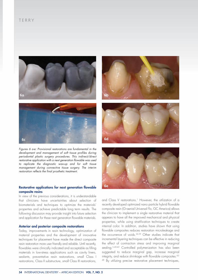
Figures 6 a-e: Provisional restorations are fundamental in thedevelopment and management of soft tissue profiles duringperiodontal plastic surgery procedures. This indirect/directrestorative application with a next generation flowable was usedto replicate the diagnostic wax-up and for soft tissuemanagement during connective tissue surgery. The interimrestoration reflects the final prosthetic treatment.
6a 6b
6d
6e
6c
T E R R Y
56 INTERNATIONAL DENTISTRY – AFRICAN EDITION VOL. 7, NO. 2
conservative adhesive preparation designs 35 and a morethorough adhesive protocol these advanced formulations (G-aenial Universal Flo, GC America) can be utilized for ClassI (Figure 01 a, b), Class II , Class III , Class IV), and ClassV restorations (Figure 02 a, b) diastema closures and directveneers (Figure 03 a, b).
Composite mock-upThe composite mock-up is an excellent tool for increasing thepatient’s understanding and education of the clinicalprocedure through a visual prototype. This compositeprototype allows the patient and the restorative team(ceramist, clinician and the surgeon) to establish parametersfor lip profile, incisal length and orientation to the gingiva,and to simulate the final result. An indirect/direct technique,which uses a clear matrix, can be used to translate thisinformation to the oral cavity. This process can be performedintra-orally without anesthesia and can provide proper lipposition and phonetic considerations.49 (Figure). A clear vinylpolysiloxane impression (Memosil 2, Heraeus Kulzer) can beused to replicate the diagnostic wax-up. The matrix can beplaced intra-orally, and used as a transfer vehicle for theflowable composite (G-aenial Universal Flo, GC America) tobe injected. This composite mock-up should occur prior tofinalization of the treatment plan to ensure that patients’ andthe restorative teams’ expectations have been addressed.(Figure 04 a-d)
Sealants and preventative resin restorationsNext generation flowables can be utilized as compositesurface sealants pit and fissure sealants and preventativeresin restorations. These highly filled nano materials can be
cured in a thin film and with a minimal air-inhibited layer andare designed to seal any cracks or microscopic porositiesthat may have formed during the finishing procedures ofdirect and indirect restorations and to seal occlusal pits andfissures. These formulations have the potential to increase thewear resistance of posterior composite resin restorations sincethe interparticle spacing is reduced and the filler particledensity is increased and thus provide more reinforcement andprotection of the resin matrix.26-28 (Figure 05)
Provisionals: fabrication, modification and repairProvisionals are the key to function, aesthetics and phoneticsand the roadmap to success in aesthetic reconstruction.6
Composite provisionals can be efficiently fabricated by makingan initial impression with a clear vinyl polysiloxane impressionmaterial (Memosil 2, Heraeus Kulzer) of the preoperative stoneor diagnostic wax-up model. After a separating medium isapplied to the preparation, the clear matrix is placed and aflowable composite resin can be injected into the coronalspace with a predetermined shape and contour. Modificationsin shape, length, and contour as well as the elimination of anysurface defects can be accomplished by the incrementalapplication of flowable composite resin after surfacepreparation. Also, long term provisionals can be utilized for theshaping and development of gingival contour for edentulousregions (ie, ovate pontic design) and for the manipulation andshaping of interproximal papillae during prosthetic implanttherapy. In addition, provisionals can be aestheticallyenhanced by cutting back the facial or buccal surface andplacing a final flowable composite layer after surfacepreparation with composite primer and any internalcharacterizations are completed. (Figure 06 a-e)
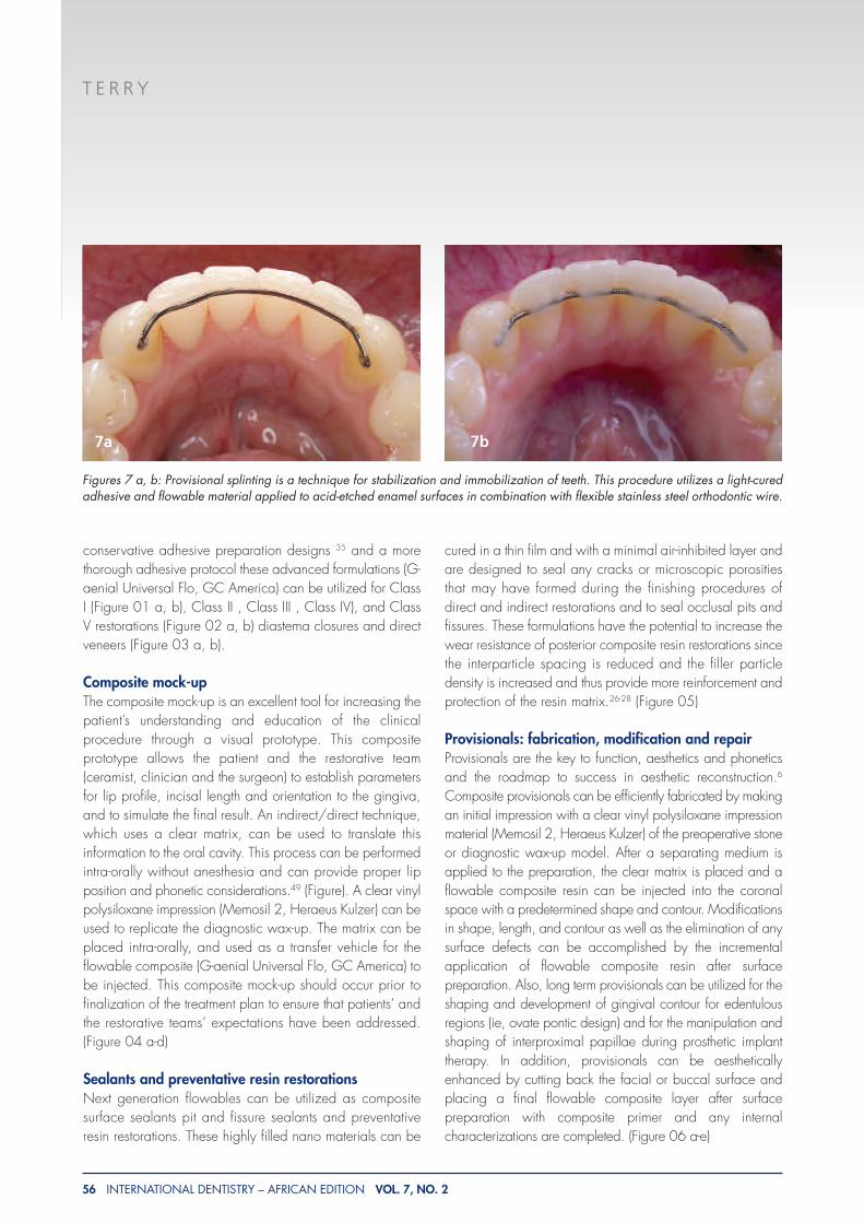
Figures 7 a, b: Provisional splinting is a technique for stabilization and immobilization of teeth. This procedure utilizes a light-curedadhesive and flowable material applied to acid-etched enamel surfaces in combination with flexible stainless steel orthodontic wire.
7a 7b
T E R R Y
VOL. 7, NO. 2 INTERNATIONAL DENTISTRY – AFRICAN EDITION 57
Composite tooth splintingProvisional splinting50 is a technique utilized for thestabilization and immobilization of teeth. This procedure haswidespread use in dentistry- orthodontics, periodontics, oralsurgery, pedodontics, and general dentistry. It provides toothfixation through application of a light-cured flowable orchemically cured composite material to acid etched enamelsurfaces in combination with flexible stainless steel wire,51
orthodontic brackets51,52 or fiber reinforced ribbon. Acceptedclinical applications include: provisional fixed partial dentureplacement,50 to prevent super-eruption of opposing teeth,50
fixed orthodontic retainer,50 periodontal splinting,50
connection of implants to natural teeth,50 support for rootresection,50 stabilization of traumatically displaced,50
transplanted and root fractured teeth,51 and orthodonticextrusion of fractured teeth.53,54 (Figure 07 a, b)
Enhancing internal adaptationIn certain cavity configurations, there are no free surfaceareas present within the cavity. Thus, the ratio between thefree and bonded restoration surfaces (C-factor)55-57 is high,creating shrinkage stresses that are higher than the bondstrength.58 This can result in partial delamination from thetooth structures interface complex generating marginal gapsand/or enamel fractures.59 The process of selective bondingcreates free surfaces within the cavity reducing theconfiguration factor of the restoration. The liner seals the
dentin yet does not adhere to the restoration, therefore thegap formation is confined to the internal aspect of the cavity,creating a free surface within the cavity and thus reducingthe C-factor. This enables more flow during polymerizationresulting in a more stress resistant marginal adaptation.60
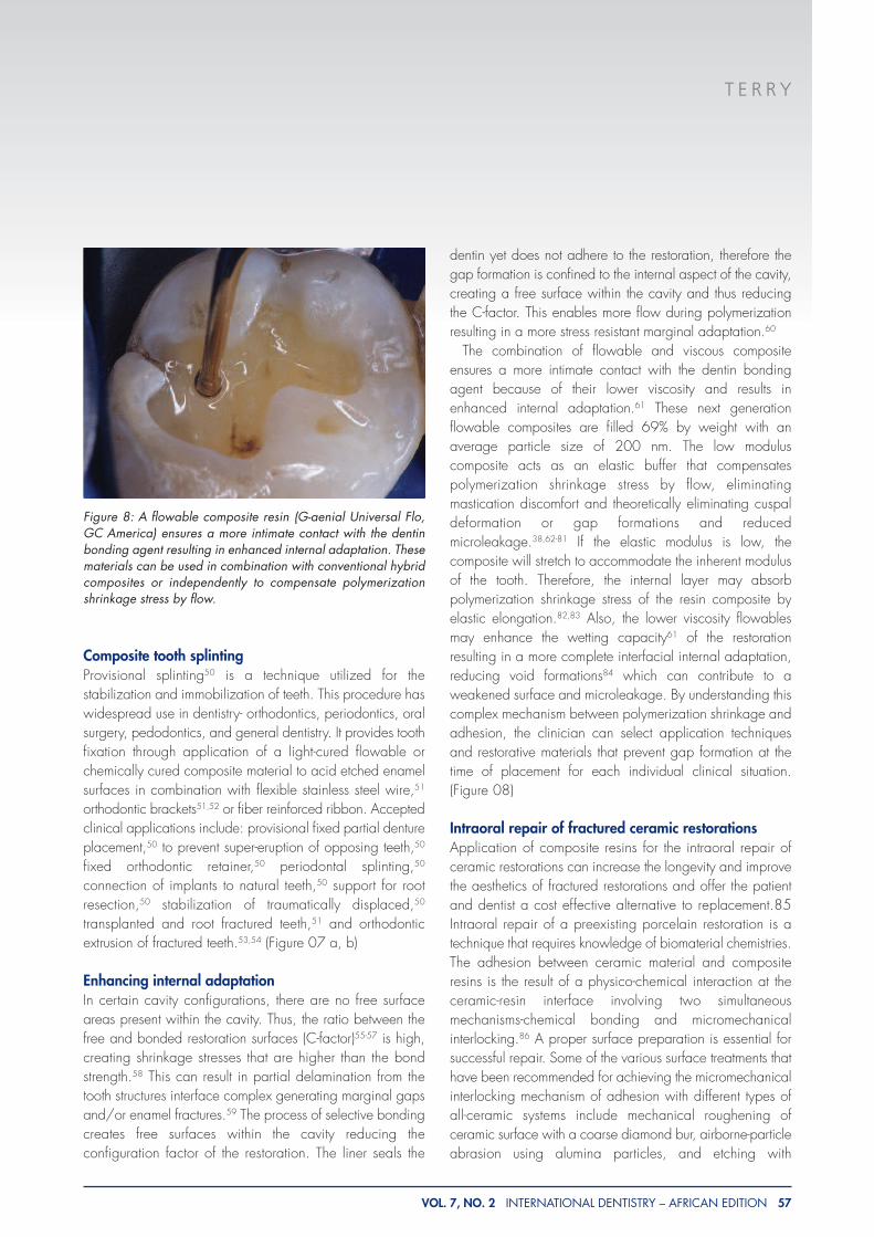
The combination of flowable and viscous compositeensures a more intimate contact with the dentin bondingagent because of their lower viscosity and results inenhanced internal adaptation.61 These next generationflowable composites are filled 69% by weight with anaverage particle size of 200 nm. The low moduluscomposite acts as an elastic buffer that compensatespolymerization shrinkage stress by flow, eliminatingmastication discomfort and theoretically eliminating cuspaldeformation or gap formations and reducedmicroleakage.38,62-81 If the elastic modulus is low, thecomposite will stretch to accommodate the inherent modulusof the tooth. Therefore, the internal layer may absorbpolymerization shrinkage stress of the resin composite byelastic elongation.82,83 Also, the lower viscosity flowablesmay enhance the wetting capacity61 of the restorationresulting in a more complete interfacial internal adaptation,reducing void formations84 which can contribute to aweakened surface and microleakage. By understanding thiscomplex mechanism between polymerization shrinkage andadhesion, the clinician can select application techniquesand restorative materials that prevent gap formation at thetime of placement for each individual clinical situation.(Figure 08)
Intraoral repair of fractured ceramic restorationsApplication of composite resins for the intraoral repair ofceramic restorations can increase the longevity and improvethe aesthetics of fractured restorations and offer the patientand dentist a cost effective alternative to replacement.85Intraoral repair of a preexisting porcelain restoration is atechnique that requires knowledge of biomaterial chemistries.The adhesion between ceramic material and compositeresins is the result of a physico-chemical interaction at theceramic-resin interface involving two simultaneousmechanisms-chemical bonding and micromechanicalinterlocking.86 A proper surface preparation is essential forsuccessful repair. Some of the various surface treatments thathave been recommended for achieving the micromechanicalinterlocking mechanism of adhesion with different types ofall-ceramic systems include mechanical roughening ofceramic surface with a coarse diamond bur, airborne-particleabrasion using alumina particles, and etching with
Figure 8: A flowable composite resin (G-aenial Universal Flo,GC America) ensures a more intimate contact with the dentinbonding agent resulting in enhanced internal adaptation. Thesematerials can be used in combination with conventional hybridcomposites or independently to compensate polymerizationshrinkage stress by flow.
T E R R Y
58 INTERNATIONAL DENTISTRY – AFRICAN EDITION VOL. 7, NO. 2
hydrofluoric acid. Because of the different chemical structurebetween silica-based and high -strength ceramics, differentchemical surface treatments are required. The following arethe author’s standard protocols for bonding composite resinto different ceramic microstructures.
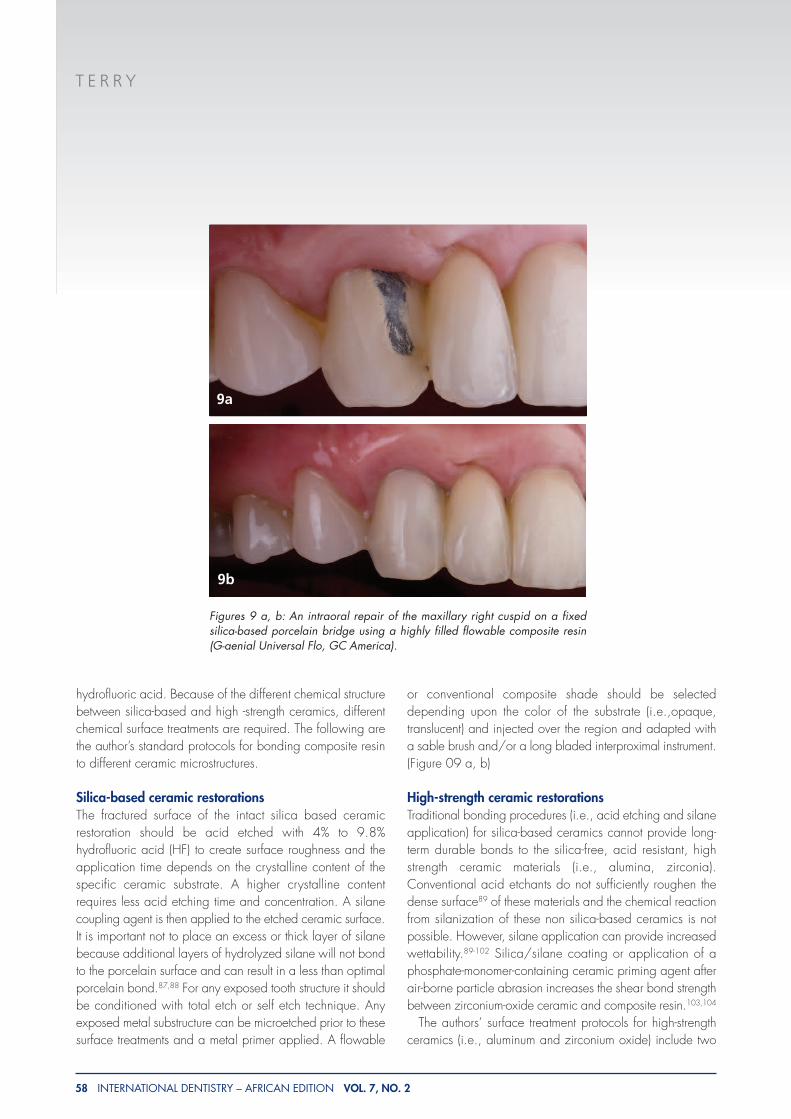
Silica-based ceramic restorations The fractured surface of the intact silica based ceramicrestoration should be acid etched with 4% to 9.8%hydrofluoric acid (HF) to create surface roughness and theapplication time depends on the crystalline content of thespecific ceramic substrate. A higher crystalline contentrequires less acid etching time and concentration. A silanecoupling agent is then applied to the etched ceramic surface.It is important not to place an excess or thick layer of silanebecause additional layers of hydrolyzed silane will not bondto the porcelain surface and can result in a less than optimalporcelain bond.87,88 For any exposed tooth structure it shouldbe conditioned with total etch or self etch technique. Anyexposed metal substructure can be microetched prior to thesesurface treatments and a metal primer applied. A flowable
or conventional composite shade should be selecteddepending upon the color of the substrate (i.e.,opaque,translucent) and injected over the region and adapted witha sable brush and/or a long bladed interproximal instrument.(Figure 09 a, b)
High-strength ceramic restorationsTraditional bonding procedures (i.e., acid etching and silaneapplication) for silica-based ceramics cannot provide long-term durable bonds to the silica-free, acid resistant, highstrength ceramic materials (i.e., alumina, zirconia).Conventional acid etchants do not sufficiently roughen thedense surface89 of these materials and the chemical reactionfrom silanization of these non silica-based ceramics is notpossible. However, silane application can provide increasedwettability.89-102 Silica/silane coating or application of aphosphate-monomer-containing ceramic priming agent afterair-borne particle abrasion increases the shear bond strengthbetween zirconium-oxide ceramic and composite resin.103,104
The authors’ surface treatment protocols for high-strengthceramics (i.e., aluminum and zirconium oxide) include two
9a
Figures 9 a, b: An intraoral repair of the maxillary right cuspid on a fixedsilica-based porcelain bridge using a highly filled flowable composite resin(G-aenial Universal Flo, GC America).
9b
T E R R Y
60 INTERNATIONAL DENTISTRY – AFRICAN EDITION VOL. 7, NO. 2
methods. One method requires silica coating of thefractured surfaces of the intact restoration with CoJet-Sand(Rocatec/CoJet System, 3M™ ESPE™) followed by anapplication of a silane coupling agent (ESPE Sil). Theapplication of a silica layer to high-strength ceramics suchas zirconia creates binding sites for the silane moleculeswhile the silane provides wettability and a chemicalcoupling with the methacrylate based composites. Anotheruser-friendly method involves an application of acommercial primer that contains phosphonate or phosphatemonomers to the fractured surfaces of the intact restoration.Phosphate monomers form covalent bonds with the zirconiasurface and have polymerizable resin terminal ends thatcopolymerize with the methacrylate based composites. Therecent developments of several special ceramic primers
indicate their importance. Currently, there are severalceramic primer systems for zirconia surface preparationavailable such as Monobond Plus (Ivoclar Vivadent);Clearfil Ceramic Primer (Kuraray); AZ-Primer (Shofu Dental);Metal/Zirconia Primer (Ivoclar Vivadent); and Z-Prime Plus(Bisco). Air-particle abrasion with small aluminum oxideparticles (e.g., 30 �m) before application of a ceramicprimer is recommended to further increase bond strengths ofcomposite resins to high-strength ceramic materials.The surface treatment for any exposed tooth structure remains
the same (i.e., self-etch or total etch). A flowable or conventionalcomposite resin shade should be selected depending upon thecolor of the substrate (i.e.,opaque, translucent) and injected orplaced over the region and adapted with a sable brush and/ora long bladed interproximal instrument.
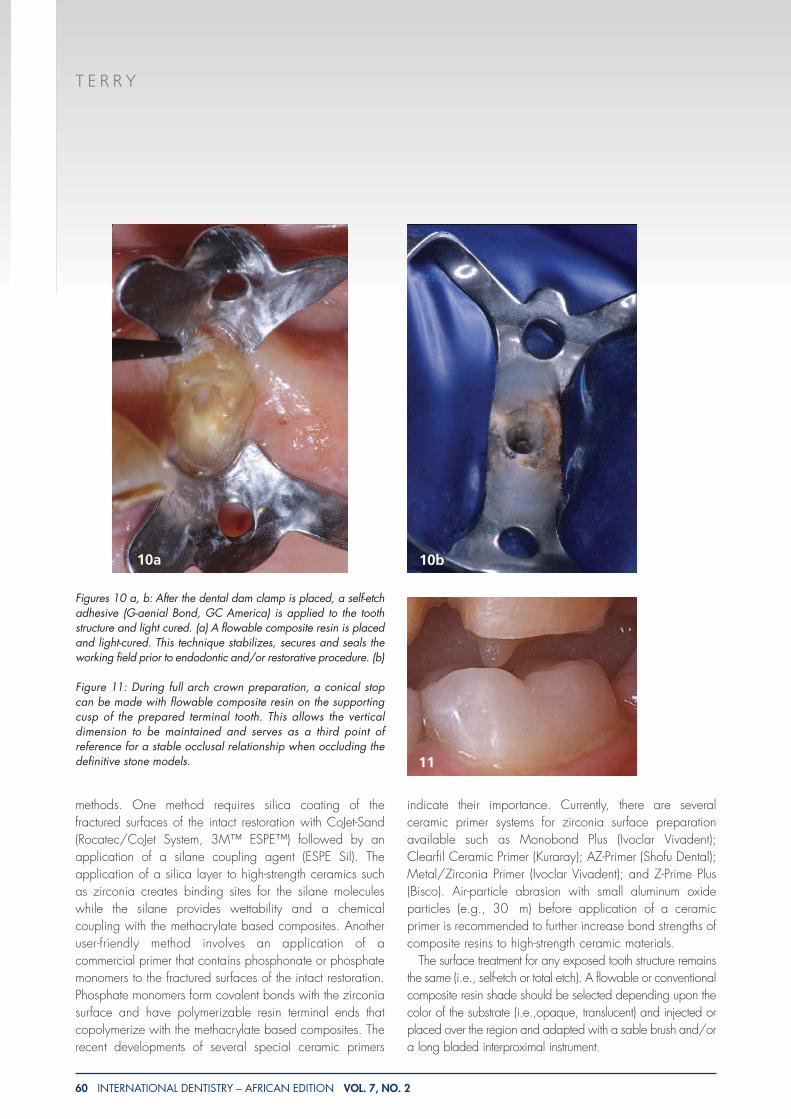
Figures 10 a, b: After the dental dam clamp is placed, a self-etchadhesive (G-aenial Bond, GC America) is applied to the toothstructure and light cured. (a) A flowable composite resin is placedand light-cured. This technique stabilizes, secures and seals theworking field prior to endodontic and/or restorative procedure. (b)
Figure 11: During full arch crown preparation, a conical stopcan be made with flowable composite resin on the supportingcusp of the prepared terminal tooth. This allows the verticaldimension to be maintained and serves as a third point ofreference for a stable occlusal relationship when occluding thedefinitive stone models.
10a 10b
11
T E R R Y
62 INTERNATIONAL DENTISTRY – AFRICAN EDITION VOL. 7, NO. 2
Stabilizing, securing, and sealing dental dam clampThis procedure allows retention and stabilization of the dentaldam clamp with flowable composite resin. The clamp isapplied before the dam, it is positioned and stabilizeddigitally and a selective etching procedure is utilized. Theenamel is spot etched for 15 seconds with a 37.5%phosphoric acid gel, rinsed for 5 seconds and air dried. Aself-etch adhesive (G-aenial Bond, GC America) is applied
to the enamel and dentin, coronal to the jaws of the clampand allowed to dwell for 10 seconds, dried for 5 secondsand light cured for 10 seconds and dried. A flowablecomposite resin (G-aenial Universal Flo, GC America) isapplied and light cured for 10 seconds. This procedureallows stabilization, isolation and seals the working fieldduring the endodontic or operative procedure.82 (Figure 10a, b)
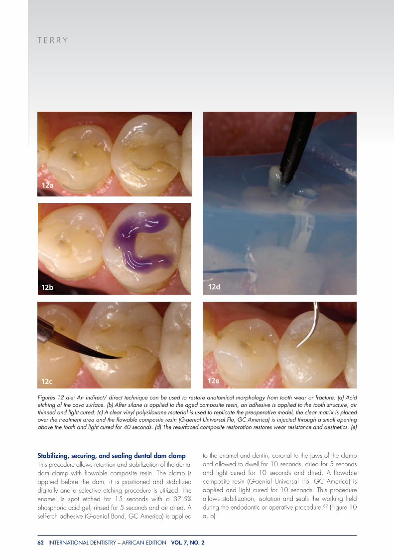
Figures 12 a-e: An indirect/ direct technique can be used to restore anatomical morphology from tooth wear or fracture. (a) Acidetching of the cavo surface. (b) After silane is applied to the aged composite resin, an adhesive is applied to the tooth structure, airthinned and light cured. (c) A clear vinyl polysiloxane material is used to replicate the preoperative model, the clear matrix is placedover the treatment area and the flowable composite resin (G-aenial Universal Flo, GC America) is injected through a small openingabove the tooth and light cured for 40 seconds. (d) The resurfaced composite restoration restores wear resistance and aesthetics. (e)
12d
12a
12b
12c 12e

T E R R Y
64 INTERNATIONAL DENTISTRY – AFRICAN EDITION VOL. 7, NO. 2
Creating a vertical stop for inter-occlusal recordsFlowable composite resin can be used as accurate inter-occlusal record for the orientation of models for fixedprosthetic restoration. This method uses conical stops ofcomposite resin prepared in the enamel of the abutment ormade of a conical composite core which maintains thevertical dimension of occlusion and acts as a third point ofreference for a stable occlusal relationship when occludingthe definitive stone models.105 (Figure 11)
Resurfacing or repairing composite restorationsCyclic tension and compressive stresses that occur in themouth during chewing or parafunctional habits can reach afatigue limit and can result in tooth structure loss.106-108 These
repeated flexural forces can also cause adhesive failure ofadhesive restorations at the dentin resin interface which canresult in microleakage, and partial or complete debondingof the restoration.109 A restorative material properly bondedto the enamel and dentin substrate will reduce marginalcontraction gaps, microleakage, marginal staining andcaries recurrence, improve retention, reinforce tooth structureand dissipate and reduce functional stresses across itsinterface throughout the entire tooth while improving thenatural aesthetics and wear resistance.110-114 Anindirect/direct technique can be used to restore anatomicalmorphology from wear or fracture. The aforementionedprocedure uses a clear matrix that can be fabricated froman preoperative or diagnostic wax-up model. After preparing
Figures 13 a-d: After endodontic treatment, the access opening(i.e., tooth, composite) is roughened with a diamond bur. (a)After silane is applied to the roughened composite surface, aself-etch adhesive (G-aenial Bond, GC America) is applied tothe surfaces and allowed to dwell for 10 seconds, air dried andlight cured for 10 seconds; (b) A light cured flowable compositeresin (G-aenial Universal Flo, GC America) is injected into theopening, contoured and light cured for 40 seconds; (c) Thetransitional seal of the endodontic access opening reduces thepotential for bacterial contamination. (d)
13b
13a
13c
13d
T E R R Y
66 INTERNATIONAL DENTISTRY – AFRICAN EDITION VOL. 7, NO. 2
the margins and surface treatment of the tooth structure andbiomaterial, the matrix can be placed over the treatment areaand the flowable composite resin (G-aenial Universal Flo,GC America) is injected through a small opening above thetooth and light cured. (Figure 12 a-e)
Sealing endodontic access openingsThe primary goal for endodontically treated teeth is toachieve long-term apical periodontal health.115 Theadvancements in endodontic materials and techniques haveallowed the clinician to attain an optimal apical seal to preventbacterial leakage.116,117 However, when the coronal portionof the root canal system is not properly sealed it can becomea potential source of bacterial invasion and failure of theendodontic treatment. 117-124 Thus, complete sealing of theendodontic access opening between appointments and afterendodontic treatment is essential for achieving endodonticsuccess.125 One study indicates that a good coronal sealresults in significantly less occurrence of periradicularinflammation.115,126 An inadequate coronal seal can allowsaliva to reach the apical region of the tooth in as little time asthree days 115,119 and endotoxins from microorganisms suchas Actinobacillus actinomycetemcomitans within 20 days.
127 There are various intermediate materials (i.e. Cavit G ,zinc oxide-eugenol, and glass ionomers)117,125,128 that areused to close the existing coronal restoration but may notprovide an adequate seal and some may interfere withrequired future adhesive procedures. One material andmethod that can be utilized for sealing the endodontic accessopening is a simple technique that involves self-etch adhesivesand/or next generation flowables. This bioadhesiveprocedure can restore or provide a transitional seal until a newrestoration can be placed. (Figure 13 a-d)
Repairing denture teeth Denture teeth are fabricated from several different materials(i.e.,ceramic, acrylic, composite) and there are a infinitenumber of shapes and sizes. Generally, fractures to dentureteeth occur as emergency situations and require replacement.Replacement can be achieved with relative ease in thelaboratory with an adequate inventory of denture teeth.However, with the proper surface treatment these fracturescan be repaired with next generation flowable compositeresins at chairside. (Figure 14 a-c)
Eliminating cervical tooth sensitivityThere are numerous and varied etiological factors andpredisposing influences to cervical dentin hypersensitivity.More than 90% of hypersensitive surfaces occur at thecervical region on the buccal and labial aspects of theinvolved teeth. In the ideal anatomical position, most teeth
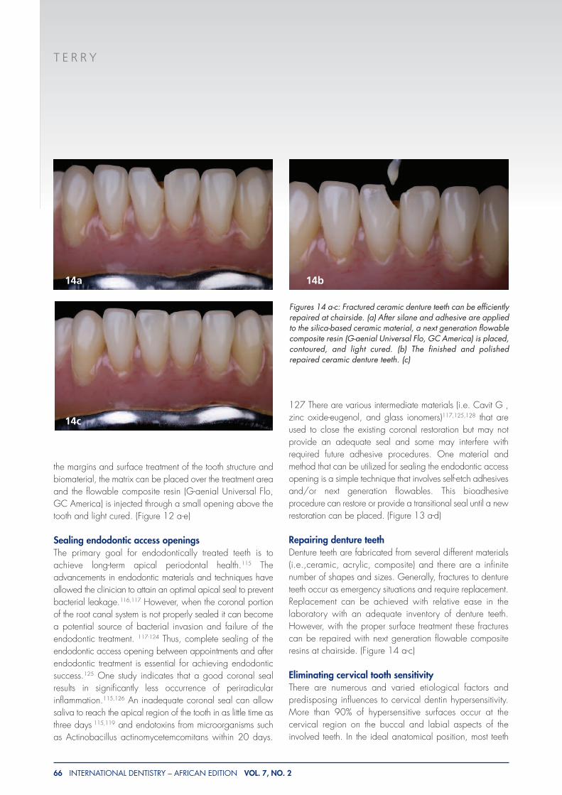
Figures 14 a-c: Fractured ceramic denture teeth can be efficientlyrepaired at chairside. (a) After silane and adhesive are appliedto the silica-based ceramic material, a next generation flowablecomposite resin (G-aenial Universal Flo, GC America) is placed,contoured, and light cured. (b) The finished and polishedrepaired ceramic denture teeth. (c)
14a 14b
14c
T E R R Y
48 INTERNATIONAL DENTISTRY – AFRICAN EDITION VOL. 7, NO. 2
have only the enamel exposed to the oral environment, anddentin that is protected by enamel or cementum is notsensitive. Cervical tooth sensitivity occurs when this enamelor cementum layer is removed and the underlying dentinaltubules are open and exposed to the oral environment.129
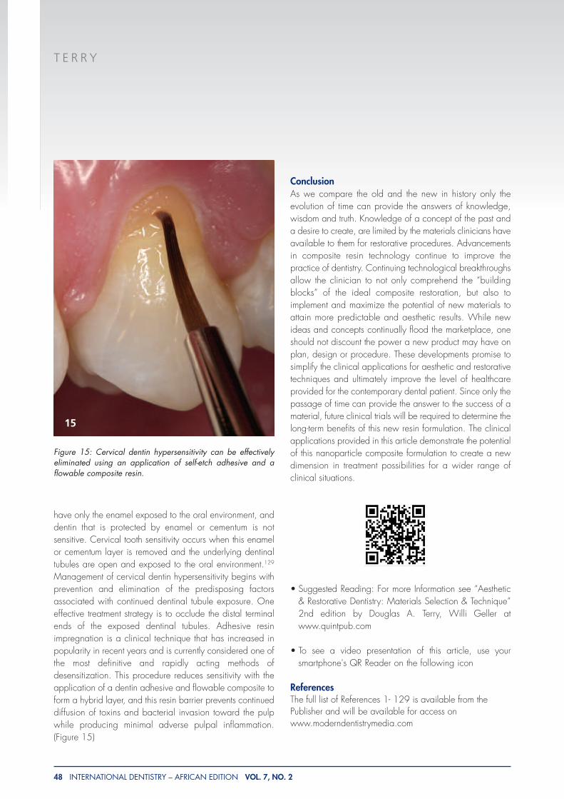
Management of cervical dentin hypersensitivity begins withprevention and elimination of the predisposing factorsassociated with continued dentinal tubule exposure. Oneeffective treatment strategy is to occlude the distal terminalends of the exposed dentinal tubules. Adhesive resinimpregnation is a clinical technique that has increased inpopularity in recent years and is currently considered one ofthe most definitive and rapidly acting methods ofdesensitization. This procedure reduces sensitivity with theapplication of a dentin adhesive and flowable composite toform a hybrid layer, and this resin barrier prevents continueddiffusion of toxins and bacterial invasion toward the pulpwhile producing minimal adverse pulpal inflammation.(Figure 15)
ConclusionAs we compare the old and the new in history only theevolution of time can provide the answers of knowledge,wisdom and truth. Knowledge of a concept of the past anda desire to create, are limited by the materials clinicians haveavailable to them for restorative procedures. Advancementsin composite resin technology continue to improve thepractice of dentistry. Continuing technological breakthroughsallow the clinician to not only comprehend the “buildingblocks” of the ideal composite restoration, but also toimplement and maximize the potential of new materials toattain more predictable and aesthetic results. While newideas and concepts continually flood the marketplace, oneshould not discount the power a new product may have onplan, design or procedure. These developments promise tosimplify the clinical applications for aesthetic and restorativetechniques and ultimately improve the level of healthcareprovided for the contemporary dental patient. Since only thepassage of time can provide the answer to the success of amaterial, future clinical trials will be required to determine thelong-term benefits of this new resin formulation. The clinicalapplications provided in this article demonstrate the potentialof this nanoparticle composite formulation to create a newdimension in treatment possibilities for a wider range ofclinical situations.
• Suggested Reading: For more Information see “Aesthetic& Restorative Dentistry: Materials Selection & Technique”2nd edition by Douglas A. Terry, Willi Geller atwww.quintpub.com
• To see a video presentation of this article, use yoursmartphone's QR Reader on the following icon
ReferencesThe full list of References 1- 129 is available from thePublisher and will be available for access onwww.moderndentistrymedia.com
Figure 15: Cervical dentin hypersensitivity can be effectivelyeliminated using an application of self-etch adhesive and aflowable composite resin.
15
T E R R Y
68 INTERNATIONAL DENTISTRY – AFRICAN EDITION VOL. 7, NO. 2
References1. Bayne SC,Thompson Jy, Swift EJ Jr, Stamatiades P,
Wilkerson M. A characterization of first-generation flowablecomposites. J Am Dent Assoc 1998;129:567-77.2. Labella R, Lambrechts P, Van Meerbeek B, Vanherle G.
Polymerization shrinkage and elasticity of flowable compositesand filled adhesives. Dent Mater 1999; 15:128-37.3. Tabassian M, Moon PC. Filler particle characterization in
flowable and packable composites. J Dent Res 1999;79:Abstract 3022, 213.4. Baroudi K, Silikas N, Watts DC. Edge -strength of
flowable resin-composites. J Dent 2008; 36:63-8.5. Ikeda I, Otsuki M, Sadr A, et al. Effect of filler content of
flowable composites on resin-cavity interface. Dent Mater J2009;28(6):679-85.6. Terry, DA. Natural Aesthetics with Composite Resin.
Montage Media Publishing, Mahwah, NJ. 2004.7. Irie M, Tjandrawinata R, E Lihua, et al. Flexural
performance of flowable versus conventional light-curedcomposite resins in a long-term in vitro study. Dent Mater J2008;27(2):300-9.8. Summitt JB, Robbins JW, Schwartz RS. Fundamental of
Operative Dentistry: A contemporary approach. Carole StreamIL: Quint Pub, 2001.9. Terry DA. Direct application of a nanocomposite resin
system: Part 1 – The evolution of contemporary compositematerials. Pract Proced Aesthet Dent 2004;16(6):417-22.10. Liebenberg WH. Assuring restorative integrity in
extensive posterior resin composite restorations: pushing theenvelope. Quintessence Int 2000; 31: 153-64.11. Taylor DF, Bayne SC, Sturdevant JR, et al. Restoration
width and complexity effects on posterior composite wear.[abstract 35]. J Dent Res 1989;68:186.12. Leinfelder KF. Composite resin in posterior teeth. Dent
Clin North Am 1981;25: 357-64.13. Ferrari M. Handling characteristics of resin composites in
posterior teeth. Compend 1998;19(9):879-93.14. Dietschi D, Spreafico R. Adhesive Metal-Free Restorations:
Current Concepts for the Esthetic Treatment of Posterior Teeth. Berlin,Germany: Quintessence Publishing; 1999.15. Sabiston CB. Etiology of cracked teeth. A review and
proposal. Iowa Dent J 1994; 80(4):13-4.16. Roulet JF. Degradation of Dental Polymers. 1st ed. Basel,
Switzerland: S. Karger AG, 1987.17. Leinfelder, KF, “Composite Resins: Properties and
Clinical Performance”, In: O’Brien WJ, and Powers, JM, ed.Dental Materials: Properties and Selection, QuintessencePublishing Company, Chicago, IL, 139-57, 1989.18. Paravina RD, Westland S, Kimura M. Powers JM, Imai
FH. Color interaction of dental materials: blending effect of
layered composites. Dent Mater 2006; 22(10): 903-8.19. Chartrand TL, Bargh JA. The chameleon effect: the
perception-behavior link and social interaction. J Pers SocPsychol 1999;76(6):893-910.20. Paravina RD, Westland S, Imai FH , et al. Evaluation of
blending effect of composites related to restoration size. DentMater 2006;22(4):299-307.21. Hall NR, Kafalias MC. Composite colour matching: The
development and evaluation of a restorative colour matchingsystem. Aust Prosthod J 1991;5:47-52.22. Jefferies SR. The art and science of abrasive finishing
and polishing in restorative dentistry. Dent Clin North Am1998;42(4):613-27.23. Wilson F, Heath JR, Watts DC. Finishing composite
restorative materials. J Oral Rehab 1990;17(1):79-87.24. Kirk RE, Othmer DF, Kroschwitz J, Howe-Grant .
Encyclopedia of Chemical technology. 4th ed. New YorkWiley;1991:397 .25. Muselmann M. Composites make large difference in
“small” medical, dental applications, Comp Tech; December2003:24-27.26. Bayne SC, Taylor DF, Heymann HO. Protection
hypothesis for composite wear. Dent Mater 1992;8(5):305-9.27. Turssi CP, Ferracane JL, Vogel K. Filler features and their
effects on wear and degree of conversion of particulate dentalresin composites. Biomater 2005;4932-7.28. Lim BS, Ferracane JL, Condon JR, Adey JD. Effect of filler
fraction and filler surface treatment on wear of microfilledcomposites. Dent Mater 2002;18:1-11.29. Venhoven BMA, de Gee Aj, Werner A, et al. Influence
of filler parameters on the mechanical coherence of dentalrestorative resin composites. Biomater 1996;17(7):735-40.30. Condon JR, Ferracane JL. In vitro wear of composite with
varied cure, filler level, and filler treatment. J Dent Res1997;76:1405-11.31. Condon JR, Ferracane JL. Factors effecting dental
composite wear in vitro. J Biomed Mater Res 1997;38:303-13.32. Beatty MW, Swartz ML, Moore BK, et al. Effect of
microfiller fraction and silane treatment on resin compositeproperties. J Biomed Mater Res 1998;40:12-23.33. Cadenaro M, Marchesi G, Antoniolli F, et al. Flowability
of composites is no guarantee for contraction stress reduction.Dent Mater 2009;25:649-54.34. Gallo JR, Burgess JO, Ripps AH, et al. Three-year clinical
evaluation of two flowable composites. Quintessence Int2010;41:497-503.35. Yamase M, Maseki T, Nitta T, et al. Mechanical
properties of various latest resin composite restoratives. J DentRes 89(Spec Iss A):abstract 464, 2010.36. Attar N, Tam LE, McComb D. Flow, strength, stiffness
T E R R Y
69 INTERNATIONAL DENTISTRY – AFRICAN EDITION VOL. 7, NO. 2
and radiopacity of flowable resin composites. J Can DentAssoc 2003;69(8):516-21.37. Payne JH 4th. The marginal seal of class II restorations:
flowable composite resin compared to injectable glassionomer. J Clin Pediatr Dent 1999;23(2):123-30.38. Ferdianakis K. Microleakage reduction from newer
esthetic restorative materials in permanent molars. J Clin PediatrDent 1998;22(3):221-9.39. Malmstrom H, Schlueter M, Roach T, et al. Effect of
thickness of flowable resins on marginal leakage in class IIcomposite restorations. Oper Dent 2002;27(4):373-80.40. Chi HH. A posterior composite case utilizing the
incremental and stratified layering technique. Oper Dent2006:31(4):512-16.41. Felix SA, Gonzalez-Lopez S, Mauricio PD, et al. Effect
of filling techniques on the regional bond strength to lateralwalls in Class I cavities. Oper Dent 2007;32(6):602-9.42. Yamazaki PC, Bedran-Russo AK, Pereira PN, et al.
Microleakage evaluation of a new low-shrinkage compositerestorative material. Oper Dent 2006;31(6):670- 76.43. Obici AC, Sinhoreti MA, de Goes MF, et al. Effect of
photo-activation method on polymerization shrinkage ofrestorative composites. Oper Dent 2002;27(2):192- 98.44. Mehl A, Hickel R, Kunzelmann KH. Physical properties
and gap formation of light-cured composites with and without‘soft-start polymerization’. J Dent 1997;25:321-30.45. Sakaguchi RL, Berge HX. Reduced light energy density
decreases post-gel contraction while maintaining degree ofconversion in composites. J Dent 1998;26(8):695-700.46. Oberholzer TG, Du Preez IC, Kidd M. Effect of LED
curing on the microleakage, shear bond strength and surfacehardness of a resin-based composite restoration. Biomater2005;26:3981-6.47. Santos AJ, Lisso MT, Aguiar FH, et al. Effect of stepped
exposure of quantitative in vitro marginal microleakage. J EsthetRestor Dent 2005;17:236-42.48. Yazici AR, Celik C, Dayangac B, et al. Effects of
different light curing units/modes on the microleakage offlowable composite resins. Eur J Dent 2008;2(4):240-6.49. Kesseler JC. Dentist and laboratory: Communication for
success. J Amer Dent Assoc 1987; (Spec No.): 97E-102E.50. Becker CM, Kraiser DA, Kaldahl WB. The evolution of
temporary fixed splint the a-splint. Int J Periodont Restor Dent1998;18(3):277-85.51. Oikarinen K. Tooth splinting: a review of the literature
and consideration of the versatility of a wire-composite splint.Endo Dent Traum 1990; 6: 237-250.52. Park SB, Son WS, Ko CC, et al. Influence of flowable
resins on the shear bond strength of orthodontic brackets. DentMater J 2009;28(6):730-4.
53. Baratieri LN. Esthetic Principles (in Portuguese) . SaoPaulo, Brazil: Quintessence Publishing, 1998:48.54. Lew KK. A removable appliance to extrude teeth. Quint
Int 1993; 24(3): 157-60.55. Roberson TM, Heymann HO, Swift EJ. Sturdevant’s The
Art and Science of Operative Dentistry. 4th ed. St. Louis:Mosby-Year Book, 2002:203.56. Davidson CL, Feilzer AJ. Polymerization shrinkage and
polymerization shrinkage stress in polymer-based restoratives. JDent 1997;25:435-40.57. Feilzer AJ, De Gee AJ, Davidson CL. Setting stress in
composite resin in relation to configuration of the restoration. JDent Res 1987;66:1636-9.58. Frankenberger R, Kramer N, Pelka M, et al. Internal
adaptation and overhang formation of direct Class II resin compositerestorations. Clin Oral Investig 1999; 3(4): 208-215.59. Pescatore C. The direct posterior esthetic restoration
using state-of-art composite resin technology. Compend ContinEduc Dent Suppl 2000; 26(Suppl):14-18.60. Baratieri LN, Ritter AV, Peridigao J, et al. Direct posterior
composite resin restorations: Current concepts for thetechnique. Pract Periodont Aesthet Dent 1998;10(7):875-886.61. Lindberg A, van Dijken JW, Horstedt P. Interfacial
adaptation of a Class II polyacid-modified resincomposite/resin composite laminate restoration in vivo. ActaOdontol Scand 2000; 58(2): 77-84.62. Van Meerbeek B, Willems G, Celis JP, et al. Assessment
by nano-indentation of the hardness and elasticity of the resin-dentin bonding area. J Dent Res 1993;72:1434-42.63. Choi KK, Condon JR, Ferracane JL. The effect of
adhesive thickness on polymerization contraction of composite.J Dent Res 2000;79:812-17.64. Hashimoto M, Sano H, Yoshida E, et al. Effect of
multiple adhesive coatings on dentin bonding. Oper Dent2004;29:416-23.65. Zheng L, Pereira PN, Nakajima M, et al. Relationship
between adhesive thickness and microtensile bond strength.Oper Dent 2001;26:97-104.66. Senawongse P, Pongprueksa P, Tagami J. The effect of
the elastic modulus of low-viscosity resins on the microleakageof Class V resin composite restorations under occlusal loading.Dent Mater J 2010;29(3):324-9.67. Tung FF, Estafan D, Scherer W. Microleakage of a
condensable resin composite: an vitro investigation.Quintessence Int 2000;31:430-4.68. Leevailoj C, Cochran MA, Matis BA, et al.
Microleakage of posterior packable resin composites with andwithout flowable liners. Oper Dent 2001;26:302-7.69. Braga RR, Hilton TJ, Ferracane JL. Contraction stress of
T E R R Y
70 INTERNATIONAL DENTISTRY – AFRICAN EDITION VOL. 7, NO. 2
flowable composite materials and their efficacy as stress-relieving layers. J Amer Dent Assoc 2003;134:721-8.70. M R, Sajjan GS, BNK, Mittal N. Effect of different
placement techniques on marginal microleakage of deep class-II cavities restored with two composite resin formulations. JConserv Dent 2010;13(1)9-15.71. Kemp-Scholte CM, Davidson CL. Complete marginal
seal of class V resin composite restorations effected byincreased flexibility. J Dent Res 1990;69:1240-3.72. Chuang SF, Liu JK, Chao CC, et al. Effects of flowable
composite lining and operator experience on microleakageand internal voids in class II composite restorations. J ProsthetDent 2001;85(2):177-83.73. Cho E, Chikawa H, Kishikawa R, et al. Influence of
elasticity on gap formation in a lining technique with flowablecomposite. Dent Mater J 2006;25(3):538-44.74. Alomari QD, Reinhardt JW, Boyer DB. Effects of liners
on cusp deflection and gap formation in compositerestorations. Oper Dent 2001;26(4):406-11.75. Kubo S, Yokota H, Hayashi Y. Effect of low viscosity
resin-based composite on the microleakage of cervicalrestorations. Am J Dent 2003;16:244-8.76. Kubo S, Yakkota H, Hayashi Y. Microleakage of
cervical cavities restored with flowable composites. Am J Dent2004;17:33-7.77. Attar N, Turgut MD, Gungor HC. The effect of flowable
resin composites as gingival increments on the microleakage ofposterior resin composites. Oper Dent 2004;29:162-7.78. Ernst CP, Cortain G, Spohn M, et al. Marginal integrity
of different resin-basedcomposites for posterior teeth: an in vitro dye-penetration
study on eight resincomposite and compomer-/adhesivecombinations with a particular look at the additional use offlow-composites. Dent Mater 2002;18(4):351-8.79. Korkmaz Y, Ozel E, Attar N. Effect of flowable
composite lining on microleakage and internal voids in Class IIcomposite restorations. J Adhes Dent 2007;9:189- 94.80. Peutzfeldt A, Asmussen E. Composite restorations:
influence of flowable and self-curing resin composite linings onmicroleakage in vitro. Oper Dent 2002;27:569-75.81. Olmez A, Oztas N, Bodur H. The effect of flowable
resin composite on microleakage and internal voids in class IIcomposite restorations. Oper Dent 2004;29:713-9.82. Liebenberg WH. Successive cusp build-up an improved
placement technique for posterior direct resin restoration. JCanada Dent Assoc 1996;62(6):501-7.83. Van Meerbeek B, Perdigao J, Lambrechts P, et al. The
clinical performance of adhesives. J Dent 1998; 26(1): 1-20.84. Kemp-Scholte CM, Davidson CL. Marginal integrity
related to bond strength and strain of composite resin
restorative systems. J Prosthet Dent 1990;64:658-64.85. Latta MA, Barkmeier WW. Approaches for intraoral
repair of ceramic restorations. Compend Contin Educ Dent2000;21(8):635-44.86. Terry DA, Leinfelder KF, Geller W, et al. Aesthetic &
Restorative Dentistry Material Selection & Technique. Houston,TX; Everest Publishing Media: 2009.87. Alex G. Preparing porcelain surfaces for optimal
bonding. Compend Educ Dent 2008;29(6):324-36.88. Matinlinna JP, Lassila LV, Ozcan M, et al. An introduction
to silanes and their clinical applications in dentistry. Int JProsthodont. 2004;17:155-64.89. Awliya W, Odén A, Yaman P, Dennison JB, et al. Shear
bond strength of a resin cement to densely sintered high-purityalumina with various surface conditions. Acta Odontol Scand1998;56(1):9–13.90. Ozcan M, Alkumru HN, Gemalmaz D. The effect of
surface treatment on the shear bond strength of luting cement toa glass-infiltrated alumina ceramic. Int J Prosthondont2001;14(4):335–9.91. Roulet JF, Söderholm KJ, Longmate J. Effects of treatment
and storage conditions on ceramic/composite bond strength. JDent Res 1995;74(1):381–7.92. Bottino MA, Valandro LF, Scotti R, Buso L. Effect of
surface treatments on the resin bond to zirconium-basedceramic. Int J Prosthodont 2005;18(1):60–5.93. Bailey JH. Porcelain-to-composite bond strengths using
four organosilane materials. J Prosthet Dent 1989;61(2):174–7.94. Phoenix RD, Shen C. Characterization of treated
porcelain surfaces via dynamic contact angle analysis. Int JProsthodont 1995;8(2):187–94.95. Foxton RM, Pereira PN, Nakajima M, et al. Durability
of the dual-cure resin cement/ceramic bond with differentcuring strategies. J Adhes Dent 2002;4(1):49–59.96. Borges GA, Sophr AM, de Goes MF, et al. Effect of
etching and airborne particle abrasion on the microstructure ofdifferent dental ceramics. J Prosthet Dent 2003;89(5):479–88.97. Kato H, Matsumura H, Atsuta M. Effect of etching and
sandblasting on bond strength to sintered porcelain of unfilledresin. J Oral Rehabil 2000;27(2):103–10.98. Thurmond JW, Barkmeier WW, Wilwerding TM. Effect
of porcelain surface treatments on bond strengths of compositeresin bonded to porcelain. J Prosthet Dent 1994;72(4):355–9.99. Chen JH, Matsumura H, Atsuta M. Effect of different
etching periods on the bond strength of composite resin to amachinable porcelain. J Dent 1998;26(1):53–8.100. Söderholm KJ, Shang SW. Molecular orientation of
silane at the surface of colloidal silica. J Dent Res1993;72(6):1050–4.
T E R R Y
71 INTERNATIONAL DENTISTRY – AFRICAN EDITION VOL. 7, NO. 2
101. Chen TM, Brauer GM. Solvent effects on bondingorgano-silane to silica surfaces. J Dent Res1982;61(12):1439–43.102. Madani M, Chu FC, McDonald AV, Smales RJ. Effects
of surface treatments on shear bond strengths between a resincement and an alumina core. J Prosthet Dent2000;83(6):644–7.103. Atsu SS, Kilicarslan MA, Kucukesmen HC, Aka PS.
Effect of zirconium-oxide ceramic surface treatments on thebond strength to adhesive resin. J Prosthet Dent2006;95(6):430–6.104. Blatz MB, Sadan A, Martin J, Lang B. In vitro
evaluation of shear bond strengths of resin to densely-sinteredhigh-purity zirconium-oxide ceramic after long-term storage andthermal cycling. J Prosthet Dent 2004;91(4):356–62.105. Sato Y, Hosokawa R, Tsuga K, et al. Creating a
vertical stop for interocclusal records. J Prosthet Dent 2000;83(5): 582-585.106. Tresher RW, Satio GE. The stress analysis of human
teeth. J Biomech1973;6(5)443-9.107. Hood JA. Experimental studies on tooth deformation:
Stress distribution on class V restorations. N Z Dent J1972;68(312):116-131.108. Powers JM, Craig RG, Ludema KC. Friction behavior
surface failure of human enamel. J Dent Res 1972;5(6):1327-31.109. Lee WC, Eakle WS. Possible role of tensile stress in
the etiology of cervical erosive lesions of teeth. J Prosthet Dent1984;52(3):374-80.110. Goracci G, Mori G. Esthetic and functional
reproduction of occlusal morphology with composite resins.Compend Contin Educ Dent 1999;20(7):643- 48.111. Touati B. Bonded ceramic restorations: achieving
predictability. Pract Periodontics Aesthet Dent 1995;7(4):33-7.112. Santos JF, Bianchi J. Restoration of severely damaged
teeth with resin bonding systems: case reports. Quintessence Int1991;22:611-5.113. Van Meerbeek B, Vanherle G, Lambrechts P, et al.
Dentin-and enamelbonding agents. Current Opin Dent1992;2:17-27.114. Eakle WS. Fracture resistance of teeth restores with
class II bonded composite resin. J Dent Res 1986; 65:149-53.115. Korasli D, Ziraman F, Ozyurt P, et al. Microleakage of
self-etch primer/adhesives in endodontically treated teeth. JAmer Dent Assoc 2007;138(5):634-40.
116. Schwartz RS, Fransman R. Adhesive dentistry andendodontics: materials, clinical, strategies and procedures forrestoration of access cavities: a review. J Endod2005;31:151-65.117. Bailon-Sanchez ME, Gonzalez-Castillo S, Gonzalez-
Rodriguez MP, et al. Intraorifice sealing ability of differentmaterials in endodontically treated teeth. Med Oral Patol OralCir Bucal 2011;16(1):e105-9.118. Saunder WP, Saunder EM. Coronal leakage as a
cause of failure in root-canal therapy: a review. Endod DentTraumatol. 1994;10:105-8.119. Swanson K, Madison S. An evaluation of coronal
microleakage in endodontically treated teeth. Part I. Timeperiods. J Endod 1987;13:56-9.120. Galvan RR Jr, West LA, Liewehr FR, Pashley DH.
Coronal microleakage of five materials used to create anintracoronal seal in endodontically treated teeth. J Endod2002;28:59-61.121. Pisano DM, DiFiore PM, McClanahan SB, et al.
Intraorifice sealing of guttapercha obturated root canals toprevent coronal microleakage. J Endod 1998;24:659-62.122. Wolcott JF, Hicks ML, Himel VT. Evaluation of
pigmented intraorifice barriers in endodontically treated teeth. JEndod 1999;25:589-92.123. Jenkins S, Kulild J, Williams K, et al. Sealing ability of
three materials in the orifice of root canal systems obturatedwith gutta-percha. J Endod 2006;32:225-7.124. Sauaia TS, Gomes BP, Pinheiro ET, et al.
Microleakage evaluation of intraorifice sealing materials inendodontically treated teeth. Oral Surg Oral Med Oral PatholOral Radiol Endod 2006;102:242-6.125. Noguera AP, McDonald NJ. Comparative in vitro
coronal microleakage study of new endodontic restorativematerials. J Endod 1990;16(11):523-7.126. Ray HA, Trope M. Periapical status of endodontically
treated teeth in relation to the technical quality of the root fillingand the coronal restoration. Int Endod J 1995;28(1): 12-8.127. Trope M, Chow E, Nissan R. In vitro endotoxin
penetration of coronally unsealed endodontically treated teeth.J Dent Res 1995;11(2):90-4.128. Ettinger RL. Evaluating the longevity of restorative
materials that seal the root canals of overdenture abutments. JAmer Dent Assoc 1995;126:1420-5.129. Orchardson R, Collins WJ. Clinical features of
hypersensitive teeth. Br Dent J 1987;162(7):253-6.