bodipy nanoparticles functionalized with lactose for
TRANSCRIPT
Page 1/21
BODIPY Nanoparticles Functionalized With Lactosefor Cancer-Targeted and Fluorescence Imaging-guided Photodynamic TherapyDuy Khuong Mai
Chonnam National UniversityChanwoo Kim
Yonsei UniversityJoomin Lee
Chosun UniversityTemmy Pegarro Vales
Caraga State UniversityIsabel Wen Baldon
Chosun UniversityCho Sung
Chonnam National UniversityJaesung Yang
Yonsei UniversityHo-Joong Kim ( [email protected] )
Chosun University
Research Article
Keywords: para-phenyl moiety, PeT process, photophysical properties, BODIPY, water-solublenanoparticles (NPs)
Posted Date: August 26th, 2021
DOI: https://doi.org/10.21203/rs.3.rs-833734/v1
License: This work is licensed under a Creative Commons Attribution 4.0 International License. Read Full License
Version of Record: A version of this preprint was published at Scienti�c Reports on February 15th, 2022.See the published version at https://doi.org/10.1038/s41598-022-06000-5.
Page 2/21
AbstractA series of four lactose-modi�ed BODIPY photosensitizers (PSs) with different substituents (-I, -H, -OCH3,and -NO2) in the para-phenyl moiety attached to the meso-position of the BODIPY core were synthesized;the photophysical properties and photodynamic anticancer activities of these sensitizers wereinvestigated, focusing on the electronic properties of the different substituent groups. Iodine substitution(BODIPY I) enhanced the intersystem crossing (ISC) to produce singlet oxygen (1O2) due to the heavyatom effect, and maintained a high �uorescence quantum yield (ΦF) of 45.3%. Substitution with the
electron-donating group (-OCH3) (BODIPY OMe) resulted in a high 1O2 generation capability and a ΦF of49.2% while substitution with the electron-withdrawing group (-NO2) led to the PeT process. Thus, insteadof assisting ISC as typically expected, this BODIPY PS induced non-radiative charge recombination,prohibiting both �uorescence emission and 1O2 generation. The BODIPY PSs formed water-solublenanoparticles (NPs) functionalized with lactose as liver cancer-targeting ligands. BODIPY I and OMe NPsshowed good �uorescence imaging and PDT activity against various tumor cells (HeLa and Huh-7 cells).Collectively, the BODIPY NPs demonstrated high 1O2 generation capability and ΦF may create a newopportunity to develop useful imaging-guided PDT agents for tumor cells.
IntroductionPhotodynamic therapy (PDT) is a promising cancer treatment that has been applied to various cancers,such as oral, lung, bladder, brain, ovarian, and esophageal cancers1,2. The PDT process requires three keycomponents: light, oxygen, and a photosensitizing agent3. In the presence of absorbed light, thephotosensitizer (PS) initiates a photochemical reaction with oxygen, resulting in cytotoxic reactive oxygenspecies (ROS), such as singlet oxygen (1O2), which directly kills tumor cells4,5. PDT can provide goodselectivity and is non-invasive at the treatment region as the activity of this medical technique is onlycarried out when the PS is combined with light of a particular wavelength. Recently, imaging-guided PDThas been assessed to develop speci�c agents for visualizing individual tumor targets, thereby enhancingtherapeutic e�ciency and reducing side effects6. However, to date, PSs that can be simultaneouslyapplied for both imaging and treatment are not available in the clinic. Accordingly, there is an urgent needto develop PSs that can effectively produce both �uorescence and ROS7,8.
Among the different PSs, 4, 4-di�uoro-4-bora-3a,4a-diaza-s-indacene (BODIPY) is a new family of�uorescent dyes with outstanding photophysical features, such as high molar extinction coe�cient, highquantum e�ciencies of �uorescence, and easy modi�cation. BODIPYs have thus been applied aspromising imaging/detection agents with high light-to-dark toxicity ratios9–12. The generation of 1O2 insolution requires PS to be converted to the triplet excited state (intersystem crossing, ISC) uponirradiation. Many dyes with a high ISC obtained from natural or synthetic sources have been employed inPDT reactions13,14. The most common design strategy employed to enhance ISC is the conjugation ofheavy halogen atoms (Br or I) to promote spin-orbit coupling (SOC), which improves the 1O2 generation
Page 3/21
capability and the population of longer-lived excited triplet states15,16. However, the incorporation ofheavy halogen atoms causes toxicity and �uorescence quenching11,17,18. Therefore, BODIPY PSs withoutheavy halogen atoms are preferred as theranostic agents.
Several approaches with heavy-atom-free PSs to improve the ISC, such as the use of dimer BODIPY19,20,spin converters21, and photoinduced electron transfer (PeT)22,23, have recently been reported. Theformation of triplet states via photoinduced electron transfer (PeT) is a well-known process that was notemployed to develop practical triplet sensitizers until recently24. The charge-transfer (CT) statescomprised the donor radical cation and the acceptor radical anion are induced by PeT, which recombinesthe ground state via different pathways23,25,26. Owing to its high dipole moment, the energy of the CTstates strongly depends on the polarity of the media. In non-polar solvents, the CT energy level is usuallysigni�cantly higher than that of the dyad's �rst excited state, resulting in very low PeT e�ciency andintense �uorescence. In contrast, for solvents with su�cient polarity, such as in an aquatic environment,the CT state energy is reduced, causing PeT and the triplet state27, which ultimately induce �uorescencequenching and forbid singlet oxygen generation.
Recently, we reported a series of water-soluble BODIPY PSs attached to heavy atoms at the 2,6-positionof the BODIPY core17. These BODIPY PSs showed excellent PDT ability, while exhibiting very low darktoxicity; however, they could not be used as imaging reagents due to their low �uorescence quantum yield(ΦF) resulting from the incorporation of heavy atoms. To overcome the above-mentioned issues, we aimto develop heavy-atom-free BODIPY PSs with imaging-guided PDT properties. Herein, we present a newfamily of heavy-atom-free BODIPY nanoparticles (NPs) (Scheme 1) with potential applications in tumor-targeted �uorescence cell imaging and PDT. We synthesized four BODIPY PSs containing differentsubstituent groups (-I, -H, -OCH3, and -NO2) in the para-phenyl moiety attached to the meso-position of theBODIPY core, and investigated their photophysical and photosensitizing properties according to thevariation in the substituents. In addition, BODIPY segments were conjugated with lactose-tetheringtriazole as a speci�c ligand for asialoglycoprotein (ASGP) in liver cancer cells28. The resulting BODIPYPSs formed NPs with a uniform size in water. Further, the cell viability, cellular imaging, andphotodynamic anticancer activities of these BODIPY NPs were evaluated using HeLa and Huh-7 cells.Overall, our �ndings indicate that BODIPY NPs are promising tumor-targeted PDT agents, with�uorescence cell imaging properties in live cancer cell lines.
MethodsMaterials Instrumentations
Almost all reagents and chemicals were obtained from Sigma Aldrich (St. Louis, MO, USA). Somesolvents such as dichloromethane (CH2Cl2), methanol (MeOH), or MgSO4, sodium azide (NaN3), sodiumascorbate (NaAsc), Copper (II) sulfate pentahydrate (CuSO4.5H2O) were purchased from Daejung
Page 4/21
chemical (Gyeonggi-do, South Korea), and used without further puri�cation. Lactose-propargyl wassynthesized in our previous literature17.
All compounds were characterized by 1H and 13C-NMR spectroscopy on a Bruker AM 250 spectrometer(Billerica, MA, USA). The impurity of the products was checked by thin-layer chromatography (TLC, silicagel 60 mesh). UV spectra were measured on a Shimadzu UV-1650PC spectrometer, and Fluorescencespectra were carried on a Hitachi F-7000 spectrometer. The size and morphology of BODIPY NPs wereanalyzed by using dynamic light scattering (DLS) on Malvern Zetasizer Nano ZS90 and transmissionelectron microscopy (TEM). We used machine JEOL- JEM 2100F at an accelerating voltage of 200 kV.The sample for TEM was prepared according to our reported literature41.
Synthesize of water-soluble BODIPY I, H, OMe, and NO2:
According to our reported literature17, the series of water-soluble BODIPY I, H, OMe, and NO2 wereprepared using the same pathway. A representative routine is presented for the compound I. Brie�y,BODIPY 2a (80 mg, 0.157 mmol), lactose propargyl (66 mg, 0.173 mmol), NaAsc (156 mg, 0.785 mol),and CuSO4.5H2O (79 mg, 0.316 mmol) were dissolved in the mixture of THF/water (15/5 mL, v/v). Theresulting mixture was stirred for one day at room temperature, extracted with THF and water three times,and dried over MgSO4. After removing the solvent by a rotary evaporator, the crude product was puri�ed
by recrystallization using MeOH/diethyl ether to afford an orange solid (yield 76 mg, 52% yield). 1H NMR(300MHz, CD3OD, δ, ppm): δ 8.02 (s, 1H), 7.97- 7.95 (d, 2H), 7.17- 7.15 (d, 2H), 5.83 (s, 2H), 4.36- 4.33 (d,2H), 3.87- 3.85 (d, 2H), 3.81- 3.80 (d, 2H), 3.76- 3.74 (d, 1H), 3.71- 3.69 (d, 1H), 3.57- 3.53 (d, 2H), 3.49-3.48 (d, 2H), 3.31- 3.29 (d, 2H), 2.57 (s, 3H), 2.42-2.39 (q, 4H), 1.44 (s, 3H), 1.36 (s, 3H), 1.28 (s, 3H), 1.05-1.00 (t, 3H). 13C NMR (75MHz, CD3OD, δ, ppm): δ 162.25, 143.55, 143.24, 142.81, 140.13, 138.60, 137.3,135.75, 134.58, 134.16, 131.48, 131.32, 105.09, 103.29, 96.18, 80.57, 77.05, 76.45, 76.29, 74.75, 74.58,72.52, 70.38, 63.00, 62.42, 61.82, 46.12, 17.57, 17.32, 14.87, 14.76, 13.2, 12.45, 11.92. HRMS (ESI):calculated for (C38H49BF2IN5O11): m/z: [M]: 928.2613; found: 928.2619.
Compound H: BODIPY H was synthesized according to the general procedure to afford the orange solid(67 mg, 57% yield). 1H NMR (300MHz, CD3OD, δ, ppm): δ 8.01 (s, 1H), 7.59- 7.57 (t, 3H), 7.37- 7.36 (d, 2H),5.84 (s, 2H), 4.37- 4.33 (d, 2H), 3.87- 3.85 (d, 2H), 3.81- 3.80 (d, 2H), 3.76- 3.73 (d, 1H), 3.69- 3.66 (d, 1H),3.59- 3.57 (d, 2H), 3.54- 3.52 (d, 2H), 3.49- 3.32 (d, 2H), 2.53 (s, 3H), 2.43-2.38 (q, 4H), 1.38 (s, 3H), 1.31 (s,3H), 1.28 (s, 3H), 1.04- 0.99 (t, 3H). 13C NMR (75MHz, CD3OD, δ, ppm): δ 161.82, 144.19, 143.61, 143.46,138.71, 136.45, 134.29, 131.7, 130.75, 130.61, 129.43, 105.16, 103.36, 80.45, 77.26, 77.15, 76.41, 74.6,72.4, 70.26, 62.96, 62.53, 61.77, 61.66, 48.07, 17.68, 14.85, 14.5, 13.16, 12.24, 11.66. HRMS (ESI):calculated for (C38H55BF2IN5O12Na): m/z: [M+Na]+: 834.3713; found: 834.3714.
Compound OMe: BODIPY OMe was synthesized according to the general procedure to afford the orangesolid (55 mg, 47 % yield). 1H NMR (300MHz, CD3OD, δ, ppm): δ 8.0 (s, 1H), 7.23- 7.2 (d, 2H), 7.13- 7.1 (d,2H), 5.87 (s, 2H), 4.37- 4.34 (d, 2H), 3.89 (s, 3H), 3.77- 3.71 (q, 3H), 3.6- 3.5 (m, 6H), 3 42- 3.39 (d, 1H), 2.56
Page 5/21
(s, 3H), 2.4-2.38 (q, 4H), 1.43 (s, 3H), 1.36 (s, 3H), 1.28 (s, 3H), 1.01- 0.95 (t, 3H). 13C NMR (75MHz, CD3OD,δ, ppm): δ 162.15, 144.48, 143.03, 138.59, 136.87, 134.71, 134.18, 132.17, 130.55, 128.29, 116.06,105.04, 103.33, 80.37, 77.08, 76.53, 76.22, 74.82, 74.64, 72.64, 70.21, 63.04, 62.43, 61.75, 55.8, 46.14,17.73, 17.44, 14.87, 14.26, 13.28, 12.53, 11.56. HRMS (ESI): calculated for (C39H52BF2N5O12Na): m/z:
[M+Na]+: 854.3571; found: 854.3572.
Compound NO2: BODIPY NO2 was synthesized according to the general procedure to afford the orangesolid (80 mg, 61 % yield). 1H NMR (300MHz, CD3OD, δ, ppm): δ 8.47- 8.44 (d, 2H), 8.01 (s, 1H), 7.69- 7.66(d, 2H), 5.83 (s, 2H), 4.37- 4.34 (d, 2H), 3.86- 3.32 (d, 2H), 3.79- 3.75 (d, 2H), 3.6- 3.5 (m, 6H), 3.31- 3.29 (d,2H), 2.59 (s, 3H), 2.41- 2.39 (q, 4H), 1.39 (s, 3H), 1.31 (s, 3H), 1.29 (s, 3H), 1.04- 0.98 (t, 3H). 13C NMR(75MHz, CD3OD, δ, ppm): δ 163.11, 149.96, 143.16, 141.37, 138.46, 137.78, 134.63, 133.69, 131.64,130.96, 125.76, 105.14, 103.6, 80.37, 77.35, 76.4, 76.24, 74.84, 74.35, 72.54, 70.36, 63.11, 62.44, 61.76,46.06, 19.39, 17.86, 14.86, 14.46, 13.09, 12.63, 11.71. HRMS (ESI): calculated for (C38H49BF2N6O13Na):
m/z: [M+Na]+ : 869.3316; found: 869.3318.
Measurement of photophysical properties
The Φf were measured by a comparative method using the standard reference with the already-knownvalue of Φf as follows:
The subscript of S and R represent sample and reference, respectively. N is the refractive index of solvent.A and D are the absorbance and the integrated �uorescence area, respectively. Solutions should beoptically dilute to avoid inner �lter effects. The Rhodamine 6G was used as the reference sample, whichpossessed a known quantum yield of 0.9435.
Detection of singlet oxygen quantum yields
The quantum yields of singlet oxygen (ΦΔ) of I, H, OMe, and NO2 were studied using
diphenylisobenzofuran (DBPF) as a chemical quencher36. Brie�y, a mixture of the BODIPY dye(absorption ~0.06 at 515 nm in EtOH) and the DPBF (absorption ~1.0 at 424 nm in EtOH) was irradiatedwith green laser light (λmax = 520 nm). The photooxidation of DPBF was monitored between 0~70 mindepending on the e�ciency of the BODIPY. The singlet oxygen quantum yield was calculated usinghematoporphyrin (HP) as the reference with a yield of 0.53 in ethanol according to the followingequation:
Page 6/21
Where k is the slope of the photodegradation rate of DPBF, S means the sample, R represents thereference, and is the reference's singlet oxygen quantum yield.
Quantum chemical calculations
Molecular structure optimizations for BODIPY derivatives were carried out using density functional theory(DFT), and their electronic states were calculated using time-dependent DFT (TD-DFT). For H, OMe,and NO2, the b3lyp functional of the Gaussian 16 program package and 6-31G(d) basis sets werechosen. For I, due to the heavy iodine atom, lanl2dz basis sets were chosen instead. All calculations werecarried out in water solvent environment. The lactose-tethered triazole moiety attached to the a-positionof the BODIPY core was con�rmed to have a negligible in�uence on calculation results (Figure S2) andtherefore was replaced by a hydrogen atom for simplicity.
Preparation of BODIPY nanoparticles (NPs)
The stock solution of each BODIPY dyes in THF (0.5 mg.ml-1) was prepared, then 50 µL of stock solutionwas slowly added to 5 mL of water. The mixture was stirred overnight to evaporate all of THF naturally toyield the designed nanoparticles for further experiment.
Cells and cell culture
HeLa (human cervix adenocarcinoma), Huh7 (human liver carcinoma) cells were obtained from theKorean Cell Line Bank. The cells were maintained in RPMI 1640 medium (Gibco, Carlsbad, CA, USA)supplemented with 10% heat-inactivated fetal bovine serum (FBS) and antibiotics (100 U/mL penicillinand 100 mg/mL streptomycin) at 37 ºC in a humidi�ed 5% CO2 incubator.
Cell proliferation assay
Cell proliferation was studied using CellTiter 96 ® Aqueous One Solution Cell Proliferation Assay(Promega, Madison, WI, USA) according to the manufacturer’s instructions. Brie�y, HeLa and Huh7 (3 ×10³ cells/well) were seeded in 96-well plates. After the cells were maintained for 24 h, cells were treatedwith BODIPY I, H, OMe, and NO2 at different concentrations (0, 0.25, 0.5, and 1 µM) for 24 h. Following 24h of incubation, 20 µL MTS [3-(4, 5-dimethylthiazol-2-yl)-5-(3-carboxymethoxyphenyl)-2-(4-sulfophenyl)-2H-tetrazolium] reagent were added to each well and incubated for 4 h at 37°C. Theabsorbance was determined at 490 nm using an ELISA plate reader (Thermo Fisher Scienti�c, Inc.,Waltham, MA, USA).
Assessment of cellular uptake and cellular imaging
To con�rm the cellular uptake and cellular imaging by using the BODIPY I, H, OMe, and NO2, Huh-7 cellswere incubated with 2 µM of BODIPY I, H, OMe, and NO2 for 2 h and washed three times with DPBS. Afterthen, the cells were subsequently counterstained with Hoechst 33342 for 10 min. After washing threetimes with DPBS, the morphologies of Huh-7 cells were taken by an automated live-cell imager (Lionheart
Page 7/21
FX, BioTek Instruments, Inc., VT, USA) with 40 x objective lens and �uorescence optics (excitation at 520nm for BODIPY I, H, OMe, and NO2 and at 377 nm for Hoechst 33342, and emission at 535 nm forBODIPY I, H, OMe, and NO2 and at 447 nm for Hoechst 33342). Cellular images were analyzed usingGen5TM imager software (Ver.3.04, BioTek Instruments, Inc., VT, USA).
Photodynamic anticancer activity assessment.
The HeLa and Huh7cells were seeded at 3 × 10³ cells/well in a 96-well plate and incubated at 37 °C in 5 %CO2. After 24 h, the cells were incubated again with various concentrations of BODIPY I, H, OMe, and NO2(0, 0.25, 0.5, and 1 µM) at 37 °C in 5 % CO2 for 2 h under dark conditions. After 2 h incubation foruptaking the BODIPY I, H, OMe, and NO2 into the cells, the media in all plates were changed with RPMI1640 media without phenol red. Irradiation of cells was performed with a green light-emitting diode (LED)using about 9 mW (530 nm, for 20 min, 80%). After irradiation, the cells were incubated for an additional24 h, and the cell proliferation was measured using CellTiter 96 ® Aqueous One Solution Cell ProliferationAssay as the same method for the cytotoxicity described above.
Cellular uptake using �ow cytometry
The cells (HeLa and Huh7) were seeded at 1×10⁵ cells/well in 6-well plate. After 24 h incubation at 37℃in 5 % CO2, the BODIPY I, H, OMe, and NO2 (2 μM) were treated with cells for 2 h. Then, the cell werewashed with phosphate buffered saline and analyzed using �ow cytometry FC500 (Beckman coulter, CA,USA).
Statistical Analysis
All results are expressed as the means ± standard deviations and were compared by one-way analysis ofvariance (ANOVA followed by Tukey’s analysis with Prism GraphPad 6 software (San Diego, CA, USA). Asigni�cance level was set at p<0.05.
Results And DiscussionSynthesis and photophysical characterization of BODIPY NPs
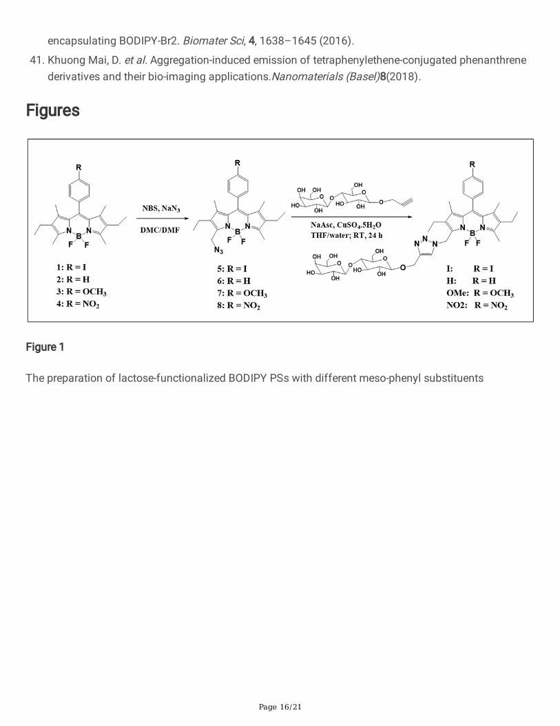
The process used to synthesize the four BODIPY PSs (I, H, OMe, and NO2) functionalized with lactose-tethering triazole is outlined in Figure 1. The detailed procedure is provided in the Supporting Informationsection.
Compounds 1a–d were synthesized via condensation reactions using 3-ethyl-2, 4-dimethyl pyrrole with 4-iodobenzoyl chloride and benzaldehyde derivatives, which yielded compounds 1a and 1b–d,respectively. Compounds 2a–d modi�ed with alkyl azide at the 3-methyl position of the BODIPY corewere obtained in the same manner29. Cu(I)-catalyzed alkyne–azide cycloaddition reactions (CuAAC) wereperformed with compounds 2a-d and propargyl lactoside to obtain the �nal compounds

Page 8/21
BODIPY I, H, OMe, and NO2, respectively. The �nal BODIPY PSs were fully characterized by 1H, 13C NMR,and HR-MS, as shown in Fig. S12-25.
A series of water-dispersible NPs, namely I-NPs, H-NPs, OMe-NPs, and NO2-NPs, were obtained from thecorresponding BODIPY PSs I, H, OMe, and NO2 in aqueous solution, respectively, after completeevaporation of tetrahydrofuran (THF).
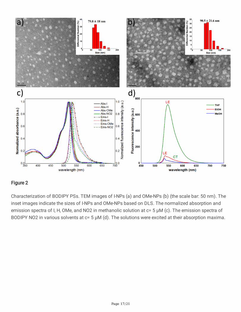
The hydrodynamic diameters of the obtained BODIPY NPs in aqueous solution were determined usingdynamic light scattering (DLS) measurements. As shown in Figure 2a,b and Figure S1, the averagediameters of the H, I, OMe, and NO2-NPs were approximately 71.3, 79.8, 90.5, and 100.7 nm,respectively. The morphology and size of BODIPY NPs were also examined by transmission electronmicroscopy (TEM), which revealed their spherical morphology and average diameter of approximately20–30 nm (Fig. 2a and 2b). Notably, the size of the fully hydrated BODIPY NPs as measured by DLSmight be larger than that determined in the dried state from TEM. Many water molecules appeared to beentrapped within the BODIPY NPs via interactions with hydrophilic lactose segments.
The optical characteristics of the BODIPY dyes were investigated using UV/Vis absorption and�uorescence spectrometry (Fig. 2c,d and Tab. 1). All of the synthesized BODIPY derivatives showedtypical spectra, with a robust S0 → S1 (π → π*) transition band around 518-524 nm, an absorption
coe�cient of 42400–49500 M-1·cm-1 from the boradiazaindacene chromophore,30,31 and a weak broadband around 350-400 nm corresponding to the S0 → S2 (π → π*) transition, which can be attributed to
the out-of-plane vibrations of the aromatic skeleton3,32. The iodine (-I) and methoxy (-OCH3)substituents incorporated into the meso-phenyl of BODIPY resulted in almost the same absorption andemission spectra as BODIPY H, indicating that these substituents have a negligible effect onthe photophysical properties of the BODIPY cores29. The BODIPY dyes with H, I,and OMe exhibited a high ΦF of approximately 45%–50% (Table 1), which is usually recorded for BODIPY
derivatives29. Of note, the values of ΦF remained high despite the BODIPY dyes being modi�ed with the
lactose-tethering triazole substituent17,33. However, the nitro substituent (-NO2) appeared to have a moresigni�cant in�uence on the photophysical properties of the BODIPY dye. The absorption spectrum ofcompound NO2 exhibited a red-shift of approximately 6 nm relative to that of H.
Table 1. The photophysical and photosensitizing properties of BODIPY I, H, OMe, and NO2 in MeOH

Page 9/21
I H OMe NO2
λab (nm)a 522 518 520 524
λem (nm)a 535 533 530 534
(103 M-1cm-1)a 45.2 42.4 44.3 49.5
ΦF (%)b 45.3 50.6 49.2 1.35
ΦΔc 10 3.7 7.3 0.9
(a) Absorption and �uorescence data measured using methanol. (b) Fluorescence quantum yields weredetermined using rhodamine 6G in methanol as the standard (Φf = 0.9435). (c) Singlet oxygen quantum
yields were determined by the DPBF bleaching method, using HP in ethanol as a reference (ΦΔ = 0.53)36.
As shown in Table S1, the emission intensities and ΦF of NO2 were strongly quenched in protic polarsolvents, exhibiting ΦF of 3.46% in ethanol and 1.35% in methanol compared with 38% in THF. Theintense emission observed in THF corresponds to the �uorescence from the local excited (LE) state of theBODIPY subunit. In contrast, �uorescence is strongly quenched in a polar methanol solution, owing to alow-emissivity CT state34. This phenomenon is expected because the PeT mechanism competes with�uorescence22,25,26. Details of the photophysical studies are presented in the theoretical calculationsection.
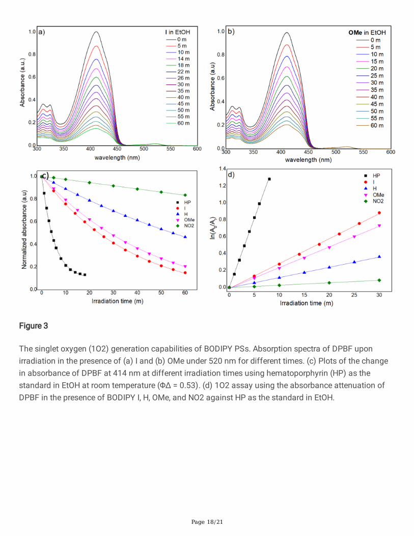
Singlet oxygen generation
The singlet oxygen (1O2) generation capabilities of I, H, OMe, and NO2 were assessed in air-saturated
ethanol under irradiation at 520 nm. A commercial 1O2 probe, 1,3-diphenylisobenzofuran (DPBF), was
used as an indicator, and hematoporphyrin (HP-ΦΔ= 0.53 in EtOH) was used as the reference37.
As shown in Figs. 3a,b,c and S3, the absorbance of DPBF at 414 nm decreased gradually in the presenceof the BODIPY dyes under continuous light irradiation. According to the linear relationship of the decaycurves (Fig. 3d), the 1O2 quantum yields (Φ△) of I, H, OMe, and NO2 were 10%, 3.7%, 7.3%, and 0.9%,
respectively (Table 1). Thus, the most robust 1O2 generation ability of I among the series ofPS suggested that the additional heavy iodine atom on the meso-phenyl of BODIPY induced spin-orbitperturbations on the molecules and signi�cantly in�uenced its superior capability to generate singletoxygen, as shown by the faster reducing rate of DPBF absorbance bands and a higher Φ△ than that ofthe other dyes. However, the incorporation of the electron-donating group (-OCH3) at the meso-phenyl of
BODIPY played an essential role in enhancing the 1O2 generation of OMe. Incontrast, the introduction of a strong electron-withdrawing group (-NO2) did not lead to an e�cient Φ△ ofBODIPY NO2 in a polar solvent (ethanolic solution). However, as shown in Figure S4a, Φ△ could be
Page 10/21
enhanced to 3% in a less polar solvent (THF). Such �nding indicates that the triplet state of BODIPYNO2 is strongly affected by the polarity of the media (the details are presented inthe theoretical calculation). These results collectively indicate that both iodinated- meso-phenyl BODIPY Iand heavy-free atom BODIPY OMe achieved an elevated 1O2 under LED illumination that facilitatedsinglet oxygen generation, demonstrating their potential use as e�cient photosensitizers for PDT.
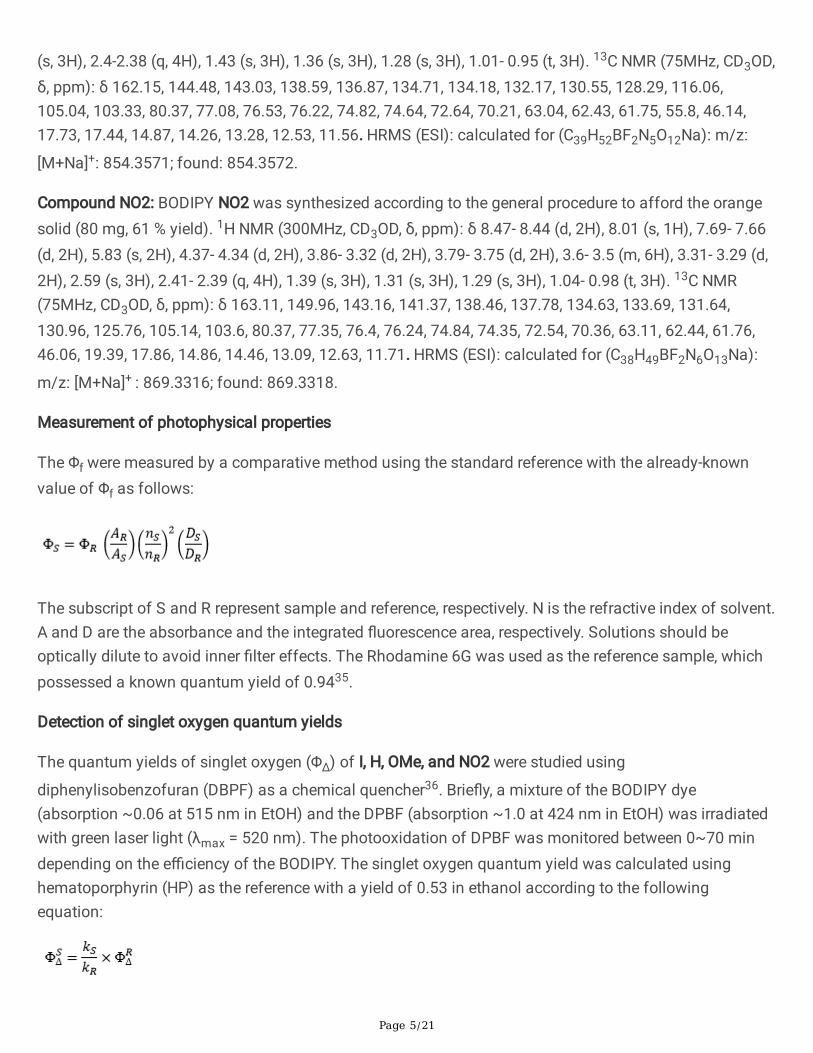
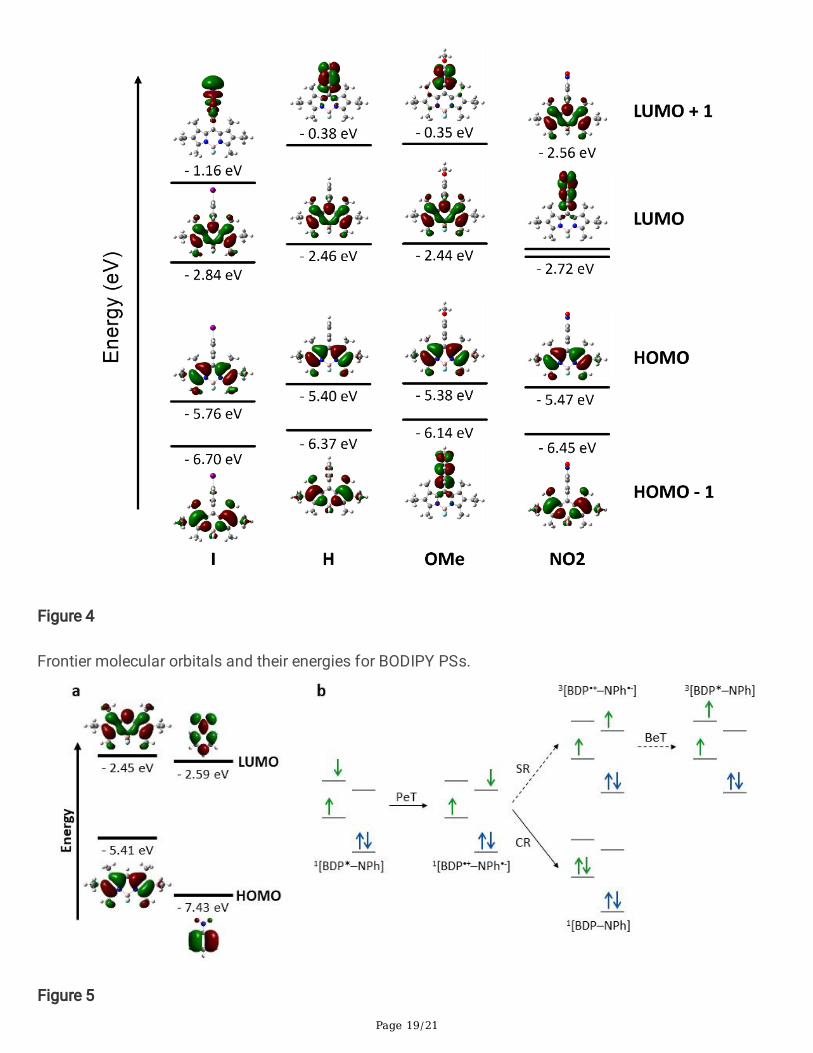
Theoretical characterization of the BODIPY derivatives
To determine the effect of meso-phenyl substituents on the electronic structure of BODIPY derivatives,density functional theory (DFT) calculations were performed (see Methods for computational details). ForBODIPY OMe, as depicted in Fig. 4, HOMO-1 is mainly located on methoxyphenyl (MPh). In contrast, theLUMO is fully localized on the BODIPY (BDP), implying that upon the generation of the singlet excitedstate by photoexcitation, PeT may occur, leading to the CT state, 1[BDP•-–MPh•+]. This process enhancesthe triplet state formation e�ciency, as demonstrated by the signi�cant increase in of OMe, comparedwith H. The CT state was approximately 0.2 eV higher in energy than the singlet excited state (Table S2).However, Filatov et al. showed that even if the energy of the CT state is greater than that of the singletexcited state, PeT and the subsequent triplet state formation can occur, with a propensity that thee�ciency of the processes reduces with increasing energy gap22. Hence, despite the reversal of the stateenergy, PeT is viewed as a valid mechanism for OMe, which facilitates the generation of the triplet stateand 1O2.
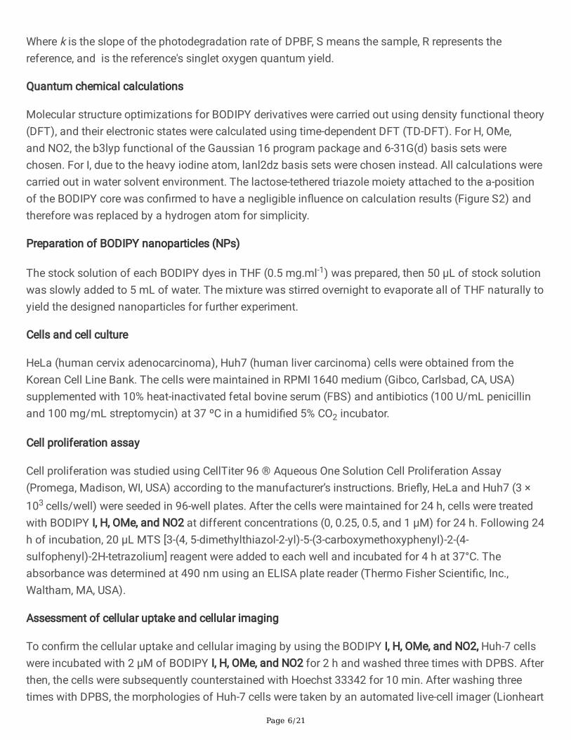
The electron-withdrawing nitro group existing in NO2 causes nitrophenyl (NPh) to act as an electronacceptor, while BODIPY becomes an electron donor, as demonstrated by the HOMO and LUMO localizedon BODIPY and nitrophenyl, respectively (Fig. 5). Therefore, in NO2, PeT may produce 1[BDP•+–NPh•-].Unlike OMe, this CT state lies between the singlet and triplet excited states (Table S2). The results of theDFT calculations, in conjunction with the solvent dependency of , clearly con�rm the existence of PeT inNO2; however, this does not lead to e�cient triplet state formation, as re�ected by the very low of thiscompound.
For donor-acceptor-type BODIPY dyads, comparing the frontier orbital energies of donor and acceptorunits is bene�cial for explaining electron movement during the PeT process34. This approach applies toNO2 because, in its optimized geometry, the two subunits are oriented almost perpendicular and can beconsidered as two independent functional groups. In this context, to elucidate the unexpected decouplingbetween PeT and the triplet state formation observed for NO2, the HOMO/LUMO energies of hexaalkyl-substituted BODIPY and nitrobenzene were calculated and compared (see Fig. 5a).
As illustrated in Fig. 5b, ISC from 1[BDP•+–NPh•-] to 3[BDP*–MPh] requires the back transfer of an electronfrom LUMONPh to LUMOBDP, following the conversion of its spin; this process is known as the radical pairISC (RP-ISC). However, DFT calculations revealed that E(LUMOBDP) > E(LUMONPh) (Fig. 5a),demonstrating that for NO2, back electron transfer (BeT) is an energetically unfavorable process. These
Page 11/21
�ndings agree with those of Zhao et al., who explored redox potentials for energetic comparison offrontier orbitals38. In addition, electron spin conversion, which must precede the above process, is oftenforbidden for directly linked donor-acceptor systems, such as NO2. Hence, 1[BDP•+–NPh•-] of NO2 is mostlikely to decay nonradiatively to the ground state. However, whether this dissipation occurs directly or viathe formation of 3[BDP–NPh*] remains unclear.
These results are consistent with those of Qi et al8. The electronic properties of the meso-substituent onthe BODIPY core, particularly the introduction of a suitable electron-donating group, could be �ne-tuned tocontrol the e�ciency of singlet oxygen formation. These results suggest that BODIPY OMe can be usedas a theranostic agent for cancer treatment.
Cellular uptake and cell-imaging of BODIPY NPs
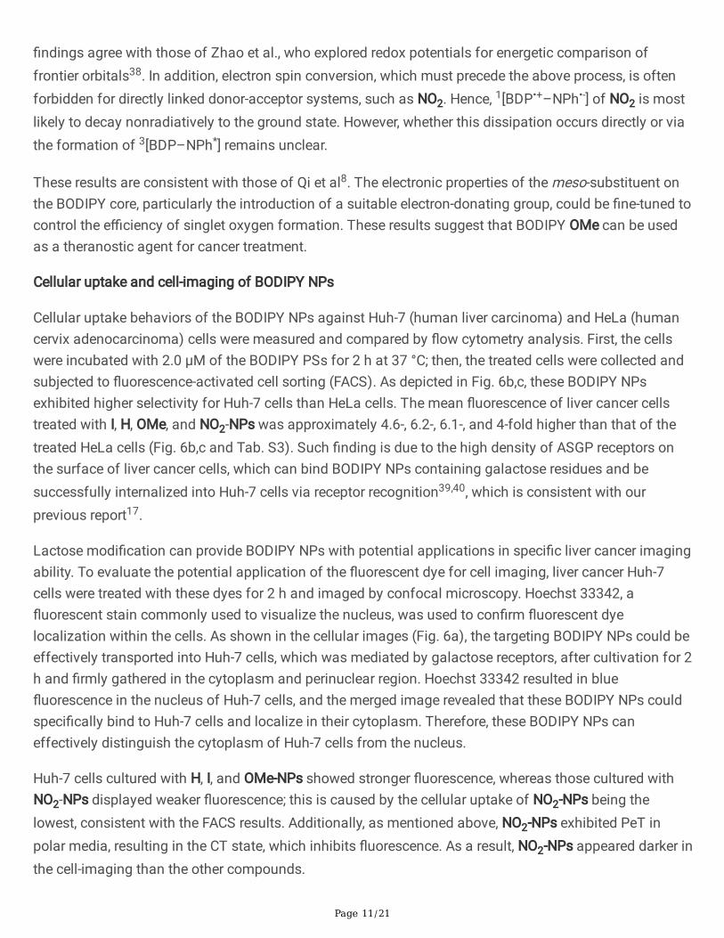
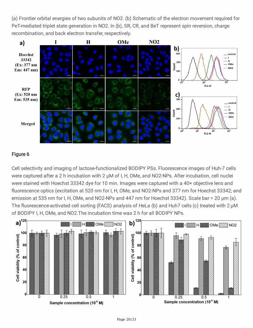
Cellular uptake behaviors of the BODIPY NPs against Huh-7 (human liver carcinoma) and HeLa (humancervix adenocarcinoma) cells were measured and compared by �ow cytometry analysis. First, the cellswere incubated with 2.0 μM of the BODIPY PSs for 2 h at 37 °C; then, the treated cells were collected andsubjected to �uorescence-activated cell sorting (FACS). As depicted in Fig. 6b,c, these BODIPY NPsexhibited higher selectivity for Huh-7 cells than HeLa cells. The mean �uorescence of liver cancer cellstreated with I, H, OMe, and NO2-NPs was approximately 4.6-, 6.2-, 6.1-, and 4-fold higher than that of thetreated HeLa cells (Fig. 6b,c and Tab. S3). Such �nding is due to the high density of ASGP receptors onthe surface of liver cancer cells, which can bind BODIPY NPs containing galactose residues and besuccessfully internalized into Huh-7 cells via receptor recognition39,40, which is consistent with ourprevious report17.
Lactose modi�cation can provide BODIPY NPs with potential applications in speci�c liver cancer imagingability. To evaluate the potential application of the �uorescent dye for cell imaging, liver cancer Huh-7cells were treated with these dyes for 2 h and imaged by confocal microscopy. Hoechst 33342, a�uorescent stain commonly used to visualize the nucleus, was used to con�rm �uorescent dyelocalization within the cells. As shown in the cellular images (Fig. 6a), the targeting BODIPY NPs could beeffectively transported into Huh-7 cells, which was mediated by galactose receptors, after cultivation for 2h and �rmly gathered in the cytoplasm and perinuclear region. Hoechst 33342 resulted in blue�uorescence in the nucleus of Huh-7 cells, and the merged image revealed that these BODIPY NPs couldspeci�cally bind to Huh-7 cells and localize in their cytoplasm. Therefore, these BODIPY NPs caneffectively distinguish the cytoplasm of Huh-7 cells from the nucleus.
Huh-7 cells cultured with H, I, and OMe-NPs showed stronger �uorescence, whereas those cultured withNO2-NPs displayed weaker �uorescence; this is caused by the cellular uptake of NO2-NPs being thelowest, consistent with the FACS results. Additionally, as mentioned above, NO2-NPs exhibited PeT inpolar media, resulting in the CT state, which inhibits �uorescence. As a result, NO2-NPs appeared darker inthe cell-imaging than the other compounds.
Page 12/21
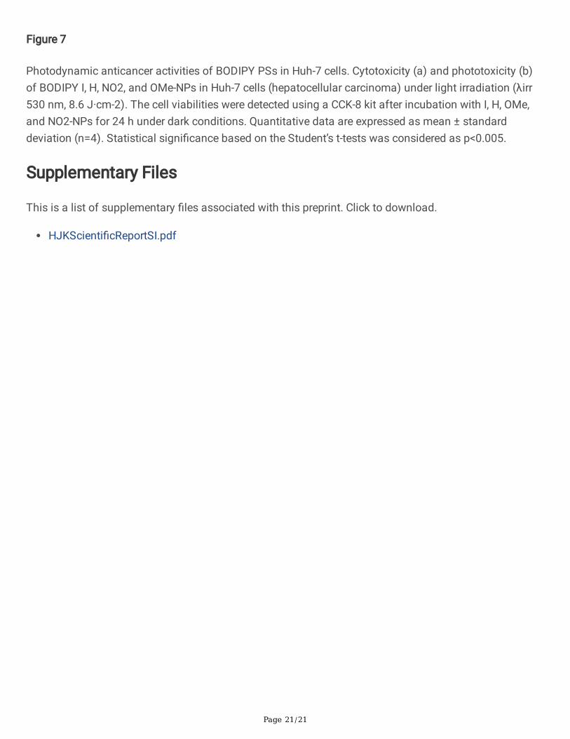
Light-induced cytotoxicity of BODIPY NPs against cancer cells
The biocompatibility of I, H, NO2, and OMe-NPs with Huh-7 and HeLa cells was determined using MTSassays. As shown in Figure 7a and Figure S5a, there was no cytotoxicity in any of the tested cells, andmore than 97% of both Huh-7 and HeLa cells survived after 24 h of incubation. Thus, all water-solubleBODIPY PSs below a dose of 1 µM had good biocompatibility and did not induce severe cytotoxicity in�broblasts and cancer cells, implying that BODIPY I, H, NO2, and OMe-NPs could be used in the lightcytotoxicity test.
For the e�cient production of ROS, HeLa and Huh-7 cells were exposed to 530 nm laser irradiation at anextremely low energy of 8.6 J·cm-2, and the in vitro phototoxicity of PDT was assessed. First, there wasno signi�cant variance when HeLa and Huh-7 cells were cultured with RPMI containing 1 μM of H andNO2-NPs, not only under dark conditions but also under irradiation; this is because of their low singletoxygen quantum yield, as mentioned above. When exposed to LED light, I and OMe-NPs resulted in highphototoxicity to tumor cells and negligible cell toxicity in the dark, as shown in Figure 7b and Figure S5b.Cell viability evidently decreased as the concentration of these BODIPY NPs increased. Furthermore, whenHeLa cells were exposed to light in the presence of 1 μM of OMe and I-NPs, cell viability decreased byapproximately 78% and 83%, respectively, while Huh-7 cells died by up to 89% and 97.7%, respectively,indicating their e�cient PDT targeting ability. Besides, the half-lethal dose (IC50) of OMe-NPs for HeLaand Huh-7 cell lines was 0.62 and 0.52 μM, respectively. Notably, when exposed to light, the lactose-tethered BODIPY OMe and I-NPs killed more Huh-7 cells than HeLa cells, with IC50 values of 0.51 and0.26 μM, respectively. These results demonstrate that BODIPY I and OMe-NPs provide better treatmente�cacy with relatively low irradiation intensity than H and NO2-NPs.
ConclusionHerein, we designed and synthesized a series of lactose-functionalized BODIPY PSs with differentsubstituent groups at the meso-position of the BODIPY core as cancer-targeted theragnostic agents forimaging and PDT. These BODIPY PSs could aggregate into nanoparticles (I-, H-, NO2-, and OMe-NPs) in anaqueous solution and displayed a uniform size and shape, as determined by TEM and DLS. The�uorescence quantum yields of BODIPY I, H, and OMe were remarkably high, whereas that of BODIPYNO2 was signi�cantly lower due to the PeT process in polar media. Among the four BODIPYphotosensitizers, BODIPY I demonstrated high e�ciency of singlet oxygen generation caused by theheavy atom effect due to the presence of an iodine atom, while BODIPY OMe containing an electron-donating methoxy group at the meso-phenyl moiety enhanced the ISC. In contrast, the withdrawingelectron (NO2) caused a marked reduction in both the �uorescence and singlet oxygen of NO2 due to PeTin the polar media. The cell experiments demonstrated that the water-soluble BODIPY NP series showedgood biocompatibility and cancer-speci�c �uorescence imaging ability. Notably, BODIPY OMe and I-NPspresented excellent phototoxicity against cancer cells, especially, liver cancer Huh-7 cells. The
Page 13/21
biocompatible BODIPY NPs have proven to be promising for developing highly e�cient theragnosticagents for imaging-guided PDT for cancer treatment.
DeclarationsAcknowledgements
This work was performed with �nancial support from the research funds provided by Chosun Universityin 2021.
Author contributions
D.K.M, H.J.K designed the protocol. D.K.M, I.W.B, and T.P.V carried out the experiments. J.L conducted cellexperiments. C.K studied computational modeling work. S.C, J.Y, and H.J.K supported and advised theexperiments. D.K.M, J.Y, and H.J.K wrote the manuscript. All authors discussed the results andcommented on the manuscript.
Competing Interests
The authors declare no competing �nancial interests.
References1. Allison, R. R. & Sibata, C. H. Oncologic photodynamic therapy photosensitizers: a clinical review.
Photodiagnosis Photodyn Ther, 7, 61–75 (2010).
2. Kamkaew, A. et al. BODIPY dyes in photodynamic therapy. Chem Soc Rev, 42, 77–88 (2013).
3. Awuah, S. G. & You, Y. Boron dipyrromethene (BODIPY)-based photosensitizers for photodynamictherapy.RSC Advances2(2012).
4. Kue, C. S. et al. Recent strategies to improve boron dipyrromethene (BODIPY) for photodynamiccancer therapy: an updated review. Photochem Photobiol Sci, 17, 1691–1708 (2018).
5. Turksoy, A., Yildiz, D. & Akkaya, E. U. Photosensitization and controlled photosensitization withBODIPY dyes. Coord. Chem. Rev, 379, 47–64 (2019).
�. Yuan, P. et al. Oxygen self-su�cient �uorinated polypeptide nanoparticles for NIR imaging-guidedenhanced photodynamic therapy. J Mater Chem B, 6, 2323–2331 (2018).
7. Liu, L., Fu, L., Jing, T., Ruan, Z. & Yan, L. pH-Triggered polypeptides nanoparticles for e�cient BODIPYimaging-guided near infrared photodynamic therapy. ACS Appl Mater Interfaces, 8, 8980–8990(2016).
�. Qi, S., Kwon, N., Yim, Y., Nguyen, V. N. & Yoon, J. Fine-tuning the electronic structure of heavy-atom-free BODIPY photosensitizers for �uorescence imaging and mitochondria-targeted photodynamictherapy. Chem Sci, 11, 6479–6484 (2020).
Page 14/21
9. Bui, H. T. et al. Effect of substituents on the photophysical properties and bioimaging application ofBODIPY rerivatives with triphenylamine substituents. J Phys Chem B, 123, 5601–5607 (2019).
10. Guo, Z. et al. Bifunctional platinated nanoparticles for photoinduced tumor ablation. Adv Mater, 28,10155–10164 (2016).
11. Takatoshi Yogo, Y. U., Ishitsuka, Y., Maniwa, F. & Nagano, T. Highly e�cient and photostablephotosensitizer based on BODIPY chromophore. J. Am. Chem. Soc. 2005, 127, 12162–12163 127,12162–12163 (2005).
12. Mark Wainwright, D. A. P., Lesley Rice, S. M. & Burrow Jack Waring. Increased cytotoxicity andphototoxicity in the methylene blue series via chromophore methylation. Journal of Photochemistryand Photobiology B: Biology, 40, 233–239 (1997).
13. Plaetzer, K., Krammer, B., Berlanda, J., Berr, F. & Kiesslich, T. Photophysics and photochemistry ofphotodynamic therapy: fundamental aspects. Lasers Med Sci, 24, 259–268 (2009).
14. Kim, B. et al. In vitro photodynamic studies of a BODIPY-based photosensitizer. European Journal ofOrganic Chemistry, 2017, 25–28 (2016).
15. Zou, J. et al. BODIPY derivatives for photodynamic therapy: in�uence of con�guration versus heavyatom effect. ACS Appl Mater Interfaces, 9, 32475–32481 (2017).
1�. Wang, Z. et al. BODIPY-doped silica nanoparticles with reduced dye leakage and enhanced singletoxygen generation. Sci Rep, 5, 12602 (2015).
17. Khuong Mai, D. et al. Synthesis and photophysical properties of tumor-targeted water-solubleBODIPY photosensitizers for photodynamic therapy.Molecules25(2020).
1�. Aoife Gorman, J. K., Caroline, O. S., Kenna, T., Gallagher, W. M. & Donal, F. O’Shea. In vitrodemonstration of the heavy-atom effect for photodynamic therapy. J. Am. Chem. Soc. 2004, 126,10619–10631 126, 10619–10631.
19. Lu, S. et al. PEGylated dimeric BODIPY photosensitizers as nanocarriers for combined chemotherapyand cathepsin B-activated photodynamic therapy in 3D tumor spheroids. ACS Applied Bio Materials,3, 3835–3845 (2020).
20. Chen, H., Bi, Q., Yao, Y. & Tan, N. Dimeric BODIPY-loaded liposomes for dual hypoxia marker imagingand activatable photodynamic therapy against tumors. J Mater Chem B, 6, 4351–4359 (2018).
21. Ucuncu, M. et al. BODIPY-Au(I): a photosensitizer for singlet oxygen generation and photodynamictherapy. Org Lett, 19, 2522–2525 (2017).
22. Filatov, M. A. et al. Control of triplet state generation in heavy atom-free BODIPY-anthracene dyads bymedia polarity and structural factors. Phys Chem Chem Phys, 20, 8016–8031 (2018).
23. Callaghan, S., Filatov, M. A., Savoie, H., Boyle, R. W. & Senge, M. O. In vitro cytotoxicity of a library ofBODIPY-anthracene and -pyrene dyads for application in photodynamic therapy. PhotochemPhotobiol Sci, 18, 495–504 (2019).
24. Filatov, M. A. Heavy-atom-free BODIPY photosensitizers with intersystem crossing mediated byintramolecular photoinduced electron transfer. Org Biomol Chem, 18, 10–27 (2019).

Page 15/21
25. Li, L., Nguyen, J. H. B. & Burgess, K. Syntheses and spectral properties of functionalized, water-soluble BODIPY derivatives. J. Org. Chem. 2008, 73, 1963– 1970 73, 1963–1970 (2008).
2�. Dorh, N. et al. BODIPY-based �uorescent probes for sensing protein surface-hydrophobicity. Sci Rep,5, 18337 (2015).
27. Hisato Sunahara, Y. U., Kojima, H. & Nagano, T. Design and synthesis of a library of BODIPY-basedenvironmental polarity sensors utilizing photoinduced electron-transfer-controlled �uorescenceON/OFF switching. J. Am. Chem. Soc. 2007, 129, 5597–5604 129, 5597–5604 (2007).
2�. Kim, T. H., Park, I. K., Nah, J. W., Choi, Y. J. & Cho, C. S. Galactosylated chitosan/DNA nanoparticlesprepared using water-soluble chitosan as a gene carrier., 25, 3783–3792 (2004).
29. Ulrich, G., Ziessel, R. & Haefele, A. A general synthetic route to 3,5-substituted boron dipyrromethenes:applications and properties. J Org Chem, 77, 4298–4311 (2012).
30. Shilei Zhu, J. Z., Vegesna, G., Luo, F. T., Green, S. A. & Liu, H. Highly water-soluble neutral BODIPYdyes with controllable �uorescence quantum yields. Org. Lett, 13, 438–441 (2011).
31. Vu, T. T. et al. Understanding the spectroscopic properties and aggregation process of a new emittingboron dipyrromethene (BODIPY). The Journal of Physical Chemistry C, 117, 5373–5385 (2013).
32. Sun, H. et al. Excellent BODIPY dye containing dimesitylboryl groups as PeT-based �uorescentprobes for �uoride. The Journal of Physical Chemistry C, 115, 19947–19954 (2011).
33. Papalia, T. et al. Cell internalization of BODIPY-based �uorescent dyes bearing carbohydrateresidues. Dyes and Pigments, 110, 67–71 (2014).
34. Filatov, M. A. et al. Generation of triplet excited states via photoinduced electron transfer in meso-anthra-BODIPY: �uorogenic response toward singlet oxygen in solution and in vitro. J Am Chem Soc,139, 6282–6285 (2017).
35. Douglas & Magde R.W.a.P.G.S. Fluorescence quantum yields and their relation to lifetimes ofrhodamine 6G and �uorescein in nine solvents: improved absolute standards for quantum yields.Photochemistry and Photobiology, 2002, 75(4): 327–334 75, 327–334 (2002).
3�. Wolfgang Spiller, H. K., Dieter Wo¨hrle, S. & Hackbarth Beater Ro¨der and Gu¨nter Schnurpfeil. Singletoxygen quantum yields of different photosensitizers in polar solvents and micellar solutions. Journalof Porphyrins and Phthalocyanines, Vol. 2, 145–158 (1998) 2, 145–158 (1998).
37. Liane, M. et al. Baptista. Protoporphyrin IX nanoparticle carrier: preparation, optical properties, andsinglet oxygen generation. Langmuir 2008, 24, 12534–12538 24, 12534–12538 (2008).
3�. Hu, W. et al. Can BODIPY-electron acceptor conjugates act as heavy atom-free excited triplet stateand singlet oxygen photosensitizers via photoinduced charge separation-charge recombinationmechanism? The Journal of Physical Chemistry C, 123, 15944–15955 (2019).
39. Lu, Z. et al. Water-soluble BODIPY-conjugated glycopolymers as �uorescent probes for live cellimaging.Polymer Chemistry4(2013).
40. Liu, L., Ruan, Z., Li, T., Yuan, P. & Yan, L. Near infrared imaging-guided photodynamic therapy underan extremely low energy of light by galactose targeted amphiphilic polypeptide micelle
Page 16/21
encapsulating BODIPY-Br2. Biomater Sci, 4, 1638–1645 (2016).
41. Khuong Mai, D. et al. Aggregation-induced emission of tetraphenylethene-conjugated phenanthrenederivatives and their bio-imaging applications.Nanomaterials (Basel)8(2018).
Figures
Figure 1
The preparation of lactose-functionalized BODIPY PSs with different meso-phenyl substituents
Page 17/21
Figure 2
Characterization of BODIPY PSs. TEM images of I-NPs (a) and OMe-NPs (b) (the scale bar: 50 nm). Theinset images indicate the sizes of I-NPs and OMe-NPs based on DLS. The normalized absorption andemission spectra of I, H, OMe, and NO2 in methanolic solution at c= 5 µM (c). The emission spectra ofBODIPY NO2 in various solvents at c= 5 µM (d). The solutions were excited at their absorption maxima.
Page 18/21
Figure 3
The singlet oxygen (1O2) generation capabilities of BODIPY PSs. Absorption spectra of DPBF uponirradiation in the presence of (a) I and (b) OMe under 520 nm for different times. (c) Plots of the changein absorbance of DPBF at 414 nm at different irradiation times using hematoporphyrin (HP) as thestandard in EtOH at room temperature (ΦΔ = 0.53). (d) 1O2 assay using the absorbance attenuation ofDPBF in the presence of BODIPY I, H, OMe, and NO2 against HP as the standard in EtOH.
Page 19/21
Figure 4
Frontier molecular orbitals and their energies for BODIPY PSs.
Figure 5
Page 20/21
(a) Frontier orbital energies of two subunits of NO2. (b) Schematic of the electron movement required forPeT-mediated triplet state generation in NO2. In (b), SR, CR, and BeT represent spin reversion, chargerecombination, and back electron transfer, respectively.
Figure 6
Cell selectivity and imaging of lactose-functionalized BODIPY PSs. Fluorescence images of Huh-7 cellswere captured after a 2 h incubation with 2 µM of I, H, OMe, and NO2-NPs. After incubation, cell nucleiwere stained with Hoechst 33342 dye for 10 min. Images were captured with a 40× objective lens and�uorescence optics (excitation at 520 nm for I, H, OMe, and NO2-NPs and 377 nm for Hoechst 33342; andemission at 535 nm for I, H, OMe, and NO2-NPs and 447 nm for Hoechst 33342). Scale bar = 20 μm (a).The �uorescence-activated cell sorting (FACS) analysis of HeLa (b) and Huh7 cells (c) treated with 2 μMof BODIPY I, H, OMe, and NO2.The incubation time was 2 h for all BODIPY NPs.
Page 21/21
Figure 7
Photodynamic anticancer activities of BODIPY PSs in Huh-7 cells. Cytotoxicity (a) and phototoxicity (b)of BODIPY I, H, NO2, and OMe-NPs in Huh-7 cells (hepatocellular carcinoma) under light irradiation (λirr530 nm, 8.6 J·cm-2). The cell viabilities were detected using a CCK-8 kit after incubation with I, H, OMe,and NO2-NPs for 24 h under dark conditions. Quantitative data are expressed as mean ± standarddeviation (n=4). Statistical signi�cance based on the Student’s t-tests was considered as p<0.005.
Supplementary Files
This is a list of supplementary �les associated with this preprint. Click to download.
HJKScienti�cReportSI.pdf