leptin receptor is involved in stat3 activation in human colorectal adenoma
TRANSCRIPT

Leptin receptor is involved in STAT3 activation inhuman colorectal adenomaTakashi Uchiyama,1 Hirokazu Takahashi,1 Michiko Sugiyama,1 Eiji Sakai,1 Hiroki Endo,1 Kunihiro Hosono,1
Kyoko Yoneda,1 Masato Yoneda,1 Masahiko Inamori,1 Yoji Nagashima,2 Yoshiaki Inayama,2 Koichiro Wada3 andAtsushi Nakajima1,4
1Gastroenterology Division, 2Pathology Division, School of Medicine, Yokohama City University, Yokohama, Kanagawa; 3Department of Pharmacology,School of Dentistry, Osaka University, Osaka, Japan
(Received August 31, 2010 ⁄ Revised November 7, 2010 ⁄ Accepted November 14, 2010 ⁄ Accepted manuscript online November 19, 2010 ⁄ Article first published online December 19, 2010)
4To whom correspondence should be addressed.E-mail: [email protected]
The possible role of leptin in colorectal tumors has been investi-gated in previous studies; however, to date, the conclusionsremain under debate. Therefore, we investigated the serum leptinlevels in colorectal adenoma patients. In addition, expression ofthe leptin receptor, and the leptin receptor-mediated signalingpathways were investigated in biopsy specimens collected fromhuman patients with colorectal adenoma. No significant differencein the mean serum leptin level was observed between the colorec-tal adenoma patients and the control subjects; however, increasedexpression and activation of the leptin receptor, as indicated byfindings such as the phosphorylation of Tyr 1141, was observed inthe colorectal adenoma tissues. In addition, activation of the JAK/STAT signaling pathway mediated by the leptin receptor andincreased transcriptional regulation of downstream target mole-cules were observed in colorectal adenomas compared with thenon-adenoma tissues. These results indicate STAT3-mediatedleptin receptor signaling pathways may be activated in humancolorectal adenomas. (Cancer Sci 2011; 102: 367–372)
C olorectal cancer is a major cause of mortality and morbid-ity worldwide;(1) however, the mechanism of colorectal
carcinogenesis remains unclear. Recently, the existence of anassociation between obesity or metabolic abnormalities and anelevated risk of colorectal cancer was reported.(2,3) Adipose tis-sue was reported to be not only an energy storage organ, but alsoan active endocrine organ that secretes important adipocytokinessuch as adiponectin, leptin, tumor necrosis factor-a (TNF-a),free fatty acid and resistin.(4,5)
Leptin is a 167-amino acid peptide that plays a central role inthe hypothalamus in relation to mammalian feeding behaviorand energy expenditure.(6) Plasma leptin levels have beenreported to be strongly correlated with the body mass index(BMI) in humans(7–9) and also to be elevated in obese subjects.Leptin exerts its activity thorough its specific membrane recep-tor, the leptin receptor (ObR), belonging to the class 1 cytokinereceptor family.(5) Two isoforms, the long and short variants ofObR, namely, ObRL and ObRS, have been identified, and onlythe long isoform of ObR has been shown to have full signalingpotential, with the short isoform showing diminished or abol-ished capacity for signaling.(5,10)
Several studies have reported the association between serumleptin levels and the presence of several cancers such as pros-tate(11–13) and breast(14,15) cancer. Similarly, previous studieshave also shown an association between serum leptin levels andthe presence of colorectal cancer.(16–23) However, the results ofthese previous studies are contradictory and difficult to interpret.While some studies have shown a decrease in the serum leptinlevels in colorectal cancer patients,(16–20) others have reportedelevated serum leptin levels in colorectal cancer patients.(21–23)
Thus, the association between leptin and the presence of
doi: 10.1111/j.1349-7006.2010.01803.xªª 2010 Japanese Cancer Association
colorectal cancer has not yet been clarified. In addition, previousstudies have shown that leptin stimulated cell proliferation inseveral types of carcinoma cell lines in vitro.(24–26) However,the molecular mechanisms underlying the promotion of humancolorectal carcinogenesis by leptin remain unclear.
In the present study we investigated the association betweenplasma leptin levels ⁄ leptin receptor-mediated signaling and thedevelopment of colorectal adenoma.
Materials and Methods
Study population. One hundred and forty-four patients whounderwent endoscopic mucosal resection for colorectal adenomabetween June 2006 and April 2009 at Yokohama City Univer-sity Hospital, and 64 control subjects who were detected to haveno colorectal polyps on colonoscopy were recruited for thisstudy. The exclusion criteria were subjects with colorectal carci-noma, familial adenomatous polyposis, inflammatory bowel dis-ease, radiation colitis or any malignant disease, and alsosubjects with a previous history of colectomy, gastrectomy orcolorectal polypectomy. Written informed consent was obtainedfrom all subjects prior to their participation in the study. Thestudy protocol was approved by the Yokohama City UniversityHospital Ethics Committee.
Collection and analysis of blood samples for determination ofthe leptin levels. Blood samples were obtained in the morningon the day of colonoscopy after the subjects had fasted over-night. Serum leptin levels were measured by enzyme-linkedimmunosorbent assay of human leptin (SRL Co., Tokyo, Japan).
Immunohistochemical analyses. The expressions of ObR andphospho-STAT3 (p-STAT3) were investigated in the colorectaladenoma and normal colorectal tissues. A total of 61 adenomatissue samples were obtained endoscopically from the study sub-jects. Formalin-fixed and paraffin-embedded samples were depa-raffinized and rehydrated. The sections were incubated withantibodies for ObR (1:50; Santa Cruz Biotechnology, SantaCruz, CA, USA) and p-STAT3 (Tyr 705) (1:50; Cell SignalingTechnology, Danvers, MA, USA) as the primary antibodies,using an LSAB2 kit (Dako Cytomation, Carpinteria, CA, USA).They were then incubated with biotinylated immunoglobulin asthe secondary antibody and treated with peroxidase-conjugatedstreptavidin. The antibody complex was visualized with 3,3¢-diaminobenzidine, tetrahydrochloride (Dojindo Laboratories,Kumamoto, Japan). The expressions of ObR and p-STAT3 wereanalyzed by light microscopy in 10 different fields of each sec-tion, and the mean percentage of adenoma cells that showedpositive staining was scored by two pathologists. The ObR andp-STAT3 expressions were classified into two categoriesdepending on the percentage of cells showing positive staining:
Cancer Sci | February 2011 | vol. 102 | no. 2 | 367–372
negative, 0–15% of all the tumor cells showing positivestaining; and positive, >15% of all tumor cells showing positivestaining, as previously described.(27)
Western blot analysis. Twenty-five colorectal adenomapatients were randomly selected, and biopsy samplesobtained from the colorectal adenomas and normal areaswere isolated. The extracted protein was separated usingsodium dodecylsulfate polyacrylamide gel electrophoresis(SDS-PAGE) and the separated proteins were transferred to apolyvinylidene difluoride (PVDF) membrane (Amersham,London, UK). The membranes were probed with primaryantibodies specific for phospho-ObR (p-ObR) (Tyr 1141), p-ObR (Tyr 985), ObR (Santa Cruz Biotechnology), phospho-JAK2 (p-JAK2), JAK2, p-STAT3 (Tyr 705) STAT3 (CellSignaling Technology) and glyceraldehyde-3-phosphatedehydrogenase (GAPDH) (Trevigen, Gaithersburg, MD,USA). Horseradish-peroxidase-conjugated secondary antibod-ies and the electrochemi-luminescence (ECL) detection kit(Amersham) were used for the detection of specific proteins.
Real-time RT-PCR. Twelve colorectal adenoma patients wererandomly selected and biopsy samples of the adenoma and adja-cent normal tissues obtained from the colorectal adenoma andnormal areas were isolated. Total RNA from the colorectal ade-
Table 1. Characteristics of the study patients
Normal Adenoma P value
N 64 144
Age (years) 62.1 ± 13.8 64.7 ± 10.2 0.12
Sex (M ⁄ F) 33 ⁄ 31 100 ⁄ 44 0.18
Waist
circumference (cm)
84.4 ± 10.3 86.5 ± 10.5 0.28
BMI (kg ⁄ m2) 22.7 ± 3.5 23.3 ± 3.2 0.20
VFA (cm2) 75.7 ± 50.8 93.5 ± 53.5 0.08
FBS (mg ⁄ dL) 112.6 ± 26.9 108.7 ± 31.1 0.44
HbA1c (%) 5.7 ± 1.2 5.6 ± 1.0 0.43
Leptin (ng ⁄ mL) 5.6 ± 4.3 5.4 ± 4.2 0.70
Data are shown as mean ± standard deviation. Statistical analysis wasperformed using the Mann–Whitney U-test. *P < 0.05. **P < 0.01.BMI, body mass index; VFA, visceral fat area; FBS, fasting blood sugar.
(a) (b)
(c)
368
noma and normal tissue biopsy specimens was extracted usingthe RNeasy mini kit (Qiagen, Hilden, Germany). For the real-time reverse-transcriptase polymerase chain reaction, total RNAwas reverse-transcribed into cDNA and amplified using the real-time quantitative polymerase chain reaction using the Step OnePlus Real Time PCR System (Applied Biosystems, Foster City,CA, USA). Probes and primer pairs specific for ObRL, ObRS,BclX, c-Myc, cyclin D1, cdc2, cyclin B1, VEGF and 18S werepurchased from Applied Biosystems. The concentrations of thetarget genes were determined using the competitive computedtomography method and the values were normalized to the inter-nal control.
Statistical analysis. Statistical analyses were performed usingthe Mann–Whitney U-test and chi-square test. All analyses wereperformed using the Stat View software (SAS Institute, Cary,NC, USA). P < 0.05 was regarded as denoting statistical signifi-cance.
Results
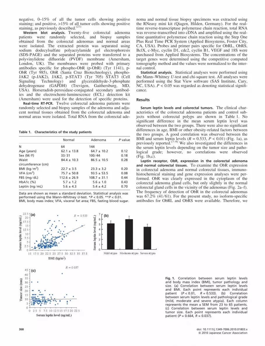
Serum leptin levels and colorectal tumors. The clinical char-acteristics of the colorectal adenoma patients and control sub-jects without colorectal polyps are shown in Table 1. Nosignificant difference in the mean serum leptin level wasobserved between the two groups. There were also no significantdifferences in age, BMI or other obesity-related factors betweenthe two groups. A good correlation was observed between theBMI and serum leptin levels (R = 0.533, P < 0.01) (Fig. 1a), aspreviously reported.(7–9) We also investigated the differences inthe serum leptin levels depending on the tumor size and patho-logical grade; however, no correlations were observed(Fig. 1b,c).
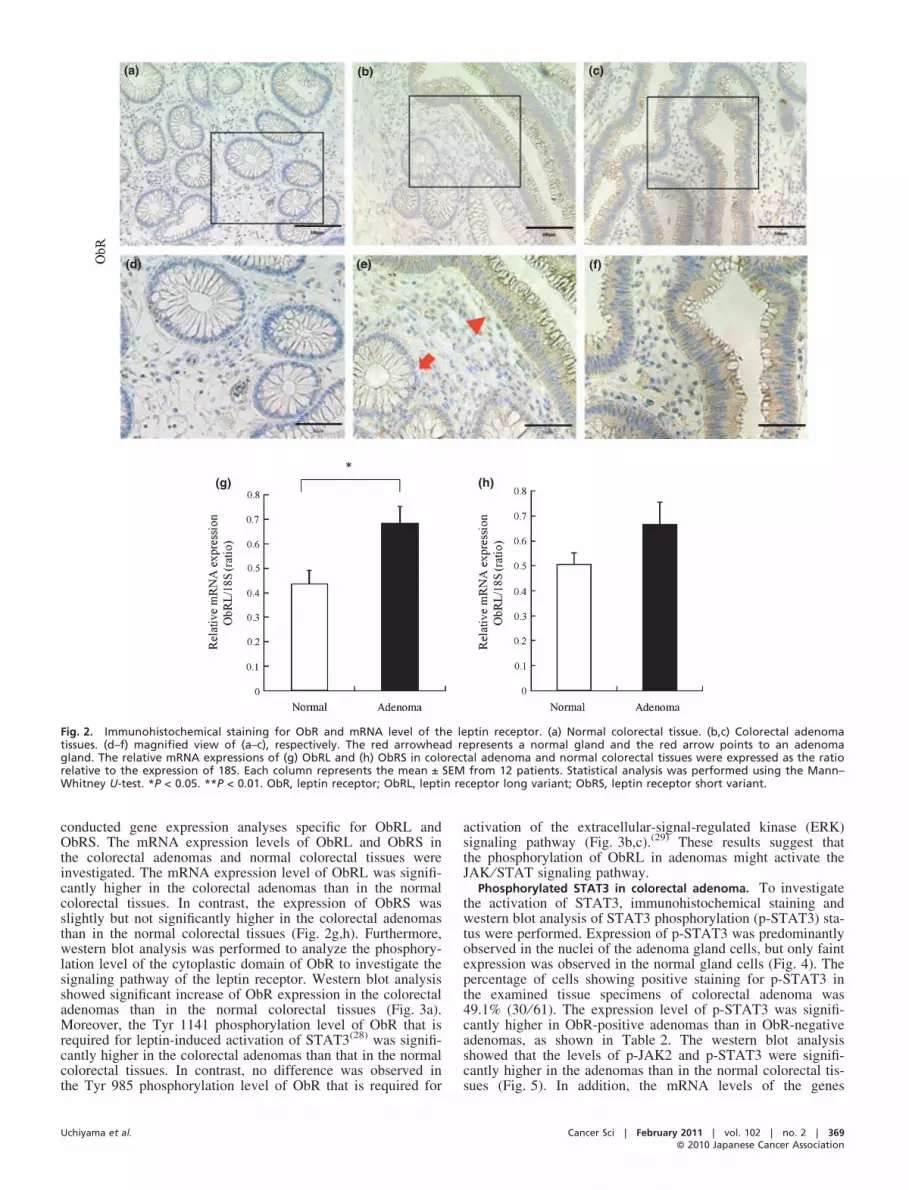
Leptin receptor, ObR, expression in the colorectal adenomaand normal colorectal tissues. To examine the ObR expressionin colorectal adenoma and normal colorectal tissues, immuno-histochemical staining and gene expression analyses were per-formed. ObR was clearly expressed in the cytoplasm of thecolorectal adenoma gland cells, but only slightly in the normalcolorectal gland cells in the vicinity of the adenomas (Fig. 2a–f).The frequency of detection of ObR in the colorectal adenomaswas 67.2% (41 ⁄ 61). For the present study, no isoform-specificantibodies for ObRL and ObRS were available. Therefore, we
Fig. 1. Correlation between serum leptin levelsand body mass index (BMI), tumor pathology andsize. (a) Correlation between serum leptin levelsand BMI. Each point represents each individualpatient (P < 0.01, R = 0.533). (b) Correlationbetween serum leptin levels and pathological grade(mild, moderate and severe atypia). Each columnrepresents the mean ± SEM from 23 to 65 patients.(c) Correlation between serum leptin levels andtumor size. Each point represents each individualpatient (P = 0.664, R = 0.037).
doi: 10.1111/j.1349-7006.2010.01803.xªª 2010 Japanese Cancer Association
(a) (b) (c)
(f)(e)(d)
(h)(g)
Fig. 2. Immunohistochemical staining for ObR and mRNA level of the leptin receptor. (a) Normal colorectal tissue. (b,c) Colorectal adenomatissues. (d–f) magnified view of (a–c), respectively. The red arrowhead represents a normal gland and the red arrow points to an adenomagland. The relative mRNA expressions of (g) ObRL and (h) ObRS in colorectal adenoma and normal colorectal tissues were expressed as the ratiorelative to the expression of 18S. Each column represents the mean ± SEM from 12 patients. Statistical analysis was performed using the Mann–Whitney U-test. *P < 0.05. **P < 0.01. ObR, leptin receptor; ObRL, leptin receptor long variant; ObRS, leptin receptor short variant.
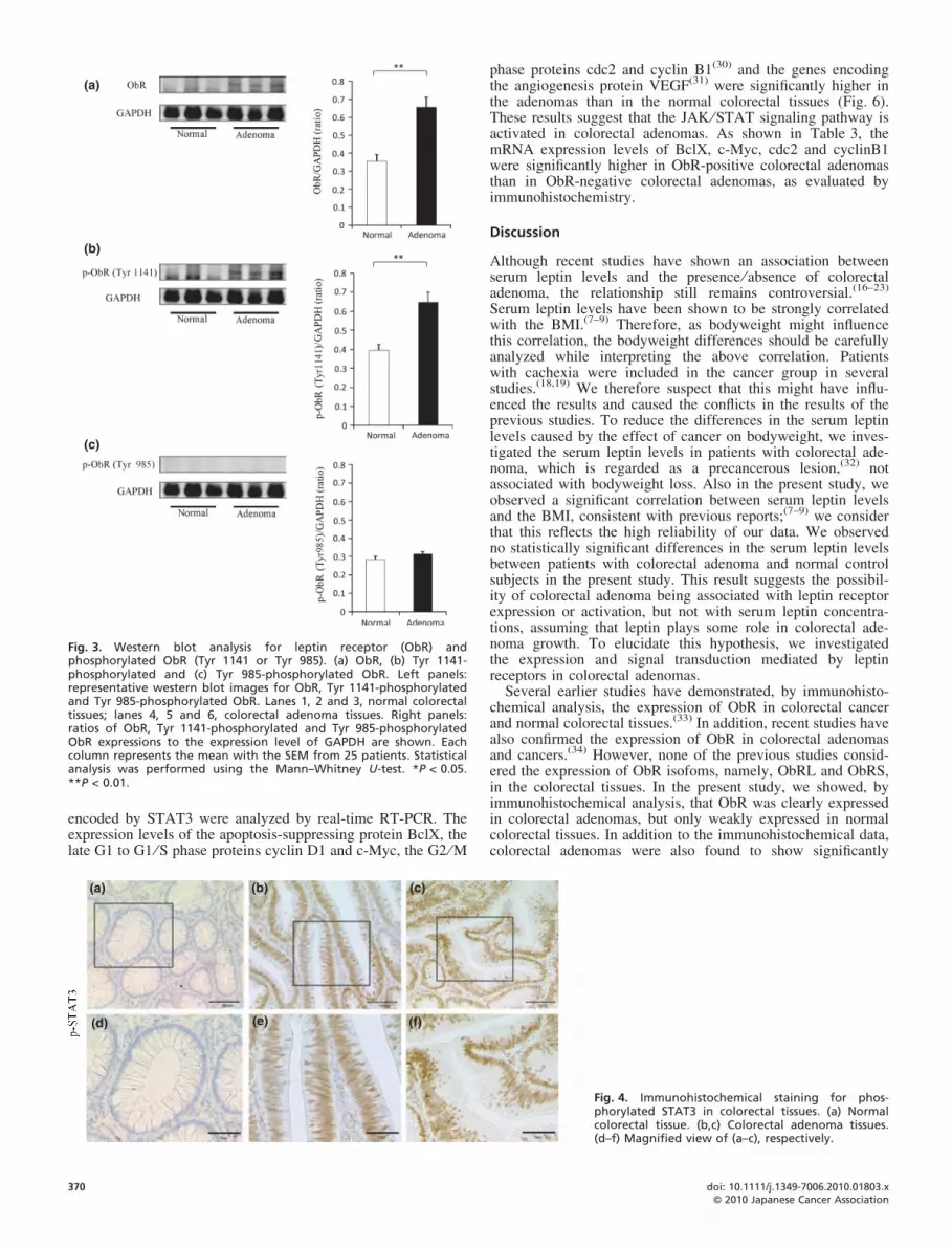
conducted gene expression analyses specific for ObRL andObRS. The mRNA expression levels of ObRL and ObRS inthe colorectal adenomas and normal colorectal tissues wereinvestigated. The mRNA expression level of ObRL was signifi-cantly higher in the colorectal adenomas than in the normalcolorectal tissues. In contrast, the expression of ObRS wasslightly but not significantly higher in the colorectal adenomasthan in the normal colorectal tissues (Fig. 2g,h). Furthermore,western blot analysis was performed to analyze the phosphory-lation level of the cytoplastic domain of ObR to investigate thesignaling pathway of the leptin receptor. Western blot analysisshowed significant increase of ObR expression in the colorectaladenomas than in the normal colorectal tissues (Fig. 3a).Moreover, the Tyr 1141 phosphorylation level of ObR that isrequired for leptin-induced activation of STAT3(28) was signifi-cantly higher in the colorectal adenomas than that in the normalcolorectal tissues. In contrast, no difference was observed inthe Tyr 985 phosphorylation level of ObR that is required for
Uchiyama et al.
activation of the extracellular-signal-regulated kinase (ERK)signaling pathway (Fig. 3b,c).(29) These results suggest thatthe phosphorylation of ObRL in adenomas might activate theJAK ⁄ STAT signaling pathway.
Phosphorylated STAT3 in colorectal adenoma. To investigatethe activation of STAT3, immunohistochemical staining andwestern blot analysis of STAT3 phosphorylation (p-STAT3) sta-tus were performed. Expression of p-STAT3 was predominantlyobserved in the nuclei of the adenoma gland cells, but only faintexpression was observed in the normal gland cells (Fig. 4). Thepercentage of cells showing positive staining for p-STAT3 inthe examined tissue specimens of colorectal adenoma was49.1% (30 ⁄ 61). The expression level of p-STAT3 was signifi-cantly higher in ObR-positive adenomas than in ObR-negativeadenomas, as shown in Table 2. The western blot analysisshowed that the levels of p-JAK2 and p-STAT3 were signifi-cantly higher in the adenomas than in the normal colorectal tis-sues (Fig. 5). In addition, the mRNA levels of the genes
Cancer Sci | February 2011 | vol. 102 | no. 2 | 369ªª 2010 Japanese Cancer Association
(a)
(b)
(c)
Fig. 3. Western blot analysis for leptin receptor (ObR) andphosphorylated ObR (Tyr 1141 or Tyr 985). (a) ObR, (b) Tyr 1141-phosphorylated and (c) Tyr 985-phosphorylated ObR. Left panels:representative western blot images for ObR, Tyr 1141-phosphorylatedand Tyr 985-phosphorylated ObR. Lanes 1, 2 and 3, normal colorectaltissues; lanes 4, 5 and 6, colorectal adenoma tissues. Right panels:ratios of ObR, Tyr 1141-phosphorylated and Tyr 985-phosphorylatedObR expressions to the expression level of GAPDH are shown. Eachcolumn represents the mean with the SEM from 25 patients. Statisticalanalysis was performed using the Mann–Whitney U-test. *P < 0.05.**P < 0.01.
encoded by STAT3 were analyzed by real-time RT-PCR. Theexpression levels of the apoptosis-suppressing protein BclX, thelate G1 to G1 ⁄ S phase proteins cyclin D1 and c-Myc, the G2 ⁄ M
(a) (b) (c)
(f)(e)(d)
370
phase proteins cdc2 and cyclin B1(30) and the genes encodingthe angiogenesis protein VEGF(31) were significantly higher inthe adenomas than in the normal colorectal tissues (Fig. 6).These results suggest that the JAK ⁄ STAT signaling pathway isactivated in colorectal adenomas. As shown in Table 3, themRNA expression levels of BclX, c-Myc, cdc2 and cyclinB1were significantly higher in ObR-positive colorectal adenomasthan in ObR-negative colorectal adenomas, as evaluated byimmunohistochemistry.
Discussion
Although recent studies have shown an association betweenserum leptin levels and the presence ⁄ absence of colorectaladenoma, the relationship still remains controversial.(16–23)
Serum leptin levels have been shown to be strongly correlatedwith the BMI.(7–9) Therefore, as bodyweight might influencethis correlation, the bodyweight differences should be carefullyanalyzed while interpreting the above correlation. Patientswith cachexia were included in the cancer group in severalstudies.(18,19) We therefore suspect that this might have influ-enced the results and caused the conflicts in the results of theprevious studies. To reduce the differences in the serum leptinlevels caused by the effect of cancer on bodyweight, we inves-tigated the serum leptin levels in patients with colorectal ade-noma, which is regarded as a precancerous lesion,(32) notassociated with bodyweight loss. Also in the present study, weobserved a significant correlation between serum leptin levelsand the BMI, consistent with previous reports;(7–9) we considerthat this reflects the high reliability of our data. We observedno statistically significant differences in the serum leptin levelsbetween patients with colorectal adenoma and normal controlsubjects in the present study. This result suggests the possibil-ity of colorectal adenoma being associated with leptin receptorexpression or activation, but not with serum leptin concentra-tions, assuming that leptin plays some role in colorectal ade-noma growth. To elucidate this hypothesis, we investigatedthe expression and signal transduction mediated by leptinreceptors in colorectal adenomas.
Several earlier studies have demonstrated, by immunohisto-chemical analysis, the expression of ObR in colorectal cancerand normal colorectal tissues.(33) In addition, recent studies havealso confirmed the expression of ObR in colorectal adenomasand cancers.(34) However, none of the previous studies consid-ered the expression of ObR isofoms, namely, ObRL and ObRS,in the colorectal tissues. In the present study, we showed, byimmunohistochemical analysis, that ObR was clearly expressedin colorectal adenomas, but only weakly expressed in normalcolorectal tissues. In addition to the immunohistochemical data,colorectal adenomas were also found to show significantly
Fig. 4. Immunohistochemical staining for phos-phorylated STAT3 in colorectal tissues. (a) Normalcolorectal tissue. (b,c) Colorectal adenoma tissues.(d–f) Magnified view of (a–c), respectively.
doi: 10.1111/j.1349-7006.2010.01803.xªª 2010 Japanese Cancer Association
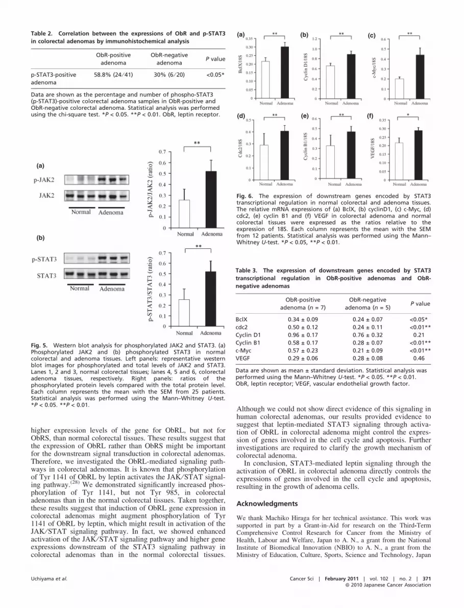
Table 2. Correlation between the expressions of ObR and p-STAT3
in colorectal adenomas by immunohistochemical analysis
ObR-positive
adenoma
ObR-negative
adenomaP value
p-STAT3-positive
adenoma
58.8% (24 ⁄ 41) 30% (6 ⁄ 20) <0.05*
Data are shown as the percentage and number of phospho-STAT3(p-STAT3)-positive colorectal adenoma samples in ObR-positive andObR-negative colorectal adenoma. Statistical analysis was performedusing the chi-square test. *P < 0.05. **P < 0.01. ObR, leptin receptor.
(a)
(b)
Fig. 5. Western blot analysis for phosphorylated JAK2 and STAT3. (a)Phosphorylated JAK2 and (b) phosphorylated STAT3 in normalcolorectal and adenoma tissues. Left panels: representative westernblot images for phosphorylated and total levels of JAK2 and STAT3.Lanes 1, 2 and 3, normal colorectal tissues; lanes 4, 5 and 6, colorectaladenoma tissues, respectively. Right panels: ratios of thephosphorylated protein levels compared with the total protein level.Each column represents the mean with the SEM from 25 patients.Statistical analysis was performed using the Mann–Whitney U-test.*P < 0.05. **P < 0.01.
(a) (b) (c)
(f)(e)(d)
Fig. 6. The expression of downstream genes encoded by STAT3transcriptional regulation in normal colorectal and adenoma tissues.The relative mRNA expressions of (a) BclX, (b) cyclinD1, (c) c-Myc, (d)cdc2, (e) cyclin B1 and (f) VEGF in colorectal adenoma and normalcolorectal tissues were expressed as the ratios relative to theexpression of 18S. Each column represents the mean with the SEMfrom 12 patients. Statistical analysis was performed using the Mann–Whitney U-test. *P < 0.05, **P < 0.01.
Table 3. The expression of downstream genes encoded by STAT3
transcriptional regulation in ObR-positive adenomas and ObR-
negative adenomas
ObR-positive
adenoma (n = 7)
ObR-negative
adenoma (n = 5)P value
BclX 0.34 ± 0.09 0.24 ± 0.07 <0.05*
cdc2 0.50 ± 0.12 0.24 ± 0.11 <0.01**
Cyclin D1 0.96 ± 0.17 0.76 ± 0.32 0.21
Cyclin B1 0.58 ± 0.17 0.28 ± 0.07 <0.01**
c-Myc 0.57 ± 0.23 0.21 ± 0.09 <0.01**
VEGF 0.29 ± 0.06 0.28 ± 0.08 0.46
Data are shown as mean ± standard deviation. Statistical analysis wasperformed using the Mann–Whitney U-test. *P < 0.05. **P < 0.01.ObR, leptin receptor; VEGF, vascular endothelial growth factor.
higher expression levels of the gene for ObRL, but not forObRS, than normal colorectal tissues. These results suggest thatthe expression of ObRL rather than ObRS might be importantfor the downstream signal transduction in colorectal adenomas.Therefore, we investigated the ObRL-mediated signaling path-ways in colorectal adenomas. It is known that phosphorylationof Tyr 1141 of ObRL by leptin activates the JAK ⁄ STAT signal-ing pathway.(28) We demonstrated significantly increased phos-phorylation of Tyr 1141, but not Tyr 985, in colorectaladenomas than in the normal colorectal tissues. Taken together,these results suggest that induction of ObRL gene expression incolorectal adenomas might augment phosphorylation of Tyr1141 of ObRL by leptin, which might result in activation of theJAK ⁄ STAT signaling pathway. In fact, we showed enhancedactivation of the JAK ⁄ STAT signaling pathway and higher geneexpressions downstream of the STAT3 signaling pathway incolorectal adenomas than in the normal colorectal tissues.
Uchiyama et al.
Although we could not show direct evidence of this signaling inhuman colorectal adenomas, our results provided evidence tosuggest that leptin-mediated STAT3 signaling through activa-tion of ObRL in colorectal adenoma might control the expres-sion of genes involved in the cell cycle and apoptosis. Furtherinvestigations are required to clarify the growth mechanism ofcolorectal adenoma.
In conclusion, STAT3-mediated leptin signaling through theactivation of ObRL in colorectal adenoma directly controls theexpressions of genes involved in the cell cycle and apoptosis,resulting in the growth of adenoma cells.
Acknowledgments
We thank Machiko Hiraga for her technical assistance. This work wassupported in part by a Grant-in-Aid for research on the Third-TermComprehensive Control Research for Cancer from the Ministry ofHealth, Labour and Welfare, Japan to A. N., a grant from the NationalInstitute of Biomedical Innovation (NBIO) to A. N., a grant from theMinistry of Education, Culture, Sports, Science and Technology, Japan
Cancer Sci | February 2011 | vol. 102 | no. 2 | 371ªª 2010 Japanese Cancer Association
(KIBAN-B) to A. N., and the grant program, ‘‘Collaborative Develop-ment of Innovative Seed’’ from the Japan Science and TechnologyAgency (JST).
372
Disclosure Statement
The authors declare no conflict of interest.
References
1 Jemal A, Siegel R, Ward E, Hao Y, Xu J, Thun MJ. Cancer statistics 2008. CACancer J Clin 2008; 58: 71–96.
2 Giovannucci E, Goldin B. The role of fat, fatty acids, and total energy intakein the etiology of human colon cancer. Am J Clin Nutr 1997; 66: 1564–71.
3 Friedenreich CM, Orenstein MR. Physical activity and cancer prevention:etiologic evidence and biological mechanisms. J Nutr 2002; 132: 3456–64.
4 Maeda K, Okubo K, Shimomura I, Mizuno K, Matsuzawa Y, Matsubara K.Analysis of an expression profile of genes in the human adipose tissue. Gene1997; 190: 227–35.
5 Kershaw EE, Flier JS. Adipose tissue as an endocrine organ. J ClinEndocrinol Metab 2004; 89: 2548–56.
6 Zhang Y, Proenca R, Maffei M, Barone M, Leopold L, Friedman JM.Positional cloning of the mouse obese gene and its human homologue. Nature1994; 372: 425–32.
7 Seck T, Englaro P, Blum WF et al. Leptin concentrations in serum from arandomly recruited sample of 50- to 80-year-old men and women: positiveassociation with plasma insulin-like growth factors (IGFs) and IGF-bindingprotein-3 in lean, but not in obese, individuals. Eur J Endocrinol 1998; 138:70–5.
8 Weigle DS, Ganter SL, Kuijper JL, Leonetti DL, Boyko EJ, Fujimoto WY.Effect of regional fat distribution and Prader–Willi syndrome on plasma leptinlevels. J Clin Endocrinol Metab 1997; 82: 566–70.
9 van Rossum CT, Hoebee B, van Baak MA, Mars M, Saris WH, Seidell JC.Genetic variation in the leptin receptor gene, leptin, and weight gain in youngDutch adults. Obes Res 2003; 11: 377–86.
10 Uotani S, Bjørbaek C, Tornøe J, Flier JS. Functional properties of leptinreceptor isoforms: internalization and degradation of leptin and ligand-induced receptor downregulation. Diabetes 1999; 48: 279–86.
11 Saglam K, Aydur E, Yilmaz M, Goktas S. Leptin influences cellulardifferentiation and progression in prostate cancer. J Urol 2003; 169: 1308–11.
12 Stattin P, Soderberg S, Hallmans G et al. Leptin is associated with increasedprostate cancer risk: a nested case-referent study. J Clin Endocrinol Metab2001; 86: 1341–5.
13 Baillargeon J, Platz EA, Rose DP et al. Obesity, adipokines, and prostatecancer in a prospective population-based study. Cancer Epidemiol BiomarkersPrev 2006; 15: 1331–5.
14 Falk RT, Brinton LA, Madigan MP et al. Interrelationships between serumleptin, IGF-1, IGFBP3, C-peptide and prolactin and breast cancer risk inyoung women. Breast Cancer Res Treat 2006; 98: 157–65.
15 Stattin P, Soderberg S, Biessy C et al. Plasma leptin and breast cancer risk: aprospective study in northern Sweden. Breast Cancer Res Treat 2004; 86: 191–6.
16 Kumor A, Daniel P, Pietruczuk M, Małecka-Panas E. Serum leptin,adiponectin, and resistin concentration in colorectal adenoma and carcinoma(CC) patients. Int J Colorectal Dis 2009; 24: 275–81.
17 Arpaci F, Yilmaz MI, Ozet A et al. Low serum leptin level in colon cancerpatients without significant weight loss. Tumori 2002; 88: 147–9.
18 Bolukbas FF, Kilic H, Bolukbas C et al. Serum leptin concentration andadvanced gastrointestinal cancers: a case controlled study. BMC Cancer 2004;24: 4.
19 Wallace AM, Sattar N, McMillan DC. Effect of weight loss and theinflammatory response on leptin concentrations in gastrointestinal cancerpatients. Clin Cancer Res 1998; 4: 2977–9.
20 Tessitore L, Vizio B, Jenkins O et al. Leptin expression in colorectal andbreast cancer patients. Int J Mol Med 2000; 5: 421–6.
21 Stattin P, Palmqvist R, Soderberg S et al. Plasma leptin and colorectal cancerrisk: a prospective study in Northern Sweden. Oncol Rep 2003; 10: 2015–21.
22 Stattin P, Lukanova A, Biessy C et al. Obesity and colon cancer: does leptinprovide a link? Int J Cancer 2004; 109: 149–52.
23 Tamakoshi K, Toyoshima H, Wakai K et al. Leptin is associated with anincreased female colorectal cancer risk: a nested case–control study in Japan.Oncology 2005; 68: 454–61.
24 Ogunwobi OO, Beales IL. The anti-apoptotic and growth stimulatory actionsof leptin in human colon cancer cells involves activation of JNK mitogenactivated protein kinase, JAK2 and PI3 kinase ⁄ Akt.Int. Int J Colorectal Dis2007; 22: 401–9.
25 Ratke J, Entschladen F, Niggemann B, Zanker K, Lang K. Leptin stimulatesthe migration of colon carcinoma cells by multiple signalling pathways.Endocr Relat Cancer 2010; 17: 179–89.
26 Cascio S, Ferla R, D’Andrea A et al. Expression of angiogenic regulators,VEGF and leptin, is regulated by the EGF ⁄ PI3K ⁄ STAT3 pathway incolorectal cancer cells. J Cell Physiol 2009; 221: 189–94.
27 Kusaba T, Nakayama T, Yamazumi K et al. Expression of p-STAT3 in humancolorectal adenocarcinoma and adenoma; correlation with clinicopathologicalfactors. J Clin Pathol 2005; 58: 833–8.
28 Cao Q, Mak KM, Ren C, Lieber CS. Leptin stimulates tissue inhibitor ofmetalloproteinase-1 in human hepatic stellate cells: respective roles of theJAK ⁄ STAT and JAK-mediated H2O2-dependant MAPK pathways. J BiolChem 2004; 279: 4292–304.
29 Burguera B, Couce ME, Long J et al. The long form of the leptin receptor(OB-Rb) is widely expressed in the human brain. Neuroendocrinology 2000;71: 187–95.
30 Bollrath J, Phesse TJ, von Burstin VA et al. gp130-mediated STAT3activation in enterocytes regulates cell survival and cell-cycle progressionduring colitis-associated tumorigenesis. Cancer Cell 2009; 15: 91–102.
31 Niu G, Wright KL, Huang M et al. Constitutive STAT3 activity up-regulatesVEGF expression and tumor angiogenesis. Oncogene 2002; 21: 2000–8.
32 Berg JW. Epidemiology, pathology, and the importance of adenomas. ProgClin Biol Res 1988; 279: 13–21.
33 Hardwick JC, Van Den Brink GR, Offerhaus GJ, Van Deventer SJ,Peppelenbosch MP. Leptin is a growth factor for colonic epithelial cells.Gastroenterology 2001; 121: 79–90.
34 Uddin S, Bavi PP, Hussain AR et al. Leptin receptor expression in MiddleEastern colorectal cancer and its potential clinical implication. Carcinogenesis2009; 30: 1832–40.
doi: 10.1111/j.1349-7006.2010.01803.xªª 2010 Japanese Cancer Association