twinning of pleomorphic adenoma: a case report | cureus
TRANSCRIPT
Received 12/23/2019 Review began 12/23/2019 Review ended 01/08/2020 Published 01/09/2020
© Copyright 2020Adiyodi et al. This is an open accessarticle distributed under the terms of theCreative Commons Attribution LicenseCC-BY 3.0., which permits unrestricteduse, distribution, and reproduction in anymedium, provided the original author andsource are credited.
Twinning of Pleomorphic Adenoma: A CaseReportNishitha V. Adiyodi , Joyce Sequeira , Anchal Mehra
1. Oral and Maxillofacial Surgery, Yenepoya Dental College, Mangalore, IND
Corresponding author: Nishitha V. Adiyodi, [email protected]
AbstractPleomorphic adenoma is a common benign salivary gland tumor which presents as a painless swelling thatgradually increases in size, if left untreated. It is often seen involving the parotid gland. However,pleomorphic adenoma has been reported to involve the minor salivary glands as well. In this report, wepresent two cases of pleomorphic adenoma originating from minor salivary glands of the upper lip,occurring in two male patients of the same age (44 years) with markedly similar history of duration, size, andsite of the lesion. The tumor was excised in both patients and sent for histopathological analysis whichshowed features of pleomorphic adenoma confirming the diagnosis.
Categories: Pathology, Oncology, OtherKeywords: pleomorphic adenoma, minor salivary gland tumor, upper lip
IntroductionPleomorphic adenoma is a common benign tumor of the salivary gland. These lesions account for two-thirds, or approximately 60%-65%, of all salivary gland tumors [1]. It shows a female predilection and ismost commonly seen in the fourth to sixth decade of life. The tumor progresses as an asymptomatic slowgrowth over a prolonged period of time. The lesion originates most commonly from the major salivaryglands, most commonly the parotid gland, although cases of occurrence in minor salivary glands have alsobeen reported. Lips and palate are the most common sites; 20%-40% of all intraoral pleomorphic adenomashave been associated with the minor salivary gland. While the etiology of pleomorphic adenoma stillremains elusive, it is known to be epithelial in origin, and clonal chromosome abnormalities withaberrations involving 8q12 and 12q15 have been implicated [2].
In this paper, we present two cases of pleomorphic adenomas involving the minor salivary glands, withstrikingly similar clinical and demographic characteristics.
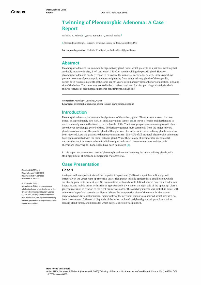
Case PresentationCase 1A 44-year-old male patient visited the outpatient department (OPD) with a painless solitary growthintraorally in the upper right lip since five years. The growth initially appeared as a small lesion, whicheventually grew to its present size. On examination, we found a well-defined, round, firm, non-tender, non-fluctuant, and mobile lesion with a size of approximately 3 × 3 cm on the right side of the upper lip. Class IIgingival recession in relation to the right canine was noted. The overlying mucosa was pinkish in color, withevidence of superficial vascularity. Figure 1 shows the preoperative view of the tumor for the above-mentioned case. Intraoral periapical radiography of the pertinent region was obtained, which revealed nobone involvement. Differential diagnosis of the lesion included peripheral giant cell granuloma, minorsalivary gland tumor, and lipoma for which surgical excision was planned.
1 1 1
Open Access CaseReport DOI: 10.7759/cureus.6608
How to cite this articleAdiyodi N V, Sequeira J, Mehra A (January 09, 2020) Twinning of Pleomorphic Adenoma: A Case Report. Cureus 12(1): e6608. DOI10.7759/cureus.6608
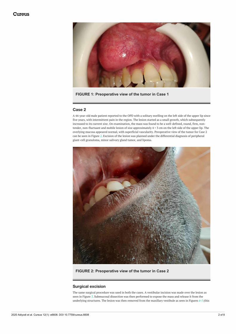
FIGURE 1: Preoperative view of the tumor in Case 1
Case 2A 44-year-old male patient reported to the OPD with a solitary swelling on the left side of the upper lip sincefive years, with intermittent pain in the region. The lesion started as a small growth, which subsequentlyincreased to its current size. On examination, the mass was found to be a well-defined, round, firm, non-tender, non-fluctuant and mobile lesion of size approximately 4 × 3 cm on the left side of the upper lip. Theoverlying mucosa appeared normal, with superficial vascularity. Preoperative view of the tumor for Case 2can be seen in Figure 2. Excision of the lesion was planned under the differential diagnosis of peripheralgiant-cell granuloma, minor salivary gland tumor, and lipoma.
FIGURE 2: Preoperative view of the tumor in Case 2
Surgical excisionThe same surgical procedure was used in both the cases. A vestibular incision was made over the lesion asseen in Figure 3. Submucosal dissection was then performed to expose the mass and release it from theunderlying structures. The lesion was then removed from the maxillary vestibule as seen in Figures 4-5 (this
2020 Adiyodi et al. Cureus 12(1): e6608. DOI 10.7759/cureus.6608 2 of 8

can be seen in Figures 7-8 as well for Case 2). Extra soft tissue present in the vestibule was excised, followedby its closure with 3 - 0 silk as seen in Figures 6-9. The excised specimens were subjected to histopathology.The histopathology of both the cases revealed features of pleomorphic adenoma. The section showed a well-encapsulated lesion surrounded by a fibrous capsule. It also displayed numerous ductal and myoepithelialcells in the form of duct-like structures, strands and sheets, lined by cuboidal cells. The ductal spaces ofvariable dimensions complete with eosinophilic coagulum was noted. The stroma consisted of extensivezones of myxoid tissue with ovoid to spindle-shaped myoepithelial cells and chondroid areas. This can beseen in Figure 10.
FIGURE 3: Surgical excision of the tumor in Case 1Vestibular incision was given to excise the tumor.
2020 Adiyodi et al. Cureus 12(1): e6608. DOI 10.7759/cureus.6608 3 of 8
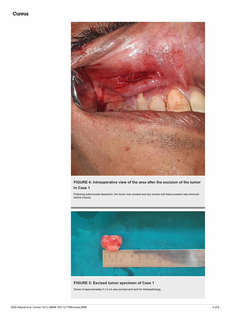
FIGURE 4: Intraoperative view of the area after the excision of the tumorin Case 1Following submucosal dissection, the tumor was excised and any excess soft tissue present was removedbefore closure.
FIGURE 5: Excised tumor specimen of Case 1Tumor of approximately 3 x 3 cm was excised and sent for histopathology.
2020 Adiyodi et al. Cureus 12(1): e6608. DOI 10.7759/cureus.6608 4 of 8
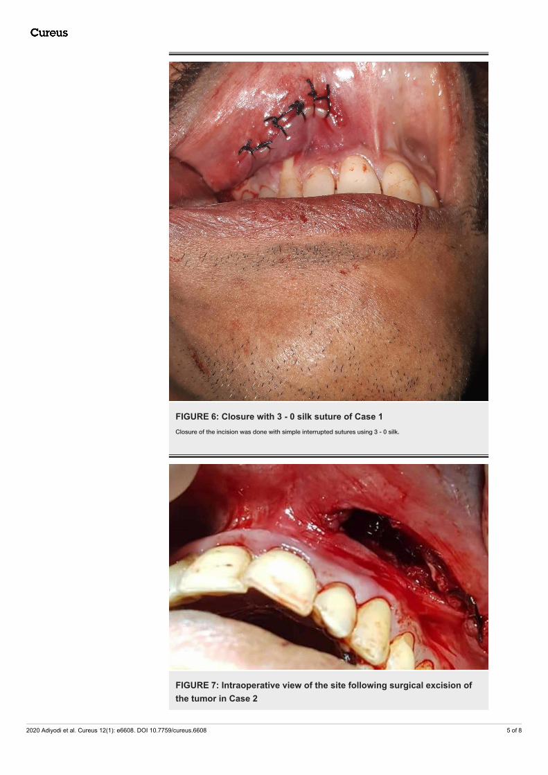
FIGURE 6: Closure with 3 - 0 silk suture of Case 1Closure of the incision was done with simple interrupted sutures using 3 - 0 silk.
FIGURE 7: Intraoperative view of the site following surgical excision ofthe tumor in Case 2
2020 Adiyodi et al. Cureus 12(1): e6608. DOI 10.7759/cureus.6608 5 of 8
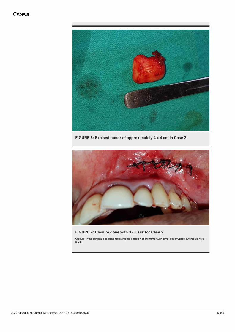
FIGURE 8: Excised tumor of approximately 4 x 4 cm in Case 2
FIGURE 9: Closure done with 3 - 0 silk for Case 2Closure of the surgical site done following the excision of the tumor with simple interrupted sutures using 3 -0 silk.
2020 Adiyodi et al. Cureus 12(1): e6608. DOI 10.7759/cureus.6608 6 of 8
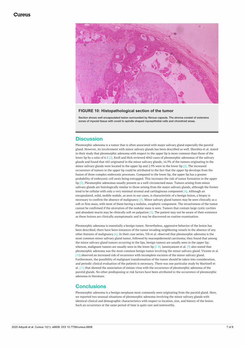
FIGURE 10: Histopathological section of the tumorSection shows well encapsulated lesion surrounded by fibrous capsule. The stroma consist of extensivezones of myxoid tissue with ovoid to spindle shaped myoepithelial cells and chondroid areas.
DiscussionPleomorphic adenoma is a tumor that is often associated with major salivary gland especially the parotidgland. However, its involvement with minor salivary glands has been described as well. Shreshta et al. statedin their study that pleomorphic adenoma with respect to the upper lip is more common than those of thelower lip by a ratio of 6:1 [2]. Kroll and Hick reviewed 4042 cases of pleomorphic adenomas of the salivaryglands and found that 445 originated in the minor salivary glands; 16.9% of the tumors originating in theminor salivary glands were located in the upper lip and 2.9% were in the lower lip [2]. The increasedoccurrence of tumors in the upper lip could be attributed to the fact that the upper lip develops from thefusion of three complex embryonic processes. Compared to the lower lip, the upper lip has a greaterprobability of embryonic cell nests being entrapped. This increases the risk of tumor formation in the upperlip [3]. Pleomorphic adenomas usually present as a well-circumcised mass. Tumors arising from minorsalivary glands are histologically similar to those arising from the major salivary glands, although the formertend to be cellular with only a very minimal stromal and cartilaginous component [4]. Although anencapsulated, solid, mobile nodule, as seen in our cases, is characteristic of a benign lesion, a biopsy isnecessary to confirm the absence of malignancy [5]. Minor salivary gland tumors may be seen clinically as asoft or firm mass, with most of them having a nodular, exophytic component. The invasiveness of the tumorcannot be confirmed if the ulceration of the nodular mass is seen. Tumors that contain large cystic cavitiesand abundant mucin may be clinically soft on palpation [5]. The patient may not be aware of their existenceas these lesions are clinically asymptomatic and it may be discovered on routine examination.
Pleomorphic adenoma is essentially a benign tumor. Nevertheless, aggressive behavior of the lesion hasbeen described; there have been instances of the tumor invading neighboring vessels in the absence of anyother features of malignancy [6]. In their case series, Yih et al. observed that pleomorphic adenoma is themost common minor salivary gland tumor, followed by mucoepidermoid carcinoma; they found that amongthe minor salivary gland tumors occurring in the lips, benign tumors are usually seen in the upper lip;whereas, malignant tumors are usually seen in the lower lip [7-8]. Jansisyanont et al. [9] also stated thatpleomorphic adenoma was the most common benign tumor involving the minor salivary gland. Vicente et al.[10] observed an increased risk of recurrence with incomplete excision of the minor salivary gland.Furthermore, the possibility of malignant transformation of the tumor should be taken into consideration,and periodic clinical evaluation of the patients is necessary. There was one particular study by Martinell etal. [11] that showed the association of simian virus with the occurrence of pleomorphic adenoma of theparotid glands. No other predisposing or risk factors have been attributed to the occurrence of pleomorphicadenoma in literature.
ConclusionsPleomorphic adenoma is a benign neoplasm most commonly seen originating from the parotid gland. Here,we reported two unusual situations of pleomorphic adenoma involving the minor salivary glands withidentical clinical and demographic characteristics with respect to location, size, and history of the lesion.Such an occurrence at the same period of time is quite rare and noteworthy.
2020 Adiyodi et al. Cureus 12(1): e6608. DOI 10.7759/cureus.6608 7 of 8

Additional InformationDisclosuresHuman subjects: Consent was obtained by all participants in this study. Conflicts of interest: Incompliance with the ICMJE uniform disclosure form, all authors declare the following: Payment/servicesinfo: All authors have declared that no financial support was received from any organization for thesubmitted work. Financial relationships: All authors have declared that they have no financialrelationships at present or within the previous three years with any organizations that might have aninterest in the submitted work. Other relationships: All authors have declared that there are no otherrelationships or activities that could appear to have influenced the submitted work.
References1. Shah A, Sayed A, Sayed H, Khutwad G: Pleomorphic adenoma: a case report. Int J Dent Res. 2048, 3:10-12.2. Shrestha A, Reddy NS, Ganguly SN: Pleomorphic adenoma of the upper lip: a case report . JCMS. 2010, 6:51-
3. 10.3126/jcmsn.v6i1.36033. Krolls SO, Hicks JL: Mixed tumors of the lower lip . Oral Surg Oral Med Oral Pathol. 1973, 35:212-7.
10.1016/0030-4220(73)90287-94. Waldron CA, El-Mofty SK, Gnepp DR: Tumors of the intraoral minor salivary glands: a demographic and
histologic study of 426 cases. Oral Surg Oral Med Oral Pathol. 1988, 66:323-33. 10.1016/0030-4220(88)90240-X
5. Dalati T, Hussein MR: Juvenile pleomorphic adenoma of the cheek: a case report and review of literature .Diagn Pathol. 2009, 4:32. 10.1186/1746-1596-4-32
6. Lingam RK, Daghir AA, Nigar E, Abbas SA, Kumar M: Pleomorphic adenoma (benign mixed tumour) of thesalivary glands: its diverse clinical, radiological, and histopathological presentation. Br J Oral MaxillofacSurg. 2011, 49:14-20. 10.1016/j.bjoms.2009.09.014
7. Yih WY, Kratochvil FJ, Stewart JC: Intraoral minor salivary gland neoplasms: review of 213 cases . J OralMaxillofac Surg. 2005, 63:805-10. 10.1016/j.joms.2005.02.021
8. Neville BW, Damm DD, Weir JC, Fantasia JE: Labial salivary gland tumors . Cancer. 1988, 61:2113-6.10.1002/1097-0142(19880515)61:10%3C2113::AID-CNCR2820611030%3E3.0.CO;2-B
9. Jansisyanont P, Blanchaert RH Jr, Ord RA: Intraoral minor salivary gland neoplasm: a single institutionexperience of 80 cases. Int J Oral Maxillofac Surg. 2002, 31:257-61. 10.1054/ijom.2002.0223
10. Pons Vicente O, Almendros-Marqués N, BeriniAytés L, Gay Escoda C: Minor salivary gland tumors: aclinicopathological study of 18 cases. Med Oral Patol Oral Cir Bucal. 2008, 13:582-588.
11. Martinelli M, Martini F, Rinaldi E, et al.: Simian virus 40 sequences and expression of the viral large Tantigen oncoprotein in human pleomorphic adenomas of parotid glands. Am J Pathol. 2002, 161:1127-33.10.1016%2FS0002-9440(10)64389-1
2020 Adiyodi et al. Cureus 12(1): e6608. DOI 10.7759/cureus.6608 8 of 8