the shape and function of solid fascias depend on ... - cureus
TRANSCRIPT
Received 02/04/2020 Review began 02/06/2020 Review ended 02/06/2020 Published 02/10/2020
© Copyright 2020Bordoni. This is an open access articledistributed under the terms of theCreative Commons Attribution LicenseCC-BY 4.0., which permits unrestricteduse, distribution, and reproduction in anymedium, provided the original author andsource are credited.
The Shape and Function of Solid Fascias Dependon the Presence of Liquid FasciasBruno Bordoni
1. Physical Medicine and Rehabilitation, Foundation Don Carlo Gnocchi, Milan, ITA
Corresponding author: Bruno Bordoni, [email protected]
AbstractScientific research is not a showcase of his own talent or own resources, it is a chance to improve commonknowledge on certain topics for the collective well-being. A researcher should use multidisciplinarity toobserve a phenomenon in its entirety and not only its alignment of thought, federations, committees, andknowledge; to get to understand it is necessary to exploit more tools and more disciplines. The articlediscusses the importance of the fluids (or liquid fascia) in maintaining the shape and function of the humanbody, as, currently, many texts forget how much body fluids are fundamental for understanding structuraldynamics (bones and muscles, fibrils, and cells). By revisiting the current literature, the text wishes tohighlight how the liquid fascia determines body adaptation in the presence of mechanical stress. Withoutfluids, there would be no body shape that we know.
Categories: Medical Education, Anatomy, Osteopathic MedicineKeywords: fascia, myofascial, osteopathic, fascintegrity, skeletal muscle, physiotherapy
Introduction And BackgroundThe mechanical model involving the skeletal muscle system and the fascial continuum is known asbiotensegrity. In the 1960s, the designer R. Buckminster Fuller conceived and put into practice the structuraland architectural concept of tensegrity (tensional integrity): continuous tension with discontinuouscompression [1]. He was inspired by a sculpture by the artist Snelson in 1948 [2]. This principle made itpossible to build architectural projects that maintained their shape, despite external mechanical stresses ortemporary changes in the external structure; the tensional balance allows the continuation of function andform [1]. In 1977, Dr. Robbie was the first to equate the concept of tensegrity in the biological field and,precisely, with the spine and with the musculoskeletal system, trying to understand the dynamics of theforces acting on the structure's living (biological), maintaining form and function [2]. According to hisvision, the muscular and bone system could be seen as a system of biological tensegrity [2]. In the late 1970sand then in 1985, Ingber equated the concept of tensegrity with the cell, where microtubules manage themechanical tensions (structures with continuous tension) produced by the actomyosin protein complex(structures with discontinuous compression) [2]. The ultimate goal is to conceptualize the phenomenon ofmechanotransduction, that is, the ability to transmit mechanical information inside and outside the cellthrough the structures themselves that make up the cell and, at the same time, obtain the maximum possibleadaptation [1-2]. In 1981, with an abstract at the 34th Annual Conference on Engineering in Medicine andBiology, Dr. Levin coined the term we universally know: biotensegrity [2]. The theoretical model ofbiotensegrity considers bones and muscle tissue (and connected) as the component of discontinuous tensionand the component in pre-stress, respectively [2]. The philosophy of biotensegrity can be applied to thewhole body, as well as to every single body area, up to the cell, as in a previous article, where I have shownin detail the different connections [3]. The model marries the definition of what is the fascial continuum,according to Fascia Nomenclature Committee: “The fascial system includes adipose tissue, adventitia,neurovascular sheaths, aponeuroses, deep and superficial fasciae, dermis, epineurium, joint capsules,ligaments, membranes, meninges, myofascial expansions, periosteum, retinacula, septa, tendons (includingendotendon/peritendon/epitendon/paratendon), visceral fasciae, and all the intramuscular andintermuscular connective tissues, including endomysium/perimysium/epimysium” [4]. What is not takeninto consideration when trying to understand how to correctly apply this model (mechanical model), is thefunction of body fluids [5]. Our vision of what should be considered a fascia also includes body fluids: “Thefascia is any tissue that contains features capable of responding to mechanical stimuli. The fascialcontinuum is the result of the evolution of the perfect synergy among different tissues, liquids, andsolids capable of supporting, dividing, penetrating, feeding, and connecting all the regions of the body, fromthe epidermis to the bone, involving all its functions and organic structures. This continuum constantlytransmits and receives mechanometabolic information that can influence the shape and function of theentire body. These afferent/efferent impulses come from the fascia and the tissues that are not considered aspart of the fascia in a biunivocal mode. In this definition, these tissues include: epidermis, dermis, fat,blood, lymph, blood and lymphatic vessels, tissue covering the nervous filaments (endoneurium,perineurium, epineurium), voluntary striated muscle fibers, and the tissue covering and permeating it(epimysium, perimysium, endomysium), ligaments, tendons, aponeurosis, cartilage, bones, meninges, andtongue” [4]. The biotensegrity model does not match the presence of body fluids, as it does not take theminto consideration. Another model called fascintegrity and created by the Foundation of Osteopathic
1
Open Access ReviewArticle DOI: 10.7759/cureus.6939
How to cite this articleBordoni B (February 10, 2020) The Shape and Function of Solid Fascias Depend on the Presence of Liquid Fascias . Cureus 12(2): e6939. DOI10.7759/cureus.6939
Research and Clinical Endorsement (FORCE) group involves blood and lymph (specialized connective tissue)[4]. The human body is made up of fluids, from its entirety to the individual cell; without the presence offluids, the human body or the single-cell could not adapt, could not survive and would not have the shape weknow [3]. In the 21st century, we are still debating whether the biotensegrity model is a valid model [6]. Themodel is valid when fluids are not taken into consideration. Dissecting the understanding of a phenomenonmeans observing a three-dimensionality of different interpenetrations of scientific concepts. Continuing topursue a theoretical model inevitably leads to confusion; the confusion does not arise from themultidisciplinarity of thought but from the marmoreal intellectual fixity. A model is used to advance and notto stop. As a model, it is a starting point that should only be defended when its total validity isdemonstrated; currently, there is no experimental scientific study that proves validity on the living. But thisis precisely the richness of a model, that is, to stimulate research and the advancement of the understandingof what happens in the biological sphere. The fascintegrity model includes fluids in its philosophy but, likethe previous model, it is a theoretical approach, which needs further study [3]. The theme of the review is tohighlight the importance of fluids on the ability of the cell and tissues to function and maintain the shapeduring biological activities. The ultimate goal is to stimulate further growth in the vision of what is the bodysystem or fascial continuum.
ReviewThe human body contains different fluids or liquid fascia with different tasks; all fluids can come intocontact with each other [7]. The same cannot be said for muscles or bones or solid fascia. We can recognizeblood (arterial and venous), lymph, cellular fluids (intracellular, extracellular), and cerebrospinal fluid [8-11]. Fluids play an extraordinary role in maintaining bodily functions and, therefore, the shape. Themovement or quantity of fluids implies constant adaptation as they generate mechanical force [12]. Forexample, every time the body changes posture, the blood system changes the space occupied, altering thepressures. The vestibule must send information to the nucleus of the solitary tract, which last receivesbaroreceptor information from the aortic arch and the carotid glomus, to better manage blood pressure andposture [13]. Blood pressures affect posture. Another example concerns breast cancer cells. AMP-activatedprotein kinase (AMPK) has the task of monitoring cellular energy status and plays a fundamental role in thebehavior of cancer cells. Generally, it is believed that the physical microenvironment primarily influencesthe response of cancer cells, a recent in vitro study has shown the importance of the fluid footprint on thetumor cell response [14]. Researchers have shown that steroid receptor coactivator (Src) and focal adhesionkinase (FAK), which are non-receptor intracellular structures, are extremely sensitive to shear stress fromintracellular fluids and are capable of influencing AMPK and tumor proliferation [14]. The flow of fluidsgenerates energy. Some chemists have developed a tiny battery that works by extracting energy from bodyfluids. It can be placed in contact with body fluids containing glucose, under the skin or in the backbone.Batteries operate using biological fluids [15]. Body fluids emit light, in particular, the blood emits biophotonsor ultra-weak photon emission (UPE) [16]. Light is an electromagnetic wave but it also acts like a stream ofcorpuscles. A study shows that the two natures of light, the corpuscular and the wave nature, can bedetected simultaneously in the same physical system [17]. The release of biophotons has a pattern, arecognizable rhythm; this biological behavior is equivalent to a communication system, probably, for thenervous system and for other body systems [16]. Fluids can make sounds like phonons. Phonons (oscillatorywaves) are "quasiparticles" that carry sound and heat and, once again, carry information from one atom toanother [18-19]. UPE and phonons interact with the biological response of the organism, but yet, there areno studies involving these notions in the theoretical model of biotensegrity.
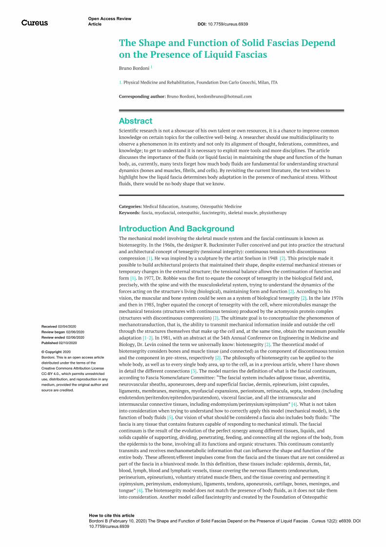
Fluids and embryogenesisThe structure adapts to fluids; the fluids with their movement, quantity, flow speed, and vector modedetermine the shape of the tissues. The form and function of the fascial continuum are primarily given byfluids. Let us see some examples. The morphogenesis of the cardiac structure is based on the thrust of thefluids. The shear stress deriving from the flow of fluids specularly determines the shape of the cell, while thepressure of the fluids themselves determine the boundaries and functions of the cell itself: the fluids shapethe form and function at the embryological level [20]. Another study confirms this concept, whereresearchers demonstrate that fluids and their behavior stimulate correct cell division during embryogenesis[21]. The precise size and symmetry of the organs, as well as their perfect position and function, depends onthe fluids. The inner ear, an extraordinary precision organ, is morphogenetically stimulated by the quantityof fluids (hydrostatic pressure) present during embryogenesis; the same pressure manages the quantity offluids that can intervene [22]. The fluids manage themselves through pressure feedback and, at the sametime, influence the duration of growth and the size of the different tissues [22-23]. The presence of fluidsinfluences the synthesis of chalones (mitotic inhibitors) [22]. The latter manages the duration of growth,such as somatostatin for skeletal muscle, bone type 3 morphogenetic protein (BMP3) for bone tissue, andprotein growth differentiation factor 11 (GDF11) for the nervous system [22]. During embryogenesis, fluidsalso influence substances that manage the size of the tissue or organ, such as insulin, target of rapamycin(TOR) signalling pathways and by the Hippo protein kinase (Hpo) [22]. The accumulation of fluids ormorphogenic gradient allows cells to synthesize these substances and create self-control of the growth,shape, and function of tissues and organs or organogenesis [22-23]. The structure, like the variouscomponents that make up the cell (proteins, microtubules, contact proteins, and more) respond to fluids(inside and outside the cell); without the constant work of fluids (blood, lymph, interstitial fluids, intra andextracellular fluids) there would be no form or function (Figure 1).
2020 Bordoni et al. Cureus 12(2): e6939. DOI 10.7759/cureus.6939 2 of 7
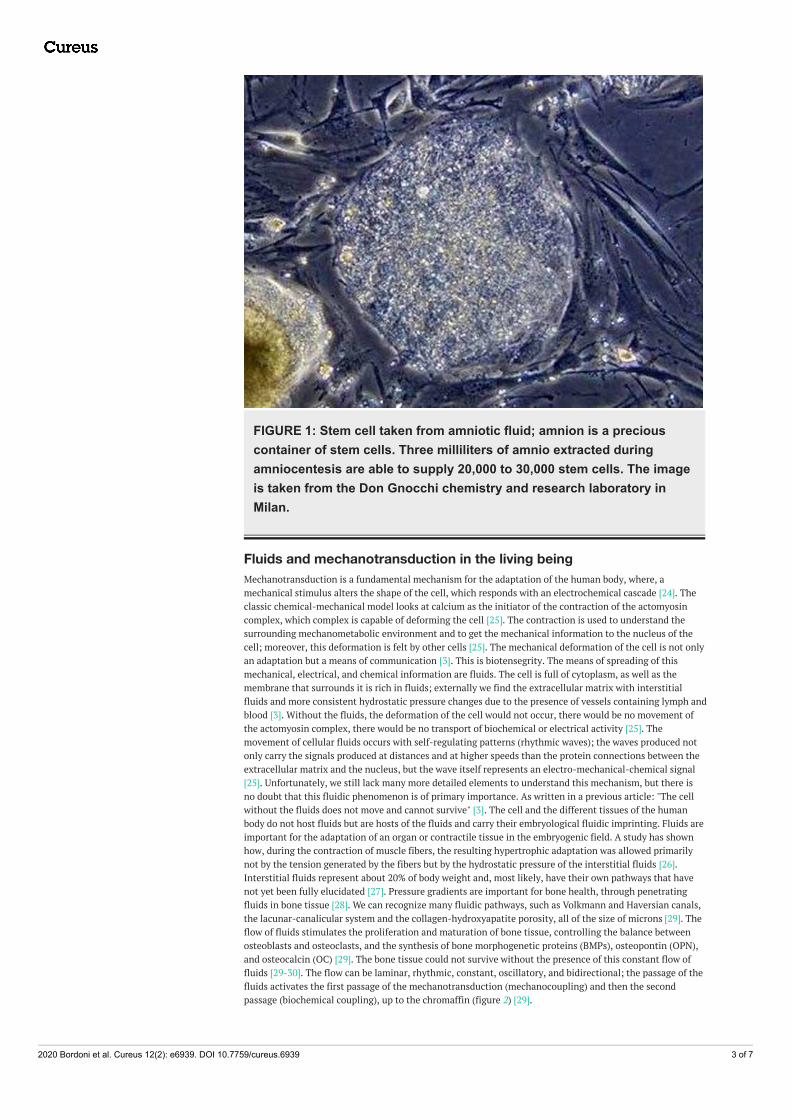
FIGURE 1: Stem cell taken from amniotic fluid; amnion is a preciouscontainer of stem cells. Three milliliters of amnio extracted duringamniocentesis are able to supply 20,000 to 30,000 stem cells. The imageis taken from the Don Gnocchi chemistry and research laboratory inMilan.
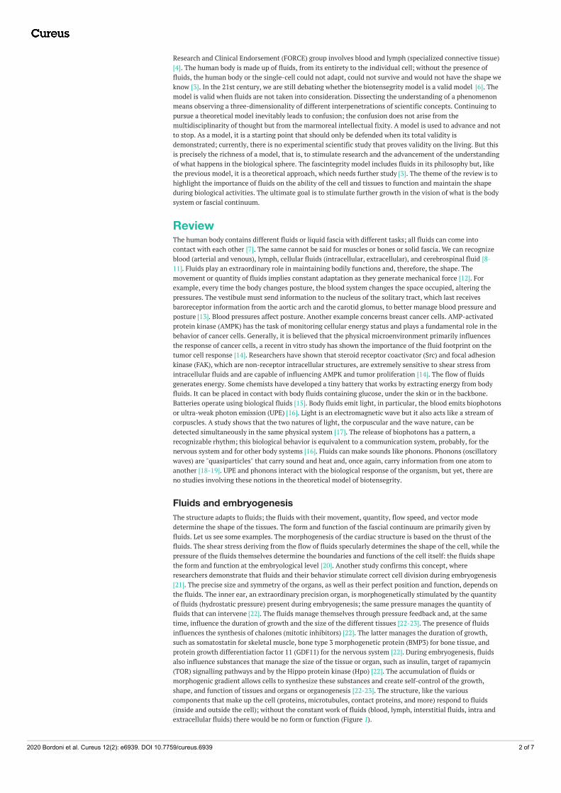
Fluids and mechanotransduction in the living beingMechanotransduction is a fundamental mechanism for the adaptation of the human body, where, amechanical stimulus alters the shape of the cell, which responds with an electrochemical cascade [24]. Theclassic chemical-mechanical model looks at calcium as the initiator of the contraction of the actomyosincomplex, which complex is capable of deforming the cell [25]. The contraction is used to understand thesurrounding mechanometabolic environment and to get the mechanical information to the nucleus of thecell; moreover, this deformation is felt by other cells [25]. The mechanical deformation of the cell is not onlyan adaptation but a means of communication [3]. This is biotensegrity. The means of spreading of thismechanical, electrical, and chemical information are fluids. The cell is full of cytoplasm, as well as themembrane that surrounds it is rich in fluids; externally we find the extracellular matrix with interstitialfluids and more consistent hydrostatic pressure changes due to the presence of vessels containing lymph andblood [3]. Without the fluids, the deformation of the cell would not occur, there would be no movement ofthe actomyosin complex, there would be no transport of biochemical or electrical activity [25]. Themovement of cellular fluids occurs with self-regulating patterns (rhythmic waves); the waves produced notonly carry the signals produced at distances and at higher speeds than the protein connections between theextracellular matrix and the nucleus, but the wave itself represents an electro-mechanical-chemical signal[25]. Unfortunately, we still lack many more detailed elements to understand this mechanism, but there isno doubt that this fluidic phenomenon is of primary importance. As written in a previous article: "The cellwithout the fluids does not move and cannot survive" [3]. The cell and the different tissues of the humanbody do not host fluids but are hosts of the fluids and carry their embryological fluidic imprinting. Fluids areimportant for the adaptation of an organ or contractile tissue in the embryogenic field. A study has shownhow, during the contraction of muscle fibers, the resulting hypertrophic adaptation was allowed primarilynot by the tension generated by the fibers but by the hydrostatic pressure of the interstitial fluids [26].Interstitial fluids represent about 20% of body weight and, most likely, have their own pathways that havenot yet been fully elucidated [27]. Pressure gradients are important for bone health, through penetratingfluids in bone tissue [28]. We can recognize many fluidic pathways, such as Volkmann and Haversian canals,the lacunar-canalicular system and the collagen-hydroxyapatite porosity, all of the size of microns [29]. Theflow of fluids stimulates the proliferation and maturation of bone tissue, controlling the balance betweenosteoblasts and osteoclasts, and the synthesis of bone morphogenetic proteins (BMPs), osteopontin (OPN),and osteocalcin (OC) [29]. The bone tissue could not survive without the presence of this constant flow offluids [29-30]. The flow can be laminar, rhythmic, constant, oscillatory, and bidirectional; the passage of thefluids activates the first passage of the mechanotransduction (mechanocoupling) and then the secondpassage (biochemical coupling), up to the chromaffin (figure 2) [29].
2020 Bordoni et al. Cureus 12(2): e6939. DOI 10.7759/cureus.6939 3 of 7
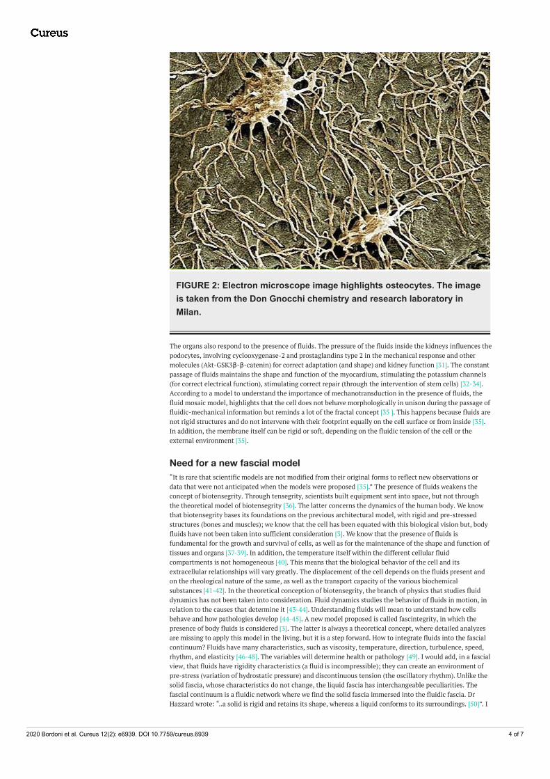
FIGURE 2: Electron microscope image highlights osteocytes. The imageis taken from the Don Gnocchi chemistry and research laboratory inMilan.
The organs also respond to the presence of fluids. The pressure of the fluids inside the kidneys influences thepodocytes, involving cyclooxygenase-2 and prostaglandins type 2 in the mechanical response and othermolecules (Akt-GSK3β-β-catenin) for correct adaptation (and shape) and kidney function [31]. The constantpassage of fluids maintains the shape and function of the myocardium, stimulating the potassium channels(for correct electrical function), stimulating correct repair (through the intervention of stem cells) [32-34].According to a model to understand the importance of mechanotransduction in the presence of fluids, thefluid mosaic model, highlights that the cell does not behave morphologically in unison during the passage offluidic-mechanical information but reminds a lot of the fractal concept [35 ]. This happens because fluids arenot rigid structures and do not intervene with their footprint equally on the cell surface or from inside [35].In addition, the membrane itself can be rigid or soft, depending on the fluidic tension of the cell or theexternal environment [35].
Need for a new fascial model“It is rare that scientific models are not modified from their original forms to reflect new observations ordata that were not anticipated when the models were proposed [35].” The presence of fluids weakens theconcept of biotensegrity. Through tensegrity, scientists built equipment sent into space, but not throughthe theoretical model of biotensegrity [36]. The latter concerns the dynamics of the human body. We knowthat biotensegrity bases its foundations on the previous architectural model, with rigid and pre-stressedstructures (bones and muscles); we know that the cell has been equated with this biological vision but, bodyfluids have not been taken into sufficient consideration [3]. We know that the presence of fluids isfundamental for the growth and survival of cells, as well as for the maintenance of the shape and function oftissues and organs [37-39]. In addition, the temperature itself within the different cellular fluidcompartments is not homogeneous [40]. This means that the biological behavior of the cell and itsextracellular relationships will vary greatly. The displacement of the cell depends on the fluids present andon the rheological nature of the same, as well as the transport capacity of the various biochemicalsubstances [41-42]. In the theoretical conception of biotensegrity, the branch of physics that studies fluiddynamics has not been taken into consideration. Fluid dynamics studies the behavior of fluids in motion, inrelation to the causes that determine it [43-44]. Understanding fluids will mean to understand how cellsbehave and how pathologies develop [44-45]. A new model proposed is called fascintegrity, in which thepresence of body fluids is considered [3]. The latter is always a theoretical concept, where detailed analyzesare missing to apply this model in the living, but it is a step forward. How to integrate fluids into the fascialcontinuum? Fluids have many characteristics, such as viscosity, temperature, direction, turbulence, speed,rhythm, and elasticity [46-48]. The variables will determine health or pathology [49]. I would add, in a fascialview, that fluids have rigidity characteristics (a fluid is incompressible); they can create an environment ofpre-stress (variation of hydrostatic pressure) and discontinuous tension (the oscillatory rhythm). Unlike thesolid fascia, whose characteristics do not change, the liquid fascia has interchangeable peculiarities. Thefascial continuum is a fluidic network where we find the solid fascia immersed into the fluidic fascia. DrHazzard wrote: “..a solid is rigid and retains its shape, whereas a liquid conforms to its surroundings. [50]”. I
2020 Bordoni et al. Cureus 12(2): e6939. DOI 10.7759/cureus.6939 4 of 7
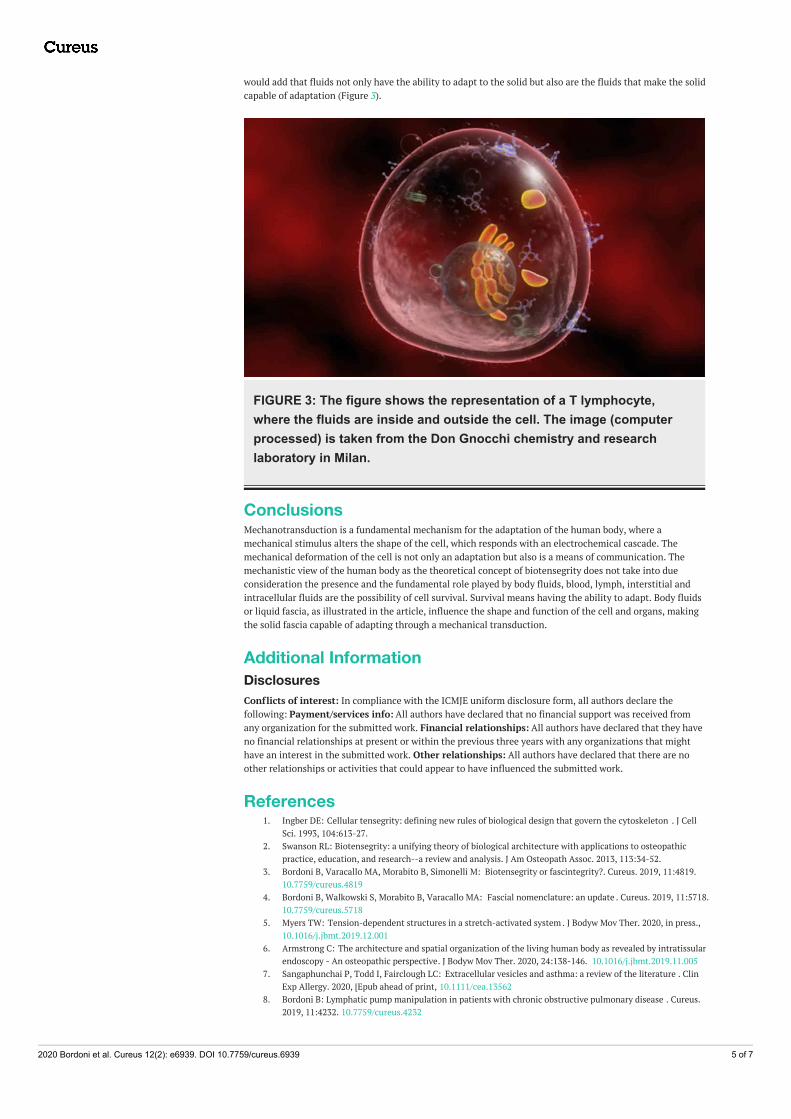
would add that fluids not only have the ability to adapt to the solid but also are the fluids that make the solidcapable of adaptation (Figure 3).
FIGURE 3: The figure shows the representation of a T lymphocyte,where the fluids are inside and outside the cell. The image (computerprocessed) is taken from the Don Gnocchi chemistry and researchlaboratory in Milan.
ConclusionsMechanotransduction is a fundamental mechanism for the adaptation of the human body, where amechanical stimulus alters the shape of the cell, which responds with an electrochemical cascade. Themechanical deformation of the cell is not only an adaptation but also is a means of communication. Themechanistic view of the human body as the theoretical concept of biotensegrity does not take into dueconsideration the presence and the fundamental role played by body fluids, blood, lymph, interstitial andintracellular fluids are the possibility of cell survival. Survival means having the ability to adapt. Body fluidsor liquid fascia, as illustrated in the article, influence the shape and function of the cell and organs, makingthe solid fascia capable of adapting through a mechanical transduction.
Additional InformationDisclosuresConflicts of interest: In compliance with the ICMJE uniform disclosure form, all authors declare thefollowing: Payment/services info: All authors have declared that no financial support was received fromany organization for the submitted work. Financial relationships: All authors have declared that they haveno financial relationships at present or within the previous three years with any organizations that mighthave an interest in the submitted work. Other relationships: All authors have declared that there are noother relationships or activities that could appear to have influenced the submitted work.
References1. Ingber DE: Cellular tensegrity: defining new rules of biological design that govern the cytoskeleton . J Cell
Sci. 1993, 104:613-27.2. Swanson RL: Biotensegrity: a unifying theory of biological architecture with applications to osteopathic
practice, education, and research--a review and analysis. J Am Osteopath Assoc. 2013, 113:34-52.3. Bordoni B, Varacallo MA, Morabito B, Simonelli M: Biotensegrity or fascintegrity?. Cureus. 2019, 11:4819.
10.7759/cureus.48194. Bordoni B, Walkowski S, Morabito B, Varacallo MA: Fascial nomenclature: an update . Cureus. 2019, 11:5718.
10.7759/cureus.57185. Myers TW: Tension-dependent structures in a stretch-activated system . J Bodyw Mov Ther. 2020, in press.,
10.1016/j.jbmt.2019.12.0016. Armstrong C: The architecture and spatial organization of the living human body as revealed by intratissular
endoscopy - An osteopathic perspective. J Bodyw Mov Ther. 2020, 24:138-146. 10.1016/j.jbmt.2019.11.0057. Sangaphunchai P, Todd I, Fairclough LC: Extracellular vesicles and asthma: a review of the literature . Clin
Exp Allergy. 2020, [Epub ahead of print, 10.1111/cea.135628. Bordoni B: Lymphatic pump manipulation in patients with chronic obstructive pulmonary disease . Cureus.
2019, 11:4232. 10.7759/cureus.4232
2020 Bordoni et al. Cureus 12(2): e6939. DOI 10.7759/cureus.6939 5 of 7
9. Bordoni B, Marelli F, Morabito B, Castagna R: A new concept of biotensegrity incorporating liquid tissues:blood and lymph. J Evid Based Integr Med. 2018, 23:2515690. 10.1177/2515690X18792838
10. Bordoni B, Lintonbon D, Morabito B: Meaning of the solid and liquid fascia to reconsider the model ofbiotensegrity. Cureus. 2018, 10:2922. 10.7759/cureus.2922
11. Orts-Del'Immagine A, Wyart C: Cerebrospinal-fluid-contacting neurons. Curr Biol. 2017 Nov 20, 27:1198-1200. 10.1016/j.cub.2017.09.017
12. Geng J, Zhao Q, Zhang T, Xiao B: In touch with the mechanosensitive piezo channels structure, ionpermeation, and mechanotransduction.. Curr Top Membr. 2017, 79:159-195. 10.1016/bs.ctm.2016.11.006
13. Holstein GR, Friedrich VL Jr, Martinelli GP: Projection neurons of the vestibulo-sympathetic reflex pathway .J Comp Neurol. 2014, 522:2053-74. 10.1002/cne.23517
14. Guo Y, Steele HE, Li BY, Na S: Fluid flow-induced activation of subcellular AMPK and its interaction withFAK and Src. Arch Biochem Biophys. 2020, 679:108208. 10.1016/j.abb.2019.108208
15. Klosterhoff BS, Tsang M, She D, Ong KG, Allen MG, Willett NJ, Guldberg RE: Implantable sensors forregenerative medicine. J Biomech Eng. 2017, 139: 10.1115/1.4035436
16. Bordoni B, Marelli F, Morabito B, Sacconi B: Emission of biophotons and adjustable sounds by the fascialsystem: review and reflections for manual therapy. J Evid Based Integr Med. 2018, 23:2515690-17750750.10.1177/2515690X17750750
17. Piazza L, Lummen TT, Quiñonez E, Murooka Y, Reed BW, Barwick B, Carbone F: Simultaneous observationof the quantization and the interference pattern of a plasmonic near-field. Nat Commun. 2015, 6:6407.10.1038/ncomms7407
18. Rowlands S, Sewchand LS, Enns EG: A quantum mechanical interaction of human erythrocytes . Can JPhysiol Pharmacol. 1982, 60:52-9. 10.1139/y82-007
19. Brezinski ME, Rupnick M: Can we advance macroscopic quantum systems outside the framework of complexdecoherence theory?. J Comput Sci Syst Biol. 2014, 7:119-136. 10.4172/jcsb.1000147
20. Sidhwani P, Yelon D: Fluid forces shape the embryonic heart: Insights from zebrafish . Curr Top Dev Biol.2019, 132:395-416. 10.1016/bs.ctdb.2018.12.009
21. Chan CJ, Costanzo M, Ruiz-Herrero T, et al.: Hydraulic control of mammalian embryo size and cell fate .Nature. 2019, 571:112-116. 10.1038/s41586-019-1309-x
22. Mosaliganti KR, Swinburne IA, Chan CU, et al.: Size control of the inner ear via hydraulic feedback. . Elife.2019, 8:39596. 10.7554/eLife.39596
23. Erdemci-Tandogan G, Clark MJ, Amack JD, Manning ML: Tissue flow induces cell shape changes duringorganogenesis. Biophys J. 2018, 115:2259-2270. 10.1016/j.bpj.2018.10.028
24. Mohammed D, Versaevel M, Bruyère C, et al.: Innovative tools for mechanobiology: unraveling outside-inand inside-out mechanotransduction. Front Bioeng Biotechnol. 2019, 7:162. 10.3389/fbioe.2019.00162
25. Julien JD, Alim K: Oscillatory fluid flow drives scaling of contraction wave with system size . Proc Natl AcadSci U S A. 2018, 115:10612-10617. 10.1073/pnas.1805981115
26. Juffer P, Bakker AD, Klein-Nulend J, Jaspers RT: Mechanical loading by fluid shear stress of myotubeglycocalyx stimulates growth factor expression and nitric oxide production. Cell Biochem Biophys. 2014,69:411-9. 10.1007/s12013-013-9812-4
27. Li H, Yin Y, Yang C, et al.: Active interfacial dynamic transport of fluid in a network of fibrous connectivetissues throughout the whole body. Cell Prolif. 2020, 12760: 10.1111/cpr.12760
28. Zhao S, Liu H, Li Y, Song Y, Wang W, Zhang C: Numerical analysis of the flow field in the lacunar-canalicular system under different magnitudes of gravity. Med Biol Eng Comput. 2020. Epub ahead of print,10.1007/s11517-019-02108-5
29. Alfieri R, Vassalli M, Viti F: Flow-induced mechanotransduction in skeletal cells . Biophys Rev. 2019, 11:729-743. 10.1007/s12551-019-00596-1
30. Hu M, Tian GW, Gibbons DE, Jiao J, Qin YX: Dynamic fluid flow induced mechanobiological modulation ofin situ osteocytic calcium oscillations. Arch Biochem Biophys. 2015, 579:55-61. 10.1016/j.abb.2015.05.012
31. Srivastava T, Dai H, Heruth DP, et al.: Mechanotransduction signaling in podocytes from fluid flow shearstress. Am J Physiol Renal Physiol. 2018, 314:22-34. 10.1152/ajprenal.00325.2017
32. Roy S, Mathew MK: Fluid flow modulates electrical activity in cardiac hERG potassium channels . J BiolChem. 2018, 293:4289-4303. 10.1074/jbc.RA117.000432
33. Castellanos G, Nasim S, Almora DM, Rath S, Ramaswamy S: Stem cell cytoskeletal responses to pulsatileflow in heart valve tissue engineering studies. Front Cardiovasc Med. 2018, 5:58. 10.3389/fcvm.2018.00058
34. Pedrizzetti G: On the computation of hemodynamic forces in the heart chambers. . J Biomech. 2019,95:109323. 10.1016/j.jbiomech.2019.109323
35. Nicolson GL: The fluid-mosaic model of membrane structure: still relevant to understanding the structure,function and dynamics of biological membranes after more than 40 years. Biochim Biophys Acta. 2014,1838:1451-66. 10.1016/j.bbamem.2013.10.019
36. Scarr G: Biotensegrity: What is the big deal? . J Bodyw Mov Ther. 2020, 24:134-137.10.1016/j.jbmt.2019.09.006
37. Tan SY, Leung Z, Wu AR: Recreating physiological environments in vitro: design rules for microfluidic-based vascularized tissue constructs. Small. 2020, 1905055: 10.1002/smll.201905055
38. Qiu J, Baik AD, Lu XL, Hillman EM, Zhuang Z, Dong C, Guo XE: A noninvasive approach to determineviscoelastic properties of an individual adherent cell under fluid flow. J Biomech. 2014, 47:1537-41.10.1016/j.jbiomech.2014.01.056
39. Zhang X, Lam WA, Graham MD: Dynamics of deformable straight and curved prolate capsules in simpleshear flow. Phys Rev Fluids. 2019, 4:043103. 10.1103/PhysRevFluids.4.043103
40. Howard R, Scheiner A, Cunningham J, Gatenby R: Cytoplasmic convection currents and intracellulartemperature gradients. PLoS Comput Biol. 2019, 15:1007372. 10.1371/journal.pcbi.1007372
41. Farutin A, Étienne J, Misbah C, Recho P: Crawling in a fluid . Phys Rev Lett. 2019, 123:118101.10.1103/PhysRevLett.123.118101
42. Mittasch M, Gross P, Nestler M, et al.: Non-invasive perturbations of intracellular flow reveal physicalprinciples of cell organization. Nat Cell Biol. 2018, 20:344-351. 10.1038/s41556-017-0032-9
2020 Bordoni et al. Cureus 12(2): e6939. DOI 10.7759/cureus.6939 6 of 7
43. Andersson N, Comer GL: Relativistic fluid dynamics: physics for many different scales . Living Rev Relativ.2007, 10:1. 10.12942/lrr-2007-1
44. Ansari A: Computational fluid dynamics in cerebral aneurysms-explaining the aneurysm's shape and thetiming of rupture with theoretical physics. World Neurosurg. 2019, 126:591-592.10.1016/j.wneu.2019.03.214
45. Camassa R, Harris DM, Hunt R, Kilic Z, McLaughlin RM: A first-principle mechanism for particulateaggregation and self-assembly in stratified fluids. Nat Commun. 2019, 10:5804. 10.1038/s41467-019-13643-y
46. Tso CP, Hor CH, Chen GM, Kok CK: Fluid flow characteristics within an oscillating lower spherical surfaceand a stationary concentric upper surface for application to the artificial hip joint. Heliyon. 2018, 4:01085.10.1016/j.heliyon.2018.e01085
47. Ishii K, Hosoda K, Nishida M, et al.: Hydrodynamic characteristics of the helical flow pump . J Artif Organs.2015, 18:206-12. 10.1007/s10047-015-0828-y
48. Yan Z, Wang P1, Wu J, et al.: Fluid shear stress improves morphology, cytoskeleton architecture, viability,and regulates cytokine expression in a time-dependent manner in MLO-Y4 cells. Cell Biol Int. 2018,42:1410-1422. 10.1002/cbin.11032
49. Fogelson AL, Neeves KB: Fluid mechanics of blood clot formation . Annu Rev Fluid Mech. 2015, 47:377-403.10.1146/annurev-fluid-010814-014513
50. Hazzard KR: Quantum physics: a solid more fluid than a fluid . Nature. 2017, 543:47-48. 10.1038/543047a
2020 Bordoni et al. Cureus 12(2): e6939. DOI 10.7759/cureus.6939 7 of 7