focal nodular hyperplasia and hepatocellular adenoma of the liver
TRANSCRIPT

IJHPD – International Journal of Hepatobiliary and Pancreatic Diseases, Vol. 4, 2014
Int J Hepatobiliary Pancreat Dis 2014;4:15–25. www.ijhpd.com
Palladino et al. 15
review article OPeN acceSS
Focal nodular hyperplasia and hepatocellular adenomas: What is new in 2013?
Elisa Palladino, Daniele Sommacale, Renaud Siboni,Christine Hoeffel, Christian Lechner, Tullio Piardi, Reza Kianmanesh
AbstrAct
Introduction: the history of benign liver cell tumors, namely, focal nodular hyperplasia (FNH), and hepatocellular adenoma (HcA), has recently progressed thanks to molecular biology and imaging studies that made it possible a new classification used in European, American and East countries. case report: A review was performed of the numerous published articles, with focus on the management and clinical outcome of benign liver cell tumors is an attempt to promote more standardized guidelines. conclusion: the discovery of genetic drivers of HcA has refined our knowledge of the life history of HcA from risk factors of malignant transformation. the clinical management of FNH and HcA have changed in the recent years. this will have an impact on the management of these lesions including surveillance.
Keywords: Focal nodular hyperplasia, benign liver cell tumors (bLct), Liver lesions, Hepatocellular adenoma
Elisa Palladino¹, Daniele Sommacale2, Renaud Siboni1,Christine Hoeffel3, Christian Lechner1,Tullio Piardi2, Reza Kianmanesh2
Affiliations: 1Department of General and Digestive Surgery, Chalons-en-Champagne Hospital, France; 2Department of General, Digestive and Endocrine Surgery, Reims University Hospital, France; 3Department of Radiology, Reims Univer-sity Hospital, France.Corresponding Author: Elisa Palladino, MD, Department of General and Digestive Surgery, Chalons-en-Champagne Hospital, 51, rue du Commandant Derrien – BP 80501, 51005 Chalons en Champagne, France; Tel:+33 (0)326696032; Fax:+33 (0)326696095; Email:[email protected]
Received: 23 August 2013Accepted: 30 September 2013Published: 14 November 2013
How to cite this article
Palladino E, Sommacale D, Siboni R, Hoeffel C, Lechner C, Piardi T, Kianmanesh R. Focal nodular hyperplasia and hepatocellular adenomas: What is new in 2013? Int J Hepatobiliary Pancreat Dis 2014;4:15–25.
Article ID: 100016IJHPDEP2014
*********
doi:10.5348/ijhpd-2014-16-RA-4
INtrODUctION
Benign liver cell tumors (BLCT) are common and their clinical management remains controversial. The differential diagnosis of liver tumors requires understanding of the clinical and imaging features of the liver lesions [1, 2]. A detailed history, physical examinations, hepatic tests and imaging studies are necessary for diagnosis. The use of liver biopsy, therapeutic options and follow-up are decided by the presentation of the lesion and associated patient characteristics. We present this review of literature with emphasis on the management of benign liver cell tumor with a multidisciplinary approach.
FOcAL NODULAr HYPErPLAsIA
This is the second most prevalent benign liver lesion (after hemangioma), with a woman preponderance, in 80–90% of the cases, in third or fourth decade and a global incidence of about 0.6–3% of the general population. It is no identifiable etiologic factor. It is, however, associated with a conditions having local or systemic vascular anomalies.

IJHPD – International Journal of Hepatobiliary and Pancreatic Diseases, Vol. 4, 2014
Int J Hepatobiliary Pancreat Dis 2014;4:15–25. www.ijhpd.com
Palladino et al. 16
Focal nodular hyperplasia (FNH) has a demographic variation with a male and children of either gender preponderance in countries where oral contraceptives use has been less prevalent (i.e., China). There is not the impact on size variation from oral contraceptives use and from pregnancy and is not indispensable, though recommended, to stop oral contraceptive use [3].
Most of the patients are not symptomatic and the diagnosis is made incidentally during surgery, autopsy or imaging procedures for others symptoms. The liver function tests is normal complications, as the rupture, bleeding or malignant transformation, although rare, are described in literature [4].
The diagnosis of FNH can be made using imaging techniques in 90% of cases in experienced centers [5, 6]. Today, surgery and biopsy are performed in some academic centers in Europe and United States for diagnosis of FNH.
Contrast-enhanced ultrasonography (CEUS) is the first modality of choice for FNH:FNH typically shows arterial increased enhancement, very marked in the first few seconds. Centrifugal (70%) or eccentric (30%) enhancement through one afferent correspondingly situated arteries is a diagnostic pointer.
In the portal venous phase FNH shows at least low-grade increased enhancement in about 95% of cases. The centrifugal filling sign is very useful for diagnosis of FNH [7, 8].
Recently in 2013, Wang et al. from the University of Guangzhou (Republic of China) published their experience with CEUS in 85 patients with 85 histological proven FNH. Enhancement, centrifugal filling, spoke-wheel arteries were reviewed and correlated with the size. Forty-seven other focal liver lesions with contrast-enhanced computed tomography (CECT) were randomly selected for comparison of diagnostic with CEUS.
The results confirm that the CECT have similar diagnostic performance for FNH (Figure 1A) and CEUS should be the first imaging technique for the diagnosis of FNH [9].
The magnetic resonance imaging (MRI) scan has the advantage of being a non-radiating technique and has the excellent contrast resolution. In MRI, a typical FNH is hyperintense in the arterial phase and isointense before contrast and in the portal venous phase (Figure 1B–C).
In 89% cases, before contrast FNH is hyperintense to isointense on T2 and isointense to hypointense on T1 [10, 11, 12].
Half of FNH has a central scar, slightly hyperintense on T2 and with late gadolinium-contrast enhancement. However, the choice between CEUS and MRI depends upon the experience and local availability. In less than 10% of cases, the differential diagnosis of FNH, HCA and HCC cannot be solved by imaging alone. A biopsy can resolve most of these problem cases, with standard and/or immunohistochemical staining [13, 14].
The histological diagnosis of FNH requires two main criteria: the lesion must be composed of benign-
appearing hepatocytes and must be supplied by altered portal tracts. The risk of complications as hemorrhage and malignant transformation is absent and surgery is not indicated. However, it is still performed for organ compression, compression of liver vessels and biliary tract, pain and especially if doubt about the nature of the tumor in the absence of histological diagnosis. To date, the unavailability of guidelines hampers the follow-up of these lesions. Regarding the management and treatment of FNH, we can summarized as follow:
If the patient is female with typical FNH on imaging, normal liver tests and no medical history of cancer, the diagnosis of FNH is considered and the biopsy is not necessary.
If the patient is a male, biopsy is proposed. In the case of atypical FNH on imaging biopsy is necessary [15]. FNH is often asymptomatic and the surgery is not indicated even if the large lesion. However, the patients with a large lesions can to develop abdominal pain or compression of adjacent structures and liver resection may be indicated. Laparoscopic enucleation without coverage of the resection margins is a contemporary approach. Relating to surveillance Cherqui D [16] published a short report on clinical management of benign liver cell tumors and it is proposed for FNH monitoring by a MRI to six and twelve months,then MRI or CEUS each year for three years; after to stop monitoring [16].
HEPAtOcELLULAr ADENOmA
This is an uncommon benign liver lesion arising from monoclonal proliferation of hepatocytes with a female preponderance before menopause and after a long- term use of oral contraception. It is associated with the duration and type of oral contraceptives usage. Other risk
Figure 1: (A) Two Focal nodular hyperplasia in the arterial phase on computed tomogaphy scan, (B) Focal nodular hyperplasia in arterial phase after gadolinium contrast injection, (C) Focal nodular hyperplasia in portalvenous phase post gadolinium contrast injection

IJHPD – International Journal of Hepatobiliary and Pancreatic Diseases, Vol. 4, 2014
Int J Hepatobiliary Pancreat Dis 2014;4:15–25. www.ijhpd.com
Palladino et al. 17
It is very important that HCA with activating mutation of β-catenin have a high risk of malignant transformation in HCC compared to other subtypes [28]. It should know that distinguishing HCA from well-differentiated HCC developed on normal liver could be challenging to diagnosis. Consequently, all patients identified with a mutation of β-catenin should be considered for liver resection to avoid the risk of malignant transformation.
The third subgroup: Hepatocellular adenomas with inflammatory features (IHCA).
Inflammatory HCA presented a cytoplasmic over-expression of SAA and CRP, two proteins of the acute phase of inflammation, in the tumor hepatocytes (Table 1) [29].
IHCA is associated frequently with high alcohol consumption and obesity, two conditions associated with chronic cytokine production and it occurs often in woman [30].
The fourth subgroup: Ten percent of HCA no identifiable etiologic factor (Table 2).
Pratical guidelines for the diagnosis of HCA subtypes are summarized in Figure 2.
factors include: glycogen storage diseases and androgen intake.
Complications as hemorrhage and more rarely malignant transformation is reported [17–20].
The major classification [21] of HCA according to molecular site [21–26] divide patients with hepatocellular adenoma (HCA) in four subgroups according to their genotype/phenotype (Table 2). The first subgroup: hepatocyte nuclear factor 1 α (HNF1 α),as the first driver gene inactived by mutation in hepatocellular adenomas and it occurs in almost exclusively all cases in females [27].
In HCA tumor cells with mutations of HNF1 α, we described complete HNF1 α inactivation by mutation of both alleles in 35–45% of the cases and by immunohistochemitry there is a complete absence of liver fatty acid binding protein (L-FABP), specific of this subtype (Table 2) [28].
The second subgroup:mutations activating β-catenin are described in 10–15% of HCA (Table 2). CTNNB1, the gene coding for β-catenin, is the most mutated oncogene in HCC (from 20–40% of the cases). The β-catenin subtype is often associated with conditions such as glycogenosis and male hormone administration and it occurs in almost exclusively in man.
Table 1: Diagnosis of FNH performed in different academic centers
FNH No Final Diagnosis
Ville Surgery Biopsy Surgery Biopsy
French
Besancon 13 3 0 0
Bordeaux 27 29 0 2
Caen 19 6 0 0
Créteil 19 13 0 0
GrenobleLilleLyon (1)Lyon (2)MontpellierNiceParis (St.Antoine)Villejuif (G.Roussy)
3251714153205
123916222525146
02403300
167 (1 with HCC)4112 (1 with HCC)4
International
Baltimore(1984-2012)
79 54 4 8
BrusselsClinique Universitarie Saint Luc(1992-2012)
22 14

IJHPD – International Journal of Hepatobiliary and Pancreatic Diseases, Vol. 4, 2014
Int J Hepatobiliary Pancreat Dis 2014;4:15–25. www.ijhpd.com
Palladino et al. 18
FNH No Final Diagnosis
Heidelberg(2007-2011)
34
London Kings(1998-2011)
NYMt Sinai(2007-2011)San FranciscoSeattleSeoulSingaporeTaiwan
15
14
1
total nb % 33554,6%
27845,4%
1732%
3668%
Table 1: (Continued)
Figure 2: Criteria used for the identification of HCA subtypes (outside the emergency context)
FNH: Focal nodular hyperplasia . HcA: Hepatocellular adenoma. IHcA: Inflammatory Hepatocellular Adenoma. H-HcA: Adenomas inactived for HNF1A. b-HcA: B-Catenin activated adenomas.

IJHPD – International Journal of Hepatobiliary and Pancreatic Diseases, Vol. 4, 2014
Int J Hepatobiliary Pancreat Dis 2014;4:15–25. www.ijhpd.com
Palladino et al. 19
Tab
le 2
: Gen
otyp
e/p
hen
otyp
e cl
assi
fica
tion
of
hep
atoc
ellu
lar
aden
omas
Gro
up
%G
en
eti
cA
lte
rati
on
Pa
thw
ay
dys
reg
ula
ted
mr
NA
m
ak
ers
Pro
tein
ma
ke
rsc
lin
ica
l A
sso
cia
tio
nH
isto
log
ica
l p
he
no
typ
e
HN
F1A
-mu
tate
d H
CA
30-4
0 %
HN
F1A
Tu
mor
su
pp
ress
or g
ene
Act
ivat
ion
of
glyc
olys
is, f
atty
aci
d
syn
thes
is a
nd
mT
or
pat
hw
ay
Dec
reas
e L
FA
BP
1 U
GT
2B7
Lac
k of
LF
AB
P1
exp
ress
ion
Ad
enom
atos
is
and
as
soci
atio
n
wit
h M
OD
Y
3 d
iabe
te
(HN
F1A
ge
rmli
ne
mu
tati
on)
Dif
fuse
ste
atos
is
CT
NN
B1-
mu
tate
d H
CA
*10
-15
%C
TN
NB
1O
nco
gen
eA
ctiv
atio
n o
f W
nt/
cate
nin
pat
hw
ayIn
crea
se
GL
UL
L
GR
5
Ove
rexp
ress
ion
of
glu
tam
ine
syn
thas
e an
d
nu
clea
r B
-cat
enin
Ris
k of
m
alig
na
tran
sfor
mat
ion
M
ale
Cel
l aty
pia
an
d
chol
esta
sis
Infl
amm
ator
y H
CA
*4
0-5
5%IL
6ST
(6
5%)
STA
T3
(6%
) G
NA
S (5
%)
Un
know
n
(24
%)
On
coge
ne
Act
ivat
ion
of
JAK
/ST
AT
Pat
hw
ay
(on
coge
ne-
ind
uce
d
infl
amm
atio
n)
Incr
ease
SA
A C
RP
SAA
an
d C
RP
O
ver
exp
ress
ion
Obe
sity
an
d
hig
h a
lcoh
ol
inta
ke
infl
amm
ator
y sy
nd
rom
e
Infl
amm
ator
y in
filt
rate
Si
nu
soid
al
dil
atat
ion
D
ystr
oph
ic
arte
ries
Un
clas
sifi
ed10
%
* 50%
of C
TNN
B1-m
utat
ed a
deno
mas
are
also
infla
mm
ator
y
IJHPD – International Journal of Hepatobiliary and Pancreatic Diseases, Vol. 4, 2014
Int J Hepatobiliary Pancreat Dis 2014;4:15–25. www.ijhpd.com
Palladino et al. 20
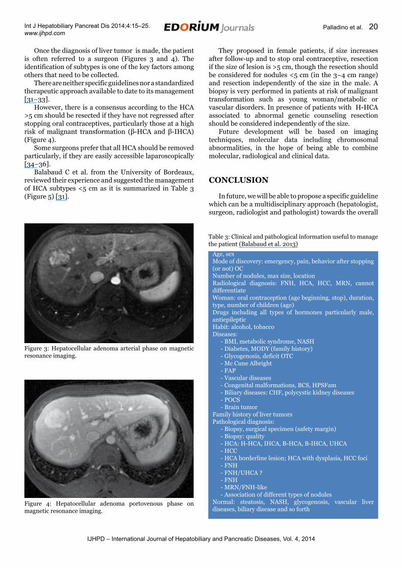
Once the diagnosis of liver tumor is made, the patient is often referred to a surgeon (Figures 3 and 4). The identification of subtypes is one of the key factors among others that need to be collected.
There are neither specific guidelines nor a standardized therapeutic approach available to date to its management [31–33].
However, there is a consensus according to the HCA >5 cm should be resected if they have not regressed after stopping oral contraceptives, particularly those at a high risk of malignant transformation (β-HCA and β-IHCA) (Figure 4).
Some surgeons prefer that all HCA should be removed particularly, if they are easily accessible laparoscopically [34–36].
Balabaud C et al. from the University of Bordeaux, reviewed their experience and suggested the management of HCA subtypes <5 cm as it is summarized in Table 3 (Figure 5) [31].
They proposed in female patients, if size increases after follow-up and to stop oral contraceptive, resection if the size of lesion is >5 cm, though the resection should be considered for nodules <5 cm (in the 3–4 cm range) and resection independently of the size in the male. A biopsy is very performed in patients at risk of malignant transformation such as young woman/metabolic or vascular disorders. In presence of patients with H-HCA associated to abnormal genetic counseling resection should be considered independently of the size.
Future development will be based on imaging techniques, molecular data including chromosomal abnormalities, in the hope of being able to combine molecular, radiological and clinical data.
cONcLUsION
In future, we will be able to propose a specific guideline which can be a multidisciplinary approach (hepatologist, surgeon, radiologist and pathologist) towards the overall
Figure 3: Hepatocellular adenoma arterial phase on magnetic resonance imaging.
Figure 4: Hepatocellular adenoma portovenous phase on magnetic resonance imaging.
Table 3: Clinical and pathological information useful to manage the patient (Balabaud et al. 2013)
Age, sexMode of discovery: emergency, pain, behavior after stopping (or not) OCNumber of nodules, max size, locationRadiological diagnosis: FNH, HCA, HCC, MRN, cannot differentiateWoman: oral contraception (age beginning, stop), duration, type, number of children (age)Drugs including all types of hormones particularly male, antiepilepticHabit: alcohol, tobaccoDiseases:
- BMI, metabolic syndrome, NASH- Diabetes, MODY (family history)- Glycogenosis, deficit OTC- Mc Cune Albright- FAP- Vascular diseases- Congenital malformations, BCS, HPSFam- Biliary diseases: CHF, polycystic kidney diseases- POCS- Brain tumor
Family history of liver tumorsPathological diagnosis:
- Biopsy, surgical specimen (safety margin)- Biopsy: quality- HCA: H-HCA, IHCA, B-HCA, B-IHCA, UHCA- HCC- HCA borderline lesion; HCA with dysplasia, HCC foci- FNH- FNH/UHCA ?- FNH- MRN/FNH-like- Association of different types of nodules
Normal: steatosis, NASH, glycogenosis, vascular liver diseases, biliary disease and so forth

IJHPD – International Journal of Hepatobiliary and Pancreatic Diseases, Vol. 4, 2014
Int J Hepatobiliary Pancreat Dis 2014;4:15–25. www.ijhpd.com
Palladino et al. 21
Figure 5: Management of HCA subtypes < 5 cm
Pa - Patients overweight/obese/with metabolic syndrome (the majority with IHCA) should be followed
In presence of multiple HCA nodules: generally all nodules are of the same subtype (i.e., H-HCA, IHCA) Among IHCA some may be B-catenin mutaded, others not. Association of H-HCA and IHCA has been rarely observed in a same liver. In glycogenosis, different types of nodules may exist (B-HCA, IHCA, B-IHCA and UHCA). The destruction (resection, radio frequency) of nodules depends on the size, on the presence of B-catenin mutation and on the disease background. OLT is exceptional and should not be considered just on the number criteria.MODY 3: maturity onset diabetes of the young type 3, GS: glutamine synthetase

IJHPD – International Journal of Hepatobiliary and Pancreatic Diseases, Vol. 4, 2014
Int J Hepatobiliary Pancreat Dis 2014;4:15–25. www.ijhpd.com
Palladino et al. 22
management of these diseases and is key factor for a good outcome.
*********
Author contributionsElisa Palladino – Substantial contriutions to conception and designs, Acquisition of data, Drafting the article, Revising it critically for important intellectual content, Final approval of the version to be publishedDaniele Sommacale – Substantial contriutions to conception and designs, Acquisition of data, Drafting the article, revising it critically for important intellectual content, Final approval of the version to be publishedRenaud Siboni – Substantial contriutions to conception and designs, Acquisition of data, Drafting the article, Revising it critically for important intellectual content, Final approval of the version to be publishedChristine Hoeffel – Substantial contriutions to conception and designs, Acquisition of data, Drafting the article, Revising it critically for important intellectual content, Final approval of the version to be publishedChristian Lechner – Substantial contriutions to conception and designs, Acquisition of data, Drafting the article, Revising it critically for important intellectual content, Final approval of the version to be publishedTullio Piardi – Substantial contriutions to conception and designs, Acquisition of data, Drafting the article, Revising it critically for important intellectual content, Final approval of the version to be publishedReza Kianmanesh – Substantial contriutions to conception and designs, Acquisition of data, Drafting the article, Revising it critically for important intellectual content, Final approval of the version to be published
GuarantorThe corresponding author is the guarantor of submission.
conflict of InterestAuthors declare no conflict of interest.
copyright© Elisa Palladino et al. 2014; This article is distributed under the terms of Creative Commons attribution 3.0 License which permits unrestricted use, distribution and reproduction in any means provided the original authors and original publisher are properly credited. (Please see www.ijhpd.com/copyright-policy.php for more information.)
AbbrEVIAtIONs:
US: UltrasoundMRI: Magnetic resonance imaging CEUS: Contrast-enhanced ultrasoundNFH: Nodular focal hyperplasiaHCA: Hepatocellular adenoma
NRH: Nodular regenerative hyperplasiaBLT: Benign liver tumourHCC: Hepatocellular CarcinomaCT: Computed TomographyOC: Oral contraceptivesGS: Glutamine synthetaseCRP: C-reactive –proteinIHCA: inflammatory adenomas
rEFErENcEs
1. Bonder A, Afdhal N. Evaluation of liver lesions. Clin Liver Dis 2012 May;16(2):271–83.
2. Mezhir JJ, Fourman LT, Do RK, et al. Changes in the management of benign liver tumours: An analysis of 285 patients. HPB (Oxford) 2013 Feb;15(2):156–3.
3. Mathieu D, Kobeiter H, Maison P, et al. Oral contraceptive use and focal nodular hyperplasia of the liver. Gastroenterology 2000;118(3):560–4.
4. Mneimneh W, Farges O, Bedossa P, Belghiti J, Paradis V. High serum level of alpha-fetoprotein in focal nodular hyperplasia of the liver. Pathol Int 2011;61(8):491–4.
5. Vilgrain V. Focal nodular hyperplasia. Eur J Radiol 2006;58(2):236–45.
6. Lewin M, Vilgrain V. Radiological diagnosis of benign liver cell tumors. Gastroenterol Clin Biol 2008 Mar;32(3):304–9.
7. Leen E. The role of contrast-enhanced ultrasound in the characterisation of focal liver lesions. Eur Radiol 2001;11 Suppl 3:E27–34.
8. Jang HJ, Yu H, Kim TK. Contrast-enhanced ultrasound in the detection and characterization of liver tumors. Cancer Imaging 2009 Nov 6;9:96–103.
9. Wang W, Chen LD, Lu MD, et al. Contrast-enhanced ultrasound features of histologically proven focal nodular hyperplasia: Diagnostic performance compared with contrast-enhanced CT. Eur Radiol 2013;23(9):2546–4.
10. Asbach P, Klessen C, Koch M, Hamm B, Taupitz M. Magnetic resonance imaging findings of atypical focal nodular hyperplasia of the liver. Clin Imaging 2007 Jul-Aug;31(4):244–52.
11. Fowler KJ, Brown JJ, Narra VR. Magnetic resonance imaging of focal liver lesions: Approach to imaging diagnosis. Hepatology 2011 Dec;54(6):2227–37.
12. Albiin N. MRI of Focal Liver Lesions. Current Medical Imaging Reviews 2012;8:107–16.
13. Balabaud C, Al-Rabih WR, Chen PJ, et al. Focal Nodular Hyperplasia and Hepatocellular Adenoma around the World Viewed through the scope of the Immunopathological Classification. International Journal of Hepatology 2013;2013:268625.
14. Bioulac-Sage P, Cubel G, Taouji S, et al. Immunohistochemical markers on needle biopsies are helpful for the diagnosis of focal nodular hyperplasia and hepatocellular adenoma subtypes.The American Journal of surgical Pathology 2012 Nov;36(11):1691–9.
15. Fabre A, Audet P, Vilgrain V, et al. Histological scoring of liver biopsy in focal nodular hyperplasia

IJHPD – International Journal of Hepatobiliary and Pancreatic Diseases, Vol. 4, 2014
Int J Hepatobiliary Pancreat Dis 2014;4:15–25. www.ijhpd.com
Palladino et al. 23
with atypical presentation. Hepatology 2002;35:414–20.
16. Cherqui D. Clinical Management of benign liver cell tumors. Gastroentérologie Clinique et Biologique 2008 Mar;32(3):310–3. [Article in French].
17. Barthelmes L, Tait IS. Liver cell adenoma and liver cell adenomatosis. HPB (Oxford) 2005;7(3):186–96.
18. Deneve JL, Pawlik TM, Cunningham S, et al. Liver cell adenoma: A multicenter analysis of risk factors for rupture and malignancy. Annals of Surgical Oncology 2009 Mar;16(3):640–8.
19. Farges O, Ferreira N, Dokmak S, Belghiti J, Bedossa P, Paradis V. Changing trends in malignant transformation of hepatocellular adenoma. Gut 2011 Jan;60(1):85-9.
20. Seo JM, Lee SJ, Kim SH, Park CK, Ha SY. Hepatocellular carcinoma arising from hepatocellular adenoma in a hepatitis B virus-associated cirrhotic liver. Clinical Radiology 2012 Apr;67(4):329–3.
21. Zucman-Rossi J, Jeannot E, Nhieu JT, et al. Genotype-phenotype correlation in hepatocellular adenoma: new classification and relationship with HCC. Hepatology 2006 Mar;43(3):515–24.
22. Bioulac-Sage P, Rebouissou S, Thomas C, et al. Hepatocellular adenoma subtype classification using molecular markers and immunohistochemistry. Hepatology 2007 Sep;46(3):740–8.
23. Rebouissou S, Bioulac-Sage P, Zucman-Rossi J. Molecular pathogenesis of focal nodular hyperplasia and hepatocellular adenoma. J Hepatol 2008;48:163–70.
24. Bioulac-Sage P, Laumonier H, Couchy G, et al. Hepatocellular adenoma management and phenotypic classification: The Bordeaux experience. Hepatology 2009 Aug;50(2):481–9.
25. Nault JC, Zucman Rossi J. Molecular Classification of Hepatocellular Adenomas. International Journal of Hepatology 2013;2013:315947.
26. Nault JC, Bioulac-Sage P, Zucman-Rossi J. Hepatocellular benign tumors-from molecular classification to personalized clinical care. Gastroenterology 2013 May,144(5):888–902.
27. Bluteau O, Jeannot E, Bioulac-Sage P, et al. Bi-allelic inactivation of TCF1 in hepatic adenomas. Nature Genetics 2002 Oct;32(2):312–5.
28. Lagana SM, Salomao M, Bao F, Moreira RK, Lefkowitch JH, Remotti HE. Utility of a immunohistochemical panel consisting of glypican-3, heat-shock protein-70,and glutamine synthetase in the distinction of low-grade hepatocellular carcinoma from hepatocellular adenoma. Appl Immunohistochemistry Molecular Morphology 2013 Mar;21(2):170–6.
29. Sa Cunha A, Blanc JF, Lazaro E, et al. Inflammatory syndrome with liver adenomatosis: The beneficial effects of surgical management. Gut 2007;56(2):307–9.
30. Paradis V, Champault A, Ronot M, et al. Telangiectatic adenoma: an entity associated with increased body mass index and inflammation. Hepatology 2007 Jul;46(1):140–6.
31. Bioulac-Sage P, Balabaud C, Zucman-Rossi J. Focal nodular hyperplasia, hepatocellular adenomas: Past, present, future. Gastroenterol Clin Biol. 2010 Aug-Sep;34(6-7):355–8.
32. Buell JF, Tranchart H, Cannon R, Dagher I. Management of benign hepatic tumor. Surg Clin North Am 2010 Aug;90(4):719–35.
33. Fonseca S, Hoton D, Dardenne S, et al. Histological and Immunohistochemical Revision of Hepatocellular Adenomas: A Learning Experience. International Journal of Hepatology 2013;2013:398308.
34. Ardito F, Tayar C, Laurent A, Karoui M, Loriau J, Cherqui D. Laparoscopic liver resection for benign disease. Arch Surg 2007;142:1188–93.
35. Dokmak S, Raut V, Aussilhou B, et al. Laparoscopic left lateral resection is the gold standard for benign liver lesions: A case-control study. HPB (Oxford) 2014 Feb;16(2):183–7.
36. Dokmak S, Paradis V, Vilgrain V, et al. A single-center surgical experience of 122 patients with single and multiple hepatocellular adenomas. Gastroenterology 2009 Nov;137(5):1698–705.

IJHPD – International Journal of Hepatobiliary and Pancreatic Diseases, Vol. 4, 2014
Int J Hepatobiliary Pancreat Dis 2014;4:15–25. www.ijhpd.com
Palladino et al. 24
christian Lechner is Chief Resident at Department of General and Digestive Surgery of Chalons-en- Champagne Hospital, France. His area of interest include gastrointestinal surgery, minimally invasive abdominal surgery.
tullio Piardi is Resident at Department of General and Digestive Surgery, University of Reims, School of Medecine, France. His area of interest include liver and pancreatic surgery, gastrointestinal surgery. He has published research papers in national and international academic journals.
reza Kianmanesh is a Professor at Department of General and Digestive Surgery, University of Reims, School of Medecine, France. His area of interest include hepatobiliary and pancreatic surgery, liver transplantation, gastrointestinal surgery. He has published research papers in national and international academic journals and authored books.
ABOUT THE AUTHORS
Article citation: Palladino E, Sommacale D, Siboni R, Hoeffel C, Lechner C, Piardi T, Kianmanesh R. Focal nodular hyperplasia and hepatocellular adenomas: What is new in 2013? Int J Hepatobiliary Pancreat Dis 2014;4:15–25.
Elisa Palladino is Resident at Department of General and Digestive Surgery of Chalons-en-Champagne Hospital, France. Her area of interest include liver surgery, gastrointestinal surgery, minimally invasive abdominal surgery. She has published research papers in national and international academic journals.
renaud siboni is Resident at Department of Surgery, University of Reims, France. His area of interest include general surgery.
Daniele sommacale is Professor at Department of General and Digestive Surgery, University of Reims, School of Medecine, France. His area of interest include hepatobiliary surgery and liver transplantation. He has published research papers in national and international academic journals and authored books.
christine Hoeffel (mD, PhD), She is Professor of Radiology at the University hospital of Reims, France. She has published or co-published over 100 papers, mainly focused on abdominal imaging and magnetic resonance imaging.
IJHPD – International Journal of Hepatobiliary and Pancreatic Diseases, Vol. 4, 2014
Int J Hepatobiliary Pancreat Dis 2014;4:15–25. www.ijhpd.com
Palladino et al. 25
Access full text article onother devices
Access PDF of article onother devices