aberrant p53 expression and the development of gallbladder carcinoma and adenoma
TRANSCRIPT

p53 Overexpression in gallbladder carcinoma
Kaohsiung J Med Sci February 2006 • Vol 22 • No 2 53
Gallbladder carcinoma (GBC) is a highly lethal but relativelyrare neoplasm of the digestive tract. Generally speaking,the overall 5-year survival rate is only 5–15% [1,2]. GBCalso shows an unusual geographic and demographicdistribution. It affects females two to six times morefrequently than males, and its incidence increases with age[3]. The highest incidences are observed in North and SouthAmerica, Poland, and northern India. Low incidences areseen in Singapore and Nigeria [4]. In Taiwan, GBC is alsouncommon, and 19.2% of cases are associated with a goodoperative prognosis [5,6].
ABERRANT P53 EXPRESSION AND THE DEVELOPMENT OF
GALLBLADDER CARCINOMA AND ADENOMA
Shen-Nien Wang,1 Shin-Chang Chung,1 Kun-Bow Tsai,2 Chee-Yin Chai,2 Wen-Tsun Chang,1
Kung-Kai Kuo,1 Jong-Shyone Chen,1 and King-Teh Lee1
1Division of Hepatobiliary Surgery, Department of Surgery and 2Department of Pathology,Kaohsiung Medical University Chung-Ho Memorial Hospital, Kaohsiung Medical
University, Kaohsiung, Taiwan.
Gallbladder carcinoma (GBC) is a highly lethal but relatively rare neoplasm of the digestive tract. Theprogression from gallbladder adenoma to carcinoma remains unclear. The p53 gene is the most frequentlymutated tumor suppressor in human cancers. In this study, we analyzed the expression patterns of the p53protein in 22 cases of GBC, 17 cases of precursor lesions (16 gallbladder adenomas and 1 cystadenoma), and15 cases of normal epithelia using immunohistochemical analysis. The results were correlated withclinicopathologic characteristics. We found that p53 expression was significantly increased in 59.1% (13/22)of GBC cases and in 17.6% (3/17) of gallbladder adenoma cases (p = 0.009). There was no p53 expression inthe 15 cases of normal epithelia, and a significant difference was shown between normal epithelium and GBCcases (p < 0.001). In addition, the expression pattern of p53 protein did not show any significant correlationwith the histologic type and the differentiation grade of GBC. In conclusion, we suggest that the aberrant p53expression may play a role in the occurrence of GBC.
Key Words: p53, immunohistochemistry, gallbladder adenoma, gallbladder carcinoma(Kaohsiung J Med Sci 2006;22:53–9)
Received: August 10, 2005 Accepted: November 25, 2005Address correspondence and reprint requests to: Dr. King-Teh Lee,Department of Surgery, Kaohsiung Medical University Hospital, No.100, Tzyou 1st Road., Kaohsiung County, Taiwan 807,Taiwan.E-mail: [email protected]
These differences in the incidence and mortality ratesof GBC suggest that genetic and environmental variablesmay be involved in its tumorigenesis. Although there havebeen improvements in diagnostic technique, there is,unfortunately, no promising biomarker that can detectGBC at an early stage. Thus, most cases are diagnosed at anadvanced stage with a poor outcome.
Recently, it has been demonstrated that the adenoma-carcinoma sequence is the usual route for the tumorigenesisof GBC [7–9]. However, there is limited evidence concerningthe possible mechanisms involved in the transformation.Tumorigenesis is regarded as a multistep process in whichgenetic alterations accumulate as premalignant lesionsprogress to invasive carcinomas. Among the many knowngenetic changes in human carcinogenesis, the most commonis an abnormality in the tumor suppressor gene p53.Numerous investigators have shown that p53 mutations areinvolved in the genesis of many human cancers, including
© 2006 Elsevier. All rights reserved.
S.N. Wang, S.C. Chung, K.B. Tsai, et al
54 Kaohsiung J Med Sci February 2006 • Vol 22 • No 2
GBC [10,11]. However, the role of p53 in the adenoma-carcinoma sequence of gallbladder tumorigenesis remainsunclear.
The accumulated literature suggests the oncogenicproperties of p53 brought about by its mutations [12,13].Mutations in p53 have been found in approximately 50% ofhuman tumors. One of the most common mechanisms ispoint mutation leading to the elevated expression ofconformationally altered and functionally defective p53proteins. Another is the deletion of one p53 allele, resultingin the absence of the wild-type p53 protein. Both mechanismscause the loss of tumor suppressor function and permitoncogenesis.
The best way to improve the prognosis of GBC is toidentify and correct the risk factors that may influence itsdevelopment. Our objectives were to explore the biologicproperties of gallbladder carcinogenesis and to improve theprognosis of GBC. In the present study, we examined theexpression pattern of the p53 protein in 54 tissue specimens,including normal epithelia, precursor lesion, and carcinoma,by using immunohistochemistry.
MATERIALS AND METHODS
Tissue specimensTissues from 22 GBC patients (9 men and 13 women, agerange 38–87 yrs, mean age 66.3 ± 11.3 yrs) and 17gallbladder adenoma patients (5 men and 12 women,age range 23–81 yrs, mean age 55.2 ± 16.2 yrs) wereobtained from the Department of Surgery of theKaohsiung Medical University Hospital. Furthermore,15 normal gallbladder tissues, harvested from patientswho underwent right lobectomy to treat hepatocellularcarcinoma, were also included in this study. All of thesespecimens were initially fixed in 10% neutral bufferedformalin and then embedded in paraffin after furtherdehydration processing.
The precursor lesions of the biliary epithelium weredefined according to Albores-Saavedra et al [14]. Adenomais defined as a benign neoplasm, typically polyploidy,well-demarcated, single, and arises from glandularepithelium. Cystadenoma is defined as a multiloculatedcystic neoplasm containing mucinous or serous fluid andlined by columnar epithelium reminiscent of bile duct orgastric epithelium. GBC is histologically classifiedaccording to the World Health Organization HistologicalClassification of Tumors of the Gallbladder and ExtrahepaticBile Ducts.
ImmunohistochemistrySections (4 µm thick) were prepared from the representativetissue blocks on a microtome. Routine immunohistochemistryprocedures were followed as described [15]. In brief, theslides were deparaffinized with xylene rinse and rehydratedinto distilled water through graded alcohol. Endogenousperoxidase activity was quenched by 10 minutes ofincubation in a 3% hydrogen peroxide–methanol buffer.Antigen retrieval was enhanced by autoclaving slides insodium citrate buffer (pH 6.0) for 15 minutes and allowingthe solution to cool slowly at room temperature for 20minutes. The slides were then incubated with primarymouse monoclonal anti-p53 antibody (Neomarkers) at adilution of 1:200 in a humidified chamber for 30 minutes, atroom temperature.
The slides were washed three times in phosphate-buffered solution and further incubated with biotinylatedsecondary antibody for 20 minutes at room temperature.Antigen-antibody complexes were detected by the avidin-biotin-peroxidase method using diaminobenzidine as achromogenic substrate (DAKO, CA, USA). Finally, the slideswere counterstained with hematoxylin and then examinedby light microscopy.
Interpretation of p53 immunostainingImmunohistochemical expression levels of p53 were scoredby a semiquantitative method evaluating the intensity andpercentage of positive cells. Only nuclear p53 staining wasregarded as positive staining. Accordingly, the intensitywas graded as absent (0), mild (1), moderate (2), and intense(3); while the incidence was categorized as absent (0), lessthan 10% (1), 10–50% (2), and more than 50% of positivecells (3). Overall staining score was obtained (range 0–6) byadding both variables. A score equal to or greater than 3 wasconsidered as positive for p53 overexpression. In addition,substituting the primary antibody with the immunoglobulinfraction of nonimmune mouse serum in grade 3 casesserved as a negative control. Nuclear staining wasdetermined separately for each specimen, estimated by twoindependent pathologists. The rare cases with discordantscores were re-evaluated and scored on the basis of theconsensual opinion.
Statistical analysisStatistical differences between categorized groups wereanalyzed by the χ2 test and the Fisher’s exact test. Allstatistical analyses were performed with SPSS softwarepackage for PC (Version 10.0, SPSS Inc, Chicago, IL, USA). Ap value less than 0.05 was considered statistically significant.

p53 Overexpression in gallbladder carcinoma
Kaohsiung J Med Sci February 2006 • Vol 22 • No 2 55
RESULTS
The expression patterns of the p53 protein in the normalepithelia, precursor lesions, and GBCs are summarized inTables 1 and 2.
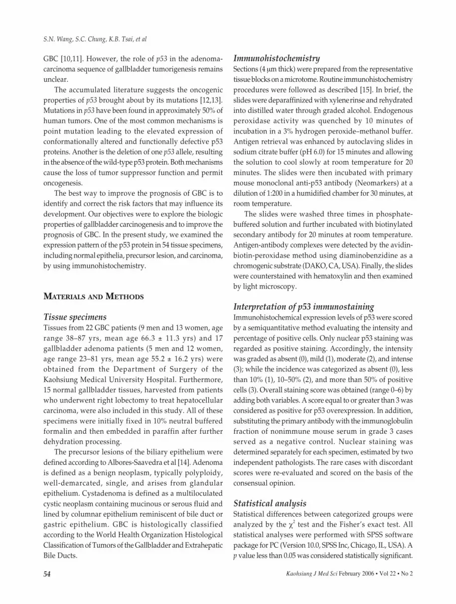
We analyzed 17 precursor lesions, including 16adenomas and 1 biliary cystadenoma (Table 1). Accordingto the growth pattern, gallbladder adenomas were dividedinto three types: tubular, papillary and tubulopapillary.In this study, all gallbladder adenomas were of tubulartype. Cytologically, of the 16 gallbladder adenomas, 12cases were pyloric gland type and 4 cases were intestinaltype. Positive p53 immunoreactivity was observed in16.7% (2/12) (Figure 1) of the pyloric gland type and in25% (1/4) of the intestinal type. In addition, there wasno p53 immunostaining in gallbladder cystadenoma.Overall, positive overexpression of p53 protein wasobserved in 3 (17.6%) of 17 precursor lesions of thegallbladder (Table 1).
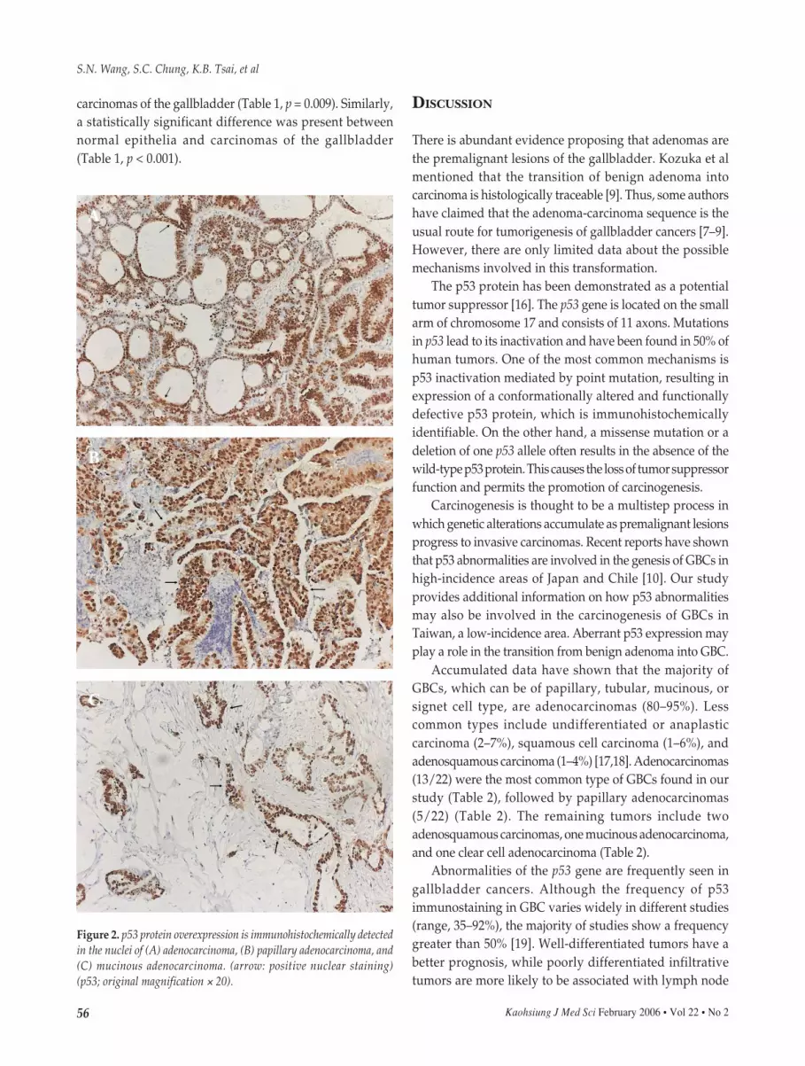
The 22 GBC cases in our study were of the followingtypes: 13 adenocarcinoma, 5 papillary adenocarcinoma,2 adenosquamous carcinoma, 1 mucinous adenocarcinoma,and 1 clear cell carcinoma (Table 2). We noted that p53overexpression was predominantly seen in two groups,adenocarcinoma (Figure 2A) and papillary adenocarcinoma(Figure 2B), which were 69.2% (9/13) and 60% (3/5),respectively (Table 2). In contrast, p53 overexpression wasnot observed in adenosquamous carcinoma or clear celladenocarcinoma specimens. p53 protein was alsooverexpressed in cases of mucinous adenocarcinoma(Figure 2C) and in 59.1% (13/22) of GBCs (Table 1).
According to the differentiation grade, gallbladderadenocarcinomas were subgrouped into well, moderately,and poorly differentiated adenocarcinomas. We foundthat p53 immunostaining was overexpressed in 50%,60%, and 83.3% of well, moderately, and poorlydifferentiated adenocarcinomas, respectively (Table 2).Although no significant correlation was shown betweenp53 overexpression and differentiation grade, p53overexpression was relatively higher in the poorlydifferentiated adenocarcinoma cases when compared withwell-differentiated cases (Table 2).
Normal gallbladder epithelium was examined in 15specimens resected from patients with hepatocellularcarcinoma. We found no positive p53 immunoreactivity inthese cases. Meanwhile, normal epithelia in the vicinity ofprecursor lesion and GBCs also showed no p53 expression.
A statistically significant difference in p53 proteinoverexpression was present between precursor lesions and
Table 2. Correlation of p53 overexpression and histologicfeatures in gallbladder carcinoma
Histologic features No. tested No. positivecases (%)
Histologic subtype 22Adenocarcinoma 13 9 (69.2)Papillary adenocarcinoma 5 3 (60)Adenosquamous carcinoma 2 0 (0)Mucinous adenocarcinoma 1 1 (100)Clear cell adenocarcinoma 1 0 (0)
Differentiation grade 13Well differentiated 2 1 (50)Moderately differentiated 5 3 (60)Poorly differentiated 6 5 (83.3)
Table 1. The incidence of p53 overexpression in gallbladderpathologic and normal epithelia
Histologic type Cases tested Positive cases (%)
Carcinoma 22 13 (59.1)*,†
Precursor lesions 17 3 (17.6)Adenoma 16 3 (18.8)
Pyloric gland type 12 2 (16.7)Intestinal type 4 1 (25)
Cystadenoma 1 0 (0)
Normal epithelium 15 0 (0)
*p < 0.01 vs normal epithelium; †p < 0.01 vs precursor lesions.
Figure 1. p53 protein overexpression is immunohistochemically detectedin the nuclei (arrow) of pyloric gland adenomas (p53; originalmagnification × 20).
S.N. Wang, S.C. Chung, K.B. Tsai, et al
56 Kaohsiung J Med Sci February 2006 • Vol 22 • No 2
carcinomas of the gallbladder (Table 1, p = 0.009). Similarly,a statistically significant difference was present betweennormal epithelia and carcinomas of the gallbladder(Table 1, p < 0.001).
DISCUSSION
There is abundant evidence proposing that adenomas arethe premalignant lesions of the gallbladder. Kozuka et almentioned that the transition of benign adenoma intocarcinoma is histologically traceable [9]. Thus, some authorshave claimed that the adenoma-carcinoma sequence is theusual route for tumorigenesis of gallbladder cancers [7–9].However, there are only limited data about the possiblemechanisms involved in this transformation.
The p53 protein has been demonstrated as a potentialtumor suppressor [16]. The p53 gene is located on the smallarm of chromosome 17 and consists of 11 axons. Mutationsin p53 lead to its inactivation and have been found in 50% ofhuman tumors. One of the most common mechanisms isp53 inactivation mediated by point mutation, resulting inexpression of a conformationally altered and functionallydefective p53 protein, which is immunohistochemicallyidentifiable. On the other hand, a missense mutation or adeletion of one p53 allele often results in the absence of thewild-type p53 protein. This causes the loss of tumor suppressorfunction and permits the promotion of carcinogenesis.
Carcinogenesis is thought to be a multistep process inwhich genetic alterations accumulate as premalignant lesionsprogress to invasive carcinomas. Recent reports have shownthat p53 abnormalities are involved in the genesis of GBCs inhigh-incidence areas of Japan and Chile [10]. Our studyprovides additional information on how p53 abnormalitiesmay also be involved in the carcinogenesis of GBCs inTaiwan, a low-incidence area. Aberrant p53 expression mayplay a role in the transition from benign adenoma into GBC.
Accumulated data have shown that the majority ofGBCs, which can be of papillary, tubular, mucinous, orsignet cell type, are adenocarcinomas (80–95%). Lesscommon types include undifferentiated or anaplasticcarcinoma (2–7%), squamous cell carcinoma (1–6%), andadenosquamous carcinoma (1–4%) [17,18]. Adenocarcinomas(13/22) were the most common type of GBCs found in ourstudy (Table 2), followed by papillary adenocarcinomas(5/22) (Table 2). The remaining tumors include twoadenosquamous carcinomas, one mucinous adenocarcinoma,and one clear cell adenocarcinoma (Table 2).
Abnormalities of the p53 gene are frequently seen ingallbladder cancers. Although the frequency of p53immunostaining in GBC varies widely in different studies(range, 35–92%), the majority of studies show a frequencygreater than 50% [19]. Well-differentiated tumors have abetter prognosis, while poorly differentiated infiltrativetumors are more likely to be associated with lymph node
Figure 2. p53 protein overexpression is immunohistochemically detectedin the nuclei of (A) adenocarcinoma, (B) papillary adenocarcinoma, and(C) mucinous adenocarcinoma. (arrow: positive nuclear staining)(p53; original magnification × 20).
A
B
C

p53 Overexpression in gallbladder carcinoma
Kaohsiung J Med Sci February 2006 • Vol 22 • No 2 57
metastases and liver invasion. In this study, we showed that59.1% of GBCs overexpressed p53 protein (Table 1).Moreover, although our results showed no significantcorrelation between p53 overexpression and differentiationgrade, p53 protein overexpression seemed to be moreprominent in cases of poor differentiation grade (Table 2).
The precursor lesions of GBCs are subgrouped by theclassification proposed by Albores-Saavedra et al [14].Adenoma, a benign glandular tumor, is one of the mostcommon precursor lesions of the gallbladder [20], with areported incidence of 0.4–1.1% in cholecystectomyspecimens. As expected, adenoma constituted the majorityof the precursor lesions (16/17) in our study (Table 1).Microscopically, there are three growth pattern types foradenomas: tubular, papillary, and mixed. All of our 16adenomas were tubular. Cytologically, adenomas can befurther classified into three subtypes: pyloric gland,intestinal, and biliary. As reported, tubular adenomas ofthe pyloric gland type are more common in the gallbladder,while the intestinal type are more common in theextrahepatic bile duct [8,21]. In the present study, the pyloricgland type predominated in 12 of 16 tubular adenomas, andthe remaining 4 adenomas were the intestinal type. Somestudies also described spindle cell metaplasia, which isoccasionally observed in these adenomas. Spindle cellmetaplasias, also called squamoid morules, are characterizedby nodular aggregates of cytologically bland spindle cellswith eosinophilic cytoplasm but without keratinization orintercellular bridges. Its incidence ranges from 5.3–33.3%[22]. In our group, we observed squamoid morules in 3(25%) of 12 pyloric gland adenomas (data not shown).
It has been suggested that two main histologic pathwaysexist for the development of gallbladder cancer: (1) thedysplasia-carcinoma in situ sequence and (2) the adenoma-carcinoma sequence. Wistuba and Albores-Saavedra [23]demonstrated the high incidence of p53 overexpression andits presence in dysplasia, even in specimens with invasivecarcinomas. Therefore, they suggested that dysplasia-carcinoma in situ is the usual route for gallbladdercarcinogenesis and that p53 abnormality is an important andearly event [19,23]. On the other hand, Watanabe et al [24]showed a low frequency of p53 overexpression even in thecarcinoma portion of carcinoma-in-adenoma cases (6.3%),indicating that the adenoma-carcinoma sequence may alsobe important. In this study, p53 overexpression was observedin 18.8% (3/16) of the adenomas, and a statistical differenceexists between adenomas and carcinomas of the gallbladder(Table 1). It indicated that gallbladder adenomas lack thep53 abnormalities frequently seen in GBCs.
In conclusion, we suggest that p53 overexpression mayplay a role in GBCs in Taiwan. However, this alteration maynot be involved in the putative pathway of gallbladdercarcinogenesis: the adenoma-carcinoma sequence.Therefore, other mechanisms or variables may be importantin this pathway.
REFERENCES
1. Cubertafond P, Mathonnet M, Gainant A, et al. Radical surgeryfor gallbladder cancer. Results of the French SurgicalAssociation Survey. Hepatogastroenterology 1999;46:1567–71.
2. Donohue JH, Stewart AK, Menck HR. The National CancerData Base report on carcinoma of the gallbladder, 1989–1995.Cancer 1998;83:2618–28.
3. Lazcano-Ponce EC, Miquel JF, Munoz N, et al. Epidemiologyand molecular pathology of gallbladder cancer. CA Cancer JClin 2001;51:349–64.
4. Misra S, Chaturvedi A, Misra NC, et al. Carcinoma of thegallbladder. Lancet Oncol 2003;4:167–76.
5. Chao TC, Wang CS, Jeng LB, et al. Primary carcinoma of thegallbladder in Taiwan. J Surg Oncol 1996;61:49–55.
6. Yang TL, Liu CL, Liu TP, et al. Primary carcinoma of thegallbladder: results of surgery—a retrospective study.Zhonghua Yi Xue Za Zhi (Taipei) 1999;62:68–75.
7. Yokoyama N, Watanabe H, Ajioka Y, et al. [Genetic alterationsin gallbladder carcinoma: a review]. Nippon Geka Gakkai Zasshi1998;99:687–95.
8. Itoi T, Watanabe H, Ajioka Y, et al. APC, K-ras codon 12mutations and p53 gene expression in carcinoma and adenomaof the gall-bladder suggest two genetic pathways in gall-bladder carcinogenesis. Pathol Int 1996;46:333–40.
9. Kozuka S, Tsubone N, Yasui A, et al. Relation of adenoma tocarcinoma in the gallbladder. Cancer 1982;50:2226–34.
10. Yokoyama N, Hitomi J, Watanabe H, et al. Mutations of p53 ingallbladder carcinomas in high-incidence areas of Japan andChile. Cancer Epidemiol Biomarkers Prev 1998;7:297–301.
11. Sessa F, Furlan D, Genasetti A, et al. Microsatellite instabilityand p53 expression in gallbladder carcinomas. Diagn MolPathol 2003;12:96–102.
12. Koda M, Yashima K, Kawaguchi K, et al. Expression of Fhit,Mlh1, and p53 protein in human gallbladder carcinoma. CancerLett 2003;199:131–8.
13. Misra S, Chaturvedi A, Goel MM, et al. Over-expression of p53protein in gallbladder carcinoma in North India. Eur J SurgOncol 2000;26:164–7.
14. Albores-Saavedra J, Henson DE, Sobin LH. The WHOHistological Classification of Tumors of the Gallbladder andExtrahepatic Bile Ducts. A commentary on the second edition.Cancer 1992;70:410–4.
15. Chung SC, Lee KT, Tsai KB, et al. Immunohistochemical studyof DPC4 and p53 proteins in gallbladder and bile duct cancers.World J Surg 2004;28:995–1000.
16. Vogelstein B, Lane D, Levine AJ. Surfing the p53 network.Nature 2000;408:307–10.
S.N. Wang, S.C. Chung, K.B. Tsai, et al
58 Kaohsiung J Med Sci February 2006 • Vol 22 • No 2
17. Misra S, Chaturvedi A, Misra NC. Carcinoma gallbladderpresenting with skeletal metastases. Indian J Gastroenterol1997:69–87.
18. Henson DE, Albores-Saavedra J, Corle D. Carcinoma of theextrahepatic bile ducts. Histologic types, stage of disease,grade, and survival rates. Cancer 1992;70:1498–501.
19. Itoi T, Watanabe H, Yoshida M, et al. Correlation of p53protein expression with gene mutation in gall-bladdercarcinomas. Pathol Int 1997;47:525–30.
20. Sasatomi E, Tokunaga O, Miyazaki K. Precancerous conditionsof gallbladder carcinoma: overview of histopathologiccharacteristics and molecular genetic findings. J Hepatobiliary
Pancreat Surg 2000;7:556–67.21. Takei K, Watanabe H, Itoi T, et al. p53 and Ki-67 immunoreactivity
and nuclear morphometry of 'carcinoma-in-adenoma' andadenoma of the gall-bladder. Pathol Int 1996;46:908–17.
22. Yim H, Jin YM, Shim C. Tubular adenoma of the gallbladderwith spindle cell metaplasia. J Korean Med Sci 1998;13:295–8.
23. Wistuba II, Albores-Saavedra J. Genetic abnormalities involvedin the pathogenesis of gallbladder carcinoma. J HepatobiliaryPancreat Surg 1999;6:237–44.
24. Watanabe H, Date K, Itoi T, et al. Histological and geneticchanges in malignant transformation of gallbladder adenoma.Ann Oncol 1999;10:136–9.

Kaohsiung J Med Sci February 2006 • Vol 22 • No 2 59
éRP=�� !"#$
�� !"#$%&'(
�� N
= =�� N
= =�� O
= =�� O
= =�� N
= =�� N
= =�� N
= =�� N
�� !"!#$%&'( )= =N
�� !"= =O
��
�� !"#$%&'()*+,-./0123456!789��:2;<=
�� !"#$%&'()*+,-./01234!éRP=�� !"#$%&'
�� !"#$%&'éRP=�� !"#$%&'()*+,-(./012'
�� !"#$%&'()*+,-./0123456789:;<=>=OO=�
�� !NT=�� !"#$=NR=�� !"#$%&'()*+%&,-./01
�� !"#$= éRP=�� !"#$%&'()*+,-./01234#56
�� !"!#$%&'=RVKNB=�� !"=NTKSB=�� !"#$=éRP=�� !
�� !"#$%&'$()*+,-./m=�� =MKMMV�� !"#$%&'(
�� !"#$%&'=éRP=�� !"#$%&'()*+,-./012345
�� !"#$%&'()*m=�� = MKMMN�� !"#$%&'(=éRP=��
�� !"#$%&'()*+,-./0 !1234�56'789:;<=>
�� !"#$%�=éRP=�� !"#$%&'()*+,-.'/01
�� !éRP=�� !"#$%&'()!*+,-!*+.
�� !"=OMMSXOOWRPJV�
�� !"VQ=�=U=�=NM=�
�� !"VQ=�=NN=�=OR=�
�� !"#$%&'
�� !"!#$%&'( )*+,-.
�� =UMT=�� !"#$=NMM=�