lentiviral vectors for delivery of genes into neonatal and adult ventricular cardiac myocytes in...
TRANSCRIPT
Basic Res Cardiol 97: 348 – 358 (2002)DOI 10.1007/s00395-002-0360-0 ORIGINAL CONTRIBUTION
Jing ZhaoGavin J. PettigrewJoan ThomasJamie I. VandenbergLuc DelriviereEleanor M. BoltonAndrew CarmichaelJody L. MartinMichael S. MarberAndrew M. L. Lever
Lentiviral vectors for delivery of genes into neonatal and adult ventricular cardiacmyocytes in vitro and in vivo
� Abstract Vectors based on lentiviruses such as human immunodeficiencyvirus (HIV) type-1 have many advantages for gene therapy, including the abil-ity to infect non-dividing cells, long-term transgene expression and theabsence of induction of an inflammatory/immune response. This study wasinitiated to determine whether lentiviruses would efficiently transfer genes toboth neonatal and adult cardiac cells in culture and, by direct injection, to theheart in vivo. A three-plasmid expression system, including a packagingdefective helper construct, a plasmid coding for a heterologous (VSV-G)envelope protein and a vector construct harboring reporter genes – E-GFP(enhanced green fluorescent protein) and puro (puromycin-resistance pro-tein) was used to generate pseudotyped HIV-1 particles by transient trans-fection of human embryonic kidney 293T cells. We demonstrated efficientgene transfer into neonatal and adult cardiac myocytes in vitro and identifiedconditions in which virtually 100% of cultured neonatal and 70% of adult car-diac myocytes express the reporter gene. Transduction of adult cardiac myo-cytes with high titre lentiviral vectors did not affect the cell number, mor-phology or viability compared to untransduced cells. We delivered HIV-1-based vectors to the intact heart by direct injection. Hearts transduced withpseudotyped HIV-1 vectors showed levels of transgene expression compara-ble to that achieved by adenovirus vectors. This study demonstrates for thefirst time that lentivirus-based vectors can successfully transduce adultcardiomyocytes both in vitro and in vivo, and opens up the prospect oflentivirus-based vectors becoming an important gene delivery system in thecardiovascular field.
� Key words HIV-1 – lentiviral vector – cardiac myocytes – gene transfer –heart
BRC
360
Prof. A. M. L. Lever (�) · J. Zhao · J. ThomasA. Carmichael Department of MedicineAddenbrooke’s HospitalHills RoadCambridge, CB2 2QQ, UKTel.: 01223/336747Fax: 01223/336846E-Mail: [email protected]
G. J. Pettigrew · L. Delriviere · E. M. BoltonUniversity of Cambridge Department of SurgeryAddenbrooke’s HospitalHills RoadCambridge, CB2 2QQ, UK
J. I. VandenbergUniversity of Cambridge Department of BiochemistryBuilding O, Downing SiteCambridge, CB2 1QW, UK
J. L. Martin · M. S. MarberDepartment of CardiologyKing’s College LondonRayne InstituteSt Thomas HospitalLondon, SE1 7EH, UK
Received: 1 October 2001Returned for 1. revision: 18 October 20011. Revision received: 19 November 2001Returned for 2. revision: 6 December 20012. Revision received: 13 February 2002Accepted: 6 March 2002
Introduction
Gene delivery to cardiac myocytes has the potential bothto therapeutically correct genetic defects and, investi-gationally, to study cardiac muscle physiology. It has
proven difficult to deliver genes to cardiac myocytes asthey are terminally differentiated, they do not divide,and, in vitro, have a relatively short life span (19). Genedelivery to cardiac myocytes is further hampered by tox-icity associated with delivery vectors; adenoviruses, forexample, may disrupt the physiology of the cell (54).

J. Zhao et al. 349Lentiviral vector gene delivery to heart
Vectors based on lentiviruses have been in use for anumber of years and have been shown to be able to trans-duce cells which are not dividing (35, 42), both in vitroand in vivo. This is due to the native capability of lenti-viruses to infect terminally differentiated cells of themonocyte/macrophage series (7, 27, 59). Transductioninvolves integration of genetic material into the targetcell and this allows for long-term stable gene expressionfrom what has now become a new cellular genetic ele-ment (41). Newer pseudotyped lentiviral vectors offer amajor advantage over other high efficiency systems inthat they appear to cause little or no disruption of the tar-get cell (26). This is concordant with the fact that retro-viruses themselves rely on perturbing cell function as lit-tle as possible for their own survival (11). Lentiviral vec-tors also have a relatively large capacity for gene transfer,potentially up to 8 kilobases of sequence.
We and others demonstrated HIV-based vector genedelivery in the early 1990s following our identification ofthe HIV-1 encapsidation signal (15, 25, 46–48). Since thattime, heterologous envelopes have been used to widenthe tropism of the vector (8) and, in doing so, have led tothe generation of a more stable particle capable of beingfrozen and thawed and with a degree of independence oflentiviral accessory gene products for infection (1, 16).We have been using three- and four-plasmid lentiviralvector systems to deliver genes to a number of cells. Wedescribe the ability of lentivirus-based systems to deliverand stably express genes in both neonatal and adult car-diac myocytes without any perturbation of cell viabilityor evidence of cytotoxicity. For the first time, we havedemonstrated that a lentiviral vector system is able totransduce the heart in vivo. These results suggest thatlentiviruses could offer a promising new approach fortreatment of cardiovascular diseases and for cardiovas-cular studies.
Materials and methods
� Plasmid constructs
The vector HVP is based on the HXBc2 isolate of HIV-1and contains a Bal 1-EcoR1 deletion (2621–5743) remov-ing the reverse transcriptase and integrase domains ofpol as previously described (48). It contains a 580 bp BglII deletion in the env gene and a promoterless puromycinresistance gene is inserted in a position analogous to thatof the viral nef gene. HVPGFP was generated by digestionof HVP with Apa I (position 2010) and Sal 1 (5786). TheE-GFP gene (a kind gift of Dr. Brian Salmons) was remo-ved from pCMVGFP using Apa1 (position 945) and Xho1(position 1716). The purified E-GFP containing fragmentwas then ligated into the purified HVP vector.
The HIV-1 gag pol expressing plasmid L�P1GPH(kindly donated by Dr. Jane Allen) is based on the HXBc2isolate (48). The 5' LTR drives expression of the gag andpol genes and the 3' LTR has been replaced by the humancytomegalovirus termination signal. It has a 19bp dele-tion in the packaging signal. The hygromycin resistancegene has been inserted at the 3' end of the env gene and aBgl II fragment (7041 – 7621) within env has been remo-ved.
The VSV-G plasmid was kindly provided by T Fried-mann (8). This contains a 1.6 kb fragment encoding VSV-G driven by a CMV promoter.
Adenovirus type 5 mutants were used in this study.EGFP fragments of the E. coli nls Lac 2 gene were clonedbetween the enhancer/promoter of the cytomegalovirusimmediate-early genes and the Simian virus 40 poly-adenylation signal of the pACCMV pLpA shuttle vector(33). Replication-deficient adenovirus was generatedthrough homologous recombination of two plasmids(pJM17, a bacterial plasmid that contains the full-lengthadenoviral genome, and the shuttle vector) after cotrans-fection into E1 transformed human embryonic kidney293 cells to produce E1-deleted adenovirus. Viral stockswere generated by infecting confluent 293 cells, harvest-ing the cells, and concentrating the cells through CsC1ultracentrifugation. Viral stocks were then desaltedthrough a Sepharose CL4B (Sigma Chemical Co) columninto a Tris-buffered solution, plaque-titered, aliquoted,and stored at –70 °C with 10% glycerol until use.
� Vector production and transduction
Pseudotyped vectors were generated by transfection ofplasmid DNA into 293T cells using a modified calciumphosphate method (9). Transfections were done in 100-mm dishes using optimized ratios of constructs: 7 µg vec-tor construct, 8 µg of the packaging construct and 4 µg ofthe VSV-G env plasmid DNA per 10 ml of Dulbecco’smodified Eagles medium (DMEM) including 10% fetalcalf serum (FCS) and 100 U/ml penicillin/streptomycin.The medium was changed 12 – 14 h later. Sixty-two hoursafter the start of transfection, the medium was removedand filtered through a 0.45 µm Millex-HA filter (Milli-pore). Vectors were concentrated by ultracentrifugationfor 90 min at 25,000 rpm (4 °C) in a Beckman centrifuge.Virion stocks were resuspended in phosphate-bufferedsaline (PBS) with 1% bovine serum albumin (BSA) andstored at –80 °C.
Virus vector titers were determined by limiting dilu-tion. SV2 target cells were split into six-well plates, whichwere plated to give approximately 50% confluence at thetime of transduction. Transductions were performed withserial dilutions of vector stock in a total of 0.5 ml medium.After 6 h at 37 °C, 1.5 ml of medium was added and theplates were incubated at 37 °C for 3 days. The medium was

then aspirated and 2 ml of medium supplemented withpuromycin (1.5 µg/ml) was placed into each well. Themedium was changed every 3 to 4 days, and the colonieswere counted on day 14 after staining with crystal violet(0.2% in 20% ethanol). In some cases FACS analysis ofGFP transduced cells was used to estimate titer.
� Isolation and culture of neonatal and adult myocytes
Neonatal rat ventricular cardiac myocytes were preparedfrom 1- to 2-day-old Sprague-Dawley rats as describedpreviously (64). Briefly, cells from neonatal rat ventricleswere dispersed in a series of incubations at 37 °C in N-(2-hydroxyethyl)piperazine-N’-(2-ethanesulfonic acid)(HEPES) buffered salt solution containing 0.6 mg/mlpancreatin and 0.5 mg/ml collagenase. The dispersedcells were preplated for at least 30 minutes to minimizefibroblast contamination and the unattached cells re-plated on six-well gelatin-coated plates at a density of 2–5 � 105 cells/well. The cardiac myocytes were culturedat 37 °C, in room air with 5% CO2 in 4:1, DMEM:medium199 (M199), supplemented with 10% horse serum (HS),5% FCS and 100 U/ml penicillin/streptomycin for thefirst 24 hours. Thereafter cells were maintained in anidentical medium with a reduced serum concentration of1% FCS. Under these conditions, in excess of 90% ofcells beat spontaneously for the duration of the experi-ment. Experiments were performed after 2 to 3 days inculture.
Adult ventricular cardiac myocytes were isolatedfrom the ventricle of rabbit hearts. Male New ZealandWhite rabbits were killed by intravenous injection ofpentobarbitone sodium (200 mg/kg). Hearts were rapidlyexcised and Langendorff perfused with a calcium-freemedium for 5 min, followed by perfusion with a collage-nase/protease digestion medium for 8 – 10 min as previ-ously described (60). Isolated myocytes were washed 3 times with DMEM supplement with 100U/ml peni-cillin/streptomycin, and then submitted to primaryculture by a modified method of Singh Kent et al. (55).Under these conditions, 60 – 70% of cells displayed rod-shaped morphology and sarcomeric cross-striations.The cells were plated on six-well plates at a density of 2–5 � 104 cells/well. The adult cardiac myocytes were cul-tured at 37 °C, in room air with 5% CO2 in M199, supple-mented with 0.2% BSA, 0.1 mM L-ascorbic acid, 5 mMcreatine, 5 mM taurine, 2 mM DL-carnitine and 100 U/mlpenicillin/streptomycin. Experiments were commenced2 h after plating.
� In vivo studies
Inbred male Lewis rats (250 – 300 g) were used as donorsand recipients. Heterotopic cardiac transplants into the
abdomen were performed as described previously (43).Rats were cared for according to the standard guidelinesusing a protocol approved by the University of Cam-bridge Central Biomedical Services.
Direct injection of HIV-1 pseudotyped vector or ade-noviral vector was performed immediately after reperfu-sion of cardiac transplants and restoration of the heart-beat. 200 µl viral vectors were injected between the twomuscle layers of the apex of the beating heart, at 3 – 4points using a 29-gauge needle.
� Histochemical staining of GFP
Recipients were killed 7 days after transplantation andthe hearts were removed and frozen in liquid nitrogen.5µm thick cryostat sections of the hearts were fixed withacetone. Non-specific binding was blocked with fatty acidfree-BSA in triethanolamine buffered saline solution.The sections were then incubated overnight with 1:50(polyclonal) rabbit anti-rat GFP antibody (AbCam, UK)in a humidified chamber at 4 °C. 1:200 donkey anti-rab-bit antibody (AbCam, UK) was used to detect boundprimary antibody.
� Histochemical staining of �-galactosidase
Recipients were killed 7 days after transplantation andthe hearts were removed and frozen in liquid nitrogen.10µm thick cryostat sections were cut along the axis ofthe hearts. The sections were then fixed with 0.25% glu-taraldehyde in PBS. �-galactosidase activity was detectedusing 5-bromo-4-chloro-3-indolyl-�-d-galactosidase (X-Gal, Sigma). All the sections were counterstained withhematoxylin and eosin.
� Analysis of transduced cells
Cells were detached from the plate by using PBS con-taining trypsin/EDTA. The cells were washed twice andresuspended in PBS, and then subjected to fluorescence-actived cell sorting (FACS) analysis. Alternatively cellswere analysed by fluorescence microscopy (Olympus1x70) with GFP exciter 435– 495 nm; barrier filter 510 –550 nm (Chroma).
� Statistical analysis
All values are expressed as mean ± SE. The “n” numbersin the result section relate to the number of animals used.Within each independent experiment, at least duplicatemeasurements were performed. The values from eachexperiment were pooled to allow statistical comparisons.
350 Basic Research in Cardiology, Vol. 97, No. 5 (2002)© Steinkopff Verlag 2002

J. Zhao et al. 351Lentiviral vector gene delivery to heart
Statistical comparisons were performed using a singlefactor analysis of variance (ANOVA) with post-hoc com-parisons made using the Fischer Protected Least Signifi-cant Difference method. All analyses were performedusing the Statview v4.0 statistical package (Abacus Con-cepts Inc, Berkley, CA). A probability value P < 0.05 wasconsidered significant.
Results
� Construction of HIV-1 based vector
To generate safe and replication-defective virus, theHIV-1 genome (Fig. 1A) was segregated into three com-ponents: a packaging construct, a vector construct, andan env-encoding plasmid. Our packaging construct(L�P1GPH) (Fig. 1C) expresses the Gag, Pol, Tat, and Revproteins, with a large deletion within the env-codingregion. The 3' LTR was replaced by a heterologous
polyadenylation signal. The packaging construct lackscis-acting sequences that have been implicated as impor-tant for efficient HIV-1 RNA packaging (21), precludingRNA transfer to target cells. The vector construct (HVP-GFP) (Fig. 1B) carries a 600 bp deletion within the env-coding region. It contains the HIV-1 packaging signaland sequences necessary for reverse transcription andvector integration. The Rev response element (RRE) wasretained in env. The truncated Gag gene and the EGFPcoding region were expressed from unspliced tran-scripts; the tat and rev genes and bacterial puro gene as areporter gene were expressed from spliced transcripts.The viral accessory proteins Vif, Vpr and Nef wereabsent. The formation of replication-competent HIV-1was precluded because a large region of the env-codingregion was missing in these vectors and only the vectorhas a 3' LTR. The envelope construct (Fig. 1D) containsthe vesicular stomatitis virus G glycoprotein (VSV-G), a heterologous env protein that leads to formation ofHIV-1 pseudotypes. Gene transfer without generation ofreplication competent virus has been validated exten-sively in our laboratory using this system (48).
� Transduction of neonatal myocytes
Primary neonatal rat ventricular myocytes were trans-duced with EGFP-encoding HIV-1 pseudotyped vectors
Fig. 1 Schematic representation of the HIV provirus (A) and components of the HIV-1 three plasmids expression system. B Transducing vector construct. C Packagingconstruct. The large open triangle symbolizes a 19-bp deletion in packaging signalbetween 5’ splice donor site and the beginning of the gag sequence. D VSV-G Envexpressing construct.
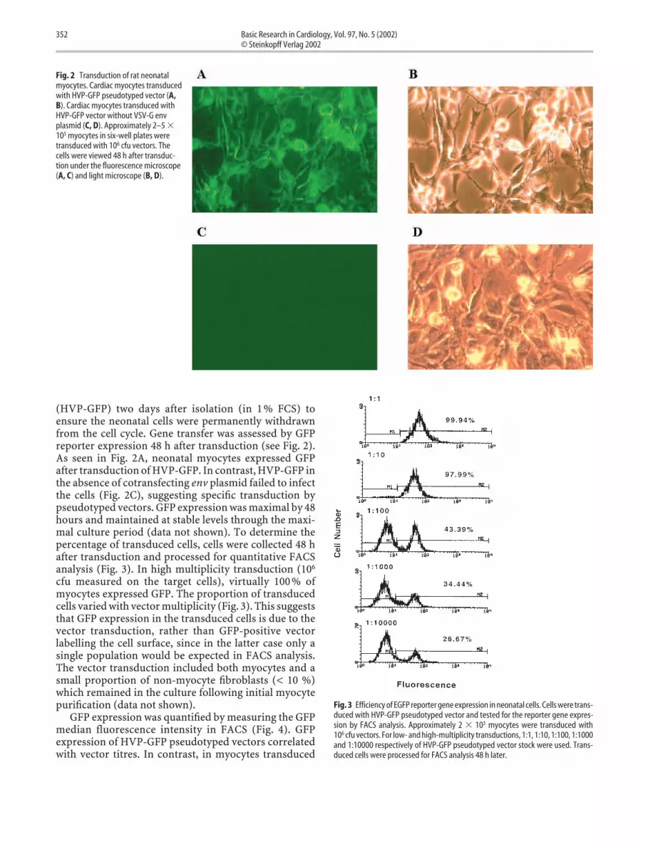
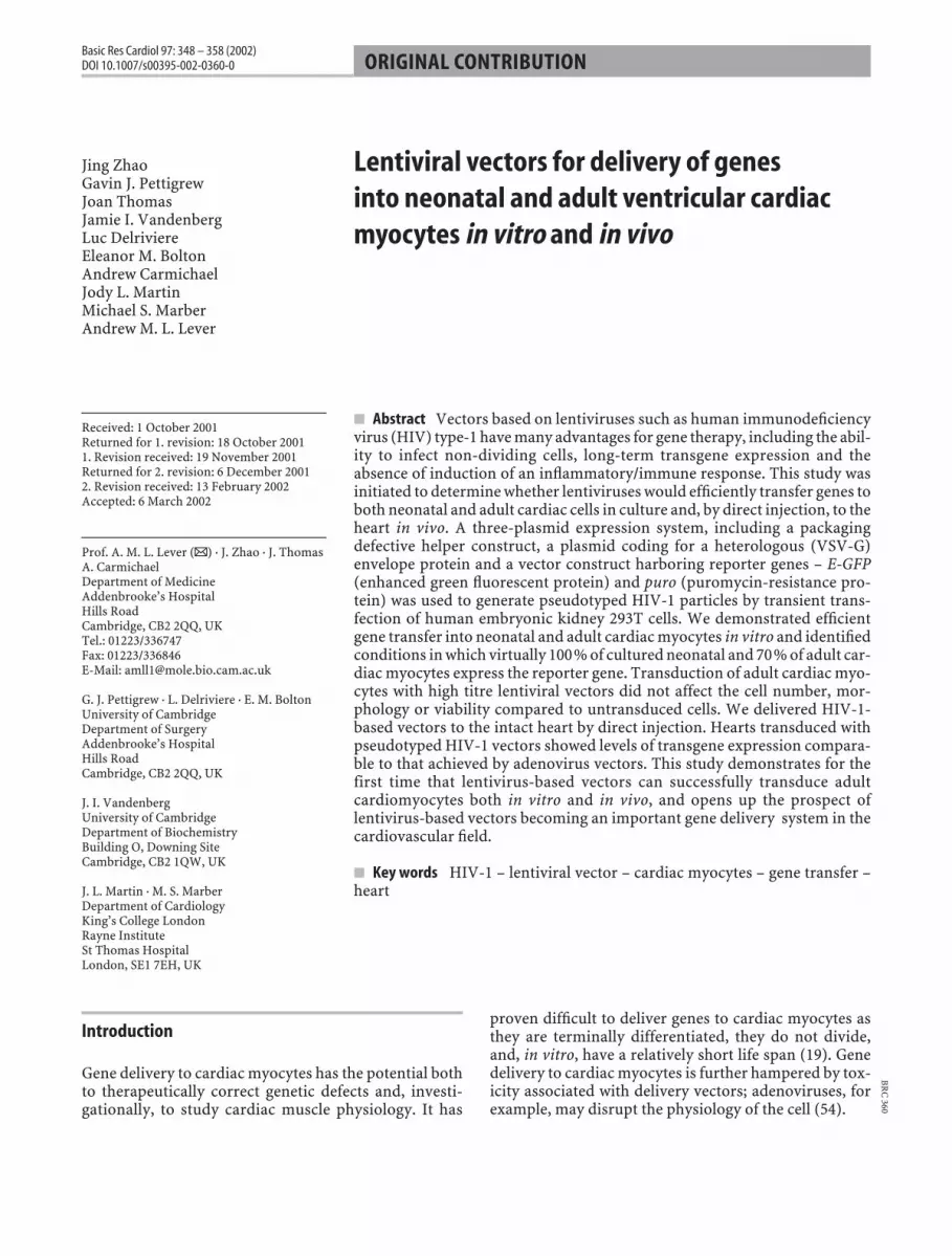
(HVP-GFP) two days after isolation (in 1 % FCS) toensure the neonatal cells were permanently withdrawnfrom the cell cycle. Gene transfer was assessed by GFPreporter expression 48 h after transduction (see Fig. 2).As seen in Fig. 2A, neonatal myocytes expressed GFPafter transduction of HVP-GFP. In contrast, HVP-GFP inthe absence of cotransfecting env plasmid failed to infectthe cells (Fig. 2C), suggesting specific transduction bypseudotyped vectors. GFP expression was maximal by 48hours and maintained at stable levels through the maxi-mal culture period (data not shown). To determine thepercentage of transduced cells, cells were collected 48 hafter transduction and processed for quantitative FACSanalysis (Fig. 3). In high multiplicity transduction (106
cfu measured on the target cells), virtually 100 % ofmyocytes expressed GFP. The proportion of transducedcells varied with vector multiplicity (Fig. 3). This suggeststhat GFP expression in the transduced cells is due to thevector transduction, rather than GFP-positive vectorlabelling the cell surface, since in the latter case only asingle population would be expected in FACS analysis.The vector transduction included both myocytes and asmall proportion of non-myocyte fibroblasts (< 10 %)which remained in the culture following initial myocytepurification (data not shown).
GFP expression was quantified by measuring the GFPmedian fluorescence intensity in FACS (Fig. 4). GFPexpression of HVP-GFP pseudotyped vectors correlatedwith vector titres. In contrast, in myocytes transduced
352 Basic Research in Cardiology, Vol. 97, No. 5 (2002)© Steinkopff Verlag 2002
Fig. 2 Transduction of rat neonatalmyocytes. Cardiac myocytes transducedwith HVP-GFP pseudotyped vector (A,B). Cardiac myocytes transduced withHVP-GFP vector without VSV-G envplasmid (C, D). Approximately 2–5 �105 myocytes in six-well plates weretransduced with 106 cfu vectors. Thecells were viewed 48 h after transduc-tion under the fluorescence microscope(A, C) and light microscope (B, D).
Fig. 3 Efficiency of EGFP reporter gene expression in neonatal cells. Cells were trans-duced with HVP-GFP pseudotyped vector and tested for the reporter gene expres-sion by FACS analysis. Approximately 2 � 105 myocytes were transduced with106 cfu vectors. For low- and high-multiplicity transductions, 1:1, 1:10, 1:100, 1:1000and 1:10000 respectively of HVP-GFP pseudotyped vector stock were used. Trans-duced cells were processed for FACS analysis 48 h later.

J. Zhao et al. 353Lentiviral vector gene delivery to heart
with HVP-GFP vector without the VSV-G expressor, GFPexpression was negative at all vector titres tested. Inorder to eliminate passive protein transfer as beingresponsible for fluorescence in the target cells, a CMV-driven GFP control plasmid was used. CMV-GFP
together with the packaging construct and the VSV-G envplasmid, were transfected into 293T cells, giving rise to a9-fold (n = 2) higher level of GFP expression than theHVP-GFP vector. However, cells transduced with thesupernatants from this transfection showed a 4-foldlower level of GFP expression compared to those trans-duced with a pseudotyped HVP-GFP vector (n = 3) (Fig.4) indicating retroviral transduction was responsible forvirtually all the gene expression in the target cells. Takentogether, the correlation between GFP expression andvector multiplicity indicated that GFP expression wasstrictly a result of the GFP vector sequence being trans-duced and reverse transcribed by HIV-1 pseudotypedparticles (Fig. 4). This was also confirmed by the obser-vation of maximum fluorescence occurring 48 h post-transduction, consistent with a viral RT and integrase-mediated event rather than passive protein transfer.
The apparent vector titre estimated from FACS analy-sis shown in Fig. 4 is less than that obtained withpuromycin selection. The FACS analysis is a gated meas-urement assessing the total GFP expression of the cellpopulation. Thus, this may not be detectable when GFPexpression is below a certain threshold. Single or lowcopy number integration might possibly give expressionbelow the sensitivity threshold of FACS analysis. This iscurrently under study.
Necrotic damage to the cultured neonatal cardiacmyocytes caused by the transduction procedure wasexamined by measuring creatine kinase (CK) release intothe culture medium. There was no difference between
Fig. 4 Dosage-dependent expression of HVP-GFP pseudotyped vector transducedneonatal cardiac myocytes. Approximately 2 � 105 myocytes were transduced. Forlow- and high-multiplicity transductions, 1:1, 1:10, 1:100, 1:1000 and 1:10000respectively of HVP-GFP pseudotyped vector stock were used. Transduced cellswere processed for FACS analysis 48 hr late GFP median fluorescence intensity wasnormalized by cells transduced with high-multiplicity of HVP-GFP pseudotypedvector particles. Cells transduced with HVP-GFP pseudotyped viral particles (closedcircle), cells transduced with plasmid pCMV-GFP and empty pseudotyped viralparticles (open triangle) and cells transduced with HVP-GFP viral particles withoutVSV-G envelope (open circle).
Fig. 5 HIV-GFP pseudotyped vectormediates efficient transfer to adult rab-bit ventricle myocytes. Representativefields are shown for (A) immediate afterisolation and (B) 48 h after isolation andGFP fluorescence 48 h after transduc-tion with (C) 106 cfu virus or with (D)control vector (HVP-GFP withoutcotransfection with VSV-G env plas-mid).
transduced and untransduced cells (CK release in trans-duced cells was 91.5 ± 7.2% of that seen in untransducedcells). Almost all cells beat spontaneously throughout theculture, even during the transduction period. Two daysafter transduction, these cells were confluent and beatingsynchronously, suggesting the HIV-1 vector had notdisturbed the electrical coupling that is essential for syn-chronous beating. The results suggest that HIV vectortransduction was not cytotoxic.
� Transduction of adult myocytes
After isolation from adult ventricular myocardium, 60 –70% of the cardiac cells displayed the rod-shaped mor-phology and sarcomeric cross-striations characteristicof myocardium in situ. The morphologic phenotype ofthe adult cardiac myocytes was stably maintained in cul-ture (Fig. 5A, B). Ventricular myocytes transduced after2 h in culture were analysed 24 – 48 h later. Ventricularmyocytes that were transduced with EGFP-encodingHIV-1 vector efficiently expressed the exogenous GFP(see Fig 5C). Myocytes which were rounded up alsostained positive for GFP. To eliminate uncertainties overwhether viral particles caused cell damage and thereforeincreased autofluorescence, EGFP-free viral vector wasused as a control and fluorescence was not observed(data not shown). Fluorescence was not due to passiveDNA transfer since HVP-GFP vector particles without
env could not successfully transduce the myocytes (Fig.5D). HVP-GFP vector transduction provides an efficientmeans of gene transfer into adult cardiomyocytes in vitrowhich has not been possible with other transfectionstrategies except adenovirus. However, due to the smallcell number, it was not possible to quantitate the GFPexpression in rod-shaped ventricular myocytes.
To address the potential impact of high titre trans-duction itself on adult ventricular myocytes, cell number,morphology and viability were compared in culturessubjected to 106 cfu of HIV-GFP vector versus controlcells. After 48 h, the total cell number was 54.0 ± 4.4% oforiginal number in untransduced cells and 58.7 ± 7.8%in transduced cells (n = 3, p > 0.05, no significant differ-ence). The proportion of myocytes retaining the differ-entiated, rod-shaped morphology was 43.4 ± 3.3% inuntransduced cells and 36.2 ± 1.9% in transduced cells(no significant difference p > 0.05). Of the cells which hadrod-shaped morphology, 75.3 ± 10.6% were viable inuntransduced cells and 67.0 ± 6.2% were viable in trans-duced cells, as determined by trypan blue exclusion(n = 3, p > 0.05, no significant difference). Necrotic dam-age to the cultured adult cardiac myocytes by the trans-duction procedure was also examined by measuring CKrelease into the culture medium. CK release in trans-duced cells was 88.9 ± 9.3% of the value in untransduced(n = 3, p > 2.05). Thus, under the conditions tested, HIVvector transduction by itself had no deleterious effects onthe survival or morphology of adult ventricular muscle
354 Basic Research in Cardiology, Vol. 97, No. 5 (2002)© Steinkopff Verlag 2002
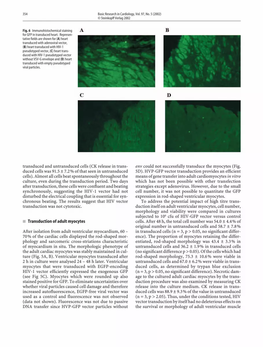
Fig. 6 Immunohistochemical stainingfor GFP in transduced heart. Represen-tative fields are shown for (A) hearttransduced with adenoviral vector,(B) heart transduced with HIV-1pseudotyped vector, (C) heart trans-duced with HIV-1 pseudotyped vectorwithout VSV-G envelope and (D) hearttransduced with empty pseudotypedviral particles.

J. Zhao et al. 355Lentiviral vector gene delivery to heart
cells. After 48 hours in culture, it is difficult to performelectrophysiological responses using standard whole-cellpatch clamping recording methods (45), even in non-transduced cells. Thus, attempts to study the contractilefunction of transduced cardiomyocytes at a single celllevel failed.
� In vivo transduction of transplanted heart
In this study, a transplantation model was used since ourultimate goal is to modulate the host immune responsein organ transplantation. Moreover, it is the only modelthat allows us to handle the heart ex vivo. It offers theunique opportunity of allowing us to precisely study dif-ferent strategies of gene transfer in the future. Due to thehigher background autofluorescence of the intact adultheart, an alternative HIV-1 vector with a stronger pro-moter for GFP, pHCMV-GFP, was used (a kind gift of Dr.Didier Trono, Department of Genetics and Microbiology,CMU, Geneve, Switzerland). In this HIV-1 vector system,the strong CMV promoter drives EGFP. Seven days afterthe transplantation, the level of gene transfer and expres-sion achieved with HIV-1 vector was comparable to thatobtained using an adenoviral vector (n = 3) (Fig. 6). Nopositive GFP stain was seen in the hearts transduced withHIV-1 vector without env or with empty vector.
There was no histological evidence in the transplantedhearts of any direct injury or inflammatory infiltrate inthe heart caused by both viral vectors (data not shown).
To confirm the results regarding gene expression withHIV-1 vector and to localise gene expression in vivo,another HIV-1 vector encoding �-galactosidase (a kindgift of Dr. N Deglon, Division of Surgical Research andGene Therapy Center, Lausanne Medical School, Switzer-land) was delivered into the heart by direct injection.Transplanted hearts were removed for histological exam-ination after 7 days. Although the injection points wereonly at the apex, the �-galactosidase activity was detectedthroughout the myocardium in all of the transplantedhearts with direct injection of HIV-1 vectors (n = 3) (Fig.7).
The efficiency of �-galactosidase expression was lessthan that of GFP for both vectors. This could be that thestaining of nuclear-localised �-galactosidase is relativelyinsensitive and is detectable only in highly expressingcells. The staining, therefore, may underestimate theextent of transgene expression. Lower levels of expres-sion may not be detected by this method but may be suf-ficient to induce physiological changes, depending onthe transgene expressed.
Fig. 7 Histochemical staining for �-galactosidase in transduced heart. Represen-tative fields are shown for (A) heart transduced with HIV-1 pseudotyped vector inthe apex of the heart, (B) heart transduced with HIV-1 pseudotyped vector in themiddle section of the heart, (C) heart transduced with HIV-1 pseudotyped vector inthe base section of the heart, (D) heart transduced with adenoviral vector in the apexof the heart, (E) heart transduced with adenoviral vector in the middle section of theheart and (F) heart transduced with adenoviral vector in the base section of theheart.
Discussion
Cardiac myocytes are terminally differentiated cells,which withdraw from the cell cycle soon after birth. Sub-sequent myocardial growth occurs through enlargementof individual cardiac myocytes, and cell loss followinginjury is not replaced by cell proliferation. This makesthem particularly problematic in terms of gene delivery.Gene therapy offers a potential valuable therapeuticavenue to treat primary cardiac diseases, to correct defec-tive cells and possibly to differentiate fibroblasts intomyocytes (36, 52).
The delivery of genetic material into intact adultmyocardium is currently achieved by one of two meth-ods, either virally mediated or non-viral (physical) deliv-ery. The latter has the advantage of involving no othercomponents of infectious organisms and being simpleand relatively cheap (38, 39) but the efficiency of deliveryis much less than that associated with viral vectors (13,37). Until recently, adenoviruses were the most favoredmethod of gene delivery to cardiac myocytes as they arehighly infectious for non-replicating mammalian cells(5, 23, 27). They can be generated as vectors to very hightitre (3, 5) and are capable of delivering genes to cardiacmyocytes by direct injection (14, 28), by perfusion intocoronary arteries (2, 6) and into the ventricular cavity(15). Their use has, however, been limited by the tran-sient nature of their expression (2, 17, 20) and by theinflammatory response that they engender under someconditions (57, 62, 63). Although the newer “gutless” ade-novirus vectors which express no adenovirus gene prod-ucts (51) may be less prone to trigger inflammatoryresponses, the adenovirus particle is nevertheless dis-ruptive of cell membranes and capable of provokinginflammation (16, 18, 54). In addition, because of theubiquitous nature of adenoviral infections, virtuallyeverybody has significant levels of pre-formed antibod-ies against adenovirus from prior infections (10, 58)which may result in unpredictable variable transfectionlevels in clinical practice. The recent problems with ade-noviral-mediated gene delivery to the liver have empha-sized that the efficiency of this system may only beachieved at a price (32). Adeno-associated viruses (AAV)have also been demonstrated as useful for gene deliveryto cardiac muscle (30, 49). While they are non-patho-genic and do not trigger inflammation, they are relativelydifficult to manipulate (56). Part of the problem is theincomplete understanding that we have of AAV replica-tion. Lentiviruses have the unique ability to introduceand integrate their DNA genome into the chromosomesof cells which are not dividing. The major concern asso-ciated with lentiviruses is bio-safety; chiefly, as with otherretroviruses, the risk of insertational mutagenesis. How-ever, it has been nearly 10 years since the first retrovirusgene therapy clinical trial (44) and there are no reportedcases of insertational mutation. Moreover this phenom-
enon has not been identified in HIV positive patients.Another concern is formation of replication-competentvirus generated during the vector preparation. To mini-mize the recombination, three-plasmid and four-plas-mid systems have been used to generate HIV-1 vectorparticles. The design of these systems is based on theconcept of segregated packaging systems that have beenavailable for retroviruses for over a decade. Naldini et al.(42) were the first to describe an HIV-1-based three-component system that involves heterologous Env pro-teins. Analogous to those used in other laboratories (22,66), we have developed an efficient three-componentpackaging system to produce HIV-1 pseudotyped vec-tors. The titers obtained were 1 � 106 cfu/ml with a sin-gle ultracentrifugation. The use of heterologous Env pro-teins such as VSV-G is assumed to preclude the forma-tion of replication-competent HIV-1, further adding tothe safety of the system.
It has been shown that lentivirus-based vectors candeliver genes to mitotically inactive neurons (4, 12, 24),and neuroglia (34, 41). There have been few reportsregarding application of lentivirus-based vectors to car-diac myocytes and all of those studies to date have usedneonatal cardiac myocytes (35, 50). There are qualitativeand quantitative differences between neonatal and adultcardiac myocytes, including the expression of contractileprotein isoforms, G-proteins and ATPase (40), growthfactor and their receptors (31, 53), and transcriptionfactors (29, 65). This is the first study demonstrating thatan HIV-1-based pseudotyped vector is able to infect adultcardiac myocytes efficiently and that efficient gene trans-fer to adult ventricular myocytes in culture can occurwithout adverse effects to the cells. For adult cardiacmyocytes, transduction studies were limited to 48 hoursdue to the difficulty in maintaining healthy differentiatedadult cardiac myocytes in culture, independent of vectortransduction. In neonatal cardiac myocytes, GFP expres-sion was maximal at 48 hours after transduction andmaintained through the maximal culture period. Wehave demonstrated, for the first time, that HIV-1 vectormediated gene transfer using direct injection of hearttransplants results in significant gene expression. Aden-oviral vector mediated gene transfer results in more �-galactosidase positive cells than HIV-1 vector in the apexof the heart. The adenoviral vector titer used in this studywas 1 � 1010 cfu/ml, whereas that of the HIV-1 vector was106 cfu/ml. The effect of viral titer on adenovirus-medi-ated gene transfer has been shown previously (61). Ade-noviral-mediated transgene expression was markedlydiminished by lower titer and the timing of peak geneexpression was also shifted with virus titer. Withimprovement in HIV-1 vector purification there arelikely to be further increases in the attainable concentra-tions and therefore increase its efficiency. Further exper-iments are currently being conducted to compare long-term stable expression between these two viral vectors
356 Basic Research in Cardiology, Vol. 97, No. 5 (2002)© Steinkopff Verlag 2002
J. Zhao et al. 357Lentiviral vector gene delivery to heart
1. Akkina RK, Walton RM, Chen ML, Li QX,Planelles V, Chen IS (1996) High effi-ciency gene transfer into CD34(+) cellswith a human immunodeficiency virustype 1-based retroviral vector pseudo-typed with vesicular stomatitis virusenvelope glycoprotein G. J Virol 70:2581–2585
2. Barr E, Carroll J, Kalynych AM, TripathySK, Kozavsky K, Wilson JM, Leidem JM(1994) Efficient catheter-mediated genetransfer into the heart using replication-defective adenovirus. Gene Ther 1: 51–58
3. Benihoud K, Yeh P, Perricaudet M (1999)Adenovirus vectors for gene delivery.Curr Opin Biotechnol 10: 440–447
4. Blomer U, Naldini L, Kafri T, Trono D,Verma IM, Gage FH (1997) Highly effi-cient and sustained gene transfer in adultneurons with a lentivirus vector. J Virol71: 6641–6649
5. Brenner M (1999) Gene transfer byadenovectors. Blood 94: 3965–3967
6. Budker V, Zhang G, Danko I, Williams P,Wolff J (1998) The efficient expression ofintravascularly delivered DNA in ratmuscle. Gene Ther 5: 272–276
7. Bukrinsky MI, Haggerty S, Dempsey MP,Sharova N, Adzhubel A, Spitz L, Lewis P,Goldfarb D, Emerman M, Stevenson M(1993) A nuclear localization signalwithin HIV-1 matrix protein that governsinfection of non-dividing cells. Nature365: 666–669
8. Burns JC, Friedmann T, Driever W,Burrascano M, Yee JK (1993) Vesicularstomatitis virus G glycoprotein pseudo-typed retroviral vectors: concentration tovery high titer and efficient gene transferinto mammalian and nonmammaliancells. Proc Natl Acad Sci USA 90: 8033–8037
9. Chen CA, Okayama H (1988) Calciumphosphate-mediated gene transfer: ahighly efficient transfection system forstably transforming cells with plasmidDNA. Biotechniques 6: 632–638
10. Dai Y, Schwarz EM, Gu D, Zhang W-W,Sarvetnick N, Verma IM (1995) Cellularand humoral immune responses to aden-oviral vectors containing factor IX gene:tolerization of factor IX and vector anti-gens allows for long-term expression.Proc. Natl Acad Sci USA 92: 1401–1405
11. Danos O, Mulligan RC (1988) Safe andefficient generation of recombinant retro-viruses with amphotropic and ecotropichost ranges. Proc Natl Acad Sci USA 85:6460–6464
12. Deglon N, Tseng JL, Bensadoun JC, ZurnAD, Arsenijevic Y, Pereira de Almeida L,Zufferey R, Trono D, Aebischer P (2000)Self-inactivating lentiviral vectors withenhanced transgene expression andpotential gene transfer system in Parkin-son's disease. Hum Gene Ther 11: 179–190
13. Felgner PL (1997) Nonviral strategies forgene therapy. Sci Am 276: 102–106
14. French BA, Mazur W, Geske RS, Bolli R(1994) Direct in vivo gene transfer intoporcine myocardium using replication-deficient adenoviral vectors. Circulation90: 2414–2424
15. Fromes Y, Salmon A, Wang X, Collin H,Rouche A, Hagege A, Schwartz K,Fiszman MY (1996) Gene delivery to themyocardium by intrapericardial injec-tion. Gene Ther 6: 683–688
16. Graham FL, Prevec L (1992) Adenovirus-based expression vectors and recombi-nant vaccines. In: Ronald W. Ellis (ed)Vaccines: New Approaches to Immuno-logical Problems. Butterworth-Heine-mann Boston, pp 363–390
17. Guzman RJ, Lemarchand P, Crystal RG,Epstein SE, Finkel T (1993) Efficient genetransfer into myocardium by direct injec-tion of adenovirus vectors. Circ Res 73:1202–1207
18. Horwitz MS (1991) Adenoviridae andtheir replication. In: Fields BN, Knipe DM(eds) Fundamental Virology. Raven PressNew York, pp 771–813
19. Jacobson SL, Piper HM (1986) Cell cul-tures of adult cardiomyocytes as modelsof the myocardium. J Mol Cell Cardiol 18:661–678
20. Kass-Eisler A, Falck-Pedersen E, AlviraM, Rivera J, Buttrick PM, Wittenberg BA,Cipriani L, Leinwand LA (1993) Quanti-tative determination of adenovirus-medi-ated gene delivery to rat cardiac myocytesin vitro and in vivo. Proc Natl Acad SciUSA 90: 11498–11502
21. Kaye JF, Richardson JH, Lever AM (1995)Cis-acting sequences involved in humanimmunodeficiency virus type 1 RNApackaging. J Virol 69: 6588–6592
22. Kim VN, Mitrophaneous K, KingsmanSM, Kingsman AJ (1998) Minimalrequirement for a lentivirus vector basedon human immunodeficiency virus type1. J Virol 72: 811–816
23. Kirshenbaum LA, MacLellan WR, MazurW, French BA, Schneider MD (1993)Highly efficient gene transfer into adultventricular myocytes by recombinantadenovirus. J Clin Invest 92: 381–387
24. Kordower JH, Bloch J, Ma SY, Chu Y, PalfiS, Roitberg BZ, Emborg M, Hantraye P,Deglon N, Aebischer P (1999) Lentiviralgene transfer to the nonhuman primatebrain. Exp Neurol 160: 1–16
25. Lever A, Gottlinger H, Haseltine W,Sodroski J (1989) Identification of asequence required for efficient packagingof human immunodeficiency virus type 1RNA into virions. J Virol 63: 4085–4087
26. Lever AM (1996) HIV and otherlentivirus-based vectors [editorial]. GeneTher 3: 470–471
27. Lewis P, Hensel M, Emerman M (1992)Human immunodeficiency virus infec-tion of cells arrested in the cell cycle.EMBO J 11: 3053–3058
28. Lin H, Parmacek MS, Morle G, Bolling S,Leiden JM (1990) Expression of recombi-nant genes in myocardium in vivo afterdirect injection of DNA. Circulation 82:2217–2221
29. Luetje CW, Tietje KM, Christian JL,Nathanson NM (1988) Differential tissueexpression and developmental regulationof guanine nucleotide binding regulatoryproteins and their messenger RNAs in ratheart. J Biol Chem 263: 13357–13365
30. Lynch CM, Hara PS, Leonard JC,Williams JK, Dean RH, Geary RL (1997)Adeno-associated virus vectors for vas-cular gene delivery. Circ Res 80: 497–505
31. Mann DL, Kent RL, Parsons B, Cooper I,G (1992) Adrenergic effects on the biol-ogy of the adult mammalian cardiocyte.Circulation 85: 790–804
32. Marshall E (1999) Gene therapy deathprompts review of adenovirus vector.Science 286: 2244–2245
33. Martin JL, Mestril R, Hilal-Dandan R,Brunton LL, Dillmann WH (1997) Smallheat shock proteins and protectionagainst ischemic injury in cardiacmyocytes. Circulation 96: 4343–4348
34. Mitrophanous K, Yoon S, Rohll J, Patil D,Wilkes F, Kim V, Kingsman S, KingsmanA, Mazarakis N (1999) Stable gene trans-fer to the nervous system using a non-primate lentiviral vector. Gene Ther 6:1808–1818
35. Mochizuki H, Schwartz JP, Tanaka K,Brady RO, Reiser J (1998) High-titerhuman immunodeficiency virus type 1-based vector systems for gene deliveryinto nondividing cells. J Virol 72:8873–8883
References
Acknowledgments This work was supported by the British Heart Foun-dation and the Sykes’ Trust. We would like to thank Dr Peter Glennonfor helpful contributions. Dr. Andrew Carmichael is supported by theLister Institute.
and to quantitate more accurately their relative efficiencyin accessing cardiac myocytes under the relevant condi-tions.
�

358 Basic Research in Cardiology, Vol. 97, No. 5 (2002)© Steinkopff Verlag 2002
36. Murry CE, Kay MA, Bartosek T,Hauschka SD, Schwartz SM (1996)Muscle differentiation during repair ofmyocardial necrosis in rats via genetransfer with MyoD. J Clin Invest 98:2209–2217
37. Nabel EG (1995) Gene therapy for cardio-vascular disease. Circulation 91: 541–548
38. Nabel EG, Gordon D, Xang ZI, Xu L, SanH, Plautz GE, Gao X, Huang L, Nabel GJ(1992) Gene transfer in vivo with DNA-liposome complexes: lack of autoimmu-nity and gonadal localisation. Hum GeneTher 3: 649–656
39. Nabel GJ, Nabel EG, Yang Z-Y, Fox BA,Plautz GE, Gao X, Huang L, Shu S,Gordon D, Chang AE (1993) Direct genetransfer with DNA-liposome complexesin melanoma: expression, biologic activ-ity, and lack of toxicity in humans. ProcNatl Acad Sci USA 90: 11307–11311
40. Nadal-Ginard B, Mahdavi V (1989) Mole-cular basis of cardiac performance.Plasticity of the myocardium generatedthrough protein isoform switches. J ClinInvest 84: 1693–1700
41. Naldini L, Blomer U, Gage FH, Trono D,Verma IM (1996) Efficient transfer, inte-gration, and sustained long-term expres-sion of the transgene in adult rat brainsinjected with a lentiviral vector. Proc NatlAcad Sci USA 93: 11382–11388
42. Naldini L, Blomer U, Gallay P, Ory D,Mulligan P, Gage FH, Verma IM, Trono D(1996) In vivo gene delivery and stabletransduction of nondividing cells by alentiviral vector. Science 272: 263–267
43. Ono K, Lindsey ES (1969) Improved tech-nique of heart transplantation in rats. JThorac Cardiovasc Surg 57: 225–229
44. Pilaro AM, Serabian MA (1999) Preclini-cal development strategies for novel genetherapeutic products. Toxicologic Patho-logy 27: 4–7
45. Powell T, Terrar DA, Twist VW (1980)Electrical properties of individual cellsisolated from adult rat ventricular myo-cardium. J Physiol 302: 131–153
46. Poznansky M, Lever A, Bergeron L,Haseltine W, Sodroski J (1991) Genetransfer into human lymphocytes by adefective human immunodeficiency virustype 1 vector. J Virol 65: 532–536
47. Richardson JH, Child LA, Lever AM(1993) Packaging of human immunodefi-ciency virus type 1 RNA requires cis-act-ing sequences outside the 5’ leader region.J Virol 67: 3997–4005
48. Richardson JH, Kaye JF, Child LA, LeverAM (1995) Helper virus-free transfer ofhuman immunodeficiency virus type 1vectors. J Gen Virol 76: 691–696
49. Rolling F, Nong Z, Pizvin S, Collen D(1997) Adeno-associated virus-mediatedgene transfer into rat carotid arteries.Gene Ther 4: 757–761
50. Sakoda T, Tasahara N, Hamamori Y,Kedes L (1999) A high-titer lentiviral pro-duction system mediates efficient trans-duction of differentiated cells includingbeating cardiac myocytes. J Mol Cell Car-diol 31: 2037–2047
51. Schiedner G, Morral N, Parks RJ, Wu Y,Koopmans SC, Langston C, Graham FL,Beaudet AL, Kochanek S (1998) GenomicDNA transfer with a high-capacity aden-ovirus vector results in improved in vivogene expression and decreased toxicity.Nat Gen 18: 180–183
52. Schneider MD (1996) Myocardial infarc-tion as a problem of growth control: cellcycle therapy for cardiac myocytes? JCard Fail 2: 259–263
53. Schneider MD, Parker TG (1991) Cardiacgrowth factors. Prog. Growth Factor Res3: 1–26
54. Schulick AH, Newman KD, Virmani R,Dichek DA (1995) In vivo gene transferinto injured carotid arteries: optimiza-tion and evaluation of acute toxicity. Cir-culation 91: 2407–2414
55. Singh Kent N, Davia K, Harding SE (1999)Characterisation of the function of adultguinea-pig ventricular myocytes follow-ing co-culture with neonatal rat myo-cytes. Basic Res Cardiol 94: 9–14
56. Sinnaeve P, Varenne O, Collen D,Janssens S (1999) Gene therapy in the car-diovascular system: an update. Cardio-vasc Res 44: 498–506
57. Tripathy SK, Black HB, Goldwasser E,Leiden JM (1996) Immune responses totransgene-encoded proteins limit the sta-bility of gene expression after injection ofreplication-defective adenovirus vectors.Nat Med 2: 545–550
58. Verma IM, Somia N (1997) Gene therapy– promises, problems and prospects.Nature 389: 239–242
59. von Schwedler U, Kornbluth RS, Trono D(1994) The nuclear localization signal ofthe matrix protein of human immunode-ficiency virus type 1 allows the establish-ment of infection in macrophages andquiescent T lymphocytes. Proc Natl AcadSci USA 91: 6992–6996
60. Wong KR, Trezise AEO, Bryant S, Hart G,Vandenberg JI (1999) Molecular andfunctional distributions of chlorideconductances in rabbit ventricle. Am JPhysiol 277: H1403–H1409
61. Wright MJ, Wightman LM, Lilley C, deAlwis M, Hart SL, Miller A, Coffin RS,Thrasher A, Latchman DS, Marber MS(2001) In vivo myocardial gene transfer:optimization, evaluation and direct com-parison of gene transfer vectors. Basic ResCardiol 96: 227–236
62. Yang Y, Ertl HCJ, Wilson JM (1994) MHCclass I-restricted cytotoxic T lymphocytesto viral antigens destroy hepatocytes inmice infected with E1-deleted recombi-nant adenoviruses. Immunity 1: 433–442
63. Yang Y, Nunes FA, Berencsi K, Furth EE,Gonczol E, Wilson JM (1994) Cellularimmunity to viral antigens limits E1-deleted adenoviruses for gene therapy.Proc Natl Acad Sci USA 91: 4407–4411
64. Zhao J, Renner O, Wightman L, SugdenPH, Stewart L, Miller AD, Latchman DS,Marber MS (1998) The expression of con-stitutively active isotypes of proteinkinase C to investigate preconditioning. JBiol Chem 273: 23072–23079
65. Zimmermann KA, Yancopoloulos GD,Collum RG, Smith RK, Kohl NE, DenisKA, Nau MM, Witte ON, Toram-AllerandD, Gee CE (1986) Differential expressionof myc family genes during murine devel-opment. Nature 319: 780–783
66. Zufferey R, Nagy D, Mandel RJ, Naldini L,Trono D (1997) Multiply attenuatedlentiviral vector achieves efficient genedelivery in vivo. Nat Biotech 15: 871–875
�