effects of cd2 locus control region sequences on gene expression by retroviral and lentiviral...
TRANSCRIPT

doi:10.1182/blood.V98.13.36072001 98: 3607-3617
AmadoriValeria Tosello, Dieter Klein, Walter H. Günzburg, Giuseppe Basso, Luigi Chieco-Bianchi and Alberto
Stefano Indraccolo, Sonia Minuzzo, Francesca Roccaforte, Rita Zamarchi, Walter Habeler, Laura Stievano, retroviral and lentiviral vectorsEffects of CD2 locus control region sequences on gene expression by
http://bloodjournal.hematologylibrary.org/content/98/13/3607.full.htmlUpdated information and services can be found at:
(523 articles)Gene Therapy � (1086 articles)Gene Expression �
Articles on similar topics can be found in the following Blood collections
http://bloodjournal.hematologylibrary.org/site/misc/rights.xhtml#repub_requestsInformation about reproducing this article in parts or in its entirety may be found online at:
http://bloodjournal.hematologylibrary.org/site/misc/rights.xhtml#reprintsInformation about ordering reprints may be found online at:
http://bloodjournal.hematologylibrary.org/site/subscriptions/index.xhtmlInformation about subscriptions and ASH membership may be found online at:
Copyright 2011 by The American Society of Hematology; all rights reserved.American Society of Hematology, 2021 L St, NW, Suite 900, Washington DC 20036.Blood (print ISSN 0006-4971, online ISSN 1528-0020), is published weekly by the
For personal use only. by guest on June 2, 2013. bloodjournal.hematologylibrary.orgFrom

GENE THERAPY
Effects of CD2 locus control region sequences on gene expression by retroviraland lentiviral vectorsStefano Indraccolo, Sonia Minuzzo, Francesca Roccaforte, Rita Zamarchi, Walter Habeler, Laura Stievano, Valeria Tosello, Dieter Klein,Walter H. Gunzburg, Giuseppe Basso, Luigi Chieco-Bianchi, and Alberto Amadori
Locus control region (LCR) sequencesare involved in the establishment of openchromosomal domains. To evaluate thepossibility of exploiting the human CD2LCR to regulate gene expression by Molo-ney murine leukemia virus (Mo-MLV)–based retroviral vectors in T cells, it wasincluded in vectors carrying the en-hanced green fluorescence protein(EGFP) reporter gene; then transductionin vitro of lymphoid and nonlymphoid celllines was performed. Deletion of the viralenhancer in the Mo-MLV long terminalrepeat was necessary to detect LCR activ-ity in the context of these retroviral vec-
tors. It was found that a full-length (2.1kb), but not a truncated (1.0 kb), CD2 LCRretained the ability to modulate reportergene expression by Mo-MLV–derived ret-roviral vectors, leading to a homoge-neous, unimodal pattern of EGFP expres-sion that remained unmodified in cultureover time, specifically in T-cell lines; onthe other hand, viral titer was stronglyreduced compared with vectors not carry-ing the LCR. Lentiviral vectors containingthe CD2 LCR could be generated at highertiters and were used to analyze its effectson gene expression in primary T cells.Subcutaneous implantation of geneti-
cally modified cells in immunodeficientmice showed that retroviral vectors carry-ing the CD2 LCR conferred an advantagein terms of transgene expression in vivo,compared with the parental vector, bypreventing the down-modulation of EGFPexpression. These findings suggest a po-tential application of this LCR to increasegene expression by retroviral and lentivi-ral vectors in T lymphocytes. (Blood. 2001;98:3607-3617)
© 2001 by The American Society of Hematology
Introduction
One of the major obstacles to gene transfer in mammalian cells isthe gap between gene expression of cellular genes from theirgenomic loci, which usually occurs at adequate levels, and therelatively poor expression levels obtained when the same gene isexpressed from viral vectors, in many cases from heterologousviral promoters. This commonly observed phenomenon clearlyconstitutes a significant limitation both of gene therapy strategiesaimed at treating genetic diseases by transduction of hematopoieticstem cells and of protocols of gene transfer into differentiatedprimary cells, such as lymphocytes and fibroblasts.1 Differences ingene expression from genomic loci and viral vectors clearlyinvolve multiple levels of complexity, including lack of introns inthe transgene, differences between viral and cellular promoters,and absence of some regulatory sequences in viral vectors—suchas silencers, insulators, and locus control regions (LCRs)—thatregulate the physiological expression of cellular genes. As aconsequence, gene expression by viral vectors is often short-term,even though it can transiently occur at higher levels than that ofcellular genes.
In some cases, the immune system also may contribute signifi-cantly to limiting transgene expression in vivo, specifically whenthe transgene itself or the viral vector used (ie, adenoviral vectors)is highly immunogenic. Furthermore, the pattern of expression of
virally carried therapeutic genes and normal cellular genes isstrikingly different: retrovector-encoded genes are expressed hetero-geneously in the transduced cells because of their random integra-tion into the host genome, whereas most cellular genes areexpressed at homogeneous levels in a defined cell subset. Giventhese limitations of current gene transfer methods, LCR sequenceshave recently attracted attention because they might improve thelevel of gene expression by retroviral vectors (reviewed in2-4).Many studies focused on theb-globin LCR, which could be usefulfor increasing erythroid-specific synthesis of this protein.5-7 How-ever, it is unclear whether selected LCR elements might be used toimprove gene expression in T lymphocytes. A retroviral vectorcarrying a T-cell–specific LCR could be useful in clinical settingsdemanding long-term expression of therapeutic genes in lymphoidcells. These might include some primary immunodeficiencies inwhich T-cell–specific, long-term expression of a relevant gene,such as that coding for ADA, is required to enable the correctdevelopment of the immune system and some acquired diseasesthat might be amenable to treatment with genetically modifiedlymphocytes that would act as the “host factory” for solubletherapeutic proteins.
To address this issue, we studied an LCR from the humanCD2gene. This 2.1-kb sequence has been largely studied in transgenic
From the IST-Viral and Molecular Oncology Section, the Department of Oncologyand Surgical Sciences, and the Department of Pediatrics, University of Padua, Italy;and the Institute of Virology, University of Veterinary Sciences, Vienna, Austria.
Submitted October 24, 2000; accepted July 15, 2001.
Supported in part by Telethon (grant A-126), ISS-AIDS Project, MURST 60%and 40%, Associazione Italiana per la Ricerca sul Cancro, Fondazione Italianaper la Ricerca sul Cancro, Fondazione Citta della Speranza, and NationalResearch Council (PF Biotechnology). S.M. is the recipient of a fellowshipfrom Fondazione Italiana per la Ricerca sul Cancro. V.T. is a recipient of an
AIRC fellowship.
Reprints: Stefano Indraccolo, IST-Viral and Molecular OncologySection and Department of Oncology and Surgical Sciences,University of Padua, Via Gattamelata, 64-35128 Padua, Italy; e-mail:[email protected].
The publication costs of this article were defrayed in part by page chargepayment. Therefore, and solely to indicate this fact, this article is herebymarked ‘‘advertisement’’ in accordance with 18 U.S.C. section 1734.
© 2001 by The American Society of Hematology
3607BLOOD, 15 DECEMBER 2001 z VOLUME 98, NUMBER 13
For personal use only. by guest on June 2, 2013. bloodjournal.hematologylibrary.orgFrom
mice, in which it leads to high-level, position-independent geneexpression in the T-cell lineage.8-10 Our findings suggested thatCD2 LCR could be useful for improving gene expression byretroviral and lentiviral vectors in T cells in vitro and in vivo.
Materials and methods
Plasmids
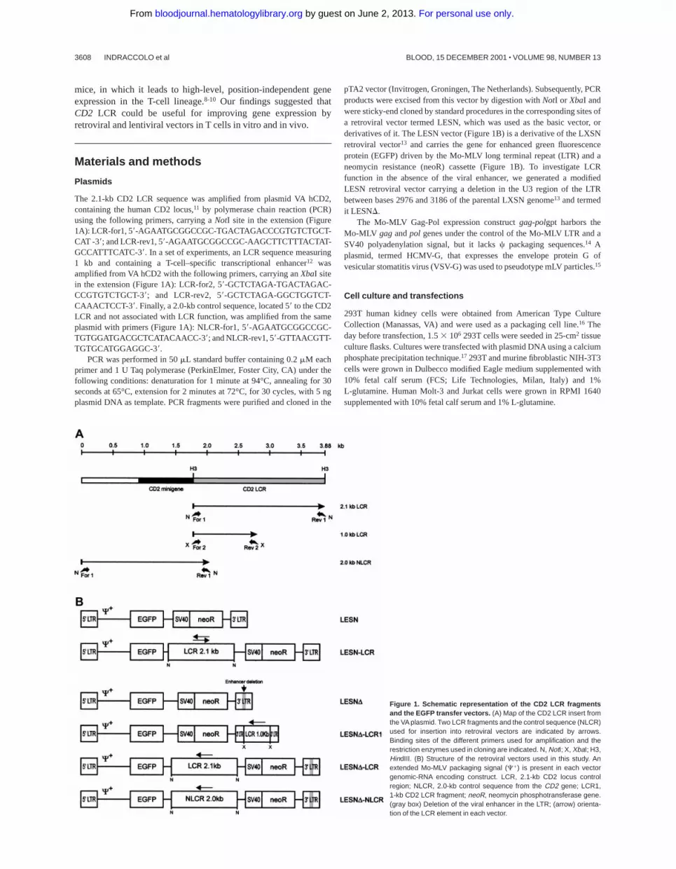
The 2.1-kb CD2 LCR sequence was amplified from plasmid VA hCD2,containing the human CD2 locus,11 by polymerase chain reaction (PCR)using the following primers, carrying aNotI site in the extension (Figure1A): LCR-for1, 59-AGAATGCGGCCGC-TGACTAGACCCGTGTCTGCT-CAT -39; and LCR-rev1, 59-AGAATGCGGCCGC-AAGCTTCTTTACTAT-GCCATTTCATC-39. In a set of experiments, an LCR sequence measuring1 kb and containing a T-cell–specific transcriptional enhancer12 wasamplified from VA hCD2 with the following primers, carrying anXbaI sitein the extension (Figure 1A): LCR-for2, 59-GCTCTAGA-TGACTAGAC-CCGTGTCTGCT-39; and LCR-rev2, 59-GCTCTAGA-GGCTGGTCT-CAAACTCCT-39. Finally, a 2.0-kb control sequence, located 59to the CD2LCR and not associated with LCR function, was amplified from the sameplasmid with primers (Figure 1A): NLCR-for1, 59-AGAATGCGGCCGC-TGTGGATGACGCTCATACAACC-39; and NLCR-rev1, 59-GTTAACGTT-TGTGCATGGAGGC-39.
PCR was performed in 50mL standard buffer containing 0.2mM eachprimer and 1 U Taq polymerase (PerkinElmer, Foster City, CA) under thefollowing conditions: denaturation for 1 minute at 94°C, annealing for 30seconds at 65°C, extension for 2 minutes at 72°C, for 30 cycles, with 5 ngplasmid DNA as template. PCR fragments were purified and cloned in the
pTA2 vector (Invitrogen, Groningen, The Netherlands). Subsequently, PCRproducts were excised from this vector by digestion withNotI or XbaI andwere sticky-end cloned by standard procedures in the corresponding sites ofa retroviral vector termed LESN, which was used as the basic vector, orderivatives of it. The LESN vector (Figure 1B) is a derivative of the LXSNretroviral vector13 and carries the gene for enhanced green fluorescenceprotein (EGFP) driven by the Mo-MLV long terminal repeat (LTR) and aneomycin resistance (neoR) cassette (Figure 1B). To investigate LCRfunction in the absence of the viral enhancer, we generated a modifiedLESN retroviral vector carrying a deletion in the U3 region of the LTRbetween bases 2976 and 3186 of the parental LXSN genome13 and termedit LESND.
The Mo-MLV Gag-Pol expression constructgag-polgpt harbors theMo-MLV gagandpol genes under the control of the Mo-MLV LTR and aSV40 polyadenylation signal, but it lacksc packaging sequences.14 Aplasmid, termed HCMV-G, that expresses the envelope protein G ofvesicular stomatitis virus (VSV-G) was used to pseudotype mLV particles.15
Cell culture and transfections
293T human kidney cells were obtained from American Type CultureCollection (Manassas, VA) and were used as a packaging cell line.16 Theday before transfection, 1.53 106 293T cells were seeded in 25-cm2 tissueculture flasks. Cultures were transfected with plasmid DNA using a calciumphosphate precipitation technique.17 293T and murine fibroblastic NIH-3T3cells were grown in Dulbecco modified Eagle medium supplemented with10% fetal calf serum (FCS; Life Technologies, Milan, Italy) and 1%L-glutamine. Human Molt-3 and Jurkat cells were grown in RPMI 1640supplemented with 10% fetal calf serum and 1% L-glutamine.
Figure 1. Schematic representation of the CD2 LCR fragmentsand the EGFP transfer vectors. (A) Map of the CD2 LCR insert fromthe VA plasmid. Two LCR fragments and the control sequence (NLCR)used for insertion into retroviral vectors are indicated by arrows.Binding sites of the different primers used for amplification and therestriction enzymes used in cloning are indicated. N, NotI; X, XbaI; H3,HindIII. (B) Structure of the retroviral vectors used in this study. Anextended Mo-MLV packaging signal (C1) is present in each vectorgenomic-RNA encoding construct. LCR, 2.1-kb CD2 locus controlregion; NLCR, 2.0-kb control sequence from the CD2 gene; LCR1,1-kb CD2 LCR fragment; neoR, neomycin phosphotransferase gene.(gray box) Deletion of the viral enhancer in the LTR; (arrow) orienta-tion of the LCR element in each vector.
3608 INDRACCOLO et al BLOOD, 15 DECEMBER 2001 z VOLUME 98, NUMBER 13
For personal use only. by guest on June 2, 2013. bloodjournal.hematologylibrary.orgFrom

Transduction of cells with retroviral vectors
Infectious particles were generated by overnight transfection of 293T cellswith 3 mg gag-polgpt, 6mg either LESN, LESN-LCR, LESND, LESND-LCR, LESND-NLCR, or LESND-LCR1 as transducing vectors, and 0.1mgVSV-G expression construct, as reported.18 Retroviral vector–containingsupernatants were collected 48 to 72 hours after transfection, filteredthrough 0.45-mm filters, and frozen at280°C until further use. Todetermine vector titer, serial dilutions of filtered supernatants were layeredover NIH-3T3 target cells that had been seeded into 6-well culture plates theday before infection at 0.63 105 cells per well. Protamine sulfate (8mg/mL) (Sigma-Aldrich, St Louis, MO) was added to the wells. After 6 to 9hours at 37°C, 3 mL medium was added; 36 hours later, the cells were split1:10 in 10-cm diameter Petri dishes, and new medium containing G418(Life Technologies; 500mg/mL active compound) was added. After 2-weekculture in G418 selective medium, the viral titer was determined asdescribed19 and was expressed as colony-forming units (cfu) per millilitersupernatant.
In experiments with lymphoid cells, because of the low titer of theLCR-carrying vectors, all the relevant supernatants were concentrated100-fold by ultracentrifugation as described.20 Transduction was performedby incubating 1 mL retroviral vector–containing supernatant with 23 105
target cells for 6 to 9 hours at 37°C in the presence of protamine sulfate (8mg/mL). Lymphoid cells were then grown for an additional 4 weeksin G418-containing medium (500mg/mL) before assessment of EGFPexpression.
Proviral DNA analysis
Genomic DNA was obtained from vector-transduced Molt-3 and NIH-3T3cells using the EasyDNA kit (Invitrogen). For Southern blot analysis, 20mgDNA was digested to completion with eitherKpnI or XhoI/HindIII (NewEngland Biolabs, Beverly, MA), electrophoresed on 0.8% agarose gels,transferred to nylon membranes (Amersham-Pharmacia Biotech, LittleChalfont, United Kingdom) by Southern blotting as described,21 andhybridized to a32P-labeled 1.32-kbHindIII/SmaI DNA probe from plasmidpSV2-neo. To determine provirus copy number in the transduced cells, theblot was stripped and rehybridized with a loading control probe (CXCR4).22
After washes under high-stringency conditions, filters were autoradio-graphed for 48 to 96 hours at270°C. Band intensities were quantified withan InstantImager (Packard, Meriden, CT) and were used to calculate therelative number of provirus copies per genome in each sample.
Lentiviral vector–mediated gene transfer in primary T cells
All lentiviral vectors were derived from ViGDBH, which is an simianimmunodeficiency virus (SIV)–based lentiviral vector carrying an internalEGFP gene driven by an MLV LTR (details of their construction areavailable on request).23 Vector-containing supernatants were generated by a3-plasmid vector packaging system based on transient transfections of 293Tcells with 6mg Hgp, a synthetic HIV gag-pol expression plasmid,24 6 mgViGDBH or its derivatives, and 0.1mg VSV-G expression construct, asreported for Mo-MLV–based retroviral vectors. NIH-3T3 cells were used inpreliminary experiments for titration of the vectors. Peripheral bloodlymphocytes from healthy donors were centrifuged on Ficoll-Hypaquegradients activated with anti-CD3 and were maintained in complete RPMImedium supplemented with 100 U/mL recombinant interleukin-2 (rIL-2;EuroCetus, Milan, Italy). Forty-eight hours after activation, 53 105 cellswere transduced with 1 mL lentiviral vector–containing supernatants for 6hours at 37°C; subsequently, peripheral blood lymphocytes were trans-ferred to 24-well plates (Becton Dickinson, Franklin Lakes, NJ) and werecultured for an additional 72 hours before EGFP detection. In a set ofexperiments, EGFP1 T lymphocytes were sorted by fluorescence-activatedcell sorter (FACS) and cultured in 96-well plates in the presence ofallogenic irradiated feeder cells (105 cells/well) and IL-2 (100 U/mL) for6 weeks.
Cytofluorometric analysis
EGFP expression in vector-transduced cells was assessed on an EPICS-Elite cytofluorometer (Coulter, Hialeah, FL) by FACS analysis. At different
times after infection, cells were pelleted, washed, fixed, and labeled, whereappropriate, with a phycoerythrin-labeled anti–HLA class I monoclonalantibody (mAb) (DAKO, Glostrup, Denmark) or with a PC5-labeledanti–human CD3 mAb (Coulter). Two-color immunofluorescence wascarried out as reported25 and was analyzed using the PRISM parameter ofthe Elite cytofluorometer; the negative control setting for each monoclonalantibody was determined by using labeled immunoglobulin of the corre-sponding isotype. Mock-transduced cell lines served as the negative controlfor EGFP-expression analysis. Mean fluorescence intensity (MFI) wascalculated by the following formula: MFI5 log10(mean3 10)3 (1024/4).Coefficient of variation (CV) was calculated by the following formula:CV 5 [SD/mean]3 100; this parameter describes the homogeneity of thebell-shaped curve of fluorescence expression.
In vivo expression studies
Female severe combined immunodeficiency (SCID) mice (6 to 8 weeks old)were purchased from Charles River (Wilmington, MA). Logarithmicallygrowing human Molt-3 cells transduced with the LESN or the LESND-LCRretroviral vectors were harvested and resuspended in fresh medium at adensity of 23 108 cells/mL. One hundred microliters of this cell suspen-sion was then injected into the flanks of the mice. Tumor growth wasmonitored by periodic observation. Mice were killed 1 month later, whenthe tumors were visible; tumor cells were recovered by microdissection andwere analyzed for HLA and EGFP expression. At least 5 animals were usedin each group, and the experiment was repeated twice.
Results
Generation of CD2 LCR–carrying retroviral vectors withunmodified LTRs
We first amplified the 2.1-kb CD2 LCR sequence from the VAplasmid11 and cloned it in both orientations downstream of theEGFP cDNA in the LESN retroviral vector (Figure 1). Vectorsgenerated were termed LESN-LCR and LESN-LCRas, and theycarried the LCR in the sense or antisense orientation, respectively,relative to theEGFP gene. Retroviral vector–containing superna-tants were generated and initially were used to transduce NIH-3T3cells for determination of the titer of the vector stocks (Table 1).Transduction of Molt-3 cells with these vectors, followed by G418selection, yielded a cell population expressing the EGFP marker athigh levels. As shown in Figure 2, the pattern of EGFP expressiondid not differ between cells transduced with the vector carrying theLCR in either orientation compared with the parental LESNretroviral vector; a broad peak of reporter gene expression wasdetected in either case, as quantitatively indicated by the CVparameter at FACS analysis, which was comparable in the differentfluorograms (Figure 2). These findings showed that CD2 LCR did
Table 1. Titration of recombinant retroviral vectors carrying CD2 LCRsequences by G418 selection
Recombinant retroviralconstruct G418-selection (cfu/mL)
LESN 6.17 3 104
LESN-LCR 4.60 3 102
LESND 7.62 3 104
LESND-LCR 1.18 3 102
LESND-NLCR 1.02 3 102
LESND-LCR1 4.17 3 104
Based on NIH-3T3 cells. Retroviral vectors were produced in 293T cells bycotransfection of gag-polgpt, HCMV-G, and a genomic RNA-encoding vector. Thenumber of neoR cfu/mL was determined after 2-week selection of NIH-3T3 cells inG418-containing medium (500 mg/mL). Average values reported refer to n 5 6-8independent titration experiments.
CD2 LCR ACTIVITY IN RETROVIRAL VECTORS 3609BLOOD, 15 DECEMBER 2001 z VOLUME 98, NUMBER 13
For personal use only. by guest on June 2, 2013. bloodjournal.hematologylibrary.orgFrom

not modulate EGFP expression when this sequence was placed inthe context of a retroviral vector carrying full-length LTRs.
Generation of CD2 LCR–carrying retroviral vectors withmodified LTRs
To investigate LCR function in the absence of the viral enhancer,we generated a modified LESN retroviral vector carrying a deletionin the U3 region of the LTR and termed it LESND (Figure 1B).Preliminary experiments indicated that its EGFP expression intransduced NIH-3T3 cells was strongly attenuated compared withLESN, in terms of MFI (data not shown); this was expectedbecause of the absence of the viral enhancer. We next inserted the2.1-kb LCR sequence in the LESNDvector in the antisenseorientation (Figure 1B) and used this vector to transduce lymphoidand nonlymphoid target cells. Furthermore, a 2.0-kb sequence fromthe humanCD2gene, presumably devoid of LCR activity, was alsoamplified by PCR from the VA plasmid (Figure 1A), cloned in theLESNDvector in the antisense orientation, and used as a control forLCR function in some experiments. The vector carrying thiscontrol sequence was termed LESND-NLCR (Figure 1B). Finally,to study whether the full-length LCR was required to exert itsactivity on gene expression, we constructed a retroviral vectorcarrying a shorter fragment of the CD2 LCR containing aT-lymphoid–specific enhancer, which was predicted to lack LCRactivity, and termed it LESND-LCR1 (Figure 1B).
We first measured the titer of the CD2 LCR–carrying vectors bytransduction of NIH-3T3 cells, followed by G418 selection.Deletion of the viral enhancer or the inclusion of a truncated LCRsequence did not reduce viral titer compared with the parentalLESN vector; however, all vectors carrying the full-length LCR orthe control sequence generated viral titers that were on average500-fold reduced compared with LESN (Table 1).
Patterns of EGFP expression in lymphoid andnonlymphoid cells
Molt-3 cells transduced by the different recombinant vectors andselected in G418-containing medium were simultaneously ana-lyzed for EGFP expression 4 weeks after gene transfer. Thepercentage of EGFP1 cells in the different pools ranged from 70%to 100%, with some variability among the experiments and with therecombinant vector used (Table 2); prolonged in vitro culture for anadditional 4 weeks did not further increase the fraction of EGFP1
cells, indicating that selection was complete at the time of analysis.We analyzed cells transduced by the LCR-containing vector and
Figure 2. Transduction of the human T-lymphoid cell line Molt-3 with retroviralvectors carrying the CD2 LCR and unmodified LTRs does not change the EGFPexpression pattern. Molt-3 cells were infected with LESN or the LCR-containingretroviral vectors generated by transient transfection of 293T cells and were selectedin G418-containing medium for 4 weeks before FACS analysis. LESN-LCR andLESN-LCRas vectors carry the LCR element in the sense or antisense orientation,respectively. Representative patterns of EGFP expression are shown. The percent-age of EGFP-positive cells for each construct and MFI are indicated, as is the CV,which describes the profile of the bell-shaped curve. The pattern of the mock-transduced lymphoid cells in each panel is also shown.
Table 2. Quantitative analysis of EGFP expression in pools of Molt-3 cells transduced by recombinant retroviral vectors
Experiment
LESN LESND-LCR LESND-NLCR LESND
% MFI CV % MFI CV % MFI CV % MFI CV
1 94.2 618 107.8 100 551 30.4 69.8 336 45.7 91.9 504 97.7
2 96.7 678 125.2 90.9 539 51.5 86.6 378 54.5 94.2 501 91
3 97.2 737 114.6 98.7 500 68.2 95.5 404 51.2 87.5 481 99.5
4 95 724 136.8 98.4 501 72.4 97 406 50.9 81.5 455 91.2
5 98.8 882 95.3 99.1 510 66.1 ND ND ND 97.1 450 78.2
6 90.5 651 172.8 98.4 464 70.6 ND ND ND 82.4 439 86.1
Mean 95.4 715* 125† 97.5 510* 59.8† 87.2 381 50.5 89.1 471 90.6
SD 2.9 93 27.2 3.3 31 16.2 12.4 32 3.6 6.3 27 7.7
Retroviral vectors were produced in 293T cells by cotransfection of gag-polgpt, HCMV-G, and a genomic RNA-encoding vector. Molt-3 cells were infected with LESN,LESND, LESND-LCR, and LESND-NLCR retroviral vectors generated by transient transfection of 293T cells and selected in G418-containing medium for 4 weeks before FACSanalysis. The percentage of EGFP1 cells for each construct (%), MFI, and CV value are indicated.
ND indicates not determined.*Statistically significant differences (P , .05) according to the Student t test in the MFI values between LESN-transduced and LESND-LCR–transduced cells.†Statistically significant differences (P , .05) according to the Student t test in the CV values between LESN-transduced and LESND-LCR–transduced cells.
3610 INDRACCOLO et al BLOOD, 15 DECEMBER 2001 z VOLUME 98, NUMBER 13
For personal use only. by guest on June 2, 2013. bloodjournal.hematologylibrary.orgFrom

observed a unimodal pattern of expression, with MFI at anintermediate level (MFI5 551) between that generated by theLESND vector devoid of LCR (MFI5 504) and the positivecontrol LESN vector (MFI5 618) (Figure 3A). The vector carry-ing the control sequence failed to improve EGFP expressioncompared with the parental LESNDvector; in fact, both percentageof EGFP1 cells and MFI were reduced compared with cellstransduced by the parental vector lacking the viral enhancer(LESND), suggesting a negative influence of the NLCR sequenceon the expression of the reporter gene (Figure 3A). Changes in thepattern of EGFP expression by LCR-carrying vectors can bequantified by the CV parameter at FACS analysis; this parameterdescribes the profile of marker gene expression by measuring thewidth of the bell-shaped curve and, thus, fluorescence homogene-ity. The CV value was dramatically reduced in T-lymphoid cellstransduced by the LCR-carrying vector (CV5 30.4) compared
with LESN-transduced cells (CV5 107.8) (Figure 3A). Similarfindings were obtained in 6 independent experiments, which aresummarized in Table 2, indicating that the CD2 LCR consistentlymodulated the EGFP expression pattern in Molt-3 cells. Theseresults were also confirmed by UV microscope observation of thetransduced cells; as shown in Figure 3B, the LCR-carrying vectorgenerated a homogeneous fluorescence pattern in transducedMolt-3 cells compared with the highly heterogeneous fluorescenceprofile generated by the parental LESN vector.
To investigate whether these effects on gene expression werespecific to Molt-3 cells, we transduced another T-cell line (Jurkat)and NIH-3T3 cells with the different retroviral vectors. As shownin Figure 4, infection of Jurkat cells with the LCR-containingvector was associated with a profile of EGFP expression fullycomparable to that obtained in Molt-3 cells, and a unimodal patternof EGFP expression was observed (Figure 4). On the contrary, theCD2 LCR was inactive in non–T cells because the LCR constructgenerated a similar pattern of EGFP expression in NIH-3T3 cells asthe parental vector LESNDwith a high CV value (Figure 4).Furthermore, the MFI was reduced by one third in NIH-3T3 cellstransduced by this vector compared with figures obtained using theLESND vector, thus indicating a possible negative effect of theLCR on gene expression in non–T cells. We also tested theretroviral vectors carrying the LCR or the control element in theopposite orientation with similar findings (data not shown). There-fore, we concluded that LCR activity was conserved in the contextof mLV-based retroviral vectors and specific for T lymphoid cells,provided the viral enhancer was removed.
Finally, to determine whether the unimodal pattern of EGFPexpression observed required the full-length LCR element, wetransduced Molt-3 cells with a retroviral vector carrying a truncatedfragment of the LCR sequence, which was expected to lack LCRactivity and to retain the enhancer function (Figure 1B). This vectoryielded increased EGFP expression compared with the LESNDconstruct, specifically in lymphoid cells (Figure 5); however, abroader pattern of reporter gene expression was observed(CV 5 115.2) compared with figures previously generated by cellstransduced by the vector carrying the full-length LCR (Figure 3A),thus confirming the lack of LCR activity in cells transduced bythis construct.
Southern blot analysis of transduced cells
To investigate whether the different patterns of EGFP expressionobserved in transduced cells might depend on nonspecific provirusrearrangements, genomic DNA samples from pools of transducedcells after G418 selection were analyzed by Southern blotting. Wefound that the CD2 LCR–carrying vector was not rearranged inMolt-3 transduced cells; digestion withKpnI restriction enzymeyielded a 5.5-kb band (Figure 6A, lane 4) that exactly matched theexpected length of an unrearranged provirus. Genomic DNA fromLESN-, LESND-, and LESND-LCR1–transduced cells digestedwith KpnI and hybridized to the same probe yielded bands of 3.6kb, 3.4 kb, and 4.4 kb, respectively, corresponding to the calculatedsizes of the vectors (Figure 6, lanes 1-2). Furthermore, quantitativeSouthern blot analysis demonstrated only limited variations in therelative number of provirus copies per genome in each sample(Figure 6).
Lack of unspecific rearrangements was also confirmed by PCRamplification of the same DNA samples with EGFP-specificprimers and primers binding to different sites of the LTR of thevectors (data not shown). Overall, these experiments indicated
Figure 3. Transduction of the human T-lymphoid Molt-3 cells with differentretroviral vectors carrying the CD2 LCR and a deletion in the viral enhancerleads to a homogeneous, unimodal pattern of EGFP expression. (A) Molt-3 cellswere infected with LESN, LESND, LESND-LCR, and LESND-NLCR retroviral vectorsgenerated by transient transfection of 293T cells and were selected in G418-containing medium for 4 weeks before FACS analysis. Representative patterns ofEGFP expression are shown. The percentage of EGFP1 cells for each construct,MFI, and CV are indicated. The profile of the mock-transduced lymphoid cells in eachpanel is shown as a continuous line. (B) Representative pattern of EGFP expressionin Molt-3 cells transduced with the vectors indicated above each panel. Same cellpopulations analyzed by FACS and shown in panel A.
CD2 LCR ACTIVITY IN RETROVIRAL VECTORS 3611BLOOD, 15 DECEMBER 2001 z VOLUME 98, NUMBER 13
For personal use only. by guest on June 2, 2013. bloodjournal.hematologylibrary.orgFrom

that retroviral vectors carrying CD2 LCR sequences are geneti-cally stable and do not undergo unwanted rearrangements intransduced cells.
Evaluation of LCR activity by analysis of EGFP expression insingle clones
To confirm the changes in EGFP expression profile observed inbulk cultures of T lymphoid cells at the level of single cells, Molt-3cells transduced by either the LESN or the LESND-LCR retroviralvectors were cloned by limiting dilution; reporter gene expressionwas analyzed by FACS at different time points and in the absenceof G418 selective pressure. As shown in Figure 7A, LESN-transduced clones showed heterogeneous levels of EGFP expres-sion that ranged from very high (as seen in clones 3 and 17) tointermediate (as in clones 8 and 12) and low or very low levels (asseen in clones 1 and 4). On the other hand, 6 of 6 analyzed clones ofLESND-LCR–transduced Molt-3 cells expressed EGFP at interme-diate levels, with a limited degree of interclonal variation of geneexpression (Figure 7A). When the transduced cell lines and theclones derived from them were cultured for 120 days in the absenceof G418 and were checked for EGFP expression at regular intervalsby FACS (Figure 7B), the transgene expression progressivelydeclined over time in LESN-transduced bulk cultures (Figure 7B).In addition, LESN-transduced clones showed down-regulation ofEGFP expression with time; in some clones, reporter gene expres-sion became heterogeneous after 30 to 60 days and was reduced to
almost background levels by day 120 (Figure 7B). On the contrary,LESND-LCR–transduced Molt-3 cells and most clones maintainedEGFP expression in vitro over the entire course of the experiment(120 days) (Figure 7B), thus showing that CD2 LCR confers anadvantage in terms of duration of gene expression in vitro, in theabsence of selective pressure. Southern blot analysis of theseclones showed that they carried only one randomly integrated copyof the vector genome (Figure 7C).
Effects of the LCR on gene expression in vivo
To study expression patterns in vivo, Molt-3 cells were trans-duced in vitro with either LESN or LESND-LCR retroviralvectors, selected in G418-containing medium for 4 weeks, andimplanted subcutaneously into SCID mice. Analysis of EGFPexpression in the bulk culture at the time of implantationrevealed similar percentages of EGFP1 cells (88.3% vs 91.6%)in LESN- or LESND-LCR–transduced cells (Figure 8); notsurprisingly, however, in view of above data (Figures 3, 4),LESN-transduced cells expressed EGFP at higher levels thanLESND-LCR–transduced cells, as indicated by the differentMFI values generated by the 2 cell populations (MFI5 604 vsMFI 5 508), but with a less homogeneous profile, as indicatedby the CV value (112.0 vs 80.5). After in vivo growth of Molt-3cells for 30 days, the animals were killed, and transduced cellswere recovered and analyzed by FACS for human HLA class 1
Figure 4. Transduction of human lymphoid Jurkat and murinefibroblastic NIH-3T3 cells with different retroviral vectors carry-ing the CD2 LCR shows that modulation of EGFP expression istissue specific. Cells were infected with LESN, LESND, LESND-LCR, and LESND-NLCR retroviral vectors generated by transienttransfection of 293T cells. Four weeks after transduction and selectionin G418-containing medium, the percentage of EGFP-expressingcells was quantified by FACS analysis. The percentage of EGFP1
cells for each construct, MFI, and CV are also reported in each panel.Profile of the mock-transduced lymphoid cells is shown as a continu-ous line.
3612 INDRACCOLO et al BLOOD, 15 DECEMBER 2001 z VOLUME 98, NUMBER 13
For personal use only. by guest on June 2, 2013. bloodjournal.hematologylibrary.orgFrom

expression and EGFP expression. The HLA antigen, expressedby this lymphoid cell line, was used as a marker of human originto restrict analysis of EGFP expression to Molt-3 cells and toverify that expression of a cellular gene did not undergo changesafter in vivo growth of the cells. EGFP expression was stronglyreduced in LESN-transduced cells compared with preimplanta-tion levels; indeed, only 11.2% of the HLA class I1 cellsexpressed detectable levels of the reporter gene, and the MFIwas also greatly reduced compared with in vitro levels(MFI 5 419 vs MFI5 604) (Figure 8). Conversely, in the caseof LESND-LCR–transduced cells, 72.4% of the Molt-3 cellsexpressed EGFP at intensity levels comparable to preimplanta-tion figures (MFI5 539 vs MFI5 508) (Figure 8). A moreheterogeneous pattern of EGFP expression, as evaluated by theCV parameter, was observed in the samples analyzed after invivo passage compared with figures generated by Molt-3 cellstransduced by the same vector and exclusively cultured in vitro;this may suggest that LCR in vivo might only partially shield thetransgene from silencing. Interestingly, the down-modulation ofgene expression in vivo, as observed with LESN-transducedcells, was restricted to the transgene, because no changes weredetected in the level of expression of the human HLA moleculeon recovered Molt-3 cells compared with preimplantation levels(Figure 8). Thus, we concluded that the CD2 LCR could at least
partially maintain its functions, also in vivo, by preventing orslowing transcriptional silencing of the transgene.
Modulation of EGFP expression by the CD2 LCR in primaryT lymphocytes
To evaluate the activity of the LCR in primary T cells, weattempted to transduce them with the different retroviral vectorsused in this study. However, because of the poor titer of theLCR-carrying vector, we could not recover EGFP-expressing Tcells using the Mo-MLV–based retroviral vectors. Therefore, weswitched to lentiviral vectors that have been reported totransduce various types of primary cells, including T lympho-cytes, with high efficiency and that might accept LCR elementswith reduced loss of titers compared with retroviral vectors.26
Various recombinant EGFP-expressing lentiviral vectors carry-ing the CD2 LCR upstream of an internal MLV promoter were,therefore, generated (Figure 9A) and used to transduce anti-CD3–activated T cells. Preliminary gene transfer experiments on
Figure 6. Quantification of gene transfer rates and analysis of proviral DNAstructure by Southern blot analysis of transduced Molt-3 cells. DNA was isolatedfrom bulk cultures of vector-transduced Molt-3 cells in the presence of G418selection, digested with KpnI that cuts once in each LTR, and analyzed by Southernblotting. The blot was first hybridized with a probe for neoR (upper panel), stripped,and rehybridized with a loading control probe, human CXCR4 (lower panel). Bandintensities were quantified with an InstantImager (Packard) and were used tocalculate the relative number of provirus copies per genome in each sample, which isindicated. DNA from untransduced Molt-3 cells was used as control (mock).
Figure 5. T ransduction of the human T-lymphoid cell line Molt-3 with aretroviral vector carrying a truncated CD2 LCR sequence indicates that LCRactivity requires the full-length 2.1-kb element. Molt-3 cells were infected withLESN, LESND, and LESND-LCR1 retroviral vectors generated by transient transfec-tion of 293T cells. Four weeks after transduction and selection in G418-containingmedium, the percentage of EGFP-expressing cells was quantified by FACS analysis.The percentage of EGFP1 cells for each construct, MFI, and CV are indicated. Profileof the mock-transduced lymphoid cells in each panel is shown as a continuous line.
CD2 LCR ACTIVITY IN RETROVIRAL VECTORS 3613BLOOD, 15 DECEMBER 2001 z VOLUME 98, NUMBER 13
For personal use only. by guest on June 2, 2013. bloodjournal.hematologylibrary.orgFrom

NIH-3T3 cells disclosed that all recombinant vectors generatedinfectious particles and that high-titer stocks of the LCR-containing vectors could be obtained (Figure 9A). Gene transferin primary T cells was followed by cytofluorometric analysis ofthe transduced cells for CD3 and EGFP expression (Figure 9B).All vectors transduced theEGFP gene in T cells, and EGFPexpression was detected in 0.1% to 16.7% of cells, depending onthe vector used and on the blood donor; results of 5 independentexperiments are listed in Table 3. The CD2 LCR did notsignificantly modify the pattern of EGFP expression whenplaced upstream of the intact MLV LTR within the lentiviralvector, as observed with retroviral vector–transduced lymphoidcells (Figure 9B, panels LTR and LTR-LCR). Furthermore, thelentiviral vector carrying a deletion in the viral enhancer showedgreatly reduced EGFP expression in the target cells comparedwith the parental vector with intact promoter-enhancer se-quences (Figure 9B, panelsD and LTR). Inclusion of the CD2LCR 59 upstream of the enhancer-deleted MLV LTR wasassociated with an increase in EGFP expression, measured bythe MFI value, mainly resulting from a reduction in the EGFP1
fraction expressing the marker at arbitrarily defined low levels(Figure 9B, panelsD and D-LCR). This phenomenon wasobserved in all experiments and was translated into morehomogeneous EGFP expression in the transduced T lymphocytes.
To determine the effects of the CD2 LCR on long-termexpression of the reporter gene in primary cells, we FACS sortedthe EGFP1 T cells transduced by either the LCR-containing vectoror the LTR vector and analyzed EGFP expression on the sortedpopulation after 6-week in vitro culture. As shown in Figure 9C,this experiment indicated that both vectors underwent a similardegree of down-modulation of EGFP expression in long-termculture, independent of the presence of the CD2 LCR. Thus, weconcluded that the CD2 LCR up-modulated gene expression inprimary T cells but could not block the decline in EGFP expressionin long-term in vitro cultures.
Figure 7. Homogeneous and long-term expression of the EGFP reporter gene by independent clones of Molt-3 cells transduced by the retroviral vector carrying theCD2 LCR. (A) Target cells were transduced with the LESN or the LESND-LCR retroviral vectors generated by transient transfection of 293T cells and selected inG418-containing medium for 4 weeks. After G418 selection, clones of both cultures were obtained by plating at low density (0.3 cells/well) and were analyzed for EGFPexpression. MFI for each clone is indicated. (B) Long-term analysis of EGFP expression in vitro in the absence of G418 selective pressure by bulk cultures and representativeLESN- or LESND-LCR–transduced Molt-3 clones. (C) Southern blot analysis of proviral DNA shows independent and unique integration sites. Genomic DNA isolated from theclones was digested with XhoI/HindIII, which cut flanking cellular sequences and (once) the proviral DNA, upstream of the neoR gene, and was analyzed by Southern blottingwith a probe for neoR.
Figure 8. CD2 LCR partially shields transplanted cells by silencing in vivo.The human T lymphoid cell line Molt-3 was transduced with the LESN or theLESND-LCR retroviral vectors generated by transient transfection of 293T cellsand was selected in G418-containing medium for 4 weeks. At that time, thepercentage of HLA class I1 and of EGFP-expressing cells was quantified by FACSanalysis (in vitro panels). Subsequently, vector-transduced cells were subcutane-ously implanted in SCID mice and were grown for 4 weeks before recovery andmeasurement of HLA-I and EGFP expression (ex vivo panels). The percentage ofpositive cells for each marker, MFI, and CV of the analyzed cell population areindicated.
3614 INDRACCOLO et al BLOOD, 15 DECEMBER 2001 z VOLUME 98, NUMBER 13
For personal use only. by guest on June 2, 2013. bloodjournal.hematologylibrary.orgFrom

Discussion
One of the major drawbacks of current gene transfer procedures isthat gene expression is often observed only transiently because oftranscriptional silencing of the transgene, even though stable genedelivery can be achieved, at least with some viral systems,including retroviral and lentiviral vectors.27 This limitation haslong been recognized, and possible solutions to the problem havebeen suggested, including replacement of viral with cellularenhancers, inclusion in the vector of LTRs from viruses that show adecreased propensity to be silenced in stem cells, such as themurine stem cell virus,28 and insertion of LCRs, matrix attachmentsites, and insulators.29-31The discovery ofb-globin LCR sequencesgreatly improved the outlook for gene therapy of human hemoglo-binopathies32 by suggesting that their insertion in retroviral vectorsmight lead to a dominant chromatin opening and thus shield alinked gene from silencing.33
We selected the human CD2 LCR, a sequence that has beenstudied in detail8-10and that is potentially useful for improving geneexpression in T-lymphoid cells. Kaptein et al34 also focused on theCD2 LCR and found that it was incapable of generating position-independent expression of the adenosine deaminase transgenedelivered by retroviral vectors, but retroviral vectors with unmodi-fied LTRs were used in this study. We observed that a deletion of
the viral enhancer might be required to detect LCR activity in thiscontext, and this finding might explain the discrepancies betweenthe 2 studies. Our finding is also strengthened by previous work intransgenic mice indicating that LCR activity might be impaired byretroviral LTRs because of silencer elements.35,36
The main finding of our study is that the CD2 LCR sequence isable to modulate gene expression specifically in T cells, and thistranslated into a homogeneous, unimodal pattern of EGFP expres-sion in cells transduced by retroviral vectors carrying the LCRcompared with controls (Figures 3, 4). A T-cell–specific increase inEGFP expression, in terms of MFI, without significant changes inthe pattern of expression was also observed in T cells transduced bya retroviral vector carrying a shortened sequence that maintainedthe CD2 enhancer but was devoid of LCR activity (Figure 5).However, different figures generated by vectors carrying full-length or shortened LCR sequences might not be directly compa-rable because of the different design of the 2 vectors. Both the LCRand the enhancer effects were tissue specific—they were found inT-lymphoid cell lines and not in other cell types, such asfibroblastic cells. Despite the use of a selection step that mighteliminate silent or unfavorable integration sites, expression amongclones varied greatly (Figure 7) and allowed us to study CD2 LCRmodulation of transgene expression. However, because of theskewing of G418-selected cell populations for permissive sites,other experiments are needed to evaluate whether the CD2 LCR
Figure 9. Lentiviral vector-mediated gene transfer of LCR-containing vectors in human T lymphocytes. (A) SIV-based lentiviral vectors carrying EGFP as thereporter gene are schematically shown; details on vector construction are available on request. The 2.1-kb LCR element was cloned in the antisense orientation in lentiviralvectors carrying either the parental MLV LTR (LTR) or an LTR lacking the viral enhancer (D) as the internal promoter. The titer of each vector, measured as the transducing unit(TU) per milliliter viral supernatant on NIH-3T3 cells, is indicated. (B) Anti-CD3–activated human T lymphocytes were transduced with the set of lentiviral vectors shown in panelA and were analyzed for CD3 and EGFP expression 72 hours later by FACS. Inclusion of the CD2 LCR 59-upstream of the enhancer-deleted MLV LTR was associated with arelative reduction of the EGFP1 fraction expressing the marker at arbitrarily defined low levels (left box, quadrant 2) compared with cells expressing EGFP at intermediatelevels (right box, quadrant 2) (panels D and D-LCR). This phenomenon was detected, but less markedly, in cells transduced by the lentiviral vectors carrying the unmodified LTR(panels LTR and LCR-LTR). Percentage values relative to each box and MFI of the entire EGFP1 cell population are indicated. (C) Effects of CD2 LCR on long-term geneexpression. Anti-CD3–activated human T lymphocytes were transduced with either the parental (LTR) or the LCR-containing (D-LCR) vector and were sorted by FACS. EGFPexpression was analyzed by FACS either immediately after sorting (day 0 panels) or after 6-week in vitro culture of the T lymphocytes in the presence of IL-2 (100 U/mL).Down-modulation of EGFP expression was detected in both samples.
Table 3. Quantitative analysis of EGFP expression in human T lymphocytes transduced by lentiviral vectors
Experiment
LTR LTR-LCR D D-LCR
% MFI CV % MFI CV % MFI CV % MFI CV
1 1.8 657 168 0.3 669 164 1.3 512 105 0.2 609 119
2 16.7 674 156 0.1 689 202 1.84 549 100 0.2 622 133
3 2.3 663 132 0.1 657 135 0.7 549 88 0.1 589 84
4 1.6 660 176 0.3 735 194 2.8 564 113 0.3 645 135
5 4.5 750 155 ND ND ND 1.1 600 92 0.5 654 87
Mean 5.38 681* 157 0.20 688 174 1.55 555† 100 0.26 624*† 112
SD 6.43 39.2 16.7 0.12 34.3 30.6 0.81 31.7 10 0.15 26.4 24.6
Lentiviral vectors were produced in 293T cells by the triple-transfection protocol. Anti-CD3-activated human T lymphocytes were infected with LTR, LTR-LCR, D, andD-LCR lentiviral vectors and analyzed by FACS 72 hours after transduction. The percentage of EGFP1 cells for each construct (%), MFI, and CV are indicated.
ND indicates not determined.*Statistically significant differences (P , .05) according to the Student t test in the MFI values between LESN-transduced and LESND-LCR–transduced cells.†Statistically significant differences (P , .05) according to the Student t test in the MFI values between LESND-transduced and LESND-LCR–transduced cells.
CD2 LCR ACTIVITY IN RETROVIRAL VECTORS 3615BLOOD, 15 DECEMBER 2001 z VOLUME 98, NUMBER 13
For personal use only. by guest on June 2, 2013. bloodjournal.hematologylibrary.orgFrom
can contribute to increase the overall rate of transgene expressionfrom unselected integration sites.
We observed that Molt-3 cells transduced by the LCR-containing vector had a lower MFI than cells transduced by theLESN retroviral vector. We attributed this to the fact that EGFP isdriven by a cellular enhancer in the former and by a viral one in thelatter. This might restrict the exploitation of this vector for genetherapy to pathologic conditions in which even intermediate levelsof transgene expression suffice for a therapeutic outcome. In thisregard, it is still unknown whether homogeneous expression of thetherapeutic gene in transduced cells, albeit at intermediate levels,would be preferable to heterogeneous expression at higher levels.
In vivo experiments, designed to test whether modulation in thepattern of EGFP expression observed in vitro would hold in vivo,disclosed that lymphoid cells transduced by a standard retroviralvector underwent marked reduction in EGFP expression in vivo, aspredicted on the basis of previous studies with other cell types,including transduced skin fibroblasts.37 Interestingly, this reductionwas specific to the transgene because the endogenous humanHLAclass I gene was not down-modulated in the same cells. Thisemphasizes the differences in gene expression between virallytransduced and physiologically expressed cellular genes in oursystem. Strikingly, the CD2 LCR could prevent transgene silencingin most of the implanted lymphoid cells; the percentage of EGFP1
cells and the MFI were similar to those observed in cultured cells.However, EGFP was expressed with a broad range of intensity byex vivo recovered cells, as opposed to the unimodal pattern ofexpression observed in cultured cells, thus suggesting that LCR-mediated shielding from silencing in vivo might be incomplete.
A limitation to the exploitation of the retroviral vectors contain-ing LCR sequences is clearly their low titer. The reduction in titer,at least in part, could depend on the increased vector size after theinsertion of long sequences (2 kb) from the CD2 locus; indeed, thetiter of the control vector, which does not carry any LCR and isapproximately the same size as LESND-LCR, is similar to that ofthe LCR-carrying vector. Furthermore, we cannot rule out that
these DNA sequences might encompass regions that are not stableas RNA molecules. A decrease in the titer of viral vectors oninclusion of LCRs was also observed by others.6,7
Inclusion of the CD2 LCR sequence in lentiviral vectors wasfollowed by a less dramatic reduction in vector titer, and it waspossible to use lentiviral vectors to transfer the CD2 LCR inprimary T cells and to detect its effects on the EGFP expressionpattern in the absence of G418 selection. The reason for thedifference in performance of retroviral and lentiviral vectorscarrying LCR sequences is unknown, but it has been observed byothers.26 In our study, titration of the vectors by a real-time PCRassay targeted at theEGFP gene indicated similar amounts ofproviral DNA in NIH-3T3 cells transduced by the LCR-containingretroviral and the lentiviral vector (data not shown). This suggeststhat the 2 vectors might be generated at similar levels by thepackaging cells but that they differ in either ability to integrate theproviral DNA or to express the transgene.
Inclusion of the CD2 LCR in lentiviral vectors led to ahomogeneous pattern of EGFP expression in the primary T cells,but it did not counteract the down-modulation of reporter geneexpression in long-term cultures, as we observed with the clonesderived from leukemia cells. This could in principle represent aproblem for the exploitation of these vectors in a clinical setting.However, the in vitro assay used might have some limitations inrelevance to the in vivo behavior of the genetically modified cells.Thus, the issue of long-term expression in primary cells will likelybe definitively addressed only by future studies in a murinetransplantation model.
Acknowledgments
We thank A. Bank for providinggag-polgpt, D. Kioussis for the VAhCD2 plasmid, and K. Uzbela and R. Wagner for ViGDBH andHgp plasmids. We also thank P. Gallo for artwork and Ms. P. Segatofor help in preparing the manuscript.
References
1. Bestor TH. Gene silencing as a threat to the suc-cess of gene therapy. J Clin Invest. 2000;105:409-411.
2. Dillon N, Grosveld F. Transcriptional regulation ofmultigene loci: multilevel control. Trends Genet.1993;9:134-137.
3. Higgs DR. Do LCRs open chromatin domains?Cell. 1998;95:299-302.
4. Li Q, Harju S, Peterson KR. Locus control re-gions: coming of age at a decade plus. TrendsGenet. 1999;15:403-408.
5. Chang JC, Liu D, Kan YW. A 36-base-pair coresequence of locus control region enhances retro-virally transferred human beta-globin gene ex-pression. Proc Natl Acad Sci U S A. 1992;89:3107-3110.
6. Novak U, Harris EA, Forrester W, Groudine M,Gelinas R. High-level beta-globin expression af-ter retroviral transfer of locus activation region-containing human beta-globin gene derivativesinto murine erythroleukemia cells. Proc Natl AcadSci U S A. 1990;87:3386-3390.
7. Plavec I, Papayannopoulou T, Maury C, Meyer F.A human beta-globin gene fused to the humanbeta-globin locus control region is expressed athigh levels in erythroid cells of mice engraftedwith retrovirus-transduced hematopoietic stemcells. Blood. 1993;81:1384-1392.
8. Festenstein R, Tolaini M, Corbella P, et al. Locuscontrol region function and heterochromatin-in-
duced position effect variegation. Science. 1996;271:1123-1125.
9. Lang G, Wotton D, Owen MJ, et al. The structureof the human CD2 gene and its expression intransgenic mice. EMBO J. 1988;7:1675-1682.
10. Greaves DR, Wilson FD, Lang G, Kioussis D. Hu-man CD2 3’-flanking sequences confer high-level, T cell-specific, position-independent geneexpression in transgenic mice. Cell. 1989;56:979-986.
11. Zhumabekov T, Corbella P, Tolaini M, Kioussis D.Improved version of a human CD2 minigenebased vector for T cell—specific expression intransgenic mice. J Immunol Methods. 1995;185:133-140.
12. Lake RA, Wotton D, Owen MJ. A 3’ transcriptionalenhancer regulates tissue-specific expression ofthe human CD2 gene. EMBO J. 1990;9:3129-3136.
13. Miller AD, Rosman GJ. Improved retroviral vec-tors for gene transfer and expression. Biotech-niques. 1989;7:980-990.
14. Markowitz D, Goff S, Bank A. A safe packagingline for gene transfer: separating viral genes ontwo different plasmids. J Virol. 1988;62:1120-1124.
15. Yee JK, Miyanohara A, LaPorte P, Bouic K, BurnsJC, Friedmann T. A general method for the gen-eration of high-titer, pantropic retroviral vectors:highly efficient infection of primary hepatocytes.Proc Natl Acad Sci U S A. 1994;91:9564-9568.
16. Pear WS, Nolan GP, Scott mL, Baltimore D. Pro-duction of high-titer helper-free retroviruses bytransient transfection. Proc Natl Acad Sci U S A.1993;90:8392-8396.
17. Sambrook J, Fritsch EF, Maniatis T. Molecularcloning: a laboratory manual (2nd ed). ColdSpring Harbor, NY: Cold Spring Harbor Labora-tory; 1989.
18. Indraccolo S, Minuzzo S, Feroli F, et al. Pseudo-typing of Moloney leukemia virus-based retroviralvectors with simian immunodeficiency virus enve-lope leads to targeted infection of human CD41
lymphoid cells. Gene Ther. 1998;5:209-217.
19. Ausubel FM. Current Protocols in Molecular Biol-ogy. New York, NY: Greene Pub Assoc andWiley-Interscience; 1988.
20. Burns JC, Friedmann T, Driever W, BurrascanoM, Yee JK. Vesicular stomatitis virus G glycopro-tein pseudotyped retroviral vectors: concentrationto very high titer and efficient gene transfer intomammalian and nonmammalian cells. Proc NatlAcad Sci U S A. 1993;90:8033-8037.
21. Indraccolo S, Simon M, Hehlmann R, Erfle V,Chieco-Bianchi L, Leib-Moesch C. Genetic insta-bility of a dinucleotide repeat-rich region in threehematologic malignancies. Leukemia. 1995;9:1517-1522.
22. Wegner SA, Ehrenberg PK, Chang G, DayhoffDE, Sleeker AL, Michael NL. Genomic organiza-tion and functional characterization of the chemo-kine receptor CXCR4, a major entry co-receptor
3616 INDRACCOLO et al BLOOD, 15 DECEMBER 2001 z VOLUME 98, NUMBER 13
For personal use only. by guest on June 2, 2013. bloodjournal.hematologylibrary.orgFrom

for human immunodeficiency virus type 1. J BiolChem. 1998;273:4754-4760.
23. Schnell T, Foley P, Wirth M, Munch J, Uberla K.Development of a self-inactivating, minimal lenti-virus vector based on simian immunodeficiencyvirus. Hum Gene Ther. 2000;11:439-447.
24. Wagner R, Graf M, Bieler K, et al. Rev-indepen-dent expression of synthetic gag-pol genes ofhuman immunodeficiency virus type 1 and simianimmunodeficiency virus: implications for thesafety of lentiviral vectors. Hum Gene Ther. 2000;11:2403-2413.
25. Indraccolo S, Mion M, Zamarchi R, et al. B cellactivation and human immunodeficiency virusinfection, V: phenotypic and functional alterationsin CD51 and CD52 B cell subsets. J Clin Immu-nol. 1993;13:381-388.
26. May C, Rivella S, Callegari J, et al. Therapeutichaemoglobin synthesis in beta-thalassaemicmice expressing lentivirus-encoded human beta-globin. Nature. 2000;406:82-86.
27. Persons DA, Nienhuis AW. Gene therapy for the
hemoglobin disorders: past, present, and future.Proc Natl Acad Sci U S A. 2000;97:5022-5024.
28. Pawliuk R, Eaves CJ, Humphries RK. Sustainedhigh-level reconstitution of the hematopoietic sys-tem by preselected hematopoietic cells express-ing a transduced cell-surface antigen. Hum GeneTher. 1997;8:1595-1604.
29. Bird AP, Wolffe AP. Methylation-induced repres-sion—belts, braces, and chromatin. Cell. 1999;99:451-454.
30. Emery DW, Yannaki E, Tubb J, Stamatoyanno-poulos G. A chromatin insulator protects retrovi-rus vectors from chromosomal position effects.Proc Natl Acad Sci U S A. 2000;97:9150-9155.
31. Rivella S, Callegari JA, May C, Tan CW, Sade-lain M. The cHS4 insulator increases the prob-ability of retroviral expression at random chro-mosomal integration sites. J Virol. 2000;74:4679-4687.
32. Tuan D, Solomon W, Li Q, London IM. The “beta-like-globin” gene domain in human erythroid cells.Proc Natl Acad Sci U S A. 1985;82:6384-6388.
33. Rivella S, Sadelain M. Genetic treatment of se-vere hemoglobinopathies: the combat againsttransgene variegation and transgene silencing.Semin Hematol. 1998;35:112-125.
34. Kaptein LC, Breuer M, Valerio D, van BeusechemVW. Expression pattern of CD2 locus control re-gion containing retroviral vectors in hemopoieticcells in vitro and in vivo. Gene Ther. 1998;5:320-330.
35. Osborne CS, Pasceri P, Singal R, Sukonnik T,Ginder GD, Ellis J. Amelioration of retroviral vec-tor silencing in locus control region beta-globin-transgenic mice and transduced F9 embryoniccells. J Virol. 1999;73:5490-5496.
36. McCune SL, Townes TM. Retroviral vector se-quences inhibit human beta-globin gene expres-sion in transgenic mice. Nucleic Acids Res. 1994;22:4477-4481.
37. Palmer TD, Rosman GJ, Osborne WR, Miller AD.Genetically modified skin fibroblasts persist longafter transplantation but gradually inactivate intro-duced genes. Proc Natl Acad Sci U S A. 1991;88:1330-1334.
CD2 LCR ACTIVITY IN RETROVIRAL VECTORS 3617BLOOD, 15 DECEMBER 2001 z VOLUME 98, NUMBER 13
For personal use only. by guest on June 2, 2013. bloodjournal.hematologylibrary.orgFrom