lentiviral-mediated rnai knockdown yields a novel mouse model for studying cyp2b function
TRANSCRIPT
Seediscussions,stats,andauthorprofilesforthispublicationat:https://www.researchgate.net/publication/51798595
Lentiviral-MediatedRNAiKnockdownYieldsaNovelMouseModelforStudyingCyp2bFunction
ArticleinToxicologicalSciences·November2011
DOI:10.1093/toxsci/kfr309·Source:PubMed
CITATIONS
4
READS
44
4authors,including:
BasmaR.Damiri
An-NajahNationalUniversity
5PUBLICATIONS4CITATIONS
SEEPROFILE
WilliamBaldwin
ClemsonUniversity
100PUBLICATIONS1,935CITATIONS
SEEPROFILE
AllcontentfollowingthispagewasuploadedbyBasmaR.Damirion02December2016.
Theuserhasrequestedenhancementofthedownloadedfile.Allin-textreferencesunderlinedinblueareaddedtotheoriginaldocument
andarelinkedtopublicationsonResearchGate,lettingyouaccessandreadthemimmediately.
TOXICOLOGICAL SCIENCES 125(2), 368–381 (2012)
doi:10.1093/toxsci/kfr309
Advance Access publication November 14, 2011
Lentiviral-Mediated RNAi Knockdown Yields a Novel Mouse Model forStudying Cyp2b Function
Basma Damiri,* Eric Holle,† Xianzhong Yu,†,‡ and William S. Baldwin*,‡,1
*Environmental Toxicology Program, Clemson University, Clemson, South Carolina 29634; †Clemson University Transgenic Facility, Greenville,
South Carolina 29605; and ‡Department of Biological Sciences, Clemson University, Clemson, South Carolina 29634
1To whom correspondence should be addressed at Department of Biological Sciences, Clemson University, 132 Long Hall, Clemson, SC 29634.
Fax: (864) 656-0439. E-mail: [email protected].
Received July 8, 2011; accepted November 8, 2011
There are few in vivo knockout models available to study the
function of Cyp2 members involved in the metabolism of
endogenous and exogenous chemicals. These models may help
provide insight into the cytochrome P450s (CYPs) responsible for
the detoxification and activation of drugs, environmental tox-
icants, and endobiotics. The aim of this work is to produce a potent
Cyp2b-knockdown (KD) mouse for subsequent study of Cyp2b
function. We made a quintuple Cyp2b-KD mouse using lentiviral-
promoted short hairpin RNA (shRNA) homologous to all five
murine Cyp2b subfamily members (Cyp2b9, 2b10, 2b13, 2b19, and
2b23). The Cyp2b-KD mice are viable, fertile, and without obvious
gross abnormalities except for an increase in liver weight.
Expression of the three hepatic Cyp2b members, 2b9, 2b10, and
2b13, is significantly repressed as demonstrated by quantitative
real-time PCR and Western blotting. The constitutive androstane
receptor activator, 1,4-Bis[2-(3,5-dichloropyridyloxy)] benzene
(TCPOBOP), was used to determine if shRNA-mediated Cyp2b10
repression could be outcompeted by Cyp2b10 induction. TCPO-
BOP-treated Cyp2b-KD mice show 80–90% less Cyp2b protein
expression than TCPOBOP-treated wild-type (WT) mice, demon-
strating that Cyp induction does not outcompete the repressive
function of the shRNA. Untreated and TCPOBOP-treated Cyp2b-
KD mice are poor metabolizers of parathion compared with WT
mice. Furthermore, Cyp2b-KD mice are sensitive to parathion, an
organophosphate insecticide primarily metabolized by Cyp2b
enzymes, when compared with WT mice. In summary, we
designed an shRNA construct that repressed the expression and
activity of multiple Cyp2b enzymes. We foresee that this novel
Cyp2b-KD mouse model will significantly improve our un-
derstanding of the role of Cyp2b enzymes in chemical sensitivity
and drug metabolism.
Key Words: cytochrome; P450; Cyp2b; transgenics;
organophosphates; metabolism.
The cytochrome P450s (CYPs) are important in lipid
metabolism, including the metabolism of fatty acids, retinoids,
eicosanoids, steroids, vitamin D, bilirubin, bile acids, and
xenobiotics. The CYPs in families 1–4 are important in the
metabolism of xenobiotic chemicals with most of the drug
metabolism being performed by CYP families 1–3 (Baldwin
et al., 2009; Hernandez et al., 2009a; Muerhoff et al., 1994;
Waxman, 1988; Waxman et al., 1991; Willingham and Keil,
2004). The CYP2 family contains several crucial subfamilies
involved in detoxification, such as CYP2A, 2B, 2C, 2D,
and 2E.
CYP2Bs participate in the metabolism of numerous xeno-
biotics, including parathion, malathion, diazinon, bupropion,
efavirenz, and cyclophosphamide (reviewed in Hodgson and
Rose, 2007; Wang and Tompkins, 2008). In some cases,
CYP2B metabolism leads to chemical activation (Foxenberg
et al., 2007; Mutch and Williams, 2006; Tang et al., 2001). The
importance of CYP2B proteins as effective monooxygenases
for environmental chemicals is illustrated by the fact that
phenotyped human microsomes show a correlation between
CYP2B6 content and increased production of metabolites of
known CYP2B substrates (Hodgson and Rose, 2007). It is
estimated that up to 12% of the available drugs on the market
are metabolized by CYP2B6 (Wang and Tompkins, 2008),
although CYP2B6 only makes up about 3–5% of the CYPs in
the human liver (Lang et al., 2004). However, CYP2B6 content
varies as much as 100-fold between individuals (Ekins et al.,1998), is sexually dimorphic (Lamba et al., 2003) and
polymorphic (Lang et al., 2004), and these variances probably
cause individual differences in the metabolism of these drugs.
Humans have one CYP2B gene, CYP2B6, whereas mice
have five Cyp2b genes, Cyp2b9, Cyp2b10, Cyp2b13,
Cyp2b19, and Cyp2b23 (Nelson et al., 2004). Cyp2b9,
Cyp2b10, and Cyp2b13 are the forms primarily expressed in
the liver (Finger et al., 2011). CYP2B6 in humans and
Cyp2b10 in mice are transcriptionally regulated by the
constitutive androstane receptor (CAR), a metabolic and
xenobiotic sensing nuclear receptor (Honkakoski et al., 1998;
Kretschmer and Baldwin, 2005; Wang et al., 2003; Zhang
et al., 2002). Perturbations in CAR activity are known to alter
the metabolism and toxicity of bile acids (Beilke et al., 2009;
Uppal et al., 2005), acetaminophen (Zhang et al., 2002), and
� The Author 2011. Published by Oxford University Press on behalf of the Society of Toxicology. All rights reserved.For permissions, please email: [email protected]
parathion (Mota et al., 2010). However, Cyp2b’s role in
protecting individuals from these endogenous and exogenous
chemicals is not fully understood as other detoxification
enzymes are also regulated by CAR. Overall, the role of
Cyp2b isoforms in mice and CYP2B6 in humans for
metabolizing endogenous and exogenous chemicals is often
overlooked and poorly understood in part due to the lack of an
in vivo model (Reschly and Krasowski, 2006; Wang and
Tompkins, 2008; Yamada et al., 2006).
Although there are in vitro models for studying many drug
metabolizing CYPs, including recombinant Cyp2b isoforms,
there are few in vivo models of CYP function, such as CYP-
knockout mice. In fact, few of the detoxifying P450s with
multiple isoforms have been knocked out. If not for the recent
production of the Cyp3a-null (van Herwaarden et al., 2007)
and Cyp2d-null mice (unpublished; available through Taconic)
produced via cre-mediated deletion in the Cyp3a and Cyp2d
clusters, there would be no CYP-null mice for P450
subfamilies with multiple isoforms. There are no CYP-null
mice for any of the Cyp2 subfamily members critical in
detoxification (i.e., Cyp2a, Cyp2b, Cyp2c, and Cyp2d) with the
exception of Cyp2e1 (Lee et al., 1996), a one-member
subfamily. There is also a Cyp2j5-null mouse (Athirakul et al.,2008); however, this CYP does not appear to have a significant
role in detoxification.
Of the 68 functional CYPs in families 1–4, only 21 have
been deleted. The primary reason that Cyp-null mice have
rarely been made is because most murine Cyps in subfamilies
2–4 have many individual isoforms that may perform re-
dundant functions. For example, the Cyp2b subfamily in mice
has five isoforms (Nelson et al., 2004). Therefore, knocking
out Cyp2b10 may have little effect on the physiology of the
mouse because Cyp2b9, Cyp2b13, Cyp2b19, and Cyp2b23
with similar structures and potentially redundant functions are
still available. In addition, the cost of making a quintuple
knockout has made such inquiries impractical. Unlike the
Cyp3a and Cyp2d clusters, the five Cyp2b genes are not found
in a tandem repeat. Instead, the Cyp2b9/10/13 repeat region is
separated from the Cy2b19/Cyp2b23 repeat region by five
genes. Knocking out all five Cyp2b genes using a cre-
recombinase system would also knockout five nontarget genes
(Supplementary file 1).
Small interfering RNAs (siRNAs) are short double-stranded
RNA molecules (21–25 nucleotides) that can form comple-
mentary sequences with single-stranded messenger RNAs
(mRNAs), and in turn target them for degradation in a process
called RNA interference (RNAi) (Elbashir et al., 2001). This
leads to a decrease, but not the absence, of the expression of the
corresponding protein. RNAi-mediated gene knockdown has
been performed through several techniques at multiple levels,
and the most successful application of RNAi has been to study
gene function in cultured human and mouse cells. The
generation of transgenic and knockout mouse models has been
constantly improved, providing researchers with a large
number of invaluable animal models. However, RNAi has
not been used to knockdown whole subfamilies of Cyps, or
produce efficient, persistent knockdown mice under the control
of a lentiviral promoter that demonstrate the repression of
multiple Cyps. Because the murine Cyp2b subfamily members
show high homology, the Cyp2b subfamily can be targeted for
short hairpin RNA (shRNA)–mediated repression. Therefore,
we can potentially knockdown all five isoforms with one
siRNA construct.
We used siRNA designed to repress the expression of each
member of the murine Cyp2b subfamily. We hypothesized that
the Cyp2b subfamily can be efficaciously knocked down in
mice using lentiviral-driven shRNA homologous to each of the
five Cyp2b subfamily members and the repression of the
hepatic Cyp2b members; Cyp2b9/10/13 was tested in the liver
of Cyp2b-knockdown (Cyp2b-KD) mice. Furthermore, we
tested whether the repression of Cyp2b function caused
changes in toxicant metabolism using the pesticide parathion.
We envision that the Cyp2b-KD model will provide a new tool
for further study of the impact of murine Cyp2b enzymes on
the in vivo metabolism of endobiotic and xenobiotic chemicals.
MATERIALS AND METHODS
Design of Cyp2b constructs and generation of shRNA-containing
lentiviruses. The five Cyp2b subfamily members were aligned with ClustalW
(Fig. 1), and shRNAs were designed based on siRNA scales (http://
gesteland.genetics.utah.edu/siRNA_scales) (Mateeva et al., 2007). Constructs
of 21–22 base pairs (bp) were designed because previous work shows that
dsRNA smaller than 23 bp do not elicit an antiviral interferon response that
causes the cessation of all protein synthesis rather than elicit specific repression
of a gene (Elbashir et al., 2001). Three different siRNA constructs (Cyp2b-
KD2, Cyp2b-KD3, and scrambled) were chemically synthesized and cloned
into the pRNAT-U6.2/Lenti plasmid at their BamH1 and Xho1 sites
(Supplementary file 2). This plasmid also contains coral green fluorescent
protein (cGFP) (Genscript, Piscataway, NJ) as a marker for expression.
Lentiviral particles were produced according to the manufacturer’s
instructions (Invitrogen, Carlsbad, CA). Human embryo kidney (293FT) cells
(Invitrogen) were cultured in complete DMEM media containing 10% fetal
bovine serum, 6mM L-glutamine, 1mM Modified Eagle’s Medium (MEM)
sodium pyruvate, 0.1mM MEM nonessential amino acids, and 500 lg/ml
Geneticin (G418). One day before transfection, 5 3 106 cells were seeded in
10-cm dishes without G418. Twenty-four hours later, the cells were transfected
with the pRNAT-U6.2/lenti plasmid from Genscript along with Virapower
plasmids and Lipofectamine 2000 (Invitrogen) following the manufacturer’s
instructions. The next day, media-containing Lipofectamine was replaced with
complete DMEM media, and 48 h later, the viral supernatant was collected,
centrifuged, filtered through 0.45-lm low protein filter, and concentrated by
ultracentrifugation (Burns et al., 1993; Ramezani and Hawley, 2002). The viral
pellets were suspended in complete DMEM media with no antibiotic for
perivitelline microinjection. Concentrated and unconcentrated viral stocks were
titered and stored at �150�C.
Viral concentrations were determined according to the manufacturer’s
instructions (Invitrogen). HT1080 cells (Invitrogen) seeded in a six-well plate at
200,000 cells per well in complete DMEM media and 1% penicillin/
streptomycin, were transduced with a serial dilution of the viral stocks, 6 lg/ll
polybrene (Invitrogen), and incubated at 37�C overnight in a humidified 5%
CO2 incubator. The following day, the media were changed to complete
DMEM media and G418 (350 lg/ml) to select the transduced cells. Media were
Cyp2b-KNOCKDOWN MOUSE MODEL 369
replaced every 3–4 days with fresh media containing G418. Titer was
determined by counting the percentage of positive cells (green cells) with an
inverted fluorescent microscope after 5–7 days of transduction, or colonies
were counted after 2 weeks of G418 exposure (Blesch, 2004; Sastry et al.,
2002). Lentiviral titers were 1.0, 1.2, and1.5 3 106 transduction units (TU) per
ml for Cyp2b-KD2, Cyp2b-KD3, and scrambled, respectively. Concentrated
lentiviral titers for Cyp2b-KD2, Cyp2b-KD3, and the scrambled construct were
3 3 108, 5 3 108, and 1 3 108 TU, respectively.
Primary hepatocyte transduction. Primary mouse hepatocytes (CellzDir-
ect, Pittsboro, NC) from male CD-1 mice plated in 12-well plates (128,000 cell
per well) were transduced with either Cyp2b-KD2, Cyp2b-KD3, or scrambled
constructs at a multiplicity of infection (MOI) of 5 or 20. Twenty-four hours after
transduction, the cells were treated with the CAR activator 1,4-Bis[2-(3,5-
dichloropyridyloxy)] benzene (TCPOBOP) (Sigma Aldrich, St Louis, MO) to
induce Cyp2b subfamily members (especially Cyp2b10). Cells were harvested for
RNA extraction and quantitative real-time PCR (QRT-PCR) 24 h after TCPOBOP
treatment. In addition, the percentage of cells infected based on the presence of
green fluorescence using fluorescent microscopy (Zeiss Axiovert 200M; Carl
Zeiss International, Gottingen, Germany) was determined so that the drop in
Cyp2b expression could be compared with the number of cells transduced.
Perivitelline injections. Donor FVB/NJ (FVB; wild-type [WT]) female
mice (The Jackson Laboratory, Bar Harbor, ME) were superovulated and mated
to FVB stud males. Donor females showing vaginal plugs were sacrificed. The
single-cell embryos were washed several times in micro drops of M16 medium
(Millipore, Billerica, MA) and used in microinjection. Concentrated Cyp2b-
KD2 lentivirus (8 plate per cell) suspended in DMEM media or PBS at
concentrations of approximately 5 3 108 TU were injected into the perivitelline
space of fertilized single-cell FVB embryos. The zygotes then were cultured
overnight and transplanted into pseudopregnant CD-1 mice the next morning.
Genotyping. Viral and siRNA integration were detected by PCR analysis
and confirmed by sequencing. Total genomic DNA was isolated from mice tail
biopsies with the DNeasy blood and tissue DNA extraction kit following the
manufacturer’s instructions (Qiagen, Valencia, CA). Primers spanning the
shRNA were used in PCR to genotype the mice. These primers, pRNAT-U.6/
Lenti-specific primers and Cyp2b-KD, amplify 956 and 359 bp targets,
respectively, within the promoter and shRNA and are available in
Supplementary file 3. Genotyping results were determined from PCR reactions
run on 1.6% agarose gels. To confirm the PCR results, the PCR product was
purified using the MinElute PCR Purification Kit (Qiagen) and sequenced.
Mice treatments. All studies were carried out according to the National
Institutes of Health guidelines for the humane use of research animals and
preapproved by Clemson University’s Institutional Animal Care and Use
Committee. Mice were provided water and fed ad libitum prior to and during
treatments. WT (FVB/NJ) mice from The Jackson Laboratory were bred to
positive Cyp2b-KD2 mice to maintain the genetic background. Untreated WT
and Cyp2b-KD2 mice were euthanized at 8–12 weeks of age (n ¼ 4–8) to
investigate differences in Cyp2b expression. In addition, 8- to 12-week-old WT
and Cyp2b-KD mice were injected ip with 3 mg/kg TCPOBOP or vehicle (corn
oil) to investigate Cyp2b expression following treatment with a CAR activator
and Cyp2b inducer (Tzameli et al., 2000; Wei et al., 2000). Mice were weighed
prior to treatment. Twenty-four hours after treatment, mice were euthanized,
livers excised and weighed, and then cut into three pieces for sample
preparation (RNA, protein, and histopathology).
Sample preparation. A portion of the liver was placed in 10% formalin
(Fisher Scientific, Pittsburgh, PA) for histology investigations. The rest of the
liver was snap frozen, diced, separated into two tubes, and placed in a freezer
at �80�C for further preparation. Total RNA was extracted from one third of
the liver using modified phenol/chloroform extraction technique with TRI-
Reagent according to the manufacturer’s instructions followed by DNase
digestion to remove residual genomic DNA (Promega Corporation, Madison,
WI). RNA concentrations were determined spectrophotometrically at 260/280 nm
(Molecular Devices, Ramsey, MN). Reverse transcription was performed to make
complementary DNA (cDNA) using 200 units Moloney murine leukemia virus-
reverse transcriptase, a 10mM deoxynucleotide triphosphate mixture, and 0.05 mg
random hexamers (Promega Corporation). RNA was stored at �80�C, and cDNA
was stored at �20�C.
For cytosol and microsome preparation, the liver was individually
homogenized with a dounce homogenizer, and protein fractions were prepared
by differential centrifugation (Van der Hoeven and Coon, 1974). Protein
concentrations were determined from cytosol and microsomes using the Bio-
Rad protein assay according to the manufacturer’s instructions (Bio-Rad
Laboratories, Hercules, CA). Microsomes and cytosol were stored at �80�C.
Quantitative real-time PCR. QRT-PCR was performed using primers for
specific isoforms to Cyp2b9, Cyp2b10, and Cyp2b13 subfamily members and
18S or b-actin as the housekeeping genes (Supplementary file 3). For primary
mouse hepatocytes, cDNA was diluted 1:5 prior to QRT-PCR. To generate
a standard curve and determine the PCR efficiency of each reaction, a composite
sample of cDNA from treated and untreated cells was made with dilutions of
FIG. 1. siRNA target areas for mouse Cyp2b genes. There are five areas of the mouse Cyp2b subfamily that are sufficiently conserved so that all the Cyp2bs
could potentially be knocked down by the same siRNA. siRNA scales estimate a greater than 70% knockdown (shown in parenthesisa) of Cyp2b expression using
two of these siRNAs (shown in gray). shRNA constructs were made to KD2 and KD3 and inserted into the pRNAT-U6.2/lenti vector. NCBI accession numbers are
provided next to the name of each Cyp2b gene. Numbers on the right indicate sequence length near the area of homology.
370 DAMIRI ET AL.
1:1, 1:5, 1:50, and 1:500. For Cyp2b10, a TaqMan probe was used, and b-actin
was the housekeeping gene; for Cyp2b9, SYBR Green qPCR Master Mix was
used, and 18S ribosomal RNA was the housekeeping gene.
For studies with mice, Cyp2b9, Cyp2b10, Cyp2b13, and CAR were
quantified from liver cDNA samples that were diluted 1:10 prior to QRT-PCR
(Supplementary file 3). To generate a standard curve and determine the PCR
efficiency of each reaction, a composite sample of cDNA from treated and
untreated WT and Cyp2b-KD mice was made, and dilutions from 1:1 to 1:10�6
were prepared. Amplification of the samples and the standard curve was
performed in triplicate using a 96-well iQ5 multicolor real-time PCR detection
system (Bio-Rad) with 0.253 SYBR Green qPCR Master Mix (SABiosciences,
Frederick, MD) as the fluorescent double strand-intercalating agent to quantify
gene expression as described previously (Hernandez et al., 2006; Mota et al.,
2010). Muller’s equation was used to determine relative quantities of each CYP
(Muller et al., 2002). A minimum of 40 cycles was run on all real-time samples
to ensure a log-based growth curve.
Immunoprecipitations and Western blots. CAR was immunoprecipitated
prior to quantification by Western blotting as described previously (Hernandez
et al., 2009b). This CAR antibody (SC-13065; Santa Cruz Biotechnology,
Santa Cruz, CA) has been demonstrated to specifically recognize CAR and
does not recognize any nuclear proteins in CAR-null mice (Hernandez et al.,
2007).
Western blots were also performed using 30–50 lg of hepatic microsomal
protein to detect and quantify Cyp2b protein levels with a primary antibody
developed in our laboratory that has increased sensitivity toward Cyp2b proteins
(Mota et al., 2010, 2011). A peptide (LHDPQYFEQPDSFN-C-Keyhole-Limpet-
Hemocyanin) that recognizes Cyp2b9, Cyp2b10, Cyp2b13, and Cyp2b19 was
used to make and purify a rabbit anti-mouse polyclonal antibody (Mota et al.,
2010). The peptide is poorly conserved between Cyp2b and other Cyp enzymes.
Cyp2b10 is highly inducible by CAR activators and slightly larger (56.7 kDa
compared with 55.7 kDa for Cyp2b9 and 55.8 kDa for Cyp2b13; www.unipro-
t.org) because it is nine amino acids larger than Cyp2b9 and Cyp2b13. Therefore,
the higher band in females and following TCPOBOP treatment is most likely
Cyp2b10, whereas the band expressed constitutively in females is predominantly
Cyp2b9 with some Cyp2b13 as these Cyps are highly female predominant
(Hashita et al., 2008; Hernandez et al., 2006; Jarukamjorn et al., 2006).
b-actin (Sigma Aldrich) was used as a housekeeper to ensure equal loading
of samples (Mota et al., 2010). Goat anti-rabbit IgG (Bio-Rad) alkaline
phosphatase–coupled secondary antibodies were used for recognizing the
Cyp2b primary antibody, and goat anti-mouse (Bio-Rad) IgG was used to
recognize b-actin primary antibodies. All bands were visualized with
chemiluminescence detection using the Immun-Star AP Chemiluminescent
Protein Detection System and quantified with the Chemi Doc XRS HQ using
Quantity One 4.6.5 software (Bio-Rad Laboratories).
Parathion metabolism. Changes in parathion metabolism were examined
with 250 lg of liver microsomes from untreated and TCPOBOP-treated WT and
Cyp2b-KD2 mice (Foxenberg et al., 2007; Mota et al., 2010; Mutch et al., 1999).
Samples were incubated in buffer (0.1M Tris-HCl and 5mM MgCl2 at pH 7.4)
and 20lM parathion at 37�C in the presence of the esterase inhibitors 1mM
EDTA and 50lM iso-OMPA. Reactions were initiated with 1mM NADPH and
stopped after 60 min with 500 ll of methanol/0.1% phosphoric acid. The assay
rates are linear in WT mouse microsomes for 30 min; however, nondetection is
common in transgenic mice with low Cyp2b expression (Mota et al., 2010).
Therefore, the assays were extended to 60 min, a time point that continues to
show increasingly greater metabolite concentrations even in WT mouse
microsomes. Metabolite concentrations from filtered (0.22 lm, PTFE filter;
Fisher Scientific) samples were measured by reverse phase high performance
liquid chromatography (HPLC) as described previously (Mota et al., 2010).
Chemical detection was determined at 275 nm for parathion and paraoxon
(POXON) and at 310 nm for para-nitrophenol (PNP). The detection limit for
POXON is 0.0275 lg/ml, and the detection limit for PNP is 0.0139 lg/ml.
Histopathology. To evaluate the histopathological effect of Cyp2b
repression on Cyp2b-KD mice, liver samples from male and female, corn oil
and TCPOBOP-treated, WT, and Cyp2b-KD mice (n ¼ 3) were fixed in 10%
formalin. Samples were processed and stained with hematoxylin and eosin at
Colorado Histo-Prep for blind histopathological evaluation (Fort Collins, CO).
Standardized toxicological pathology criteria and nomenclature for the mouse
were used to categorize microscopic tissue changes (Banks, 1993; Percy and
Barthold, 2001). Parameters examined were hepatocellular swelling, necrosis,
hypertrophy, hyperplasia, inflammation, bile duct hyperplasia, and mineraliza-
tion. The individual parameters were scored 0–4 and then summed. There is
considered minimal pathology if the mouse liver scored less than 5,
mild pathology if the total score is 5–10, moderate pathology if the total score
is 10–15, and marked pathology if the total score is 15–20. Total scores for the
hepatic histopathology lesions in each mouse were ranked, and statistical
significance determined by two-way ANOVA followed by Bonferroni posttest
using GraphPad Prism 4.0 (GraphPad Software, San Diego, CA).
Zoxazolamine. Untreated male and female mice from WT and Cyp2b-KD
mice (n ¼ 5–15) were injected ip with 400 mg/kg zoxazolamine (Zox).
Paralysis time was measured by placing paralyzed mice on their backs and
measuring the time until they were able to right themselves consistently
(Hernandez et al., 2007). Mice that did not recover from Zox-induced paralysis
within 8 h were euthanized.
Parathion toxicity. Both male and female, WT, and Cyp2b-KD mice (n ¼5–15 per treatment) were injected ip in the morning with 5 mg/kg/day of
parathion, and behavioral changes observed over the next 6 h as described
previously (Mota et al., 2010). The severity of toxicity was quantified based on
these symptoms: 0 ¼ not toxic, 1 ¼ eye leakage, 2 ¼ slow tremors, 3 ¼morbid, and 4 ¼ death. Mice showing severe toxicity were immediately
euthanized.
Statistical analysis. Results are expressed as mean ± SEM. Tests of
significance (GraphPad Prism 4.0) were conducted by unpaired Student’s t-test
or ANOVA followed by Tukey’s post hoc test when multiple treatments were
compared. A p value < 0.05 is regarded as statistically significant. Differences
in the number of mice paralyzed by Zox were determined by the Mann-
Whitney rank sum test. Parathion toxicity was determined statistically with
behavioral rankings as described previously (Mota et al., 2010) using the
Kruskal-Wallis nonparametric test for independent variables followed by
Dunn’s post hoc test.
RESULTS
Design and Determine the Efficacy of the Cyp2b shRNAConstructs
ClustalW alignments of the five Cyp2b subfamily members
demonstrated that there are five highly homologous areas of the
murine Cyp2bs. shRNA constructs that recognize all five
Cyp2b subfamily members in mice were designed based on
these homologous areas. Two of these sites, designated KD2
and KD3, respectively (Fig. 1), are potential targets for shRNA
based on siRNA scales (Mateeva et al., 2007) that indicates
these constructs would repress Cyp2b expression greater than
70% in cell culture.
In addition, each construct was compared with several other
CYP genes to make sure that they were not homologous to
other CYPs and cause the repression of their expression.
The homologous region of Cyp2a4 is 57% identical to the
Cyp2b-KD2 construct; all other Cyps examined showed less
than 50% identity to the Cyp2b-KD2 shRNA construct.
Homologous regions of Cyp2c29 and Cyp2c37 showed 82
and 73% identity to the Cyp2b-KD3 construct (Supplementary
Cyp2b-KNOCKDOWN MOUSE MODEL 371

file 4). All other Cyps examined showed less than 60% identity
to the Cyp2b-KD3 shRNA constructs.
The efficacy of Cyp2b-KD2 and -KD3 lentiviral shRNA
constructs to repress Cyp2b9 and Cyp2b10 expression was
tested using primary mouse hepatocytes. The percentage of
cells infected based on the presence of green fluorescence using
fluorescent microscopy was approximately 80% in KD2-
transduced cells at an MOI of 5 and about 70% in KD3-
transduced cells at an MOI of 5. Cyp2b-KD2 reduced Cyp2b9
and Cyp2b10 expression 73–98% (Table 1). This suggests that
the cells that were infected showed nearly a complete
abolishment of these Cyp2b subfamily members. Cyp2b-KD3
was not as efficacious. It reduced Cyp2b10 expression
50% following infection of 70% of the cells; however, KD3
did not repress Cyp2b9 expression relative to cells treated
with the scrambled shRNA (Table 1). Because Cyp2b-KD2
is more efficacious, and Cyp2b-KD3 does not reduce Cyp2b9
expression and shows higher homology to other Cyp2
members, transgenic mice were made with the Cyp2b-KD2
construct.
Generation of RNAi Transgenic Mice
Engineered lentiviral Cyp2b-KD2 particles were micro-
injected into the perivitelline space of FVB/NJ mouse zygotes.
Perivitelline injection of FVBs produced 134 pups, of which
100% were positive, by PCR genotyping of the tail clippings
(Supplementary file 5). DNA sequencing confirmed the
existence of the Cyp2b-KD2 construct in our mice. In addition,
newborn mice were screened for GFP under an ultraviolet light,
and only three mice visually expressed GFP 7 days after birth.
Several of these mice continued to show fluorescence in the
ears, tail, feet, and especially the eyes when adults. None of the
F1 or F2 generation mice tested showed brilliant green skin
expression, but some of them expressed green teeth. Five
transgenic founders (four males and one female) identified
based on genotyping and sequencing results were mated to
FVB/NJ mice to obtain F1 progeny. All F0 mice were able to
give rise to transgenic offspring. Nearly 86% of the F1 mice are
positive after mating positive F1 to FVB/NJ mice. The high
percentage of positive F1 mice indicates that our F0 Cyp2b-KD
mice had multiple integrants. All the transgenic mice have
developed and bred normally. In addition, none of the mice
show obvious spontaneous abnormalities except for a significant
increase in liver/body weight ratios (hepatosomatic index)
compared with WT mice. The hepatosomatic index increased
21–22% (p < 0.0001) in males and females, respectively.
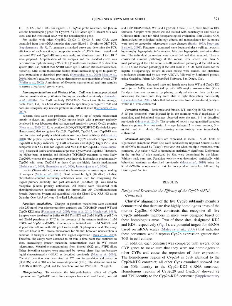
Efficacy of Cyp2b Repression in Cyp2b-KD Mice
QRT-PCR and Western blots were performed to measure
changes in the expression of individual Cyp2b isoforms in WT
and Cyp2b-KD mice. Cyp2b9, Cyp2b10, and Cyp2b13 are
female predominant Cyps (Hashita et al., 2008; Jarukamjorn
et al., 2006) in FVB mice (Hernandez et al., 2006), although
there is some disagreement as to whether Cyp2b10 is female
predominant (Jarukamjorn et al., 1999), and this may be caused
by strain differences (Hernandez et al., 2009b). QRT-PCR of
untreated adult WT and Cyp2b-KD mice indicate that most but
not all the hepatic Cyp2b members are repressed in the KD
mice. The expression of Cyp2b9, Cyp2b10, and Cyp2b13 are
repressed in male Cyp2b-KD mice relative to WT mice. In
contrast, only Cyp2b10 and Cyp2b13 are repressed in female
Cyp2b-KD mice relative to their WT controls (Fig. 2). Cyp2b9,
a female predominant Cyp that is expressed at greater levels
than the other Cyp2b members constitutively (Lee et al., 2011;
Sutton et al., 2010), is not repressed in females.
Western blots demonstrate that Cyp2b protein expression is
also repressed. Two distinct Cyp2b members, thought to be
Cyp2b9 and Cyp2b10, are repressed in females (Fig. 2),
indicating that Cyp2b protein expression is reduced in the
female Cyp2b-KD mice. Untreated male mice have low Cyp2b
expression (Hernandez et al., 2006, 2009b), and our antibody
was not sensitive enough to consistently quantify Cyp2b levels
in the WT or Cyp2b-KD mice. Therefore, we also measured
Cyp2b expression in TCPOBOP-treated mice.
The CAR activator, TCPOBOP (Tzameli et al., 2000), was
used to determine the efficacy of our Cyp2b-KD construct at
reducing Cyp2b levels following treatment with a powerful
inducer. The primary purpose of this experiment was to
determine if TCPOBOP treatment and the subsequent Cyp2b
induction would outcompete lentiviral-promoted shRNA re-
pression of Cyp2b. None of the hepatic Cyp2bs showed lower
transcript expression after TCPOBOP treatment in the Cyp2b-
KD mice compared with the WT mice (Fig. 3). Cyp2b10 and
Cyp2b13 showed greater expression in the TCPOBOP-treated
Cyp2b-KD female mice than the WT mice by 2.63, indicating
a compensatory mechanism. However, protein levels as
determined by Western blots did not confirm the QRT-PCR
results and actually showed significant decreases in Cyp2b
expression (about 5–103) in the Cyp2b-KD mice compared
with the WT mice (Fig. 3). This demonstrates that TCPOBOP-
mediated Cyp2b induction did not outcompete the shRNA’s
ability to repress Cyp2b protein expression. Therefore, the
Cyp2b-KD mouse was still functionally repressing Cyp2b
protein concentrations even after the addition of a potent
Cyp2b inducer.
CAR constitutively regulates the expression of Cyp2b10 and
Cyp2b13 (Hernandez et al., 2009b; Mota et al., 2010) and may
TABLE 1
Relative Cyp2b Expression in Primary Hepatocytes Transduced
with Cyp2b-KD2 and -KD3 shRNA Compared with
Hepatocytes Transduced with a Scrambled Construct
CYP Scrambled KD2 (5 MOI) KD2 (20 MOI) KD3 (20 MOI)
Cyp2b9 1.0 0.11 0.02 1.41
Cyp2b10 1.0 0.27 0.30 0.36
372 DAMIRI ET AL.
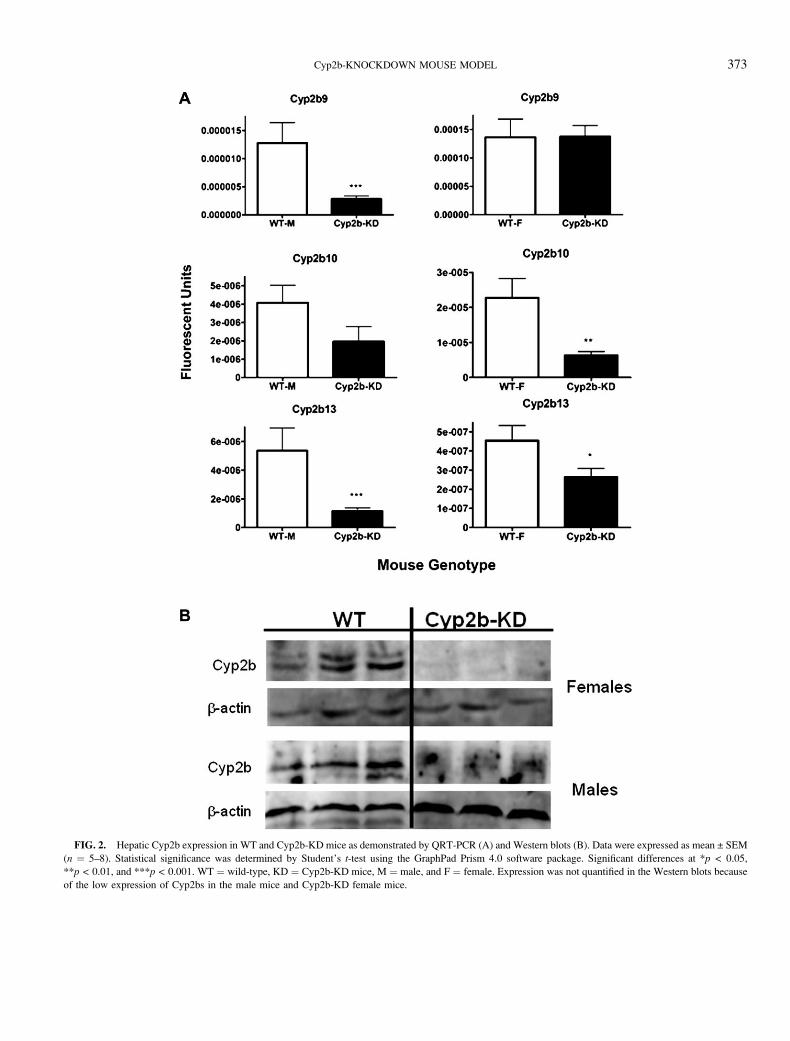
FIG. 2. Hepatic Cyp2b expression in WT and Cyp2b-KD mice as demonstrated by QRT-PCR (A) and Western blots (B). Data were expressed as mean ± SEM
(n ¼ 5–8). Statistical significance was determined by Student’s t-test using the GraphPad Prism 4.0 software package. Significant differences at *p < 0.05,
**p < 0.01, and ***p < 0.001. WT ¼ wild-type, KD ¼ Cyp2b-KD mice, M ¼ male, and F ¼ female. Expression was not quantified in the Western blots because
of the low expression of Cyp2bs in the male mice and Cyp2b-KD female mice.
Cyp2b-KNOCKDOWN MOUSE MODEL 373
constitutively regulate the expression of Cyp2b9 in males
(Mota et al., 2010). Therefore, we hypothesized that CAR
transcript expression may be increased in the Cyp2b-KD mice
as a compensatory mechanism that increases CAR’s sensitivity
to endogenous ligands or its constitutive activity and in turn
increases Cyp2b expression, especially Cyp2b10. QRT-PCR
and Western blotting were performed (Fig. 4), and QRT-PCR
suggested that CAR may be increased in female Cyp2b-KD
mice relative to WT mice, but the data were not statistically
significant (p ¼ 0.061). Western blots were performed to
confirm QRT-PCR results and ascertain whether there was
a trend suggesting increased CAR in females. CAR protein
expression was significantly increased (2.93) in the TCPO-
BOP-treated Cyp2b-KD female mice but not the untreated
mice compared with the corresponding WT mice, suggesting
that CAR may be involved in a compensatory mechanism that
helps Cyp2b-KD mice respond to a chemical insult, such as
TCPOBOP (Fig. 4); however, the compensatory mechanism
did not overcome the ability of the shRNA construct to repress
Cyp2b protein expression in the mice.
FIG. 3. Hepatic Cyp2b expression in WT and Cyp2b-KD mice treated
with the Cyp2b10 inducer, TCPOBOP. (A) RNA expression of Cyp2b9,
Cyp2b10, and Cyp2b13 as measured by QRT-PCR (n ¼ 5–8). (B) Protein
expression of hepatic Cyp2b subfamily members (n ¼ 3–4). Data were
expressed as mean ± SEM. Statistical significance was determined by Student’s
t-test using the GraphPad Prism 4.0 software package. *p < 0.05 and
**p < 0.01. WT ¼ wild-type, KD ¼ Cyp2b-KD mice, M ¼ male, and
F ¼ female.
FIG. 4. Hepatic expression of CAR in WT and Cyp2b-KD mice treated
with TCPOBOP or corn oil (carrier) as measured by QRT-PCR (A) or Western
blots (B, C). (A) WT mice are shown in white bars, and Cyp2b-KD mice are
shown in black bars. Statistical significance was determined by ANOVA
followed by Dunnett’s post hoc test with the GraphPad Prism 4.0 software
package. Western blots in corn oil (B) or TCPOBOP-treated mice (C).
Statistical significance of the Western blots was determined by Student’s t-test.
An asterisk indicates significant difference with a p < 0.05.
374 DAMIRI ET AL.
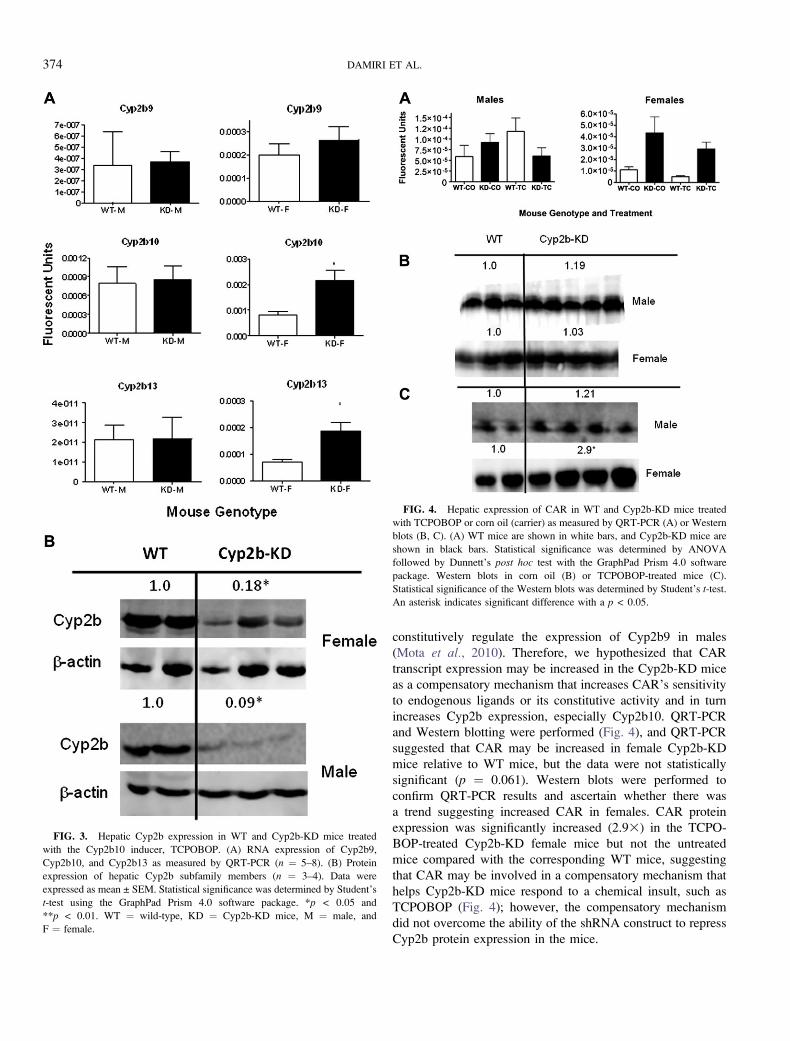
Histopathology
Because CAR is critical in hepatic responses to toxicants, we
investigated whether Cyp2b-KD mice may show histopatho-
logical changes especially after TCPOBOP treatment. Sum-
mation of the different histopathology parameters examined
indicates that only the TCPOBOP-treated Cyp2b-KD female
mice responded in an atypical manner (Fig. 5). For example, all
the TCPOBOP-treated male and female WT mice showed
increased combined histopathology scores primarily because of
increased hyperplasia, typical of TCPOBOP-treated mice
(Blanco-Bose et al., 2008; Wei et al., 2000). However,
TCPOBOP-treated Cyp2b-KD female mice had significantly
decreased combined histopathology scores compared with
TCPOBOP-treated WT mice and control (corn oil) Cyp2b-KD
mice (Fig. 5). Only eight mice showed a score of 6 or less; this
includes all three female TCPOBOP-treated Cyp2b-KD
mice. The other mice with scores of less than 6 were all
control mice (three male control WT and two control Cyp2b-
KD mice). The two-way ANOVA indicates that low
mineralization and hyperplasia are the primary reasons for
low combined histopathology scores in the TCPOBOP-treated
Cyp2b-KD female mice (p < 0.05). Hypertrophy is also
decreased in the TCPOBOP-treated Cyp2b-KD mice but not
significantly. There were no significant differences between the
groups when examining swelling, inflammation, bile duct
hyperplasia, or necrosis.
Cyp2b-Mediated Metabolism Is Compromised in the Cyp2b-KD Mice
The in vitro metabolism of parathion was examined in liver
microsomes from untreated and TCPOBOP-treated mice to test
whether Cyp2b-KD mice demonstrated perturbed metabolism
of parathion relative to WT mice. Cyp2b enzymes have a high
affinity for parathion and in turn are probably key enzymes in
the metabolism of parathion to its toxic form paraoxon
(POXON) and its nontoxic form PNP (Foxenberg et al.,2007, 2011; Mota et al., 2010). Parathion metabolism was
severely compromised (down 3–73) in the untreated male and
female Cyp2b-KD mice compared with the untreated WT mice
(Figs. 6A and 6B). Parathion metabolism was compromised
even more in the TCPOBOP-treated Cyp2b-KD mice com-
pared with the TCPOBOP-treated WT mice (down 5–83)
(Figs. 6C and 6D) consistent with the Western blot results
demonstrating lower Cyp2b expression in TCPOBOP-treated
CYP2b-KD mice than WT mice. Overall, PNP production was
compromised 42.9% more than POXON production in the
Cyp2b-KD mice, which means the ratio of PNP over POXON
was higher in the WT mice than the Cyp2b-KD mice. A higher
ratio indicates greater production of the nontoxic metabolite
and may provide a protective effect. Untreated male and
female WT mice had PNP/POXON ratios of 6.2 and 7.9,
respectively; and TCPOBOP-treated WT mice had PNP/
POXON ratios of 7.2 and 6.6, respectively. Untreated male
FIG. 5. Histopathology of WT and Cyp2b-KD (KD) mice treated with corn oil or TCPOBOP (TC). (A) Histopathology was measured as described in the
‘‘Materials and Methods’’ section using a combined histopathology score from several different measures. Asterisks indicate statistical differences (*p < 0.05;
**p < 0.01; and ***p < 0.001) as determined by a two-way ANOVA followed by a Bonferroni posttest (n ¼ 3). (B) Female WT mouse showing hyperplasia after
TC treatment (2003). (C) Female Cyp2b-KD mouse showing no discernable hyperplasia after TC treatment (2003).
Cyp2b-KNOCKDOWN MOUSE MODEL 375
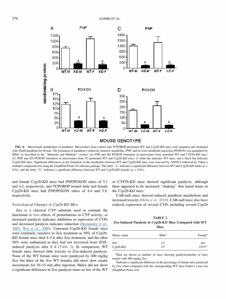
and female Cyp2b-KD mice had PNP/POXON ratios of 5.1
and 4.2, respectively, and TCPOBOP-treated male and female
Cyp2b-KD mice had PNP/POXON ratios of 4.4 and 5.8,
respectively.
Toxicological Changes in Cyp2b-KD Mice
Zox is a classical CYP substrate used to estimate the
functional in vivo effects of perturbations in CYP activity, as
increased paralysis indicates inhibition or repression of CYPs
and decreased paralysis indicates induction (Hernandez et al.,2007; Wei et al., 2000). Untreated Cyp2b-KD female mice
were extremely sensitive to Zox treatment as 50% of Cyp2b-
KD female mice died 4–5 h after Zox treatment, and the other
50% were euthanized as they had not recovered from ZOX-
induced paralysis after 8 h (Table 2). In comparison, WT
female mice showed little toxicity to Zox-induced paralysis.
None of the WT female mice were paralyzed by 400 mg/kg
Zox but three of the five WT females did show slow erratic
movements for 10–15 min after injection. Males did not show
a significant difference in Zox paralysis times as few of the WT
or CYP2b-KD mice showed significant paralysis, although
there appeared to be increased ‘‘shaking’’ that lasted hours in
the Cyp2b-KD mice.
CAR-null mice showed reduced parathion metabolism and
increased toxicity (Mota et al., 2010). CAR-null mice also have
reduced expression of several CYPs including several Cyp2b
FIG. 6. Microsomal metabolism of parathion. Microsomes from control and TCPOBOP-pretreated WT and Cyp2b-KD mice were prepared and incubated
with 20lM parathion for 60 min. The formation of parathion’s relatively nontoxic metabolite, PNP, and its toxic metabolite paraoxon (POXON) was quantified by
HPLC as described in the ‘‘Materials and Methods’’ section. (A) PNP and (B) POXON formation in microsomes from untreated WT and CYP2b-KD mice.
(C) PNP and (D) POXON formation in microsomes from TC-pretreated WT and Cyp2b-KD mice. A white bar indicates WT mice, and a black bar indicates
Cyp2b-KD mice. Significant differences in the formation of the metabolites between WT and Cyp2b-KD mice were assessed by ANOVA followed by Tukey’s
multiple comparison test using the GraphPad Prism 4.0 software package. The letter ‘‘a’’ indicates a significant difference between WT and Cyp2b-KD males (p <
0.01), and the letter ‘‘b’’ indicates a significant difference between WT and Cyp2b-KD females (p < 0.01).
TABLE 2
Zox-Induced Paralysis in Cyp2b-KD Mice Compared with WT
Mice
Mouse strain Malea Femalea
WT 1/7 0/5
Cyp2b-KD 1/7 15/15*
aData are shown as number of mice showing paralysis/number of mice
treated with 400 mg/kg Zox.
*Indicates a significant difference in the percentage of female mice paralyzed
by Zox when compared with the corresponding WT mice Fisher’s exact test
(GraphPad Prism 4.0).
376 DAMIRI ET AL.
members (Hernandez et al., 2009b; Mota et al., 2010). Because
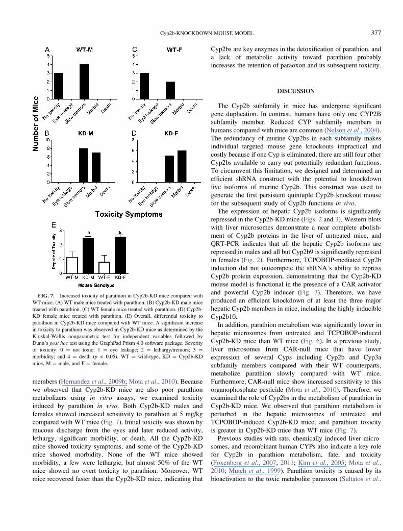
we observed that Cyp2b-KD mice are also poor parathion
metabolizers using in vitro assays, we examined toxicity
induced by parathion in vivo. Both Cyp2b-KD males and
females showed increased sensitivity to parathion at 5 mg/kg
compared with WT mice (Fig. 7). Initial toxicity was shown by
mucous discharge from the eyes and later reduced activity,
lethargy, significant morbidity, or death. All the Cyp2b-KD
mice showed toxicity symptoms, and some of the Cyp2b-KD
mice showed morbidity. None of the WT mice showed
morbidity, a few were lethargic, but almost 50% of the WT
mice showed no overt toxicity to parathion. Moreover, WT
mice recovered faster than the Cyp2b-KD mice, indicating that
Cyp2bs are key enzymes in the detoxification of parathion, and
a lack of metabolic activity toward parathion probably
increases the retention of paraoxon and its subsequent toxicity.
DISCUSSION
The Cyp2b subfamily in mice has undergone significant
gene duplication. In contrast, humans have only one CYP2B
subfamily member. Reduced CYP subfamily members in
humans compared with mice are common (Nelson et al., 2004).
The redundancy of murine Cyp2bs in each subfamily makes
individual targeted mouse gene knockouts impractical and
costly because if one Cyp is eliminated, there are still four other
Cyp2bs available to carry out potentially redundant functions.
To circumvent this limitation, we designed and determined an
efficient shRNA construct with the potential to knockdown
five isoforms of murine Cyp2b. This construct was used to
generate the first persistent quintuple Cyp2b knockout mouse
for the subsequent study of Cyp2b functions in vivo.
The expression of hepatic Cyp2b isoforms is significantly
repressed in the Cyp2b-KD mice (Figs. 2 and 3). Western blots
with liver microsomes demonstrate a near complete abolish-
ment of Cyp2b proteins in the liver of untreated mice, and
QRT-PCR indicates that all the hepatic Cyp2b isoforms are
repressed in males and all but Cyp2b9 is significantly repressed
in females (Fig. 2). Furthermore, TCPOBOP-mediated Cyp2b
induction did not outcompete the shRNA’s ability to repress
Cyp2b protein expression, demonstrating that the Cyp2b-KD
mouse model is functional in the presence of a CAR activator
and powerful Cyp2b inducer (Fig. 3). Therefore, we have
produced an efficient knockdown of at least the three major
hepatic Cyp2b members in mice, including the highly inducible
Cyp2b10.
In addition, parathion metabolism was significantly lower in
hepatic microsomes from untreated and TCPOBOP-induced
Cyp2b-KD mice than WT mice (Fig. 6). In a previous study,
liver microsomes from CAR-null mice that have lower
expression of several Cyps including Cyp2b and Cyp3a
subfamily members compared with their WT counterparts,
metabolize parathion slowly compared with WT mice.
Furthermore, CAR-null mice show increased sensitivity to this
organophosphate pesticide (Mota et al., 2010). Therefore, we
examined the role of Cyp2bs in the metabolism of parathion in
Cyp2b-KD mice. We observed that parathion metabolism is
perturbed in the hepatic microsomes of untreated and
TCPOBOP-induced Cyp2b-KD mice, and parathion toxicity
is greater in Cyp2b-KD mice than WT mice (Fig. 7).
Previous studies with rats, chemically induced liver micro-
somes, and recombinant human CYPs also indicate a key role
for Cyp2b in parathion metabolism, fate, and toxicity
(Foxenberg et al., 2007, 2011; Kim et al., 2005; Mota et al.,2010; Mutch et al., 1999). Parathion toxicity is caused by its
bioactivation to the toxic metabolite paraoxon (Sultatos et al.,
FIG. 7. Increased toxicity of parathion in Cyp2b-KD mice compared with
WT mice. (A) WT male mice treated with parathion. (B) Cyp2b-KD male mice
treated with parathion. (C) WT female mice treated with parathion. (D) Cyp2b-
KD female mice treated with parathion. (E) Overall, differential toxicity to
parathion in Cyp2b-KD mice compared with WT mice. A significant increase
in toxicity to parathion was observed in Cyp2b-KD mice as determined by the
Kruskal-Wallis nonparametric test for independent variables followed by
Dunn’s post hoc test using the GraphPad Prism 4.0 software package. Severity
of toxicity: 0 ¼ not toxic; 1 ¼ eye leakage; 2 ¼ lethargy/tremors; 3 ¼morbidity; and 4 ¼ death (p < 0.05). WT ¼ wild-type, KD ¼ Cyp2b-KD
mice, M ¼ male, and F ¼ female.
Cyp2b-KNOCKDOWN MOUSE MODEL 377

1984), but we observed reduced paraoxon production in the
Cyp2b-KD mice that is not consistent with decreased toxicity.
Similar but stronger results were obtained with the CAR-null
mice (Mota et al., 2010). This suggests that toxicity in the
Cyp2b-KD mice is due to poor metabolism of parathion to PNP
(lower PNP/POXON ratio), which is also catalyzed by Cyp2b
(Foxenberg et al., 2007, 2011; Mutch and Williams, 2006),
extrahepatic metabolism of parathion, or higher clearance of
paraoxon from the liver of Cyp2b-KD mice compared with WT
mice because of poor metabolism of parathion and paraoxon.
Liver perfusion studies indicate that parathion metabolized to
paraoxon may exit the liver as paraoxon and cause toxicity
(Sultatos et al., 1985). Overall, this study demonstrates that
in vivo Cyp2b isoforms play a key role in parathion metabolism
and toxicity, and this is the first study to demonstrate that
individuals with compromised Cyp2b are susceptible to the
toxic effects of parathion and suggests that active phase I
metabolism of parathion is important for further metabolic
deactivation and elimination.
ZOX paralysis time is a key indicator of perturbations in Cyp
activity in vivo. Female Cyp2b-KD mice did not recover from
Zox injection indicating poor metabolism and clearance and
indicating a key role of Cyp2b in Zox metabolism. In contrast,
male Cyp2b-KD mice did not demonstrate a significant
difference in Zox paralysis time (Table 2). Most Cyp2bs
(Cyp2b9, Cyp2b13, and may be Cyp2b10) are female pre-
dominant (Hernandez et al., 2006, 2009a; Wiwi et al., 2004), and
therefore, reducing Cyp2b levels in female mice may cause
a more pronounced effect. FVB (WT) mice also metabolize Zox
better than B6 mice, and a higher dose is needed to cause
paralysis (Hernandez et al., 2006). A 450 mg/kg dose appeared to
be too high in a previous study (Hernandez et al., 2006), and
therefore, we performed a pilot study and based on those results
decided to use 400 mg/kg. However, this dose did not
significantly affect the FVB male mice as few of them (WT
and Cyp2b-KD) were fully paralyzed by Zox.
Histopathology demonstrated that female Cyp2b-KD mice
did not respond to TCPOBOP treatment as expected and in turn
showed perturbed histopathology parameters. Overall, though,
the Cyp2b-KD mice are viable, fertile, and did not exhibit
obvious gross abnormalities with the exception of an increase
in liver weight. Liver enlargement has been regarded as
a marker of drug associated enzyme induction and suggests
a compensatory mechanism (Amacher et al., 2001; Webber
et al., 1994), probably to adapt to increased concentrations of
an endobiotic. Similar increases in hepatosomatic indices have
been observed in other transgenic mice with low Cyp activity,
including mice that lack key hepatic transcription factors
that regulate Cyps. Examples include the hepatic POR-null
mouse model (Henderson et al., 2003) and HNF4a-null mice
(Hayhurst et al., 2001) where the liver/body weight ratios
probably increased because of the accumulation of lipids.
However, similar effects have not been reported in the Cyp3a-
null mouse (van Herwaarden et al., 2007).
Interestingly, there also appears to be a molecular compen-
satory reaction to the repression of Cyp2bs in the Cyp2b-KD
mice. For example, Cyp2b9 mRNA expression was not
repressed in female Cyp2b-KD mice. Furthermore, no
TCPOBOP-treated male mice showed repression of Cyp2b
mRNA transcript levels, and female mice actually demon-
strated greater expression of Cyp2b10 and Cyp2b13 in Cyp2b-
KD mice following TCPOBOP treatment than WT mice. This
suggests that there is some type of compensatory mechanism
trying to overcome the repressive effects of the shRNA. CAR
basally regulates Cyp2b10 and Cyp2b13 and may in part
regulate Cyp2b9 (Hernandez et al., 2009b; Mota et al., 2010).
Therefore, we hypothesized that CAR may be induced in order
to adapt to the lack of Cyp2b members, especially Cyp2b10, in
the Cyp2b-KD mice. CAR protein levels are increased in
TCPOBOP-treated females, and this may help the mice adapt
to lower Cyp2b expression and in turn increase Cyp2b mRNA
expression. In addition to CAR, forkhead box protein A2
(FoxA2 also known as hepatic nuclear factor 3b), a female
predominant transcription factor, regulates Cyp2b9 (Hashita
et al., 2008), and therefore, this transcription factor may also
play a role in the lack of Cyp2b9 repression in untreated and
TCPOBOP-treated Cyp2b-KD female mice. Other transcrip-
tion factors that in part regulate the sexually dimorphic
expression of Cyp2b9, such as HNF4a and Stat5b, cannot be
ruled out (Wiwi et al., 2004). Lastly, the compensatory
induction of Cyp2b10 in males and females may just be due to
increased retention of TCPOBOP caused by the lack of Cyp2b
proteins, leading to greater activation of CAR and in turn
higher Cyp2b10 transcript levels.
Even though Cyp2b transcript levels were increased in
Cyp2b-KD mice to levels equal to or greater than WT mice
following TCPOBOP treatment, protein expression was still
much lower in the Cyp2b-KD mice. It has been suggested that
Western blots are more reliable to confirm the efficacy of
siRNA (Holmes et al., 2010). There are several potential
reasons for this including the extracted RNA is nuclear or not
available for siRNA degradation, or the 3#mRNA cleavage
products resulting from siRNA-mediated cleavage accumulate
within the cell (Holen et al., 2002), but are still large enough
fragments that they result in small templates for cDNA
synthesis and give rise to a false signal of mRNA detection
by QRT-PCR (Holmes et al., 2010).
The long-term stability of the Cyp2b-KD transgene is not
known. Lentiviral-mediated transgenesis following perivitel-
line injection has been shown to be stable for a couple of
generations (Park, 2007); however, there have been few studies
that have examined multigenerational stability of shRNAs.
There are several potential problems that can arise as a shRNA
transgenic line is propagated, including selective pressures
from integration into transcriptionally active genes, methyla-
tion of the promoter, or a reduction in insert copy number, and
all of these may lead to reduced functional transgene
expression (Kong et al., 2009; Sauvain et al., 2008). Dilution
378 DAMIRI ET AL.
of the number of insert sites will occur as mice are mated to
WT mice (Sauvain et al., 2008), and this may diminish the
phenotype in subsequent generations. Conversely, one of the
major advantages of viral-mediated transgenesis is the ability to
produce mice with multiple integrants and therefore greater
shRNA expression. In addition, shRNA can knockdown
multiple genes with one construct or produce a large number
of mice in a short period of time without the necessity of
breeding (Lois et al., 2002; Park, 2007). For example, all 134
mice produced from our initial injections were positive,
providing a vast number of mice for experimentation in a short
period of time.
The shRNA generated Cyp2b-KD mice demonstrate low
expression of hepatic Cyp2b members in untreated and
TCPOBOP-treated mice. They also poorly metabolize the
Cyp2b substrates Zox and parathion and in turn are sensitive to
these toxicants, indicating that Cyp2bs play a key role in
protecting individuals from select chemicals. Therefore,
Cyp2b-KD mice may be able to act as a sentinel for individuals
with low Cyp2b expression or limited metabolic capacity
because of Cyp2b polymorphisms. Furthermore, this model can
be built upon to form even better models for human disease,
human metabolism, and human genetic polymorphisms by
making CYP2B6/7-humanized mice. This study provides
a new platform for studying Cyp2b function and especially
its role in the metabolism of distinct pharmaceuticals and
environmental chemicals.
SUPPLEMENTARY DATA
Supplementary data are available online at http://toxsci.
oxfordjournals.org/.
FUNDING
National Institutes of Health grant R15-ES017321; a South
Carolina EPSCoR/IDEA CCD project; a grant from the
Clemson University Research Foundation; and Clemson
University start-up funds.
REFERENCES
Amacher, D. E., Schomaker, S. J., and Burkhardt, J. E. (2001). The relationship
among microsomal enzyme induction, liver weight and histological change
in beagle dog toxicology studies. Food Chem. Toxicol. 39, 817–825.
Athirakul, K., Bradbury, J. A., Graves, J. P., DeGraff, L. M., Ma, J., Zhao, Y.,
Couse, J. F., Quigley, R., Harder, D. R., Zhao, X., et al. (2008). Increased
blood pressure in mice lacking cytochrome P450 2j5. FASEB J. 22,
4096–4108.
Baldwin, W. S., Marko, P. B., and Nelson, D. R. (2009). The cytochrome P450
(CYP) gene superfamily in Daphnia Pulex. BMC Genomics 10, 169.
Banks, W. J. (1993). In Applied Veterinary Histology, 3rd ed. C V Mosby, St
Louis, MO.
Beilke, L. D., Aleksunes, L. M., Holland, R. D., Besselsen, D. G., Beger, R. D.,
Klaassen, C. D., and Cherrington, N. J. (2009). Constitutive androstane
receptor-mediated changes in bile acid composition contributes to hepato-
protection from lithocholic acid-induced liver injury in mice. Drug Metab.
Dispos. 37, 1035–1045.
Blanco-Bose, W. E., Murphy, M. J., Ehninger, A., Offner, S., Dubey, C.,
Huang, W., Moore, D. D., and Trumpp, A. (2008). C-myc and its target
foxm1 are critical downstream effectors of constitutive androstane receptor
(CAR) mediated direct liver hyperplasia. Hepatology 48, 1302–1311.
Blesch, A. (2004). Lentiviral and MLV based retroviral vectors for ex vivo and
in vivo gene transfer. Methods 33, 164–172.
Burns, J. C., Friedmann, T., Driever, W., Burrascano, M., and Yee, J. K.
(1993). Vesicular stomatitis virus G glycoprotein pseudotyped retroviral
vectors: Concentration to very high titer and efficient gene transfer into
mammalian and nonmammalian cells. Proc. Natl. Acad. Sci. U.S.A. 90,
8033–8037.
Ekins, S., Vandenbranden, M., Ring, B. J., Gillespie, J. S., Yang, T. J.,
Gelboin, H. V., and Wrighton, S. A. (1998). Further characterization of the
expression in liver and catalytic activity of CYP2B6. J. Pharmacol. Exp.
Ther. 286, 1253–1259.
Elbashir, S. M., Lendeckel, W., and Tuschl, T. (2001). RNA interference is
mediated by 21- and 22-nucleotide RNAs. Genes Dev. 15, 188–200.
Finger, J. H., Smith, C. M., Hayamizu, T. F., McCright, I. J., Eppig, J. T.,
Kadin, J. A., Richardson, J. E., and Ringwald, M. (2011). The mouse Gene
Expression Database (GXD): 2011 update. Nucleic Acids Res. 39(Suppl. 1),
D835–D841.
Foxenberg, R. J., Ellison, C. A., Knaak, J. B., Ma, C., and Olson, J. R. (2011).
Cytochrome P450-specific human PBPK/PD models for the organophos-
phorus pesticides: Chlorpyrifos and parathion. Toxicology 285, 57–66.
Foxenberg, R. J., McGarrigle, B. P., Knaak, J. B., Kostyniak, P. J., and
Olson, J. R. (2007). Human hepatic cytochrome P450-specific metabolism of
parathion and chlorpyrifos. Drug Metab. Dispos. 35, 189–193.
Hashita, T., Sakuma, T., Akada, M., Nakajima, A., Yamahara, H., Ito, S.,
Takesako, H., and Nemoto, N. (2008). Forkhead box A2-mediated regulation
of female-predominant expression of the mouse cyp2b9 gene. Drug Metab.
Dispos. 36, 1080–1087.
Hayhurst, G. P., Lee, Y. H., Lambert, G., Ward, J. M., and Gonzalez, F. J.
(2001). Hepatocyte nuclear factor 4alpha (nuclear receptor 2a1) is essential
for maintenance of hepatic gene expression and lipid homeostasis. Mol. Cell.
Biol. 21, 1393–1403.
Henderson, C. J., Otto, D. M., Carrie, D., Magnuson, M. A., McLaren, A. W.,
Rosewell, I., and Wolf, C. R. (2003). Inactivation of the hepatic cytochrome
P450 system by conditional deletion of hepatic cytochrome P450 reductase.
J. Biol. Chem. 278, 13480–13486.
Hernandez, J. P., Chapman, L. M., Kretschmer, X. C., and Baldwin, W. S.
(2006). Gender specific induction of cytochrome P450s in nonylphenol-
treated FVB/NJ mice. Toxicol. Appl. Pharmacol. 216, 186–196.
Hernandez, J. P., Huang, W., Chapman, L. M., Chua, S., Moore, D. D., and
Baldwin, W. S. (2007). The environmental estrogen, nonylphenol,
activates the constitutive androstane receptor (CAR). Toxicol. Sci. 98,
416–426.
Hernandez, J. P., Mota, L. C., and Baldwin, W. S. (2009a). Activation of CAR
and PXR by dietary, environmental and occupational chemicals alters drug
metabolism, intermediary metabolism, and cell proliferation. Curr. Pharma-
cogenomics Person. Med. 7, 81–105.
Hernandez, J. P., Mota, L. C., Huang, W., Moore, D. D., and Baldwin, W. S.
(2009b). Sexually dimorphic regulation and induction of P450s by the
constitutive androstane receptor (CAR). Toxicology 256, 53–64.
Hodgson, E., and Rose, R. L. (2007). The importance of cytochrome P450 2B6
in the human metabolism of environmental chemicals. Pharmacol. Ther.
113, 420–428.
Cyp2b-KNOCKDOWN MOUSE MODEL 379
Holen, T., Amarzguioui, M., Wiiger, M. T., Babaie, E., and Prydz, H. (2002).
Positional effects of short interfering RNAs targeting the human coagulation
trigger tissue factor. Nucleic Acids Res. 30, 1757–1766.
Holmes, K., Williams, C. M., Chapman, E. A., and Cross, M. J. (2010).
Detection of siRNA induced mRNA silencing by RT-qPCR: Considerations
for experimental design. BMC Res. Notes 3, 53.
Honkakoski, P., Zelko, I., Sueyoshi, T., and Negishi, M. (1998). The nuclear
orphan-receptor CAR-retinoid X receptor heterodimer activates the pheno-
barbital-responsive module of the CYP2B gene. Mol. Cell. Biol. 18,
5652–5658.
Jarukamjorn, K., Sakuma, T., Jaruchotikamol, A., Ishino, Y., Oguro, M., and
Nemoto, N. (2006). Modified expression of cytochrome P450 mRNAs by
growth hormone in mouse liver. Toxicology 219, 97–105.
Jarukamjorn, K., Sakuma, T., Miyaura, J. I., and Nemoto, N. (1999). Different
regulation of the expression of mouse hepatic cytochrome P450 2B enzymes
by glucocorticoid and phenobarbital. Arch. Biochem. Biophys. 369, 89–99.
Kim, D. O., Lee, S. K., Jeon, T. W., Jin, C. H., Hyun, S. H., Kim, E. J.,
Moon, G. I., Kim, J. A., Lee, E. S., Lee, B. M., et al. (2005). Role of
metabolism in parathion-induced hepatotoxicity and immunotoxicity.
J. Toxicol. Environ. Health A 68, 2187–2205.
Kong, Q., Wu, M., Huan, Y., Zhang, L., Liu, H., Bou, G., Luo, Y., Mu, Y., and
Liu, Z. (2009). Transgene expression is associated with copy number and
cytomegalovirus promoter methylation in transgenic pigs. PLoS One 4,
e6679. doi:10.1371/journal.pone.0006679.
Kretschmer, X. C., and Baldwin, W. S. (2005). CAR and PXR: Xenosensors of
endocrine disrupters? Chem. Biol. Interact. 155, 111–128.
Lamba, V., Lamba, J., Yasuda, K., Strom, S., Davila, J., Hancock, M. L.,
Fackenthal, J. D., Rogan, P. K., Ring, B., Wrighton, S. A., et al. (2003).
Hepatic CYP2B6 expression: Gender and ethnic differences and relationship
to CYP2B6 genotype and CAR (constitutive androstane receptor) expres-
sion. J. Pharmacol. Exp. Ther. 307, 906–922.
Lang, T., Klein, K., Richter, T., Zibat, A., Kerb, R., Eichelbaum, M.,
Schwab, M., and Zanger, U. M. (2004). Multiple novel nonsynonymous
cyp2b6 gene polymorphisms in Caucasians: Demonstration of phenotypic
null alleles. J. Pharmacol. Exp. Ther. 311, 34–43.
Lee, J. S., Ward, W. O., Liu, J., Ren, H., Vallanat, B., Delker, D., and
Corton, J. C. (2011). Hepatic xenobiotic metabolizing enzyme and
transporter gene expression through the life stages of the mouse. PLoS
One 6, e24381.
Lee, S. S. T., Buters, J. T. M., Pineau, T., Fernandez-Salguero, P., and
Gonzalez, F. J. (1996). Role of CYP2E1 in the hepatotoxicity of
acetaminophen. J. Biol. Chem. 271, 12063–12067.
Lois, C., Hong, E. J., Pease, S., Brown, E. J., and Baltimore, D. (2002).
Germline transmission and tissue-specific expression of transgenes delivered
by lentiviral vectors. Science 295, 868–872.
Mateeva, O., Nechipurenko, Y., Rossil, L., Moore, B., Saetrom, P.,
Aleksey, Y., Ogurtsov, Y., Atkins, J. F., and Shabalina, S. A. (2007).
Comparison of approaches for rational siRNA design leading to a new
efficient and transparent method. Nucleic Acids Res. 35, e63.
Mota, L. C., Hernandez, J. P., and Baldwin, W. S. (2010). CAR-null mice are
sensitive to the toxic effects of parathion: Association with reduced CYP-
mediated parathion metabolism. Drug Metab. Dispos. 38, 1582–1588.
Mota, L. C., Hernandez, J. P., Barfield, C., and Baldwin, W. S. (2011).
Nonylphenol-mediated CYP induction is PXR-dependent: The use of
humanized mice and human hepatocytes suggests that hPXR is less sensitive
than mouse PXR to nonylphenol treatment. Toxicol. Appl. Pharmacol. 252,
259–267.
Muerhoff, A. S., Griffin, K. J., and Johnson, E. F. (1994). The peroxisome
proliferator-activated receptor mediates the induction of CYP4A6, a cyto-
chrome P450 fatty acid omega-hydroxylase, by clofibric acid. J. Biol. Chem.
267, 19051–19053.
Muller, P. Y., Janovjak, H., Miserez, A. R., and Dobbie, Z. (2002). Processing
of gene expression data generated by quantitative real-time RT-PCR.
Biotechniques 32, 1372–1379.
Mutch, E., Blain, P. G., and Williams, F. M. (1999). The role of metabolism in
determining susceptibility to parathion toxicity in man. Toxicol. Lett. 107,
177–187.
Mutch, E., and Williams, F. M. (2006). Diazinon, chlorpyrifos and parathion
are metabolised by multiple cytochrome P450 in human liver. Toxicology
224, 22–32.
Nelson, D. R., Zeldin, D. C., Hoffman, S. M. G., Maltais, L. J., Wain, H. M.,
and Nebert, D. W. (2004). Comparison of cytochrome P450 (CYP) genes
from the mouse and human genomes, including nomenclature recommen-
dations for genes, pseudogenes and alternative splice variants. Pharmaco-
genetics 14, 1–18.
Park, F. (2007). Lentiviral vectors: Are they the future of animal transgenesis?
Physiol. Genomics 31, 159–173.
Percy, D. H., and Barthold, S. W. (2001). Pathology of Laboratory Rodents
and Rabbits. Iowa State University Press, Ames, IA.
Ramezani, A., and Hawley, R. G. (2002). Generation of HIV-1-based lentiviral
vector particles. Curr. Protoc. Mol. Biol. Chapter 16, Unit 16.22.
Reschly, E. J., and Krasowski, M. D. (2006). Evolution and function of the
NR1I nuclear hormone receptor subfamily (VDR, PXR, and CAR) with
respect to metabolism of xenobiotics and endogenous compounds. Curr.
Drug Metab. 7, 349–365.
Sastry, L., Johnson, T., Hobson, M. J., Smucker, B., and Cornetta, K. (2002).
Titering lentiviral vectors: Comparison of DNA, RNA and marker
expression methods. Gene Ther. 9, 1155–1162.
Sauvain, M. O., Dorr, A. P., Stevenson, B., Quazzola, A., Naef, F.,
Wiznerowicz, M., Schutz, F., Jongeneel, V., Duboule, D., Spitz, F., et al.
(2008). Genotypic features of lentivirus transgenic mice. J. Virol. 82,
7111–7119.
Sultatos, L. G., Minor, L. D., and Murphy, S. D. (1985). Metabolic activation
of phosphorothioate pesticides: Role of the liver. J. Pharmacol. Exp. Ther.
232, 624–628.
Sultatos, L. G., Shao, M., and Murphy, S. D. (1984). The role of hepatic
biotransformation in mediating the acute toxicity of the phosphorothionate
insecticide chlorpyrifos. Toxicol. Appl. Pharmacol. 73, 60–68.
Sutton, C. W., Sutherland, M., Shnyder, S., and Patterson, L. H. (2010).
Improved preparation and detection of cytochrome P450 isoforms using MS
methods. Proteomics 10, 327–331.
Tang, J., Cao, Y., Rose, R. L., Brimfield, A. A., Dai, D., Goldstein, J. A., and
Hodgson, E. (2001). Metabolism of chlorpyrifos by human cytochrome P450
isoforms and human, mouse and rat liver microsomes. Drug Metab. Dispos.
29, 1201–1204.
Tzameli, I., Pissios, P., Schuetz, E. G., and Moore, D. D. (2000). The
xenobiotic compound 1,4-bis[2-(3,5-dichloropyridyloxy)]benzene is an
agonist ligand for the nuclear receptor CAR. Mol. Cell. Biol. 20, 2951–2958.
Uppal, H., Toma, D., Saini, S. P., Ren, S., Jones, T. J., and Xie, W. (2005).
Combined loss of orphan receptors PXR and CAR heightens sensitivity to
toxic bile acids in mice. Hepatology 41, 168–176.
Van der Hoeven, T. A., and Coon, M. J. (1974). Preparation and properties of
partially purified cytochrome P450 and NADPH-cytochrome P450 reductase
from rabbit liver microsomes. J. Biol. Chem. 249, 6302–6310.
van Herwaarden, A. E., Wagenaar, E., van der Kruijssen, C. M. M., van
Waterschoot, R. A. B., Smit, J. W., Song, J. Y., van der Valk, M. A., van
Tellingen, O., van der Hoorn, J. W. A., Rosing, H., et al. (2007). Knockout
of cytochrome P450 3A yields new mouse models for understanding
xenobiotic metabolism. J. Clin. Invest. 117, 3583–3592.
Wang, H., Faucette, S., Sueyoshi, T., Moore, R., Ferguson, S., Negishi, M., and
LeCluyse, E. L. (2003). A novel distal enhancer module regulated by
pregnane X receptor/constitutive androstane receptor is essential for the
380 DAMIRI ET AL.
maximal induction of CYP2B6 gene expression. J. Biol. Chem. 278,
14146–14152.
Wang, H., and Tompkins, L. M. (2008). CYP2B6: New insights into a historically
overlooked cytochrome P450 isozyme. Curr. Drug Metab. 9, 598–610.
Waxman, D. J. (1988). Interactions of hepatic cytochromes P-450 with steroid
hormones: Regioselectivity and stereoselectivity of steroid metabolism and
hormonal regulation of rat P-450 enzyme expression. Biochem. Pharmacol.
37, 71–84.
Waxman, D. J., Pampori, N. A., Ram, P. A., Agrawal, A. K., and
Shapiro, B. H. (1991). Interpulse interval in circulating growth hormone
patterns regulates sexually dimorphic expression of hepatic cytochrome
p450. Proc. Natl. Acad. Sci. U.S.A. 88, 6868–6872.
Webber, E. M., Wu, J. C., Wang, L., Merlino, G., and Fausto, N. (1994).
Overexpression of transforming growth factor-alpha causes liver enlarge-
ment and increased hepatocyte proliferation in transgenic mice. Am.
J. Pathol. 145, 398–408.
Wei, P., Zhang, J., Egan-Hafley, M., Liang, S., and Moore, D. D. (2000). The
nuclear receptor car mediates specific xenobiotic induction of drug
metabolism. Nature 407, 920–923.
Willingham, A. T., and Keil, T. (2004). A tissue specific cytochrome P450
required for the structure and function of Drosophila sensory organs. Mech.Dev. 121, 1289–1297.
Wiwi, C. A., Gupte, M., and Waxman, D. J. (2004). Sexually dimorphic P450
gene expression in liver-specific hepatocyte nuclear factor 4a-deficient mice.
Mol. Endocrinol. 18, 1975–1987.
Yamada, H., Ishii, Y., Yamamoto, M., and Oguri, K. (2006). Induction of the
hepatic cytochrome P450 2B subfamily by xenobiotics: Research history,
evolutionary aspect, relation to tumorigenesis, and mechanism. Curr. Drug
Metab. 7, 397–409.
Zhang, J., Huang, W., Chua, S. S., Wei, P., and Moore, D. D. (2002).
Modulation of acetaminophen-induced hepatotoxicity by the xenobiotic
receptor CAR. Science 298, 422–424.
Cyp2b-KNOCKDOWN MOUSE MODEL 381