retroviral pseudotypes – from scientific tools to clinical utility
TRANSCRIPT

Retroviral Pseudotypes –From Scientific Tools toClinical UtilityNigel J Temperton, Viral Pseudotype Unit, University of Kent, Medway, UK
Edward Wright, Viral Pseudotype Unit, University of Westminster, London, UK
Simon D Scott, Viral Pseudotype Unit, University of Kent, Medway, UK
Based in part on the previous version of this eLS article ‘RetroviralPseudotypes’ (2009) by Nigel J Temperton and Edward Wright.
Advanced article
Article Contents• Introduction
• General Method
• Analysis of Receptor Binding and Virus Entry andExit
• Antiviral Screening
• Pseudotype Neutralisation Assays andImmunogenicity Testing
• Pseudotypes as Immunogens and for GeneTherapy
• Notes
• Acknowledgements
Online posting date: 15th June 2015
Retroviral pseudotypes are important researchand diagnostic tools for basic and clinical virol-ogy studies, facilitating the detailed investigationof individual genes, cellular receptors, antibodyresponses, serosurveillance and antiviral thera-pies. Importantly, pseudotypes enable the studyof highly pathogenic viruses, without the needfor high containment. Their use as gene therapyvectors is widely documented, but other uses, onceless well known, are becoming more prominent.The substitution of envelope proteins expressedon the virion surface enables pseudotypes to beemployed as surrogates for wildtype viruses inantibody neutralisation or antiviral screeningassays and for the study of cell–virus receptorinteractions. In addition, they are increasinglybeing utilised as vaccine immunogens, expressingthe antigen either on the particle surface or as atransfer gene for cellular expression. These studiesdemonstrate the potential for using pseudotypesfor both scientific and clinical applications.
Introduction
The standard definition of a pseudotype is a hybrid virus particleconsisting of a protein nucleocapsid (‘core’) encasing a ribonu-cleic acid (RNA) genome, with the core itself being encapsu-lated in a lipid ‘envelope’ membrane derived from the host cell.
eLS subject area: Virology
How to cite:Temperton, Nigel J; Wright, Edward; and Scott, Simon D (June2015) Retroviral Pseudotypes – From Scientific Tools to ClinicalUtility. In: eLS. John Wiley & Sons, Ltd: Chichester.DOI: 10.1002/9780470015902.a0021549.pub2
Importantly, this envelope gained when cores exit from the cellby ‘budding’ is studded with proteins derived from other viruses.Many of these heterologous envelope proteins are antigenic tar-gets for the host immune system. In pseudotypes, one or moreof these envelope proteins may derive from study viruses. Manypseudotypes also carry foreign genes, called ‘transfer’ genes,engineered into their genome. When in the presence of suscep-tible cells, the envelope proteins bind to cell receptors permittingcellular entry, eventually resulting in transfer gene expression.Retroviruses have been extensively exploited as cores for pseudo-typing. The name retrovirus derives from the Latin for ‘behind’or ‘backward’ (retro-) and this highlights their key characteristic,the ability to reverse transcribe their dimeric single-strandedRNAgenome into a double-stranded deoxyribonucleic acid (dsDNA)copy, which is subsequently integrated into the cell genome viathe use of viral and cellular enzymes. For retroviral pseudotypes,this usually leads to expression of the transfer/reporter gene, thelatter being readily quantifiable. Thus, reporter gene expressiondirectly correlates with efficiency of viral envelope/receptor inter-action, and conversely whether individual antibody responses orantiviral agents could interfere with the entry and replicationprocess of the native virus. See also: Retroviral Replication;Vesicular Stomatitis VirusCurrent pseudotyping protocols are based on several decades of
vector development, often for use in gene therapy. The first retro-viral vectors were based on native gammaretroviruses, such asMoloney mouse leukaemia virus (MLV). To produce such retro-virus vectors, a stable cell line containing the retroviral genomeminus the packaging signal needed for genome encapsidationwasfirst generated. These cells were then transfected with a plas-mid construct containing the packaging signal and all cis-actingsequences necessary for virus particle assembly, but in which thegenes required for genome replication and nucleocapsid forma-tion had been removed (often replaced by a gene of interest – thetransfer gene). Thus, all genes needed for retrovirus particleassemblywere provided by the cell line in trans. However, recom-bination between newly produced retroviral vector and inte-grated helper virus sequences occasionally led to production ofreplication-competent viruses using these systems (Sakuma et al.,2012). In order to reduce the production of replication-competent
eLS © 2015, John Wiley & Sons, Ltd. www.els.net 1

Retroviral Pseudotypes – From Scientific Tools to Clinical Utility
virus, novel packaging cell lines were developed where thehelper virus elements were introduced and genomically inte-grated separately, requiring only a final transient transfection ofthe transfer gene construct to generate the replication-deficientretrovirus particles required (Pear et al., 1993). Nevertheless,replication-competent virus remained detectable. This situationwas notably improved by adopting a triple-plasmid system wherethe structural and replication/integration genes (gag-pol plusregulatory/accessory proteins), envelope protein gene (env) andmarker/reporter genes were present on separate plasmids whichwere transiently transfected into producer cell lines (Soneokaet al., 1995; Naldini et al., 1996). Simultaneous expressionof these genes permits assembly of new viral particles, butmultiple recombination events would be necessary to producereplication-competent virus. The particles are able to ‘transduce’susceptible cells, integrate the marker gene, but are unable toproduce new virus progeny (hence viral transduction rather thaninfection).A myriad of more refined systems have since been developed,
often aimed at improving safety, particularly for the use of humanimmunodeficiency virus (HIV) vectors in gene therapy (Sakumaet al., 2012). Adaptations have included the removal of viralaccessory genes (HIV-1 Vif, Vpr, Vpu and Nef), important innatural infection, but not in vectors. Subsequently, to limit inter-ference with host gene expression following vector integration,‘self-inactivating’ systems were engineered. Next, to avoid theneed for transient transfection in large-scale vector production,continuous retrovirus producer cell lines were established (Ikedaet al., 2003). Finally, to further reduce recombinational events, afour-plasmid system to generate vectors was designed, where theHIV rev regulatory gene was placed on a separate plasmid. Thetat regulatory gene was also deleted from the gag-pol construct,as the tat-dependent U3 promoter driving transgene expression isreplacedwith a strong constitutive promoter. The latter adaptationhas been adopted in the three-plasmid system, which remains themost widely used to generate most retroviral pseudotypes to thisday (Figure 1).Retroviruses have proven to be particularly useful for pseudo-
typing, and owing to their ability to stably integrate ‘therapeu-tic’ transfer genes into a target cell, they have been extensivelydeveloped as gene therapy vectors. Furthermore, the exchangeof surface envelope proteins has enabled altered tropism or‘tropism switch’ in pseudotypes, permitting targeting of specificcell types. See also: Gene Therapy: Technology; RetroviralVectors in Gene Therapy;Retroviruses in HumanGene Ther-apy; Lentiviral Vectors in Gene TherapyRepresentatives of all seven genera of retroviruses have been
employed in pseudotyping studies but to date only gammaretro-viral or lentiviral pseudotypes are widely used. Lentiviruses area genus of the Retroviridae family, which unlike gammaretro-viruses, can infect non-proliferating cells, which makes themamenable for gene therapy applications involving highly differ-entiated or quiescent cells (e.g. in G0 cell cycle phase) includ-ing muscle or neurons. The most common lentivirus vectorused for pseudotyping is HIV-type 1 (HIV-1), although simianimmunodeficiency virus (Schnell et al., 2000) and equine infec-tious anaemia virus (Mitrophanous et al., 1999), among others,have occasionally been employed. Lastly, Rhabdoviridae, such
as vesicular stomatitis virus, have also been extensively utilisedas pseudotype cores, but is not the subject of this review.Pseudotypes have frequently been employed as tools for basic
virus research, as they possess a number of distinct advantagesover using wildtype viruses:
1. Binding and entry of cells is entirely receptor-mediated;therefore, pseudotyped particles can be engineered to specif-ically target certain cell types, which have been extensivelyexploited in gene therapy studies. For example, pseudotyp-ing with the vesicular stomatitis virus G protein serves towiden the host range (Naldini et al., 1996), whereas pseudo-typing with the hepatitis B envelope confers hepatotropism(Sung and Lai, 2002). As only foreign envelope proteins areexpressed on the pseudotype envelope, a more specific studyof virus–cell receptor interaction can be undertaken with-out the influence of other proteins from the wildtype virus.Research using pseudotypes has lead to the elucidation ofvirus entry mechanisms and identification of virus receptors(see Table 1).
2. A dsDNA copy of a chosen transfer gene is integrated into thehost chromosome resulting in relatively stable and long-termgene expression, useful for short-term investigations, screen-ing assays and gene therapy.
3. Using the envelope proteins of highly pathogenic virusesin the pseudotype system removes the need for handlingwildtype viruses making it a much safer alternative, andrequiring a much lower level of biological containment.
4. Pseudotypes are relatively stable, having a half-life of severaldays at 37 ∘C and of several weeks at room temperature. Thisstability can be influenced by the combination of vector andenvelope glycoprotein (Wright et al., 2009; Mather et al.,2014; Sawoo et al., 2014).
General Method
Generation of retroviral pseudotypes is achieved through theintroduction of cloned versions of foreign envelope proteingene(s), core retroviral genes and transfer gene (e.g. reporteror therapeutic gene) concurrently into producer cells, normallyhighly transfectable cell lines such as human embryo kidney(HEK) 293 clone 17 T cells (American Type Culture Collection#CRL-11268) (Pear et al., 1993).
1. The envelope plasmid. Envelope gene(s) of the study virusare cloned into an appropriate expression plasmid. Genesare usually derived via polymerase chain reaction amplifi-cation of viral cDNA using specific primers or from cus-tom gene synthesis. Some expression vectors are commer-cially available and utilise different, usually strong constitu-tive gene promoters [e.g. from the human cytomegalovirus(CMV) immediate early gene], which can influence the effi-cacy of pseudotype generation. In general, simple RNAviruses require the cloning of the envelope gene alone,whereas more complex viruses require additional accessoryenvelope sequences. To date, attempts to pseudotype DNA
2 eLS © 2015, John Wiley & Sons, Ltd. www.els.net

Retroviral Pseudotypes – From Scientific Tools to Clinical UtilityTa
ble1Selected
exam
ples
ofthegeneratio
nandapplicationof
retroviralpseudotypesfrom
basicscientificresearch
totranslationalstudies
Virus
family
Virus
species
(envelopeprotein)
Celltropism
Receptor
identifi
catio
nandstudy
PVantib
ody
neutralisation
Seroprevalance
Antiviral
screening
Vaccine
evaluatio
nExperim
ental
vaccine
Arena
virida
eLy
mph
otropic
chori-
omeningitis
virus(M
iletic
etal.,19
99),Lassa
virus
(Radoshitzky
etal.,2007)
Mile
ticetal.
(1999)
Reignieretal.
(200
6),
Radoshitzky
etal.(2007)
Mile
ticetal.
(199
9),R
odrigo
etal.(2011)
Rod
rigo
etal.
(201
1),M
adrid
etal.(20
13),
Wangetal.(2014)
Zhang
etal.
(2014)
Bun
yavirida
eHantaan
virus,LaCrosse
virus(M
aetal.,1999)
Maetal.(19
99)
Maetal.(1999)
Gehring
etal.(2014)
Yuetal.
(2013)
Coron
aviridae
SARS
virus
(Sim
mon
set
al.,2004;Wanget
al.,
2004
),MERSvirus(Zhao
etal.,2013)
Simmonsetal.
(2004)
Wangetal.
(200
4),H
ouetal.(2010)
Temperton
etal.
(2005),Z
hao
etal.(2013)
Perera
etal.
(201
3)(Zhao
etal.,
2013)
Filoviridae
Ebo
lavirus(W
ool-Lew
isand
Bates,1998;Chan
etal.,
2000),
Marburg
virus(C
hanetal.,2000)
Woo
l-Lew
isand
Bates
(199
8),
Chanetal.
(2000)
Chanetal.
(2000)
Madridetal.(2013),
Gehring
etal.
(2014),W
ang
etal.(2014)
Flavivirida
eHepatitis
Cvirus
(Bar-
toschetal.,2003b)
Bartoschetal.
(200
3b),
Cormieretal.
(2004)
Cormieretal.
(2004)
Bartoschetal.
(200
3a),Cormier
etal.(2004)
Bartoschetal.
(2003a)
Yuetal.(2012)
Orthomyxoviridae
Influ
enza
Aviruses(D
ong
etal.,
1992;Temperton
etal.,2007)
Zhang
etal.
(2010)
Sheltonetal.
(2013)
Cortietal.(20
11),
Yangetal.
(2014)
Wallerström
etal.(2014)
Suetal.(20
08),
Basuetal.(2014),
Wangetal.(2014)
(Alberini
etal.,
2009)
Szécsietal.
(2006),T
aoetal.(2012)
Paramyxoviridae
Measles
virus
(Fun
keet
al.,2008),
Nipah
and
Hendra
viruses
(Aljo
fan
etal.,2009)
Khetawatand
Broder(20
10)
Funkeetal.
(2008)
Aljo
fanetal.(2009)
Retrovirida
eHum
anT
lymphotropic
virus
(Landau
etal.,
1991
),MLV
(Watson
etal.,
2002)HIV
(Kim
etal.,2001)
Watsonetal.
(2002)
Dengetal.
(1997)
Kim
etal.(20
01),
McC
oyetal.
(2012)
Simek
etal.
(200
9)Fo
rsman
etal.
(2008)
(Pegu
etal.,
2014)
Breckpoteta
l.(2010)
Rha
bdoviridae
Vesicular
stom
atitisvirus
(Naldini
etal.,
1996),
rabies
virus
(Wright
etal.,
2008),
Mokola
lyssaviruses
(Desmaris
etal.,2001)
Desmarisetal.
(200
1),
Mentis
etal.
(2006)
Wrighteta
l.(2008)
Wrighteta
l.(201
0)Bothetal.(2013)
(Wright
etal.,
2009)
eLS © 2015, John Wiley & Sons, Ltd. www.els.net 3
Retroviral Pseudotypes – From Scientific Tools to Clinical Utility
(a)
(c)
(b)
PRO
PRO
LTR
Pseudotype virus
HIV gag-pol
Envelope glycoprotein
Reporter gene
Plasmid co-transfection ontoHEK293T producer cells
PRO
env
gag pol
LTR Reporter
Harvest and titrateonto target cells
e.g. HEK293T orBHK-21 cells
Neutralisation
VNAbpresent in
serum
VNAbabsent in
serum
Pseudotype neutralisationassay (PNA)
Reporter gene expression
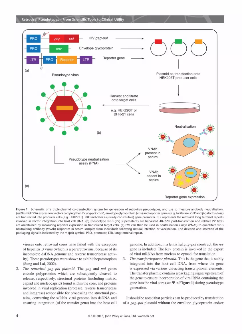
Figure 1 Schematic of a triple-plasmid co-transfection system for generation of retrovirus pseudotypes, and use to measure antibody neutralisation.(a) Plasmid DNA expression vectors carrying the HIV gag-pol ‘core’, envelope glycoprotein (env) and reporter genes (e.g. luciferase, GFP and β-galactosidase)are transfected into producer cells (e.g. HEK293T). PRO indicates a (usually constitutive) gene promoter. LTR represents the retroviral long terminal repeatsinvolved in vector integration into host cell DNA. (b) Pseudotype virus (PV) supernatants are harvested 48–72 h post-transfection and relative PV titresare ascertained by measuring reporter expression in transduced target cells. (c) PVs can then be used in neutralisation assays (PNAs) to quantitate virusneutralising antibody (VNAb) responses in serum samples from individuals following natural infection or vaccination. The deletion and insertion of thepackaging signal is indicated by the Ψ (psi) symbol. PRO, promoter; LTR, long terminal repeat.
viruses onto retroviral cores have failed with the exceptionof hepatitis B virus (which is a pararetrovirus, because of itsincomplete dsDNA genome and reverse transcriptase activ-ity). These pseudotypes were shown to exhibit hepatotropism(Sung and Lai, 2002).
2. The retroviral gag–pol plasmid. The gag and pol genesencode polyproteins which are subsequently cleaved torelease, respectively, structural proteins (including matrix,capsid and nucleocapsid) found within the core, and proteinsinvolved in viral replication (protease, reverse transcriptaseand integrase) responsible for processing the structural pro-teins, converting the ssRNA viral genome into dsDNA andensuring integration (of the transfer gene) into the host cell
genome. In addition, in a lentiviral gag–pol construct, the revgene is included. The Rev protein is involved in the exportof viral mRNAs from nucleus to cytosol for translation.
3. The transfer/reporter plasmid. This is the gene that is stablyintegrated into the host cell DNA, from where the geneis expressed via various cis-acting transcriptional elements.The transfer plasmid contains a packaging signal upstream ofthe gene to ensure incorporation of viral RNA containing thegene into the viral core (seeΨ inFigure 1) during pseudotypegeneration.
It should be noted that particles can be produced by transfectionof a gag–pol plasmid without the envelope glycoprotein and/or
4 eLS © 2015, John Wiley & Sons, Ltd. www.els.net

Retroviral Pseudotypes – From Scientific Tools to Clinical Utility
transfer gene. Those lacking the glycoprotein (often termed deltaorΔ env or ‘bald’) will bud but will be non-infectious and are thusroutinely used as control viruses in transduction experiments.In addition to increasing safety by reducing recombination
events that can lead to replication-competent virus produc-tion, using three plasmids also offers increased flexibility. Forexample, as individual plasmids can be replaced, this permitseasy swapping of retrovirus core, envelope glycoprotein orreporter.Once the cellular machinery has transcribed and translated the
transfected genes, an RNA dimer of the transfer gene (regionbetween the long terminal repeats; LTR on Figure 1) is incor-porated into the pseudotype via the packaging signal. As thetransfer plasmid is the only one engineered to contain a packag-ing signal, no other nucleic acids are incorporated into the maturepseudotype particle. A domain at the N-terminus of Gag targetsthe nucelocapsid to the cell plasma membrane, into which theenvelope protein(s) has been inserted. The pseudotype particlesbudding from the cell are encapsulated in the cell membrane,which forms the viral envelope.Pseudotyped viruses are released into the producer cell cul-
ture medium. This supernatant can be titrated onto target cells tomeasure the concentration of functional particles. These attach tothe cells via envelope protein–receptor interaction, followed bymembrane fusion and internalisation. The pseudotype genome,bearing the transfer/reporter gene is integrated into the host cellDNA, from where it is expressed. The level of reporter geneexpression correlates with the level of transduction by viableparticles. As only the transfer gene is present in the pseudo-type, no viral proteins are produced in target cells, so furtherpseudotype production and propagation does not occur. This isa key feature that renders safety in working with pseudotypescompared to working with the wildtype virus. Green fluorescentprotein (GFP)-based pseudotypes are readily titrated using fluo-rescence microscopy or flow cytometry, luciferase pseudotypesby luminometry and beta-galactosidase (β-gal) pseudotypes bycolour reaction (detailed below). The titres obtained are highlydependent on the cell type chosen for the assay as receptor den-sity may differ between cell types. As an example, transductionof influenza haemagglutinin (HA)-based retroviral pseudotypesdepends on sialic acid receptor type (α-2,3 sialic acid recep-tors are predominantly used by avian influenza strains and α-2,6sialic acid receptors by mammalian strains) and density [differ-entMadin–Darby canine kidney (MDCK) cell lines have differentratios of 2,3 and 2,6 receptors]. Also important is the density ofHA on the surface of the pseudotype particle that may differ fromstrain to strain (Tao et al., 2012). Other methods for standardis-ation of retroviral pseudotypes include qRT-PCR to determinethe number of genome copies, ELISA measurement of retrovi-ral core p24, Gag or envelope glycoprotein, RT activity assays tosemiquantitate virus titre and Nanoparticle Tracking Analysis toascertain viral particle numbers.Once successfully generated, pseudotypes can be exploited as
tools to study both basic virology and for translational studies inserology and the development and assessment of novel antiviraltherapies. A number of such applications are detailed in thefollowing sections (see also Table 1).
Analysis of Receptor Binding andVirus Entry and Exit
Retroviral and lentiviral vectors pseudotyped with foreignenvelopes have many advantages over the wildtype virus whenused as tools to study viral entry. First and foremost, the use ofretroviral pseudotypes can have major safety advantages whenstudying cellular attachment and entry of high-containment viralpathogens such as Ebola, Severe Acute and Middle East Res-piratory Syndrome-coronaviruses (SARS-CoV, MERS-CoV),lyssaviruses (e.g. rabies) and highly pathogenic (avian) influenzaviruses (H5N1, H7N1 and H7N9) (see Table 1). Pseudotypedparticles can also be safely and easily modified; nucleotidesubstitutions, deletions and insertions in envelope glycoproteingenes can be introduced (by site-directed mutagenesis) andchanges subsequently evaluated. Functional studies of the enve-lope proteins in a foreign context allow the isolation of the viralentry process from the other steps in viral replication. It alsoenables the study of individual envelope proteins to be studied(e.g. influenza virus HA or rabies virus G) or in combinationwith others (influenza neuraminidase (NA) and its cognate HA).Retroviral pseudotypes can also be used for the identification
of virus–cell receptor targets and for the detailed study of recep-tor binding (see Table 1). The first step in the identification of aviral receptor using this technology involves the construction of aretroviral pseudotype as described earlier, where the transfer geneis a selectablemarker (e.g. neomycin/G418). The second step is toidentify two cell lines; a cell line that is refractory to entry by thispseudotyped virus and another cell line that is susceptible. A com-plementary deoxyribonucleic acid (cDNA) expression library isconstructed from the susceptible cells and transfected into therefractory cells. Any resulting cells that display the receptor pro-tein(s) correctly in the plasma membrane will now be permissiveto transduction by the pseudotype and can be selected for via themarker gene. The cDNAs within these populations can subse-quently be sequenced to determine which genes are present. TheSARS-CoV receptor was identified as the angiotensin-convertingenzyme 2 (ACE2) using a modification of this expression cloningtechnique (Wang et al., 2004). See also: Virus Host Cell Recep-tors; Severe Acute Respiratory Syndrome (SARS)Retroviral pseudotypes have also been used to study specific
aspects of viral binding and entry (Table 1). For example, aSARS pseudotype was utilised to investigate tropism in a rangeof primate cells, and the pH-dependence of entry mediated via Sglycoprotein-endosome fusion (Simmons et al., 2004).In addition to virus entry studies, retroviral pseudotypes have
been widely used to study innate antiviral responses such as thepost-entry restriction factors, TRIM5α (tripartite motif 5α) (Baeand Jung, 2014) and apolipoprotein B mRNA-editing enzymecatalytic polypeptide (APOBEC) (Bae and Jung, 2014) thathave been shown to be potent antiviral proteins. In these cases,retroviral function is studied by taking advantage of a foreignenvelope, VSV-G ,which has a wider host range and cell tropismthan those of parental retroviral envelopes. Recently, retroviralpseudotypes bearing envelope glycoproteins from lyssavirusesand influenza viruses have been used to study the activity ofinterferon-inducible transmembrane protein 3 (IFITM3), an
eLS © 2015, John Wiley & Sons, Ltd. www.els.net 5

Retroviral Pseudotypes – From Scientific Tools to Clinical Utility
effector protein of the innate immune system, in many animalspecies including chickens (Smith et al., 2013), bats and pigs(Benfield et al., 2015).In addition, by incorporating the gag–pol genes from
HIV-infected patients into the pseudotype system, resistance toantiretroviral drugs such as protease and integrase inhibitors canbe measured (Parry et al., 2009). Retroviral pseudotypes havealso used to study Tetherin, a cellular protein that inhibits therelease of HIV by anchoring the virus to the cell (Bae and Jung,2014). See also: Retroviral Replication; HIV Life Cycle andInherited Co-Receptors
Antiviral Screening
Retroviral and lentiviral pseudotypes are readily exploitable forthe development of novel entry and exit assays which can beused for the screening of new antiviral compounds and for thestudy of antiviral drug resistance (seeTable 1). Cell-based antivi-ral assays have been developed for influenza virus that enablethe measurement of inhibition of pseudotype virus (PV) entry(for HA inhibitors) (Basu et al., 2014) and PV exit (for NAinhibitors) (Su et al., 2008). In the former study, pseudotypeswere used in a high-throughput assay to screen 100 000 uniquesmall molecules for agents that could inhibit the transductionof target cells by these particles, identifying two potent novelcompounds. Subsequent characterisation revealed that they bothbound to the HA stem, in a non-overlapping manner, to inhibitendosomal fusion. They also showed synergy with the currentlylicensed neuraminidase inhibitor (NAI), oseltamivir. Owing tothe relative sequence conservation in the HA stem (comparedto the head), the possibility exists that such compounds mayinhibit many strains and subtypes of influenza, for example,oseltamivir-resistant H1N1 isolates.For the influenza exit assay, the requirement of NA for the
release of HA-pseudotyped particles from the surface of pro-ducer cells was exploited for the development of an assay toevaluate NAI activities by measuring the inhibition of PV pro-duction at different NAI concentrations. This assay was used todetermine the IC50 values of oseltamivir and zanamivir againstN1 group NAs and oseltamivir-resistant mutants. IC50 valuesobtained correlated strongly with those determined using theMUNANA ‘gold standard’ assay (Su et al., 2008). Similar assayshave been developed for HIV, rabies, Lassa and Marburg viruses(Saeed et al., 2006; Wang et al., 2014). New antiviral drugs arebeing developed that target the proteases that cleave the influenzaHA (namely HAT and TMPRSS2), preventing its activation andsubsequent virus infectivity. Influenza pseudotypes that neces-sitate co-transfection of such protease genes in order to effecttransduction ability (those with a single arginine at the cleavagesite) could play a significant role in the evaluation of such drugs(Ferrara et al., 2012).Given the unexpected occurrence of some virus outbreaks
and the frequent lack of licensed vaccines or therapeutics, rapidscreening of existing approved drug libraries is becoming morecommon place. Pseudotypes have been employed in smallmolecule screening in order to determine efficacy against the
new pathogen. For example, shortly before the recent unprece-dented Ebola virus outbreak in West Africa, one group usedlentiviral pseudotypes to screen >1000 FDA approved com-pounds for antiviral activity (Madrid et al., 2013). Pseudotypeshave additionally been used for high-throughput screening of(llama-derived) antibodies, to assess neutralising potency againstinfluenza (Hultberg et al., 2011) and HIV (McCoy et al., 2012).See also: Antiviral Drugs
Pseudotype Neutralisation Assaysand Immunogenicity Testing
Virus-neutralising antibodies do not exclusively bind to proteinsexpressed on the surface of virus particles, but inhibit the processof virus multiplication. Studies on influenza have shown that neu-tralising antibodies can interfere with virus infectivity at a numberof stages in the virus life cycle, both extracellular and intracellular(Brandenburg et al., 2013). Unfortunately, many standard sero-logical assays measure only antibody binding (HI and ELISA),rather than the inhibition of viral infectivity. As a result, neutrali-sation assays have been developed to allow for sensitive detectionof functional antibody responses. For high-containment viruses(such as SARS-CoV, influenza H5N1, rabies and Ebola), how-ever, these assays are not widely applicable owing to the require-ment for high biosafety laboratory facilities and specially trainedpersonnel. Using retroviral and lentiviral particles pseudotypedwith the envelopes of such pathogens as ‘surrogate viruses’ foruse in neutralisation assays is one way of circumventing thisissue. Using a pseudotype strategy, only the envelope protein(s)of the virus is required, with no possibility of recombination ornative virus escape. These pseudotypes undergo abortive replica-tion and are unable to give rise to replication-competent progeny.See also: Antibody FunctionSafety considerations aside, pseudotypes are excellent serolog-
ical reagents for virus neutralisation assays as the virions cancontain a reporter gene and bear heterologous viral envelopeproteins on the surface. The transfer of these reporter genes totarget cells depends on the function of the viral envelope protein;therefore, the titre of neutralising antibodies against the envelopecan be measured by a reduction in reporter gene transfer andexpression. PV neutralisation assays have now been developedfor a wide range of RNA viruses, from numerous virus families(Table 1). For HIV, Ross River virus, hepatitis C, SARS-CoV,influenza and rabies virus, these assays have been shown to bevery sensitive and specific, and the neutralising antibody titresobtained correlate strongly with the respective wildtype virusassays (Kim et al., 2001; Bartosch et al., 2003a; Temperton et al.,2005, 2007; Wang et al., 2008; Wright et al., 2009; Scott et al.,2012; Molesti et al., 2013, 2014; Yang et al., 2014). These assaysare also readily adaptable to a high-throughput format for use inlarge sero-surveillance studies and vaccine immunogenicity eval-uation. For this purpose, an automated pseudotype productionfacility has been described, which has the capacity to produce1000mL of virus per week under Good Clinical Laboratory Prac-tice conditions (Schultz et al., 2012). See also: Animal VirusesPathogenic for Humans; Virus Discovery in the 21st Century
6 eLS © 2015, John Wiley & Sons, Ltd. www.els.net
Retroviral Pseudotypes – From Scientific Tools to Clinical Utility
Pseudotype-based influenza neutralisation assays havebeen shown to be highly efficient for the measurement ofbroadly-neutralising antibodies making them ideal serologicaltools for the study of cross-reactive responses against multiplesubtypes with pandemic potential (Corti et al., 2011). Antibodiesdirected specifically against the HA stalk have been shownto confer protection against multiple HA subtypes and formthe basis of new strategies for universal vaccine development(Krammer et al., 2015). The production of PVs with chimericHAs made from mismatched globular heads and stalks allows thereadout of antibody responses directed specifically against eitherHA1 or HA2 making them instrumental for immunogenicitytesting of universal vaccines. These assays will also be crucialfor the study of homo- and hetero-subtypic responses within anindividual, or at the population level.In order for these pseudotype assays to have wide applicability
within laboratories with different resource levels, the availabilityof multiple reporter systems is highly desirable. The HIV GFPreporter plasmid pCSGW described for use in viral neutral-isation assays (Temperton et al., 2005, 2007; Wright et al.,2008) has been modified to express alternative reporters. Theseinclude firefly luciferase (Wright et al., 2008), renilla luciferase(Alberini et al., 2009) and β-gal (Wright et al., 2009). Of these,the luciferase reporter-based assays are the easiest to use in termsof user interaction and downstream data analysis. However,owing to the cost of the substrate reagents and necessity forspecialised equipment (i.e. a luminometer), these assays mayhave reduced applicability for laboratories in resource-limitedregions. GFP-based assays do not require any extra reagents butdo necessitate specialised equipment (fluorescent microscope,96-well plate reader/flow cytometry facility). The β-gal-basedassays are most cost effective as β-galactoside reagents arereadily available at low cost and specialised equipment is unnec-essary (just a light microscope). The expression of β-gal canbe quantified by counting infected cells following staining withX-gal (5-bromo-4-chloro-3-indolyl-β-D-galactopyranoside).However, there are two colorimetric substrates, ONPG(o-nitrophenyl-β-D-galactopyranoside) and CPRG (chlorophe-nol red-β-D-galactopyranoside) that allow the assays to beevaluated using a microplate reader. Recent reports describemultiplex assays for the study of neutralising antibodies thatare directed against two viruses using pseudotypes carryingdifferent luciferase reporters, firefly and renilla. Lyssavirus,influenza and coronavirus pseudotypes have been exploited forthe development of such assays (Wright et al., 2010; Molestiet al., 2014; Zhou et al., 2011). These permit the measurement ofneutralising antibody responses against two antigenically distinctglycoproteins in the same serum/plasma sample thus increasingthe amount and quality of serological data that can be acquiredfrom valuable sera.The pseudotypes constructed from pathogenic viruses of
global health importance lend themselves to the establishment(pre-outbreak) of a pseudotype resource library consisting ofvirus envelope glycoprotein expression vectors and their cog-nate retroviral pseudotypes. In order to prioritise the strains toinclude in a library, the following could be utilised: data min-ing of WHO/OiE/ProMed news feeds, conference abstracts,social networks, peer-reviewed literature (human/animal
outbreaks/surveillance data/pre-clinical vaccine studies),bioinformatics/phylogenetic analysis and new antigenic car-tography tools (Fouchier and Smith, 2010). This information cansubsequently inform custom gene synthesis of surface glyco-proteins from candidate viruses and subcloning into expressionvectors. Once nucleic acid sequence and expression has beenconfirmed, retroviral vector systems can be used for pseudotyp-ing as detailed above. A comprehensive retroviral pseudotyperesource would lend itself to optimisation of pseudotype neu-tralisation assay protocols and standardisation of PV stocksdepending on end-user requirements and laboratory facilitiesand to the training of specialist users. While there may a degreeof amino acid mismatch, a comprehensive pseudotype librarywould permit a surrogate cross-reactive strain to be employedas a preliminary measure until the outbreak strain had beensequenced and deposited in the NCBI sequence database toallow for synthetic gene synthesis. For the shipping and deploy-ment of pseudotype neutralisation assays in resource-limitedregions, their encapsulation into kit form is desirable. An exper-imental system containing lyophilised viruses has recently beendescribed for influenza, Marburg and rabies (Mather et al., 2014).In summary, there are major advantages to using pseudotypes
for the study of humoral immune responses to viral infection:
1. The assays are serum-sparing: typically 2-10 μL of serum isrequired for each replicate, 5–25 fold less than for wildtypevirus assays.
2. No virus culture is necessary as the serological antigen isentirely synthetic, derived by custom synthesis or PCR.
3. There is a wide choice of available reporters depending onend-user requirement.
4. Pseudotypes are easily amenable to high-throughput plat-forms.
5. Using pseudotypes as a surrogate for wildtype virus removesthe need to handle dangerous pathogens in high biocontain-ment facilities.
6. As the pseudotyped glycoprotein is the only viral antigenroutinely incorporated into the pseudotype particle, what ismeasured by this assay is antibody solely directed against thisprotein. This assay can thus be used as a tool for dissectingout (or delineating) responses observed using wildtype virusneutralisation assays in parallel.
7. Multiple studies have demonstrated high sensitivity, speci-ficity and strong correlation of antibody titres when neutral-isation assays using pseudotypes are compared with thoseemploying wildtype viruses.
However, there are also potential disadvantages in using pseu-dotypes over wildtype viruses in neutralisation assays:
1. The density of envelope protein on the pseudotype surfaceis unknown and may not be analogous to that found on therespective wildtype virus. If the envelope protein densityis significantly lower on the pseudotype, it may be morereadily neutralised by antibodies. However, as there is ahigh correlation of neutralising antibody titres among theassays using wildtype virus or pseudotypes, this suggeststhat this may not be an issue when using polyclonal sera.
eLS © 2015, John Wiley & Sons, Ltd. www.els.net 7

Retroviral Pseudotypes – From Scientific Tools to Clinical Utility
For influenza, however, this issue paradoxically presents asignificant advantage as it has been demonstrated that pseu-dotypes are more sensitive than wildtype viruses at detectingbroadly neutralising antibodies that bind to the stalk regionof HA (Rappuoli and Dormitzer, 2012). Pseudotypes are thusplaying a central role in the development of universal vac-cines/antiviral drugs.
2. Some viruses bud internally so that their envelopes areretained on the endoplasmic reticulum (e.g. flaviviruses). Forthis reason, these viruses may produce low viral titres whentheir envelopes are used in the pseudotype system. This canbe rectified in some cases by concentrating the virus via cen-trifugation, or via the use of “leaky” overexpression whicheffects expression of the glycoprotein at the plasma mem-brane (Simmons et al., 2004; Perera et al., 2013).
3. The ability of several innate host restriction factors, such asthe TRIM and IFITM family of proteins, has been well doc-umented to inhibit lentiviral or retroviral replication. Thisraises the possibility that cell lines routinely used for assayswith wildtype virus will be refractory for infection usingpseudotypes. The effect of this altered tropism on neutrali-sation assay titres is as yet unknown.
Pseudotypes as Immunogens andfor Gene Therapy
Retroviral pseudotypes have recently been used as a source ofantigen in vaccines. Retroviral vectors carrying the surface pro-teins of influenza (H5N1 and H7N1) and HIV-1 have been shownto function as immunogens resulting in the induction of humoral(influenza and HIV) and cellular responses (HIV) in animal mod-els (Szécsi et al., 2006; Breckpot et al., 2010). For the influenzavaccines, retroviral virus-like particles were made which incor-porated HA, NA and M2 from H5N1 and H7N1 subtypes. Theseparticles induced rapid, subtype-specific neutralising antibodyproduction in mice.For the HIV-1 vaccine, a novel MLV/HIV-1 pseudotype vector
was constructed and shown to induce HIV-1-specific humoral(HIV-1 Env-specific antibodies) and cellular immune responses[Rev and Env-specific cytotoxic T-lymphocyte (CTL)-response]in rhesus macaques (Szécsi et al., 2006).These novel vaccine immunogens can be prepared rapidly
upon availability of viral RNA and could potentially be used asanimal vaccines (influenza; pigs and domestic poultry), or for thegeneration of panels of neutralising antibodies targeted againstnewly emerging viral strains (influenza and HIV).The use of retroviral pseudotypes for gene delivery has already
had major impact on both experimental and clinical gene ther-apies. They can also be used to study the effect of introducingforeign proteins into a cell, as pseudotype transduction resultsin the long-term stable expression of the integrated gene. Theforeign gene may be a reporter gene (GFP, luciferase or lacZ), asdiscussed earlier, a therapeutic gene (as is the case for gene ther-apy applications) or indeed any gene that is required to be stablyexpressed in a given cell line/type. To achieve efficient and sus-tained expression of foreign genes delivered by retroviral vectors,
there are several options for the design of the plasmid. The foreigngene may be directly expressed from the retroviral promoter inthe construct’s 5′-LTR or from a heterologous promoter down-stream of the 5′-LTR, such as from the CMV immediate earlyor human β-actin genes. If expression of two genes is required,then a spliced message, an internal ribosome entry site (IRES),or even a dual promoter vector may be used. Lastly, cell-specificpromoters can be used to regulate gene expression and alter-native envelopes can be used which allow targeting of specificcells (Bischof and Cornetta, 2010). See also: Lentiviral Vec-tors in Gene Therapy; Retroviral Vectors in Gene Therapy;Retroviruses in Human Gene Therapy
Notes
‘Plasmid’ and ‘construct’ are used interchangeably throughoutthis article.
Acknowledgements
The authors would like to thank Stuart Mather for providing theoriginal artwork for Figure 1.
References
Alberini I, Del Tordello E, Fasolo A, et al. (2009) Pseudopar-ticle neutralization is a reliable assay to measure immunityand cross-reactivity to H5N1 influenza viruses. Vaccine 27:5998–6003.
Aljofan M, Sganga M, Lo M, et al. (2009) Antiviral activity ofgliotoxin, gentian violet and brilliant green against Nipah andHendra virus in vitro. Virology Journal 6: 187–200.
Bae EH and Jung Y (2014) Comparison of the effects of retroviralrestriction factors involved in resistance to porcine endogenousretrovirus. Journal of Microbiology and Biotechnology 24 (4):577–583.
Bartosch B, Bukh J, Meunier J, et al. (2003a) In vitro assay forneutralizing antibody to hepatitis C virus: evidence for broadlyconserved neutralization epitopes. Proceedings of the NationalAcademy of Sciences of the United States of America 100 (24):14199–14204.
Bartosch B, Dubuisson J and Cosset F (2003b) Infectious hepatitisC virus pseudo-particles containing functional E1-E2 envelopeprotein complexes. The Journal of Experimental Medicine 197 (5):633–642.
Basu A, Antanasijevic A, WangM, et al. (2014) New small moleculeentry inhibitors targeting hemagglutinin-mediated influenza a virusfusion. Journal of Virology 88 (3): 1447–1460.
Benfield C, Smith S, Wright E, et al. (2015) Bat and pigInterferon-Induced Transmembrane Protein 3 restrict cell entry byinfluenza virus and lyssaviruses. Journal of General Virology 96(1): DOI: 10.1099/vir.0.000058.
Bischof D and Cornetta K (2010) Flexibility in cell targeting bypseudotyping lentiviral vectors. Methods in Molecular Biology614: 53–68.
8 eLS © 2015, John Wiley & Sons, Ltd. www.els.net

Retroviral Pseudotypes – From Scientific Tools to Clinical Utility
Both L, van Dolleweerd C, Wright E, et al. (2013) Production,characterization, and antigen specificity of recombinant 62-71-3, acandidate monoclonal antibody for rabies prophylaxis in humans.FASEB Journal 27 (5): 2055–2065.
Brandenburg B, Koudstaal W, Goudsmit J, et al. (2013) Mechanismsof hemagglutinin targeted influenza virus neutralization. PLoSONE 8 (12): 1–14.
Breckpot K, Escors D, Arce F, et al. (2010) HIV-1 lentiviral vectorimmunogenicity is mediated by Toll-like receptor 3 (TLR3) andTLR7. Journal of Virology 84 (11): 5627–5636.
Chan SY, Speck RF andMaMC (2000) Distinct mechanisms of entryby envelope glycoproteins of Marburg and Ebola (Zaire) viruses.Journal of Virology 74 (10): 4933–4937.
Cormier EG, Tsamis F, Kajumo F, et al. (2004) CD81 is an entrycoreceptor for hepatitis C virus. Proceedings of the NationalAcademy of Sciences of the United States of America 101:7270–7274.
Corti D, Voss J, Gamblin S, et al. (2011) A neutralizing antibodyselected from plasma cells that binds to group 1 and group 2influenza A hemagglutinins. Science 333 (6044): 850–856.
Deng HK, Unutmaz D, KewalRamani VN and Littman DR (1997)Expression cloning of new receptors used by simian and humanimmunodeficiency viruses. Nature 388 (6639): 296–300.
Desmaris N, Bosch A, Salaün C, et al. (2001) Production and neu-rotropism of lentivirus vectors pseudotyped with lyssavirus enve-lope glycoproteins. Molecular Therapy 4 (2): 149–156.
Dong J, Roth MG and Hunter E (1992) A chimeric avian retroviruscontaining the influenza virus hemagglutinin gene has an expandedhost range. Journal of Virology 66 (12): 7374–7382.
Ferrara F, Molesti E, Böttcher-Friebertshäuser E, et al. (2012) Thehuman Transmembrane Protease Serine 2 is necessary for theproduction of Group 2 influenza A virus pseudotypes. Journal ofMolecular and Genetic Medicine 7: 309–314.
Forsman A, Beirnaert E, Aasa-Chapman M, et al. (2008) Llamaantibody fragments with cross-subtype human immunodeficiencyvirus type 1 (HIV-1)-neutralizing properties and high affinity forHIV-1 gp120. Journal of Virology 82 (24): 12069–12081.
Fouchier RAM and Smith DJ (2010) Use of antigenic cartographyin vaccine seed strain selection. Avian diseases 54 (1 Suppl):220–223.
Funke S, Maisner A, Mühlebach M, et al. (2008) Targeted cell entryof lentiviral vectors. Molecular Therapy 16 (8): 1427–1436.
Gehring G, Rohrmann K, Atenchong N, et al. (2014) The clinicallyapproved drugs amiodarone, dronedarone and verapamil inhibitfilovirus cell entry. Journal of Antimicrobial Chemotherapy 69:2123–2131.
Hou Y, Peng C, Yu M, et al. (2010) Angiotensin-converting enzyme2 (ACE2) proteins of different bat species confer variable sus-ceptibility to SARS-CoV entry. Archives of Virology 155 (10):1563–1569.
Hultberg A, Temperton N, Rosseels V et al. (2011). Llama-derivedsingle domain antibodies to build multivalent, superpotent andbroadened neutralizing anti-viral molecules. PloS ONE 6(4)e17665.
Ikeda Y, Takeuchi Y, Martin F, et al. (2003) Continuous high-titerHIV-1 vector production. Nature Biotechnology 21 (5): 569–572.
Khetawat D and Broder C (2010) A functional henipavirus envelopeglycoprotein pseudotyped lentivirus assay system. Virology Jour-nal 7: 312–326.
KimYB, LeeM,HanD, et al. (2001) Development of a safe and rapidneutralization assay usingmurine leukemia virus pseudotypedwith
HIV type 1 envelope glycoprotein lacking the cytoplasmic domain.AIDS Research and Human Retroviruses 17 (18): 1715–1724.
Krammer F, Palese P and Steel J (2015) Advances in universalinfluenza virus vaccine design and antibody mediated therapiesbased on conserved regions of the hemagglutinin. Current Topicsin Microbiology and Immunology 386: 301–321.
Landau NR, Page KA and Littman DR (1991) Pseudotyping withhuman T-cell leukemia virus type I broadens the human immun-odeficiency virus host range. Journal of Virology 65 (1): 162–169.
Ma M, Kersten D, Kamrud K, et al. (1999) Murine leukemia viruspseudotypes of La Crosse and Hantaan Bunyaviruses: a system foranalysis of cell tropism. Virus Research 64 (1): 23–32.
Madrid PB, Chopra S, Manger I et al. (2013) A systematic screenof FDA-approved drugs for inhibitors of biological threat agents.PloS ONE 8 (4) e60579.
Mather ST, Wright E, Scott SD, et al. (2014) Lyophilisation ofinfluenza, rabies and Marburg lentiviral pseudotype viruses forthe development and distribution of a neutralisation -assay-baseddiagnostic kit. Journal of Virological Methods 210: 51–58.
McCoy LE, Quigley A, Strokappe N, et al. (2012) Potent and broadneutralization of HIV-1 by a llama antibody elicited by immuniza-tion. The Journal of Experimental Medicine 209 (6): 1091–1103.
Mentis GZ, Gravell M, Hamilton R, et al. (2006) Transduction ofmotor neurons and muscle fibers by intramuscular injection ofHIV-1-based vectors pseudotyped with select rabies virus glyco-proteins. Journal of Neuroscience Methods 157 (2): 208–217.
Miletic H, Bruns M, Tsiakas K, et al. (1999) Retroviral vectorspseudotyped with lymphocytic choriomeningitis virus. Journal ofVirology 73 (7): 6114–6116.
Mitrophanous K, Yoon S, Rohll JB, et al. (1999) Stable gene transferto the nervous system using a non-primate lentiviral vector. GeneTherapy 6: 1808–1818.
Molesti E, Milani A, Terregino C, et al. (2013) Comparative sero-logical assays for the study of h5 and h7 avian influenza viruses.Influenza Research and Treatment 2013: 286158.
Molesti E, Wright E, Terregino C, et al. (2014) Multiplex evaluationof influenza neutralizing antibodies with potential applicability toin-field serological studies. Journal of Immunology Research 2014:457932.
Naldini L, Blömer U, Gallay P, et al. (1996) In vivo gene deliveryand stable transduction of nondividing cells by a lentiviral vector.Science 272 (5259): 263–267.
Parry CM, Kohli A, Boinett C, et al. (2009) Gag determinantsof fitness and drug susceptibility in protease inhibitor-resistanthuman immunodeficiency virus type 1. Journal of Virology 83 (18):9094–9101.
Pear WS, Nolan G, Scott M, et al. (1993) Production of high-titerhelper-free retroviruses by transient transfection. Proceedings ofthe National Academy of Sciences of the United States of America90: 8392–8396.
Pegu A, Yang Z, Boyington J, et al. (2014) Neutralizing antibodies toHIV-1 envelope protect more effectively in vivo than those to theCD4 receptor. Science Translational Medicine 6 (243): 243ra88.
Perera RA, Wang P, Gomaa M, et al. (2013) Seroepidemiology forMERS coronavirus using microneutralisation and pseudoparticlevirus neutralisation assays reveal a high prevalence of antibodyin dromedary camels in Egypt, June 2013. Euro Surveillance:European Communicable Disease Bulletin 18 (36): 20574.
Radoshitzky SR, Abraham J, Spiropoulo C, et al. (2007) Transferrinreceptor 1 is a cellular receptor for NewWorld haemorrhagic feverarenaviruses. Nature 446 (7131): 92–96.
eLS © 2015, John Wiley & Sons, Ltd. www.els.net 9

Retroviral Pseudotypes – From Scientific Tools to Clinical Utility
Rappuoli R and Dormitzer PR (2012) Influenza: options to improvepandemic preparation. Science 336 (6088): 1531–1533.
Reignier T, Oldenburg J, Noble B, et al. (2006) Receptor use bypathogenic arenaviruses. Virology 353: 111–120.
Rodrigo WWSI, de la Torre JC and Martínez-Sobrido L (2011) Useof single-cycle infectious lymphocytic choriomeningitis virus tostudy hemorrhagic fever arenaviruses. Journal of Virology 85 (4):1684–1695.
SaeedMF, Kolokoltsov AA and Davey RA (2006) Novel, rapid assayfor measuring entry of diverse enveloped viruses, including HIVand rabies. Journal of Virological Methods 135 (2): 143–150.
Sakuma T, Barry MA and Ikeda Y (2012) Lentiviral vectors: basic totranslational. Biochemical Journal 443: 603–618.
Sawoo O, Dublineau A, Batéjat C, et al. (2014) Cleavage ofhemagglutinin-bearing lentiviral pseudotypes and their use in thestudy of influenza virus persistence. PloS ONE 9 (8): e106192.
Schnell T, Foley P, Wirth M, et al. (2000) Development ofa self-inactivating, minimal lentivirus vector based on simianimmunodeficiency virus. Human Gene Therapy 11 (3): 439–447.
Schultz A, Koch S, Fuss M et al. (2012). An automated HIV-1Env-pseudotyped virus production for global HIV vaccine trials.PloS ONE 7(12): e51715.
Scott S, Molesti E, Temperton N, et al. (2012) The use of equineinfluenza pseudotypes for serological screening. Journal of Molec-ular and Genetic Medicine 6: 304–308.
Shelton, H. et al., 2013 Mutations in haemagglutinin thataffect receptor binding and pH stability increase replica-tion of a PR8 influenza virus with H5 HA in the upperrespiratory tract of ferrets and may contribute to transmis-sibility. The Journal of general virology, 94 (Pt 6), 1220–9.http://www.pubmedcentral.nih.gov/articlerender.fcgi?artid=3709624&tool=pmcentrez&rendertype=abstract [AccessedJanuary 20, 2015].
Simek MD, Rida W, Priddy F, et al. (2009) Human immunodefi-ciency virus type 1 elite neutralisers: individuals with broad andpotent neutralizing activity identified by using a high-throughputneutralization assay together with an analytical selection algo-rithm. Journal of Virology 83 (14): 7337–7348.
Simmons G, Reeves J, Rennecamp A, et al. (2004) Characteriza-tion of severe acute respiratory syndrome-associated coronavirus(SARS-CoV) spike glycoprotein-mediated viral entry. Proceed-ings of the National Academy of Sciences of the United States ofAmerica 101: 4240–4245.
Smith SE, Gibson M, Wash R, et al. (2013) Chickeninterferon-inducible transmembrane protein 3 restricts influenzaviruses and lyssaviruses in vitro. Journal of Virology 87 (23):12957–12966.
Soneoka Y, Cannon P, Ramsdale E, et al. (1995) A transientthree-plasmid expression system for the production of high titerretroviral vectors. Nucleic Acids Research 23 (4): 628–633.
Su CY, Wang S, Shie J, et al. (2008) In vitro evaluation of neu-raminidase inhibitors using the neuraminidase-dependent releaseassay of hemagglutinin-pseudotyped viruses. Antiviral Research79: 199–205.
Sung VMH and Lai MMC (2002) Murine retroviral pseudotypevirus containing hepatitis B virus large and small surface anti-gens confers specific tropism for primary human hepatocytes: apotential liver-specific targeting system. Journal of Virology 76 (2):912–917.
Szécsi J, Boson B, Johnsson P, et al. (2006) Induction of neutralisingantibodies by virus-like particles harbouring surface proteins from
highly pathogenic H5N1 and H7N1 influenza viruses. VirologyJournal 3: 70–77.
Tao L, Chen J, Zheng Z, et al. (2012) H5N1 influenza virus-likeparticles produced by transient expression in mammalian cellsinduce humoral and cellular immune responses in mice. CanadianJournal of Microbiology 58 (4): 391–401.
Temperton NJ, Chan P, Simmons G, et al. (2005) Longitudinallyprofiling neutralizing antibody response to SARS coronavirus withpseudotypes. Emerging Infectious Diseases 11 (3): 411–416.
Temperton NJ, Hoschler K,Major D, et al. (2007) A sensitive retrovi-ral pseudotype assay for influenza H5N1-neutralizing antibodies.Influenza and other Respiratory Viruses 1: 105–112.
Wallerström S, Lagerqvist N, Temperton N, et al. (2014) Detectionof antibodies against H5 and H7 strains in birds: evaluation ofinfluenza pseudovirus particle neutralization tests. Infection Ecol-ogy & Epidemiology 4: 23011–23019.
Wang J, Cheng H, Ratia K, et al. (2014) A comparativehigh-throughput screening protocol to identify entry inhibitorsof enveloped viruses. Journal of Biomolecular Screening 19 (1):100–107.
Wang P, Chen J, Zheng A, et al. (2004) Expression cloning offunctional receptor used by SARS coronavirus. Biochemical andBiophysical Research Communications 315: 439–444.
Wang W, Butler E, Veguilla V, et al. (2008) Establishment of retro-viral pseudotypes with influenza hemagglutinins from H1, H3, andH5 subtypes for sensitive and specific detection of neutralizingantibodies. Journal of Virological Methods 153 (2): 111–119.
Watson DJ, Kobinger G, Passini M, et al. (2002) Targeted transduc-tion patterns in the mouse brain by lentivirus vectors pseudotypedwith VSV, Ebola, Mokola, LCMV, or MuLV envelope proteins.Molecular Therapy 5 (5): 528–537.
Wool-Lewis RJ and Bates P (1998) Characterization of Ebolavirus entry by using pseudotyped viruses: identificationof receptor-deficient cell lines. Journal of Virology 72 (4):3155–3160.
Wright E, Temperton N, Marston D, et al. (2008) Investigatingantibody neutralization of lyssaviruses using lentiviral pseudo-types: a cross-species comparison. Journal of General Virology 89:2204–2213.
Wright E, McNabb S, Goddard T, et al. (2009) A robust lentiviralpseudotype neutralisation assay for in-field serosurveillance ofrabies and lyssaviruses in Africa. Vaccine 27 (51): 7178–7186.
Wright E, Hayman D, Vaughan A, et al. (2010) Virus neutralisingactivity of African fruit bat (Eidolon helvum) sera against emerginglyssaviruses. Virology 408 (2): 183–189.
Yang J, Li W, Long Y, et al. (2014) Reliability of pseudotypedinfluenza viral particles in neutralizing antibody detection. PloSONE 9 (12): e113629.
Yu L, Bai W, Wu X, et al. (2013) A recombinant pseudotypedlentivirus expressing the envelope glycoprotein of Hantaan virusinduced protective immunity in mice. Virology Journal 10 (1):301–310.
Yu X, Sainz B, Petukhov P, et al. (2012) Identification of hepatitisC virus inhibitors targeting different aspects of infection using acell-based assay. Antimicrobial Agents and Chemotherapy 56 (12):6109–6120.
Zhang C, Hu B, Xiao L, et al. (2014) Pseudotyping lentiviral vectorswith lymphocytic choriomeningitis virus glycoproteins for trans-duction of dendritic cells and in vivo immunization. Human GeneTherapy Methods 25 (6): 328–338.
10 eLS © 2015, John Wiley & Sons, Ltd. www.els.net

Retroviral Pseudotypes – From Scientific Tools to Clinical Utility
Zhang Y, Lin X, Wang G, et al. (2010) Neuraminidase and hemag-glutinin matching patterns of a highly pathogenic avian and twopandemic H1N1 influenza A viruses. PloS ONE 5 (2): e9167.
Zhao G, Du L, Ma C, et al. (2013) A safe and convenientpseudovirus-based inhibition assay to detect neutralizing antibod-ies and screen for viral entry inhibitors against the novel humancoronavirus MERS-CoV. Virology Journal 10 (1): 266–274.
Zhou Y, Agudelo J, Lu K, et al. (2011) Inhibitors of SARS-CoVentry–identification using an internally-controlled dual envelopepseudovirion assay. Antiviral Research 92 (2): 187–194.
Further Reading
Centers for Disease Control and Prevention (2015) Diseases andConditions. http://www.cdc.gov/DiseasesConditions/
Escors D, Breckpot K, Arce F, Kochan G and Stephenson H (2012)Lentiviral Vectors and Gene Therapy. Springer: Science.
Pöhlmann S and Simmonds G (2013) Viral Entry into Host Cells.Landes Bioscience (Austin, Texas) & Springer Science. New York:New York.
Temperton NJ andWright E (2009) Retroviral Pseudotypes. Encyclo-pedia of Life Sciences. Chichester: John Wiley & Sons Ltd. DOI:10.1002/9780470015902.a0021549.
Viral Pseudotype Unit (2015) http://www.viralpseudotypeunit.info
eLS © 2015, John Wiley & Sons, Ltd. www.els.net 11