lentiviral vectors express chondroitinase abc in cortical projections and promote sprouting of...
TRANSCRIPT

Lp
RJJJa
b
c
d
e
a
ARRA
KLCCSA
1
(eaA
cggp
(a((((
0d
Journal of Neuroscience Methods 201 (2011) 228– 238
Contents lists available at ScienceDirect
Journal of Neuroscience Methods
j o ur nal homep age: www.elsev ier .com/ locate / jneumeth
entiviral vectors express chondroitinase ABC in cortical projections andromote sprouting of injured corticospinal axons
ong-Rong Zhaoa,1, Elizabeth M. Muirb,1, João Nuno Alvesa,b, Hannah Rickmanb, Anna Y. Allanb,essica C. Kwoka, Kasper C.D. Roetc, Joost Verhaagenc, Bernard L. Schneiderd,ean-Charles Bensadound,2, Sherif G. Ahmede, Rafael J. Yánez-Munoze, Roger J. Keynesb,ames W. Fawcetta, John H. Rogersb,∗
Cambridge Centre for Brain Repair, Forvie Site, Robinson Way, Cambridge CB2 0PY, UKDepartment of Physiology Development and Neuroscience, University of Cambridge, Downing St., Cambridge CB2 3EG, UKLaboratory for Neuroregeneration, Netherlands Institute for Neuroscience, Meibergdreef 47, 1105BA Amsterdam, The NetherlandsBrain Mind Institute, Ecole Polytechnique Fédérale de Lausanne, 1015 Lausanne, SwitzerlandSchool of Biological Sciences, Royal Holloway, University of London, Egham, Surrey TW20 0EX, UK
r t i c l e i n f o
rticle history:eceived 14 June 2011eceived in revised form 2 August 2011ccepted 3 August 2011
a b s t r a c t
Several diseases and injuries of the central nervous system could potentially be treated by delivery of anenzyme, which might most effectively be achieved by gene therapy. In particular, the bacterial enzymechondroitinase ABC is beneficial in animal models of spinal cord injury. We have adapted the chondroiti-nase gene so that it can direct secretion of active chondroitinase from mammalian cells, and inserted it
eywords:entiviral vectorshondroitinase ABCorticospinal tractpinal cord injuryxon regeneration
into lentiviral vectors. When injected into adult rat brain, these vectors lead to extensive secretion ofchondroitinase, both locally and from long-distance axon projections, with activity persisting for morethan 4 weeks. In animals which received a simultaneous lesion of the corticospinal tract, the vectorreduced axonal die-back and promoted sprouting and short-range regeneration of corticospinal axons.The same beneficial effects on damaged corticospinal axons were observed in animals which receivedthe chondroitinase lentiviral vector directly into the vicinity of a spinal cord lesion.
. Introduction
For several diseases and injuries of the central nervous systemCNS), potential forms of treatment would involve delivery of an
nzyme, which might most effectively be achieved by a gene ther-py approach. One example is the bacterial enzyme chondroitinaseBC, which is under consideration as a possible treatment for spinalAbbreviations: BDA, biotinylated dextran amine; CMV, cytomegalovirus; CNS,entral nervous system; CS, chondroitin sulfate; CSPG, chondroitin sulfate proteo-lycan; CST, corticospinal tract; DMEM, Dulbecco’s modified Eagle’s medium; GAG,lycosaminoglycan; LV-ChABC, lentiviral vector encoding chondroitinase ABC; PBS,hosphate buffered saline; PGK, phosphoglycerate kinase.∗ Corresponding author. Tel.: +44 1223 3 33865; fax: +44 1223 3 33840.
E-mail addresses: [email protected] (R.-R. Zhao), [email protected]. Muir), [email protected] (J.N. Alves), [email protected] (H. Rickman),[email protected] (A.Y. Allan), [email protected] (J.C. Kwok), [email protected]. Roet), [email protected] (J. Verhaagen), [email protected]. Schneider), [email protected] (J.-C. Bensadoun), [email protected]. Ahmed), [email protected] (R.J. Yánez-Munoz), [email protected]. Keynes), [email protected] (J.W. Fawcett), [email protected] (J.H. Rogers).
1 R.-R.Z. and E.M.M. made equal contributions.2 Present address: Merck Serono S.A., 1202 Geneva, Switzerland.
165-0270/$ – see front matter © 2011 Elsevier B.V. All rights reserved.oi:10.1016/j.jneumeth.2011.08.003
© 2011 Elsevier B.V. All rights reserved.
cord injury. Because of the potential risks of prolonged or repeatedinfusions, we wished to adapt the chondroitinase gene so that itcould direct secretion of active chondroitinase from mammaliancells, and to deliver it using lentiviral vectors so that both neuronsand glia could secrete the enzyme.
The aim in using chondroitinase is to remove chondroitin sul-fate proteoglycans (CSPGs), one of the major classes of inhibitorymolecule present in the scar that forms after injury and preventsregeneration of damaged axons (Kwok et al., 2008). CSPGs consistof a core protein with attached glycosaminoglycan (GAG) chains.After CNS injury, CSPGs are synthesized by glial cells and createa barrier impenetrable for axon regeneration. CSPGs also exist inthe uninjured CNS, particularly in matrix structures found aroundthe cell body and neurites of some neurons, known as perineu-ronal nets. Perineuronal nets have a role in restricting plasticity;removing CSPGs has been shown to restore CNS plasticity.
Chondroitinase ABC is a bacterial enzyme from Proteus vulgaris,which degrades CSPGs by cleaving GAG chains, which are responsi-
ble for most of the inhibitory effect. Injection of chondroitinase intothe brain or spinal cord promotes circuit plasticity, due to sproutingof axons and remodelling of intact circuits. Also, following spinalcord injury, chondroitinase treatment can promote long-distance
R.-R. Zhao et al. / Journal of Neuroscience Methods 201 (2011) 228– 238 229
Table 1Lentiviral vectors encoding modified chondroitinase ABC.
Name Promoter Lab Concentrationa Minimum titreb References
LV-C CMV Amsterdam 99 �g/ml of P24 6 × 10⁵ TU/�l (HEK cells) Naldini et al. (1996a), Hendriks et al. (2007),LV-D CMV London 1.44 × 10⁹ gen/ml 8 × 10⁵ TU/�l (HEK cells) Dull et al. (1998), Yánez-Munoz et al., 2006LV-P PGK Lausanne 479 �g/ml of P24 ∼106 TU/�l (SCTM cells) Déglon et al. (2000)
a gen: genomes, by Q-PCR.mber
s
re2
lres(e2iolFsipr
agcco
mssctafr(bwitpd
tLg12tws2dgtr
b TU: transducing units. These titres are approximate values derived from the nuubconfluent monolayer, quoted for the cell type which gave the highest titre.
egrowth of axons. Both local sprouting and long-distance regen-ration are accompanied by functional recovery (Bradbury et al.,002; Kwok et al., 2008; Bradbury and Carter, 2011).
Delivery of the enzyme can entail practical problems. The half-ife of the enzyme at 37 ◦C can vary considerably. Although it caneach 2–3 weeks with stabilizers such as albumin or trehalose (Chaut al., 2004; Lee et al., 2010), it is much shorter in the absence ofuch agents, and the half-life was only 6 days in injected rat brainLin et al., 2008). A single injection can produce functional recov-ry by local axon growth and circuit reorganization (Galtrey et al.,007; Cafferty et al., 2008), but longer-range regrowth of axons
s likely to need more prolonged treatment, by chronic infusionr repeated injections, as has been given in experiments whereong-distance tracts were severed (Bradbury and Carter, 2011).or eventual treatment of human patients, given the much largerize of the spinal cord and of the injured region, and the greatermportance of the corticospinal tract, chondroitinase activity wouldrobably be required over several months. Chronic delivery carriesisks of tissue damage, inflammation, and infection.
Gene delivery of chondroitinase to the CNS could have severaldvantages. A long-lasting effect could be achieved with only a sin-le injection, and any side-effects of injection, including the risk ofausing further trauma and infection, would be minimized. Thus itould represent a powerful approach for promoting regenerationf the spinal cord after injury.
However, achieving secretion of a bacterial protein byammalian cells can be problematic. As the newly synthe-
ized chondroitinase polypeptide passes through the eukaryoticecretion pathway, cryptic N-glycosylation sites can become gly-osylated, and because the bacterial sequence is not adapted forhe eukaryotic pathway, this process can interfere with foldingnd secretion of the protein. We found that this process led toailure of functional enzyme production from the original bacte-ial chondroitinase gene when transfected into mammalian cellsMuir et al., 2010). We therefore modified the chondroitinase geney directed mutagenesis of up to 6 selected N-glycosylation siteshich mapped to positions important for structure or ligand bind-
ng. These mutations did not impair enzyme activity in vitro, andhey allowed the translated protein to pass through the secretionathway unimpaired, resulting in efficient secretion of active chon-roitinase from transfected cells in vitro (Muir et al., 2010).
For greater efficiency of expression in vivo, we have now insertedhe modified chondroitinase gene into three lentiviral vectors.entiviral vectors have been shown to transduce both neurons andlia efficiently in the CNS (Naldini et al., 1996b; Bloemer et al.,997; Baekelandt et al., 2002; Zhao et al., 2003; Hendriks et al.,007). In brain, expression is mainly in glia and leukocytes ini-ially (Baekelandt et al., 2002), but also occurs in neurons from 2eeks onwards (Bloemer et al., 1997; Baekelandt et al., 2002). In
pinal cord, injections into dorsal column white matter (Zhao et al.,003) or into post-injury glial scar (Hendriks et al., 2007) trans-
uce all classes of glia, as well as many neurons in the surroundingray matter, for ≥14 days. Expression is sustained in some popula-ions of neurons for up to a year, and there is minimal immuneesponse (Bloemer et al., 1997; Baekelandt et al., 2002). Suchof cells positive by chondroitinase immuno-staining 3 days after transduction of a
vectors have been used successfully to deliver therapeutic trans-genes to the rodent CNS in models of neurological disease (e.g. Dullet al., 1998; Yánez-Munoz et al., 2006). Lentiviral vectors are thusthe present vector of choice for high-level long-term local expres-sion of a transgene, both in glia which produce the axon-inhibitoryCSPGs, and in neurons which are affected by them.
Here we report that cells transduced with these vectors secreteactive chondroitinase at high levels in tissue culture and in ratbrain, and that the chondroitinase action in a model of spinal cordinjury protects corticospinal axons from die-back and promotessprouting. These results suggest that lentiviral vector-mediatedexpression of modified chondroitinase could form a useful com-ponent of a future treatment for spinal cord injury.
2. Materials and methods
2.1. Chondroitinase gene
Previous studies in vitro and in tissue culture (Muir et al., 2010)were used to design an optimized chondroitinase gene for secre-tion of enzyme from mammalian cells. The best gene from thosestudies was Y133, with 5 mutations, but we also found that muta-tion N751Q (present in Y133) was irrelevant as this residue is notglycosylated, whereas mutation N338Q (present in a different con-struct) appeared to have a slight extra beneficial effect (data notshown). We therefore synthesized gene Y1330 with the mutationsN282K, N338Q, N345Q, S517A, N675Q (S517A abolishes glycosy-lation at N515), with the coding sequence replaced with preferredmammalian codons (Eurofins MWG Operon, Germany). (The fullsequence is in Supplementary Data.) The signal sequence was frommouse matrix metalloproteinase 2 (Muir et al., 2010). This genewas cloned into vector pcDNA3.1 and was confirmed to producehighly active chondroitinase by in vitro transcription/translationand enzyme assay (data not shown).
2.2. Lentiviral vectors
The optimized chondroitinase gene was inserted into threelentiviral vectors (Table 1). The vectors are collectively referredto as LV-ChABC, and individually named LV-C and LV-D (withthe cytomegalovirus immediate-early [CMV] promoter) and LV-P(with the mouse phosphoglycerate kinase [PGK] promoter for morelong-term expression in neurons). All the vectors are integratingself-inactivating vectors and pseudotyped with VSV-G. The vectorswere made by standard techniques, after first subcloning the chon-droitinase transgene into a transfer plasmid such that the transgenecould be transcribed into a packageable RNA upon transfection intoHEK293T cells along with non-recombining plasmids that expresslentiviral and VSV-G genes (Naldini et al., 1996a; Dull et al., 1998).
Each transfer plasmid specifies a vector RNA which contains,from 5′ to 3′, the RU5 fragment of the long terminal repeat (LTR),
HIV-1 packaging signal in a Gag gene fragment, Rev response ele-ment, central polypurine tract/central termination sequence, CMVor PGK promoter with transgene, woodchuck hepatitis virus post-transcriptional regulatory element, and self-inactivating (SIN) 3′
2 oscien
Lmcibbtpesawfapwt
tpELEbc
2
Tft(cs
vw1aws21wsp
trbagsibfistcs
bdra
30 R.-R. Zhao et al. / Journal of Neur
TR. The vector RNAs are essentially identical except for the pro-oter, polylinker and transgene. Vectors LV-C and LV-D were both
reated with the transfer plasmid pRRL (Dull et al., 1998), contain-ng the CMV promoter, with the chondroitinase transgene insertedetween the XbaI and NheI sites. Vector particles were generatedy cotransfection of HEK293T cells with the transfer plasmid andwo other plasmids (for LV-C: the VSV-G envelope protein vectorMD.G.2 and the viral core packaging construct pCMV�R8.74; Dullt al., 1998) or three other plasmids (for LV-D: Rev encoded on aeparate plasmid; Dull et al., 1998). Vector LV-P was created with
transfer plasmid derived from pRRL via the SIN-W-PGK vector,ith the CMV promoter replaced with the PGK promoter and dif-
erent polylinkers which includes an additional polypurine tract,nd vector particles were generated by cotransfection with thislus two other plasmids (Déglon et al., 2000). Thus, LV-C and LV-Pere produced with a second-generation system and LV-D with a
hird-generation system.Viral particles were concentrated by ultracentrifugation and
he viral particle-containing pellet was resuspended in 0.1 Mhosphate-buffered saline pH 7.4 (PBS) or in Dulbecco’s modifiedagle’s medium (DMEM), and stored at −80 ◦C until further use.entiviral vectors were titered by a p24 antigen ELISA assay (Perkinlmer), or by quantitative PCR (Yánez-Munoz et al., 2006), and theny determining the transgene-expressing units on either HEK293Tells or SCTM41 cells (Table 1).
.3. Cell culture and transduction
Cell lines and primary astrocytes were as in Muir et al. (2010).o provide conditioned medium containing CSPGs as a substrateor secreted chondroitinase, just-confluent Neu7 cells were main-ained in DMEM with Insulin/transferrin/selenium supplementITS3+; Sigma) for 48 h, and the medium collected. Otherwise, allells were grown in DMEM containing 10% fetal bovine serum plustandard concentrations of penicillin, streptomycin and fungazone.
For transduction with LV-ChABC, cells were passaged the pre-ious day onto 35-mm dishes so as to be ∼50% to 70% confluenthen LV was added. LV was added (0.2–2.5 �l of stock for titration,
.0 �l ≈ 6–10 × 105 transducing units for western blots), diluted in small volume of medium plus polybrene to 8 �g/ml. The mediumas changed after 24 h. After another 24 h, it was replaced with
erum-free Neu7 conditioned medium. This was collected after4 h, centrifuged to remove detached cells, and concentrated 5- to0-fold by centrifugation in a Centricon-50 unit (Millipore), mixedith protease inhibitor cocktail (Sigma P8340), and frozen for sub-
equent electrophoresis. Meanwhile the cells were fixed with 4%araformaldehyde for immunocytochemistry.
Fixed cells were ‘blocked’ with PBS/2% sheep serum/0.3% Tri-on X-100 for 2 h, then immunocytochemistry was performed withabbit anti-chondroitinase ABC (Acorda Inc., 1:2000, pre-absorbedy incubation with conditioned medium from Neu7 cells for 3 ht room temperature). After washing with PBS, peroxidase-linkedoat anti-(rabbit Ig) (Vector Labs) was added at 1:200 in PBS/1%heep serum/0.1% Triton X-100 for 1–2 h. After further washes, pos-tive cells were visualized either by diaminobenzidine staining, ory tyramide signal amplification (1% Tyramide-488 green in ampli-cation buffer plus 0.0015% H2O2 for 5 min). Some dishes wereubsequently counterstained with bisbenzimide (Hoechst-33258)o visualize all cell nuclei. Cell counts of diaminobenzidine-stainedells were performed using a light microscope on >4 randomlyelected 1-mm2 fields per dish.
Western blots were performed as in Muir et al. (2010). Anti-
odies were: mouse anti-NG2 (Santa Cruz sc33666 = mcAb 132.38),iluted 1:1000; mouse anti-‘stub’ (Seikagaku, mcAb 1B5, 1:250);abbit anti-chondroitinase ABC (Acorda Inc., 1:2000, pre-absorbeds above). The anti-‘stub’ antibodies 1B5 and 2B6 sensitively detectce Methods 201 (2011) 228– 238
products of chondroitinase activity, as they recognize disaccharideunits (1B5: �-di-0S; 2B6: �-di-4S) that remain attached to theCSPG core proteins after chondroitinase ABC has cleaved off mostof the GAG chains.
2.4. Animal surgery, tract tracing, and immunohistochemistry
2.4.1. Animal surgeryMale adult Lister Hooded rats aged 3 months from Charles
River were housed in groups in standard cages under a 12 h:12 hlight/dark cycle, with food and water ad libitum. All procedureswere performed in accordance with the UK Animals (Scientific Pro-cedures) Act (1986). All surgical procedures were performed underinhalation anesthetic isoflurane (1–2% in a mixture of 25% nitricoxide and 50% oxygen). Body temperature was maintained at 37 ◦Cduring surgery using a heating pad.
2.4.2. Cortical injectionRats were placed in a stereotactic head frame, and the bregma
was exposed as reference. One hole was drilled through the skull,and vector was injected into the left cortex (coordinates: AP,−0.5 mm; ML, −2.0 mm; DV, +1.5 mm). Volume injected was 1.0 �l(LV-P being diluted 2-fold) ≈5–8 × 10⁵ transducing units; salinewas injected in controls. No obvious ill effects attributable to vec-tor injection were observed. Expression was examined at 2, 4 and8 weeks. All data are from at least 3 rats unless otherwise stated.
2.4.3. Spinal cord lesionThe spinal cord was exposed with a laminectomy at level C4,
a small slit was made on the dura, and a pair of fine forceps (FineScience Tool, #11253-20) was positioned on either side of the dorsalcolumns and pushed down vertically into the cord for 1.5 mm. Theforceps were then held tightly together for 20 s before being raisedback out of the cord creating a crush lesion of the dorsal columns.In animals given concurrent operations, the spinal cord lesion wasdone ∼15 min before the cortical injection of vector.
2.4.4. Intraspinal injectionIn an additional series of animals, LV-C was injected into the
spinal cord lesion region instead of into the brain. Immediatelyafter the spinal cord lesion, two injections of 1 �l of either LV-Cor surgical saline were carried out, 1 mm below and 1 mm abovethe lesion site. The injections were made through a pulled glasscapillary (borosilicate thin wall with O.D. 1.0 mm and I.D. 0.78 mm,Harvard Instruments BS4 30-0039) of which the tip end that entersthe spinal cord has a diameter of ∼20 �m. The opposite end wasconnected to 30 cm of polyethylene tubing (I.D., 0.40 mm) whichwas backfilled with mineral oil (Sigma) and linked to a syringedriven by a microdrive pump (Syringe Infusion Pump 22, HarvardApparatus, Kent, UK). The glass capillary was first lowered verti-cally for 2 mm then raised for 0.5 mm, then infusion started andlasted for 5 min. Infusion rate was set at 200 nl/min. The capillarywas then left to stand in place for another 3 min before being lifted,to allow complete absorption.
2.4.5. Anterograde axon tract tracingThe corticospinal tract (CST) was traced with cortically injected
biotinylated dextran amine (BDA). The BDA injection was widerthan the original LV-ChABC injection to ensure good uptake, whichmight be impeded by glial scarring in the original injection track.About 2 weeks before termination of experiments, the animalswere deeply anesthetized with isoflurane and secured in a stereo-
taxic frame. With a dental drill, three holes were made in the skullover the forelimb representation in the sensorimotor cortex, and1 �l of 10% BDA (MW 10,000, Molecular Probes, in 10 mM PBS) wasinjected slowly using a 26-gauge Hamilton syringe at a depth of
R.-R. Zhao et al. / Journal of Neuroscience Methods 201 (2011) 228– 238 231
Fig. 1. Western blots showing that active chondroitinase is secreted following transduction of tissue culture cells with the lentiviral vectors. Neu7 conditioned medium (asource of CSPGs) was placed on transfected cells for 24 h, then analyzed by western blotting. (a) LV-C and LV-D. The first two lanes are positive and negative controls with Neu7medium not exposed to transfected cells; lane 1 was digested in vitro with commercial chondroitinase (Sigma, 20 mU, 37◦ , 3 h). (Top panel) Probed for carbohydrate ‘stub’epitope produced by chondroitinase action (antibody 1B5). Neu7 conditioned medium (lane 2) shows little immunoreactivity, but incubation with cells after transductionwith LV-ChABCs generates extensive reactivity. (Middle) Probed for NG2. Undigested NG2 (lane 2) appears largely as a characteristic ‘smear’ above the core protein band dueto the heterodispersed high-Mr GAG chains, and this is all converted to core protein by digestion with commercial chondroitinase (lane 1) or by incubation with cells aftertransduction with LV-ChABC. (Bottom) Probed for chondroitinase ABC. Commercial chondroitinase (lane 1) shows both full-length band (Ch) and a shorter band (Ch**) dueto proteolytic activity during incubation with medium. LV-ChABC all generate a diffuse chondroitinase band (GlyCh: partially glycosylated): this migrates more slowly thanbacterial chondroitinase, confirming that it has some glycosylation at sites which were not mutated. Some lanes are overexposed as the experiment was principally intendedto detect digestion of CSPGs; also see (c). Black bar or star indicates intense bands which partially bleached during imaging. (b) LV-GFP (control) and LV-P. (Top) Probed forN n vari( on. (1L tion.
1−wc
2
(fboo(sfh
2
oSb2is3fFwF
2r
g
G2; the high-Mr smear due to GAG chains (marked NG2:CSPG), whose distributioBottom) Probed for chondroitinase ABC. (c) Confirmation of chondroitinase secretiV-C; (3) the same after treatment with N-glycosidase to remove residual glycosyla
.5 mm at each site. (Stereotaxic coordinates: AP +0.5, ML −2; AP0.5, ML −3; AP −0.5, ML −1.8.) After the last injection, the skinas sutured and the animals were returned to standard housing
onditions for 14–19 days.
.4.6. Tissue preparationAnimals were sacrificed by sodium pentobarbital overdose
200 mg/kg intraperitoneally), and transcardially perfused with PBSollowed by 4% paraformaldehyde in 0.1 M phosphate buffer. Therains and spinal cords were then post-fixed in the same fixativevernight at 4 ◦C, followed by 30% sucrose in phosphate buffervernight at 4 ◦C. Tissue was frozen in optimized cutting agentOCT) before cutting. For brain tissue, and spinal cord at C1, coronalections 40 �m thick were cut with a freezing microtome and keptree-floating at 4 ◦C. Spinal cord below C1 was cut in parasagittal ororizontal plane at 30 �m.
.4.7. Fluorescent immunostainingTissue sections were stained with antibodies 2B6 (as above)
r CS56 (Sigma; 1:400), or Wisteria floribunda agglutinin (WFA;igma; 1:150). Sections were ‘blocked’ for 1 h with TBST (Tris-uffered saline with 0.1% Triton-X100) plus 10% goat serum, then00–300 �l primary antibody at appropriate dilution in block-
ng buffer was added to each well and the plates were gentlyhaken overnight at 4 ◦C. Sections were then washed in TBST for× 30 min at RT before secondary antibodies were added to wellsor 2 h at RT. For BDA, final detection was with Streptavidin Alexaluor 488 conjugate (Invitrogen). After further washes, sectionsere mounted onto 1% gelatin coated glass slides, covered with
luorSaveTM mounting medium (Merck, #345787), and coverslip.
.4.8. Quantification of axon sprouting, retraction, andegeneration
Axon sprouting was quantified by drawing 6 lines in theray matter, parallel and 100 �m apart, from 1.0 mm to 1.5 mm
es according to cell type, is all reduced to core protein following LV-P transduction.) Commercial chondroitinase (Sigma); (2) medium from HEK cells transduced with
rostral to the lesion front (the rostral edge of the lesion cavity), asshown in Fig. 5a. The number of axons that crossed each line wascounted on 4–7 sections per animal; the number of crossings ofall 6 lines were summed as the total sprouting number; the axonnumber in a transverse section at C1, ∼2 mm rostral to the lesion,was counted as the total labeled axon number (to account for theinter-animal BDA tracing variability); and sprouting number wasnormalized against the total labeled axon number and the numberof sections to give the axon sprouting index. To quantify retrac-tion and regeneration, 10 lines were drawn transversely (Fig. 5a),and the number of traced axons crossing each line was counted,and calculated as the percentage of the total labeled axon number.The same procedures were used for animals with LV injected intobrain (Fig. 5) or spinal cord (Fig. 6). Controls with saline injectionsin either location showed indistinguishable axon distributions andwere therefore combined for analysis. Significance was assessed bytwo-tailed t-tests.
2.5. Enzyme activity assay
Animals received LV-ChABC injection to cerebral cortex asabove. After 2 or 4 weeks they were sacrificed by sodium pentobar-bital overdose, and a 3-mm cube of cortex from the injected area,and an equivalent sample from the contralateral side, were dis-sected and frozen and subsequently homogenized in enzyme bufferfor total enzyme activity assay (Lin et al., 2008). Polyacrylamidediscs of 3.5 mm diameter, each containing 7.5 �g of chondroitin sul-fate (CS-A, Sigma), were incubated with the homogenates for 1 h at37 ◦C. A range of standard quantities of commercial chondroitinaseABC (Seikagaku) was used as a positive control. (One unit generates
one micromole of unsaturated disaccharide from chondroitin-6-sulfate per minute at 37 ◦C, pH8.0.) After incubation, the discs wererinsed briefly with water, fixed in 40% methanol:8% acetic acid, andstained with 0.2% Alcian blue 8GX (Sigma) for 30 min. Discs were
232 R.-R. Zhao et al. / Journal of Neuroscience Methods 201 (2011) 228– 238
Fig. 2. Immunocytochemistry showing expression of the vectors in tissue culture cells. Cells were transduced with lentiviral vectors, fixed, reacted with antibody againstc s, 100i cultu
tt
3
3
tut(
ra2s
(
c
hondroitinase, and visualized with peroxidase diaminobenzidine reaction. Scale barn HEK293T cells, Neu7 cells, and primary astrocytes. Bottom row: negative control
hen examined for loss of color due to the digestion of CS relativeo the negative control.
. Results
.1. Validation of lentiviral vectors in tissue culture
To investigate whether the lentiviral vectors could successfullyransfect cells and secrete functional chondroitinase enzyme, wesed them to transduce several cell lines and primary astrocyte cul-ures. Chondroitinase production was assessed by western blottingFig. 1) and immunocytochemistry (Fig. 2).
For western blotting, 2 days after adding vector, the medium waseplaced with conditioned medium from Neu7 cells, which containsbundant CSPGs to serve as a substrate. This medium was collected4 h later and chondroitinase activity was assessed by western blotshowing degradation of CSPGs (Fig. 1):
(i) by degradation of NG2 proteoglycan;(ii) by production of disaccharide ‘stub’ immunoreactivity (anti-
body 1B5);
iii) by direct detection with antibody against chondroitinase ABC.All three vectors showed successful expression in HEK293Tells, rat glial cell lines, and primary astrocytes. The NG2 and
�m. Top row: LV-D in HEK293T cells, Neu7 cells, and SCTM41 cells. Middle rowLV-Pres with no vector.
1B5 antibodies showed essentially complete digestion of the gly-cosaminoglycans of the CSPGs in each sample. The chondroitinaseantibody showed that the amount of secreted protein was con-siderably greater than with earlier experiments using plasmidtransfection (Muir et al., 2010). It was especially prolific in HEK293Tcell cultures transduced with LV-C or LV-D, which employ the CMVpromoter. The amount of chondroitinase seen on a western blotfrom LV-C-transduced HEK293T cells was equivalent to ∼1 mU ofcommercial chondroitinase per 5 �l of medium (Fig. 1c).
Transduction was also visualized directly by immunocytochem-istry with antibody against chondroitinase ABC (Fig. 2). Positivecells were clearly identified by stained cytoplasm contrasting withwell-defined unstained nuclei. There was a wide range of inten-sities and it is possible that some cells secreting chondroitinasewere not visualized, especially in the more confluent cultures.Therefore, the titres deduced from these cultures (Table 1) are con-sidered to be lower limits. LV-P (with chondroitinase under the PGKpromoter) gave similar high transduction efficiencies in several dif-ferent cell types: HEK293T cells, Neu7 cells (astrocyte-derived),SCTM41 cells (Schwann cell-derived), and primary astrocytes.LV-C and LV-D (with the CMV promoter) also transduced all
these cell types well but the titres were severalfold higher onHEK293T cells than the others. As expected with integrating lentivi-ral vectors, expression persisted after passaging the transducedcells.
R.-R. Zhao et al. / Journal of Neuroscience Methods 201 (2011) 228– 238 233
Fig. 3. In vivo LV-ChABC injection into the rat cortex results in chondroitinase activity and the activity is also transported along axons. Injection was into deep layers of cortexon the left. Scale bar, 1 mm. (a–c) LV-C, 2 weeks: adjacent coronal sections from one brain stained for chondroitinase-produced stub (a), perineuronal net (b) and chondroitinsulfate (c). Chondroitinase digestion (area outlined with red arrows) is seen around the injection site and in the cortical white matter and adjacent hippocampus. On theoriginal of (a), a faint column of reactivity can also be seen in the contralateral cortex. M, midline. (d–f) LV-C, 4 weeks: a similar series of sections, showing that chondroitinasedigestion has extended along axon tracts and to the contralateral cortex. (g,h) LV-GFP, 4 weeks: similar sections from a brain injected with a lentivector encoding farnesyl-GFP;4 weeks; stained with antibody against GFP. (g) Cortex near injection site, showing strong expression in astrocytes at surface (A), and oligodendrocytes in white matter/corpuscallosum (O), and large numbers of neuronal fibers (N). (h) High-power views of contralateral side showing GFP-positive axons that have crossed in the corpus callosum and(inset) ascending in the cortex opposite the injection site. (i) Enzyme activity assay of functional chondroitinase level 4 weeks after LV injection into the cortex. Disks wereloaded with equal amounts of chondroitin sulfate (CS) and each was incubated with extract of cortex from one brain to allow digestion by chondroitinase expressed in thesample. They were then stained for CS. Top row: incubated with extract of control brain (0), and with various amounts of commercial chondroitinase (12.5–100 mU). Bottompanels: incubated with extracts of LV-injected brains. All samples from the injected side gave some digestion, especially the 4-week samples, as did the 4-week samples fromthe opposite side. (j,k) 2B6 staining on spinal cord after LV-P cortical injection. (j) 2 weeks: horizontal section at level C3-C4. (k) 4 weeks: transverse section at level C1. M,midline. (For interpretation of the references to color in this figure legend, the reader is referred to the web version of this article.)

2 oscience Methods 201 (2011) 228– 238
3c
tadSdgtwpiimbwa(hiatcwttC
sascitnotip(cd
o2efapataiar
tctta(C
Fig. 4. Expression of chondroitinase in rat brain from different LVs at differenttimes: coronal sections stained with Ab-2B6 as in Fig. 3. Scale bar, 1 mm. (a) LV-P, 2weeks: there is widespread digestion on both sides (red arrows). White arrow marksthe approximate position of the injection. (b) LV-D, 4 weeks: more circumscribeddigestion within ∼0.5 mm of the injection track, but also strong digestion within
34 R.-R. Zhao et al. / Journal of Neur
.2. Injection of lentiviral vectors into rat cortex produceshondroitinase activity locally and along axons
To assess in vivo activity, each LV-ChABC was injected intohe left cortex of adult rat brain, and the effects were examinedt 2, 4 and 8 weeks, using antibody 2B6 to detect the carbohy-rate ‘stub’ residue of chondroitinase digestion (Figs. 3 and 4).imilar results were seen with all three vectors and so they areescribed together, although the spatial extent of digestion wasenerally greatest with LV-P and least with LV-D. CSPG degrada-ion was evident by 2 weeks (n = 5): the digestion region stainedith Ab-2B6 spanned all cortical layers and part of the hippocam-us (Fig. 3a). In the same region there was a strong reduction
n chondroitin sulfate level detected by antibody CS56 stain-ng (Fig. 3c), and elimination of the perineuronal nets whose
ain components are CSPGs linked to hyaluronan, as detectedy Wisteria hemagglutinin (Fig. 3b). In a typical brain injectedith 1 �l of LV-C, the digested region measured 3.5 mm in the
nterior–posterior direction at 2 weeks. Four weeks post-injectionn = 6), the chondroitinase degradation zone stained with Ab-2B6ad spread laterally in the injected cortex, and was also present
n the corpus callosum (cortical axon tract crossing the midline),nd in the contralateral cortex in a column opposite the injec-ion site (Fig. 3d). This extended distribution indicates secretion ofhondroitinase from commissural axons. The degradation residueas still present by week 8 (n = 3) with even broader distribu-
ion but uneven and generally weaker (Fig. 4c), presumably dueo a balance between induction, degradation, and resynthesis ofSPGs.
To test directly whether a lentiviral vector could lead to expres-ion of its product in axons crossing to the contralateral cortex,n animal was injected with a lentiviral vector encoding farne-yl green fluorescent protein (GFP) into the same position of theortex, and GFP expression was examined by immunohistochem-stry at 4 weeks. GFP was expressed at high levels in glia aroundhe injection track and in the cortical white matter, and also inumerous nerve fibers throughout the thickness of the cortexn the injected side (Fig. 3g). Some GFP-positive axons extendedhrough the corpus callosum, and some were also seen ascendingn the contralateral region of cortex, confirming that the transgeneroduct can be distributed to that location by commissural axonsFig. 3h). No 2B6 staining was present in the GFP-positive regions,onfirming that the lentiviral vector itself does not induce CSegradation.
The enzyme activity level at 2 and 4 weeks was studied inne set of brains using CS digestion assay in gel circles (Lin et al.,008). A 3-mm cube of cortex from the injected area and thequivalent volume from the contralateral side were dissected androzen and subsequently homogenized for total enzyme activityssay. Lighter color indicates that the enzyme activity of that sam-le is stronger (Fig. 3i). LV-C already produced active enzymet 2 weeks, and by week 4 the activity was also detectable inhe contralateral cortex. LV-P produced a similar pattern but thectivity level was lower. The total amount present in the LV-C-njected cortex at 4 weeks is estimated as ∼25 mU from this assay,nd the distribution is consistent with the 2B6 immunostainingesults.
Since CS degradation in brain was observed to have spreadhrough the corpus callosum axons, we also looked for it in theorticospinal tract (CST) in two animals with LV-P injection intohe left cortex. Sections of spinal cord stained with Ab-2B6 showedhat degradation residue was present in the right CST, i.e. the tract
rising from the injected area, and it extended down to C4 levelFig. 3j and k). Specific 2B6 staining was not seen outside theST.the corpus callosum extending to the contralateral side. (c) LV-C, 8 weeks: patchydigestion in various regions, especially ipsilateral hippocampus (Hipp) and thala-mus (Thal). (d) PBS-injected control, 4 weeks: no specific immunoreactivity. (Forinterpretation of the references to color in this figure legend, the reader is referredto the web version of this article.)

R.-R. Zhao et al. / Journal of Neuroscience Methods 201 (2011) 228– 238 235
Fig. 5. In animals given a spinal cord lesion, LV-ChABC injected into cerebral cortex enhances corticospinal axon growth towards the lesion and lateral axon sprouting.Horizontal sections of spinal cord at the level of the dorsal columns are shown, 4 weeks after lesion ± LV-P cortical injection, stained for BDA, which had been injected intocortex to trace the CST. Cranial at top; lesion cavity at bottom in (a–c). M, midline. Images are laterally inverted, so the right CST (labeled from the left cortex) is shown to theleft. Scale is indicated in (a); scale bars 100 �m for other panels.(a) Methods to quantify retraction, regeneration and sprouting. Spinal cord at C3-C4 from a lesioned animal with no vector treatment. At bottom, grid for measuringlongitudinal retraction/regeneration relative to the lesion front (the rostral edge of the lesion cavity). At top, grid for measuring lateral sprouting in gray matter (gm) relativeto the edge of the white matter (wm). (b) Control spinal cord lesion without LV injection: axons have retracted from the cranial edge of the lesion cavity (retraction bulbsarrowed). (c) In animals which received LV-P cortical injection, axons grow along the lesion edge caudally (arrowed). (d) Spinal cord 1.0–1.5 mm above the lesion, just leftof the midline, in a control lesioned animal; axons show little lateral sprouting. (e) Similar section in a LV-P-treated animal shows substantial lateral sprouting into the graym ons wL s of laL
3p
wesdwftfwiiFttfc
atter. (f) Measurements of longitudinal retraction/regeneration: percentage of axV-treated CST (±SEM). Significance by t-test: *P < 0.05; **P < 0.01. (g) MeasurementV-treated CST. Significance, P < 0.05. (h) Diagram of the surgical procedures.
.3. Cortical injection of lentiviral vector enables axonreservation and sprouting after spinal cord injury
The effect of the LV-ChABC cortical injection on lesioned axonsas studied with the CST of spinal cord injured rats (Fig. 5). A bilat-
ral dorsal column crush lesion at C4 level was made, then LV-P, oraline in the control group, was injected unilaterally into the cortexuring the same surgery (n = 3 per group). Subsequently, the axonsere traced with cortical BDA injection, then animals were per-
used at 4 weeks post-lesion. This operation totally severs the mainract of CST axons, in the dorsal columns, and a roughly oval cavityorms in the lesion area. Effects on the axons on the rostral sideere evaluated by two criteria: the number of axons regenerated
n the caudal direction, and the number of axons sprouted laterallynto the gray matter. The method of quantification is illustrated inig. 5a. In the control saline-injected group, axons retracted ros-
rally for several hundred microns and formed retraction bulbs athe tips (Fig. 5b and f). In contrast, in the LV-P group, the axons hadewer retraction bulbs, and some extended along the edge of theavity and grew caudally (Fig. 5c and f), reaching up to 500 �m alonghich remain at various distances above and below the lesion front, for control andteral axon sprouting index (per section per 100 axons labeled, ±SD), for control and
the edge. The difference in distribution was statistically significant(Fig. 5).
Axon sprouting laterally into the gray matter was also enhancedby the LV-P treatment. Compared to the saline control group,a larger number of axons crossed the gray matter/white matterboundary, and the fiber length within the gray matter was increased(Fig. 5d,e, and g). The sprouting index in the LV-P group was3.15 ± 0.48 (SD), significantly more than that in the control groupwhich was 1.32 ± 0.07 (P < 0.05).
3.4. Spinal cord injection of lentiviral vector after spinal cordinjury produces similar improvements
We next asked whether local injection of LV-ChABC into theinjured spinal cord, which would generate chondroitinase locallyfrom transduced glia as well as possibly from neurons, would have
similar effects to cortical injection, which delivered chondroitinaseto the injury site specifically via the corticospinal axons. For thispurpose, the same bilateral dorsal column crush lesion was madeat C4 level, and immediately afterwards, two injections of LV-C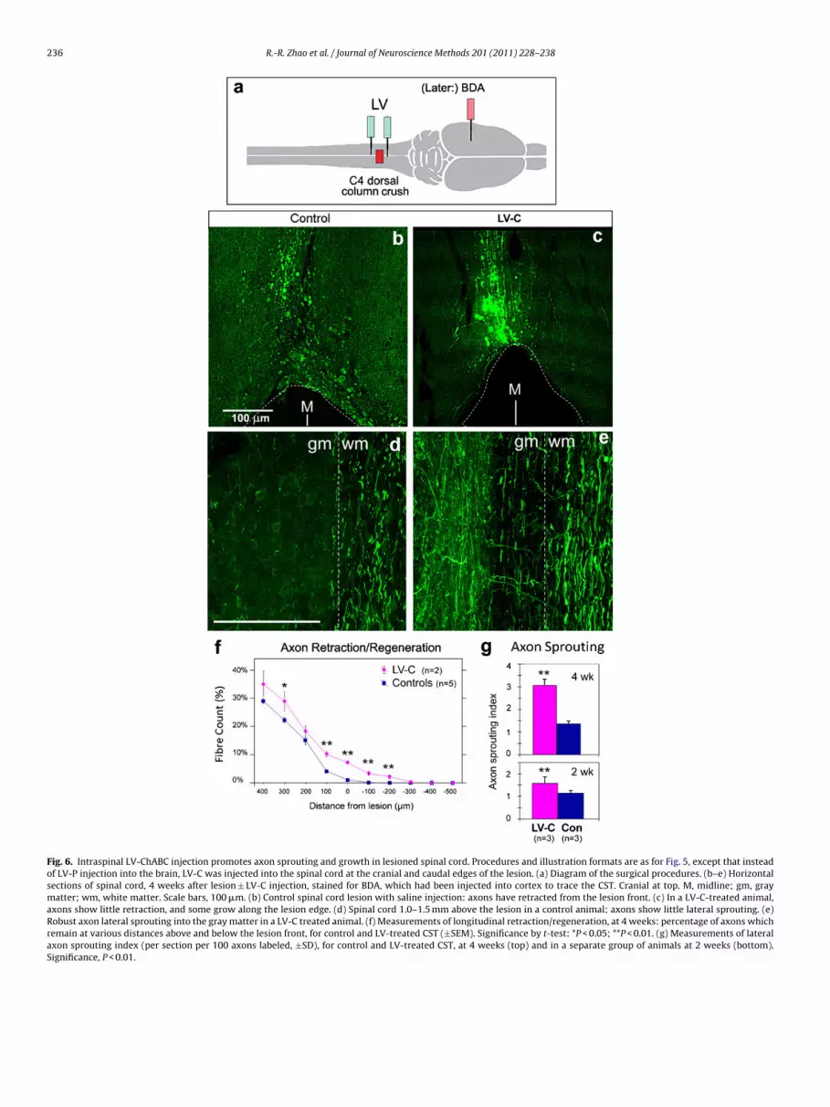
236 R.-R. Zhao et al. / Journal of Neuroscience Methods 201 (2011) 228– 238
Fig. 6. Intraspinal LV-ChABC injection promotes axon sprouting and growth in lesioned spinal cord. Procedures and illustration formats are as for Fig. 5, except that insteadof LV-P injection into the brain, LV-C was injected into the spinal cord at the cranial and caudal edges of the lesion. (a) Diagram of the surgical procedures. (b–e) Horizontalsections of spinal cord, 4 weeks after lesion ± LV-C injection, stained for BDA, which had been injected into cortex to trace the CST. Cranial at top. M, midline; gm, graymatter; wm, white matter. Scale bars, 100 �m. (b) Control spinal cord lesion with saline injection: axons have retracted from the lesion front. (c) In a LV-C-treated animal,axons show little retraction, and some grow along the lesion edge. (d) Spinal cord 1.0–1.5 mm above the lesion in a control animal; axons show little lateral sprouting. (e)Robust axon lateral sprouting into the gray matter in a LV-C treated animal. (f) Measurements of longitudinal retraction/regeneration, at 4 weeks: percentage of axons whichremain at various distances above and below the lesion front, for control and LV-treated CST (±SEM). Significance by t-test: *P < 0.05; **P < 0.01. (g) Measurements of lateralaxon sprouting index (per section per 100 axons labeled, ±SD), for control and LV-treated CST, at 4 weeks (top) and in a separate group of animals at 2 weeks (bottom).Significance, P < 0.01.

oscien
w1rwCg
aawlfWttab53
sitgwCPcp
eitgmcsonmw(slaLm
4
mse
iwcewaubt(
R.-R. Zhao et al. / Journal of Neur
ere given at 1 mm caudal and 1 mm rostral to the lesion, totalling �l (6 × 10⁵ units). Control animals were given the same lesion buteceived saline injections instead of vector. Subsequent stainingith Ab-2B6 antibody confirmed that there was a wide zone ofSPG degradation product around the lesion in the LV-C injectionroup but not in control animals (data not shown).
The CST axons were traced with bilateral BDA cortical injection,nimals were perfused 4 weeks post-lesion, and axon retractionnd sprouting were examined as before (Figs. 5a and 6). The resultsere the same as with cortical LV-ChABC injection and spinal cord
esion. In the control group, axons had retracted from the lesionront, and retraction bulbs were found at the axon endings (Fig. 6b).
ith the LV-C injection, the CST axons were present right up tohe cavity, and some extended a few hundred micrometers alonghe lesion edge past the lesion front (Fig. 6c). These fibers showed
tortuous shape instead of a smooth path, consistent with theireing regenerated fibers. Local delivery of LV-C allowed more than% axon fibers to grow pass the lesion front and extended for up to00 �m (Fig. 6f).
Local delivery of LV-C also significantly enhanced the lateralprouting of axons above lesion. At 4 weeks, the sprouting indexn the LV-C group was 3.06 ± 0.27 (SD), more than double that inhe control group which was 1.36 ± 0.11 (Fig. 6g; P < 0.01). Anotherroup of animals was operated in the same way and examined at 2eeks, and they also showed significantly more sprouting in the LV-
group (1.58 ± 0.09) than the control group (1.14 ± 0.09; Fig. 6g; < 0.01). The sprouting index at 2 weeks was lower than at 4 weeks,onsistent with the axon sprouts continuing to grow during thiseriod, while the chondroitinase gene is still being expressed.
Although lentiviral vectors generally produce few adverseffects, we wished to check whether the LV-ChABC vectors specif-cally produced inflammation following injection into the CNS. Forhis purpose, sections of spinal cord from several animals in eachroup were stained with antibody OX42, to visualize microglia andacrophages, and with antibody against GFAP, to visualize astro-
ytes (data not shown). The same pattern was seen in lesionedpinal cords that had been injected with saline (2 or 4 weeks, n = 4),r with LV-C (2 or 4 weeks, n = 3), or with LV-P (2 or 4 weeks,
= 3; these animals were not BDA traced). They showed that bothicrogliosis and astrocytosis were restricted to a zone ∼0.5 to 1 mmide around the lesion edge, except in the dorsal columns rostrally
and occasionally, to a lesser extent, caudally) where microglio-is was observed for a long distance; this is normally seen in suchesioned animals due to the degeneration of myelinated axons dam-ged by the crush lesion. Thus, the extent of inflammation in theV-ChABC injected animals was similar to or less than the inflam-ation in the controls.
. Discussion
These experiments demonstrate that a combination of selectiveutagenesis to ablate cryptic N-glycosylation sites, and expres-
ion in lentiviral vectors, allows the bacterial chondroitinase ABCnzyme to be copiously expressed in mammalian CNS.
Chondroitinase activity (as shown by the presence of 2B6 stubmmunoreactivity) was detected in injected brains for up to 8
eeks, the longest time point assessed. This probably representedontinuing production of active enzyme up to that time, as itxtended well beyond the areas labeled at 4 weeks. Active enzymeas detected directly from brains at 4 weeks by enzyme activity
ssay. We estimate that one �l of vector (6–10 × 105 transducing
nits) generated several tens of milliunits of enzyme in injectedrain in this assay (Fig. 3g), and a similar quantity in condi-ioned medium of cultured glial cells after 3 days by western blotsFig. 1).ce Methods 201 (2011) 228– 238 237
Prior studies with lentiviral vectors in the CNS have shown thatthey transduce all types of glial cells and neurons, with neuronalexpression predominating from 2 weeks onwards. In the case ofthe chondroitinase vectors, the pattern of chondroitinase diges-tion indicates that the enzyme is being secreted from neurons,because glial transduction could not account for the widespreadchondroitinase activity, including in long-distance axonal projec-tions: the corpus callosum and the corticospinal tract. It was notpossible to visualize chondroitinase production directly as theantibody, which stained transduced subconfluent cells in tissueculture, did not stain transduced cells in over-confluent culturesnor in sections from injected brains. However, a GFP-transducedbrain confirmed that a lentiviral vector can indeed express itsproduct in the decussating axons that pass through the corpuscallosum.
Lentiviral vectors do not themselves produce significant inflam-mation nor an immune response (Bloemer et al., 1997; Baekelandtet al., 2002), if a purified preparation is used, and we did not observeastrocytosis in the chondroitinase-expressing areas of brain beyondthat attributable to the injection itself. Likewise in spinal cord,astrocytosis and microgliosis covered a region no greater thanthat seen in control lesioned spinal cords, the glial reactions beingattributable to the crush lesion rather than the vector. There werealso no obvious behavioral anomalies attributable to the vectorinjections.
The results in spinal cord injury can be compared with those ofBradbury et al. (2002), who performed the same spinal cord lesionfollowed by intermittent infusion of 300 mU of chondroitinaseintrathecally over 10 days. Both treatments prevented retraction ofthe severed CST axons from the lesion (at the time-points assessed),and produced significant regeneration of CST axons along the edgeof the lesion cavity, and lateral sprouting of CST into the gray matter.Thus, within the 4-week timescale of our axon tracing experiments,the results appear to be similar, whether the vector is delivered tothe cortex or to the spinal cord.
Bradbury et al. (2002) also found more extensive regeneration ofaxons, for >4 mm, at 8–10 weeks, and this was accompanied by sig-nificant behavioral recovery. We are now undertaking longer-termexperiments to directly compare lentiviral treatment with serialinfusion, and to assess behavioral as well as anatomical recovery,and to test whether lentiviral vector delivery is equally (or more)effective if given some time after the lesion.
The present results indicate that delivery of chondroitinase by alentiviral vector to the cerebral cortex generates substantial chon-droitinase activity locally, and significant activity in long-rangeaxon projections, attributable to enzyme transport down axons. Inanimals with lesioned CST, the enzyme has a beneficial effect onthe severed axons that is comparable in effect to direct infusion,and likely to be much more suitable for therapeutic use. In con-trast to injection or infusion, the lentiviral vector only has to beadministered once; it confers greatly reduced risks of mechanicaldamage, inflammation, and infection at the infusion site; it gen-erates enzyme continuously within the CNS tissue, including axontracts; and it is effective for a long period. It can now be combinedwith other treatments in long-term animal models, to overcomeother inhibitory agents in the injured CNS and to enhance the regen-erative ability of neurons, in order to work towards a combinationtherapy suitable for human patients.
Acknowledgements
Parts of this work were supported by the Wellcome Trust(Project Grant to R.J.K., J.W.F., and E.M.M., 2007-10) and the NewtonTrust (Grant to R.J.K., 2010). Parts were supported by the EuropeanUnion FP7 projects Plasticise (Project Number 223524, to B.L.S.)
2 oscien
aR
sDCb
A
m
A
t
R
B
B
B
B
C
38 R.-R. Zhao et al. / Journal of Neur
nd PERSIST (Project Number 222878, to R.J.Y.-M.) and Spinal Cordepair (to J.W.F.).
R.R.Z. and J.W.F. were also supported by the Cambridge Univer-ity Trinity College Krishnan-Ang Studentship, the Christopher andana Reeve Foundation, the NIHR Cambridge Biomedical Researchentre, and the Medical Research Council. E.M.M. is also supportedy the International Spinal Research Trust.
The antibody against chondroitinase ABC was kindly given bycorda Inc.
E.M.M. and J.H.R. declare an interest in a patent application sub-itted for the modified chondroitinase ABC gene used in this work.
ppendix A. Supplementary data
Supplementary data associated with this article can be found, inhe online version, at doi:10.1016/j.jneumeth.2011.08.003.
eferences
aekelandt V, Claeys A, Eggermont K, Lauwers E, De Strooper B, Nuttin B, et al. Char-acterization of lentiviral vector-mediated gene transfer in adult mouse brain.Hum Gene Ther 2002;13:841–53.
loemer U, Naldini L, Kafri T, Trono D, Verma IM, Gage FH. Highly efficient andsustained gene transfer in adult neurons with a lentivirus vector. J Virol1997;71:6641–9.
radbury EJ, Moon LDF, Popat RJ, King VR, Bennett GS, Patel PN, et al. Chon-droitinase ABC promotes functional recovery after spinal cord injury. Nature2002;416:636–40.
radbury EJ, Carter LM. Manipulating the glial scar: chondroitinase ABC as a therapyfor spinal cord injury. Brain Res Bull 2011;84:306–16.
afferty WB, Bradbury EJ, Lidierth M, Jones M, Duffy PJ, Pezet S, et al. Chon-droitinase ABC-mediated plasticity of spinal sensory function. J Neurosci2008;28:11998–2009.
ce Methods 201 (2011) 228– 238
Chau CH, Shum DK, Li H, Pei J, Lui YY, Wirthlin L, et al. Chondroitinase ABC enhancesaxonal regrowth through Schwann cell-seeded guidance channels after spinalcord injury. FASEB J 2004;18:194–6.
Déglon N, Tseng JL, Bensadoun J-C, Zurn AD, Arsenijevic Y, Pereira de AlmeidaL, et al. Self-inactivating lentiviral vectors with enhanced transgene expres-sion as potential gene transfer system in Parkinson’s disease. Hum Gene Ther2000;11:179–90.
Dull T, Zufferey R, Kelly M, Mandel RJ, Nguyen M, Trono D, et al. A third-generationlentivirus vector with a conditional packaging system. J Virol 1998;72:8463–71.
Galtrey CM, Asher RA, Nothias F, Fawcett JW. Promoting plasticity in the spinal cordwith chondroitinase improves functional recovery after peripheral nerve repair.Brain 2007;130:926–39.
Hendriks WTJ, Eggers R, Verhaagen J, Boer GJ. Gene transfer to the spinal cord neuralscar with lentiviral vectors: predominant transgene expression in astrocytes butnot in meningeal cells. J Neurosci Res 2007;85:3041–52.
Kwok JC, Afshari F, García-Alías G, Fawcett JW. Review: proteoglycans in the centralnervous system: plasticity, regeneration and their stimulation with chondroiti-nase ABC. Restor Neurol Neurosci 2008;26:131–45.
Lee H, McKeon RJ, Bellamkonda RV. Sustained delivery of thermostabilized chABCenhances axonal sprouting and functional recovery after spinal cord injury.PNAS 2010;107:3340–5.
Lin R, Kwok JC, Crespo D, Fawcett JW. Chondroitinase ABC has a long lasting effecton chondroitin sulphate glycosaminoglycan content in the injured rat brain. JNeurochem 2008;104:400–8.
Muir EM, Fyfe I, Gardiner S, Li L, Warren P, Fawcett JW, et al. Modification ofN-glycosylation sites allows secretion of bacterial chondroitinase ABC frommammalian cells. J Biotechnol 2010;145:103–10.
Naldini L, Bloemer U, Gallay P, Ory D, Mulligan R, Gage FH, et al. In vivo gene deliv-ery and stable transduction of non-dividing cells by a lentiviral vector. Science1996a;272:263–7.
Naldini L, Bloemer U, Gage FH, Trono D, Verma IM. Efficient transfer, integra-tion, and sustained long-term expression of the transgene in adult rat brainsinjected with a lentiviral vector. Proc Natl Acad Sci USA 1996b;93:11382–8.
Yánez-Munoz RJ, Balaggan KS, MacNeil A, Howe S, Schmidt M, Smith AJ,et al. Effective gene therapy with nonintegrating lentiviral vectors. Nat Med2006;12:348–53.
Zhao C, Strappe PM, Lever AML, Franklin RJM. Lentiviral vectors for gene delivery tonormal and demyelinated white matter. Glia 2003;42:59–67.