jiafm-38(4) - indian academy of forensic medicine
TRANSCRIPT
EditorDr Dasari HarishProfessor & HeadDepartment of Forensic Medicine & ToxicologyGovernment Medical College & HospitalChandigarh, INDIA 1 60030Ph: 0172-2665253-59 ext 1 064Cell: +91-9646121551Email :editoriaf m @ qmail.com,dasariharish @ qmail.com
Joint EditorDr Manish NigamProfessor & HeadDepartment of Forensic Medicine and ToxicologyDeputy Medical superintendentSriAurobindo MedicalCollege and PG lnstituteAnd associated hospitals, lndore (MP)Cell: +91-9826213412Email: iurimanish @ qmail.com
Assr'sfa nt EditorDr Amandeep SinghAssociate ProfessorDepartment of Forensic Medicine & ToxicologyGovernment Medical College & HospitalChandigarh, INDIA 1 60030Ph: 0172-26525&59 ext 1 048Cell: +91-9646121610E mai I :dqrn aldeep-B o mai,Lcom
Assistant EditorDr Satinder Pal SinghAssistant ProfessorDepartment of Forensic Medicine & ToxicologyGovernment Medical College & HospitalChandigarh, INDIA 1 60030Ph: 0172-266525$59 ext 1064Cell: +91-9646121484Emai l:spsinoh9988 @ vah
J Indian Acad Forensic Med. October – December 2016, Vol. 38, No. 4 ISSN 0971-0973
i
Indian Academy of Forensic Medicine (IAFM)
(Registration No.349, 12th May, 1972, Panji, Goa)
Governing Council 2016-2019
President Dr. Kalpesh Shah
General Secretary Treasurer Dr.Madhu Godhikirikar Dr.S.K.Daddu Vice Presidents North Zone: Dr. Pankaj Gupta South Zone: Dr. Cyriac Job East Zone: Dr. A.J.Patowary West Zone: Dr. Sudhir Ninave Central Zone: Dr. Shiv Ratan Kochar
Joint Secretaries North Zone: Dr. Rajeev Joshi South Zone: Dr. Krishna Rao GM East Zone: Dr. Putul Mahanta West Zone: Dr. Ganesh Govekar Central Zone: Dr. Manish Kumath
Editor Joint Editor Dr.Dasari Harish Dr. Manish Nigam
Executive Members
Dr. S.D. Nanadkar (Ex. President, IAFM) Dr. Ajay Kumar Dr. Sudha R. Dr.T. K. K. Naidu Dr. Rai Sudhir Prasad Dr. Tulsi Mahto
Dr. C.B.Jani (Ex. Secretary, IAFM) Dr. Rajesh C. Dere
Dr. M. I. Sheikh Dr. O.P.Murthy Dr. Abhishek Yadav
J Indian Acad Forensic Med. October – December 2016, Vol. 38, No. 4 ISSN 0971-0973
ii
Journal of Indian Academy of Forensic Medicine
(JIAFM) The Official Publication of Indian Academy of Forensic Medicine
Editor Dr Dasari Harish Professor & Head Department of Forensic Medicine & Toxicology Government Medical College & Hospital Chandigarh, INDIA 160030 Residence: House No 1112, Sector 32 B, Chandigarh 160030 Ph: 0172-2665253-59 ext 1064 Cell: +91-9646121551 Email: [email protected], [email protected]
Joint Editor Dr Manish Nigam Professor & Head
Forensic Medicine and Toxicology Deputy Medical superintendent
Sri Aurobindo Medical College and PG Institute And associated hospitals, Indore (MP)
Cell: +91-9826213412 Email: [email protected]
Editorial Team
Dr Amandeep Singh (GMCH, Chandigarh) Dr Satinder Pal Singh (GMCH, Chandigarh)
International Advisory Board
Prof. Derrick J Pounder, Dundee, UK Prof. D N Vieira, Coimbra Portugal Prof. Dan Dermengiu, Romania Prof. Peter Vanezis, London, UK Prof. Roger Byard, Australia Dr. Michael S. Pollanen, Canada Prof. Leandro Duarte De Carvalho, Brazil Dr. Shubhakar K.P. UK
Dr. BL Meel, South Africa Dr. John Clark, Glasgow, UK Dr. George Paul, Singapore Dr. Serap Annette AKGUR, Turkey Dr. Clifford Perera, Sri Lanka
Dr. B.N. Yadav, Nepal Dr. K. P. Saha, Bangladesh Dr. Gorea R.K KSA
National Advisory Board
Srivastava A.K. (U.P.) Pillay V.V. (Kerala) Jani C.B. (Gujarat) Bose T.K (West Bengal) Pradeep Kumar G. (Karnatka) Verma S.K. (New Delhi) Kumar Shantha B. (Tamil Nadu) Gupta B.D. (Gujrat) S.C. Mahapatra (Odisha) Manju Nath K.H, (Karnatka) Das Sanjoy, (Uttarakhand) Mahtoo Tulsi, (Jharkhand)
Ravindran K. (Puducherry) Rastogi Prateek (Karnatka) Potwary AJ (Assam) Singh R.K. (Chhatisgarh) Dongre A.P. (Maharastra) Sharma Aditya (H.P.) Yogendra Bansal (Chandigarh) Khanagwal V. (Haryana) Rastogi Pooja (U.P.) Khaja Shaikh (A.P.) P.P. Mukhopadhyay (W.B.) Naik R.S. (Maharastra)
Job Cyriac (Kerala) Vinita K. (U.P.) Mohite Shailesh (Mumbai) Yadav Jayanti (M.P.) Kochar S.R. (Rajasthan) L. Fimate (Manipur) K H Chavali (Raipur) Gaurav Sharma (Haryana) R S Bangal (Maharashtra) S S Oberoi (Punjab)
Printed and published by Dr. D. Harish, Editor, JIAFM and Dr. Manish Nigam, Joint Editor, JIAFM on behalf of
Indian Academy of Forensic Medicine at name of the press
J Indian Acad Forensic Med. October - December 2016, Vol. 38, No. 4 ISSN 0971-0973
386
Journal of Indian Academy of Forensic Medicine
Contents Sr. Page
I. From the Editor’s Desk 388-388
II. Editorial 389-392
Original Research Paper
1. An Analysis of Fractures of Hyoid Bone and Thyroid Cartilage in Deaths Due to Hanging Nityanand Kumar, Niranjan Sahoo, Bibhuti Bhusana Panda, Mohan Kumar
Hansda
393-396
2. A Prospective Study of Poisoning Cases due to Paraquat at a Tertiary Care Centre - Chennai J James Rajesh, J Gerard Rakesh,U Jagdish Kamal Chander, P Sampath
Kumar
397-399
3. Analysis of Fatal burns cases in a Metropolitan city of South India. P. Shruthi,
R. K. Varma, B. Viswakanth
400-403
4. Pregnancy Related Deaths: A Ten Year Retrospective Study in the Mortuary of a Tertiary Care Teaching Hospital of Northeast India. Memchoubi Ph., Th.
Meera, John Deb Barma, Kh. Pradipkumar Singh
404-407
5. An Epidemiological Study of Fracture Related Deaths in a Tertiary Care Hospital in West Bengal Alakesh Halder, Kaushik Mukhopadhyay, Shailesh V Parate, Ashok
Kumar Samanta
408-411
6. Growth Report of “The End” – Autopsy Statistics in a Developing Medical College Jigesh V. Shah, Gaurang J. Patel, Ankur Patel, Dharmesh S. Patel, Sanjay Jadav, Rajesh
Jakhar
412-415
7. Dental Age Estimation by Radiographic Evaluation of Pulp/Tooth Ratio in Mandibular Canines and Premolars Usha Jambunath, Balaji. P, Poornima. G, Vinitra
Vasan, Ashish Gupta, Peeyush Shivhare
416-419
8. Footprint Breadth Dimensions: Is it possible to determine sex and/or side? Ashish Badiye, Hansi Bansal, Anjali Rahatgaonkar, Mukesh Yadav
420-422
9. Analysis of Sudden Death Cases Brought for Postmortem Examination at Sir T. General Hospital, Bhavnagar Love R. Bhagora, Amit P. Parmar, Dipti C. Parmar,
Tejas C. Patel
423-425
10. A Study of Victims of fall from Height from 2006 to 2015 in Imphal Memchoubi
Ph, Th. Meera, Pulak Chakma 426-428
11. Role of Hand Anthropometry for Stature Estimation in Females of Bhavnagar – Gujarat T.C. Patel, A.P. Parmar, D.C. Parmar, S.I. Patel, L.R. Bhagora
429-432
12. Histopathological examination in routine medicolegal autopsy: 3-years retrospective analysis Vipul N. Ambade, Balwant D. Kowe, Ajay N. Keoliya
433-436
13. Study of Undergraduate Students’ Perceptions Towards Organ Donation Pragnesh B. Parmar, Kunjan Bharpoda, Vidhi Bhensdadia, Pragnesh Bhokan, Parth Bhut, Bharat Chaudhary
437-440
14. Stature Estimation Using Per-Cutaneous Tibial Length in Malwa Population of Punjab Rajiv Joshi, Monika Bhardwaj, Ramandeep Singh
441-445
15. Profile of Scorpion Envenomation in Rural India Venkatesh Maled, Umesh Dixit 446-448
Volume: 38 • Number: 4 • Oct. – Dec. 2016
J Indian Acad Forensic Med. October - December 2016, Vol. 38, No. 4 ISSN 0971-0973
387
16. Forensic Anthropological Examination of Lip-Print Pattern Types Among Northwest Indians Subjects: A Pilot Study Jagmahender Singh Sehrawat
449-454
17. Fatal Burn Injuries: A Five Year Retrospective Autopsy Study in Temple Town, Tirupati M. Abdul Khalid, Bijili Venkatesulu, B. Lakshmi Narayana
455-459
18 A study of Unnatural Female Deaths reported to a tertiary care hospital in Khammam, Telangana Bharath Kumar Guntheti, Uday Pal Singh
460-464
19. Epidemiological analysis of drowning deaths: A 10 year Study. Sujan Kumar
Mohanty, Virendra Kumar, Jaffar Hussain A.P, V. Bhuvan 465-470
20. Age estimation and eruption of Permanent Teeth between two Genders - A Cross sectional study
Manjot Kaur, Chandrakanth HV, Dasari Harish
471-475
21. To Appraise the Medical Negligence Complaints and Liable Specialties with respect to CPA Megha Rani, Manushi Srivastava, Ratan K. Srivastava
476-480
Review Research Paper
22. Postmortem interval (PMI) estimations from vitreous humor: a review of current status, limitations and probabilities Nikita Gupta, JS Sehrawat, Dasari Harish
481-484
23. RAJ – TAJ and MODI. The life & times of Dr. J. P. Modi (1875 - 1954). (A leaf in the history of forensic medicine in India) A.M.M.Patnaik, M.Jagadeesh Naik,
Ch.Lakshmi Kumar
485-488
24. Child Sexual Abuse (CSA): India & the World. Raghvendra Kumar Vidua, Sweta
Patel, B.L.Chaudhary, Alok K. Mishra, Arneet Arora 489-494
Case Reports
25. Sexual Assault and Murder of a 3 year Old Female Child: A Case Report. Preet Inder Singh, S. Valliappan, Amandeep Singh, Ajay Kumar
495-498
26. Revising the current MTP Act : Hasn’t the Time Come Yet?? Jyoti Barwa,
Amandeep Singh, Ajay Kumar, Dasari Harish 499-502
27. Fatal Paint Thinner Ingestion - A Case Report Jitendra Singh Tomar, Pradeep Kr
Mishra, Mandar Ramchandra Sane, Divyesh Saxena, Manish Kumar 503-505
28. Breast Feeding Pulmonary Aspiration Death: A Case Report Bhullar DS,
Aggarwal KK, Mehta N 506-508
29 Bicuspid Aortic Valve: Cause for Sudden Cardiac Death - A Case Report Bhagavan.S, Punitha.R, Pradeep Kumar, Jagadeesh.N.H.
509-511
30 Biomechanics of fatal head injury by the wheel rim of a tractor trailer - A Case Report Siddhartha Das, Manoj Kumar Jena
513-515
Address request for reprint or further information relating to any article may please be made with
author and in case of multi authored article, please communicate to Corresponding Author or the
First Author
Copy Right © All rights reserved: No part of this publication may be reprinted or publish without the prior permission of the Editor, JIAFM. Submission of all manuscripts to the journal is understood to imply that it is not being considered for publication elsewhere. Submission of multi authored papers implies that the consent of each author has been obtained. In this journal, every effort has been made NOT to publish inaccurate or misleading information. However, the Editor, Joint Editor, Peer Review Group and Advisory Board accept NO liability in consequences of such statements. The Journal of Indian Academy of Forensic Medicine is indexed in Index Copernicus [Poland] and IndMED [India]
J Indian Acad Forensic Med. October - December 2016, Vol. 38, No. 4 ISSN 0971-0973
388
From Editor’s Desk
JIAFM A Quarterly Publication
Volume 38, Number 4, October - December, 2016
Dear Friends, The IVth issue of the JIAFM is now in your hands. We have made sincere efforts to see that both the standard and the quality of the articles published improve to meet the international standards. However, much of the standard depends on the quality of manuscripts received. We receive manuscripts of every quality - some so good that we fall in love with them after the initial reading and some so bad that we are forced to reject them outright. I, on behalf of the Editorial Team request you to please submit manuscripts of some standard. Most of the manuscripts with the so called original work are nothing but counting of dead bodies. Pl do some good research and send us some good manuscripts. One year has gone by in your service. Time flies so fast. It's your love and affection that sustained us through this mammoth Endeavour. We now have almost 180 subscriptions from various libraries. The Journal and the manuscripts are extensively quoted in the literature. The one major task now is to get PUBMED indexing. If any one of you has some resources for this task, Pl help us to get your Journal indexed in the PUBMED.
Jai Hind &Long Live IAFM Dr Dasari Harish
Editor
Subscription Information • Members of IAFM will receive the free of cost.
• Non Members and Institutions (Annual Subscription rates) • Personal: In India, Rs. 1000/ (Rest of the world: US$ 200/ or equivalent) • Institutions: In India, Rs. 5000/ (Rest of the world: US$ 400/ or equivalent) • We Accept: Bank Cheque / Demand Drafts (Add Rs. 50/- for outstation Cheques)
• The Scope of the Journal covers all aspects of Forensic Medicine and allied fields, research and
applied.
Subscription orders and payments should be made in favour of
“Editor, JIAFM, payable at Greater Noida”
Claims for missing issue: A copy will be sent free to the member / subscriber provided the claim is made within 2 months of publication of the issue & self addressed envelop of the size 9” x 12” is sent to the Editor. (Those who want the journals to be dispatched by Registered Post must affix Rs. 50/ worth postage stamps).
The journal is indexed with IndMed and made available online by following website:
www.jiafm.com www.medind.nic.in www.indianjournals.com http://indmed.nic.in
www.iafmonline.in
J Indian Acad Forensic Med. October - December 2016, Vol. 38, No. 4 ISSN 0971-0973
389
Editorial
Increasing the Legal limits of Gestation Period as Prescribed in the MTP Act, 1971 in Light of the Recent Supreme Court Judgements
1Ajay Kumar,
2Dasari Harish,
1Amandeep Singh
1Associate Professor,
2Professor & head, Dept. of Forensic Medicine, GMCH, Chandigarh
Abstract A 22-year-old woman was allowed to abort her foetus in its 24th week of gestation period after the Supreme Court observed that it suffered from anencephaly, a condition incompatible with life and in which the foetus is said to be suffering from a defect of skull and brain tissue development. In July 2016 also, the Supreme Court allowed a woman to undergo abortion in her 24th week of pregnancy at one hospital in Mumbai, granting her the benefit under Section 5 of Medical Termination of Pregnancy (MTP) Act, 1971, that allows abortion despite the 20-week ceiling. However, in one case, on 24 weeks routine check up the doctor discovered that the fetus had a severe abnormality. They approached the Bombay High Court which denied abortion Under the MTP Act. Another petitioner, Mrs Y in her 19th week of pregnancy, was told by the doctors that her fetus may have had a congenital malformation characterized by partial absence of brain tissue. Under the limits imposed by the MTP Act, Mrs. Y was forced to make the excruciating decision to terminate her pregnancy. It is clear that in some cases MTP was allowed, while in the others, it was not. All the judgements were on a case to case basis, as the legal period of gestation prescribed by the Act is only 20 weeks, except in dire emergencies. The time has come to amend the 45 years old Act in the light of new advancements in science and in light of new judgements by the Hon'ble Supreme Court. This would go a long way in helping the affected ladies, who are forced to rush to the courts for redressal, if their pregnancy crosses the arbitrarily drawn limit of 20 weeks.
Key Words: Supreme Court Judgements, Medical Termination of Pregnancy, Gestation Period, MTP
Act 1971
.Introduction: Recently, a 22-year-old woman, Mrs A, was allowed to abort her foetus in its 24th week of gestation period after the Supreme Court observed that it suffered from anencephaly. The hospital, based on radiology tests, diagnosed the foetus to be anencephalic. In such situations, the foetus can only survive In Utero and death is inevitable after birth. The Law, under the Medical Termination of Pregnancy Act 1971, does not allow abortions beyond 20 weeks of pregnancy, except in cases where there is an imminent threat to the mother’s life. Any person/ doctor breaching the provisions of the said Act would invite imprisonment up to seven years.[1]
The Court allowed her to undergo abortion in her 24th week of pregnancy, granting her the benefit under Section 5 of MTP Act, that allows abortion despite the 20-week ceiling. The woman found out about the defect in her 21st week of pregnancy when she underwent sonography. In her 23rd week, she approached Doctors for an abortion, who then requested the court to intervene.[2] Again, the Supreme Court, in July 2016, allowed a rape victim based in Mumbai to abort her 24-week-old abnormal foetus after the Centre clarified that a 20-week cap on termination of pregnancy is not
applicable if the pregnant woman’s life is found to be in grave danger. She alleged that the pregnancy was a result of an act of rape perpetrated on her, and sought to terminate it for this reason. The Supreme Court set up a medical board which opined independently that the pregnancy posed a serious risk to her health and for that reason, could be justifiably terminated in accordance with the MTP Act.[3] The judgment questioned the constitutional validity of the Medical Termination of Pregnancy (MTP) Act 1971, which currently allows abortion only up to the 20th week. A bench headed by Justice J.S. Khehar said: “In view of the clear findings of the medical board whose examination showed that contained pregnancy could endanger the petitioner’s life, we are satisfied that it may be permissible to terminate pregnancy.”[4] In 2009, an appeal petition was filed by the Human Rights Law Network group in the Supreme Court on behalf of a gynaecologist, practicing in Mumbai against the High Court order which denied access to an abortion Under the MTP Act 1971. At the first antenatal checkup, doctors told one Mrs. C that her fetus had severe abnormalities and would not survive more than a few hours after delivery. Mrs. C was 26 weeks pregnant and therefore could not legally obtain a medical termination of pregnancy under the MTP Act. Mrs. C was forced to continue the pregnancy, visit the hospital
J Indian Acad Forensic Med. October - December 2016, Vol. 38, No. 4 ISSN 0971-0973
390
regularly, and participate in social events to celebrate the birth. After three days of excruciating labor pains, Mrs. C delivered a baby that ultimately died less than three hours later. In her affidavit Mrs. C states, “The whole process was extremely painful. In normal circumstances a mother goes through all the discomfort just for the joy of giving birth to the baby. However in my case there was no joy as I was aware of the poor outcome of the baby. All this could have been avoided if my pregnancy was terminated in time.” The Supreme Court petition argued that the current Act violates women’s rights to health, life, dignity, and equality. The case is pending in the Supreme Court.[5] Another petitioner Mrs D in her 19th week of pregnancy, was told by the doctors that her fetus may have had a congenital malformation characterized by partial absence of brain tissue. Additional test results would not be available until after the 20th week of pregnancy. Under the limits imposed by the Medical Termination of Pregnancy Act, Mrs. D was forced to make the painful decision to terminate her pregnancy without a full understanding of the medical facts.[6] In another case from Chandigarh in 2009, a woman, Ms. E, had become pregnant as a result of an alleged rape that took place while she was an inmate at a government-run welfare institution located in Chandigarh. After the discovery of her pregnancy, the Chandigarh Administration approached the High Court seeking approval for the termination of her pregnancy, keeping in mind that in addition to being mentally retarded she was also an orphan who did not have any parent or guardian to look after her or her prospective child. The High Court directed the termination of the pregnancy in spite of the Medical Expert Body's findings which show that the victim had expressed her willingness to bear a child.[7] The High Court gave permission. An appeal was made against this order before Apex Court. After perusing the experts opinions and after hearing arguments, the 3 judge bench of the Supreme Court gave the opinion that the Consent of the victim was not taken and in their opinion, and the MTP was not in the Best interest of the victim.[8]
The law at Present: Under the MTP Act 1971,[9] pregnancies may be terminated in India when, Notwithstanding anything contained in the Indian Penal Code (45 of 1860), a registered medical practitioner shall not be guilty of any offence under that Code or under any other law for the time being in force, if any pregnancy is terminated by him in accordance with the provisions of this Act. Subject to the provisions of sub-section (4), a pregnancy may be terminated by a registered medical practitioner,- Where the length of the pregnancy does not exceed twelve weeks if such medical practitioner is, or
Where the length of the pregnancy exceeds twelve weeks but does not exceed twenty weeks, if not less than two registered medical practitioners are. Of opinion, formed in good faith, that,- (i) The continuance of the pregnancy would involve a risk to the life of the pregnant woman or of grave injury physical or mental health ; or (ii) There is a substantial risk that if the child were born, it would suffer from such physical or mental abnormalities as to be seriously handicapped. Explanation 1.-Where any, pregnancy is alleged by the pregnant woman to have been caused by rape, the anguish caused by such pregnancy shall be presumed to constitute a grave injury to the mental health of the pregnant woman. Explanation 2.-Where any pregnancy occurs as a result of failure of any device or method used by any married woman or her husband for the purpose of limiting the number of children, the anguish caused by such unwanted pregnancy may be presumed to constitute a grave injury to the mental health of the pregnant woman. (3) In determining whether the continuance of pregnancy would involve such risk of injury to the health as is mentioned in sub-section (2), account may be taken of the pregnant woman's actual or reasonable foreseeable environment. (4) (a) No pregnancy of a woman, who has not attained the age of eighteen years, or, who, having attained the age of eighteen years, is a lunatic, shall be terminated except with the consent in writing of her guardian. 5(b) Save as otherwise provided in C1.(a), no pregnancy shall be terminated except with the consent of the pregnant woman5. Discussion and Conclusion As per the cases discussed above, sometimes the abortions were allowed and sometimes they were refused on basis of the MTP Act. A woman can procure abortion legally up to 20 weeks in ordinary circumstances, but to save the life of the woman, abortion can be done at any time of gestation. The need to raise the upper limit of abortion is the need of the hour as newer diagnostic tests are readily available which can diagnose rare but serious abnormalities in the foetus after the 20 weeks of gestation. So in cases where a congenital or rare but serious abnormality in the foetus is diagnosed, the woman is left with no choice but to continue the pregnancy. Not every pregnant woman who has crossed the deadline of 20 weeks has access to file a petition in the apex court. One cannot imagine the anxiety the pregnant woman goes through when she learns that her baby has a fatal condition but she cannot undergo abortion because she has crossed 20 weeks of pregnancy.[1] An important question which comes to the mind is that when Law does not dictate or specify when should a women to become pregnant then why does she need the law to decide whether to
J Indian Acad Forensic Med. October - December 2016, Vol. 38, No. 4 ISSN 0971-0973
391
continue the pregnancy or not? She should have a constitutional right to have complete autonomy over her own body in the context of a pregnancy. The Supreme Court’s order of 26 July 2016 in case of “Ms X” v Union of India failed to deal with important issue of the constitutional right of a woman to have complete autonomy over her own body in the context of a pregnancy.[10] While allowing the petitioner to terminate her pregnancy on the basis of medical advice by a board of doctors, that pointed out that her life would be in danger if the pregnancy was continued, the Court refused to engage with the larger questions that the petition raised i.e. to change the provisions of the Medical Termination of Pregnancy Act, 1971. It was the doctors who decided for the patient, what was in her best interest and not the patient herself (paternalism). The abortion law in India deprives the woman of choice and control over her body whether to complete the pregnancy or not. The question of choice, autonomy over one’s body and how to balance these two against the issue of female foeticide have to be debated and is to be addressed by the law makers. In the case of Ms E of Chandigarh, the rationale behind the Supreme Court judgement not to allow MTP after the 20 weeks period was based on two broad considerations. The first consideration was whether it was correct on part of the High Court to direct the termination of pregnancy without the consent of the woman in question. This was the foremost issue since a plain reading of the relevant provision in the MTP Act clearly indicates that consent is an essential condition for performing an abortion on a woman who has attained the age of majority and does not suffer from any `mental illness'. There is a clear distinction between `mental illness' and `mental retardation' for the purpose of this statute. The second consideration was that even if the said woman was assumed to be mentally incapable of making an informed decision, what are the appropriate standards for a Court to exercise `Parens Patriae' jurisdiction? If the intent was to ascertain the `best interests' of the woman in question, it was the considered opinion of SC judges that the direction for termination of pregnancy did not serve that objective. Of special importance is the fact that at the time of hearing, the woman had already been pregnant for more than 19 weeks and there is a medico-legal consensus that a late-term abortion can endanger the health of the woman who undergoes the same3. The State could claim that it was the guardian of the pregnant victim since she is an orphan and has been placed in government-run welfare institutions. However, the State's claim to guardianship was not mechanically extended in order to make decisions about the termination of her pregnancy. An ossification test revealed that the physical age of the victim is around 19-20
years. This conclusively shows that she is not a minor. Furthermore, her condition has been described as that of `mild mental retardation' which is clearly different from the condition of a `mentally ill person' as contemplated by Section 3(4)(a) of the MTP Act. It is pertinent to note that the MTP Act was amended in 2002, by way of which the word `lunatic' was replaced by the expression `mentally ill person' in Section 3(4)(a) of the said statute. The said amendment also amended Section 2(b) of the MTP Act, where the erstwhile definition of the word `lunatic' was replaced by the definition of the expression `mentally ill person' which reads as `mentally ill person' means a person who is in need of treatment by reason of any mental disorder other than mental retardation"
3. The doctrine of `Parens Patriae' was evolved in common law and is applied in situations where the State must make decisions in order to protect the interests of those persons who are unable to take care of themselves. Traditionally this doctrine is applied in cases involving the rights of minors and those persons who have been found to be mentally incapable of making informed decisions for themselves. Courts in other common law jurisdictions have developed two distinct standards while exercising `Parens Patriae' jurisdiction for the purpose of making reproductive decisions on behalf of mentally retarded persons. These two standards are the `Best interests' test and the `Substituted judgment' test3. It was the `Best Interests' test alone which governed the inquiry in the present case of Ms E and not the `Substituted Judgment' test3. Prior to MTP Act of 1971, abortions were criminalised under Section 312 of the Indian Penal Code, 1860,[11] and notwithstanding the 1971 Act, continue to be criminalised as of date. In fact, even the pregnant woman could be found guilty if she self-aborts the child she is carrying. This position was considered unsatisfactory, and on the basis of the recommendations of the Abortion Study Committee in 1966, the MTP Act was introduced and passed in Parliament. The Act provides a clearer set of guidelines and time frames within which a doctor carrying out an abortion was protected from criminal sanction under the law.[9] So in situations where the question of whether an abortion is to be undertaken or not, is left purely in the hands of a medical practitioner, with no say for the woman who is actually pregnant. The woman who is actually carrying the foetus has no agency over her body under Indian law. If the doctor thinks the continuance of the pregnancy would involve a risk to the life of the pregnant woman or if it causes grave injury physical or mental health of the women only then abortion is allowed, that to under 20 weeks.[12] Legal and medical experts feel that a revision of the legal limit for abortion is long overdue. Foetal abnormalities show up only by 18
J Indian Acad Forensic Med. October - December 2016, Vol. 38, No. 4 ISSN 0971-0973
392
weeks, so just a two-week window after that is too small for the would-be parents to take the difficult call on whether to keep their baby and for the medical practitioner to exhaust all possible options before advising the patient to take the extreme step9. Some of the cardiac and renal diseases are evident only after 24 to 26 weeks. The rising incidence of sex crimes and the urgent need to empower women with sexual rights and choices both in their own interest and for the sake of reducing the fertility rate as a whole, have made it imperative that the law be changed. Also, since lack of legal approval does not prevent abortions from being carried out beyond 20 weeks, women are put under risk since the abortions then are often conducted in shady, unhygienic conditions by untrained, unqualified quacks.[13] If abortion is not allowed, as in case of Mrs C and Ms E after 20 weeks, then some more complex questions also arise in front of the would be parents. Mrs. C knew that her fetus had severe abnormalities and would not survive more than a few hours after delivery, but was forced to continue the pregnancy, visit the hospital regularly, and participate in social events to celebrate the birth, bear excruciating labor pains, delivered a baby that ultimately died less than three hours later. This whole sequence of events under the current MTP Act violated women’s rights to health, life, dignity, and equality. Who will have borne the cost of raising a malformed/ deformed/ mentally ill child/ unwanted child of Mrs C if it had survived? Who will have borne the cost of raising of unwanted child born due to rape as in case of Ms E? What if the foetus survives the ordeal of abortion and is delivered live and survives for few hours or days as in one of the above cases? Doesn’t the foetus/newborn, intended to be aborted, have human rights? In the US, Supreme Court sought to strike a balance between the rights of the woman over her own body and the interests of the state in the birth of the foetus. It struck down a whole draft of State and local laws which had criminalised abortions in various parts of the United States, as being in violation of the right to privacy, without due process protected under the 14th Amendment to the US Constitution. Roe v Wade was
subsequently modified by the Supreme Court in Planned Parenthood v Casey where the legality of the abortion law is now linked to the viability of the foetus rather than the rigid third trimester test laid down in Roe v Wade.[7] The Government of India has proposed amendments in the MTP Act that would extend the legal time limit for abortion. The amendments should expand access to abortion to all. Supreme Court should extend the upper time limit on abortion to 24 weeks and exclude time limits all together where doctors have detected substantial foetal abnormalities. The draft Bill[6] allows abortion up to 24 weeks if A healthcare provider “in good faith” can make the decision of aborting a pregnancy between 20 and 24 weeks if: - The pregnancy involves substantial risks to the mother or child - If the pregnant woman has alleged the pregnancy was caused by rape Let us only hope that good sense prevails, in the end.
Reference: 1. http://indianexpress.com/article/india/sc-allows-24-week-pregnant-
woman-to-abort-foetus-with-undeveloped-skull/
2. http://www.newindianexpress.com/nation/2017/jan/16/supreme-court-permits-termination-of-pregnancy-at-24-weeks-1560127.html
3. http://www.thebetterindia.com/62683/abortion-laws-india-supreme-court-rape-survivor/
4. http://www.thehindu.com/news/national/SC-allows-rape-victim-to-abort-24-week-old-foetus/article14508050.ece
5. http://www.hrln.org/hrln/images/stories/pdf/xandy-petition-8-3-14.pdf 6. http://www.hrln.org/hrln/reproductive-rights/pils-a-cases/1646-
government-of-india-announces-proposed-amendments-to-the-mtp-act.html
7. http://www.wbja.nic.in/wbja_adm/files/Sc%20title%20-%2044.pdf 8. http://indiankanoon.org/doc/1500783/
9. The MTP Act 1971, Act 34 of 1971. Available at: http://tcw.nic.in/Acts/MTP-Act-1971.pdf
10. http://www.firstpost.com/india/abortion-law-in-24-week-pregnancy-case-supreme-court-failed-to-address-womans-right-to-her-body-
2916174.html 11. The Indian Penal Code, 1860. Act 45 of 1860. Available at:
http://www.advocatekhoj.com/library/bareacts/indianpenalcode/index.php?Title=Indian%20Penal%20Code,%201860
12. https://indiankanoon.org/doc/237570/ 13. http://indianexpress.com/article/india/sc-allows-24-week-pregnant-
woman-to-abort-foetus-a-glimpse-of-the-trajectory-of-indias-abortion-laws-4480004/
J Indian Acad Forensic Med. October - December 2016, Vol. 38, No. 4 ISSN 0971-0973
393
Original Research Paper
An Analysis of Fractures of Hyoid Bone and Thyroid Cartilage in Deaths Due to Hanging
1Nityanand Kumar,
2Niranjan Sahoo,
3Bibhuti Bhusana Panda,
4Mohan Kumar Hansda
Abstract Hanging is one of the most common unnatural deaths due to mechanical asphyxia encountered
in the mortuary. The present study was undertaken with the aim of studying the incidence & frequency of
fracture in hyoid bone and thyroid cartilage in deaths due to hanging in the Department of FMT, RIMS,
Ranchi, prospectively from 1st March, 2013 to 30
th May, 2014. Highest number of deaths due to hanging
was in the age group of 21-30 years. In maximum cases, the nature of suspension was of Complete &
Atypical type. Most commonly used ligature was Jute Rope and Dupatta. In majority cases, the ligature
mark was above the thyroid cartilage followed by at and above the thyroid cartilage and only few cases
the ligature mark was at or overriding the thyroid cartilage. In maximum cases, the position of knot was
present at occipital region of the neck, followed by left mastoid region. In majority of cases, the ligature
mark was prominent and discontinuous. Hyoid fracture was found in only 5.2% cases. There was no case
with thyroid cartilage fracture.
Key Words: Hanging, fracture hyoid bone, thyroid cartilage, ligature material, knot position.
Introduction: Hanging is that form of asphyxia which is caused by suspension of the body by ligature which encircles the neck, the constriction force being the weight of the body. It may be partial or complete, depending on the position or posture of the body at the time of hanging. Hanging is a common method of suicide around the world. In India hanging is among the top 5 methods of choice for committing suicide.[1] More than 800,000 people die due to suicide every year and there are many more who attempt suicide. Hence, millions of people are affected or experience suicide bereavement every year. Suicide occurs throughout the lifespan and is the second leading cause of death among the age group of 15-29 year globally.[2]
Corresponding Author: 3Assistant Professor,
Department of FMT, Hi-Tech Medical College, Bhubaneswar, Odisha
1Assistant Professor,
Department of FMT, LLAM Govt Medical College Hospital, Raigarh, Chattishgarh, 2,4
Assistant Professor, Department of FMT, IMS & SUM Hospital, SOA University, Bhubaneswar, Odisha, DOR: 29/02/2016 DOA: 25/12/2016 DOI: 10.5958/0974-0848.2016.00128.7
Suicide by hanging is the most frequent method in India.[2] The profile of victims comprises of married females or unmarried males in the age group of 21-30 years, faced with stress in the form of unemployment, harassment for dowry, prolonged illness, failure in examinations, financial duress, or interpersonal problems.[3]
During the autopsy of hanging, strangulation, throttling case, hyoid bone and thyroid cartilage becomes the most integral part of examination. This fact has been highlighted by many workers.[9] Some workers also claimed that a hard ligature material can cause fracture of hyoid bone and thyroid cartilage.[12] Various authors have reported the incidence of hyoid bone fracture being from nil/rare to as high as 67% in hanging cases.[4]
Aims and Objectives: The present study was undertaken with the aim of studying the presence of fracture in hyoid bone and thyroid cartilage in hanging cases and to determine their frequency along with it to review the anatomo-pathological findings over the neck.
Material and Methods: The present study was carried out in the Department of FMT, RIMS, Ranchi, prospectively from 1st March, 2013 to 30th May, 2014. The materials for the present study were
J Indian Acad Forensic Med. October - December 2016, Vol. 38, No. 4 ISSN 0971-0973
394
the deceased brought for autopsy from various police stations. Cases which died due to asphyxia as a result of hanging either alone or in association with other injuries were included for the study. Information relating to age, type of suspension, type of ligature used and localization of the knot were gathered from the police records like inquest report, dead body, challan etc., and detailed history from relatives, neighbours, friends, and police officials accompanying the dead bodies. In case of hospital deaths, hospital records were also examined. A detailed and thorough post-mortem examination was carried on every case to examine the fracture hyoid bone and thyroid cartilages. The findings noted were carefully compiled, tabulated and analyzed.
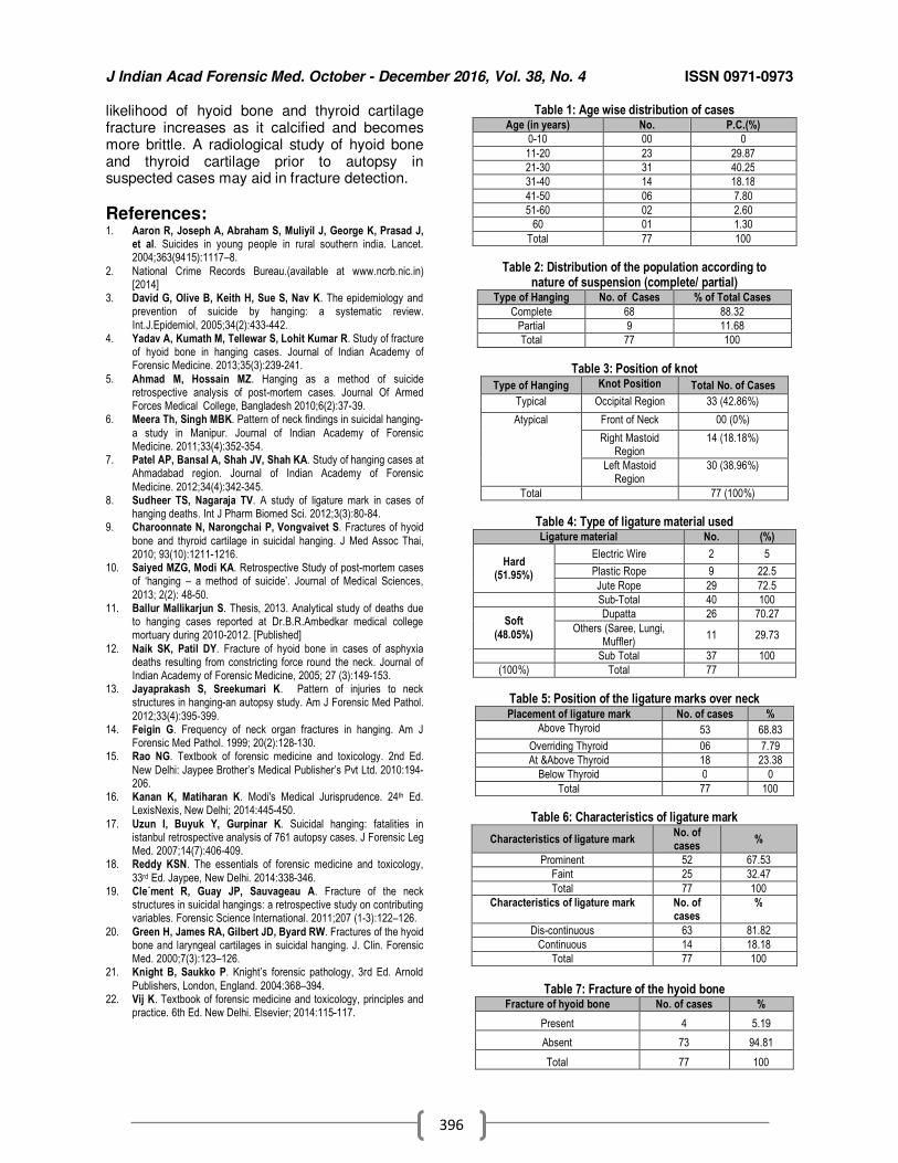
Observations and Results: There were 3307 cases of post-mortem examinations conducted during the study period and among those 77 cases were hanging i.e. 2.32 %. The most vulnerable age for hanging was observed to be between 21-30 years followed by age group 11-20 years. In the age group more than 50 years only few cases had been reported. No case found within 10 year age group. (Table:1) In maximum cases the nature of suspension was complete type, where as partial type of hanging was few in number. (Table:2) In maximum cases the position of Knot was present at occipital region of the neck, followed by left mastoid region of the neck least over right mastoid region of the neck. There was not a single case found where the position of knot at the front of the neck. (Table:3) Considering the information gathered from the police records and from the relatives of the deceased and taking the examination findings of the ligature material whenever it has been sent along with the dead body, it was observed that the maximum victims used hard ligature material like jute rope, plastic rope, and electric wire. While, in soft ligatures (48%), Dupatta, Saree, Muffler and Lungi were used. This showed that most commonly used ligature among hard ligature material was the Jute Rope, whereas Dupatta was the most commonly used among soft ligature material.(Table:4) It was observed from Table 5 that in 69% cases, the ligature mark was above the thyroid cartilage and in about 23% cases, the ligature mark was at or below the thyroid cartilage. Only 6 cases in which, the ligature mark was at overriding the thyroid cartilage
(about 8%). In majority of case the ligature mark is prominent (67.53%) and discontinuous (81.82%).(Table:6) It was observed that the hyoid bone fracture was found in only 4 cases (5.19%). (Table:7) The findings of the present study showed that there was not a single case amongst total cases studied in which the thyroid cartilage fractures was present.
Discussion: The present study was conducted to analyse the frequency of hyoid and thyroid fractures in hanging in relation to the ligature material, position of the knot, age etc. A study showed that the largest group was found to be 21-30 years, followed by 11-20 years and 31- 40 years, respectively.[4] These findings are very much similar to our findings. More previous studies have also reported similar results, with 21-30 years age group being the most commonly involved by different other authors.[5,6,7] The above findings can easily be explained by the fact that 21-30 years of age group is most susceptible to frustration in life because of many factors like stressful marital life, dowry, financial crunch, failure of love affairs, and pressure of making a good career after completion of studies etc. In present study it was found that in the maximum cases, the nature of suspension was complete type, 88.3%, where as partial type of hanging was in few cases, 11.7%. The present findings are comparable with those of the other authors - complete hanging cases were about 64% and partial about 36%;[8] complete hanging in about 99% of cases studied;[7] about 88% complete and 12 % incomplete.[6] Authors from Thailand observed higher number of incomplete hanging cases, (55%) as compared to complete hanging cases, (45%).[9] Hanging also differed with respect to being typical/ atypical. In the present study, typical hanging was seen in about 43% and atypical in 57% of cases. There are some other studies reported similar findings - atypical hanging in about 97% and only 3% were typical of total cases studied.[7]; in about 89% cases the hanging was atypical and in 11% it was typical,[8] and the atypical hanging was seen in about 96% of the cases and typical hanging in only 4%.[6] Similar findings were also reported by the various authors from different countries that about 82% cases showed right or left sided knot that indicate atypical hanging while only 13 cases about 18% showed knot was located on nape of neck that indicates typical hanging.[10]
J Indian Acad Forensic Med. October - December 2016, Vol. 38, No. 4 ISSN 0971-0973
395
In the present study maximum victims used hard ligature, (52%) like jute rope and soft ligature like Dupatta (48%); while others reported that the most common ligature material used was Dupatta (Soft) in 40 (54%) cases, followed by Nylon rope (Hard) in 18 (24.3%) cases.[10] Similarly, another study reported that 57% used cloth as a ligature material which was either scarf, towel, Lungi etc (Soft) and about 43% were used ropes - nylon or jute ( Hard).[6] In our study, in 69% cases, the ligature mark was above the thyroid cartilage. These are comparable with those of others like; 83% the ligature mark was above the thyroid cartilage, followed by about 12% overriding the thyroid cartilage and about 5% below the thyroid cartilage;[11] in about 88% cases the mark was present above thyroid cartilage, in 10% it was present over the thyroid cartilage and in 2% it was present below the thyroid cartilage.[8] Similar observation was made by another author that in about 62% of cases where the ligature mark was above the thyroid cartilage followed by in about 20% cases over the thyroid cartilage, about 13% on and above the thyroid cartilage and about 5% cases below the thyroid cartilage.[12] The present study showed that the in majority of cases, the ligature mark was prominent (67.5%) and discontinuous (81.8%). Similar observations were observed by others.[8,11] Again, of 77 cases in our study, fracture of hyoid bone was present in only 5.2% cases, all on the unilateral Greater Cornu. Comparable findings are reported by authors in 1.6% of cases,[4] in 4% of total cases,[8] and in about 4% of cases,[11] in 2.7%,[13] in 3.6%;[6] while other reported the incidence of fracture of hyoid bone was nil in hanging cases in their study.[7,12] There are some studies conducted in other countries and their observation are comparable to our findings - 3.2% cases to be having fractures.[14] According to various authors, the hyoid bone was intact in 90-95% cases of hanging.[15] One author opined that due to direct lateral compression of the neck, fractures of hyoid bone are rare.[16] In contrast, there are some other studies that have reported higher incidence of hyoid bone fracture among the hanging cases.[5,9,17,18] They have mentioned about the variation of incidence of fractures of hyoid bone from 0-60%, with an average being 15-20% cases. In majority of cases, isolated hyoid bone fractures were in about 12% and combined thyroid cartilage and hyoid bone fracture was 7% in hanging cases.[19] In a prospective study of 40 cases of
suicidal hanging in Australia the hyoid bone and/or thyroid cartilage fractures in 47.5%.[20] Opinion varies regarding the frequency of fracture of the hyoid bone. Estimates range from 0 to 60%, but the average is 15 to 20%. Fractures are rare below 40 years because of the elasticity of the cartilage and mobility of the joints. The fracture is common in persons above 40 years and involves the great horns, at the junction of inner two-thirds and outer one-third. Hyoid bone is a U-shaped structure and lies at the root of the tongue. The bone haves a central body, two greater horns which sweep backwards and upwards and two lesser horns on the upper surface of the body that have no forensic anatomical significance. The bone is having natural joints between the body and the greater horns.[21] It is calcified at variable times: the body is usually calcified, but the horns may calcify irregularly, both in space and time. In teenagers and young adults they are usually cartilaginous and the joints mobile. In middle and later life, the hyoid and thyroid horns calcify and become more brittle. These natural joints may be mistaken as fractures, if dissection is not done meticulously. There is also the possibility of fractures being post-mortem, due to incorrect autopsy techniques, inexperienced forensic pathologist, body transit trauma, improper handling in the mortuary etc. [21,22] There was not a single case amongst total cases studied in which the fracture of thyroid cartilage was found. Similar findings are also reported by other in their study conducted at Ahmedabad.[7] However, in another study it was found that thyroid cartilage were fractured in a small percentage of cases (5.3%).[13] In contrast, there are some other studies which have reported higher incidence of thyroid cartilage fractures in hanging cases. According to one author, the cases of isolated thyroid cartilage fractures were about 15% and combined thyroid cartilage and hyoid bone fracture was 7% in hanging cases.[19] Thyroid cartilage fracture is dependent on the age of calcification, type of ligature, position of ligature over neck, nature of suspension etc.
Conclusion: Hyoid fracture was found in only 5.19% cases studied in this series and there was no case detected with thyroid cartilage fracture. With majority of the cases studied, the age group were young, the two structures so mentioned are not yet calcified and are still quite flexible and likely to be able to withstand compression. As the age increases, the
J Indian Acad Forensic Med. October - December 2016, Vol. 38, No. 4 ISSN 0971-0973
396
likelihood of hyoid bone and thyroid cartilage fracture increases as it calcified and becomes more brittle. A radiological study of hyoid bone and thyroid cartilage prior to autopsy in suspected cases may aid in fracture detection.
References: 1. Aaron R, Joseph A, Abraham S, Muliyil J, George K, Prasad J,
et al. Suicides in young people in rural southern india. Lancet. 2004;363(9415):1117–8.
2. National Crime Records Bureau.(available at www.ncrb.nic.in) [2014]
3. David G, Olive B, Keith H, Sue S, Nav K. The epidemiology and prevention of suicide by hanging: a systematic review.
Int.J.Epidemiol, 2005;34(2):433-442. 4. Yadav A, Kumath M, Tellewar S, Lohit Kumar R. Study of fracture
of hyoid bone in hanging cases. Journal of Indian Academy of Forensic Medicine. 2013;35(3):239-241.
5. Ahmad M, Hossain MZ. Hanging as a method of suicide retrospective analysis of post-mortem cases. Journal Of Armed Forces Medical College, Bangladesh 2010;6(2):37-39.
6. Meera Th, Singh MBK. Pattern of neck findings in suicidal hanging-
a study in Manipur. Journal of Indian Academy of Forensic Medicine. 2011;33(4):352-354.
7. Patel AP, Bansal A, Shah JV, Shah KA. Study of hanging cases at Ahmadabad region. Journal of Indian Academy of Forensic
Medicine. 2012;34(4):342-345. 8. Sudheer TS, Nagaraja TV. A study of ligature mark in cases of
hanging deaths. Int J Pharm Biomed Sci. 2012;3(3):80-84. 9. Charoonnate N, Narongchai P, Vongvaivet S. Fractures of hyoid
bone and thyroid cartilage in suicidal hanging. J Med Assoc Thai, 2010; 93(10):1211-1216.
10. Saiyed MZG, Modi KA. Retrospective Study of post-mortem cases of ‘hanging – a method of suicide’. Journal of Medical Sciences,
2013; 2(2): 48-50. 11. Ballur Mallikarjun S. Thesis, 2013. Analytical study of deaths due
to hanging cases reported at Dr.B.R.Ambedkar medical college mortuary during 2010-2012. [Published]
12. Naik SK, Patil DY. Fracture of hyoid bone in cases of asphyxia deaths resulting from constricting force round the neck. Journal of Indian Academy of Forensic Medicine, 2005; 27 (3):149-153.
13. Jayaprakash S, Sreekumari K. Pattern of injuries to neck structures in hanging-an autopsy study. Am J Forensic Med Pathol.
2012;33(4):395-399. 14. Feigin G. Frequency of neck organ fractures in hanging. Am J
Forensic Med Pathol. 1999; 20(2):128-130. 15. Rao NG. Textbook of forensic medicine and toxicology. 2nd Ed.
New Delhi: Jaypee Brother’s Medical Publisher’s Pvt Ltd. 2010:194-206.
16. Kanan K, Matiharan K. Modi's Medical Jurisprudence. 24th Ed. LexisNexis, New Delhi; 2014:445-450.
17. Uzun I, Buyuk Y, Gurpinar K. Suicidal hanging: fatalities in istanbul retrospective analysis of 761 autopsy cases. J Forensic Leg Med. 2007;14(7):406-409.
18. Reddy KSN. The essentials of forensic medicine and toxicology,
33rd Ed. Jaypee, New Delhi. 2014:338-346. 19. Cle´ment R, Guay JP, Sauvageau A. Fracture of the neck
structures in suicidal hangings: a retrospective study on contributing variables. Forensic Science International. 2011;207 (1-3):122–126.
20. Green H, James RA, Gilbert JD, Byard RW. Fractures of the hyoid bone and laryngeal cartilages in suicidal hanging. J. Clin. Forensic Med. 2000;7(3):123–126.
21. Knight B, Saukko P. Knight’s forensic pathology, 3rd Ed. Arnold
Publishers, London, England. 2004:368–394. 22. Vij K. Textbook of forensic medicine and toxicology, principles and
practice. 6th Ed. New Delhi. Elsevier; 2014:115-117.
Table 1: Age wise distribution of cases Age (in years) No. P.C.(%)
0-10 00 0
11-20 23 29.87
21-30 31 40.25
31-40 14 18.18
41-50 06 7.80
51-60 02 2.60
60 01 1.30
Total 77 100
Table 2: Distribution of the population according to nature of suspension (complete/ partial)
Type of Hanging No. of Cases % of Total Cases
Complete 68 88.32
Partial 9 11.68
Total 77 100
Table 3: Position of knot
Type of Hanging Knot Position Total No. of Cases
Typical Occipital Region 33 (42.86%)
Atypical Front of Neck 00 (0%)
Right Mastoid Region
14 (18.18%)
Left Mastoid Region
30 (38.96%)
Total 77 (100%)
Table 4: Type of ligature material used Ligature material No. (%)
Hard (51.95%)
Electric Wire 2 5
Plastic Rope 9 22.5
Jute Rope 29 72.5
Sub-Total 40 100
Soft
(48.05%)
Dupatta 26 70.27
Others (Saree, Lungi, Muffler)
11 29.73
Sub Total 37 100
(100%) Total 77
Table 5: Position of the ligature marks over neck Placement of ligature mark No. of cases %
Above Thyroid 53 68.83
Overriding Thyroid 06 7.79
At &Above Thyroid 18 23.38
Below Thyroid 0 0
Total 77 100
Table 6: Characteristics of ligature mark
Characteristics of ligature mark No. of cases
%
Prominent 52 67.53
Faint 25 32.47
Total 77 100
Characteristics of ligature mark No. of cases
%
Dis-continuous 63 81.82
Continuous 14 18.18
Total 77 100
Table 7: Fracture of the hyoid bone
Fracture of hyoid bone No. of cases %
Present 4 5.19
Absent 73 94.81
Total 77 100
J Indian Acad Forensic Med. October - December 2016, Vol. 38, No. 4 ISSN 0971-0973
397
Original Research Paper
A Prospective Study of Poisoning Cases due to Paraquat at a Tertiary Care Centre - Chennai
1J James Rajesh,
2J Gerard Rakesh,
3U Jagdish Kamal Chander,
4P Sampath Kumar
Abstract Drugs and chemicals are a great danger to human lives but most of the poisoning in our country
is due to pesticides, herbicides and insecticides. This paper is a study conducted in the department of Forensic Medicine and Toxicology in Sri Ramachandra Medical College and Research Institute, Chennai, from June 2014- June 2015. All cases of poisoning were analysed, and in particular, Paraquat was analysed according to socio-demographic pattern, target organ damage, histo-pathological changes and forensic science report. 6 deaths were reported out of 12 paraquat cases which accounts to 50% fatality. Paraquat, which has both corrosive as well as systemic effects, causes rapid death a result of which, it is considered more dangerous and fatal when compared to other agricultural poisons. When compared to other studies, the incidence of paraquat has increased drastically in recent days, which in turn increases the number of fatalities.
Key Words: Poison, Paraquat, Multi organ damage, Fatalities.
Introduction: Poison is a substance which has deleterious effect on living organisms and produces ill health or death by direct contact or by absorption in the body. India as such a agricultural country largely depends upon agriculture for its major income.[1] Today, India ranks second worldwide in farm output. Many pesticides like insecticides and herbicides are used widely by them for better results. The availability of it to the farmers is a must for their day to day use, which makes it very easily accessible. Paraquat and diquat are widely used herbicides which belong to the bipyridyl group. The chemical formula for paraquat is [(C6 H7 N)2]CL2. It is classified as a viologen, a family of
Corresponding Author: 1Assistant Professor,
Department of Forensic Medicine and Toxicology, 2Assistant Professor,
Department of Microbiology, Sri Venkateshwaraa Medical College Hospital and Research Centre, Ariyur, Puducherry. 3Assistant Professor;
4Professor and Head,
Department of Forensic Medicine and Toxicology, Sri Ramachandra Medical College and Research Institute, Porur, Chennai Email: [email protected] DOR: 06/02/2016 DOA: 25/12/2016 DOI: 10.5958/0974-0848.2016.00099.3
redox – active heterocycles of similar structure. The salt is one of the most widely used herbicide. It is available either in granular form or as water soluble concentrates which is a odourless brown liquid.[2] In India, most of the concentrates of paraquat are available as 10% - 20% solutions and 10ml of 20% solutions contain about 2gms of paraquat. It is a highly toxic weed killer, once promoted in foreign countries, and now it is classified as a “restricted commercial use” and people must obtain a license to the use of this product. When compared to Organophosphorus Compounds (OPCs) and Aluminium Phosphides, which are the commonest agricultural poisons consumed in India, it is much more dangerous.[2]
We carried out this study for its severity and to evaluate the various features of the it.
Materials and Methods: This study was carried out in the
department of Forensic Medicine and Toxicology, Sri Ramachandra Medical College and Research Institute, Chennai. All the cases of alleged paraquat poisoning that reported for a period of one year between June 2014 – June 2015 were considered for this study. The study included cases which were directly admitted in the hospital and also which were referred from other hospitals as well, with a similar history.
Detailed information regarding the age, sex, occupation, educational status, socio – economic status, marital status, family pattern, time of intake, evidence produced for intake, manner of poisoning, cause of consumption,
J Indian Acad Forensic Med. October - December 2016, Vol. 38, No. 4 ISSN 0971-0973
398
final outcome, target organ damage, histopathological changes and forensic science report were analysed.
Results: Epidemiological profile
Of a total number of 353 poisoning cases that reported, 12 were of paraquat poisoning, which accounted to 3.4%. When compared to previous year’s studies, the incidence had increased three folds. This shows that the abuse of the herbicide has increased a lot.
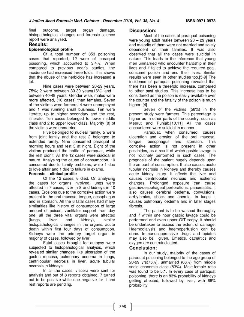
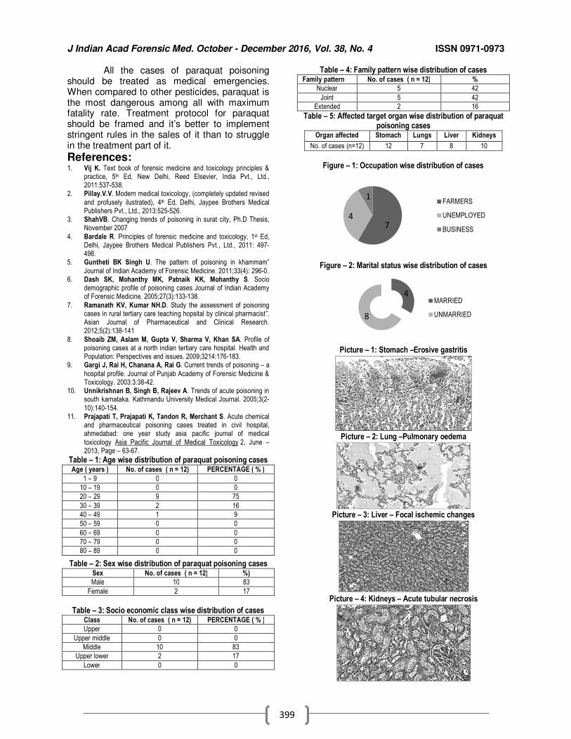
Nine cases were between 20-29 years, 75%; 2 were between 30-39 years(16%) and 1 between 40-49 years. Gender wise, males were more affected, (10 cases) than females. Seven of the victims were farmers, 4 were unemployed and 1 was running small business. Ten were literate, up to higher secondary and the rest, illiterate. Ten cases belonged to lower middle class and 2 to upper lower class. Majority (8) of the victims were unmarried.
Five belonged to nuclear family, 5 were from joint family and the rest 2 belonged to extended family. Nine consumed paraquat at morning hours and rest 3 at night. Eight of the victims produced the bottle of paraquat, while the rest didn’t. All the 12 cases were suicidal in nature. Analysing the cause of consumption, 10 consumed due to family problems, while 1 due to love affair and 1 due to failure in exams. Forensic – clinical profile
Of the 12 cases, 6 died. On analysing the cases for organs involved, lungs were affected in 7 cases, liver in 8 and kidneys in 10 cases. Erosions due to the corrosive action were present in the oral mucosa, tongue, oesophagus and in stomach. All the 6 fatal cases had many similarities like history of consumption of large amount of poison, ventilator support from day one, all the three vital organs were affected (lungs, liver and kidney), similar histopathological changes in the organs, acute death within first four days of consumption. Kidneys were the primary target organ in majority of cases, followed by liver.
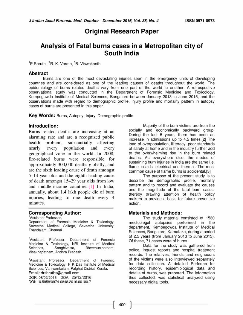
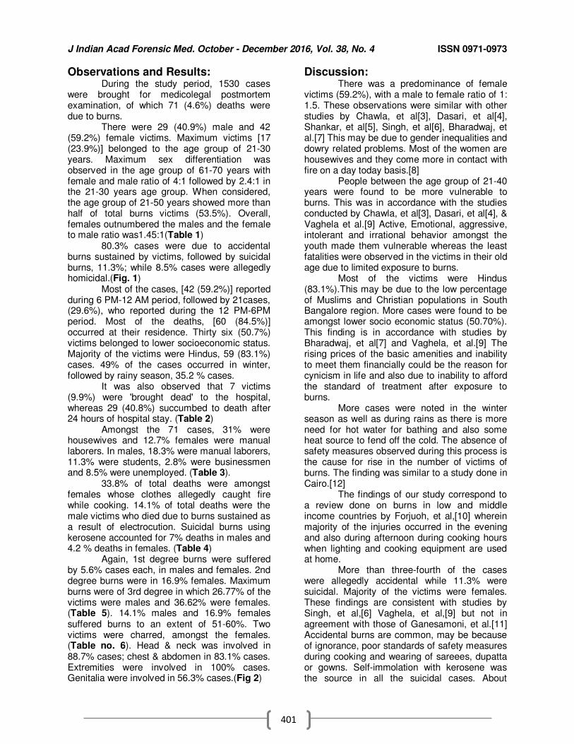
Fatal cases brought for autopsy were subjected to histopahological analysis, which revealed similar changes like ulceration of the gastric mucosa, pulmonary oedema in lungs, centrilobular necrosis in liver, acute tubular necrosis in kidneys.
In all the cases, viscera were sent for analysis and out of 8 reports obtained, 7 turned out to be positive while one negative for it and rest reports are pending.
Discussion: Most of the cases of paraquat poisoning
were young adult males between 20 – 29 years and majority of them were not married and solely dependent on their families. It was also observed that all the cases were suicidal in nature. This leads to the inference that young men unmarried who encounter hardship in their lives and if failed to achieve the required goal, consume poison and end their lives. Similar results were seen in other studies too.[5-9] The incidence of paraquat poisoning revealed that there has been a threefold increase, compared to other past studies. This increase has to be considered as the poison is easily available over the counter and the fatality of the poison is much higher. [4]
Seven of the victims (58%) in the present study were farmers. This percentage is higher as in other parts of the country, such as Meerut and Punjab.[10,11] All the cases encountered were suicidal in manner.
Paraquat, when consumed, causes ulceration and erosion of the oral mucosa, tongue, oesophagus and stomach. This corrosive action is not present in other pesticides, as a result of which gastric lavage is not routinely performed in such cases. The prognosis of the patient hugely depends upon the amount of consumption. It also causes acute tubular necrosis in kidneys and thereby causes acute kidney injury. It affects the liver and causes centrilobular necrosis and ischemic changes. Prolonged exposure can cause gastric/oesophageal perforations, pancreatitis. It also causes cerebral oedema, convulsions, arrhythmias, shock and anemia. In lungs it causes pulmonary oedema and in later stages fibrosis.
The patient is to be washed thoroughly and if within one hour gastric lavage could be performed and even upper GIT scopy, it should be undertaken to assess the extent of damage. Haemodialysis and haemoperfusion can be done. Immunosuppressive drugs and opiates may also be given. Emetics, cathartics and oxygen are contraindicated.
Conclusion: In our study, majority of the cases of
paraquat poisoning belonged to the age group of 20-29 yrs(75%), unmarried (66%) from middle socio economic class (83%). Male-female ratio was found to be 5:1. In every case of paraquat poisoning, there is an 83% probability of kidneys getting affected, followed by liver, with 66% probability.
J Indian Acad Forensic Med. October - December 2016, Vol. 38, No. 4 ISSN 0971-0973
399
All the cases of paraquat poisoning should be treated as medical emergencies. When compared to other pesticides, paraquat is the most dangerous among all with maximum fatality rate. Treatment protocol for paraquat should be framed and it’s better to implement stringent rules in the sales of it than to struggle in the treatment part of it.
References: 1. Vij K. Text book of forensic medicine and toxicology principles &
practice, 5th Ed, New Delhi, Reed Elsevier, India Pvt., Ltd., 2011:537-538.
2. Pillay.V.V. Modern medical toxicology, (completely updated revised
and profusely ilustrated), 4th Ed, Delhi, Jaypee Brothers Medical Publishers Pvt., Ltd., 2013:525-526.
3. ShahVB. Changing trends of poisoning in surat city, Ph.D Thesis, November 2007
4. Bardale R. Principles of forensic medicine and toxicology, 1st Ed, Delhi, Jaypee Brothers Medical Publishers Pvt., Ltd., 2011: 497-498.
5. Guntheti BK Singh U. The pattern of poisoning in khammam”
Journal of Indian Academy of Forensic Medicine. 2011;33(4): 296-0. 6. Dash SK, Mohanthy MK, Patnaik KK, Mohanthy S. Socio
demographic profile of poisoning cases Journal of Indian Academy of Forensic Medicine. 2005;27(3):133-138.
7. Ramanath KV, Kumar NH.D. Study the assessment of poisoning cases in rural tertiary care teaching hopsital by clinical pharmacist”. Asian Journal of Pharmaceutical and Clinical Research. 2012;5(2):138-141
8. Shoaib ZM, Aslam M, Gupta V, Sharma V, Khan SA. Profile of poisoning cases at a north indian tertiary care hospital. Health and Population: Perspectives and issues. 2009;3214:176-183.
9. Gargi J, Rai H, Chanana A, Rai G. Current trends of poisoning – a
hospital profile. Journal of Punjab Academy of Forensic Medicine & Toxicology. 2003:3:38-42.
10. Unnikrishnan B, Singh B, Rajeev A. Trends of acute poisoning in south karnataka. Kathmandu University Medical Journal. 2005;3(2-
10):140-154. 11. Prajapati T, Prajapati K, Tandon R, Merchant S. Acute chemical
and pharmaceutical poisoning cases treated in civil hospital, ahmedabad: one year study asia pacific journal of medical
toxicology Asia Pacific Journal of Medical Toxicology 2, June – 2013, Page – 63-67.
Table – 1: Age wise distribution of paraquat poisoning cases Age ( years ) No. of cases ( n = 12) PERCENTAGE ( % )
1 – 9 0 0
10 – 19 0 0
20 – 29 9 75
30 – 39 2 16
40 – 49 1 9
50 – 59 0 0
60 – 69 0 0
70 – 79 0 0
80 – 89 0 0
Table – 2: Sex wise distribution of paraquat poisoning cases Sex No. of cases ( n = 12) %)
Male 10 83
Female 2 17
Table – 3: Socio economic class wise distribution of cases
Class No. of cases ( n = 12) PERCENTAGE ( % )
Upper 0 0
Upper middle 0 0
Middle 10 83
Upper lower 2 17
Lower 0 0
Table – 4: Family pattern wise distribution of cases
Family pattern No. of cases ( n = 12) %
Nuclear 5 42
Joint 5 42
Extended 2 16
Table – 5: Affected target organ wise distribution of paraquat poisoning cases
Organ affected Stomach Lungs Liver Kidneys
No. of cases (n=12) 12 7 8 10
Figure – 1: Occupation wise distribution of cases
Figure – 2: Marital status wise distribution of cases
Picture – 1: Stomach –Erosive gastritis
Picture – 2: Lung –Pulmonary oedema
Picture – 3: Liver – Focal ischemic changes
Picture – 4: Kidneys – Acute tubular necrosis
74
1FARMERS
UNEMPLOYED
BUSINESS
4
8
MARRIED
UNMARRIED
J Indian Acad Forensic Med. October - December 2016, Vol. 38, No. 4 ISSN 0971-0973
400
Original Research Paper
Analysis of Fatal burns cases in a Metropolitan city of South India
1P.Shruthi,
2R. K. Varma,
3B. Viswakanth
Abstract Burns are one of the most devastating injuries seen in the emergency units of developing
countries and are considered as one of the leading causes of deaths throughout the world. The epidemiology of burns related deaths vary from one part of the world to another. A retrospective observational study was conducted in the Department of Forensic Medicine and Toxicology, Kempegowda Institute of Medical Sciences, Bangalore between January 2013 to June 2015, and the observations made with regard to demographic profile, injury profile and mortality pattern in autopsy cases of burns are presented in this paper.
Key Words: Burns, Autopsy, Injury, Demographic profile
Introduction: Burns related deaths are increasing at an
alarming rate and are a recognized public
health problem, substantially affecting
nearly every population and every
geographical zone in the world. In 2008,
fire-related burns were responsible for
approximately 300,000 deaths globally, and
are the sixth leading cause of death amongst
5–14 year olds and the eighth leading cause
of death amongst 15–29 year olds from low
and middle-income countries.[1] In India,
annually, about 1.4 lakh people die of burn
injuries, leading to one death every 4
minutes.
Corresponding Author: 1Assistant Professor,
Department of Forensic Medicine & Toxicology, Saveetha Medical College, Saveetha University, Thandalam, Chennai.
2Assistant Professor, Department of Forensic
Medicine & Toxicology, NRI Institute of Medical Sciences, Sanghivalsa, Bheemunipatnam, Visakhapatnam, Andhra Pradesh.
3Assistant Professor, Department of Forensic
Medicine & Toxicology, P K Das Institute of Medical Sciences, Vaniyamkulam, Palghat District, Kerala.
Email: [email protected] DOR: 08/02/2016 DOA: 25/12/2016 DOI: 10.5958/0974-0848.2016.00100.7
Majority of the burn victims are from the socially and economically backward group. During the last 5 years, there has been an increase in admissions up to 4.5 times.[2] The load of overpopulation, illiteracy, poor standards of safety at home and in the industry further add to the overwhelming rise in the burn related deaths. As everywhere else, the modes of sustaining burn injuries in India are the same i.e. flame, scalds, electrical and thermal. The most common cause of flame burns is accidental.[3] The purpose of the present study is to describe the demographic profile, mortality pattern and to record and evaluate the causes and the magnitude of the fatal burn cases, thereby drawing attention of health policy makers to provide a basis for future preventive action.
Materials and Methods: The study material consisted of 1530 medicolegal autopsies performed in the department, Kempegowda Institute of Medical Sciences, Bangalore, Karnataka, during a period of 2.5 years (from January 2013 to June 2015). Of these, 71 cases were of burns. Data for the study was gathered from police, inquest reports and hospital treatment records. The relatives, friends, and neighbours of the victims were also interviewed separately for data collection. A detailed Performa for recording history, epidemiological data and details of burns, was prepared. The information thus collected, was statistical analyzed using necessary digital tools.
J Indian Acad Forensic Med. October - December 2016, Vol. 38, No. 4 ISSN 0971-0973
401
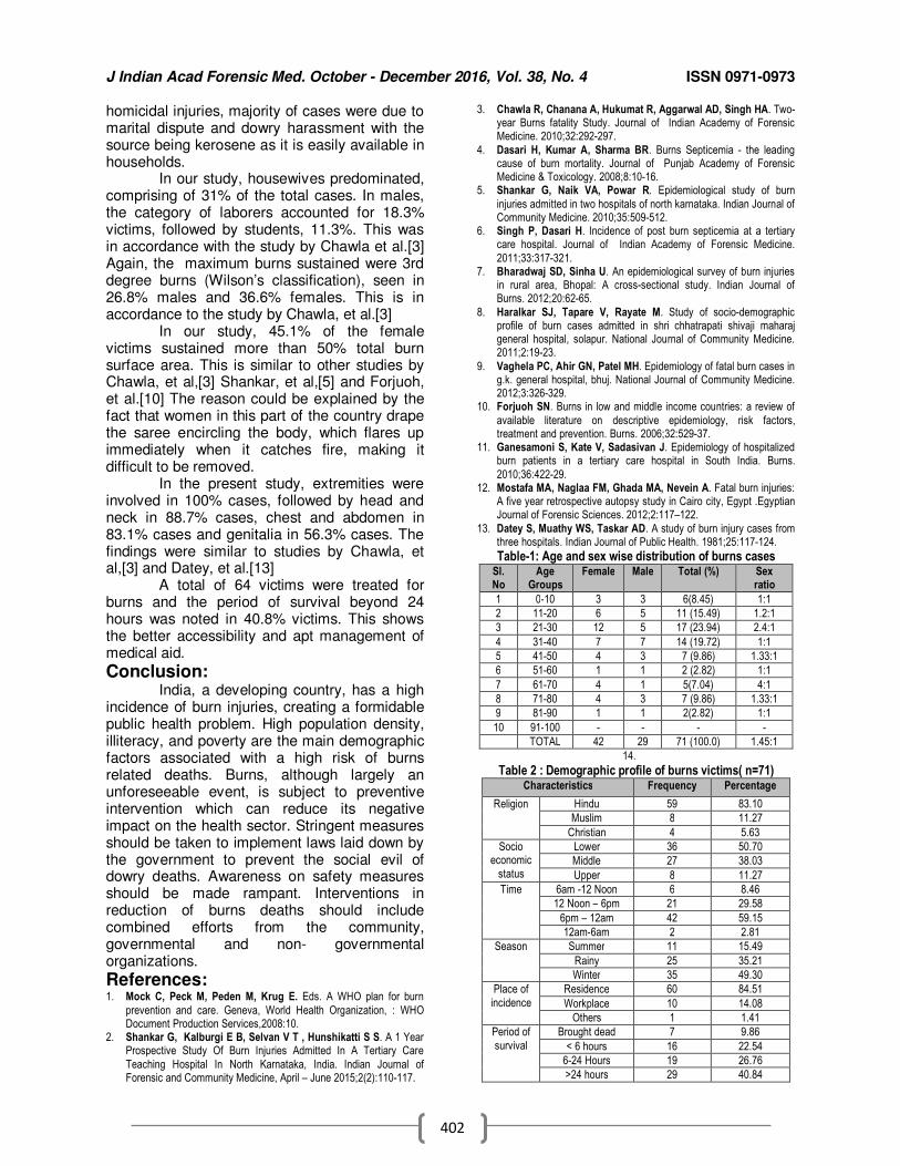
Observations and Results: During the study period, 1530 cases were brought for medicolegal postmortem examination, of which 71 (4.6%) deaths were due to burns. There were 29 (40.9%) male and 42 (59.2%) female victims. Maximum victims [17 (23.9%)] belonged to the age group of 21-30 years. Maximum sex differentiation was observed in the age group of 61-70 years with female and male ratio of 4:1 followed by 2.4:1 in the 21-30 years age group. When considered, the age group of 21-50 years showed more than half of total burns victims (53.5%). Overall, females outnumbered the males and the female to male ratio was1.45:1(Table 1) 80.3% cases were due to accidental burns sustained by victims, followed by suicidal burns, 11.3%; while 8.5% cases were allegedly homicidal.(Fig. 1) Most of the cases, [42 (59.2%)] reported during 6 PM-12 AM period, followed by 21cases, (29.6%), who reported during the 12 PM-6PM period. Most of the deaths, [60 (84.5%)] occurred at their residence. Thirty six (50.7%) victims belonged to lower socioeconomic status. Majority of the victims were Hindus, 59 (83.1%) cases. 49% of the cases occurred in winter, followed by rainy season, 35.2 % cases. It was also observed that 7 victims (9.9%) were 'brought dead' to the hospital, whereas 29 (40.8%) succumbed to death after 24 hours of hospital stay. (Table 2) Amongst the 71 cases, 31% were housewives and 12.7% females were manual laborers. In males, 18.3% were manual laborers, 11.3% were students, 2.8% were businessmen and 8.5% were unemployed. (Table 3). 33.8% of total deaths were amongst females whose clothes allegedly caught fire while cooking. 14.1% of total deaths were the male victims who died due to burns sustained as a result of electrocution. Suicidal burns using kerosene accounted for 7% deaths in males and 4.2 % deaths in females. (Table 4) Again, 1st degree burns were suffered by 5.6% cases each, in males and females. 2nd degree burns were in 16.9% females. Maximum burns were of 3rd degree in which 26.77% of the victims were males and 36.62% were females. (Table 5). 14.1% males and 16.9% females suffered burns to an extent of 51-60%. Two victims were charred, amongst the females. (Table no. 6). Head & neck was involved in 88.7% cases; chest & abdomen in 83.1% cases. Extremities were involved in 100% cases. Genitalia were involved in 56.3% cases.(Fig 2)
Discussion: There was a predominance of female victims (59.2%), with a male to female ratio of 1: 1.5. These observations were similar with other studies by Chawla, et al[3], Dasari, et al[4], Shankar, et al[5], Singh, et al[6], Bharadwaj, et al.[7] This may be due to gender inequalities and dowry related problems. Most of the women are housewives and they come more in contact with fire on a day today basis.[8] People between the age group of 21-40 years were found to be more vulnerable to burns. This was in accordance with the studies conducted by Chawla, et al[3], Dasari, et al[4], & Vaghela et al.[9] Active, Emotional, aggressive, intolerant and irrational behavior amongst the youth made them vulnerable whereas the least fatalities were observed in the victims in their old age due to limited exposure to burns. Most of the victims were Hindus (83.1%).This may be due to the low percentage of Muslims and Christian populations in South Bangalore region. More cases were found to be amongst lower socio economic status (50.70%). This finding is in accordance with studies by Bharadwaj, et al[7] and Vaghela, et al.[9] The rising prices of the basic amenities and inability to meet them financially could be the reason for cynicism in life and also due to inability to afford the standard of treatment after exposure to burns. More cases were noted in the winter season as well as during rains as there is more need for hot water for bathing and also some heat source to fend off the cold. The absence of safety measures observed during this process is the cause for rise in the number of victims of burns. The finding was similar to a study done in Cairo.[12] The findings of our study correspond to a review done on burns in low and middle income countries by Forjuoh, et al,[10] wherein majority of the injuries occurred in the evening and also during afternoon during cooking hours when lighting and cooking equipment are used at home. More than three-fourth of the cases were allegedly accidental while 11.3% were suicidal. Majority of the victims were females. These findings are consistent with studies by Singh, et al,[6] Vaghela, et al,[9] but not in agreement with those of Ganesamoni, et al.[11] Accidental burns are common, may be because of ignorance, poor standards of safety measures during cooking and wearing of sareees, dupatta or gowns. Self-immolation with kerosene was the source in all the suicidal cases. About
J Indian Acad Forensic Med. October - December 2016, Vol. 38, No. 4 ISSN 0971-0973
402
homicidal injuries, majority of cases were due to marital dispute and dowry harassment with the source being kerosene as it is easily available in households. In our study, housewives predominated, comprising of 31% of the total cases. In males, the category of laborers accounted for 18.3% victims, followed by students, 11.3%. This was in accordance with the study by Chawla et al.[3] Again, the maximum burns sustained were 3rd degree burns (Wilson’s classification), seen in 26.8% males and 36.6% females. This is in accordance to the study by Chawla, et al.[3] In our study, 45.1% of the female victims sustained more than 50% total burn surface area. This is similar to other studies by Chawla, et al,[3] Shankar, et al,[5] and Forjuoh, et al.[10] The reason could be explained by the fact that women in this part of the country drape the saree encircling the body, which flares up immediately when it catches fire, making it difficult to be removed. In the present study, extremities were involved in 100% cases, followed by head and neck in 88.7% cases, chest and abdomen in 83.1% cases and genitalia in 56.3% cases. The findings were similar to studies by Chawla, et al,[3] and Datey, et al.[13] A total of 64 victims were treated for burns and the period of survival beyond 24 hours was noted in 40.8% victims. This shows the better accessibility and apt management of medical aid.
Conclusion: India, a developing country, has a high incidence of burn injuries, creating a formidable public health problem. High population density, illiteracy, and poverty are the main demographic factors associated with a high risk of burns related deaths. Burns, although largely an unforeseeable event, is subject to preventive intervention which can reduce its negative impact on the health sector. Stringent measures should be taken to implement laws laid down by the government to prevent the social evil of dowry deaths. Awareness on safety measures should be made rampant. Interventions in reduction of burns deaths should include combined efforts from the community, governmental and non- governmental organizations.
References: 1. Mock C, Peck M, Peden M, Krug E. Eds. A WHO plan for burn
prevention and care. Geneva, World Health Organization, : WHO Document Production Services,2008:10.
2. Shankar G, Kalburgi E B, Selvan V T , Hunshikatti S S. A 1 Year Prospective Study Of Burn Injuries Admitted In A Tertiary Care
Teaching Hospital In North Karnataka, India. Indian Journal of Forensic and Community Medicine, April – June 2015;2(2):110-117.
3. Chawla R, Chanana A, Hukumat R, Aggarwal AD, Singh HA. Two-year Burns fatality Study. Journal of Indian Academy of Forensic Medicine. 2010;32:292-297.
4. Dasari H, Kumar A, Sharma BR. Burns Septicemia - the leading cause of burn mortality. Journal of Punjab Academy of Forensic Medicine & Toxicology. 2008;8:10-16.
5. Shankar G, Naik VA, Powar R. Epidemiological study of burn
injuries admitted in two hospitals of north karnataka. Indian Journal of Community Medicine. 2010;35:509-512.
6. Singh P, Dasari H. Incidence of post burn septicemia at a tertiary care hospital. Journal of Indian Academy of Forensic Medicine.
2011;33:317-321. 7. Bharadwaj SD, Sinha U. An epidemiological survey of burn injuries
in rural area, Bhopal: A cross-sectional study. Indian Journal of Burns. 2012;20:62-65.
8. Haralkar SJ, Tapare V, Rayate M. Study of socio-demographic profile of burn cases admitted in shri chhatrapati shivaji maharaj general hospital, solapur. National Journal of Community Medicine. 2011;2:19-23.
9. Vaghela PC, Ahir GN, Patel MH. Epidemiology of fatal burn cases in g.k. general hospital, bhuj. National Journal of Community Medicine. 2012;3:326-329.
10. Forjuoh SN. Burns in low and middle income countries: a review of
available literature on descriptive epidemiology, risk factors, treatment and prevention. Burns. 2006;32:529-37.
11. Ganesamoni S, Kate V, Sadasivan J. Epidemiology of hospitalized burn patients in a tertiary care hospital in South India. Burns.
2010;36:422-29. 12. Mostafa MA, Naglaa FM, Ghada MA, Nevein A. Fatal burn injuries:
A five year retrospective autopsy study in Cairo city, Egypt .Egyptian Journal of Forensic Sciences. 2012;2:117–122.
13. Datey S, Muathy WS, Taskar AD. A study of burn injury cases from three hospitals. Indian Journal of Public Health. 1981;25:117-124.
Table-1: Age and sex wise distribution of burns cases Sl. No
Age Groups
Female Male Total (%) Sex ratio
1 0-10 3 3 6(8.45) 1:1
2 11-20 6 5 11 (15.49) 1.2:1
3 21-30 12 5 17 (23.94) 2.4:1
4 31-40 7 7 14 (19.72) 1:1
5 41-50 4 3 7 (9.86) 1.33:1
6 51-60 1 1 2 (2.82) 1:1
7 61-70 4 1 5(7.04) 4:1
8 71-80 4 3 7 (9.86) 1.33:1
9 81-90 1 1 2(2.82) 1:1
10 91-100 - - - -
TOTAL 42 29 71 (100.0) 1.45:1
14.
Table 2 : Demographic profile of burns victims( n=71) Characteristics Frequency Percentage
Religion Hindu 59 83.10
Muslim 8 11.27
Christian 4 5.63
Socio economic
status
Lower 36 50.70
Middle 27 38.03
Upper 8 11.27
Time 6am -12 Noon 6 8.46
12 Noon – 6pm 21 29.58
6pm – 12am 42 59.15
12am-6am 2 2.81
Season Summer 11 15.49
Rainy 25 35.21
Winter 35 49.30
Place of
incidence
Residence 60 84.51
Workplace 10 14.08
Others 1 1.41
Period of
survival
Brought dead 7 9.86
< 6 hours 16 22.54
6-24 Hours 19 26.76
>24 hours 29 40.84
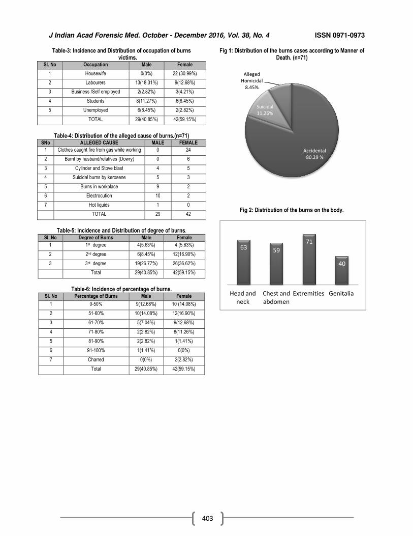
J Indian Acad Forensic Med. October - December 2016, Vol. 38, No. 4 ISSN 0971-0973
403
Table-3: Incidence and Distribution of occupation of burns victims.
Sl. No Occupation Male Female
1 Housewife 0(0%) 22 (30.99%)
2 Labourers 13(18.31%) 9(12.68%)
3 Business /Self employed 2(2.82%) 3(4.21%)
4 Students 8(11.27%) 6(8.45%)
5 Unemployed 6(8.45%) 2(2.82%)
TOTAL 29(40.85%) 42(59.15%)
Table-4: Distribution of the alleged cause of burns.(n=71)
SNo ALLEGED CAUSE MALE FEMALE
1 Clothes caught fire from gas while working 0 24
2 Burnt by husband/relatives (Dowry) 0 6
3 Cylinder and Stove blast 4 5
4 Suicidal burns by kerosene 5 3
5 Burns in workplace 9 2
6 Electrocution 10 2
7 Hot liquids 1 0
TOTAL 29 42
Table-5: Incidence and Distribution of degree of burns.
Sl. No Degree of Burns Male Female
1 1st degree 4(5.63%) 4 (5.63%)
2 2nd degree 6(8.45%) 12(16.90%)
3 3rd degree 19(26.77%) 26(36.62%)
Total 29(40.85%) 42(59.15%)
Table-6: Incidence of percentage of burns.
Sl. No Percentage of Burns Male Female
1 0-50% 9(12.68%) 10 (14.08%)
2 51-60% 10(14.08%) 12(16.90%)
3 61-70% 5(7.04%) 9(12.68%)
4 71-80% 2(2.82%) 8(11.26%)
5 81-90% 2(2.82%) 1(1.41%)
6 91-100% 1(1.41%) 0(0%)
7 Charred 0(0%) 2(2.82%)
Total 29(40.85%) 42(59.15%)
Fig 1: Distribution of the burns cases according to Manner of Death. (n=71)
Fig 2: Distribution of the burns on the body.
Accidental
80.29 %
Suicidal
11.26%
Alleged
Homicidal
8.45%
63 59
71
40
Head and
neck
Chest and
abdomen
Extremities Genitalia
J Indian Acad Forensic Med. October - December 2016, Vol. 38, No. 4 ISSN 0971-0973
404
Original Research Paper
Pregnancy Related Deaths: A Ten Year Retrospective Study in the Mortuary of a Tertiary Care Teaching Hospital of
Northeast India
1Memchoubi Ph.,
2Th. Meera,
3John Deb Barma,
4Kh. Pradipkumar Singh
Abstract An audit of autopsies of pregnancy related deaths is important for the establishment of accurate
cause of delivery deaths and to determine whether there is existence of negligence or not to the various cases. The present study was aimed at finding out the factors which are associated with pregnancy related deaths that underwent autopsy in the mortuary of a tertiary care teaching hospital of northeast India during the period of 2006 to 2015. Autopsy records with clinical notes were retrieved and histopathology slides were studied to establish the accurate cause of delivery deaths. The variables like age (years), gravidity, pregnancy outcome, obstetrical characteristics, period of gestation, methods of delivery, death of patients after delivery, and place of deaths were used to classify and analyze the data from autopsy. There were altogether 14 cases of pregnancy related deaths and their age ranged from 18 years to 39 years. Of this, 12 were related with delivery. Four died due to shock and haemorrhage caused by uterine atony (28.6%), 4 due to cervical tear (28.6%) which could have been prevented with proper obstetrical procedure. Two died of uterine rupture (14.3%) at 40 weeks and 38 weeks, respectively, indicating a possibility of intervention at about 36 weeks or so. One died due to slippage of ligature (7.1%) and one due to amniotic fluid embolism (7.1%). The remaining 2 were of MTP and a natural death, (bronchopneumonia associated with pregnancy) respectively. Eight died in Hospital, 3 in Nursing home, 2 at home and 1 on the way to hospital. In conclusion, delay in diagnosis, treatment and decision to transfer, delay in transport for reaching to proper hospital and delayed therapy are the main factors these maternal deaths.
Key Words: Pregnancy, death, hospital, negligence, autopsy
Introduction: According to the World Health
Organization (WHO), maternal death is defined as “the death of a woman while pregnant (or) within 42 days of termination of pregnancy, irrespective of the duration & site of pregnancy, from any cause related to (or) aggravated by the pregnancy or its management but not from accidental (or) incidental causes.”[1]
Corresponding Author: 1Associate Professor,
2Professor,
3P.G Student,
4Assistant Professor
Dept. of Forensic Medicine, Regional Institute of Medical Sciences, Imphal E-mail:- [email protected] DOR: 16/04/2016 DOA: 25/12/2016 DOI: 10.5958/0974-0848.2016.00101.9
An estimated five lakh women die as a result of pregnancy each year, worldwide, and approximately one quarter of all pregnancy and delivery related maternal deaths worldwide occur in India. Reasons why woman dies in pregnancy and child birth have many layers. Beside direct, indirect, and co-incidental causes, there are also logistic causes - that is failure in the health care system, lack of transport, lack of manpower and apathy towards patient care.[2] On the other hand, any maternal death is shrouded in mystery. Immediately, the doctor is seen as the culprit. A doctor is considered to be a life saver, but when his alleged negligence has resulted in a physical injury, a mental condition and/death, the Consumer Protection Act (CPA), 1986
3 helps one to dispute a case of
negligence against the doctor. However, the role of contributory negligence from the patient’s side cannot be completely ignored. The aim of the present study was to analyse the pregnancy related deaths that underwent autopsy in the department of Forensic Medicine of a tertiary care teaching hospital of northeast India so as to
J Indian Acad Forensic Med. October - December 2016, Vol. 38, No. 4 ISSN 0971-0973
405
find out the factors which are associated with such deaths.
Materials and Methods: This retrospective study was of a period of 10 years, from 2006 to 2015. The autopsy records with clinical notes were retrieved and histopathology slides were studied to establish the accurate cause of delivery deaths. The variables like age (years), gravidity, pregnancy outcome, obstetrical characteristics, period of gestation, methods of delivery, death of patients after delivery, and place of deaths were used to classify and analyze the data from autopsy.
Results: There were altogether 14 cases of pregnancy related deaths. As shown in Table 1, maximum incidence was seen in > 30-35 yr (28.6%), followed by 15-20yr (21.4%). Of this, 12 (85.7%) were related with delivery and 2 (14.3%) with medical termination of pregnancy (MTP). Regarding foetal outcome, 9 (64.3%) were live-born, 3 (21.4%) were undelivered, 2 (14.3%) were aborted (Fig 1). It is evident from Table 2 that, 8 (57.1%) were primigravida and 6 (42.9%) were multiparous; 57.1% were normal vaginal deliveries (including the MTP cases) and 42.9% were caesarian deliveries. Three cases (21.4%) were home deliveries, while the remaining were hospital and nursing home deliveries. Eight cases (57.1%) died 1 day after delivery, 2 (14.3%) cases each died during delivery and during MTP (Table 3). Atony of uterus was the cause of death in 28.6% of the cases, 3 (21.4%) due to cervical and vaginal wall tear, 2 (14.3%) due to uterine rupture; 1 case (7.1%) died due to amniotic fluid embolism and 1 (7.1%) due to bronchopneumonia (Table 4). Eleven cases (78.6%) died in hospital and nursing home, while 2 (14.3%) died at home and 1 (7.1%) on the way to hospital (Table 5).
Discussion: In the present study, there were altogether 14 cases of pregnancy related deaths. This figure is lower than that of other parts of the country and various parts of the world.[4-6]
The reason could be the lesser
population catered to by our institute, as compared to other places. In our study, maximum incidence was seen in >30-35 years followed by 15-20 years. This could be due to the higher risk inherent in the increasing age of the mother. The age group of 19 to 24 years was mainly affected in one
study.[4] Majority of deaths in rural India occurred in age group 20-30 years due to the prevailing custom of early marriage.[7-10] Again, 57.1% of the victims were primigravida and 42.9% were multiparous in our study. Unpredictable outcome in the primigravida women associated with inadequate monitoring could be a factor. This finding is contrary to that of other workers where too many & too close pregnancies together adversely affect the mother's health, and have its roots in the social status of the woman.[7,11] Apart from the other established circumstances of death, complications of abortion is a well known cause of pregnancy related death.[12]
Some workers believe that admissions of moribund cases referred from periphery have inflated mortality rate in various teaching institutions of India.[7] This may explain the higher number of hospital and nursing home deaths in the present study. Regarding time of death, 57.1% died 1 day after delivery, 14.3% during delivery, 14.3% during MTP, 7.1% died 2 days after delivery and 7.1% died 3 days after delivery, in our study. About 50-70% maternal deaths occur in postpartum period, of which 45% of deaths occur in the first 24 hours after delivery and more than two-thirds during the first week. Between 11-17 percent of maternal deaths occur during child birth itself.[13] Maternal deaths mostly occur from the third trimester to the first week after birth. Studies show that mortality risks for mothers are particularly elevated within the first two days after birth. One study showed that maximum deaths occurred nearly equally in antenatal (47%) & post-partum period (43%),[7]
which is different from another study in a referral centre which showed 73.3% deaths in post-partum period.[14] In our study, 28.6% died due to uterine atony, 21.4% due to cervical and vaginal wall tear, 14.3% due to uterine rupture, 14.3% due to cervical and perineal tear. Some of these causes could have been prevented with proper obstetrical procedure. Most maternal deaths are related to obstetric complications including post partum haemorrhage (most common cause), infections, eclampsia, prolonged (or) obstructed labour and complications of abortion.[12] Most clinical and verbal autopsy studies conducted in other developing countries found haemorrhage as the leading direct obstetric cause of maternal deaths.[4,6,7,15,16] Most postpartum hemorrhages were due to uterine atony, especially in high-risk women who belonged to the high-parity group.[6] These women were also more likely to be in the older
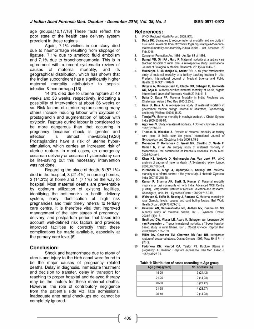
J Indian Acad Forensic Med. October - December 2016, Vol. 38, No. 4 ISSN 0971-0973
406
age groups.[12,17,18] These facts reflect the
poor state of the health care delivery system prevalent in these regions. Again, 7.1% victims in our study died due to haemorrhage resulting from slippage of ligature, 7.1% due to amniotic fluid embolism and 7.1% due to bronchopneumonia. This is in agreement with a recent systematic review of causes of maternal mortality, and its geographical distribution, which has shown that the Indian subcontinent has a significantly higher maternal mortality attributable to sepsis, infection & hemorrhage.[13] 14.3% died due to uterine rupture at 40 weeks and 38 weeks, respectively, indicating a possibility of intervention at about 36 weeks or so. Risk factors of uterine rupture among many others include inducing labour with oxytocin or prostaglandin and augmentation of labour with oxytocin. Rupture during labour is considered to be more dangerous than that occurring in pregnancy because shock is greater and infection is almost inevitable.[19,20]
Prostaglandins have a risk of uterine hyper-stimulation, which carries an increased risk of uterine rupture. In most cases, an emergency cesarean delivery or cesarean hysterectomy can be life-saving but this necessary intervention was not done. Regarding the place of death, 8 (57.1%) died in the hospital, 3 (21.4%) in nursing homes, 2 (14.3%) at home and 1 (7.1%) on the way to hospital. Most maternal deaths are preventable by optimum utilization of existing facilities, identifying the bottlenecks in health delivery system, early identification of high risk pregnancies and their timely referral to tertiary care centre. It is therefore vital that improved management of the later stages of pregnancy, delivery, and postpartum period that takes into account well-defined risk factors and provides improved facilities to correctly treat these complications be made available, especially at the primary care level.[6]
Conclusion: Shock and haemorrhage due to atony of uterus and injury to the birth canal were found to be the major causes of pregnancy related deaths. Delay in diagnosis, immediate treatment and decision to transfer, delay in transport for reaching to proper hospital and delayed therapy may be the factors for these maternal deaths. However, the role of contributory negligence from the patient’s side viz. late admissions, inadequate ante natal check-ups etc. cannot be completely ignored.
References: 1. WHO, Regional Health Forum, 2005; 9(1).
2. Dutta DK. Strategies to reduce maternal mortality and morbidity in rural india. Available from:http://www.fogsi.org/strategies-to-reduce-maternal-mortality-and-morbidity-in-rural-india/. Last accessed 20 Feb 2016.
3. Consumer Protection Act, 1986 - Act No. 68 of 1986. 4. Bangal VB, Giri PA , Garg R. Maternal mortality at a tertiary care
teaching hospital of rural india: a retrospective study. International Journal of Biological & Medical Research. 2011;2(4):1043– 6.
5. Mukherjee S, Mukherjee S, Sarkar RR. A six year retrospective study of maternal mortality at a tertiary teaching institute in Uttar Pradesh. International Journal of Medical Science and Public Health. 2014;3(11):1407-9.
6. Dinyain A, Omoniyi-Esan O, Olaofe OO, Sabageh D, Komolafe AO, Sojo O. Autopsy-certified maternal mortality at Ile-Ife, Nigeria International Journal of Women’s Health 2014:6 41–6
7. Datta D, Datta PP. Maternal Mortality in India: Problems and
Challenges. Asian J Med Res 2013;2:33-5. 8. Kaur D, Kaur A. A retrospective study of maternal mortality in
government medical college. Journal of Obstetrics, Gynaecology and family Welfare 1999;5:18-22.
9. Taneja PV. Maternal mortality in madhya pradesh. J Obstet Gynaec India 2000;50:59-61.
10. Aggrawal V. Study of maternal mortality. J Obstetric Gynaecol India 1982;32:686-90.
11. Thomas B, Mhaskar A. Review of maternal mortality at tertiary care hosp of India over ten years. International Journal of Gynaecology and Obstetrics India 2006;9:19-21
12. Menéndez C, Romagosa C, Ismail MR, Carrilho C, Saute F,
Osman N, et al. An autopsy study of maternal mortality in Mozambique: the contribution of infectious diseases. PLoS Med. 2008;5(2):e44.
13. Khan KS, Wojdyla D, Gulmezoglu Am, Van Look PF. WHO
analysis of causes of maternal death : A Systematic review, Lancet 2006;367:1066-74.
14. Purandare N, Singh A, Upadhyae S, Saraogi RM. Maternal mortality at a referral centre : a five year study. J obstetric Gynaecol
India 2007;57:248-50. 15. Kumar R, Sharma AK, Barik S, Kumar V. Maternal mortality
inquiry in a rural community of north India. Advanced MCH Centre (ICMR), Postgraduate Institute of Medical Education and Research,
Chandigarh, India. Int J Gynaecol Obstet.1989;29:313-319. 16. Walraven G, Telfer M, Rowley J, Romans C. Maternal mortality in
rural Gambia: levels, causes and contributing factors. Bull World Health Organ. 2000;78:603-613.
17. Kavatkar AN, Sahasrabudhe NS, Jadhav MV, Deshmukh SD. Autopsy study of maternal deaths. Int J Gynaecol Obstet. 2003;81(1):1–8.
18. Geelhoed DW, Visser LE, Asare K, Schagen van Leeuwen JH,
van Roosmalen J. Trends in maternal mortality: a 13-year hospital-based study in rural Ghana. Eur J Obstet Gynecol Reprod Biol. 2003;107(2): 135–139.
19. Miller DA, Goodwin TM, Gherman RB Paul RH. Intrapartum rupture of unscarred uterus. Obstet Gynecol 1997; May: 89 (5 Pt 1),
671-3. 20. Fedorkow DM, Nimrod CA, Taylor PJ. Rupture Uterus in
pregnancy: A Canadian Hospital’s experience. Can Med Assoc J. 1987;137:27-31.
Table 1: Distribution of cases according to Age group
Age group (years) No. of cases (%)
15-20 3 (21.42)
21-25 2 (14.28)
26-30 3 (21.42)
31-35 4 (28.57)
36-40 2 (14.28)
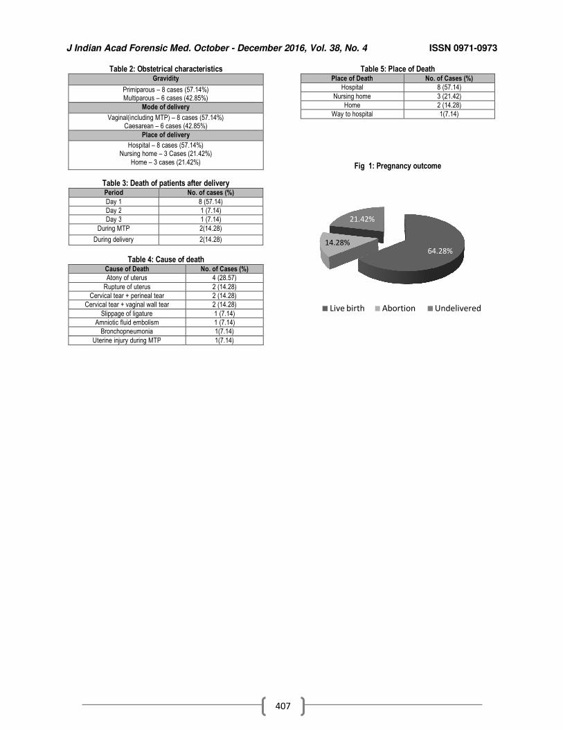
J Indian Acad Forensic Med. October - December 2016, Vol. 38, No. 4 ISSN 0971-0973
407
Table 2: Obstetrical characteristics Gravidity
Primiparous – 8 cases (57.14%) Multiparous – 6 cases (42.85%)
Mode of delivery
Vaginal(including MTP) – 8 cases (57.14%) Caesarean – 6 cases (42.85%)
Place of delivery
Hospital – 8 cases (57.14%) Nursing home – 3 Cases (21.42%)
Home – 3 cases (21.42%)
Table 3: Death of patients after delivery Period No. of cases (%)
Day 1 8 (57.14)
Day 2 1 (7.14)
Day 3 1 (7.14)
During MTP 2(14.28)
During delivery 2(14.28)
Table 4: Cause of death
Cause of Death No. of Cases (%)
Atony of uterus 4 (28.57)
Rupture of uterus 2 (14.28)
Cervical tear + perineal tear 2 (14.28)
Cervical tear + vaginal wall tear 2 (14.28)
Slippage of ligature 1 (7.14)
Amniotic fluid embolism 1 (7.14)
Bronchopneumonia 1(7.14)
Uterine injury during MTP 1(7.14)
Table 5: Place of Death Place of Death No. of Cases (%)
Hospital 8 (57.14)
Nursing home 3 (21.42)
Home 2 (14.28)
Way to hospital 1(7.14)
Fig 1: Pregnancy outcome
64.28%14.28%
21.42%
Live birth Abortion Undelivered
J Indian Acad Forensic Med. October - December 2016, Vol. 38, No. 4 ISSN 0971-0973
408
Original Research Paper
An Epidemiological Study of Fracture Related Deaths in a Tertiary Care Hospital in West Bengal
1Alakesh Halder,
2Kaushik Mukhopadhyay,
3Shailesh V Parate,
4Ashok Kumar Samanta
Abstract Fractures due to Road Traffic Accidents or fall, are an extremely common occurrence and often
lead to death. Aim of this study was to find out the epidemiology of fracture related deaths in a medical college mortuary in eastern India. For this, an autopsy-based cross-sectional study was conducted from 1st April, 2013 to 31st March, 2014 in the department of Forensic and State Medicine, Nil Ratan Sircar Medical College, Kolkata. During this period a total of 2813 autopsies were carried out. Of these, 613 cases were associated with fractures, of which 100 cases of fracture were selected randomly and analyzed. RTA and fall were the commonest cause of fracture related deaths. Fractures among RTA victims were common in male (78.9%), in middle age persons (38.6%) and during rainy season. Greater number of victims died within first 24 hours of the event, both in case of RTA (43.8%) and fall (57.6%). Skull (48%) and spine (11%) fractures were present in most of the cases. This study can help concerned authorities to plan and implement safety measures to avoid preventable death
Key Words: Fracture, Road Traffic Accident, Fall from height
Introduction: Fracture of a bone is defined as a breach in
the continuity of a bone or a complete or incomplete discontinuity of bone caused by direct or indirect violence.[1]. Fractures or breaking of bones usually happens when too much force is applied to a bone, usually during a fall from height or a Road Traffic accident. The global status report on road safety 2015, by WHO, reflecting information from 180 countries, indicates that worldwide the total number of road traffic deaths has reached 1.25 million per year, with the highest road traffic fatality rates in low-economic countries.[2] Over 1,37,000 people were died in India in road traffic accidents in 2013 alone; which is more than the number of people killed in all our wars put together.[3]. But fractures may also occur due to fall in the bathroom or stair case, gunshot injury etc.
Corresponding Author: 1Tutor,
Department of Forensic Medicine, NRS Medical College, Kolkata
2Asst. Prof.,
Department of Pharmacology,
3Asst. Prof.,
4Prof. & Head,
Department of Forensic Medicine, ESIC Medical College, Kolkata Email: [email protected] DOR: 26/09/2016 DOA: 25/12/2016 DOI: 10.5958/0974-0848.2016.00102.0
Majority of fractures occur in children or in elderly people, as young and middle-aged adults generally have very strong bones. An autopsy surgeon encounters different types of mechanical injuries during autopsy examination like abrasion, laceration, contusion etc. Fracture of bone is a common form of mechanical injury which may sometimes be the only cause or a contributory factor in causation of death. Usually, fractures occur accidentally, though they may also be present in suicidal or homicidal deaths. They are important medicolegally for manner of production of fracture, the type of weapon used, the gravity of the trauma, the direction of infliction, etc. The aim of this study was to study the fractures in different parts of the body, their seasonal variation, survivability and to determine the relation between the fractures and their manner of production.
Materials and Methods: We conducted a cross-sectional autopsy-based study in association with the clinical records available from investigating agencies. This hospital and mortuary based study was conducted from 1st April, 2013 to 31st March, 2014. During this period a total of 2813 autopsies were carried out in the department of Forensic and State Medicine, Nil Ratan Sircar Medical College, Kolkata. Of these, 613 cases were associated with fractures, among which 100 cases of fractures were selected randomly
J Indian Acad Forensic Med. October - December 2016, Vol. 38, No. 4 ISSN 0971-0973
409
and analyzed. Demographic data was collected, including the age, sex, religion, place of incidence, seasonal variation, body parts involvement, manner of production, duration of survival etc.
Results: A total 100 cases of fracture related deaths were analyzed in this study. Table 1 shows that fractures due to RTA was common in males (78.9%), as compared to females (21.1%) but the percentage of fractures due to fall was almost the same in both genders - males, 51.5% and females, 49.5%, respectively. Fractures due to both fall and RTA was more prevalent in the age group 21 to 60 years. Most of the deaths occurred within the first 6 hours of the incident while very few victims survived more than one month. Time of the day and duration of the survival has been detailed in Table 1. Figure 1 shows the percentage of fracture related deaths due to different causes. Majority of fractures were due to RTA (57%) and fall (33%). Hanging (3%) and gunshot injuries (4%) were the cause in certain cases. Among fall category, fall from height (31%) and fall in bathroom (27%) were the important contributors. Figure 2 shows seasonal variation among the deaths due to fracture. Higher number of incidents occurred in rainy season, especially July and August. Most of the victims died due to fracture in the axial skeleton - skull (48%), vertebra (11%) and Thoracic fracture (13%). A few cases showed multiple fractures, which is detailed in Table 2.
Discussion: Age wise distribution: Fractures due to both fall and RTA were more prevalent in the age group 21 to 60 years. The findings of this study were consistent with the study from New Delhi, which revealed that the incidence of traffic injury per 1000 population was high in the age group of 15 to 55 years.[4] Sinha S.N., et al retrospectively studied road traffic accident fatalities in Port Moresby for a period of 10 years spanning from 1976 to 1985 and observed that the highest fatality rates were in the age group between 15-54 years.[5] Kumar A, et al, found that most of the victims were adults; age group involved being 41-50 years (29.5%) and 31-40 years (27.9%).[6] Chandra J, et al found that the most common group involved in road traffic accidents was the 21-60 years. Findings of these studies were similar to our study.[7]
Sex distribution: Fractures due to RTA were more common in males (78.9%) but there was no difference in the gender in fractures due to fall - males, 51.5% and females, 49.5%. Reddy, et al found that 92% of the victims were males.[8] Adeyemo A.O, et al found 85.8% male victims among the 148 cases that he studied.[9] Santhiyasekaran observed that 82.5% of the victims were male.[10] Similar male prevalence was found in several studies conducted in different parts of India.[11,12]. The findings of our study are consistent with these observations. Causes of fracture related death: In 2006, Pathak, et al found that RTA (66%), followed by fall (23%) were the main external factors causing fracture related deaths.[13] Igho, et al. reported that RTA (58%) and domestic/sport related fall (27%) cause most of the fracture related incidents.[14] In our study also, majority of fractures were due to RTA (57%) and fall (33%). Fall from height (31%) and fall in bathroom (27%) were the two common mechanisms for fractures in the fall category. The time of the day when incidence of fracture occurred The peak time of the day, contributing highest number of RTA vary greatly in different studies. Pillay VV, in his study, encountered the maximum number of traffic fatalities in the late evening hours of 8 PM to 12 AM (31%) and the second highest peak from 12 PM to 4 PM, while the least number was recorded for the period from 12 AM to 4 AM.[15] Sinha SN, et al found that in 40.2% of all the fatalities, the accidents occurred between 6 PM to 6 AM and in 35.3%, between 6 AM to 6 PM.[5]. Kumar A, et al, observed that the time of occurrence and mortality was maximum, between 12-6 PM while mortality rate was highest during 6 PM -12 AM. In this study, majority of RTA took place between 6 AM to 12pPM but fall was frequent from 12 – 6 PM.[6] Duration of survival after fracture As per Table IV, most of the victims survived less than 6 hours, both in RTA (24.6%) and in fall from height (33.3%) and very few survived for more than a month 3.4% and 6.6%, respectively. Patil, et al reported that 63% of the victims died within first 24 hours of the injury.[16] In the present study, 62.3% of the victims died within the first 24 hours. Reddy, et al found that 59% of the victims died within first 24 hours and only 23% victims survived more than 3 weeks.[8] Seasonal variation We found that higher number of incidents occurred in the rainy season, especially, July (26%) and August (23%). Similar
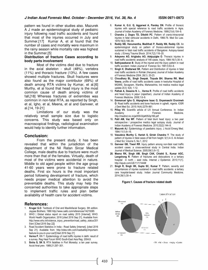
J Indian Acad Forensic Med. October - December 2016, Vol. 38, No. 4 ISSN 0971-0973
410
pattern ws found in other studies also. Mazurek A J made an epidemiological study of pediatric injury following road traffic accidents and found that most of the injuries occurred in July and Summer.[17] Kumar A, et al found that the number of cases and mortality were maximum in the rainy season while mortality rate was highest in the Summer.[5] Distribution of fracture Cases according to body parts involvement Most of the victims died due to fracture in the axial skeleton - skull (48%), vertebra (11%) and thoracic fracture (13%). A few cases showed multiple fractures. Skull fractures were also found as the major contributor (69%) of death among RTA victims by Kumar, et al.[6] Murthy, et al found that head injury is the most common cause of death among victims of fall,[18] Whereas fractures of extremities was common in non-fatal RTA, as reported by Singh, et al, Igho, et al, Meena, et al and Ganveer, et al.[14, 19-21] Limitations of our study was the relatively small sample size due to logistic concerns. This study was based only on macroscopical findings, radiological examination would help to identify further information.
Conclusion: From the present study, it has been revealed that within the jurisdiction of the department of the Nil Ratan Sircar Medical College, male deaths due to fracture were much more than that of the females. Virtually, death of most of the victims were accidental in nature. Middle to old aged people within the age group 41-60 years were prone to fracture related deaths. First six hours is the most important period following development of fracture, which needs proper medical attention to avoid the preventable deaths. This study may help the concerned authorities to take appropriate steps to implement traffic rules and plan better availability of health care for accident victims.
References: 1. Kruger G.O. Textbook of Oral and Maxillofacial Surgery. 6th edition.
Jaypee Brothers; 1990 http://www.oalib.com/references/14219385 2. WHO | Global status report on road safety 2015 [Internet]. WHO.
World Health Organization; 2016 [cited 2016 Sep 21]. Available from:
http://www.who.int/violence_injury_prevention/road_safety_status/2015/en/ [Cited 2016 Sep 22]
3. Road Accident Statistics in India - Road Safety [Internet]. [cited 2016 Sep 21]. Available from: http://sites.ndtv.com/roadsafety/important-
feature-to-you-in-your-car/[Cited 2016 Sep 21] 4. Verma P, KN T. Epidemiology of road traffic injuries in delhi: result of
a survey. Reg Heal Forum WHO South-East Asia Reg. 2004;8. 5. Sinha S, SK S. RTA fatalities in Port Moresby: a ten year survey.
Accid Anal prev. 1989;21:297–301.
6. Kumar A, G.U. Q, Aggarwal A, Pandey DN. Profile of thoracic injuries with special reference to road traffic accidents in Agra. Journal of Indian Academy of Forensic Medicine. 1999;21(4):104–9.
7. Chandra J, Dogra TD, Dikshit PC. Pattern of cranio-intracranial injuries in fatal vehicular accidents in Delhi, 1966-76. Med Sci Law. 1979;19(3):186–94.
8. Reddy NB, Hanumantha, Madithati P, Reddy NN, Reddy CS. An
epidemiological study on pattern of thoraco-abdominal injuries sustained in fatal road traffic accidents of Bangalore: Autopsy-based study. J Emerg Trauma Shock. 2014;7(2):116–20.
9. Adeyemo AO, Arigbabu AO, Adejuyigbe O. Thoracic injuries in
road traffic accidents: analysis of 148 cases. Injury. 1984;16(1):30–4. 10. Sathiyasekaran B. Study of the injured and the injury pattern in road
traffic accident. Indian Journal of Forensic Sci. 1991;5:63–8. 11. Singh H, Dhattarwal SK, Pattern and Distribution of injuries in fatal
road traffic accidents in Rohtak (Haryana) Journal of Indian Academy of Forensic Medicine 2004; 26(1): 20-23.
12. Choudhary BL, Singh Deepak, Tirpude BH, Sharma RK, Meel Veena, profile of road traffic accidents cases in kesturba Hospital of
MGIMS, Sevagram, Wardha, Maharashtra, Ind medica medico legal update 20,5; 5(4): 1-12.
13. Pathak A, Desania N. L , Verma R. Profile of road traffic accidents and head injury in jaipur (rajasthan). Journal of Indian Academy of
Forensic Medicine. 2008;1:6–9. 14. Emmanuel Igho O, Akpoghene Isaac O, Onyemaechi Eronimeh
O. Road traffic accidents and bone fractures in ughelli, nigeria. IOSR J Dent Med Sci. 2015;14(4):2279–861.
15. Pillay VV. Scientific article of IJV Annual Conference. In: Indian Academy of Forensic Medicine. http://medind.nic.in/jal/t05/i3/jalt05i3p166.pdf
16. Patil AM, Vaz WF. Pattern of fatal blunt head injury: a two year
retrospective / prospective medico legal autopsy study. Journal of Indian Academy of Forensic Medicine. 2010;32(2):144–9.
17. Mazurek AJ. Epidemiology of paediatric injury. J Accid Emerg Med. 1994;11(1):9–16.
18. Vasudeva Murthy C, Harish S, Girish Chandra Y. The study of pattern of injuries in fatal cases of fall from height. 5(1):4–5. Al Ameen J Med Sci; Volume 5, No.1, 2012
19. Ganveer GB, Tiwari RR. Injury pattern among non-fatal road traffic
accident cases: a cross-sectional study in Central India. Indian Journal of Medical Science. 2005;59(1):9–12.
20. Meena RK, Singh AM, Singh CAK, Chishti S., Kumar AGC, Langshong R. Pattern of fractures and dislocations in a tertiary
hospital in north – east india. Internet J Epidemiol. 2013;11(1). http://ispub.com/IJE/11/1/1444#
21. Singh R, Singh HK, Gupta SC, Kumar Y. Pattern, severity and circumtances of injuries sustained in road traffic accidents: a tertiary
care hospital-based study. Indian Journal Community Medicine. 2014;39(1):30–4.
Figure:1. Causes of fracture related death
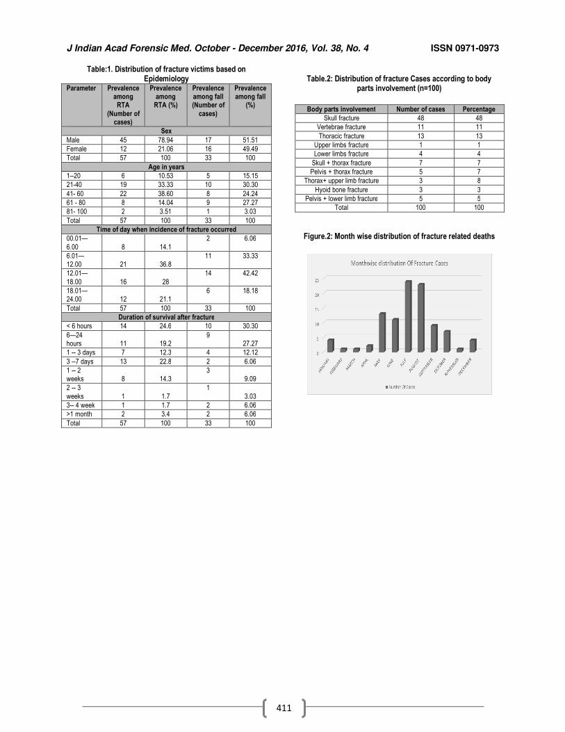
J Indian Acad Forensic Med. October - December 2016, Vol. 38, No. 4 ISSN 0971-0973
411
Table:1. Distribution of fracture victims based on Epidemiology
Parameter Prevalence among
RTA
(Number of cases)
Prevalence among
RTA (%)
Prevalence among fall (Number of
cases)
Prevalence among fall
(%)
Sex
Male 45 78.94 17 51.51
Female 12 21.06 16 49.49
Total 57 100 33 100
Age in years
1--20 6 10.53 5 15.15
21-40 19 33.33 10 30.30
41- 60 22 38.60 8 24.24
61 - 80 8 14.04 9 27.27
81- 100 2 3.51 1 3.03
Total 57 100 33 100
Time of day when incidence of fracture occurred
00.01—
6.00 8 14.1
2 6.06
6.01—12.00 21 36.8
11 33.33
12.01—
18.00 16 28
14 42.42
18.01—24.00 12 21.1
6 18.18
Total 57 100 33 100
Duration of survival after fracture
< 6 hours 14 24.6 10 30.30
6—24 hours 11 19.2
9 27.27
1 -- 3 days 7 12.3 4 12.12
3 --7 days 13 22.8 2 6.06
1 -- 2 weeks 8 14.3
3 9.09
2 -- 3
weeks 1 1.7
1
3.03
3-- 4 week 1 1.7 2 6.06
>1 month 2 3.4 2 6.06
Total 57 100 33 100
Table.2: Distribution of fracture Cases according to body
parts involvement (n=100)
Figure.2: Month wise distribution of fracture related deaths
Body parts involvement Number of cases Percentage
Skull fracture 48 48
Vertebrae fracture 11 11
Thoracic fracture 13 13
Upper limbs fracture 1 1
Lower limbs fracture 4 4
Skull + thorax fracture 7 7
Pelvis + thorax fracture 5 7
Thorax+ upper limb fracture 3 8
Hyoid bone fracture 3 3
Pelvis + lower limb fracture 5 5
Total 100 100

J Indian Acad Forensic Med. October - December 2016, Vol. 38, No. 4 ISSN 0971-0973
412
Original Research Paper
Growth Report of “The End” – Autopsy Statistics in a Developing Medical College
1Jigesh V. Shah,
2Gaurang J. Patel,
3Ankur Patel,
4Dharmesh S. Patel,
5Sanjay Jadav,
5Rajesh Jakhar
Abstract The present prospective study was carried out at the mortuary of GMERS Medical College and Associated Civil Hospitals, Sola, Ahmedabad. In the year 2011, the institution got permission from the MCI to start the medical college with an annual intake of 150 students. During the 5 calendar years from 2011 to 2015, we received a total of 1,561 corpses at the mortuary of the hospital for the autopsy. It was seen that the numbers of autopsy cases were rising year by year.No specific pattern of distribution was noticed regarding month-wise distribution. 1183 dead bodies (75.8%) were of males and the remaining were of females. 85 dead bodies (73 males & 12 females,5.45% of total)were unidentified. 791 victims (50.7%) were from the age groups of 21-40 years. A variety of cases were examined with regard to cause of death. Road traffic accidents (460 cases, 29.5%) were the maximum.In majority of the cases (762 cases, 48.8%), the manner of death was accidental, followed by natural/pathological (363 cases, 23.3%) & suicidal (343 cases, 22 %), respectively.
Key Words: Autopsy cases, Statistics, Cause of Death, Manner of Death
Introduction: The case of injury or ailment, in which
investigations by the law enforcing agencies are required to fix the responsibility regarding its causation, is known as Medico Legal Case.[1] These cases are an integral part of medical practice. Basically, all cases of medico-legal examination are divided into two broad categories. One is the medico-legal examination of the living, which is frequently encountered by Medical Officers working in Casualty Department; and the second is the medico-legal examination of the dead, which is known as autopsy or post-mortem examination.[1]
Corresponding Author: 1Associate Professor,
4Professor & Head,
5Tutors
Forensic Medicine Department, GMERS Medical College, Sola, Ahmedabad. 2Associate Professor,
3Assistant Professor,
Forensic Medicine Department, GMERS Medical College, Dharpur, Patan. E-mail – [email protected] DOR: 29.2.16 DOA: 25/12/2016 DOI: 10.5958/0974-0848.2016.00103.2
Studying the pattern & magnitude of the autopsy cases is an important aspect to study the crime rate in the particular areas.[2]Suicidal cases reveal the social and mental status of the residents of the society. Accidental cases expose the disciplines and awareness prevailing within the civilization. Cobalt RC,[3] in 1912,observed that many common diseases such as cirrhosis, acute endocarditis, bronchopneumonia, and acute nephritis were missed in clinical diagnosis in more than 50 percent of the cases. Obviously, clinical medicine has made massive strides since 1912. Some clinicians may believe that our current sophisticated high-tech diagnostic tests render the autopsy superfluous. However, many pathologists do not agree that the autopsy has been rendered needless because of modern technology. Landefeld, et al,[4] Shanks, et al,[5] Sarode, et al,[6] Veress, et al[7] as well as Manzini, et al,[8] found “major discrepancies” between the autopsy findings and clinical diagnosis. A pre-mortem diagnosis of these findings would probably have improved survival of the patient. These studies raised the important question, "Did the patient receive the correct treatment for the correct disease?"
Autopsy information has long been an essential tool for quality control of medical care and for enhancing the quality of cause-of-death information reported on the death certificate. The motto behind this study was to illustrate the
J Indian Acad Forensic Med. October - December 2016, Vol. 38, No. 4 ISSN 0971-0973
413
continued value of autopsy in the practice of medicine, in health care and health statistics.The study provides a snapshot about burden of autopsy cases at a tertiary care hospital of a megacity. Efforts were made to identify the magnitude & trends of different autopsy cases at one tertiary center ofAhmedabad; the biggest and most populous city of Gujarat.
Material and Methods Present prospective study was carried
out at GMERS Medical College and Associated Civil Hospital, Sola, Ahmedabad, Gujarat. The study material comprised of1561 autopsies carried out at the mortuary over a period of 5calendar years from 1st January 2011 to 31st December, 2015. Since the inception of the college in the year 2011, 5 police stations of Ahmedabad city were attached to the Civil Hospital, Sola. A few cases were referred by the Medical Officers of nearby Community Health Centres and Primary Health Centres for expert opinion. The bodies were received for autopsy with a requisition letter, 'maranottar form' and copy of inquest panchnama.
In all cases, a detailed history was elicited from the police and near relatives. Preliminaries of the deceased including his sex, age etc. with a short history of the incident and circumstances were noted on a specially designed Performa. In case of unidentified bodies, all necessary data was noted and DNA samples preserved to ensure precise individuality in the future. Positive findings of post-mortem examination with laboratory reports and final cause of death were also recorded. The data so received was tabulated and statistically analyzed.
Observations and Discussion: The load of medico-legal cases at a
tertiary care hospital not only caters to the needs of patients who report for their illness, but also to carry out various legal responsibilities to examine, document and certify medico-legal cases. Autopsy cases are one of the main concern, second to the medico-legal examination of living, at the casualty.It can be seen from Table no. 1 that 234 medico-legal autopsies were carried out in 2011, which went on rising year by year. The data of 2015 showed the number to be 390. The obvious reason is the increased urbanization in the catchment area of the hospital. However, while looking at the monthly average, no specific pattern could be established. The number of cases were not affected by weather. Overall, it can be said that the post-mortem examination was carried out as and when dead body was received for the
purpose. In contrast, Garg V, et al,[2], Bhagora,et al,[9] and Malik Y, et al,[10] have observed maximum autopsy cases in October followed by June.
Table – 2 shows the year and gender wise distribution of cases. Of the 1561cases under study, 1183 (75.8%) were males and the remaining (24.2%) were females. In comparison, Bhagora RV, et al,[9]observed 65.2% male victims. This is because males are more exposed to outdoor activities as well as disputes in family matters. Also, males, by nature, indulge in more violent activities as compared to females. They are also the main bread-earners of the family. Collectively, therefore, they are more vulnerable and exposed to such situations. 1476 cases (94.6%) were identified by the near relatives which included 1110 males (71.1%) and 366 females (23.5%). Remaining 85 corpses (5.5 %) had no relatives available to identify them; they included 73 males and 12 females.
Age and gender wise distribution with detailing on identification of the deceased is described in Table no. 3. It was observed that out of 85 unidentified bodies, 8 were foetuses (5 male and 3 female) of age 4 to 7 months of intra-uterine life. Bulk of the victims (452 cases, 29 %) was from the age group of 21–30 years, which is the young work force group. Bhagora RV, et al,[9]noted 22.4% victims between 21 to 30 years. Similar findings were noted by Bhullar DS, et al[11] and Aggarwal KK, et al.[12]This was followed by 31-40 years group[339 cases (21.7%)] of, 41-50 years group [248 cases (15.9%)].
A variety of cases were received for post-mortem examination with regard to cause of death as elicited from Table no. 4. In all these cases, the manner of death (Table no. 5) was determined on the basis of post-mortem findings and history elicited from police and relatives of the deceased. Road Traffic Accidents (RTA) were the maximum, with 460 cases (29.5%), followed by 358 natural/pathological deaths (22.9%) and 195 cases of hanging (12.5%). All hanging and poisoning cases (71, 4.6%) were suicidal in nature. With regard to the nature of poisonous substance consumed, majority were organophosphorus compound, aluminium phosphide and acids. A few had committed suicide by jumping from height, running in front of trains, drowning, burns and a few deaths were accidental. One victim had committed suicide by cutting his cut throat. The basis for classifying these cases as suicidal in nature was the history given by the eyewitness and its correlation to the post-mortem findings and circumstantial
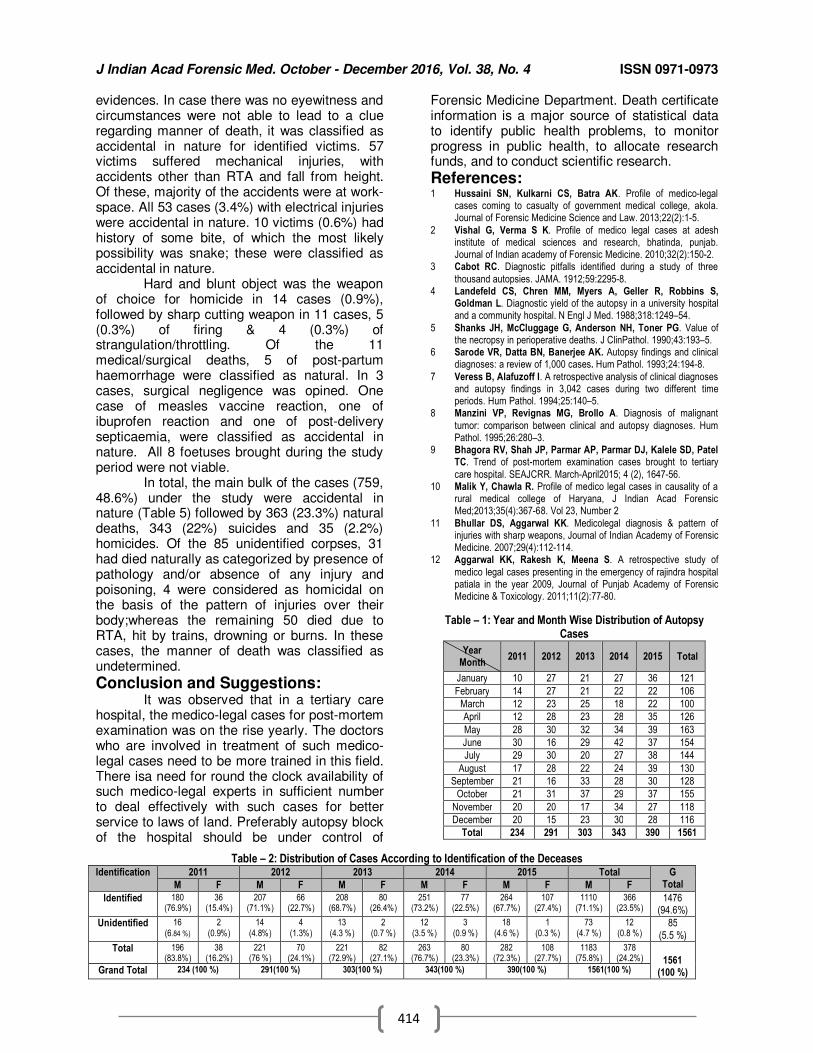
J Indian Acad Forensic Med. October - December 2016, Vol. 38, No. 4 ISSN 0971-0973
414
evidences. In case there was no eyewitness and circumstances were not able to lead to a clue regarding manner of death, it was classified as accidental in nature for identified victims. 57 victims suffered mechanical injuries, with accidents other than RTA and fall from height. Of these, majority of the accidents were at work-space. All 53 cases (3.4%) with electrical injuries were accidental in nature. 10 victims (0.6%) had history of some bite, of which the most likely possibility was snake; these were classified as accidental in nature.
Hard and blunt object was the weapon of choice for homicide in 14 cases (0.9%), followed by sharp cutting weapon in 11 cases, 5 (0.3%) of firing & 4 (0.3%) of strangulation/throttling. Of the 11 medical/surgical deaths, 5 of post-partum haemorrhage were classified as natural. In 3 cases, surgical negligence was opined. One case of measles vaccine reaction, one of ibuprofen reaction and one of post-delivery septicaemia, were classified as accidental in nature. All 8 foetuses brought during the study period were not viable.
In total, the main bulk of the cases (759, 48.6%) under the study were accidental in nature (Table 5) followed by 363 (23.3%) natural deaths, 343 (22%) suicides and 35 (2.2%) homicides. Of the 85 unidentified corpses, 31 had died naturally as categorized by presence of pathology and/or absence of any injury and poisoning, 4 were considered as homicidal on the basis of the pattern of injuries over their body;whereas the remaining 50 died due to RTA, hit by trains, drowning or burns. In these cases, the manner of death was classified as undetermined.
Conclusion and Suggestions: It was observed that in a tertiary care
hospital, the medico-legal cases for post-mortem examination was on the rise yearly. The doctors who are involved in treatment of such medico-legal cases need to be more trained in this field. There isa need for round the clock availability of such medico-legal experts in sufficient number to deal effectively with such cases for better service to laws of land. Preferably autopsy block of the hospital should be under control of
Forensic Medicine Department. Death certificate information is a major source of statistical data to identify public health problems, to monitor progress in public health, to allocate research funds, and to conduct scientific research.
References: 1 Hussaini SN, Kulkarni CS, Batra AK. Profile of medico-legal
cases coming to casualty of government medical college, akola. Journal of Forensic Medicine Science and Law. 2013;22(2):1-5.
2 Vishal G, Verma S K. Profile of medico legal cases at adesh institute of medical sciences and research, bhatinda, punjab. Journal of Indian academy of Forensic Medicine. 2010;32(2):150-2.
3 Cabot RC. Diagnostic pitfalls identified during a study of three
thousand autopsies. JAMA. 1912;59:2295-8. 4 Landefeld CS, Chren MM, Myers A, Geller R, Robbins S,
Goldman L. Diagnostic yield of the autopsy in a university hospital and a community hospital. N Engl J Med. 1988;318:1249–54.
5 Shanks JH, McCluggage G, Anderson NH, Toner PG. Value of the necropsy in perioperative deaths. J ClinPathol. 1990;43:193–5.
6 Sarode VR, Datta BN, Banerjee AK. Autopsy findings and clinical diagnoses: a review of 1,000 cases. Hum Pathol. 1993;24:194-8.
7 Veress B, Alafuzoff I. A retrospective analysis of clinical diagnoses and autopsy findings in 3,042 cases during two different time periods. Hum Pathol. 1994;25:140–5.
8 Manzini VP, Revignas MG, Brollo A. Diagnosis of malignant
tumor: comparison between clinical and autopsy diagnoses. Hum Pathol. 1995;26:280–3.
9 Bhagora RV, Shah JP, Parmar AP, Parmar DJ, Kalele SD, Patel TC. Trend of post-mortem examination cases brought to tertiary
care hospital. SEAJCRR. March-April2015; 4 (2), 1647-56. 10 Malik Y, Chawla R. Profile of medico legal cases in causality of a
rural medical college of Haryana, J Indian Acad Forensic Med;2013;35(4):367-68. Vol 23, Number 2
11 Bhullar DS, Aggarwal KK. Medicolegal diagnosis & pattern of injuries with sharp weapons, Journal of Indian Academy of Forensic Medicine. 2007;29(4):112-114.
12 Aggarwal KK, Rakesh K, Meena S. A retrospective study of
medico legal cases presenting in the emergency of rajindra hospital patiala in the year 2009, Journal of Punjab Academy of Forensic Medicine & Toxicology. 2011;11(2):77-80.
Table – 1: Year and Month Wise Distribution of Autopsy Cases
Year Month
2011 2012 2013 2014 2015 Total
January 10 27 21 27 36 121
February 14 27 21 22 22 106
March 12 23 25 18 22 100
April 12 28 23 28 35 126
May 28 30 32 34 39 163
June 30 16 29 42 37 154
July 29 30 20 27 38 144
August 17 28 22 24 39 130
September 21 16 33 28 30 128
October 21 31 37 29 37 155
November 20 20 17 34 27 118
December 20 15 23 30 28 116
Total 234 291 303 343 390 1561
Table – 2: Distribution of Cases According to Identification of the Deceases Identification 2011 2012 2013 2014 2015 Total G
Total M F M F M F M F M F M F
Identified 180 (76.9%)
36 (15.4%)
207 (71.1%)
66 (22.7%)
208 (68.7%)
80 (26.4%)
251 (73.2%)
77 (22.5%)
264 (67.7%)
107 (27.4%)
1110 (71.1%)
366 (23.5%)
1476 (94.6%)
Unidentified 16 (6.84 %)
2 (0.9%)
14 (4.8%)
4 (1.3%)
13 (4.3 %)
2 (0.7 %)
12 (3.5 %)
3 (0.9 %)
18 (4.6 %)
1 (0.3 %)
73 (4.7 %)
12 (0.8 %)
85
(5.5 %)
Total 196 (83.8%)
38 (16.2%)
221 (76 %)
70 (24.1%)
221 (72.9%)
82 (27.1%)
263 (76.7%)
80 (23.3%)
282 (72.3%)
108 (27.7%)
1183 (75.8%)
378 (24.2%)
1561
(100 %) Grand Total 234 (100 %) 291(100 %) 303(100 %) 343(100 %) 390(100 %) 1561(100 %)
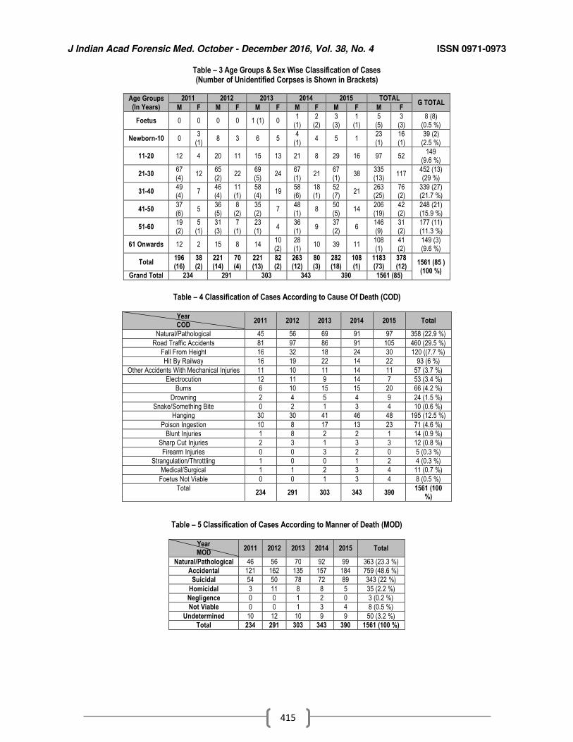
J Indian Acad Forensic Med. October - December 2016, Vol. 38, No. 4 ISSN 0971-0973
415
Table – 3 Age Groups & Sex Wise Classification of Cases (Number of Unidentified Corpses is Shown in Brackets)
Table – 4 Classification of Cases According to Cause Of Death (COD)
Year COD
2011 2012 2013 2014 2015 Total
Natural/Pathological 45 56 69 91 97 358 (22.9 %)
Road Traffic Accidents 81 97 86 91 105 460 (29.5 %)
Fall From Height 16 32 18 24 30 120 ((7.7 %)
Hit By Railway 16 19 22 14 22 93 (6 %)
Other Accidents With Mechanical Injuries 11 10 11 14 11 57 (3.7 %)
Electrocution 12 11 9 14 7 53 (3.4 %)
Burns 6 10 15 15 20 66 (4.2 %)
Drowning 2 4 5 4 9 24 (1.5 %)
Snake/Something Bite 0 2 1 3 4 10 (0.6 %)
Hanging 30 30 41 46 48 195 (12.5 %)
Poison Ingestion 10 8 17 13 23 71 (4.6 %)
Blunt Injuries 1 8 2 2 1 14 (0.9 %)
Sharp Cut Injuries 2 3 1 3 3 12 (0.8 %)
Firearm Injuries 0 0 3 2 0 5 (0.3 %)
Strangulation/Throttling 1 0 0 1 2 4 (0.3 %)
Medical/Surgical 1 1 2 3 4 11 (0.7 %)
Foetus Not Viable 0 0 1 3 4 8 (0.5 %)
Total 234 291 303 343 390
1561 (100 %)
Table – 5 Classification of Cases According to Manner of Death (MOD)
Year MOD
2011 2012 2013 2014 2015 Total
Natural/Pathological 46 56 70 92 99 363 (23.3 %)
Accidental 121 162 135 157 184 759 (48.6 %)
Suicidal 54 50 78 72 89 343 (22 %)
Homicidal 3 11 8 8 5 35 (2.2 %)
Negligence 0 0 1 2 0 3 (0.2 %)
Not Viable 0 0 1 3 4 8 (0.5 %)
Undetermined 10 12 10 9 9 50 (3.2 %)
Total 234 291 303 343 390 1561 (100 %)
Age Groups
(In Years)
2011 2012 2013 2014 2015 TOTAL G TOTAL
M F M F M F M F M F M F
Foetus 0 0 0 0 1 (1) 0 1
(1) 2
(2) 3
(3) 1
(1) 5
(5) 3
(3) 8 (8)
(0.5 %)
Newborn-10 0 3
(1) 8 3 6 5
4
(1) 4 5 1
23
(1)
16
(1)
39 (2)
(2.5 %)
11-20 12 4 20 11 15 13 21 8 29 16 97 52 149
(9.6 %)
21-30 67 (4)
12 65 (2)
22 69 (5)
24 67 (1)
21 67 (1)
38 335 (13)
117 452 (13) (29 %)
31-40 49 (4)
7 46 (4)
11 (1)
58 (4)
19 58 (6)
18 (1)
52 (7)
21 263 (25)
76 (2)
339 (27) (21.7 %)
41-50 37 (6)
5 36 (5)
8 (2)
35 (2)
7 48 (1)
8 50 (5)
14 206 (19)
42 (2)
248 (21) (15.9 %)
51-60 19
(2)
5
(1)
31
(3)
7
(1)
23
(1) 4
36
(1) 9
37
(2) 6
146
(9)
31
(2)
177 (11)
(11.3 %)
61 Onwards 12 2 15 8 14 10 (2)
28 (1)
10 39 11 108 (1)
41 (2)
149 (3) (9.6 %)
Total 196 (16)
38 (2)
221 (14)
70 (4)
221 (13)
82 (2)
263 (12)
80 (3)
282 (18)
108 (1)
1183 (73)
378 (12)
1561 (85 ) (100 %)
Grand Total 234 291 303 343 390 1561 (85)
J Indian Acad Forensic Med. October - December 2016, Vol. 38, No. 4 ISSN 0971-0973
416
Original Research Paper
Dental Age Estimation by Radiographic Evaluation of Pulp/Tooth Ratio in Mandibular Canines and Premolars
1Usha Jambunath,
2Balaji P,
3Poornima G,
4Vinitra Vasan,
5Ashish Gupta,
6Peeyush Shivhare
Abstract Forensic age estimation is important in clarifying issues pertaining to unknown or disputed ages of living individuals as well as reconstructive identification of the deceased.Teeth are considered as useful tools in age estimation because of their ability to resist to various physical and chemical agents. Aim of the study was to estimate dental age in adults by Cameriere’s method using pulp/tooth area ratio of mandibular canines and premolars. Study group comprised digital panoramic radiographs of 250 subjects, between 18 to 75 years. Adobe Photoshop CS4 image editing software program was used to measure the tooth and pulp area in pixels. Pulp/tooth ratio was calculated and subjected to statistical analysis. Multiple regression analysis, with age as the dependent variable and P/T ratio as predictor, yielded formulae for age estimation. Among the three teeth studied, 1
st PM showed best age correlation,
followed by canine and the combined teeth.
Key Words: Forensic Science, Age estimation, Pulp/tooth Area, Panoramic radiograph, Single rooted teeth.
Introduction: Age estimation of an individual, whether
living or dead, is an intimidating task in forensic nvestigations and is the most difficult to achieve.[1] Age estimation of adults may be essential in post-mortem identification, verifying age of immigrants, in refugees with disputed birth records, and in persons of criminal antecedents reluctant to reveal their age.[2]
It is known that teeth are the most durable and resilient parts of the skeleton, with their physiologic variations, pathoses and effects of therapy, they usually resist the influence of many factors, taphonomic process and disintegrate very slowly.
Corresponding Authors: 1,4
Practicing Oral Medicine & Radiologist, Bangalore;
2Professor & H O D,
3Reader,
Dept. Oral Medicine & Radiology, Rajarajeswari Dental College & Hospital, Bangalore;
5Practicing Oral Medicine & Radiologist, Jammu;
6Lecturer, Dept. Oral Medicine & Radiology,
Narasinhbhai Patel Dental College & Hospital, Visnagar, Gujurat Email – [email protected] DOR: 21.2.16 DOA: 25/12/2016 DOI: 10.5958/0974-0848.2016.00104.4
They are often the only body part available for studies and this makes teeth very suitable for dental age estimation.[3] A variety of methods have been used for dental age estimation. Radiography being a non-invasive method, plays a vital role in forensic dentistry to uncover the hidden facts, which cannot be seen by means of physical examination.[4] Radiographic evaluation of teeth requires neither tooth extraction nor processing. The present study was undertaken to estimate dental age in adults by Camerier’s method, using pulp/tooth area ratio of mandibular canines & premolars, using panoramic radiographs.
Materials and methods: The present study was conducted in the Department of Oral Medicine and Radiology, Rajarajeswari Dental College and Hospital, Bangalore. Digital panoramic dental radiographs of 250 subjects were collected randomly from the database (SIRONA Orthophos XG5 Digital OPG Machine),which included 125 males and 125 females in the age range of 18 to 75 years. Patients with lost or extracted mandibular canines and premolars, teeth with root fillings, crown restorations, teeth with severe caries, attrited teeth, badly rotated teeth, and other abnormal dental anatomy, which might cause difficulty with measurement were excluded. Panoramic radiographs showing teeth with the large areas of enamel overlap between
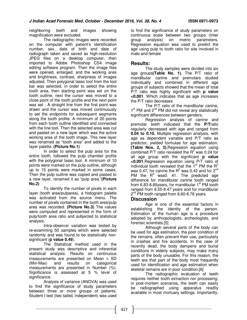
J Indian Acad Forensic Med. October - December 2016, Vol. 38, No. 4 ISSN 0971-0973
417
neighboring teeth and images showing magnification were excluded. The radiographic images were recorded on the computer with patient’s identification number, sex, date of birth and date of radiograph taken and saved as high-resolution JPEG files on a desktop computer, then imported to Adobe Photoshop CS4 image editing software program. Then the image files were opened, enlarged; and the working area and brightness, contrast, sharpness of images adjusted. Then polygonal lasso tool from the tool bar was selected, in order to select the entire tooth area, then starting point was set on the tooth outline, next the cursor was moved to a close point of the tooth profile and the next point was set . A straight line from the first point was drawn and the cursor was moved continuously to set the endpoints for subsequent segments along the tooth profile. A minimum of 20 points from each tooth outline identified and connected with the line tool. Then the selected area was cut and pasted on a new layer which was the active working area of the tooth image. This new layer was renamed as “tooth area” and added to the layer palette. (Picture No.1) In order to select the pulp area for the entire tooth, followed the pulp chamber profile with the polygonal lasso tool. A minimum of 10 points were marked on the pulp outline, although up to 15 points were marked in some cases. Then the pulp outline was copied and pasted to a new layer, renamed it as “pulp area.”(Picture No.2) To identify the number of pixels in each layer (tooth area/pulparea), a histogram palette was activated from the source menu. The number of pixels contained in the tooth area/pulp area was recorded. (Picture No.3) The values were computed and represented in the form of pulp/tooth area ratio and subjected to statistical analysis. Intra-observer variation was tested by re-examining 50 samples which were selected randomly and was found to be statistically non-significant (p value 0.49). The Statistical method used in the present study was descriptive and inferential statistical analysis. Results on continuous
measurements are presented on Mean ± SD (Min-Max) and results on categorical measurements are presented in Number (%). Significance is assessed at 5 % level of significance. Analysis of variance (ANOVA) was used to find the significance of study parameters between three or more groups of patients. Student t test (two tailed, independent) was used
to find the significance of study parameters on continuous scale between two groups (Inter group analysis) on metric parameters. Regression equation was used to predict the age using pulp to tooth ratio for site involved in male and female
Results: The study samples were divided into six age groups(Table No. 1). The P/T ratio of mandibular canine and premolars studied individually and combined in different age groups of subjects showed that the mean of total P/T ratio was highly significant with p value <0.001. Which indicates that as age increases the P/T ratio decreases. The P/T ratio of the mandibular canine, 1
st PM and 2
nd PM did not reveal any statistically
significant differences between genders. Regression analysis of canine and premolar teeth indicated that the P/Tratio regularly decreased with age and ranged from 0.04 to 0.10. Multiple regression analysis, with age as dependent variable and P/T ratio as predictor, yielded formulae for age estimation. (Table Nos. 2, 3).Regression equation using combined P/T ratio revealed the R
2 was 0.24 for
all age group with the significant p value <0.001.Regression equation using P/T ratio of individual tooth revealed that the 1
st PM the R
2
was 0.47, for canine the R2 was 0.42 and for 2
nd
PM the R2 was0. 41. The predicted age
difference for mandibular canine tooth ranged from 6.83-8.85years, for mandibular 1
st PM tooth
ranged from 6.55-8.47 years and for mandibular 2
nd PM tooth ranged from 6.60-8.76 years.
Discussion Age is one of the essential factors in establishing the identity of the person. Estimation of the human age is a procedure adopted by anthropologists, archeologists, and forensic scientists.[5] Although several parts of the body can be used for age estimation, the poor condition of the remains, often prevent their use, particularly in crashes and fire accidents. In the case of recently dead, the body dampens and burial conditions in elderly subjects, may make many parts of the body unusable. For this reason, the teeth are that part of the body most frequently used for identification and age estimation when skeletal remains are in poor condition.[6] The radiographic evaluation of teeth requires neither tooth extraction nor processing; in post-mortem scenarios, the teeth can easily be radiographed using apparatus readily available in most mortuary settings. Importantly,
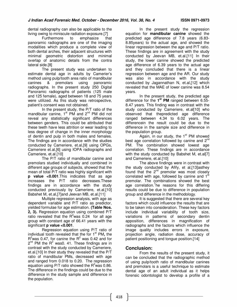
J Indian Acad Forensic Med. October - December 2016, Vol. 38, No. 4 ISSN 0971-0973
418
dental radiography can also be applicable to the living owing to miniscule radiation exposure.[7] Furthermore to emphasize that panoramic radiographs are one of the imaging modalities which produce a complete view of both dental arches, their adjacent structures with minimal geometric distortion and minimal overlap of anatomic details from the contra lateral side.[8] The present study was undertaken to estimate dental age in adults by Camerier’s method using pulp/tooth area ratio of mandibular canines & premolars using panoramic radiographs. In the present study 250 Digital Panoramic radiographs of patients (125 male and 125 female), aged between 18 and 75 years were utilized. As this study was retrospective, patient’s consent was not obtained. In the present study, the P/T ratio of the mandibular canine, 1
st PM and 2
nd PM did not
reveal any statistically significant differences between genders. This could be attributed that these teeth have less attrition or wear leading to less degree of change in the inner morphology of dentin and pulp in both males and females. The findings are in accordance with the studies conducted by Cameriere, et.al,[9] using OPGs, Cameriere et.al,[6] using IOPA radiographs and Cameriere, et.al.[10] The P/T ratio of mandibular canine and premolars studied individually and combined in different age groups of subjects, showed that the mean of total P/T ratio was highly significant with p value <0.001.This indicates that as age increases the P/T ratio decreases.These findings are in accordance with the study conducted previously by Cameriere, et.al,[10] Babshet M, et.al,[7]and Jeevan MB, et.al.[11] Multiple regression analysis, with age as dependent variable and P/T ratio as predictor, yielded formulae for age estimation. (Table Nos. 2, 3). Regression equation using combined P/T ratio revealed that the R
2was 0.24 for all age
group with constant age of 66.41 years with the significant p value <0.001. Regression equation using P/T ratio of individual tooth revealed that the for 1
st PM, the
R2was 0.47, for canine the R
2 was 0.42 and for
2nd
PM the R2 was0. 41. These findings are in
contrast with the study conducted by Cameriere, et.al.[10] In their study they revealed that the P/T ratio of mandibular PMs, decreased with age and ranged from 0.018 to 0.20. The regression equation using P/T ratio showed the R
2was 0.86.
The difference in the findings could be due to the difference in the study sample and difference in the population.
In the present study the regression equation for mandibular canine showed the predicted age difference of 7.8 years (6.83-8.85years) to the actual age, and showed the linear regression between the age and P/T ratio. These findings are in agreement with the study conducted by Jeevan MB, et.al.[11] In their study, the lower canine showed the predicted age difference of 6.39 years to the actual age and they concluded that there is a linear regression between age and the AR. Our study was also in accordance with the study conducted by Jagannathan N, et.al,[12] which revealed that the MAE of lower canine was 8.54 years.
In the present study, the predicted age difference for the 1
st PM ranged between 6.55-
8.47 years. This finding was in contrast with the study conducted by Cameriere, et.al[10] who observed that thepredicted age difference ranged between 4.34 to 6.02 years. The differencein the result could be due to the difference in the sample size and difference in the population group. Again, in our study, the 1
st PM showed
best age correlation followed by canine and 2nd
PM. The combination showed lowest age correlation. These findings are in accordance with the study conducted by Babshet M, et.al[7] and Cameriere, et.al.[10] The above findings were in contrast with the study conducted by Afify, et al,[13]asthey found that the 2
nd premolar was most closely
correlated with age, followed by canine and 1st
premolar. The combinations showed the best age correlation.
The reasons for this differing
results could be due to difference in population group and difference in the methodology. It is suggested that there are several key factors which could influence the results that are to be taken into consideration. These key factors include individual variability of tooth size, variations in patterns of secondary dentin apposition, differences in magnification of radiographs and the factors which influence the image quality includes errors in exposure, projection angle, radiation dose, accuracy of patient positioning and tongue position.[14]
Conclusion: From the results of the present study, it can be concluded that the radiographic method of using pulp/tooth ratio of mandibular canines and premolars is a useful technique to estimate dental age of an adult individual as it helps forensic odontologist to develop a profile of a
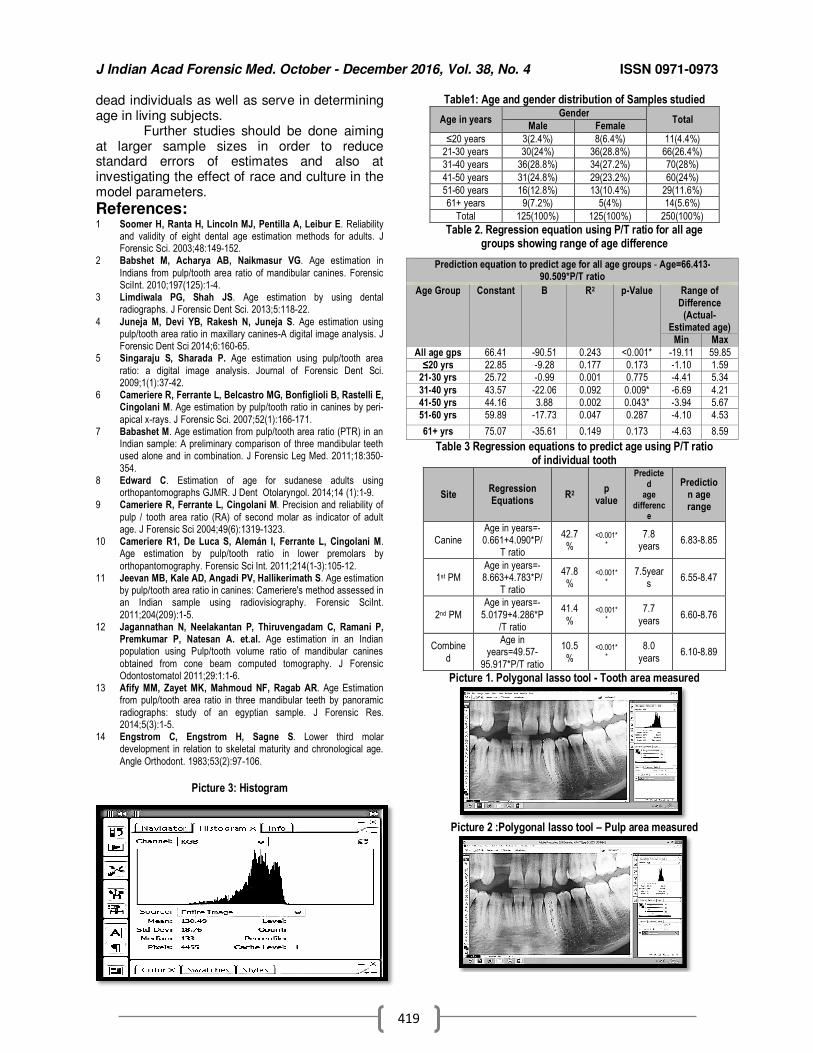
J Indian Acad Forensic Med. October - December 2016, Vol. 38, No. 4 ISSN 0971-0973
419
dead individuals as well as serve in determining age in living subjects. Further studies should be done aiming at larger sample sizes in order to reduce standard errors of estimates and also at investigating the effect of race and culture in the model parameters.
References: 1 Soomer H, Ranta H, Lincoln MJ, Pentilla A, Leibur E. Reliability
and validity of eight dental age estimation methods for adults. J Forensic Sci. 2003;48:149-152.
2 Babshet M, Acharya AB, Naikmasur VG. Age estimation in
Indians from pulp/tooth area ratio of mandibular canines. Forensic SciInt. 2010;197(125):1-4.
3 Limdiwala PG, Shah JS. Age estimation by using dental radiographs. J Forensic Dent Sci. 2013;5:118-22.
4 Juneja M, Devi YB, Rakesh N, Juneja S. Age estimation using pulp/tooth area ratio in maxillary canines-A digital image analysis. J Forensic Dent Sci 2014;6:160-65.
5 Singaraju S, Sharada P. Age estimation using pulp/tooth area
ratio: a digital image analysis. Journal of Forensic Dent Sci. 2009;1(1):37-42.
6 Cameriere R, Ferrante L, Belcastro MG, Bonfiglioli B, Rastelli E, Cingolani M. Age estimation by pulp/tooth ratio in canines by peri-
apical x-rays. J Forensic Sci. 2007;52(1):166-171. 7 Babashet M. Age estimation from pulp/tooth area ratio (PTR) in an
Indian sample: A preliminary comparison of three mandibular teeth used alone and in combination. J Forensic Leg Med. 2011;18:350-
354. 8 Edward C. Estimation of age for sudanese adults using
orthopantomographs GJMR. J Dent Otolaryngol. 2014;14 (1):1-9. 9 Cameriere R, Ferrante L, Cingolani M. Precision and reliability of
pulp / tooth area ratio (RA) of second molar as indicator of adult age. J Forensic Sci 2004;49(6):1319-1323.
10 Cameriere R1, De Luca S, Alemán I, Ferrante L, Cingolani M. Age estimation by pulp/tooth ratio in lower premolars by
orthopantomography. Forensic Sci Int. 2011;214(1-3):105-12. 11 Jeevan MB, Kale AD, Angadi PV, Hallikerimath S. Age estimation
by pulp/tooth area ratio in canines: Cameriere's method assessed in an Indian sample using radiovisiography. Forensic SciInt.
2011;204(209):1-5. 12 Jagannathan N, Neelakantan P, Thiruvengadam C, Ramani P,
Premkumar P, Natesan A. et.al. Age estimation in an Indian population using Pulp/tooth volume ratio of mandibular canines
obtained from cone beam computed tomography. J Forensic Odontostomatol 2011;29:1:1-6.
13 Afify MM, Zayet MK, Mahmoud NF, Ragab AR. Age Estimation from pulp/tooth area ratio in three mandibular teeth by panoramic
radiographs: study of an egyptian sample. J Forensic Res. 2014;5(3):1-5.
14 Engstrom C, Engstrom H, Sagne S. Lower third molar development in relation to skeletal maturity and chronological age.
Angle Orthodont. 1983;53(2):97-106.
Picture 3: Histogram
Table1: Age and gender distribution of Samples studied
Age in years Gender
Total Male Female
≤20 years 3(2.4%) 8(6.4%) 11(4.4%)
21-30 years 30(24%) 36(28.8%) 66(26.4%)
31-40 years 36(28.8%) 34(27.2%) 70(28%)
41-50 years 31(24.8%) 29(23.2%) 60(24%)
51-60 years 16(12.8%) 13(10.4%) 29(11.6%)
61+ years 9(7.2%) 5(4%) 14(5.6%)
Total 125(100%) 125(100%) 250(100%)
Table 2. Regression equation using P/T ratio for all age groups showing range of age difference
Table 3 Regression equations to predict age using P/T ratio of individual tooth
Site Regression Equations
R2 p
value
Predicted
age differenc
e
Prediction age range
Canine Age in years=-0.661+4.090*P/
T ratio
42.7%
<0.001**
7.8 years
6.83-8.85
1st PM Age in years=-8.663+4.783*P/
T ratio
47.8%
<0.001**
7.5years
6.55-8.47
2nd PM
Age in years=-
5.0179+4.286*P/T ratio
41.4
% <0.001*
*
7.7
years 6.60-8.76
Combine
d
Age in years=49.57-
95.917*P/T ratio
10.5
% <0.001*
*
8.0
years 6.10-8.89
Picture 1. Polygonal lasso tool - Tooth area measured
Picture 2 :Polygonal lasso tool – Pulp area measured
Prediction equation to predict age for all age groups - Age=66.413-90.509*P/T ratio
Age Group Constant Β R2 p-Value Range of
Difference (Actual-
Estimated age)
Min Max
All age gps 66.41 -90.51 0.243 <0.001* -19.11 59.85
≤20 yrs 22.85 -9.28 0.177 0.173 -1.10 1.59
21-30 yrs 25.72 -0.99 0.001 0.775 -4.41 5.34
31-40 yrs 43.57 -22.06 0.092 0.009* -6.69 4.21
41-50 yrs 44.16 3.88 0.002 0.043* -3.94 5.67
51-60 yrs 59.89 -17.73 0.047 0.287 -4.10 4.53
61+ yrs 75.07 -35.61 0.149 0.173 -4.63 8.59
J Indian Acad Forensic Med. October - December 2016, Vol. 38, No. 4 ISSN 0971-0973
420
Original Research Paper
Footprint Breadth Dimensions: Is it possible to determine sex and/or side?
1Ashish Badiye,
2Hansi Bansal,
3Anjali Rahatgaonkar,
4Mukesh Yadav
Abstract Determination of the sex of an unidentified individual from footprint or its part may contribute
towards the establishment of identity and elimination of the suspects of the non-contributing sex from the investigation. The main aim of the present work was to study the possible existence of sexual dimorphism and bilateral differences in the footprint breadth dimensions (at Heel and Ball region) and in the indices derived from these dimensions. This study included 400 healthy adult subjects comprising of 200 males and 200 females in the age group of 18-35 years. Thus, a total of 800 foot prints (200 x 2 of males and females each) were obtained using standard ink method. The breadth at ball (BBAL) and breadth at heel (BHEL) regions were measured and tabulated. Heel ball index [(BHEL÷ BBAL) × 100] and Ball heel index [(BBAL÷ BHEL) × 100] were derived for both males and females and t-test was applied to test any statistical significance (at p<0.05 level). The study concludes that, breadth dimensions of footprints can aid in the side (left/right) and sex determination. However, HB & BH indices do not serve as reliable indicators for determining sex but will be helpful in determination of the side
Key Words: Heel - ball index; Forensic; Ball - Heel index; Partial footprint.
Introduction: Footprints, like various other
impressions (fingerprints, thumb prints, lip prints, palmprints etc.), are often encountered at the scene of crime as important evidence. In countries like India, where majority of people still prefer to go bare-footed, footprints often afford good clues.[1] Partial or complete footprints may be found on rain covered surfaces, freshly waxed floors, wet cemented surfaces, moist surfaces, in soil, mud, sand, paint, blood, etc.[2] Determining the identity of a person is of utmost importance in forensic cases. Even if complete identification is not possible, establishment of certain facts like age, sex, stature or ethnicity may lead to partial identification thereby reducing the burden on the investigating officer.[2,3]
Corresponding Author: 1Assistant Prof. & Head,
2Assistant Prof
Department of Forensic Science, Govt. Institute of Forensic Science, Nagpur.
3Joint Director, Higher Education (Nagpur Region),
Nagpur, Maharashtra
4Principal & Professor, N.C. Medical College, Panipat,
Haryana, India Email: [email protected] DOR: 25.2.16 DOA: 25/12/2016 DOI: 10.5958/0974-0848.2016.00105.6
Individualistic characteristics of footprint can help the investigators in the identification of the suspect.[4-6] By determining the sex of the accused from the prints left at the crime scene, the onus of the investigator is lessened. Footprints[7-9] and footprint ridge density[10] have been studied by many researchers for their usefulness in sex determination.
Various studies have been done
for stature estimation through footprints and its various parts.[11-15]
Regression models and correlations have been obtained by few researchers on the basis of footprints and other body parts.[17,18] The ability
of HB Index for determination of sex
is very less explored. Kanchan, et al.[22] derived heel ball index from footprint measurements
to
differentiate between sexes while Krishna K, et al[19] used foot measurements for the same. In general, males have larger body size, more massive joints and stronger musculature compared with females. However, the degree of this sexual dimorphism varies between populations [20].
The present work aims to infer whether sexual dimorphism and bilateral differences exist in footprint breadth dimensions (i.e. breadth at ball and heel regions) and the indices derived [i.e. Heel-Ball (HB) index and Ball-Heel (BH) index] from these dimensions.
J Indian Acad Forensic Med. October - December 2016, Vol. 38, No. 4 ISSN 0971-0973
421
Material and methods: Sample size and exclusion criteria: The
study was conducted on 400 healthy adult individuals of Central Indian population comprising of 200 males and 200 females aged between 18-35 yrs. The subjects with any sign of disease, pathological condition, injury &/or defect of feet were excluded from the study.
Method: A clean metallic plate was uniformly smeared with black impression ink (Kores India Limited) with the help of a rubber roller. Each individual was asked to wash their feet and after drying they were asked to apply their feet to the smeared plate and then place them onto white paper by standing upright with equal pressure on both feet. Thus, a total of 800 prints were obtained of both left and right feet.
Analysis: Foot breadth at the ball (BBAL) was measured as the widest part of the foot at the ball whilst the foot breadth at heel (BHEL) was measured as the widest part of the heel [Fig 1]. The measurements thus obtained, were computed and analyzed. The HB Index (Heel Ball Index) was derived as (BHEL÷ BBAL) × 100 and BH index (Ball Heel Index) was derived as (BBAL÷BHEL) × 100. Bilateral differences were also calculated.
Statistical Analysis: The obtained results were statistically analyzed using t-test with alpha level of significance at p<0.05 & p<0.01 levels. Kappa (K) value was also calculated to check the inter-observer agreement strength.
Ethical approval: Informed consent was taken from all the participating individuals. Necessary ethical approval was obtained from the institute ethics committee for the study.
Results and Discussion: Descriptive statistics of breadth
dimensions at the ball and heel regions in male and female footprints are shown in Table 1. The measurements of footprints at ball region are higher as compared to the measurements at heel region in both males and females. Also, both the breadth measurements i.e. at heel and ball regions are significantly higher in males than females which are in accordance with various study results of other researchers.[7,8,16,19,21] The calculated Kappa (K) values showed that the strength of agreement between both the observers was ‘good’.
In males, the mean HB indices on right and left sides were 55.56 and 54.07 respectively, while in females; the mean HB index was 55.69 on the right and 53.10 on the left side (Table 2). The sex differences were insignificant. The HB index on the right footprint in females was marginally higher than in males.
The result is in partial accordance with the study conducted by Kanchan, et al,[22] who reported higher HB index in males. Our results are in accordance with Krishna, et al,[19] and Kanchan,
et al,[22] who also reported
insignificant sex differences in the HB index. Our results are in contrast with the findings by Krishna, et al,[19] who calculated HB index on foot measurements and obtained higher indices for females. The contrast in the study proved that the index values which are derived on foot measurements can’t be applied on foot print measurements.
Ball Heel Index (BH Index) values were higher for females. In males, the mean BH indices on right and left sides were 180.98 & 185.86 resp., while in females; the mean BH index was 180.37 on the right and 189.46 on the left side (Table 3). The sex differences were not significant.
In males, bilateral (Right/Left) differences were significant for ball breadth, HB index and BH index but were insignificant for heel breadth (Table 4). Whereas in females, the bilateral differences were significant for all the four parameters (i.e. ball & heel breadths, HB and BH indices). Bilateral asymmetry is a complex phenomenon and variations have been observed in different studies as well. The exact cause for asymmetry on a particular side of males and females in our study conversely, cannot be explained and is a matter of further investigation as suggested by Kanchan, et al.[22]
A comparison of HB index derived from foot print measurements in the present study, and only two such similar available studies by Kanchan, et.al,[22]
on footprint measurements
and by Krishna, et.al,[19] on foot measurements is done (Table 5). In accordance to the findings of Kanchan, et al,[22] the comparison confirms that differences do exist between the indices derived on the footprint measurements and foot measurements. The HB index on foot measurements is higher than those derived on footprint measurements. The observed variations in the current and earlier studies may be accredited to the differences in study populations and probably the method used. This difference needs to be studied and explored further.
Identification of sex is an important parameter contributing to the biological profile of the unidentified individual. While determining sex from incomplete or partial foot prints, ball and heel are the areas which are of much concern as they are more commonly found at a crime scene. Even if a complete footprint is not
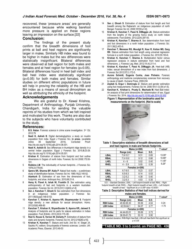
J Indian Acad Forensic Med. October - December 2016, Vol. 38, No. 4 ISSN 0971-0973
422
recovered, these ‘pressure areas’ are generally encountered because while walking barefoot more pressure is applied on these regions leaving an impression on the surface.[22]
Conclusion: The findings of the present study
confirm that the breadth dimensions of foot prints at ball and heel regions are significantly larger in males. Similarly HB index was found to be higher in males but the sex differences were statistically insignificant. Bilateral differences were observed at ball region for both males and females and at heel region for females (p<0.05). Bilateral differences found in heel ball index and ball heel index were statistically significant (p<0.05) for both males and females. Similar studies on different ethnic populations in future will help in proving the reliability of the HB and BH index as a means of sexual dimorphism as well as attributing the ethnicity of the footprint.
Acknowledgements We are grateful to Dr. Kewal Krishna,
Department of Anthropology, Punjab University, Chandigarh, India for sending the valuable reprints of his studies from which we felt inspired and motivated for this work. Thanks are also due to the subjects who have voluntarily contributed to the study.
References: 1 B.S. Nabar. Forensic science in crime scene investigation. 3rd Ed.
2012:79
2 Neeti K, Ashish B. Digital dermatoglyphics: a study on muslim population from india. Egypt J Forensic Sci, in press. (available online 06 September 2014). Corrected Proof: http://dx.doi.org/10.1016/j.ejfs.2014.08.001.
3 Neeti K, Ashish B. Sex differences in thumbprint ridge density in a central Indian population. Egypt J Forensic Sci. 2015;5:23-29. http://dx.doi.org/10.1016/j.ejfs.2014.05.001.
4 Krishan K. Estimation of stature from footprint and foot outline
dimensions in Gujjars of north India. Forensic Sci Int 2008;175:93-101.
5 Robbins LM. The individuality of human footprints. J Forensic Sci. 1978;23:778-85.
6 Qamra SR, Sharma BP, Kaila P. Naked foot marks – a preliminary study of identification factors. Forensic Sci Int. 1980;16(2):145-52.
7 Atamturk D. Estimation of sex from the dimensions of foot, footprints, and shoe. Anthropol Anz. 2010;68:21-9.
8 Hemy N, Flavel A, Ishak NI, Franklin D. Sex estimation using anthropometry of feet and footprints in a western Australian population. Forensic Sci Int. 2013;231(1-3)[402.e1-6]
9 Sen J, Kanchan T, Ghosh S. Sex estimation from foot dimensions
in an indigenous Indian population. J Forensic Sci. 2011;56(Suppl):148-S153.
10 Kanchan T, Krishan K, Aparna KR, Shyamsunder S. Footprint ridge density: a new attribute for sexual dimorphism. Homo.
2012;63(6):468-80 11 Kanchan T, Krishan K, ShyamSunder S, Aparna KR, Jaiswal S.
Analysis of footprints and its parts for stature estimation in Indian population. Foot (Edinb). 2012;22(3):175–80.
12 Reel S, Rouse S, Vernon W, Doherty P. Estimation of stature from
static and dynamic footprints. Forensic Sci Int. 2012;219[283.e15]. 13 Krishan K, Kanchan T. Stature and build. 2nd Ed. In: Siegel JA,
Saukko PJ, editors. Encyclopedia of forensic sciences. London, UK: Academic Press, Elsevier; 2013:49-53
14 Sen J, Ghosh S. Estimation of stature from foot length and foot breadth among the Rajbanshi: an indigenous population of north Bengal. Forensic Sci Int. 2008;181[55.e1-6].
15 Krishan K, Kanchan T, Passi N, DiMaggio JA. Stature estimation from the lengths of the growing foot-a study on north Indian adolescents. Foot (Edinb). 2012;22(4):287-93.
16 Krishan K, Kanchan T, Sharma A. Sex determination from hand
and foot dimensions in a north Indian population. J Forensic Sci. 2011;56(2):453-9.
17 Kanchan T, Menezes RG, Moudgil R, Kaur R, Kotian MS, Garg RK. Stature estimation from foot length using universal regression
formula in a north Indian population. J Forensic Sci. 2010;55:163-6. 18 Krishan K, Kanchan T, Sharma A. Multiplication factor versus
regression analysis in stature estimation from hand and foot dimensions. J. Forensic Leg. Med. 2012;19 (4):211-4.
19 Krishan K, Kanchan T, Passi N, DiMaggio JA. Heel–ball (HB) index: sexual dimorphism of a new index from foot dimensions. J Forensic Sci 2012;57(1):172-5.
20 Aurore Schmitt, Eugenia Cunha, Joao Pinheiro. Forensic
anthropology and medicine complementary sciences from recovery to cause of death. Humana Press, 2006:234
21 Zeybek G, Ergur I, Demiroglu Z. Stature and gender estimation using foot measurements. Forensic Sci Int. 2008;181(1-3) [54.e1-5].
22 Kanchan K, Krishan k, Prusty D, Machado M. Heel–Ball index: An analysis of foot print dimensions for determination of sex, Egypt J Forensic Sci. 2014, http://dx.doi.org/10.1016/j.ejfs.2014.02.002
Figure 1: Representation of the landmarks used for measurements on the footprint. (Not to scale)
Table 1: Descriptive statistics of breadth dimensions at ball
and heel regions in male and female footprints. Males (n=200) T value#
Range (cm) Mean S.D.
13.09* 10.89* 11.49* 9.28*
LBAL 7.8-10.7 9.31 0.69
RBAL 7.5-10.6 9.12 0.66
LHEL 4 – 6 5.02 0.40
RHEL 4 – 5.9 5.05 0.39
Females (n=200)
LBAL 6.7-9.5 8.23 0.57
RBAL 6.5-9.3 8.14 0.53
LHEL 3.2-5.2 4.38 0.40
RHEL 3.2-5.3 4.53 0.39 S.D. – Standard Deviation, LBAL – Left footprint breadth at ball, RBAL – Right
footprint breadth at ball, RHEL – Right footprint breadth at heel, LHEL – Left footprint breadth at heel. * Significant at p ≤ 0.05P value <0.00001
Table 2: Descriptive Statistics of Heel ball indices derived for males and females.
Males (n=200) T value#
Range (cm) Mean S.D.
0.24 -1.75
RBH Index 45.92-65.33 55.56 4.04
LBH Index 42.55-64.1 54.07 3.77
Females (n=200)
RBH Index 44.44-63.01 55.69 3.73
LBH Index 38.55-61.73 53.10 3.95 S.D.-standard deviation, RHB-Right Heel Ball, LHB-Left Heel Ball # Not significant at
p ≤ 0.05
**TABLE NO. 3 to 5 contd. on PAGE NO. 436
J Indian Acad Forensic Med. October - December 2016, Vol. 38, No. 4 ISSN 0971-0973
423
Original Research Paper
Analysis of Sudden Death Cases Brought for Postmortem Examination at Sir T. General Hospital, Bhavnagar
1Love R. Bhagora,
2Amit P. Parmar,
3Dipti C. Parmar,
4 Tejas C. Patel
Abstract Background: Sudden death, especially when it occurs in an apparently healthy person, can have a great impact on society. Sudden deaths undoubtedly constitute a significant portion of deaths which undergo autopsy for investigation of death. Material and Methodology: The present prospective study was conducted at the Department of Forensic Medicine & Toxicology, Government Medical College & Sir T. General Hospital, Bhavnagar, during the period from 1st May 2014 to 30th April 2015. Aims and Objectives: The aim of the study was to include the scenario of sudden death cases with respect to socio economic class, activity of deceased at the time of onset of symptoms, past and personal history like food habit and any bad habit, time of onset of symptoms, period of survival etc. Results & Observations: More than half numbers of cases [139 (52.1%)] belonged to middle class. In majority of cases, [136 (50.9%)], the symptoms started, when person was engaged in routine day to day activity. In majority of sudden death cases [86 (32.2%)], time of onset of symptoms was seen during 06:01 a.m. to 12:00 p.m. In our study, majority of sudden death victims were of mixed diet (48.3%), while 44.2% victims were pure vegetarians. Conclusions: Health policy research must be strengthened. Health education of public and patients is a priority. Although strategies for behaviour modification in relation to specific dietary practices may require further research within each population.
Key Words: Sudden Death, Coronary Artery Disease, Autopsy
Introduction: Virtually all Forensic Experts deal not
only with suspicious, accidental and suicidal deaths, but with a wide range of deaths from natural causes. Many of these are sudden, unexpected, clinically unexplained, or otherwise obscure, even though there need be no unnatural element in their causation.[1]
Corresponding Author: 1Associate Professor,
Dept. of Forensic Medicine & Toxicology, Ananta institute of Medical science & Research centre, Kaliwas (Rajasthan)
2Professor & Head,
Dept. of Forensic Medicine & Toxicology, 3Associate Professor,
Dept. of Obstetrics and Gynaecology Govt. Medical College, Bhavnagar.
4Tutor,
Dept. of Forensic Medicine and Toxicology, GMERS Medical College, Patan. Email: [email protected] DOR: 5.6.16 DOA: 25/12 2016 DOI: 10.5958/0974-0848.2016.00106.8
The most widely accepted definition of sudden death is a death which is not known to have been caused by any trauma, poisoning or violent asphyxia, and where death occurs all on a sudden or within 24 hours of the onset of terminal symptoms.[2]
Sudden natural death, especially when it occurs in an apparently healthy person, can have a great impact on society.[3] Sudden natural deaths undoubtedly constitute a significant portion of deaths which undergo autopsy for investigation of death.
Material and Methodology: The present prospective study was
conducted at the Department of Forensic Medicine & Toxicology, Government Medical College & Sir T. General Hospital, Bhavnagar, during the period from 1
st May 2014 to 30
th April
2015. The material for the present study consisted of all the cases of sudden death brought to the mortuary complex of Sir T. General Hospital, Bhavnagar, for post mortem examination. A total of 1254 medicolegal autopsies were conducted during this period, of which 267 cases (21.3%) were of sudden death.
Results & Observations: In our study we observed that more than
half number of cases [139 (52.1%)] belonged to
J Indian Acad Forensic Med. October - December 2016, Vol. 38, No. 4 ISSN 0971-0973
424
the middle class, followed by lower class [84 cases (31.5%)]. Upper class constituted only 24 cases (9%).[Table - 1]
In majority of cases, [136 (50.9%)], the symptoms started when the patient was engaged in the routine day to day activity, which ultimately lead to sudden death; while in 83 cases (31.1%), symptoms started when the deceased was resting. Only in 19 cases, (7.1%), symptoms started during strenuous activity. [Table - 2]
Again, H/o alcohol intake, smoking & chewing tobacco was present in 123 victims (46.1%), 111 cases (41.6%) & 65 cases (25.5%), respectively. While the same was absent in 124 cases (46.4%), 136 cases (50.9%) & 179 cases (67%), respectively. [Table - 3] 15 victims (5.6%) were known cases of diabetes, while 29 (10.9%) were known cases of hypertension. [Table - 4]
In majority of the cases, [86 (32.2%)], time of onset of symptoms was seen during 06:01 a.m. to 12:00 p.m., while the least number of cases [36 (13.48%)] were seen during 00:00 a.m. to 06:00 a.m. [Table - 5]
20 victims (7.5%) were found dead. These were unknown/ unidentified; while, 115 victims (43.1%) died within 1 hour or were brought dead to the hospital & 105 (39.3%) died within 1-6 hours. [Table - 6]
Majority of the victims were of mixed diet [129 (48.3%)], while 118 victims (44.2%) were pure vegetarians. [Table - 7]
Discussion: In our study, most of the victims were
from the middle class families (52.1%), followed by the lower class families (31.5%). This is more or less similar with the findings of Neerav Rana [4] & Chaudhary VA [5]. The more preponderance in middle class and lower class family is due to low affordability to medical facilities. Due to financial problem, they might not get regular medical checkups for early detection of their diseases or even treatment for their diseases.
Again, in majority of cases (50.9%), the symptoms started when the victim was engaged in the routine day to day activity, which ultimately lead to their death. This finding is similar to study by Neerav Rana [4]. This concludes that sudden death can happen during day to day activity as well as during rest/sleep, which is very well highlighted in the present study. So we can say that, strenuous activity does not always exaggerate sudden death.
History of alcohol intake, smoking & chewing tobacco was present in 46.1%, 41.6% &
25.5% respectively. Nearly similar findings were observed in the study done by Mukhopadhyay S[6].
However,
association between drinking
alcohol and smoking with sudden death was not found in studies by Madhavan S [7]
& Neerav
Rana [4] as these habits might have been under reported due to social taboos.
In our study, hypertension was present in 10.9% cases, which was quite similar with the study of Neerav Rana[4], while it was in contrast with the studies of Mukhopadhyay S[6]
&
Madhavan S [7]. In the present study, diabetes was present in 5.6% cases, which was quite similar with the study of Mukhopadhyay S [6], while it was in contrast with the studies of Neerav Rana [4]
& Madhavan S [7]. Diabetes
increases the risk of coronary artery disease, a condition that is commonly found in association with sudden death. However, there may be diabetes specific accelerated forms of atherosclerosis with enhanced thrombogenicity. Hypertension is also a major risk factor for sudden death due to cardio vascular system involvement.
Again, in majority of the cases (32.2%), time of onset of symptoms was seen during 06:01 a.m. to 12:00 p.m., followed by 12:01 p.m. to 18:00 p.m. These findings are similar to those of Chaudhary VA [5] and Neerav Rana [4]. It is suggest that, in the morning hours, because there is more stress of daily schedule, transportation, fulfilment of duties etc. There is a higher frequency of the deaths.
In our study, 43.1% victims died within 1 hour or were brought dead to the hospital. This coincides with the observations of Neerav Rana [4] & Thomas A [8]. Many sudden deaths are declared only after arrival to hospital and that could be the reason for higher number of these cases.
Majority of the cases were of mixed diet, 48.3%, while 44.2% were pure vegetarians. Food habits of 20 victims (7.5%) were not known. Saturated and trans fats in the diet tend to increase LDL cholesterol in the blood. LDL cholesterol can lead to plaque forming on arteries. Common sources of saturated fats include animal products (butter, meat fat, beef, lamb, chicken skin and full cream dairy foods) and processed foods like pastries and biscuits. So, mix vegetarian person are at increased risk of cardio vascular diseases leading to sudden death.[9]
Conclusion:
Health policy research must be strengthened. Health education of public and patients is a priority. Some of the knowledge and
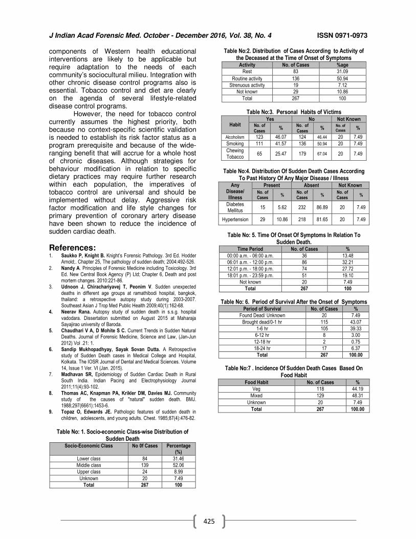
J Indian Acad Forensic Med. October - December 2016, Vol. 38, No. 4 ISSN 0971-0973
425
components of Western health educational interventions are likely to be applicable but require adaptation to the needs of each community’s sociocultural milieu. Integration with other chronic disease control programs also is essential. Tobacco control and diet are clearly on the agenda of several lifestyle-related disease control programs.
However, the need for tobacco control currently assumes the highest priority, both because no context-specific scientific validation is needed to establish its risk factor status as a program prerequisite and because of the wide-ranging benefit that will accrue for a whole host of chronic diseases. Although strategies for behaviour modification in relation to specific dietary practices may require further research within each population, the imperatives of tobacco control are universal and should be implemented without delay. Aggressive risk factor modification and life style changes for primary prevention of coronary artery disease have been shown to reduce the incidence of sudden cardiac death.
References: 1. Saukko P, Knight B. Knight’s Forensic Pathology. 3rd Ed. Hodder
Arnold;. Chapter 25, The pathology of sudden death; 2004:492-526.
2. Nandy A. Principles of Forensic Medicine including Toxicology. 3rd Ed. New Central Book Agency (P) Ltd; Chapter 6, Death and post mortem changes. 2010:221-86.
3. Udnoon J, Chirachariyavej T, Peonim V. Sudden unexpected
deaths in different age groups at ramathibodi hospital, bangkok, thailand: a retrospective autopsy study during 2003-2007. Southeast Asian J Trop Med Public Health 2009;40(1):162-68.
4. Neerav Rana. Autopsy study of sudden death in s.s.g. hospital
vadodara. Dissertation submitted on August 2015 at Maharaja Sayajirao university of Baroda.
5. Chaudhari V A, D Mohite S C. Current Trends in Sudden Natural Deaths. Journal of Forensic Medicine, Science and Law, (Jan-Jun
2012) Vol .21: 1. 6. Sandip Mukhopadhyay, Sayak Sovan Dutta. A Retrospective
study of Sudden Death cases in Medical College and Hospital, Kolkata. The IOSR Journal of Dental and Medical Sciences. Volume
14, Issue 1 Ver. VI (Jan. 2015). 7. Madhavan SR, Epidemiology of Sudden Cardiac Death in Rural
South India. Indian Pacing and Electrophysiology Journal 2011;11(4):93-102.
8. Thomas AC, Knapman PA, Krikler DM, Davies MJ. Community study of the causes of "natural" sudden death. BMJ. 1988;297(6661):1453-6.
9. Topaz O, Edwards JE. Pathologic features of sudden death in
children, adolescents, and young adults. Chest. 1985;87(4):476-82.
Table No: 1. Socio-economic Class-wise Distribution of
Sudden Death Socio-Economic Class No 0f Cases Percentage
(%)
Lower class 84 31.46
Middle class 139 52.06
Upper class 24 8.99
Unknown 20 7.49
Total 267 100
Table No:2. Distribution of Cases According to Activity of the Deceased at the Time of Onset of Symptoms
Activity No. of Cases %age
Rest 83 31.09
Routine activity 136 50.94
Strenuous activity 19 7.12
Not known 29 10.86
Total 267 100
Table No:3. Personal Habits of Victims
Habit Yes No Not Known
No. of Cases
% No. of Cases
% No. of Cases %
Alcoholism 123 46.07 124 46.44 20 7.49
Smoking 111 41.57 136 50.94 20 7.49
Chewing
Tobacco 65 25.47 179 67.04 20 7.49
Table No:4. Distribution Of Sudden Death Cases According
To Past History Of Any Major Disease / Illness Any
Disease/ Illness
Present Absent Not Known
No. of Cases
% No. of Cases
% No. of Cases
%
Diabetes Mellitus
15 5.62 232 86.89 20 7.49
Hypertension 29 10.86 218 81.65 20 7.49
Table No: 5. Time Of Onset Of Symptoms In Relation To Sudden Death.
Time Period No. of Cases %
00:00 a.m. - 06:00 a.m. 36 13.48
06:01 a.m. - 12:00 p.m. 86 32.21
12:01 p.m. - 18:00 p.m. 74 27.72
18:01 p.m. - 23:59 p.m. 51 19.10
Not known 20 7.49
Total 267 100
Table No: 6. Period of Survival After the Onset of Symptoms Period of Survival No. of Cases %
Found Dead/ Unknown 20 7.49
Brought dead/0-1 hr 115 43.07
1-6 hr 105 39.33
6-12 hr 8 3.00
12-18 hr 2 0.75
18-24 hr 17 6.37
Total 267 100.00
Table No:7 . Incidence Of Sudden Death Cases Based On Food Habit
Food Habit No. of Cases %
Veg 118 44.19
Mixed 129 48.31
Unknown 20 7.49
Total 267 100.00
J Indian Acad Forensic Med. October - December 2016, Vol. 38, No. 4 ISSN 0971-0973
426
Original Research Paper
A Study of Victims of fall from Height from 2006 to 2015 in Imphal
1Memchoubi Ph,
2Th. Meera,
3Pulak Chakma
Abstract In falls from height, a major determining factor of injury is the height. This study aims at finding
out the pattern of injuries in fatal cases of fall, and the factors associated with such cases in this part of the country. A retrospective study was conducted over a 10-year period (2006 - 2015) on cases brought for autopsy to Regional Institute of Medical Sciences (RIMS), Imphal. The information contained in this study was derived from a review of autopsy records and police reports. During the 10 year period from 2006 to 2015, of the 3986 autopsies, only 24 cases were of fall from height. Males (95.8%) outnumbered the female victims and maximum number of cases was observed in the age group 31- 40years (41.6%). Primary impact was most commonly seen on the head (79.2%), followed by hand & upper limb (8.3%). Head injury was the commonest cause of death (66.6%) with fracture of skull observed in 56.3% of the cases. The manner of death was mostly accidental (83.3%). To conclude, an analysis of pattern of injuries on a dead body found on the ground with blunt trauma on various parts of the body, would help in differentiating vehicular accident cases from ‘fall from height’ cases. Safety structures like railings, handrail, etc. to prevent such fatal falls are needed since majority of the cases in the study were accidental deaths.
Key Words: Fall From Height, Injury Patterns, Cause Of Death.
Introduction: In falls from height, a frequently encountered setting in forensic practice, it is the height, which is a major determining factor of injury, as the velocity of impact is intrinsically related to the distance of the fall to the point of a terminal velocity.[1] Upon contact with a surface, usually the ground, the falling body undergoes a deceleration and the kinetic energy is transformed into work.[2]
Therefore, by virtue of
a calculable energy transmission to the body, such cases are an ideal model for the assessment of the effects of blunt trauma to a human body. A great deal has been written of the injury pattern, i.e. the distribution of injuries on the body, in cases of falls from great height.[3,4]
Corresponding Author: 2Professor,
1Associate Professor,
3PG Trainee,
Department of Forensic Medicine, Regional Institute of Medical Sciences, Imphal. E-mail: [email protected] DOR: 03/04/16 DOA: 25/12/2016 DOI: 10.5958/0974-0848.2016.00107.X
However, although these authors have described the injury patterns in great detail, only one study[5] correlates the injury distribution with the energy involved in the fall and thus the circumstances of causation can be deduced. This study aims at finding out the pattern of injuries in fatal cases of fall, and the factors associated with such cases in this part of the country, and an attempt is also made to find out whether the death of a body recovered on the ground is the result of a fall or some other type of trauma from the distribution of injuries on the body.
Materials and methods: This retrospective autopsy study was
conducted over a 10-year period (from 2006 to 2015). The information contained in this study was derived from a review of autopsy records and police reports. All cases and injuries were caused by the free-fall model of high fall (both jumps and accidental falls were included in the study) onto a solid surface. Also, all cases in which cause of death was other than blunt force injury (e.g., drowning) were excluded from the study in order to make the sample as homogenous as possible in terms of injury mechanism. The findings were statistically analyzed in relation to demographic profile of the victims, pattern of injuries, and the factors associated with such cases of fatal fall.
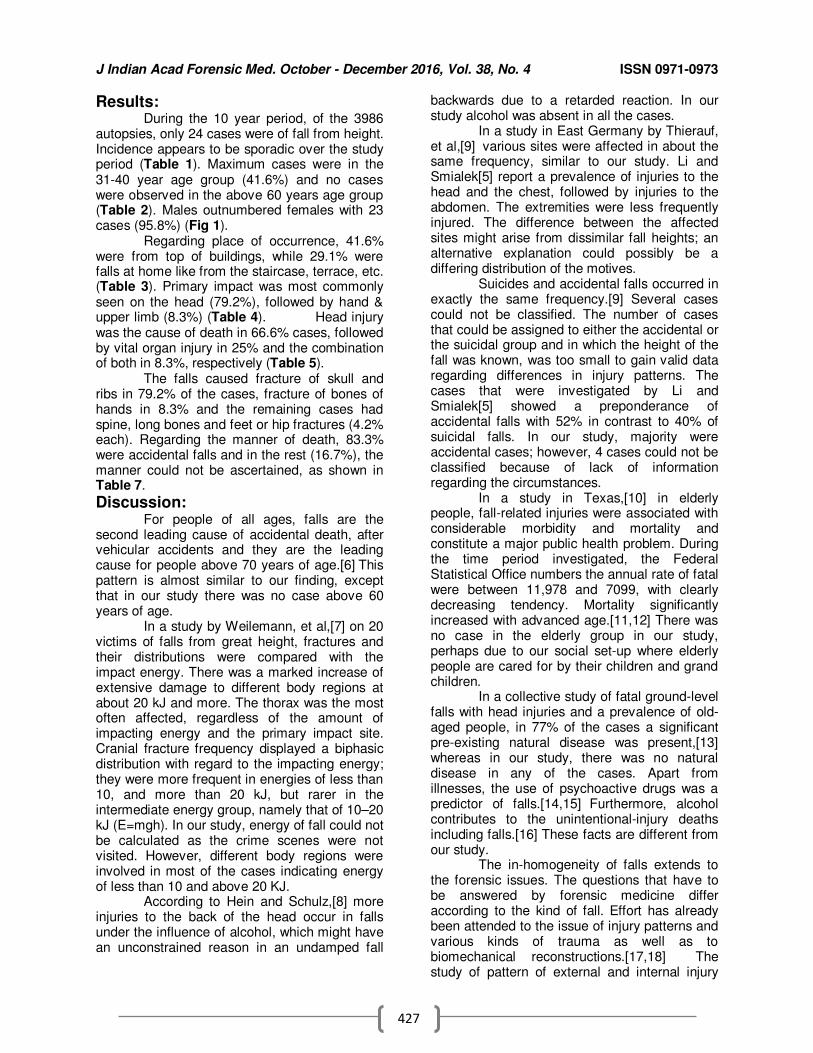
J Indian Acad Forensic Med. October - December 2016, Vol. 38, No. 4 ISSN 0971-0973
427
Results: During the 10 year period, of the 3986
autopsies, only 24 cases were of fall from height. Incidence appears to be sporadic over the study period (Table 1). Maximum cases were in the 31-40 year age group (41.6%) and no cases were observed in the above 60 years age group (Table 2). Males outnumbered females with 23 cases (95.8%) (Fig 1).
Regarding place of occurrence, 41.6% were from top of buildings, while 29.1% were falls at home like from the staircase, terrace, etc. (Table 3). Primary impact was most commonly seen on the head (79.2%), followed by hand & upper limb (8.3%) (Table 4). Head injury was the cause of death in 66.6% cases, followed by vital organ injury in 25% and the combination of both in 8.3%, respectively (Table 5).
The falls caused fracture of skull and ribs in 79.2% of the cases, fracture of bones of hands in 8.3% and the remaining cases had spine, long bones and feet or hip fractures (4.2% each). Regarding the manner of death, 83.3% were accidental falls and in the rest (16.7%), the manner could not be ascertained, as shown in Table 7.
Discussion: For people of all ages, falls are the
second leading cause of accidental death, after vehicular accidents and they are the leading cause for people above 70 years of age.[6]
This
pattern is almost similar to our finding, except that in our study there was no case above 60 years of age.
In a study by Weilemann, et al,[7] on 20 victims of falls from great height, fractures and their distributions were compared with the impact energy. There was a marked increase of extensive damage to different body regions at about 20 kJ and more. The thorax was the most often affected, regardless of the amount of impacting energy and the primary impact site. Cranial fracture frequency displayed a biphasic distribution with regard to the impacting energy; they were more frequent in energies of less than 10, and more than 20 kJ, but rarer in the intermediate energy group, namely that of 10–20 kJ (E=mgh). In our study, energy of fall could not be calculated as the crime scenes were not visited. However, different body regions were involved in most of the cases indicating energy of less than 10 and above 20 KJ.
According to Hein and Schulz,[8] more injuries to the back of the head occur in falls under the influence of alcohol, which might have an unconstrained reason in an undamped fall
backwards due to a retarded reaction. In our study alcohol was absent in all the cases.
In a study in East Germany by Thierauf, et al,[9]
various sites were affected in about the
same frequency, similar to our study. Li and Smialek[5] report a prevalence of injuries to the head and the chest, followed by injuries to the abdomen. The extremities were less frequently injured. The difference between the affected sites might arise from dissimilar fall heights; an alternative explanation could possibly be a differing distribution of the motives.
Suicides and accidental falls occurred in exactly the same frequency.[9] Several cases could not be classified. The number of cases that could be assigned to either the accidental or the suicidal group and in which the height of the fall was known, was too small to gain valid data regarding differences in injury patterns. The cases that were investigated by Li and Smialek[5] showed a preponderance of accidental falls with 52% in contrast to 40% of suicidal falls. In our study, majority were accidental cases; however, 4 cases could not be classified because of lack of information regarding the circumstances.
In a study in Texas,[10] in elderly people, fall-related injuries were associated with considerable morbidity and mortality and constitute a major public health problem. During the time period investigated, the Federal Statistical Office numbers the annual rate of fatal were between 11,978 and 7099, with clearly decreasing tendency. Mortality significantly increased with advanced age.[11,12] There was no case in the elderly group in our study, perhaps due to our social set-up where elderly people are cared for by their children and grand children.
In a collective study of fatal ground-level falls with head injuries and a prevalence of old-aged people, in 77% of the cases a significant pre-existing natural disease was present,[13] whereas in our study, there was no natural disease in any of the cases. Apart from illnesses, the use of psychoactive drugs was a predictor of falls.[14,15]
Furthermore, alcohol
contributes to the unintentional-injury deaths including falls.[16] These facts are different from our study.
The in-homogeneity of falls extends to the forensic issues. The questions that have to be answered by forensic medicine differ according to the kind of fall. Effort has already been attended to the issue of injury patterns and various kinds of trauma as well as to biomechanical reconstructions.[17,18]
The
study of pattern of external and internal injury
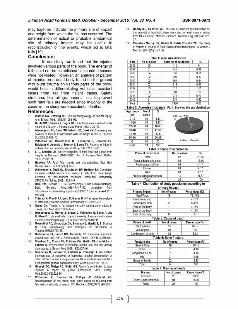
J Indian Acad Forensic Med. October - December 2016, Vol. 38, No. 4 ISSN 0971-0973
428
may together indicate the primary site of impact and height from which the fall has occurred. The determination of actual or probable anatomical site of primary impact may be useful in reconstruction of the events, which led to fatal falls.[19]
Conclusion: In our study, we found that the injuries
involved various parts of the body. The energy of fall could not be established since crime scenes were not visited. However, an analysis of pattern of injuries on a dead body found on the ground with blunt trauma on various parts of the body, would help in differentiating vehicular accident cases from ‘fall from height’ cases. Safety structures like railings, handrail, etc. to prevent such fatal falls are needed since majority of the cases in the study were accidental deaths.
References: 1. Warner KG. Demling RH. The pathophysiology of free-fall injury,
Ann. Emerg. Med. 1986;15:1088–93. 2. Gupta SM, Chandra J, Dogra TD. Blunt force lesions related to the
height of a fall, Am J Forensic Med Pathol.1982; 3:35–43. 3. Atanasijevic TC, Savic SN, Nikolic SD, Djoki VM. Frequency and
severity of injuries in correlation with the height of fall, J. Forensic Sci.2005;50:608–12.
4. Velhamos GC, Demetriades D, Theodorou D, Cornwell EE, Belzberg H, Asensio J, Murray J, Berne TV. Patterns of injury in
victims of urban free-falls, World J Surg. 1997;21:816–21. 5. Li L, Smialek JE, The investigation of fatal falls and jumps from
heights in Maryland (1987–1992), Am. J. Forensic Med. Pathol. 1994;15:295-99.
6. Hoskins AF. Fatal falls: trends and characteristics, Stat. Bull. Metrop. Insur. Co.1998;79(2):10–5.
7. Weilemann Y, Thali MJ, Kneubuehl BP, Bolliger SA. Correlation between skeletal trauma and energy in falls from great height
detected by post-mortem multislice computed tomography (MSCT).For Sci Int. 2008;180:81–5.
8. Hein PM, Schulz E. Die sturzbedingte Scha¨delhirnverletzung, Beitr. Gerichtl. Med.1989:47:447–50. Available from
https://www.ncbi.nlm.nih.gov/pubmed/2818517 [Last accessed 2015 Mar 20].
9. Thierauf A, PreuB J, Lignitz E, Madea B. A Retrospective analysis of fatal falls. Forensic Science International.2010;198:92–6.
10. Orces CH. Trends in fall-related mortality among older adults in Texas, Tex. Med.2008;104(5):55-9.
11. Demetriades D, Murray J, Brown C, Velmahos G, Salim A, Alo K, Rhee P. High level falls: type and severity of injuries and survival
outcome according to age, J.Trauma.2005;58(2):342-45. 12. Mosenthal AC, Livingston DH, Elcavage J, Merritt S, S. Stucker
S. Falls: epidemiology and strategies for prevention. J. Trauma.1995;38:753-56.
13. Hartshorne NJ, Harruff RC, Alvord Jr. EC. Fatal head injuries in ground-level falls, Am. J. Forensic Med. Pathol. 1997;18(3):258-64.
14. Sheahan SL, Coons SJ, Robbins CA, Martin SS, Hendricks J, Latimer M. Psychoactive medication, alcohol use and falls among
older adults, J. Behav. Med.1995;18(2):127-40. 15. Stenbacka M, Jansson B, Leifman A, Romelsjo A. Association
between use of sedatives or hypnotics, alcohol consumption or other risk factors and a single injurious fall or multiple injurious falls:
a longitudinal general population study, Alcohol.2002;28(1):9-16. 16. Girasek DC, Gielen AC, Smith GS. Alcohol’s contribution to fatal
injuries: a report on public perceptions, Ann. Emerg. Med.2002;39(2):622-30.
17. O’Riordain K, Thomas PM, Phillips JP, Gilchrist MD. Reconstruction of real world head injury accidents resulting from falls using multibody dynamics, Clin.Biochem. 2003;18(7):590-600.
18. Doorly MC, Gilchrist MD. The use of accident reconstruction for the analysis of traumatic brain injury due to head impacts arising from falls, Comput. Methods Biomech. Biomed. Eng.2006;9(6):371-
77. 19. Vasudeva Murthy CR, Harish S, Girish Chandra YP. The Study
of Pattern of Injuries in Fatal Cases of fall from Height. Al Ameen J Med Sci (20 12)5 (1):45 -52.
Table 1: Year- Wise Incidence
Year No of Cases Total no of autopsies %
2006 00 410 0
2007 01 545 0.18
2008 04 650 0.61
2009 02 591 0.33
2010 02 364 0.54
2011 02 313 0.63
2012 05 310 1.61
2013 05 301 1.66
2014 02 271 0.73
2015 01 231 0.43
Total 24 3986 0.60
Table 2: Age-wise incidence Fig 1. Showing the sex distribution
Age range (yrs)
No of cases
%
0-10 01 4.17
11-20 01 4.17
21-30 04 16.66
31-40 10 41.67
41-50 03 12.5
51-60 05 20.83
>61 00 00
Total : 24 100
Table 3: Place of occurrence Place of occurrence No. of cases %
Home 07 29.16
Road side(electric pole) 04 16.66
Public places(office) 01 4.17
Flyover 01 4.17
Tree 01 4.17
From building(bazaar,etc) 10 41.67
Total 24 100
Table 4: Distribution of study population according to primary impact
Primary Impact No. of cases Percentage (%)
Head/Face 19 79.16%
Feet/Lower limb 1 4.16%
Hand/Upper limb 2 8.33%
Front of the body 1 4.16%
Back of the body 1 4.16%
Side of the body 0 0
Table 5: Cause of death Cause of death No of cases Percentage (%)
Head injuries 16 66.67
Vital organs 06 25
Combination of both 02 8.33
Table 6: Bone fracture Fracture site No of cases Percentage (%)
Skull & Ribs 19 79.16
Spine 01 4.16
Long bone & Feet 01 4.16
Hip 01 4.16
Bones of hands 02 8.33
Total 24 100
Table 7: Manner of death Manner No of cases Percentage (%)
Accident 20 83.33
Others (unascertained) 04 16.67
Total: 24 100
95.8
%
4.17
%
Males Females
J Indian Acad Forensic Med. October - December 2016, Vol. 38, No. 4 ISSN 0971-0973
429
Original Research Paper
Role of Hand Anthropometry for Stature Estimation in Females of Bhavnagar - Gujarat
1T.C. Patel,
2A.P. Parmar,
3D.C. Parmar,
4S.I. Patel,
5L.R. Bhagora
Abstract Introduction: Anthropometry, the typical and traditional tool of physical anthropology, provides
the scientific methods and techniques for taking various measurements for observations of living and their skeleton. Therefore, it is being widely used in medical science, especially in forensic sciences, for identification, which is an important step in crime investigation. Estimation of stature from different body parts and their relationship is an area of interest to forensic experts, anthropologists and anatomists. Objectives: To find out the relationship between stature and hand measurements and derive population specific regression equations for estimation of stature in females. Methods: In present study, a total of 250 healthy female individuals between 18 - 25 years of age belonging to Bhavnagar region, were selected. All the individuals were measured for hand length & hand breadth separately on the right and left side and stature. All the data was subjected to statistical analysis using Microsoft Excel 2007 Software (Data Analysis). Results: The present study showed statistically significant and positive correlation between stature and measurements of hand. Stature had statistically significant (p < 0.001) and higher value of correlation(r) as 0.607, 0.594 and 0.601 for right, left and mean hand length and 0.498, 0.492 and 0.496 for right, left and mean hand breadth, respectively. Simple and multiple linear regression equations were derived for specific study population to estimate stature from hand measurements. Interpretation & Conclusion: Hand length exhibited statistically significant and positive correlation with stature as compared to hand breadth. It is also concluded that the multiple regression formulae was the better predictor for stature estimation as compared to simple regression equation.
Key Words: Stature, Hand Length & Breadth, Identification, Correlation, Regression Equation
Introduction: Anthropometry, the typical and traditional tool of physical anthropology, provides the scientific methods and the techniques for taking various measurements for observations of living and their skeleton.[1] Therefore, it is being widely used in medical science, especially in forensic sciences, for the identification which is an important step in crime investigation.
Corresponding Author: 1Tutor,
Dept. Of Forensic Medicine and Toxicology, GMERS Medical College, Dharpur, Patan, Gujarat
2Professor and Head, Dept. of Forensic Medicine,
3Associate Professor,Dept. of Obs. & Gynaecology,
Govt. Medical College, Bhavnagar.
4Tutor, Dept. of physiology,
GMERS Medical College, Valsad, Gujarat 5Assistant Professor, Dept. of Forensic Medicine,
Ananta institute of Medical science & Research centre, Kaliwas (Rajasthan) E-mail: [email protected] DOR: 05/03/16 DOA: 25/12/2016 DOI: 10.5958/0974-0848.2016.00108.1
In medico-legal autopsies, when an unidentified complete body is found, determination of stature for establishing the identity is rather an easy task; but in cases of mutilated, decomposed bodies and dismembered/ skeletal remains, it is a very difficult and challenging task for autopsy surgeon to establish personal identification. The forensic experts/ anthropologists can measure dimensions of available body parts for examination and put these measurements into a mathematical formula as per gender and ancestry group. Such regression equation formulae have limited application as they are specially computed for a particular population for estimation of stature. Computation of stature from this formula is not appropriate to other population as climate, heredity and nutritional status of population has been reported to have an effect on stature and length of long bones. Thus, the study on residents of one region is not necessarily applicable to other regions. Keeping this in mind, this study was carried out for estimation of stature of 250 healthy female individuals between 18 - 25 years of age
J Indian Acad Forensic Med. October - December 2016, Vol. 38, No. 4 ISSN 0971-0973
430
belonging to Bhavnagar region by regression equation formula from hand measurements.
Objectives: 1. To find out the relationship between stature
(Dependable Variable) and hand measurements (Independent Variable).
2. To derive population specific regression equations for estimation of stature from the measurements of hand in female population (mainly in 18 to 25 years age group) of Bhavnagar region.
Methodology: The present cross sectional study was conducted during 2014-15 in which a total of 250 asymptomatic, healthy female individuals between 18 - 25 years of age belonging to Bhavnagar region, were selected irrespective of their caste, religion, dietary habits or socio-economic status. Exclusion Criteria: Individuals with growth disorders, deformities, bony anomalies and h/o fracture of long bones were excluded. IRB Ethical Committee permission was taken prior to study. The procedure, aims & objectives of the study were explained to and written informed consent was obtained from each of the participants before taking measurements. Height (stature): Each individual was asked to stand barefoot on Standard anthropometric measuring instrument Anthropometer (stadiometer) in anatomical position with head being oriented in the Frankfurt's plane as the distance between the standing surface and vertex - highest point of head. Hand Length: With the help of sliding vernier caliper, hand length was measured as a straight distance from midpoint of a line connecting the styloid process of radius and ulna to the most anterior projecting point of middle finger. Hand Breadth: It was measured as the distance between the most prominent point outside of the lower epiphyses of the 2
nd
metacarpal (metacarpal radiale: point most laterally projected on the head of the 2
nd
metacarpal) to the most prominent inside point of the lower epiphyses of 5
th metacarpal
(metacarpal ulnare: the most medially projecting point of the head of the 5
th metacarpal) using of
sliding vernier caliper. A small group of individuals were selected for taking various measurements each day at a fixed time in afternoon (12:00 pm to 3:00 pm) to avoid diurnal variations. All measurements were recorded by one single investigator, to avoid inter-observer error in
methodology. All the measurements were recorded thrice and their mean was calculated for accuracy. All the measurements were taken in centimeters. All measurements of hand were taken separately for right and left side for each individual. All the data was subjected to statistical analysis using Microsoft Excel 2007 Software (Data Analysis).
Results: The results of present study showed that the mean stature ± SD was 157.89 ± 4.68, right hand length ± SD was 17 ± 0.78, left hand length ± SD was 16.95 ± 0.78, right hand breadth ± SD was 7.22 ± 0.42, left hand breadth ± SD was 7.18 ± 0.41 in female individuals of Bhavnagar region. (Table 1) Table 2 illustrates the correlation coefficients between stature and measurements of hand in females. In the present study, the highest correlation coefficient was observed between stature and right hand length as 0.607 and lowest value of correlation as 0.492 was for left hand breadth which was statistically highly significant (p < 0.001) and this relationship was also explained by graph 1 & 2 due to strong association as straight line in scatter diagrams. So, from this relationship, the regression equation formulae were derived for stature estimation from hand measurements. Paired t test was performed to analyse statistical difference between right and left side of observations for females which showed statistically highly significant bilateral variation (p < 0.001) (Table 3). Table 4 shows simple linear regression equation derived for stature estimation from hand length and hand breadth and also the values of R
2, Adjusted R
2 and SEE mentioned in
this table. The higher value of R, R2, Adjusted R
2
and lower value of SEE indicate relative accuracy and reliability of the formulae. So, the best simple linear regression formulae was derived for stature estimation in females from right hand length was as follows: Stature = 96.10 + 3.63 (RTHL) ± 3.728 Table 5 shows multiple regression equation derived for stature estimation from hand length and hand breadth measurements. So, the best multiple regression formulae was derived for stature estimation in females from right hand length and breadth measurements was as follows:Stature = 91.728 + 2.877(RTHL) + 2.387(RTHB) ± 3.645 As mentioned earlier that the reliability and accuracy of formulae depends on higher value of R, R
2, Adjusted R
2 and Lower value of
SEE. Thus, the observations of present study
J Indian Acad Forensic Med. October - December 2016, Vol. 38, No. 4 ISSN 0971-0973
431
exhibit that multiple regression formulae were better predictor of stature as compared to simple linear equations.
Discussion: The analysis of the present study shows that measurements of hand can be successfully used for estimation of stature in females by forensic experts and anthropologists in concerned study population. In present study, bilateral variations were statistically significant ( p < 0.001) for all the measurements of hand, which was supported by study by Sunil et al,[2] while on the other hand, some previous studies conducted by Pal, et al[3] and Jasuja, et al[4] reported no bilateral variation for hand length. In our study, the mean stature of females was 157.89 ± 4.68 cms and ranged between 140.2 cms to 172.4 cms which was also consistent with previous studies like Patel, et al,[5] Patel, et al,[6] Pandey, et al.[7] Table 6 shows comparison of various studies to estimate stature from hand length in females. In the present study, stature had correlation(r) with right, left and mean hand length were 0.605, 0.594 and 0.601 respectively, which were statistically highly significant (p < 0.001). Various previous studies conducted by Pandey, et al,[7] Khanapurkar, et al,[8] Rastogi, et al,[9] showed similar value of correlation coefficient while Patel, et al,[6] exhibited highest value of 'r' as 0.943. Present study also revealed the lower value of SEE as 3.728, 3.774 and 3.746 for stature estimation from right, left and mean hand length respectively, which was also observed in Patel, et al[6] and Rastogi, et al,[9] mentioned in table 6. Table 7 shows comparison of various studies to estimate stature from hand breadth in females. In present study, stature had correlation(r) with right, left and mean hand breadth were 0.498, 0.492 and 0.496 respectively, which were statistically highly significant (p < 0.001). All previous studies mentioned in table 7 showed similar lower value of 'r' and higher value of SEE as compared to present study. In present study, Stature had positive and statistically significant higher value of correlation with hand length as compared to hand breadth but hand breadth measurements could be useful to calculate stature, only When (1) whole length of hand is not available (e.g. in mutilated body parts in mass disasters). (2) Breadth measurements used in combination with length measurements to formulate multiple regression equations to enhance the accuracy of the predicted stature.
Conclusion: The procedure of measuring hand length, to use as important tool for determining the stature is simple in mass disasters or accidents, where fragmented or mutilated remains are recovered. Hand length measurement exhibited statistically significant and higher value of correlation as compared to hand breadth measurement in females (p < 0.001). In present study, it was concluded that the reliability and accuracy for estimation of stature were better in multiple linear regression equations than simple linear regression formulae. In cases of mass disasters and war casualties, the foot or hand may be found intact and well preserved in many cases as compared to other body parts. The identification and evaluation of dead bodies or dismembered body parts is become a challenging task for forensic experts. DNA analysis could be the most accurate procedure for identification but it lacks in terms of reasonableness, affordability and time consuming in developing countries like India. Hence, the identification from anthropometry like present study, involving foot and hand measurements to estimate stature could be reasonable, convenient and simple.
References: 1. Singh IP, Bhasin MK. Anthropometry. a laboratory manual on
biological anthropometry. Delhi: Kamla-raj Enterprises. 1968:18,39 2. Sunil, Dikshit PC, Aggrawal A, Rani M. Estimation of stature from
hand length. Journal of Indian Academy of Forensic Medicine. 2005;27(4):219-221.
3. Pal A, Aggarwal P, Bharati S, Panda M, Datta I, Roy PW. Anthropometric measurements of hand length and their correlation with the stature in eastern indian population. National Journal of Medical Research. 2014;4(4):303-305.
4. Jasuja OP, Singh G. Estimation of stature from hand and phalangeal length. Journal of Indian Academy of Forensic Medicine. 2004;26(3):100-106.
5. Patel SM, Shah GV, Patel SV. Estimation of height from
measurements of foot length in gujarat region. Journal of Anatomical Society of India. 2007;56(1):25-27.
6. Patel R, Parekh U and Patel P. A study of estimation of stature from hand length in Gujarat region. NHL Journal of Medical
Sciences. 2014;3(2):32-35. 7. Pandey N, Ujwal NS. Estimation of stature from dimensions of
hand in medical students. International Journal of Scientific Study. 2015; 3(1): 35-38.
8. Khanapurkar S, Radke A Estimation of stature from the measurement of foot length, hand length and head length in maharashtra region. Indian Journal of Basic & Applied Medical Research, 2012;1(2):77-85.
9. Rastogi P, Nagesh KR, Yoganarasimha K. Estimation of stature from hand dimensions of North and South Indians. Leg. Med. 2008; 10: 185-89.
10. Kaur M, Singh B, Mahajan A, Khurana BS. Anthropometric
measurements of hand length for estimation of stature in north indians. International Journal of Applied Biology and Pharmaceutical Technology. 2013;4(2):251-255.
11. Patel JP, Patel BG, Shah RK, Bhojak NR, Desai JN. Estimation of
stature from Hand Length in Gujarat region. NHL Journal of Medical Sciences. 2014;3(1):41-44.
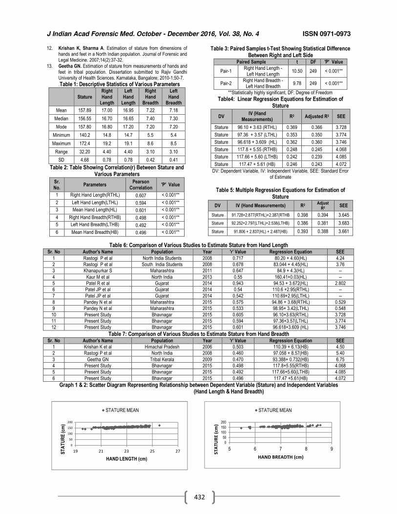
J Indian Acad Forensic Med. October - December 2016, Vol. 38, No. 4 ISSN 0971-0973
432
12. Krishan K, Sharma A. Estimation of stature from dimensions of hands and feet in a North Indian population. Journal of Forensic and Legal Medicine. 2007;14(2):37-32.
13. Geetha GN. Estimation of stature from measurements of hands and feet in tribal population. Dissertation submitted to Rajiv Gandhi University of Health Sciences. Karnataka, Bangalore; 2010-1:50-7.
.Table 1: Descriptive Statistics of Various Parameters
Stature
Right
Hand Length
Left
Hand Length
Right
Hand Breadth
Left
Hand Breadth
Mean 157.89 17.00 16.95 7.22 7.18
Median 156.55 16.70 16.65 7.40 7.30
Mode 157.80 16.80 17.20 7.20 7.20
Minimum 140.2 14.8 14.7 5.5 5.4
Maximum 172.4 19.2 19.1 8.6 8.5
Range 32.20 4.40 4.40 3.10 3.10
SD 4.68 0.78 0.78 0.42 0.41
Table 2: Table Showing Correlation(r) Between Stature and Various Parameters
Sr.
No. Parameters
Pearson
Correlation 'P' Value
1 Right Hand Length(RTHL) 0.607 < 0.001**
2 Left Hand Length(LTHL) 0.594 < 0.001**
3 Mean Hand Length(HL) 0.601 < 0.001**
4 Right Hand Breadth(RTHB) 0.498 < 0.001**
5 Left Hand Breadth(LTHB) 0.492 < 0.001**
6 Mean Hand Breadth(HB) 0.496 < 0.001**
Table 3: Paired Samples t-Test Showing Statistical Difference Between Right and Left Side
Paired Sample t DF 'P' Value
Pair-1 Right Hand Length -
Left Hand Length 10.50 249 < 0.001**
Pair-2 Right Hand Breadth -
Left Hand Breadth 9.78 249 < 0.001**
**Statistically highly significant, DF: Degree of Freedom
Table4: Linear Regression Equations for Estimation of Stature
DV IV (Hand
Measurements) R2 Adjusted R2 SEE
Stature 96.10 + 3.63 (RTHL) 0.369 0.366 3.728
Stature 97.36 + 3.57 (LTHL) 0.353 0.350 3.774
Stature 96.618 + 3.609 (HL) 0.362 0.360 3.746
Stature 117.8 + 5.55 (RTHB) 0.248 0.245 4.068
Stature 117.66 + 5.60 (LTHB) 0.242 0.239 4.085
Stature 117.47 + 5.61 (HB) 0.246 0.243 4.072
DV: Dependent Variable, IV: Independent Variable, SEE: Standard Error
of Estimate
Table 5: Multiple Regression Equations for Estimation of
Stature
DV IV (Hand Measurements) R2 Adjust
R2 SEE
Stature 91.728+2.877(RTHL)+2.387(RTHB 0.398 0.394 3.645
Stature 92.252+2.797(LTHL)+2.538(LTHB) 0.386 0.381 3.683
Stature 91.806 + 2.837(HL) + 2.487(HB) 0.393 0.388 3.661
Table 6: Comparison of Various Studies to Estimate Stature from Hand Length Sr. No Author's Name Population Year 'r' Value Regression Equation SEE
1 Rastogi P et al North India Students 2008 0.717 80.20 + 4.60(HL) 4.24
2 Rastogi P et al South India Students 2008 0.678 83.044 + 4.45(HL) 3.76
3 Khanapurkar S Maharashtra 2011 0.647 84.9 + 4.3(HL) --
4 Kaur M et al North India 2013 0.55 160.41+0.03(HL) --
5 Patel R et al Gujarat 2014 0.943 94.53 + 3.672(HL) 2.802
6 Patel JP et al Gujarat 2014 0.54 110.6 +2.95(RTHL) --
7 Patel JP et al Gujarat 2014 0.542 110.69+2.95(LTHL) --
8 Pandey N et al Maharashtra 2015 0.575 94.86 + 3.68(RTHL) 0.529
9 Pandey N et al Maharashtra 2015 0.533 98.95+ 3.42(LTHL) 0.548
10 Present Study Bhavnagar 2015 0.605 96.10+3.63(RTHL) 3.728
11 Present Study Bhavnagar 2015 0.594 97.36+3.57(LTHL) 3.774
12 Present Study Bhavnagar 2015 0.601 96.618+3.609 (HL) 3.746
Table 7: Comparison of Various Studies to Estimate Stature from Hand Breadth Sr. No Author's Name Population Year 'r' Value Regression Equation SEE
1 Krishan K et al Himachal Pradesh 2006 0.503 110.39 + 6.13(HB) 4.50
2 Rastogi P et al North India 2008 0.460 97.058 + 8.57(HB) 5.40
3 Geetha GN Tribal Kerala 2009 0.470 93.388+ 0.732(HB) 6.75
4 Present Study Bhavnagar 2015 0.498 117.8+5.55(RTHB) 4.068
5 Present Study Bhavnagar 2015 0.492 117.66+5.60(LTHB) 4.085
6 Present Study Bhavnagar 2015 0.496 117.47 +5.61(HB) 4.072
Graph 1 & 2: Scatter Diagram Representing Relationship between Dependent Variable (Stature) and Independent Variables (Hand Length & Hand Breadth)
0
50
100
150
200
5 6 7 8 9
ST
AT
UR
E (
cm)
HAND BREADTH (cm)
STATURE MEAN
0
50
100
150
200
19 21 23 25 27ST
AT
UR
E (c
m)
HAND LENGTH (cm)
STATURE MEAN
J Indian Acad Forensic Med. October - December 2016, Vol. 38, No. 4 ISSN 0971-0973
433
Original Research Paper
Histopathological examination in routine medicolegal autopsy: 3-years retrospective analysis
1Vipul N. Ambade,
2Balwant D. Kowe,
3Ajay N. Keoliya
Abstract Histopathological examination is one of the important ancillary investigations to be done in
medicolegal autopsies, particularly in sudden and natural deaths. It not only helps to know or to confirm the cause of death at autopsy but also helps in knowing the circumstances of death by judging the pathology/ pregnancy in suspected deaths. In the present study, out of a total of 2134 medico-legal autopsies carried out in the apex medical centre during 3 year period, histopathological examination (HPE) was done in 12.3% cases. The histopathological examination was predominantly carried out in males with peak incidence at age of 21-40 years. Lung was the commonest organ sent for histopathological examination followed by brain, liver, spleen, kidney and heart. Usually, the tissue was sent in combination of two or more organs, mostly in small pieces. The findings of histopathological examination report were specific in relation to cause of death or pathology of the organ in 44.5% cases. The manner of death was not altered in any of the case after HPE, but it helped in giving the cause of death in 4.6% cases where the cause of death was not apparent at autopsy.
Key Words: Histopathological examination; medicolegal autopsy; postmortem; analysis; ancillary investigation
Introduction: In certain medicolegal autopsies, the
tissues are preserved for microscopic examination either to know the cause of death or to confirm the cause of death at autopsy or to know the pathology / disease of the organ irrespective of the cause of death. Such histopathological examination is commonly carried out whenever any morbid anatomical changes in the tissue are suspected, particularly in the natural death. The forensic application of histological examination is important in determination of age of injuries,[1] antemortem and postmortem injury, electrocution injury,[2] hanging,[3] antemortem and postmortem fracture, estimation of age from long bone[4] and dating of deep vein thrombosis[5] apart from the
Corresponding Author: 1Professor and Head,
3Ex-Professsor and Head,
Department of Forensic Medicine, Government Medical College, Yavatmal, Maharashtra. 2Associate Professor, Department of Pathology,
Government Medical College, Nagpur, Maharashtra. Email: [email protected] DOR: 17/05/2016 DOA: 25/12/2016 DOI: 10.5958/0974-0848.2016.00109.3
conclusion of cause of death in sudden and natural deaths. The utility of histopathological findings in death due to poisoning has been reported by Job, et al[6] and Sutay, et al.[7] However, there are conflicting reports regarding the benefits and utility of histopathological examination in routine medicolegal autopsy. In this background, the present study was undertaken with a view to make an analysis of HPE in routine medicolegal postmortems.
Material and methods: The present study attempted to review all medicolegal postmortems in which the tissues were preserved for histopathological examination during the period of January 2003 to December 2005. All the autopsies had been carried out in the mortuary of the Department of Forensic Medicine at the apex medical centre, Yeotmal. Of the total 2134 medicolegal autopsies conducted during the study period, histopathological examinations were done in 263 cases and were included in the present study. All the relevant information was gathered from the autopsy records, histopathology forms, and histopathological reports along with the police documents.
Results: Histopathological examination was done in 12.3% of the total 2134 medicolegal autopsies carried out during the study period.
J Indian Acad Forensic Med. October - December 2016, Vol. 38, No. 4 ISSN 0971-0973
434
Preponderance of males was seen in HPE with peak incidence seen in the age group of 31-40 year (20.9%) followed by 21-30 years (20.2%). In males, most number of cases was noticed in age group of 31-50 years as compared to 11-30 years in female. (Table 1 & 2). As per Table 3, lung was the commonest organ preserved for HPE, 59.7% cases; followed by brain (48.7%), spleen (41.4%), liver (41.1%), kidney (32.7%), and heart/ coronaries (32.7%). Other structures and tissues like skin, intestine, esophagus, uterus, lymph nodes, respiratory tract, etc, were preserved in very few cases. Table 4 shows the distribution of number of organs for histopathological examination in individual case. Multiple organs were preserved for HPE in most of the cases (61.5%). Tissue of only one organ was preserved in 35.4% cases. This was followed by preservation of five organs for HPE in 17.9% and two organs in 15.2% cases. As per table 5, small pieces of the organs were kept for HPE in 79.5% cases. Whole of the organs was preserved in 23.2% cases; usually the heart and uterus. In 7.2% cases, part of the organ was kept for histopathological examination. Table 6 shows the distribution of cases as per the reason for histopathological examination in medicolegal autopsies. In 64.3% cases, the tissue was preserved to know the cause of death from related pathology in the organs. In 18.3% cases, the tissue was preserved for HPE just to confirm the cause of death given at autopsy. In 14.4% cases, the tissue was preserved to know the pathology unrelated to the cause of death. In 3% cases, the tissue was preserved in obscure autopsy where no obvious pathological findings or cause of death was available at autopsy. As per table 7, the opinion given at histopathological examination was specific in relation to cause of death or pathology of the organs in 44.5% cases. In 52.5% cases, the findings at HPE were non-specific. The report shows autolysis of tissue in 3% cases. Table 8 shows the distribution of HPE in relation to manner of death. The histopathological examination was commonly done in natural death seen in 77.2% cases. This is followed by accidental death in 10.6% and suicidal death in 8.7% cases. The manner of death was undetermined in 1.9% cases. As per table 9, HPE was most commonly done in natural cases or sudden death cases (63.1%). The histopathological examination of the tissue was also done in
alcoholic intoxication (6.5%) and maternal death (2.7%). It was also done in different unnatural deaths like poisoning (9.5%), burns (2.3%), others (6.9%). Table 10 & 11 show the comparison of manner of death and cause of death respectively at autopsy and after histopathological examination. The manner of death at autopsy remains the same in all cases after HPE. So the manner of death was not altered by HPE. However the cause of death which was given or apparent at autopsy (32.7%) was confirmed after HPE in 26.6% cases. Where the cause of death was apparent but the opinion was not given at autopsy (43%), the HPE helps in framing the cause of death in 13.3% cases. Also, where the cause of death was not apparent at autopsy (24.3%), the histopathological examination helps in framing the cause of death in 4.6% cases. Thus, out of the total 64 cases, where cause of death was not apparent at autopsy, histopathological examination was useful in giving cause of death in 12 cases (18.8%). Overall, it was beneficial in giving cause of death in 17.9% cases where the opinion about cause of death was not given at autopsy.
Discussion: Histopathology and forensic pathology are amongst the major subdivisions of the pathology[8] dealing with the histological analysis of various changes at cellular/ tissue level. In medicolegal autopsies, one is related with the pathological changes in the organ to form opinion as to the cause of death and other is related with the forensic application in solving crime mystery in different cases like hanging, poisoning, electrocution, antemortem and postmortem injury, etc. Sometimes, the HPE is done in obscure autopsy to find out the cause of death or to rule out any pathology if any as to the cause of death. A few studies highlight the importance of HPE in different medicolegal autopsies, but few reported that histopathological examination is not of much useful modality in medicolegal autopsies.[8,9] In the present study, histopathological examination was done in 12.3% cases out of the total medicolegal autopsies carried out in 3 years. Predominance of males was seen with higher incidence at 3
rd and 4
th decade. These
findings are consistence with that of Jani, et al,[8] Gupta and Jani,[9] and Pathak and Mangal.[10] In routine medicolegal autopsies, lung was the commonest organ preserved for HPE followed by brain, spleen, liver, kidney and heart. Usually the multiple organs were preserved for HPE, mostly in pieces. Jani, et
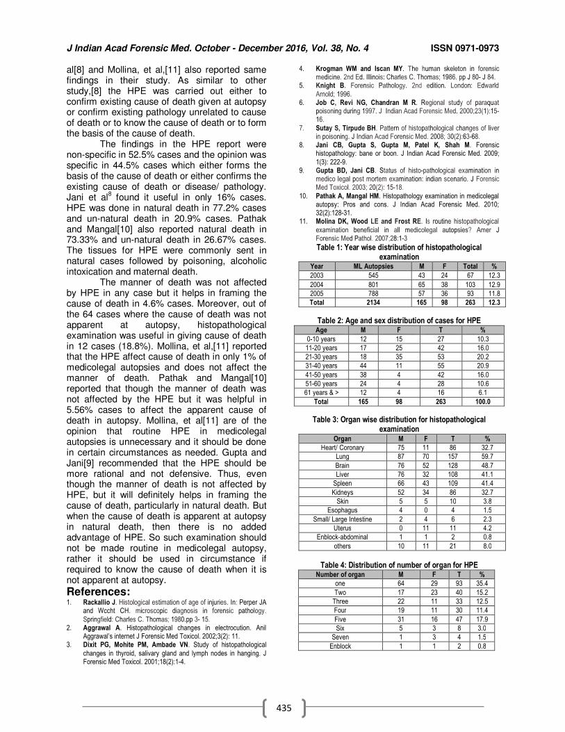
J Indian Acad Forensic Med. October - December 2016, Vol. 38, No. 4 ISSN 0971-0973
435
al[8] and Mollina, et al,[11] also reported same findings in their study. As similar to other study,[8] the HPE was carried out either to confirm existing cause of death given at autopsy or confirm existing pathology unrelated to cause of death or to know the cause of death or to form the basis of the cause of death. The findings in the HPE report were non-specific in 52.5% cases and the opinion was specific in 44.5% cases which either forms the basis of the cause of death or either confirms the existing cause of death or disease/ pathology. Jani et al
8 found it useful in only 16% cases.
HPE was done in natural death in 77.2% cases and un-natural death in 20.9% cases. Pathak and Mangal[10] also reported natural death in 73.33% and un-natural death in 26.67% cases. The tissues for HPE were commonly sent in natural cases followed by poisoning, alcoholic intoxication and maternal death. The manner of death was not affected by HPE in any case but it helps in framing the cause of death in 4.6% cases. Moreover, out of the 64 cases where the cause of death was not apparent at autopsy, histopathological examination was useful in giving cause of death in 12 cases (18.8%). Mollina, et al,[11] reported that the HPE affect cause of death in only 1% of medicolegal autopsies and does not affect the manner of death. Pathak and Mangal[10] reported that though the manner of death was not affected by the HPE but it was helpful in 5.56% cases to affect the apparent cause of death in autopsy. Mollina, et al[11] are of the opinion that routine HPE in medicolegal autopsies is unnecessary and it should be done in certain circumstances as needed. Gupta and Jani[9] recommended that the HPE should be more rational and not defensive. Thus, even though the manner of death is not affected by HPE, but it will definitely helps in framing the cause of death, particularly in natural death. But when the cause of death is apparent at autopsy in natural death, then there is no added advantage of HPE. So such examination should not be made routine in medicolegal autopsy, rather it should be used in circumstance if required to know the cause of death when it is not apparent at autopsy.
References: 1. Rackallio J. Histological estimation of age of injuries. In: Perper JA
and Wccht CH. microscopic diagnosis in forensic pathology.
Springfield: Charles C. Thomas; 1980.pp 3- 15. 2. Aggrawal A. Histopathological changes in electrocution. Anil
Aggrawal’s internet J Forensic Med Toxicol. 2002;3(2): 11. 3. Dixit PG, Mohite PM, Ambade VN. Study of histopathological
changes in thyroid, salivary gland and lymph nodes in hanging. J Forensic Med Toxicol. 2001;18(2):1-4.
4. Krogman WM and Iscan MY. The human skeleton in forensic medicine. 2nd Ed. Illinois: Charles C. Thomas; 1986. pp J 80- J 84.
5. Knight B. Forensic Pathology. 2nd edition. London: Edwarld
Arnold; 1996. 6. Job C, Revi NG, Chandran M R. Regional study of paraquat
poisoning during 1997. J Indian Acad Forensic Med. 2000;23(1):15-16.
7. Sutay S, Tirpude BH. Pattern of histopathological changes of liver in poisoning. J Indian Acad Forensic Med. 2008; 30(2):63-68.
8. Jani CB, Gupta S, Gupta M, Patel K, Shah M. Forensic histopathology: bane or boon. J Indian Acad Forensic Med. 2009;
1(3): 222-9. 9. Gupta BD, Jani CB. Status of histo-pathological examination in
medico legal post mortem examination: indian scenario. J Forensic Med Toxicol. 2003; 20(2): 15-18.
10. Pathak A, Mangal HM. Histopathology examination in medicolegal autopsy: Pros and cons. J Indian Acad Forensic Med. 2010; 32(2):128-31.
11. Molina DK, Wood LE and Frost RE. Is routine histopathological
examination beneficial in all medicolegal autopsies? Amer J Forensic Med Pathol. 2007;28:1-3
Table 1: Year wise distribution of histopathological examination
Year ML Autopsies M F Total %
2003 545 43 24 67 12.3
2004 801 65 38 103 12.9
2005 788 57 36 93 11.8
Total 2134 165 98 263 12.3
Table 2: Age and sex distribution of cases for HPE Age M F T %
0-10 years 12 15 27 10.3
11-20 years 17 25 42 16.0
21-30 years 18 35 53 20.2
31-40 years 44 11 55 20.9
41-50 years 38 4 42 16.0
51-60 years 24 4 28 10.6
61 years & > 12 4 16 6.1
Total 165 98 263 100.0
Table 3: Organ wise distribution for histopathological examination
Organ M F T %
Heart/ Coronary 75 11 86 32.7
Lung 87 70 157 59.7
Brain 76 52 128 48.7
Liver 76 32 108 41.1
Spleen 66 43 109 41.4
Kidneys 52 34 86 32.7
Skin 5 5 10 3.8
Esophagus 4 0 4 1.5
Small/ Large Intestine 2 4 6 2.3
Uterus 0 11 11 4.2
Enblock-abdominal 1 1 2 0.8
others 10 11 21 8.0
Table 4: Distribution of number of organ for HPE
Number of organ M F T %
one 64 29 93 35.4
Two 17 23 40 15.2
Three 22 11 33 12.5
Four 19 11 30 11.4
Five 31 16 47 17.9
Six 5 3 8 3.0
Seven 1 3 4 1.5
Enblock 1 1 2 0.8
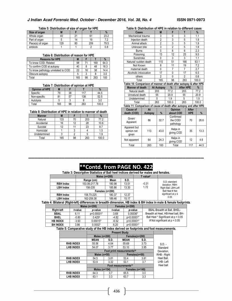
J Indian Acad Forensic Med. October - December 2016, Vol. 38, No. 4 ISSN 0971-0973
436
Table 5: Distribution of size of organ for HPE Size of organ M F T %
Whole organ 40 21 61 23.2
Part of organ 5 14 19 7.2
Piece(s) of organ 129 80 209 79.5
enblock 1 1 2 0.8
Table 6: Distribution of reason for HPE
Reasons for HPE M F T %
To know COD- Related 98 71 169 64.3
To confirm COD at autopsy 40 8 48 18.3
To know pathology unrelated to COD 22 16 38 14.4
Obscure autopsy 5 3 8 3.0
Total 165 98 263 100
Table 7: Distribution of opinion at HPE
Opinion of HPE M F T %
Specific 79 38 117 44.5
Non-specific 81 57 138 52.5
Autolysis 5 3 8 3.0
Total 165 98 263 100.0
Table 8: Distribution of HPE in relation to manner of death
Manner M F T %
Natural 133 70 203 77.2
Accidental 16 12 28 10.6
Suicidal 12 11 23 8.7
Homicidal 1 3 4 1.5
Undetermined 3 2 5 1.9
Total 165 98 263 100.0
Table 9: Distribution of HPE in relation to different cases Cases M F T %
Mechanical trauma 3 0 3 1.1
Injection death 2 3 5 1.9
Animal attack 2 3 5 1.9
Unknown bite 3 2 5 1.9
Burns 0 6 6 2.3
Poisoning 13 12 25 9.5
Sunstroke 0 3 3 1.1
Natural/ sudden death 115 51 166 63.1
Not Known 8 11 19 7.2
maternal death 0 7 7 2.7
Alcoholic intoxication 17 0 17 6.5
others 2 0 2 0.8
Total 165 98 263 100.0
Table 10: Comparison of manner of death after autopsy & after HPE
Manner of death At Autopsy % After HPE %
Natural death 203 77.2 203 77.2
Unnatural death 55 20.9 55 20.9
Undetermined 5 1.9 5 1.9
Total 263 100.0 263 100.0
Table 11: Comparison of cause of death after autopsy and after HPE
Cause of death (COD)
At Autopsy %
Opinion about COD
After HPE %
Given/ Apparent
86 32.7
Confirmed
the COD/ pathology
70 26.6
Apparent but
opinion not given
113 43.0 Helps in
giving COD 35 13.3
Not apparent 64 24.3 Helps in
giving COD 12 4.6
Total 263 100 Total 117 44.5
__________________________________________________________
**Contd. from PAGE NO. 422 Table 3: Descriptive Statistics of Ball heel indices derived for males and females. Males (n=200) T value#
Range (cm) Mean S.D. -0.31 1.72
S.D.-standard deviation, RBH-
Right Ball, LBH-Left Ball Heel # Not
significant at p ≤ 0.05
RBH Index 153.06-217.78 180.98 13.51
LBH Index 156-235 185.86 13.30
Females (n=200)
RBH Index 158.70-225 180.37 12.37
LBH Index 162-259.38 189.46 15.17
Table 4: Bilateral (Right-left) differences in breadth dimension, HB index & BH index in male & female footprints.
Males (n=200) Females (n=200)
Right-left t-value p-value t-value p-value BBAL-Breadth at Ball, BHEL-
Breadth at Heel, HB-Heel ball, BH-Ball Heel * Significant at p < 0.05
# Not significant at p < 0.05
BBAL 6.11 p<0.00001* 3.69 0.00036*
BHEL -0.80 0.425# -4.82 p<0.00001*
HB INDEX -3.93 0.00015* -6.52 p<0.00001*
BH INDEX 3.86 0.0002* 6.20 p<0.00001*
Table 5: Comparative study of the HB index derived on footprints and foot measurements.
Present Study
Males (n=200) Females(n=200)
S.D. - Standard Deviation,
RHB - Right Heel Ball,
LHB- Left Heel ball
MEAN S.D. MEAN S.D.
RHB INDEX 55.56 4.04 55.69 3.73
LHB INDEX 54.07 3.77 53.10 3.95
Foot print measurements20
Males (n=50) Females(n=50)
RHB INDEX 54.5 3.81 53.4 3.81
LHB INDEX 53.9 3.32 53.1 3.7
Foot measurements17
Males (n=154) Females (n=149)
RHB INDEX 64.3 3.7 65.5 3.0
LHB INDEX 63.1 3.3 63.7 3.3
J Indian Acad Forensic Med. October - December 2016, Vol. 38, No. 4 ISSN 0971-0973
437
Original Research Paper
Study of Undergraduate Students’ Perceptions towards Organ Donation
1Pragnesh B. Parmar,
2Kunjan Bharpoda,
2Vidhi Bhensdadia,
2Pragnesh Bhokan,
2Parth Bhut,
2Bharat
Chaudhary
Abstract Background: In India, the concept and awareness of organ donation is still new for many. There
is enormous shortage of organs in India and that is due to lack of awareness among general population. Keeping this in mind, we have conducted present study to assess perceptions of undergraduate students’ towards organ donation. Materials and methods: A cross-sectional study was conducted among randomly selected 112 students aged 18 years and above, in SumandeepVidyapeeth campus, Vadodara, Gujarat, who were willing to participate in survey, after obtaining their informed written consent. The students were subjected to a pretested and validated questionnaire (Likert type scale) for organ donation. Responses given by the students were assessed and analyzed via SPSS. Frequency and median score of each question was calculated. Results: Out of 112 participants, only 100 were considered for analysis and remaining 12 were rejected because they had not responded completely or had given multiple responses. Total 61% of the candidates were aware of the concept of organ donation. There was no statistically significant difference between perceptions of male and female. Conclusion: Though knowledge and practice is far lacking in the field of organ donation, attitude is much more better which can help to make future of organ donation more optimistic.
Key Words: Organ donation, Students, Perceptions, Trends
Introduction: Organ shortageis a global problem of
current era, but Asia lags behind much of the rest of the world as of now.[1] Organ donation following brain stem death is infrequent in India and still a new concept for general population. The current organ donation for cadaver in India is 0.08 per million while Spain tops the list with 35 per million.[1] There is an enormous shortage of organs in India, and patients die while on the waiting list, as they do not get an organ on time due to non existence of proper system. Only 5% of all patients with end-stage kidney disease are successful in undergoing kidney transplantation.[2] The current demand in the country for kidney transplant is 150,000; liver, 200,000 and heart, 150,000 which is very significant [3]. Human organ donation was legalized in India since 1994 through The Transplantation of Human Organs Act, 1994.[4]
Corresponding Author: 1Associate Professor,
Forensic Medicine, GMERS Medical College,Valsad, Gujarat
2Final MBBS Student, SBKS MI & RC,
SumandeepVidyapeeth, Vadodara, Gujarat E mail – [email protected] DOR: 06/04/2016 DOA: 25/12/2016 DOI: 10.5958/0974-0848.2016.00110.X
Even after 18 years, only kidney donations by live donors are in vogue - cadaver donations have still not picked, up as compared to western countries. Certification and declaration of brain death have been made mandatory in transplant hospitals and in non-transplant organ retrieval centres registered under the Human Organs Transplant Act, 1994. To further this, Tamil Nadu passed an order in 2008, making the certification mandatory.[5] Every year, close to six lakh people die due to organ failure.[5] Conversely, with 70 per cent of India's 1.4 lakh accident victims diagnosed as brain dead annually, the country has 80,000 potential organ donors, who remain unutilized. Organs from only about 120 are retrieved, making the percentage of cadaver donations a dismal 0.08 per million of the population, according to MOHAN Foundation.[6] On the other hand, in most developed countries, the cadaveric conversion is approximately 25% to 30%. This results in 90% of all organs for transplants coming from brain-dead donors.[6] The greatest impediment to organ donation is the refusal of family consent.[7] Organ donation rates could be increased by enhancing the quality of hospital care and ensuring that the request for donation is handled in a way that meets the families' informational and emotional needs.[8]
J Indian Acad Forensic Med. October - December 2016, Vol. 38, No. 4 ISSN 0971-0973
438
Materials and methods: A cross-sectional study was conducted among randomly selected 112 students, aged 18 years and above in SumandeepVidyapeeth campus, Vadodara, Gujarat, studying in various faculties like Physiotherapy, Nursing, Dental, Pharmacy, who were willing to participate in survey after obtaining their informed written consent. The students were given a pretested and validated questionnaire (Likert type scale) for organ donation. The questionnaire contained 20 questions with answers varying from Disagree, Not sure and Agree. Responses given by the students were assessed and analyzed via SPSS. Frequency and median score of each question was calculated. Study Design: Cross Sectional Study Size: Randomly selected 112 studentsStudy population: Undergraduate students of Sumandeep Vidyapeeth.Statistical methods: Frequency and median score of each question. Ethical issue: Study dealt with only perceptions of participants and there was no ethical issue involved as the identity of participants was not disclosed. Likely outcomes/ benefits: Such type of study had not been conducted in SumandeepVidyapeeth campus before, so it was unique in its nature. Participants were exposed to various questions pertaining to organ donation which was very helpful to know the trend for organ donation.
Results: A total of 112 students were given the questionnaire regarding organ donation. Of these, only 100 were considered for analysis and remaining 12 were rejected because they had not responded completely or had given multiple responses. Frequency and median score of responses was as per Table – 1. Total 61% were aware of the concept of organ donation. 53% accepted that organ donation is of use to the needy individual and the society as a whole. 43% were not sure that organ donation can be done easily after death. 58% were not aware of the rules and legislation about organ donation. 53% didn’t agree that organ donation is against religious beliefs. 69% preferred donating organs to close relatives/known to, only. 60% were not willing to donate each and every organ of body. 65% were note sure that their family would support decision of organ donation. 82% agreed that there is a lack of information about organ donation in our country among the masses. 39% were willing to educate the masses about significance of organ donation. 76% feared that
the organ donated may be misused. 81% agreed that organ donation and transplantation is good and should be encouraged. 61% disagreed that there are enough willing organ donors for those in need in this community. 73% were willing to receive a transplant if necessary. 50% agreed to encourage family and friends to donate organs, if required. 73% were not sure that people in this community are willing to donate organs, if needed. 76% trusted the health worker to handle appropriately the donated organ. 74% opposed commercialization of organ donation. 68% were not happy with current scenario of organ donation. 80% believed that future of organ donation is optimistic (Table – 1). Total 57 were male and 43 were female participants. There was no statistically significant difference between perceptions of male and female as per Table - 2. Faculty wise distribution of participants was as per Table – 3.
Discussion: According to the results generated from the study, 61% of participants were aware of the concept of Organ Donation which is comparable with other study.[9]Organ Donation can be considered as a Humanitarian Project. It is clearly an act for welfare of Mankind and should be considered as a holy task if needed.There are millions of people out there in need of Organs to live a normal life, and there are more than millions out there who can provide them with the same opportunity as of others in their life. 53%participants were of the opinion that organ donation is for needy individuals and the society as a whole, which is really a positive thought process, as it can help uplift the community as to educate them to opt for organ donation.Even though it may be a stereotype in the community that the donated organs may not be treated with the deserved Dignity and Respect. Education about the process is vital. But as of now there really is a lack of vital and basic knowledge about the process of Organ Donation and its Legislation. Only 3% of Participants knew about the proper Legislationassociated with the process of organ donation which is comparable with other study.[10]But looking at a positive side, 39% were ready to educate the masses about the organ donation if given the opportunity, and only 11% were willing to donate each and every organ of their body. 15% of respondents thought that Organ Donation can be easily done after death. Sometimes there may be a thought that certain religion may bind individuals and stop
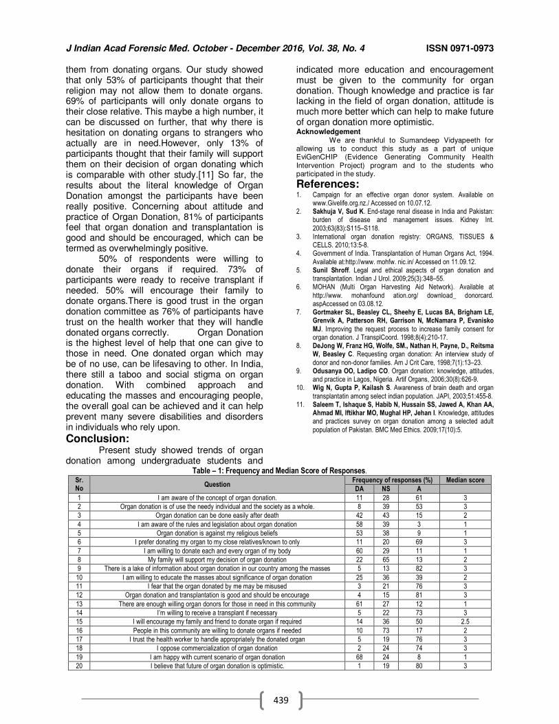
J Indian Acad Forensic Med. October - December 2016, Vol. 38, No. 4 ISSN 0971-0973
439
them from donating organs. Our study showed that only 53% of participants thought that their religion may not allow them to donate organs. 69% of participants will only donate organs to their close relative. This maybe a high number, it can be discussed on further, that why there is hesitation on donating organs to strangers who actually are in need.However, only 13% of participants thought that their family will support them on their decision of organ donating which is comparable with other study.[11] So far, the results about the literal knowledge of Organ Donation amongst the participants have been really positive. Concerning about attitude and practice of Organ Donation, 81% of participants feel that organ donation and transplantation is good and should be encouraged, which can be termed as overwhelmingly positive. 50% of respondents were willing to donate their organs if required. 73% of participants were ready to receive transplant if needed. 50% will encourage their family to donate organs.There is good trust in the organ donation committee as 76% of participants have trust on the health worker that they will handle donated organs correctly. Organ Donation is the highest level of help that one can give to those in need. One donated organ which may be of no use, can be lifesaving to other. In India, there still a taboo and social stigma on organ donation. With combined approach and educating the masses and encouraging people, the overall goal can be achieved and it can help prevent many severe disabilities and disorders in individuals who rely upon.
Conclusion: Present study showed trends of organ donation among undergraduate students and
indicated more education and encouragement must be given to the community for organ donation. Though knowledge and practice is far lacking in the field of organ donation, attitude is much more better which can help to make future of organ donation more optimistic. Acknowledgement We are thankful to Sumandeep Vidyapeeth for allowing us to conduct this study as a part of unique EviGenCHIP (Evidence Generating Community Health Intervention Project) program and to the students who participated in the study.
References: 1. Campaign for an effective organ donor system. Available on
www.Givelife.org.nz./ Accessed on 10.07.12. 2. Sakhuja V, Sud K. End-stage renal disease in India and Pakistan:
burden of disease and management issues. Kidney Int.
2003;63(83):S115–S118. 3. International organ donation registry: ORGANS, TISSUES &
CELLS. 2010;13:5-8. 4. Government of India. Transplantation of Human Organs Act, 1994.
Available at:http://www. mohfw. nic.in/ Accessed on 11.09.12. 5. Sunil Shroff. Legal and ethical aspects of organ donation and
transplantation. Indian J Urol. 2009;25(3):348–55. 6. MOHAN (Multi Organ Harvesting Aid Network). Available at
http://www. mohanfound ation.org/ download_ donorcard. aspAccessed on 03.08.12.
7. Gortmaker SL, Beasley CL, Sheehy E, Lucas BA, Brigham LE, Grenvik A, Patterson RH, Garrison N, McNamara P, Evanisko
MJ. Improving the request process to increase family consent for organ donation. J TransplCoord. 1998;8(4):210-17.
8. DeJong W, Franz HG, Wolfe, SM., Nathan H, Payne, D., Reitsma W, Beasley C. Requesting organ donation: An interview study of
donor and non-donor families. Am J Crit Care, 1998;7(1):13–23. 9. Odusanya OO, Ladipo CO. Organ donation: knowledge, attitudes,
and practice in Lagos, Nigeria. Artif Organs, 2006;30(8):626-9. 10. Wig N, Gupta P, Kailash S. Awareness of brain death and organ
transplantatin among select indian population. JAPI, 2003;51:455-8. 11. Saleem T, Ishaque S, Habib N, Hussain SS, Jawed A, Khan AA,
Ahmad MI, Iftikhar MO, Mughal HP, Jehan I. Knowledge, attitudes and practices survey on organ donation among a selected adult
population of Pakistan. BMC Med Ethics. 2009;17(10):5.
Table – 1: Frequency and Median Score of Responses.
Sr. No
Question Frequency of responses (%) Median score
DA NS A
1 I am aware of the concept of organ donation. 11 28 61 3
2 Organ donation is of use the needy individual and the society as a whole. 8 39 53 3
3 Organ donation can be done easily after death 42 43 15 2
4 I am aware of the rules and legislation about organ donation 58 39 3 1
5 Organ donation is against my religious beliefs 53 38 9 1
6 I prefer donating my organ to my close relatives/known to only 11 20 69 3
7 I am willing to donate each and every organ of my body 60 29 11 1
8 My family will support my decision of organ donation 22 65 13 2
9 There is a lake of information about organ donation in our country among the masses 5 13 82 3
10 I am willing to educate the masses about significance of organ donation 25 36 39 2
11 I fear that the organ donated by me may be misused 3 21 76 3
12 Organ donation and transplantation is good and should be encourage 4 15 81 3
13 There are enough willing organ donors for those in need in this community 61 27 12 1
14 I’m willing to receive a transplant if necessary 5 22 73 3
15 I will encourage my family and friend to donate organ if required 14 36 50 2.5
16 People in this community are willing to donate organs if needed 10 73 17 2
17 I trust the health worker to handle appropriately the donated organ 5 19 76 3
18 I oppose commercialization of organ donation 2 24 74 3
19 I am happy with current scenario of organ donation 68 24 8 1
20 I believe that future of organ donation is optimistic. 1 19 80 3
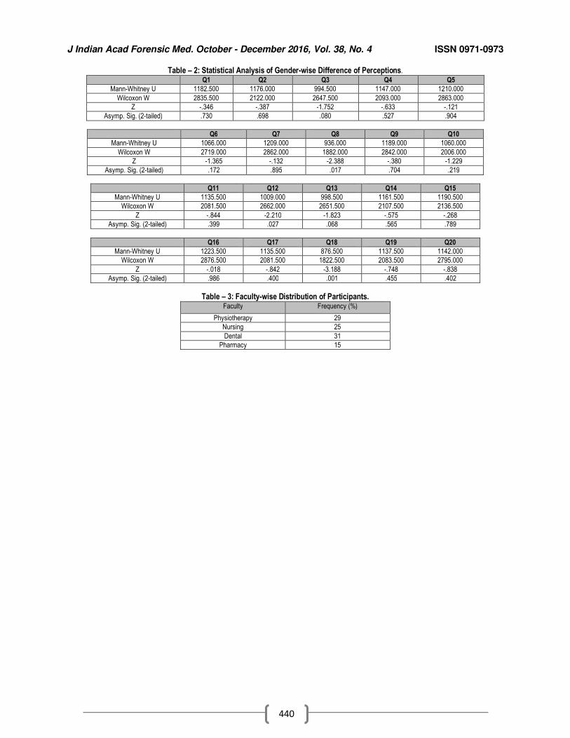
J Indian Acad Forensic Med. October - December 2016, Vol. 38, No. 4 ISSN 0971-0973
440
Table – 2: Statistical Analysis of Gender-wise Difference of Perceptions.
Q1 Q2 Q3 Q4 Q5
Mann-Whitney U 1182.500 1176.000 994.500 1147.000 1210.000
Wilcoxon W 2835.500 2122.000 2647.500 2093.000 2863.000
Z -.346 -.387 -1.752 -.633 -.121
Asymp. Sig. (2-tailed) .730 .698 .080 .527 .904
Q6 Q7 Q8 Q9 Q10
Mann-Whitney U 1066.000 1209.000 936.000 1189.000 1060.000
Wilcoxon W 2719.000 2862.000 1882.000 2842.000 2006.000
Z -1.365 -.132 -2.388 -.380 -1.229
Asymp. Sig. (2-tailed) .172 .895 .017 .704 .219
Q11 Q12 Q13 Q14 Q15
Mann-Whitney U 1135.500 1009.000 998.500 1161.500 1190.500
Wilcoxon W 2081.500 2662.000 2651.500 2107.500 2136.500
Z -.844 -2.210 -1.823 -.575 -.268
Asymp. Sig. (2-tailed) .399 .027 .068 .565 .789
Q16 Q17 Q18 Q19 Q20
Mann-Whitney U 1223.500 1135.500 876.500 1137.500 1142.000
Wilcoxon W 2876.500 2081.500 1822.500 2083.500 2795.000
Z -.018 -.842 -3.188 -.748 -.838
Asymp. Sig. (2-tailed) .986 .400 .001 .455 .402
Table – 3: Faculty-wise Distribution of Participants. Faculty Frequency (%)
Physiotherapy 29
Nursing 25
Dental 31
Pharmacy 15
J Indian Acad Forensic Med. October - December 2016, Vol. 38, No. 4 ISSN 0971-0973
441
Original Research Paper
Stature Estimation Using Per-Cutaneous Tibial Length in Malwa Population of Punjab
1Rajiv Joshi,
2Monika Bhardwaj,
3Ramandeep Singh
Abstract Background: Stature estimation is important as it provides a Forensic Anthropological measurement which may be used in identification of individuals from grossly mutilated skeletal remains, especially in mass disasters. Objective of the Study: The present study was designed to estimate the height of an individual from the per-cutaneous tibial length (PCTL) by formulating simple regression equation and multiplication factor (M.F.). Materials and Methodology: The study was conducted on a random sample of 300 students (150 male and 150 female) in the age group of 18 to 24 years from the Malwa region of Punjab. PCTL of right and left were measured with the help of spreading vernier calipers. The data was tabulated and analyzed statistically. Result: A positive correlation (r= 0.842 in males and r= 0.733 in females) was found between length of Tibia and the estimated height. A linear regression formula was derived for estimation of height from the length of right and left side of Tibia in both the genders. The regression formula derived for males was y0=80.160 +2.260x (PCTL) + 6.837 and for females was y0 = 93.302 +1.647 x (PCTL) =6.816. Conclusion: It was concluded that the stature of a deceased person from Malwa region of Punjab can be estimated by using the regression equations derived from the present investigation fairly accurately to some extent.
Key Words: Stature, Tibial Length, PCTL, Malwa Region, Regression Formula
Introduction: Stature estimation is an indispensable
part of the identification process of human skeletal remains or body parts.[1,2]
.It is defined
as “Height of body in standing position”.[3] Stature of an individual is an inherent character and is considered as one of the most important parameters of personal identification. Long bones that make up the greatest proportion of the stature, i.e. femur and tibia are more accurate than the other bones as they have direct co-relation with the height of the body.[4] Landmarks on other long bones are more difficult to identify and are more variable than that of tibia, hence the per-cutaneous tibial length (PCTL). The growth of the bones and hence, stature of an individual, are influenced by
Corresponding Author: 2Assistant Professor,
Department of Life Sciences, B.B.K, D.A.V College, Amritsar, Punjab 1Professor & Head, Department of Forensic Medicine,
3MBBS Student, G.G.S, Medical College, Faridkot,
Punjab Email:[email protected] DOR: 09/06/2016 DOA: 25/12/2016 DOI: 10.5958/0974-0848.2016.00111.1
numerous factors as age, gender, race, geographical climate, nutrition and genetic factors.[5,6]
.These factors are unique to different
types of demography. Extensive work has been done on correlation of measurements of various body parts with stature of a person in India and abroad.[8-15] Many studies from north and south Africa have summarized that long bones of lower extremity usually give a close estimate of stature than long bones of upper extremity.[10,16,17] Most of these studies measured height of the subject in standing position & per-cutaneous tibial length on both sides and found positive correlation between them.[7,25,26]
.
Stature estimation can be specifically derived from each population. Specific regression for specific population is important to account for inherent population variation,[4] such as genetic and environment factors.[18] India is a sub continent where population consists of various ethnic groups. The stature not only differs state-wise, but also varies according to different castes and tribes.[19-22] It is also seen that the stature of a person differs in different region of the same state.[23-26] Little work has been done in measuring stature using per-cutaneous tibial length in the state of Punjab. The lack of
J Indian Acad Forensic Med. October - December 2016, Vol. 38, No. 4 ISSN 0971-0973
442
anthropometric data concerning the Malwa population of Punjab was felt for various medicolegal purposes.
Objectives of the Study: 1. To evaluate a possible co-relation between
stature of an individual and his per-cutaneous tibial length in Malwa population of Punjab.
2. To drive regression formula and multiplication factor for estimation of stature from the per-cutaneous tibial length.
3. To compare the per-cutaneous length of right and left tibia in both genders.
Materials and Methodology: Stature was estimated by employing a mathematical method. This method employs bone length, stature table and regression formulae to estimate total skeletal height from long bones.[27]
.
The regression formula is statistical measure of average relationship between two variables, one independent (tibia length in this study) and other dependent (Height of an individual). Study design: - cross sectional study Study population and Sample size: - A random sample of 300 students (150 males and 150 females) in the age group between 18-24 years of paramedical and nursing courses of Baba Farid University of Health Sciences (BFUHS), Faridkot was taken. This age group was selected because multiplication factor {M.F.} remains more or less same in this age group.[28]
Inclusion criteria: - only those students were included who were born and brought up in Malwa region of Punjab and with ancestral origin from this region. Exclusive criteria: - subjects with history of major trauma or fracture of the leg, achondroplasia or any other congenital or hereditary bone disease were excluded A formal consent was obtained from all of the students, including proper approval from the Institutional Ethics Committee. It normally took 5-10 minutes to measure the stature and per cutaneous tibial length of both sides. Parameters noted were: Age, Gender, Height in cm, Per-cutaneous tibial length (P.C.T.L.) of right and left side in cms All the measurements were taken by the same observer and with the same instrument, to avoid any technical and/or inter-observer error and to maintain reproducibility. The measurements were taken three times and their
mean value was considered to find correlation between stature and per cutaneous tibial length. Measurement of height {stature}: standing height of the subject was measured in standing position on a standard stadiometer with both feet in close contact with each other with the trunk straight along the vertical board, and the head adjusted in Frankfurt plane. The measurements were taken in centimeters by bringing the horizontal sliding bar to the vertex. Measurement of the per-cutaneous tibial length (P.C.T.L): subject was asked to stand and keep his/her foot on a stool to maintain the angle between the flexor surface of leg and that of thigh at 90 degree. Then the distance was measured between the following the points:- Upper point: the most prominent palpable portion of the medial condyle of the tibia. Lower point: tip of medial malleolus of the tibia. Distance between the two points was measured with the help of spreading caliper to determine tibial length. The data obtained after measurements was computed, tabulated and statistically analyzed using the 16 version of SPSS Statistics.
Results: Tables 1 and 2 depict the distribution of subjects according to height and the range of Tibial length, respectively. Majority of the subjects (36%), had height ranging from 155-164 cms. The most common Tibial Length was 37 - 42 cms, observed in 74% of the subjects. Table 3 highlights the Statistical analysis of PCTL of right and left side of tibia in males and females. There was no significant difference (p>0.05) in the per-cutaneous length of right and left tibia in both genders, thus showing bilateral symmetry in the length of Tibia in both the genders. The mean PCTL for males was 40.29 cm and for females, was 38.06 cm. The study revealed that standing height of many individuals were same, but their PCTL differed, i.e. the contribution of tibial length to the stature of a person varied from person to person, even for a given height.[5] Keeping this in view, Mean of stature and PCTL were taken into consideration and the data was calculated and analyzed (Table 4). The observed mean height was 171.22 cm and 156 cm, and mean PCTL was 40.29 cm and 38.06 cm in males and females, respectively, which was significantly (p<0.01) greater for males compared with females.
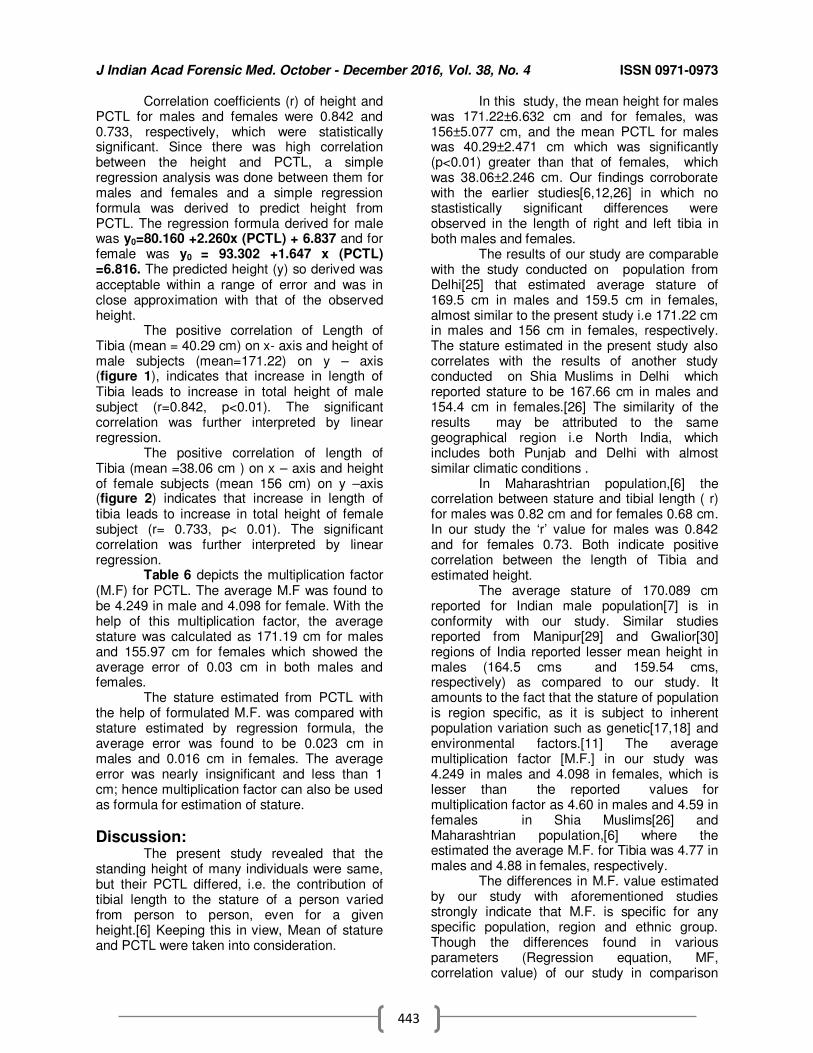
J Indian Acad Forensic Med. October - December 2016, Vol. 38, No. 4 ISSN 0971-0973
443
Correlation coefficients (r) of height and PCTL for males and females were 0.842 and 0.733, respectively, which were statistically significant. Since there was high correlation between the height and PCTL, a simple regression analysis was done between them for males and females and a simple regression formula was derived to predict height from PCTL. The regression formula derived for male was y0=80.160 +2.260x (PCTL) + 6.837 and for female was y0 = 93.302 +1.647 x (PCTL) =6.816. The predicted height (y) so derived was acceptable within a range of error and was in close approximation with that of the observed height. The positive correlation of Length of Tibia (mean = 40.29 cm) on x- axis and height of male subjects (mean=171.22) on y – axis (figure 1), indicates that increase in length of Tibia leads to increase in total height of male subject (r=0.842, p<0.01). The significant correlation was further interpreted by linear regression. The positive correlation of length of Tibia (mean =38.06 cm ) on x – axis and height of female subjects (mean 156 cm) on y –axis (figure 2) indicates that increase in length of tibia leads to increase in total height of female subject (r= 0.733, p< 0.01). The significant correlation was further interpreted by linear regression. Table 6 depicts the multiplication factor (M.F) for PCTL. The average M.F was found to be 4.249 in male and 4.098 for female. With the help of this multiplication factor, the average stature was calculated as 171.19 cm for males and 155.97 cm for females which showed the average error of 0.03 cm in both males and females. The stature estimated from PCTL with the help of formulated M.F. was compared with stature estimated by regression formula, the average error was found to be 0.023 cm in males and 0.016 cm in females. The average error was nearly insignificant and less than 1 cm; hence multiplication factor can also be used as formula for estimation of stature.
Discussion: The present study revealed that the standing height of many individuals were same, but their PCTL differed, i.e. the contribution of tibial length to the stature of a person varied from person to person, even for a given height.[6] Keeping this in view, Mean of stature and PCTL were taken into consideration.
In this study, the mean height for males was 171.22±6.632 cm and for females, was 156±5.077 cm, and the mean PCTL for males was 40.29±2.471 cm which was significantly (p<0.01) greater than that of females, which was 38.06±2.246 cm. Our findings corroborate with the earlier studies[6,12,26] in which no stastistically significant differences were observed in the length of right and left tibia in both males and females. The results of our study are comparable with the study conducted on population from Delhi[25] that estimated average stature of 169.5 cm in males and 159.5 cm in females, almost similar to the present study i.e 171.22 cm in males and 156 cm in females, respectively. The stature estimated in the present study also correlates with the results of another study conducted on Shia Muslims in Delhi which reported stature to be 167.66 cm in males and 154.4 cm in females.[26] The similarity of the results may be attributed to the same geographical region i.e North India, which includes both Punjab and Delhi with almost similar climatic conditions . In Maharashtrian population,[6] the correlation between stature and tibial length ( r) for males was 0.82 cm and for females 0.68 cm. In our study the ‘r’ value for males was 0.842 and for females 0.73. Both indicate positive correlation between the length of Tibia and estimated height. The average stature of 170.089 cm reported for Indian male population[7] is in conformity with our study. Similar studies reported from Manipur[29] and Gwalior[30] regions of India reported lesser mean height in males (164.5 cms and 159.54 cms, respectively) as compared to our study. It amounts to the fact that the stature of population is region specific, as it is subject to inherent population variation such as genetic[17,18] and environmental factors.[11] The average multiplication factor [M.F.] in our study was 4.249 in males and 4.098 in females, which is lesser than the reported values for multiplication factor as 4.60 in males and 4.59 in females in Shia Muslims[26] and Maharashtrian population,[6] where the estimated the average M.F. for Tibia was 4.77 in males and 4.88 in females, respectively. The differences in M.F. value estimated by our study with aforementioned studies strongly indicate that M.F. is specific for any specific population, region and ethnic group. Though the differences found in various parameters (Regression equation, MF, correlation value) of our study in comparison
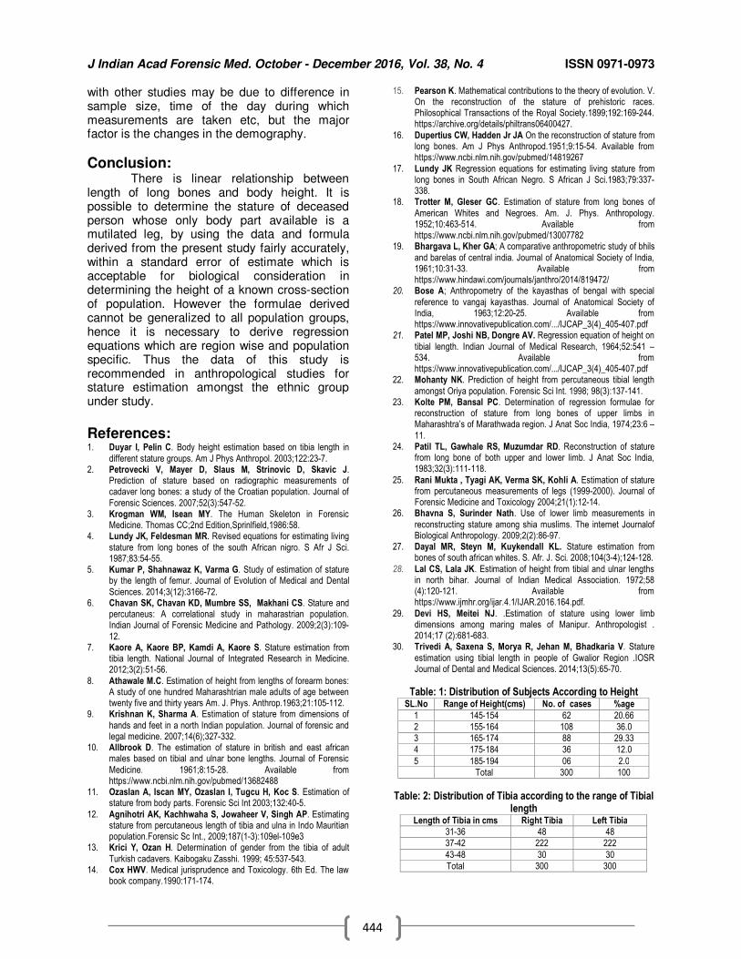
J Indian Acad Forensic Med. October - December 2016, Vol. 38, No. 4 ISSN 0971-0973
444
with other studies may be due to difference in sample size, time of the day during which measurements are taken etc, but the major factor is the changes in the demography.
Conclusion: There is linear relationship between length of long bones and body height. It is possible to determine the stature of deceased person whose only body part available is a mutilated leg, by using the data and formula derived from the present study fairly accurately, within a standard error of estimate which is acceptable for biological consideration in determining the height of a known cross-section of population. However the formulae derived cannot be generalized to all population groups, hence it is necessary to derive regression equations which are region wise and population specific. Thus the data of this study is recommended in anthropological studies for stature estimation amongst the ethnic group under study.
References: 1. Duyar I, Pelin C. Body height estimation based on tibia length in
different stature groups. Am J Phys Anthropol. 2003;122:23-7. 2. Petrovecki V, Mayer D, Slaus M, Strinovic D, Skavic J.
Prediction of stature based on radiographic measurements of cadaver long bones: a study of the Croatian population. Journal of
Forensic Sciences. 2007;52(3):547-52. 3. Krogman WM, Isean MY. The Human Skeleton in Forensic
Medicine. Thomas CC;2nd Edition,Sprinlfield,1986:58. 4. Lundy JK, Feldesman MR. Revised equations for estimating living
stature from long bones of the south African nigro. S Afr J Sci. 1987;83:54-55.
5. Kumar P, Shahnawaz K, Varma G. Study of estimation of stature by the length of femur. Journal of Evolution of Medical and Dental
Sciences. 2014;3(12):3166-72. 6. Chavan SK, Chavan KD, Mumbre SS, Makhani CS. Stature and
percutaneus: A correlational study in maharastrian population. Indian Journal of Forensic Medicine and Pathology. 2009;2(3):109-
12. 7. Kaore A, Kaore BP, Kamdi A, Kaore S. Stature estimation from
tibia length. National Journal of Integrated Research in Medicine. 2012;3(2):51-56.
8. Athawale M.C. Estimation of height from lengths of forearm bones: A study of one hundred Maharashtrian male adults of age between twenty five and thirty years Am. J. Phys. Anthrop.1963;21:105-112.
9. Krishnan K, Sharma A. Estimation of stature from dimensions of
hands and feet in a north Indian population. Journal of forensic and legal medicine. 2007;14(6);327-332.
10. Allbrook D. The estimation of stature in british and east african males based on tibial and ulnar bone lengths. Journal of Forensic
Medicine. 1961;8:15-28. Available from https://www.ncbi.nlm.nih.gov/pubmed/13682488
11. Ozaslan A, Iscan MY, Ozaslan I, Tugcu H, Koc S. Estimation of stature from body parts. Forensic Sci Int 2003;132:40-5.
12. Agnihotri AK, Kachhwaha S, Jowaheer V, Singh AP. Estimating stature from percutaneous length of tibia and ulna in Indo Mauritian population.Forensic Sc Int., 2009;187(1-3):109el-109e3
13. Krici Y, Ozan H. Determination of gender from the tibia of adult
Turkish cadavers. Kaibogaku Zasshi. 1999; 45:537-543. 14. Cox HWV. Medical jurisprudence and Toxicology. 6th Ed. The law
book company.1990:171-174.
15. Pearson K. Mathematical contributions to the theory of evolution. V. On the reconstruction of the stature of prehistoric races. Philosophical Transactions of the Royal Society.1899;192:169-244. https://archive.org/details/philtrans06400427.
16. Dupertius CW, Hadden Jr JA On the reconstruction of stature from long bones. Am J Phys Anthropod.1951;9:15-54. Available from https://www.ncbi.nlm.nih.gov/pubmed/14819267
17. Lundy JK Regression equations for estimating living stature from long bones in South African Negro. S African J Sci.1983;79:337-338.
18. Trotter M, Gleser GC. Estimation of stature from long bones of
American Whites and Negroes. Am. J. Phys. Anthropology. 1952;10:463-514. Available from https://www.ncbi.nlm.nih.gov/pubmed/13007782
19. Bhargava L, Kher GA; A comparative anthropometric study of bhils
and barelas of central india. Journal of Anatomical Society of India, 1961;10:31-33. Available from https://www.hindawi.com/journals/janthro/2014/819472/
20. Bose A; Anthropometry of the kayasthas of bengal with special
reference to vangaj kayasthas. Journal of Anatomical Society of India, 1963;12:20-25. Available from https://www.innovativepublication.com/.../IJCAP_3(4)_405-407.pdf
21. Patel MP, Joshi NB, Dongre AV. Regression equation of height on
tibial length. Indian Journal of Medical Research, 1964;52:541 – 534. Available from https://www.innovativepublication.com/.../IJCAP_3(4)_405-407.pdf
22. Mohanty NK. Prediction of height from percutaneous tibial length
amongst Oriya population. Forensic Sci Int. 1998; 98(3):137-141. 23. Kolte PM, Bansal PC. Determination of regression formulae for
reconstruction of stature from long bones of upper limbs in Maharashtra’s of Marathwada region. J Anat Soc India, 1974;23:6 –
11. 24. Patil TL, Gawhale RS, Muzumdar RD. Reconstruction of stature
from long bone of both upper and lower limb. J Anat Soc India, 1983;32(3):111-118.
25. Rani Mukta , Tyagi AK, Verma SK, Kohli A. Estimation of stature from percutaneous measurements of legs (1999-2000). Journal of Forensic Medicine and Toxicology 2004;21(1):12-14.
26. Bhavna S, Surinder Nath. Use of lower limb measurements in
reconstructing stature among shia muslims. The internet Journalof Biological Anthropology. 2009;2(2):86-97.
27. Dayal MR, Steyn M, Kuykendall KL. Stature estimation from bones of south african whites. S. Afr. J. Sci. 2008;104(3-4);124-128.
28. Lal CS, Lala JK. Estimation of height from tibial and ulnar lengths in north bihar. Journal of Indian Medical Association. 1972;58 (4):120-121. Available from https://www.ijmhr.org/ijar.4.1/IJAR.2016.164.pdf.
29. Devi HS, Meitei NJ. .Estimation of stature using lower limb dimensions among maring males of Manipur. Anthropologist . 2014;17 (2):681-683.
30. Trivedi A, Saxena S, Morya R, Jehan M, Bhadkaria V. Stature
estimation using tibial length in people of Gwalior Region .IOSR Journal of Dental and Medical Sciences. 2014;13(5):65-70.
Table: 1: Distribution of Subjects According to Height SL.No Range of Height(cms) No. of cases %age
1 145-154 62 20.66
2 155-164 108 36.0
3 165-174 88 29.33
4 175-184 36 12.0
5 185-194 06 2.0
Total 300 100
Table: 2: Distribution of Tibia according to the range of Tibial length
Length of Tibia in cms Right Tibia Left Tibia
31-36 48 48
37-42 222 222
43-48 30 30
Total 300 300
J Indian Acad Forensic Med. October - December 2016, Vol. 38, No. 4 ISSN 0971-0973
445
Table: 3 Descriptive statistics of right and left side of Tibial Length
Statistics Tibia Male(n=150) Female(n=150)
Rt PCTL LtPCTL RtPCTL LtPCTL
Range 34.5-46 34.5-46 32-43 32-43
Mean 40.285 40.357 38.06 38.067
Std.Deviation 2.450 2.478 2.246 2.238
Std. Error 0.283 0.286 0.259 0.258
Coefficient of variation(CV) 6.00% 6.143% 5.047% 5.01%
t-value t=0.1789 t=0.0191
p-value 0.8582 0.9848
P value summary Ns Ns
Average mean(rt+lt) 40.29 cm 38.06 cm
PCTL = per –cutaneous tibial length; RT=right, Lt=left Ns=not significant
Table: 4 Descriptive statistics of observed Height and Tibial Length of male and female
Statistics Male Female
Height PCTL Height PCTL
Range 160-190 34.5-46 146-170.5 32-43cm
Mean 171.22 40.29 156.0 38.06
Std.Deviation 6.632 2.471 5.077 2.246
Std.Error 0.765 0.285 0.582 0.259
Coefficient of variation(CV) 43.99% 6.108% 25.473 5.047%
Student t-test between male and female tibial length:
t-value t=5.784
p-value P<0.01
P value summary ** Significant
Difference between means 2.230 ± 0.385
Are means significantly Different?(P<0.05) Yes
95% confidence interval 1.468 to 2.992
Average mean PCTL(M+F) 39.775 cm
** Significant at P<0.01
Table: 5 Formulation of Regression equation for calculating the stature from PCTL in male and female
Regression Statistics of Tibia
Male (observed ht=171.22 cm) Female (observed ht=156.0 cm)
Rt Lt Average
PCTL(rt+lt) Rt Lt
Average
PCTL(rt+lt)
Independent variable(x)=PCTL X1=40.285 X2=40.357 X0=40.290 X1=38.06 X238.067 X0=38.060
Intercept(a) 79.215 80.420 80.160 93.302 93.231 93.302
Regression coefficient(b) 2.854 2.250 2.260 1.647 1.649 1.647
Correlation coefficient (r) 0.844 0.841 0.842 0.733 0.731 0.733
Correlation of determination R2 0.712 0.707 0.709 0.538 0.535 0.531
Std.error of estimate(SEE) 6.858 6.856 6.837 6.816 6.864 6.816
Significance(p) ** ** ** ** ** **
Regression formula(y=a+bx) Y1=79.215+2.284(x)x1
Y2=80.420+2.250(x)x2
Y0=80.160+2.260(x)x0
Y1=93.302+1.647(x)x1
Y2=93.231+1.649(x)x2
Y0=93.302+1.647(x)x0
Predicted ht(y) 171.225 171.223 171.215 155.986 156.003 155.986
** Significant at P<0.01: rt=right; lt= Left.
Table 6: Multiplication factor (M.F.) in both genders for Tibial length
Tibia Male Female
Right Left Right Left
PCTL 40.285 cm 40.357 cm 38.06 cm 38.067
M.F. 4.255 4.243 4.098 4.098
Average M.F. 4.249 4.098
Calculated avg. stature 171.192 cm 155.97 cm
Figure 1: Correlation between length of tibia (PCTL) and Figure 2: Correlation between length of tibia (PCTL) and Height in Height in females males
J Indian Acad Forensic Med. October - December 2016, Vol. 38, No. 4 ISSN 0971-0973
446
Original Research Paper
Profile of Scorpion Envenomation in Rural India 1Venkatesh Maled,
2Umesh Dixit
Abstract Scorpion sting incidence is common worldwide. Morbidity and mortality of scorpion sting is
considered as a public health problem in rural India. Although the exact incidence of scorpion stings is not known, it is estimated that the annual number of scorpion stings exceeds 1.2 million with 2.3 billion populations at risk world-wide. Present study highlights the demographic characteristics of scorpion sting cases in rural India. In this study, all scorpion sting cases admitted during last 6 years were selected. Total 70 cases were recorded and analyzed. Nearly 75% of the victims were in the age group of 1-30 years and males comprised 74% of cases. Majority of incidences took place in outdoor workers. Upper limbs were the most common site of sting, nearing 51% of cases. First aid measures were not taken in 95% of cases. The study highlights the sociological impact and suggests certain preventive measures to reduce scorpion sting morbidity and mortality.
Key Words: Scorpion Envenomation, Demography, India, Incidence, Prevention
Introduction: Scorpion sting incidence is common worldwide. Majority of cases reported from rural areas belonging to tropical and subtropical countries.[1,2] Although the exact incidence of scorpion stings is not known, it is estimated that the annual number of scorpion stings exceeds 1.2 million with 2.3 billion population at risk world-wide.[3] Among more than a thousand known species of scorpion, only 25 have venom that is deadly to humans; most of these belong to the family Buthidae. Mesobuthustamulusor, the Indian red scorpion, is the most lethal of all scorpion species.[4] These are found abundantly in Western Maharashtra, parts of Karnataka, Andhra Pradesh, Pondicherry and Tamil Nadu.[5] Children are at greater risk of developing severe envenomation. India records a good number of scorpion stings every year, but these figures reflect only the tip of iceberg because many cases go unreported due to popular traditional methods of treatment. It is recognized as a public health problem in rural India.[6]
Corresponding Author:
1Associate Prof.,
Dept. of Forensic Medicine, 2Associate Prof.,
Dept. of Community Medicine, SDM College of Medical Sciences & Hospital, Dharwad, Karnataka. Email: [email protected] DOR:08/03/2016 DOA: 25/12/2016 DOI: 10.5958/0974-0848.2016.00112.3
Scorpion sting may not produce mortality in majority of cases, but the death is more common in children. It remains an important cause of unnatural death in modern India.[6] The present study was undertaken to highlight the demographic characteristics of scorpion sting cases in rural India.
Materials & Methods: The present study is a retrospective one, conducted in the department of Forensic Medicine, SDM College of Medical Sciences & Hospital, Dharwad. All the scorpion sting cases admitted to the hospital from June 2007 to June 2013 were included in the study. A total of 70 cases were recorded in detail from hospital records regarding the incidence with respect to time, place, site of sting and the first aid with the specific treatment given. The data was analyzed statistically by using SPSS software and conclusion was drawn.
Results: Individuals in the age group of 11-20 years were the most common victims (25.7%), followed by the age group of 1-10years (24.3%) and 21-30 years (24.3%), respectively. Persons above 60 years comprised 2.9% cases. The youngest victim was1year old and the oldest was 80 years old. (Table 1) Males were the most common sufferers, 52 (74%) cases; with a male to female ratio of 3:1. Majority of incidences took place outdoors, compared to other locations. Most of the bites were recorded during night time i.e. between 18-24 hours (44.3%). (Table 2) Students were the
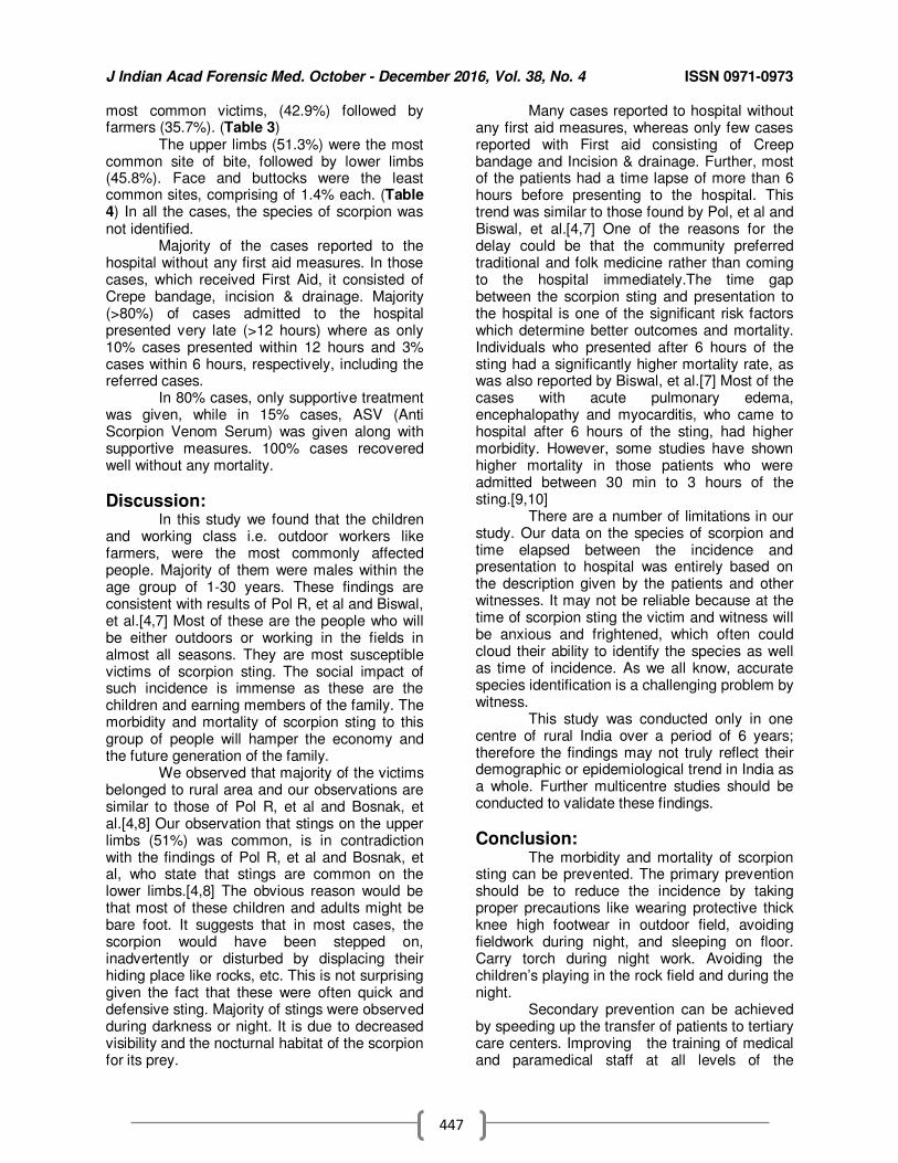
J Indian Acad Forensic Med. October - December 2016, Vol. 38, No. 4 ISSN 0971-0973
447
most common victims, (42.9%) followed by farmers (35.7%). (Table 3) The upper limbs (51.3%) were the most common site of bite, followed by lower limbs (45.8%). Face and buttocks were the least common sites, comprising of 1.4% each. (Table 4) In all the cases, the species of scorpion was not identified. Majority of the cases reported to the hospital without any first aid measures. In those cases, which received First Aid, it consisted of Crepe bandage, incision & drainage. Majority (>80%) of cases admitted to the hospital presented very late (>12 hours) where as only 10% cases presented within 12 hours and 3% cases within 6 hours, respectively, including the referred cases. In 80% cases, only supportive treatment was given, while in 15% cases, ASV (Anti Scorpion Venom Serum) was given along with supportive measures. 100% cases recovered well without any mortality.
Discussion: In this study we found that the children and working class i.e. outdoor workers like farmers, were the most commonly affected people. Majority of them were males within the age group of 1-30 years. These findings are consistent with results of Pol R, et al and Biswal, et al.[4,7] Most of these are the people who will be either outdoors or working in the fields in almost all seasons. They are most susceptible victims of scorpion sting. The social impact of such incidence is immense as these are the children and earning members of the family. The morbidity and mortality of scorpion sting to this group of people will hamper the economy and the future generation of the family. We observed that majority of the victims belonged to rural area and our observations are similar to those of Pol R, et al and Bosnak, et al.[4,8] Our observation that stings on the upper limbs (51%) was common, is in contradiction with the findings of Pol R, et al and Bosnak, et al, who state that stings are common on the lower limbs.[4,8] The obvious reason would be that most of these children and adults might be bare foot. It suggests that in most cases, the scorpion would have been stepped on, inadvertently or disturbed by displacing their hiding place like rocks, etc. This is not surprising given the fact that these were often quick and defensive sting. Majority of stings were observed during darkness or night. It is due to decreased visibility and the nocturnal habitat of the scorpion for its prey.
Many cases reported to hospital without any first aid measures, whereas only few cases reported with First aid consisting of Creep bandage and Incision & drainage. Further, most of the patients had a time lapse of more than 6 hours before presenting to the hospital. This trend was similar to those found by Pol, et al and Biswal, et al.[4,7] One of the reasons for the delay could be that the community preferred traditional and folk medicine rather than coming to the hospital immediately.The time gap between the scorpion sting and presentation to the hospital is one of the significant risk factors which determine better outcomes and mortality. Individuals who presented after 6 hours of the sting had a significantly higher mortality rate, as was also reported by Biswal, et al.[7] Most of the cases with acute pulmonary edema, encephalopathy and myocarditis, who came to hospital after 6 hours of the sting, had higher morbidity. However, some studies have shown higher mortality in those patients who were admitted between 30 min to 3 hours of the sting.[9,10] There are a number of limitations in our study. Our data on the species of scorpion and time elapsed between the incidence and presentation to hospital was entirely based on the description given by the patients and other witnesses. It may not be reliable because at the time of scorpion sting the victim and witness will be anxious and frightened, which often could cloud their ability to identify the species as well as time of incidence. As we all know, accurate species identification is a challenging problem by witness. This study was conducted only in one centre of rural India over a period of 6 years; therefore the findings may not truly reflect their demographic or epidemiological trend in India as a whole. Further multicentre studies should be conducted to validate these findings.
Conclusion: The morbidity and mortality of scorpion sting can be prevented. The primary prevention should be to reduce the incidence by taking proper precautions like wearing protective thick knee high footwear in outdoor field, avoiding fieldwork during night, and sleeping on floor. Carry torch during night work. Avoiding the children’s playing in the rock field and during the night. Secondary prevention can be achieved by speeding up the transfer of patients to tertiary care centers. Improving the training of medical and paramedical staff at all levels of the
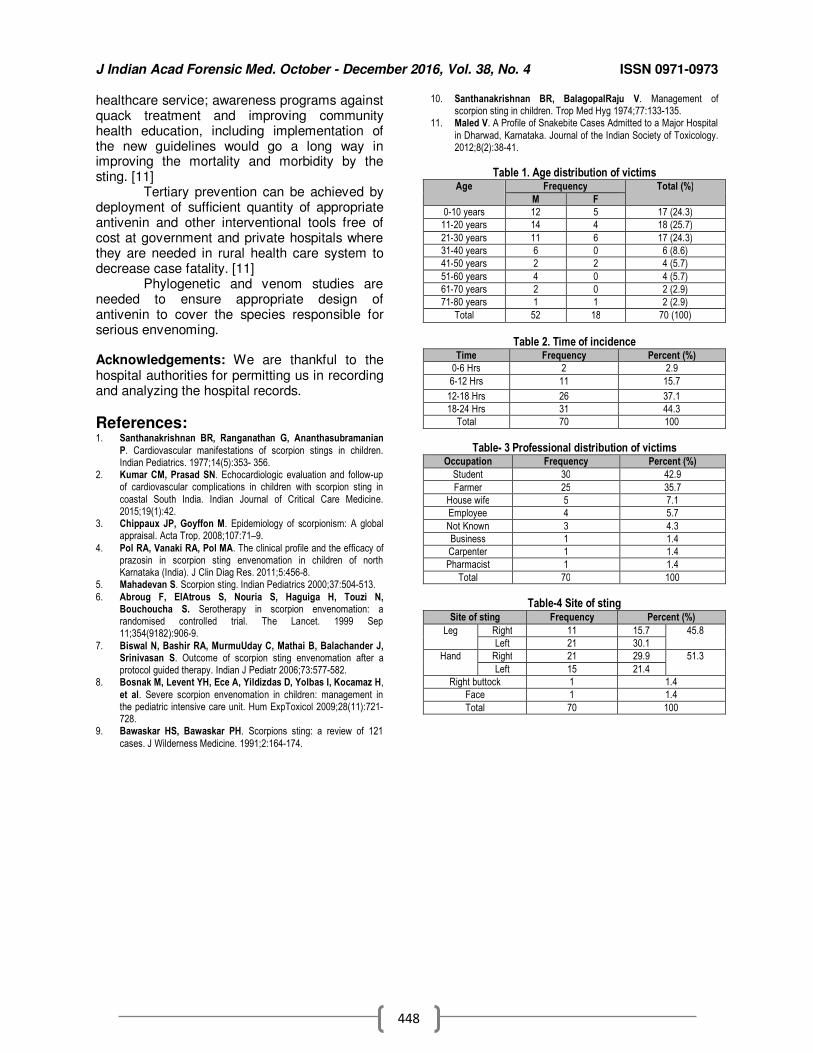
J Indian Acad Forensic Med. October - December 2016, Vol. 38, No. 4 ISSN 0971-0973
448
healthcare service; awareness programs against quack treatment and improving community health education, including implementation of the new guidelines would go a long way in improving the mortality and morbidity by the sting. [11] Tertiary prevention can be achieved by deployment of sufficient quantity of appropriate antivenin and other interventional tools free of cost at government and private hospitals where they are needed in rural health care system to decrease case fatality. [11] Phylogenetic and venom studies are needed to ensure appropriate design of antivenin to cover the species responsible for serious envenoming. Acknowledgements: We are thankful to the hospital authorities for permitting us in recording and analyzing the hospital records.
References: 1. Santhanakrishnan BR, Ranganathan G, Ananthasubramanian
P. Cardiovascular manifestations of scorpion stings in children. Indian Pediatrics. 1977;14(5):353- 356.
2. Kumar CM, Prasad SN. Echocardiologic evaluation and follow-up of cardiovascular complications in children with scorpion sting in
coastal South India. Indian Journal of Critical Care Medicine. 2015;19(1):42.
3. Chippaux JP, Goyffon M. Epidemiology of scorpionism: A global appraisal. Acta Trop. 2008;107:71–9.
4. Pol RA, Vanaki RA, Pol MA. The clinical profile and the efficacy of prazosin in scorpion sting envenomation in children of north Karnataka (India). J Clin Diag Res. 2011;5:456-8.
5. Mahadevan S. Scorpion sting. Indian Pediatrics 2000;37:504-513.
6. Abroug F, ElAtrous S, Nouria S, Haguiga H, Touzi N, Bouchoucha S. Serotherapy in scorpion envenomation: a randomised controlled trial. The Lancet. 1999 Sep 11;354(9182):906-9.
7. Biswal N, Bashir RA, MurmuUday C, Mathai B, Balachander J, Srinivasan S. Outcome of scorpion sting envenomation after a protocol guided therapy. Indian J Pediatr 2006;73:577-582.
8. Bosnak M, Levent YH, Ece A, Yildizdas D, Yolbas I, Kocamaz H,
et al. Severe scorpion envenomation in children: management in the pediatric intensive care unit. Hum ExpToxicol 2009;28(11):721-728.
9. Bawaskar HS, Bawaskar PH. Scorpions sting: a review of 121
cases. J Wilderness Medicine. 1991;2:164-174.
10. Santhanakrishnan BR, BalagopalRaju V. Management of scorpion sting in children. Trop Med Hyg 1974;77:133-135.
11. Maled V. A Profile of Snakebite Cases Admitted to a Major Hospital
in Dharwad, Karnataka. Journal of the Indian Society of Toxicology. 2012;8(2):38-41.
Table 1. Age distribution of victims Age Frequency Total (%)
M F
0-10 years 12 5 17 (24.3)
11-20 years 14 4 18 (25.7)
21-30 years 11 6 17 (24.3)
31-40 years 6 0 6 (8.6)
41-50 years 2 2 4 (5.7)
51-60 years 4 0 4 (5.7)
61-70 years 2 0 2 (2.9)
71-80 years 1 1 2 (2.9)
Total 52 18 70 (100)
Table 2. Time of incidence Time Frequency Percent (%)
0-6 Hrs 2 2.9
6-12 Hrs 11 15.7
12-18 Hrs 26 37.1
18-24 Hrs 31 44.3
Total 70 100
Table- 3 Professional distribution of victims Occupation Frequency Percent (%)
Student 30 42.9
Farmer 25 35.7
House wife 5 7.1
Employee 4 5.7
Not Known 3 4.3
Business 1 1.4
Carpenter 1 1.4
Pharmacist 1 1.4
Total 70 100
Table-4 Site of sting Site of sting Frequency Percent (%)
Leg Right 11 15.7 45.8
Left 21 30.1
Hand Right 21 29.9 51.3
Left 15 21.4
Right buttock 1 1.4
Face 1 1.4
Total 70 100
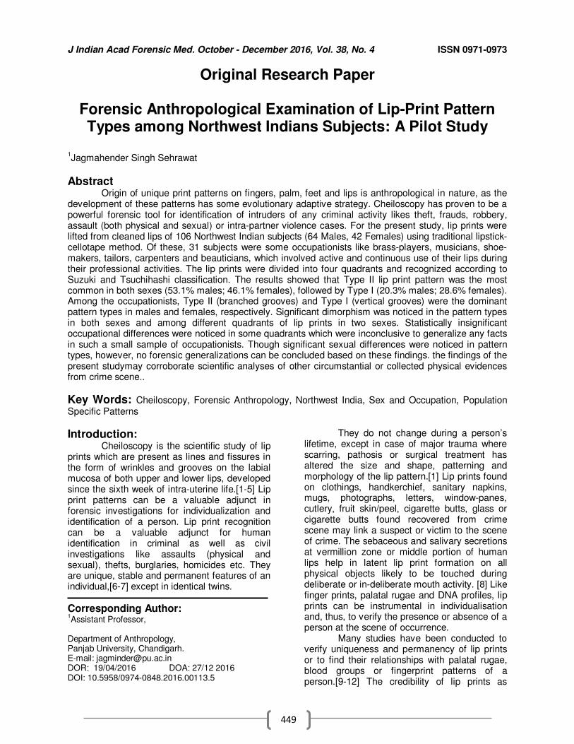
J Indian Acad Forensic Med. October - December 2016, Vol. 38, No. 4 ISSN 0971-0973
449
Original Research Paper
Forensic Anthropological Examination of Lip-Print Pattern Types among Northwest Indians Subjects: A Pilot Study
1Jagmahender Singh Sehrawat
Abstract Origin of unique print patterns on fingers, palm, feet and lips is anthropological in nature, as the
development of these patterns has some evolutionary adaptive strategy. Cheiloscopy has proven to be a powerful forensic tool for identification of intruders of any criminal activity likes theft, frauds, robbery, assault (both physical and sexual) or intra-partner violence cases. For the present study, lip prints were lifted from cleaned lips of 106 Northwest Indian subjects (64 Males, 42 Females) using traditional lipstick-cellotape method. Of these, 31 subjects were some occupationists like brass-players, musicians, shoe-makers, tailors, carpenters and beauticians, which involved active and continuous use of their lips during their professional activities. The lip prints were divided into four quadrants and recognized according to Suzuki and Tsuchihashi classification. The results showed that Type II lip print pattern was the most common in both sexes (53.1% males; 46.1% females), followed by Type I (20.3% males; 28.6% females). Among the occupationists, Type II (branched grooves) and Type I (vertical grooves) were the dominant pattern types in males and females, respectively. Significant dimorphism was noticed in the pattern types in both sexes and among different quadrants of lip prints in two sexes. Statistically insignificant occupational differences were noticed in some quadrants which were inconclusive to generalize any facts in such a small sample of occupationists. Though significant sexual differences were noticed in pattern types, however, no forensic generalizations can be concluded based on these findings. the findings of the present studymay corroborate scientific analyses of other circumstantial or collected physical evidences from crime scene..
Key Words: Cheiloscopy, Forensic Anthropology, Northwest India, Sex and Occupation, Population Specific Patterns
Introduction: Cheiloscopy is the scientific study of lip
prints which are present as lines and fissures in the form of wrinkles and grooves on the labial mucosa of both upper and lower lips, developed since the sixth week of intra-uterine life.[1-5] Lip print patterns can be a valuable adjunct in forensic investigations for individualization and identification of a person. Lip print recognition can be a valuable adjunct for human identification in criminal as well as civil investigations like assaults (physical and sexual), thefts, burglaries, homicides etc. They are unique, stable and permanent features of an individual,[6-7] except in identical twins.
Corresponding Author: 1Assistant Professor,
Department of Anthropology, Panjab University, Chandigarh. E-mail: [email protected] DOR: 19/04/2016 DOA: 27/12 2016 DOI: 10.5958/0974-0848.2016.00113.5
They do not change during a person’s lifetime, except in case of major trauma where scarring, pathosis or surgical treatment has altered the size and shape, patterning and morphology of the lip pattern.[1] Lip prints found on clothings, handkerchief, sanitary napkins, mugs, photographs, letters, window-panes, cutlery, fruit skin/peel, cigarette butts, glass or cigarette butts found recovered from crime scene may link a suspect or victim to the scene of crime. The sebaceous and salivary secretions at vermillion zone or middle portion of human lips help in latent lip print formation on all physical objects likely to be touched during deliberate or in-deliberate mouth activity. [8] Like finger prints, palatal rugae and DNA profiles, lip prints can be instrumental in individualisation and, thus, to verify the presence or absence of a person at the scene of occurrence. Many studies have been conducted to verify uniqueness and permanency of lip prints or to find their relationships with palatal rugae, blood groups or fingerprint patterns of a person.[9-12] The credibility of lip prints as
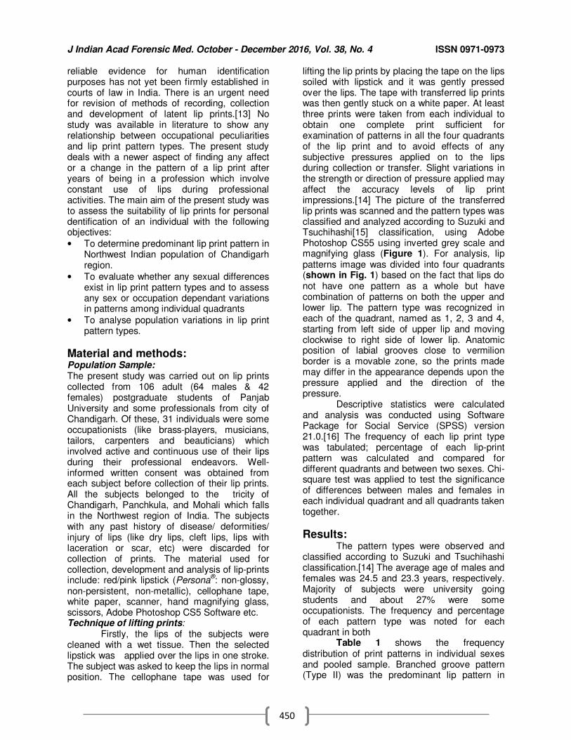
J Indian Acad Forensic Med. October - December 2016, Vol. 38, No. 4 ISSN 0971-0973
450
reliable evidence for human identification purposes has not yet been firmly established in courts of law in India. There is an urgent need for revision of methods of recording, collection and development of latent lip prints.[13] No study was available in literature to show any relationship between occupational peculiarities and lip print pattern types. The present study deals with a newer aspect of finding any affect or a change in the pattern of a lip print after years of being in a profession which involve constant use of lips during professional activities. The main aim of the present study was to assess the suitability of lip prints for personal dentification of an individual with the following objectives:
• To determine predominant lip print pattern in Northwest Indian population of Chandigarh region.
• To evaluate whether any sexual differences exist in lip print pattern types and to assess any sex or occupation dependant variations in patterns among individual quadrants
• To analyse population variations in lip print pattern types.
Material and methods: Population Sample: The present study was carried out on lip prints collected from 106 adult (64 males & 42 females) postgraduate students of Panjab University and some professionals from city of Chandigarh. Of these, 31 individuals were some occupationists (like brass-players, musicians, tailors, carpenters and beauticians) which involved active and continuous use of their lips during their professional endeavors. Well-informed written consent was obtained from each subject before collection of their lip prints. All the subjects belonged to the tricity of Chandigarh, Panchkula, and Mohali which falls in the Northwest region of India. The subjects with any past history of disease/ deformities/ injury of lips (like dry lips, cleft lips, lips with laceration or scar, etc) were discarded for collection of prints. The material used for collection, development and analysis of lip-prints include: red/pink lipstick (Persona
®: non-glossy,
non-persistent, non-metallic), cellophane tape, white paper, scanner, hand magnifying glass, scissors, Adobe Photoshop CS5 Software etc. Technique of lifting prints: Firstly, the lips of the subjects were cleaned with a wet tissue. Then the selected lipstick was applied over the lips in one stroke. The subject was asked to keep the lips in normal position. The cellophane tape was used for
lifting the lip prints by placing the tape on the lips soiled with lipstick and it was gently pressed over the lips. The tape with transferred lip prints was then gently stuck on a white paper. At least three prints were taken from each individual to obtain one complete print sufficient for examination of patterns in all the four quadrants of the lip print and to avoid effects of any subjective pressures applied on to the lips during collection or transfer. Slight variations in the strength or direction of pressure applied may affect the accuracy levels of lip print impressions.[14] The picture of the transferred lip prints was scanned and the pattern types was classified and analyzed according to Suzuki and Tsuchihashi[15] classification, using Adobe Photoshop CS55 using inverted grey scale and magnifying glass (Figure 1). For analysis, lip patterns image was divided into four quadrants (shown in Fig. 1) based on the fact that lips do not have one pattern as a whole but have combination of patterns on both the upper and lower lip. The pattern type was recognized in each of the quadrant, named as 1, 2, 3 and 4, starting from left side of upper lip and moving clockwise to right side of lower lip. Anatomic position of labial grooves close to vermilion border is a movable zone, so the prints made may differ in the appearance depends upon the pressure applied and the direction of the pressure. Descriptive statistics were calculated and analysis was conducted using Software Package for Social Service (SPSS) version 21.0.[16] The frequency of each lip print type was tabulated; percentage of each lip-print pattern was calculated and compared for different quadrants and between two sexes. Chi-square test was applied to test the significance of differences between males and females in each individual quadrant and all quadrants taken together.
Results: The pattern types were observed and classified according to Suzuki and Tsuchihashi
classification.[14] The average age of males and females was 24.5 and 23.3 years, respectively. Majority of subjects were university going students and about 27% were some occupationists. The frequency and percentage of each pattern type was noted for each quadrant in both Table 1 shows the frequency distribution of print patterns in individual sexes and pooled sample. Branched groove pattern (Type II) was the predominant lip pattern in
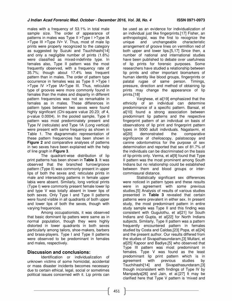
J Indian Acad Forensic Med. October - December 2016, Vol. 38, No. 4 ISSN 0971-0973
451
males with a frequency of 53.1% in total male sample size. The order of appearance of patterns in males was Type II ˃Type I ˃Type IA ˃Type III ˃Type IV˃ V. Thus, most of male lip prints were properly recognized to the category as suggested by Suzuki and Tsuchihashi[14]
and only a negligible number of prints (1.6%) were classified as mixed-indefinite type. In females also, Type II pattern was the most frequently observed, with a frequency rate of 35.7%; though about 17.4% less frequent pattern than in males. The order of pattern type occurrence in females was as Type II ˃Type I ˃Type IV ˃Type IA˃Type III. Thus, reticulate type of grooves were more commonly found in females than the males and disparity in different pattern frequencies was not so remarkable in females as in males. These differences in pattern types between two sexes were found highly significant (Chi-square value 20.23; df 4, p-value 0.0004). In the pooled sample, Type II pattern was most predominately present and Type IV (reticulate) and Type 1A (Long vertical) were present with same frequency as shown in Table 1. The diagrammatic representation of these pattern frequencies has been shown in Figure 2 and comparative analyses of patterns in two sexes have been explained with the help of line graph in Figure 3. The quadrant-wise distribution of lip print patterns has been shown in Table 3. It was observed that the branched furrow/groove pattern (Type II) was commonly present in upper lips of both the sexes and; reticulate prints in male and intersecting patterns in female upper labia were absent. Similarly, long vertical prints (Type I) were commonly present female lower lip and type V was totally absent in lower lips of both sexes. Only Type I and Type II patterns were found visible in all quadrants of both upper and lower lips of both the sexes, though with varying frequencies. Among occupationists, it was observed that basic dominant lip patters were same as in normal population, though they were highly distorted in lower quadrants in both sexes particularly among tailors, shoe-makers, barbers and brass-players. Type I and Type II patterns were observed to be predominant in females and males, respectively.
Discussion and conclusions: Identification or individualization of unknown victims of some homicidal, accidental or mass disaster incidents becomes necessary due to certain ethical, legal, social or sometimes political issues concerned with it. Lip prints can
be used as an evidence for individualization of an individual just like fingerprints.[17] Fisher, an anthropologist, was the first to recognize the unique and unchangeable characteristic arrangement of groove lines on vermillion red of both upper and lower lips.[5,17] Since then, a number of national and international studies have been published to debate over usefulness of lip prints for forensic purposes. Some researchers have studied a relationship between lip prints and other important biomarkers of human identity like blood groups, fingerprints or palatal rugae of same person. Timings, pressure, direction and method of obtaining lip prints may change the appearance of lip prints.[18]
.
Varghese, et al[19] emphasized that the ethnicity of an individual can determine predominance of a specific pattern. Bansal, et al[10] found a strong association between predominant lip patterns and the respective fingerprint pattern of an individual on basis of observations of lip print and fingerprint pattern types in 5000 adult individuals. Nagalaxmi, et al[20] demonstrated the comparative significance of cheiloscopy, palatoscopy and canine odontometrics for the purpose of sex determination and reported that sex of 81.7% of the individuals can be discriminated on the basis of lip-prints only. Verma, et al[9] found that Type II pattern was the most prominent among South Indians but no relationship could be established between them and blood groups or inter-commissural distance. Statistically significant sex differences were noticed in pattern types and these results were in agreement with some previous studies.[5] Analysis of results of various studies presented in Table 3 revealed that certain patterns were prevalent in either sex. In present study, the most predominant pattern in entire study sample was Type II and this finding was consistent with Gugulothu, et al[21] for South Indians and Gupta, et al[22] for North Indians subjects. Similarly, Type II pattern was a pattern frequently encountered pattern in females studied by Costa and Caldas,[23] Popa, et al[24]
and the present author. Our results differed from the studies of Sivapathasundaram,[3] Multani, et al[25] Kapoor and Badiye,[5] who observed that Type III pattern was most predominant in females. Type V was found as the least predominant lip print pattern which is in agreement with previous studies by Tsuchihashi[14] and Sivapathasundaram,[3] though inconsistent with findings of Type IV by Manipadya[26] and Jain, et al.[27] It may be clarified here that Type V pattern is ‘mixed and
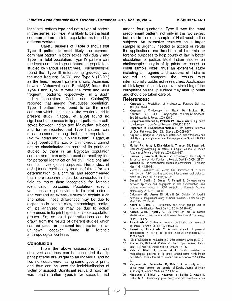
J Indian Acad Forensic Med. October - December 2016, Vol. 38, No. 4 ISSN 0971-0973
452
indefinite’ pattern type and not a type of pattern in true sense, so Type IV is likely to be the least common pattern in total population as found by different workers. Careful analysis of Table 3 shows that Type II pattern is most likely the common dominant pattern in both sexes individually and Type I in total population. Type IV pattern was the least common lip print pattern in populations studied by various researchers. Tsuchihashi[14] found that Type III (intersecting grooves) was the most frequent (64.6%) and Type V (13.9%) as the least frequent pattern among Japanese, however Vahanwalla and Parekh[28] found that Type I and Type IV were the most and least frequent patterns, respectively in a South Indian population. Costa and Caldas[23]
reported that among Portuguese population, Type II pattern was found to be the most common which is similar to the results found in present study. Nagpal, et al[29] found no significant differences in lip print patterns in both sexes between Indian and Malaysian subjects and further reported that Type I pattern was most common among both the populations (42.7% Indian and 56.1% Malaysian). Ragab, et al[30] reported that sex of an individual cannot not be discriminated on basis of lip prints as studied by them in an Egyptian population sample and it can only be used an ancillary tool for personal identification for civil litigations and criminal investigation purposes. Hernandez, et al[31] found cheiloscopy as a useful tool for sex determination of a criminal and recommended that more research should be conducted in this field to make them acceptable for forensic identification purposes. Population- specific variations are quite evident in lip print patterns and demand an extensive study to explain such anomalies. These differences may be due to disparities in sample size, methodology, portion of lips analysed or may be due to actual differences in lip print types in diverse population groups. So, no valid generalisations can be drawn from the results of different studies which can be used for personal identification of an unknown cadaver found in forensic anthropological contexts.
Conclusion: From the above discussions, it was observed and thus can be concluded that lip print patterns are unique to an individual and no two individuals were having same types of prints and thus can be used for individualisation of victim or suspect. Significant sexual dimorphism was noted in pattern types in two sexes but not
among four quadrants. Type II was the most predominant pattern, not only in the two sexes, but also in the total sample of Northwest Indian subjects. An extensive research on a larger sample is urgently needed to accept or refute the applications and thresholds of lip prints for forensic purposes to help courts of law in better elucidation of justice. Most Indian studies on cheiloscopic analysis of lip prints are based on small sample sizes, thus an extensive study including all regions and sections of India is required to compare the results with internationally published researches. Application of thick layer of lipstick and over stretching of the cellophane on the lip surface may alter lip prints and should be taken care of.
References: 1. Kasprzak J. Possibilities of cheiloscopy. Forensic Sci Intl.
1990;46:145-51. 2. Kasprzak J. Cheiloscopy. In: Siegel JA, Saukko, PJ,
Knupfer, GC. E d s . Encyclopedia of Forensic Sciences. 2nd Ed. Academic Press., 2000:358-61.
3. Sivapathasundharam B, Prakash PA, Sivakumar G. Lip prints (cheiloscopy). Indian Dental Research 2001;12:234-37.
4. Rajendran R, Sivapathasundharam B. In: Shafer’s Textbook of Oral Pathology. Sixth Ed, Elsevier. 2006:896-897.
5. Kapoor N, Badiye A. A study of distribution, sex differences and stability of lip print patterns in an Indian population. Saudi J Biol Sci
2015:1-6. 6. Murkey PN, Sutay S, Khandekar IL, Tirpude, BH, Pawar VG.
Cheiloscopy-everything in nature is unique. Journal of Indian Academy of Forensic Medicine. 2008;30:194-97.
7. Sharma P, Saxena S, Rathod V. Cheiloscopy: the study of lip prints in sex identification. J Forensic Dent Sci.2009;1:24-27.
8. Williams TR. Lip prints-another means of identification. J Forensic Ident 1991;41:190-94.
9. Verma P, Sachdeva SC, Verma KG. Correlation of lip prints with gender, ABO blood groups and inter-commissural distance. North Am J Med Sci. 2013;5(7):427-31
10. Bansal P, Sheikh S, Bansal R, Pallgati S. Correspondence
between lip-prints and fingerprints in sex determination and pattern predominance in 5000 subjects. J Forensic Odonto-stomatology 2014;31(1):8-14.
11. Eldomiaty MA, Anwar RI, Algaidi SA. Stability of lip-print
patterns: a longitudinal study of Saudi females. J Forensic legal Med. 2014; 22:154-58.
12. Karim B, Gupta D. Cheiloscopy and blood groups: aid in forensic identification. Saudi Dent J. 2 0 1 4 ; 26:176-80.
13. Kaleem A KH, Tripathy C. Lip Print: an aid to human identification. Indian Journal of Forensic Medicine & Toxicology. 2015;9(1):64-67.
14. Tsuchihashi Y. Studies on personal identification by means of
lip prints. Forensic Sci Intl. 1974;3:233-48. 15. Suzuki K, Tsuchihashi Y. A new attempt of personal
identification by means of lip print. Can Soc Forensic Sci J. 1971;4:54-58.
16. IBM SPSS Science Inc.Statistics 21.0 for Windows. Chicago, 2012 17. Prabhu RV, Dinkar A, Prabhu V. Cheiloscopy: revisited. Indian
Journal of Forensic Dental Science. 2012;4(1):47-52 18. Vats Y, Dhall JK, Kapoor A K. Gender variation in
morphological patterns of lip prints among some north Indian populations. Indian Journal of Forensic Dental Science. 2014;4:19–23.
19. Varghese AJ, Somesekar M, Babu UR. A study on lip
prints types among the people of Kerala. Journal of Indian Academy of Forensic Medicine. 2010;32:6-7.
20. Nagalaxmi V, Sridevi U, Nagajyothi M, Lalitha C, Nayak K, Srikanth K. Cheiloscopy, palatoscopy and odontometrics in sex
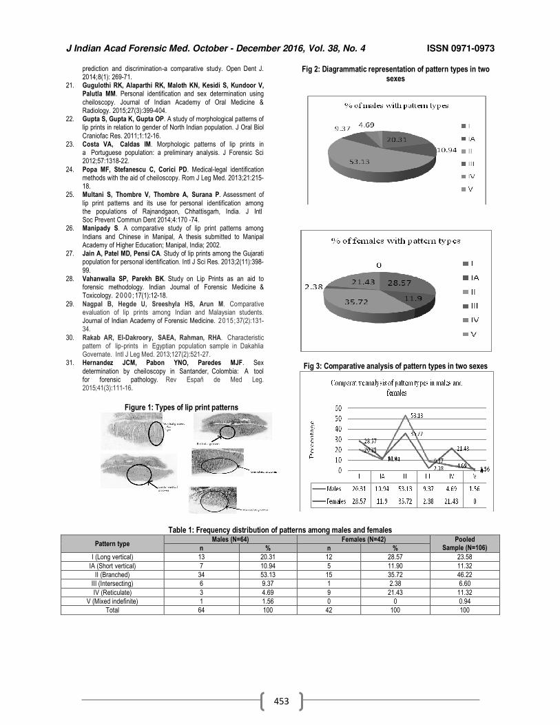
J Indian Acad Forensic Med. October - December 2016, Vol. 38, No. 4 ISSN 0971-0973
453
prediction and discrimination-a comparative study. Open Dent J. 2014;8(1): 269-71.
21. Gugulothi RK, Alaparthi RK, Maloth KN, Kesidi S, Kundoor V, Palutla MM. Personal identification and sex determination using
cheiloscopy. Journal of Indian Academy of Oral Medicine & Radiology. 2015;27(3):399-404.
22. Gupta S, Gupta K, Gupta OP. A study of morphological patterns of lip prints in relation to gender of North Indian population. J Oral Biol
Craniofac Res. 2011;1:12-16. 23. Costa VA, Caldas IM. Morphologic patterns of lip prints in
a Portuguese population: a preliminary analysis. J Forensic Sci 2012;57:1318-22.
24. Popa MF, Stefanescu C, Corici PD. Medical-legal identification methods with the aid of cheiloscopy. Rom J Leg Med. 2013;21:215-18.
25. Multani S, Thombre V, Thombre A, Surana P. Assessment of
lip print patterns and its use for personal identification among the populations of Rajnandgaon, Chhattisgarh, India. J Intl Soc Prevent Commun Dent 2014;4:170 -74.
26. Manipady S. A comparative study of lip print patterns among
Indians and Chinese in Manipal, A thesis submitted to Manipal Academy of Higher Education; Manipal, India; 2002.
27. Jain A, Patel MD, Pensi CA. Study of lip prints among the Gujarati population for personal identification. Intl J Sci Res. 2013;2(11):398-
99. 28. Vahanwalla SP, Parekh BK. Study on Lip Prints as an aid to
forensic methodology. Indian Journal of Forensic Medicine & Toxicology. 2 0 0 0 ; 17(1):12-18.
29. Nagpal B, Hegde U, Sreeshyla HS, Arun M. Comparative evaluation of lip prints among Indian and Malaysian students. Journal of Indian Academy of Forensic Medicine. 2015;37(2):131-34.
30. Rakab AR, El-Dakroory, SAEA, Rahman, RHA. Characteristic pattern of lip-prints in Egyptian population sample in Dakahlia Governate. Intl J Leg Med. 2013;127(2):521-27.
31. Hernandez JCM, Pabon YNO, Paredes MJF. Sex
determination by cheiloscopy in Santander, Colombia: A tool for forensic pathology. Rev Españ de Med Leg. 2015;41(3):111-16.
Figure 1: Types of lip print patterns
Fig 2: Diagrammatic representation of pattern types in two sexes
Fig 3: Comparative analysis of pattern types in two sexes
Table 1: Frequency distribution of patterns among males and females
Pattern type Males (N=64) Females (N=42) Pooled
Sample (N=106) n % n %
I (Long vertical) 13 20.31 12 28.57 23.58
IA (Short vertical) 7 10.94 5 11.90 11.32
II (Branched) 34 53.13 15 35.72 46.22
III (Intersecting) 6 9.37 1 2.38 6.60
IV (Reticulate) 3 4.69 9 21.43 11.32
V (Mixed indefinite) 1 1.56 0 0 0.94
Total 64 100 42 100 100
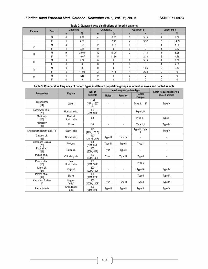
J Indian Acad Forensic Med. October - December 2016, Vol. 38, No. 4 ISSN 0971-0973
454
Table 2: Quadrant wise distributions of lip print patterns
Pattern Sex Quadrant 1 Quadrant 2 Quadrant 3 Quadrant 4
n % n % n % n %
I M 6 9.38 4 6.25 2 3.13 1 1.56
F 1 2.38 1 2.38 4 9.52 6 14.29
IA M 4 6.25 2 3.13 0 0 1 1.56
F 1 2.38 0 0 0 0 4 9.52
II M 16 25.00 12 18.75 2 3.13 4 6.25
F 7 16.67 5 11.90 1 2.38 2 4.76
III M 3 4.69 0 0 2 3.13 1 1.56
F 0 0 0 0 0 0 1 2.38
IV M 0 0 0 0 1 1.56 2 3.13
F 5 11.90 3 7.14 1 2.38 0 0
V M 1 1.56 0 0 0 0 0 0
F 0 0 0 0 0 0 0 0
Table 3: Comparative frequency of pattern types in different population groups in individual sexes and pooled sample
Researcher Region No. of
subjects
Most frequent pattern type Least frequent pattern in
pooled sample Males Females Pooled Sample
Tsuchihashi [14]
Japan 1364
(757 M, 607 F)
- - Type III, I , IA Type V
Vahanwalla et al., [28]
Mumbai,India, 100
(50M, 50 F) - - Type I, IA -
Manipady [26]
Manipal South India
50 - - Type II , I Type III
Manipady [26]
China 50 - - Type II, I Type IV
Sivapathasundaram et al., [3] South India 196
(96M, 100 F)
Type III, Type
I Type V
Gupta et al., [22]
North India, 150
(75 M, 75F) Type II Type IV - -
Costa and Caldas
[23] Portugal
50
{25M, 25 F) Type III Type II Type II -
Popa et al., [24]
Romania 100
(50M, 50F) Type I Type II - -
Multani et al., [25]
Chhatishgarh 200
(100M, 100F) Type I Type III Type I -
Prabhu et al., [16]
Goa, South India
100 (50M, 50 F)
- - Type V -
Jain et al., [27]
Gujarat 200
(100M, 100F) - - Type IA Type IV
Peeran et al.,
[32] Libiya
104
(37M, 67 F) - - Type I Type IA
Kapur and Badiye [5]
Nagpur (India)
200 (100M, 100F)
Type I Type III Type I Type IA
Present study Chandigarh
India 106
(64M, 42 F) Type II Type II Type II, Type V
J Indian Acad Forensic Med. October - December 2016, Vol. 38, No. 4 ISSN 0971-0973
455
Original Research Paper
Fatal Burn Injuries: A Five Year Retrospective Autopsy Study In Temple Town, Tirupati
1M. Abdul Khalid,
2Bijili Venkatesulu,
3B Lakshmi Narayana
Abstract Burn deaths have tremendous medicolegal importance as they are one of the commonest causes
of unnatural deaths in India. The purpose of this study was to record and evaluate the epidemiological and medicolegal aspects of fatal burn injuries retrospectively. An analysis of autopsy records revealed 742 (18.3%) cases of burn injuries among total autopsies done over 5 years period (2010 – 2014) in the mortuary of department of Forensic Medicine, S V Medical College, Tirupati. The majority of victims were females (62.8%), with the age group 21-40 years (57.4%) being predominantly affected. Married victims (78.3%) outnumbered unmarried. Thermal burns, 673 cases (90.7%) was commonly noted with explosion of kerosene stove in 238 (32.1%) being the predominant cause of fire. Mortality was higher in victims with 51-75% of TBSA burn with septicemia, 219 cases (29.5%) being the leading cause of death, followed by hypovolemic shock, 192cases (25.9%). Majority of the cases were accidental, 605 (81.5%), followed by suicidal, 89 (12%) and homicidal deaths 46(6.5%), respectively.
Key Words: Fatal Burns, Epidemiology, Types of Burns, Cause of Death, Manner of Death.
Introduction: Fire was perhaps, man’s first double edged sword for throughout history, it has served as well as destroyed mankind. [1]Traumadue to burns is as old as the discovery of fire in the history of mankind. If used with precaution, it is of great help to mankind and when precautions are not observed, it creates disaster, resulting in injuries to human body and damage to the property. Burns are injuries produced by application of dry heat such as flame, radiant heat or some heated solid substance like metal or glass to the body. Local injury to the body by heat may result from dry heat, application of hot bodies, licking by flames resulting in simple burns, moist heat leading to scalds and corrosive poisons resulting in burns.
Corresponding Author:
1Assistant Professor,
Department of Forensic Medicine, Apollo Institute of Medical Sciences and Research, Murakumbattu, Chittoor. 2Assistant Professor,
Department of Forensic Medicine, Sri Venkateswara Medical College, Tirupati. 3Professor and Head, Department of Forensic
Medicine, Kurnool Medical College, Kurnool Email: [email protected] DOR:03/03/2016 DOA: 27/12 2016 DOI: 10.5958/0974-0848.2016.00114.7
Electric spark discharges, flashes and lightening leads to electric burns. [2]Burn injury is one of the common medical emergencies admitted to any hospital and is an important public health problem throughout the world. Burns have always been considered as one of the most destructive injuries, causing not only deaths but also major economic and psychological impacts and long term somatic sequela as well.[3,4] The obnoxious and ubiquitous practice of dowry has perpetuated a new and alarming rise in mortality from burns, the so called “dowry deaths”. Homicidal burning of married women in India is a major concern for the government, law enforcing authorities, the judiciary, the police and medicolegal experts all over the country. A number of studies on various aspects of burns have been reported from various parts of India, but this is the first study to be carried out on fatal burns cases in the temple town of Tirupati. In the present study, an attempt is made to analyze the data collected on such cases, statistically, to find out the incidence and circumstances of death due to fire, age and sex distribution, marital status, etiology of burn, manner of death, surface area burns, period of survival and cause of death, together highlighting the various epidemiological and medicolegal aspects in the community which will help to plan the preventive programs and in
J Indian Acad Forensic Med. October - December 2016, Vol. 38, No. 4 ISSN 0971-0973
456
order to let the legal system and medico-legalist take necessary action.
Materials and Methods Material for the present study comprised of a total number of 742cases of death due to burns brought to the mortuary of Government General Hospital attached to the Department of Forensic Medicine, Sri Venkateswara Medical College, Tirupati. All case records in the mortuary files were studied for fatalities among male and female victims due to burns trauma over a period of 5 years i.e., from 1
st January
2010 to 31st
December 2014, collecting all the data required. An in-depth examination of the epidemiological features and medicolegal aspects like age and sex of deceased, marital status, type of burn, etiology of burn, extent of surface area of burn, period of survival, socioeconomic status, cause of death and manner of death were ascertained from In-patient case sheets in case of hospital admission, Inquest reports, perusal of police papers, records from medical records section of Sri Venkateswara Ram Narayan Ruia Government General Hospital, Tirupati, post-mortem reports of the cases, Information collected from the police officer, relatives and friends of the deceased accompanying the dead bodies. The resulting data was tabulated and analyzed. On the basis of analysis and observation, results were drawn, discussed and compared with other relevant literature. During the study period, a total number of 4047 medico legal autopsies were carried out by the department, of which742 cases were victims of bums. Among these, there was a definite female preponderance with 466 females against 276 males, constituting a ratio of F: M of 1.7:1.
Observations & Results: The incidence of burns whether accidental, homicidal or suicidal are not uncommon to Indian society since time immemorial. Incidence, etiology and nature of burns vary from one community to another and depend mainly upon age, sex, customs and economic status, environmental and social circumstances. A total of 742 cases were studied during the period 2010 to 2014, which forms about 18.3% of total postmortem examinations done during that period(4047 cases) (Table 1). In the present study, a female preponderance of 1.7:1 was seen.The age group
of 21 – 30 years (36.1%) was the most commonly affected, followed by 31 – 40 years, 21.3%. (Table 2) Married victims, 581 (78.3%) outnumbered the unmarried. (Table 3). Among the married, females (65.2%) outnumbered the males (34.8%). In married females, 30.2% of victims sustained burns within 5 years of marriageand 69.2% of cases sustained burns within 7 years of marriage. The higher incidence of female victims and incidents in the early years of marriage is because of marital disharmony, dowry problems, alcoholism of husband and wife beating behavior and illegal relations. Death due to burns was more commonly seen in lower socioeconomic strata, 395 cases (53.2%), followed by middle socioeconomic strata 336 (45.3%), (Table 4). Thermal burns 673 (90.70%) was most common type of burn followed by electrical burns, 63 (8.49%) and scalds, 6 (0.8%), (Table 5). The major cause for burns was alleged explosion of kerosene stove, 238 cases (32.1%), followed by kerosene lamp spillage 184 (24.8%), flame burns 175 (23.6%) (Table 6). Mortality was higher in victims with burnt surface area of 51 - 75 % of body surface, 296 cases (39.9%), followed by 26 - 50 % burns 176 cases (23.7%), (Table 7). 193 cases (26%) survived for 3 -7days, followed by 108 victims surviving for 24-36 hrs (14.6%), (Table 8). In the present study, septicemic shock was the most common cause of death, 219 cases (29.5%), followed by hypovolemicshock 192 (25.9%).(Table 9) Most common manner of death was accidental, 605 cases, (81.5%), followed by suicidal 89 (12%) and homicidal 48 (6.5%), (Table 10).
Discussion: Burn injury occurs universally and has plagued mankind since antiquity till the present day. In all societies, including developed or developing countries, burns constitute not only a medical and psychological problem, but also have severe economic and social consequences, not only to victims, but also to their family and society in general.[5] 742 burns cases were analysed during the study period, which forms about 18.3% of the total case load. Similar results were found by Batra A K [6] (23.3%) and Ambade V N, [7] (21.6%). In the present study, female preponderance of 1:1.68 was seen over males, which was similar to studies of Ambade V N, [7] Dhiraj, et al [8] and Zanjad [9] and in contrast with the study of Mostafa M. [10] In India, as
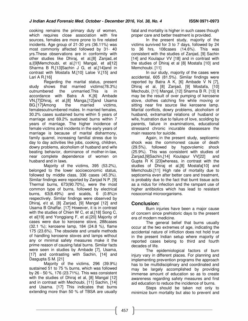
J Indian Acad Forensic Med. October - December 2016, Vol. 38, No. 4 ISSN 0971-0973
457
cooking remains the primary duty of women, which requires close association with fire sources, females are more prone to fire related incidents. Age group of 21-30 yrs (36.11%) was most commonly affected followed by 31- 40 yrs.These observations are in conformity with other studies like Dhiraj, et al,[8] Zanjad,.et a,l[9]Memchoubi, et al,[11] Mangal, et al[12] Sharma B R,[13]Sachin J G, et al,[14]and in contrast with Mostafa M,[10] Laloe V,[15] and Lari A R.[16] Regarding the marital status, present study shows that married victims(78.3%) outnumbered the unmarried.This is in accordance with Batra A K,[6] Ambade VN,[7]Dhiraj, et al,[8] Manga,[12]and Usama BG.[17]Among the married victims, femalesoutnumbered males. In married females, 30.2% cases sustained burns within 5 years of marriage and 69.2% sustained burns within 7 years of marriage. The higher incidence of female victims and incidents in the early years of marriage is because of marital disharmony, family quarrel, increasing familial stress due to day to day activities like jobs, cooking, children, dowry problems, alcoholism of husband and wife beating behavior, dominance of mother-in-law, near complete dependence of women on husband and in laws. Majority of the victims, 395 (53.2%), belonged to the lower socioeconomic status, followed by middle class, 336 cases (45.3%). Similar findings were reported by Zanjad N P. [9] Thermal burns, 673(90.70%), were the most common type of burns, followed by electrical burns, 63(8.49%) and scalds, 6 (0.8%)n respectively. Similar findings were observed by Dhiraj, e\t al, [8] Zanjad, [9] Mangal [12] and Usama B Ghaffar. [17] However, it is in contrast with the studies of Chien W C, et al,[18] Song C, et al[19] and Yonggiang F, et al.[20] Majority of cases were due to kerosene stove, 288cases (32.1 %); kerosene lamp, 184 (24.8 %), flame 175 (23.6%). The obsolete and unsafe methods of handling kerosene stoves and lamps without any or minimal safety measures make it the prime reason of causing fatal burns. Similar facts were seen in studies by Ambade [7], Usama, [17] and contrasting with Sachin, [14] and Dasgupta S M. [21] Majority of the victims, 296 (39.9%) sustained 51 to 75 % burns, which was followed by 26 - 50 %, 176 (23.71%). This was consistent with the studies of Dhiraj et al, [8] Mangal [12] and in contrast with Mechoubi, [11] Sachin, [14] and Usama. [17] This indicates that burns extending more than 50 % of TBSA are usually
fatal and mortality is higher in such cases though proper care and better treatment is provided. In the present study, majority of the victims survived for 3 to 7 days, followed by 24 to 36 hrs, 108cases (14.6%). This was consistent with the studies of Zanjad, [9] Sachin [14] and Koulapur VV [18] and in contrast with the studies of Dhiraj et al [8] Mostafa [10] and Memchoubi. [11] In our study, majority of the cases were accidental, 605 (81.5%). Similar findings were reported by Batra A K, [6] Ambade V N [7], Dhiraj et al, [8] Zanjad, [9] Mostafa, [10] Mechoubi, [11] Mangal, [12] Sharma B R. [13] It may be the result of over pumping of kerosene stove, clothes catching fire while moving or sitting near fire source like kerosene lamp. Marital conflicts, dowry problems, alcoholism of husband, extramarital relations of husband or wife, frustration due to failure of love, scolding by parents, failure in examinations, educational stressand chronic incurable diseasesare the main reasons for suicide. Again, in the present study, septicemic shock was the commonest cause of death (29.5%), followed by hypovolemic shock (25.9%). This was consistent with studies of Zanjad,[9]Sachin,[14] Koulapur VV[22] and Gupta R K [23]whereas, in contrast with the studies of Dhiraj et al,[8] Mostafa[10] and Memchoubi.[11] High rate of mortality due to septicemia even after better care and treatment, is probably due to the fact that burnt tissue acts as a nidus for infection and the rampant use of higher antibiotics which has lead to resistant nosocomial microorganisms.
Conclusion: Burn injuries have been a major cause of concern since prehistoric days to the present era of modern medicine. The general belief that burns usually occur at the two extremes of age, indicating the accidental nature of infliction does not hold true in the present Indian setup where majority of reported cases belong to third and fourth decades of life. The epidemiological factors of burn injury vary in different places. For planning and implementing prevention programs the approach has to be multidisciplinary and coordinated and may be largely accomplished by providing immense amount of education so as to create awareness regarding safety measures and first aid education to reduce the incidence of burns. Steps should be taken not only to minimize burn mortality but also to prevent and
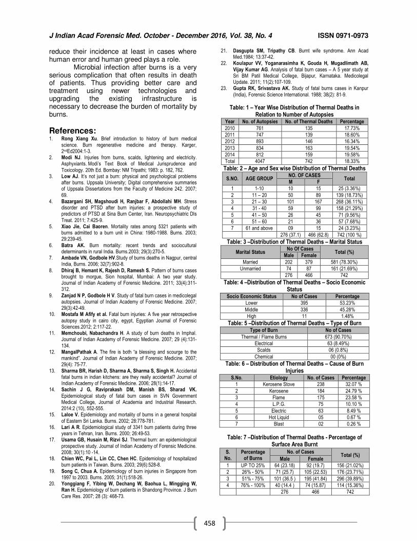
J Indian Acad Forensic Med. October - December 2016, Vol. 38, No. 4 ISSN 0971-0973
458
reduce their incidence at least in cases where human error and human greed plays a role. Microbial infection after burns is a very serious complication that often results in death of patients. Thus providing better care and treatment using newer technologies and upgrading the existing infrastructure is necessary to decrease the burden of mortality by burns.
References: 1. Rong Xiang Xu. Brief introduction to history of burn medical
science. Burn regenerative medicine and therapy. Karger, 2ndEd2004:1-3.
2. Modi NJ. Injuries from burns, scalds, lightening and electricity. Asphyxiants. Modi’s Text Book of Medical Jurisprudence and
Toxicology. 20th Ed. Bombay: NM Tripathi; 1983: p. 182, 762. 3. Low AJ. It’s not just a burn: physical and psychological problems
after burns. Uppsala University; Digital comprehensive summaries of Uppsala Dissertations from the Faculty of Medicine 242. 2007;
69. 4. Bazargani SH, Magshoudi H, Ranjbar F, Abdollahi MH. Stress
disorder and PTSD after burn injuries: a prospective study of predictors of PTSD at Sina Burn Center, Iran. Neuropsychiatric DIs
Treat. 2011; 7:425-9. 5. Xiao Jie, Cai Baoren. Mortality rates among 5321 patients with
burns admitted to a burn unit in China: 1980-1988. Burns. 2003; 29:239-45.
6. Batra AK. Burn mortality: recent trends and sociocultural determinants in rural India. Burns.2003; 29(3):270-5.
7. Ambade VN, Godbole HV.Study of burns deaths in Nagpur, central India, Burns. 2006; 32(7):902-8.
8. Dhiraj B, Hemant K, Rajesh D, Ramesh S. Pattern of burns cases brought to morgue, Sion hospital, Mumbai: A two year study. Journal of Indian Academy of Forensic Medicine. 2011; 33(4):311-312.
9. Zanjad N P, Godbole H V. Study of fatal burn cases in medicolegal autopsies. Journal of Indian Academy of Forensic Medicine. 2007; 29(3):42-49.
10. Mostafa M Afify et al. Fatal burn injuries: A five year retrospective
autopsy study in cairo city, egypt, Egyptian Journal of Forensic Sciences.2012; 2:117-22.
11. Memchoubi, Nabachandra H. A study of burn deaths in Imphal. Journal of Indian Academy of Forensic Medicine. 2007; 29 (4):131-
134. 12. MangalPathak A. The fire is both “a blessing and scourge to the
mankind”. Journal of Indian Academy of Forensic Medicine. 2007; 29(4): 75-77.
13. Sharma BR, Harish D, Sharma A, Sharma S, Singh H. Accidental fatal burns in indian kitchens: are they really accidental? Journal of Indian Academy of Forensic Medicine. 2006; 28(1):14-17.
14. Sachin J G, Raviprakash DM, Manish BS, Sharad VK.
Epidemiological study of fatal burn cases in SVN Government Medical College, Journal of Academia and Industrial Research. 2014:2 (10), 552-555.
15. Laloe V. Epidemiology and mortality of burns in a general hospital
of Eastern Sri Lanka. Burns. 2002; 28:778-781. 16. Lari A R. Epidemiological study of 3341 burn patients during three
years in Tehran, Iran. Burns. 2000; 26:49-53. 17. Usama GB, Husain M, Rizvi SJ. Thermal burn: an epidemiological
prospective study. Journal of Indian Academy of Forensic Medicine. 2008; 30(1):10 -14.
18. Chien WC, Pai L, Lin CC, Chen HC. Epidemiology of hospitalized burn patients in Taiwan. Burns. 2003; 29(6):528-8.
19. Song C, Chua A. Epidemiology of burn injuries in Singapore from 1997 to 2003. Burns. 2005; 31(1):518-26.
20. Yonggiang F, Yibing W, Dechang W, Baohua L, Mingging W, Ran H. Epidemiology of burn patients in Shandong Province. J Burn
Care Res. 2007; 28 (3): 468-73.
21. Dasgupta SM, Tripathy CB. Burnt wife syndrome. Ann Acad Med.1984; 13:37-42.
22. Koulapur VV, Yoganarasimha K, Gouda H, Mugadlimath AB,
Vijay Kumar AG. Analysis of fatal burn cases – A 5 year study at Sri BM Patil Medical College, Bijapur, Karnataka. Medicolegal Update. 2011; 11(2):107-109.
23. Gupta RK, Srivastava AK. Study of fatal burns cases in Kanpur
(India), Forensic Science International. 1988; 38(2): 81-9.
Table: 1 – Year Wise Distribution of Thermal Deaths in Relation to Number of Autopsies
Year No. of Autopsies No. of Thermal Deaths Percentage
2010 761 135 17.73%
2011 747 139 18.60%
2012 893 146 16.34%
2013 834 163 19.54%
2014 812 159 19.58%
Total 4047 742 18.33%
Table: 2 – Age and Sex wise Distribution of Thermal Deaths
S.NO. AGE GROUP NO. OF CASES
Total M F
1 1-10 10 15 25 (3.36%)
2 11 – 20 50 89 139 (18.73%)
3 21 – 30 101 167 268 (36.11%)
4 31 - 40 59 99 158 (21.29%)
5 41 – 50 26 45 71 (9.56%)
6 51 – 60 21 36 57 (7.68%)
7 61 and above 09 15 24 (3.23%)
276 (37.1) 466 (62.8) 742 (100 %)
Table: 3 –Distribution of Thermal Deaths – Marital Status
Marital Status No Of Cases
Total (%) Male Female
Married 202 379 581 (78.30%)
Unmarried 74 87 161 (21.69%)
276 466 742
Table: 4 –Distribution of Thermal Deaths – Socio Economic Status
Socio Economic Status No of Cases Percentage
Lower 395 53.23%
Middle 336 45.28%
High 11 1.48%
Table: 5 –Distribution of Thermal Deaths – Type of Burn Type of Burn No of Cases
Thermal / Flame Burns 673 (90.70%)
Electrical 63 (8.49%)
Scalds 06 (0.8%)
Chemical 00 (0%)
Table: 6 – Distribution of Thermal Deaths – Cause of Burn Injuries
S.No. Etiology No. of Cases Percentage
1 Kerosene Stove 238 32.07 %
2 Kerosene 184 24.79 %
3 Flame 175 23.58 %
4 L.P.G. 75 10.10 %
5 Electric 63 8.49 %
6 Hot Liquid 05 0.67 %
7 Blast 02 0.26 %
Table: 7 –Distribution of Thermal Deaths - Percentage of Surface Area Burnt
S. No.
Percentage of Burns
No. of Cases Total (%)
Male Female
1 UP TO 25% 64 (23.18) 92 (19.7) 156 (21.02%)
2 26% - 50% 71 (25.7) 105 (22.53) 176 (23.71%)
3 51% - 75% 101 (36.5 ) 195 (41.84) 296 (39.89%)
4 76% - 100% 40 (14.4 ) 74 (15.87) 114 (15.36%)
276 466 742
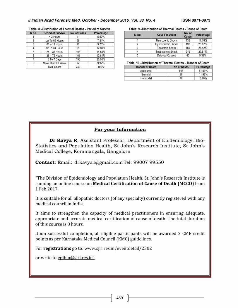
J Indian Acad Forensic Med. October - December 2016, Vol. 38, No. 4 ISSN 0971-0973
459
Table: 8 –Distribution of Thermal Deaths - Period of Survival S.No. Period of Survival No. of Cases Percentage
1 < 2 Hours 41 5.52%
2 Up To 06 Hours 58 7.81%
3 06 – 12 Hours 72 9.70%
4 12 To 24 Hours 95 12.80%
5 24 – 36 Hours 108 14.55%
6 36 – 72 Hours 101 13.61%
7 3 To 7 Days 193 26.01%
8 More Than 01 Week 74 9.97%
Total Cases 742 100%
Table: 9 –Distribution of Thermal Deaths - Cause of Death
S. No. Cause of Death No. of Cases
Percentage
1 Neurogenic Shock 132 17.78%
2 Hypovolemic Shock 192 25.87%
3 Toxaemic Shock 159 21.42%
4 Septicaemic Shock 219 29.51%
5 Delayed Causes 40 5.39%
Table: 10 –Distribution of Thermal Deaths – Manner of Death Manner of Death No of Cases Percentage
Accidental 605 81.53%
Suicidal 89 11.99%
Homicidal 48 6.46%
For your Information
Dr Kavya R, Assistant Professor, Department of Epidemiology, Bio-Statistics and Population Health, St John's Research Institute, St John's
Medical College, Koramangala, Bangalore
Contact: Email: [email protected] Tel: 99007 99550
"The Division of Epidemiology and Population Health, St. John's Research Institute is
running an online course on Medical Certification of Cause of Death (MCCD) from
1 Feb 2017.
It is suitable for all allopathic doctors (of any specialty) currently registered with any
medical council in India.
It aims to strengthen the capacity of medical practitioners in ensuring adequate,
appropriate and accurate medical certification of cause of death. The total duration
of this course is 8 hours.
Upon successful completion, all eligible participants will be awarded 2 CME credit
points as per Karnataka Medical Council (KMC) guidelines.
For registrations go to: www.sjri.res.in/eventdetail/2302
or write to [email protected]"
J Indian Acad Forensic Med. October - December 2016, Vol. 38, No. 4 ISSN 0971-0973
460
Original Research Paper
A study of Unnatural Female Deaths reported to a tertiary care hospital in Khammam, Telangana
1Bharath Kumar Guntheti,
2Uday Pal Singh
Abstract A total of 112 unnatural female deaths were studied for one year during 2015-2016 at the Dept. of
Forensic Medicine, Mamata Medical College, attached with MGH, Khammam. The most common age group affected was 21-30 years. Most of the victims were married, 75% of them died within the first 3 years of marriage. Hindu brides{87.5%} rural, illiterate {72.3%}and of low socioeconomic group {58%} were the most common victims. Most of the victims were housewives,79{70.5%} and living in nuclear families {82.1%}.Maximum cases were encountered in summer and in day time. As regards the pattern of unnatural deaths, suicides accounted for 86{76.8%} cases, followed by accidental - 14{12.5%}and homicidal 12{10.7%}.Unnatural female deaths encountered in our study were RTAs, poisoning, hanging, burns, snake bite and drowning. Poisoning was the most common method of suicidal death followed by hanging and burns. Suicidal deaths were mainly precipitated by dowry demand, chronic diseases, and psychiatric illnesses. Homicidal deaths were due to dowry, disputes, extra marital relationships, revenge, and jealousy. 35% of total deaths were alleged dowry deaths. The main objective of this study was to find out the various socio –cultural causesof unnatural female deaths and their preventive measures.
Key Words: Unnatural, Female, Death, Suicide, Accident, Poisoning, Hanging.
Introduction: A death is named unnatural when it is caused prematurely against the order of nature by injury, poison, or other means of violence.[1] This could be homicidal, suicidal, accidental or of unexplained origin. Manner of death is divided into two types: 1.Natural- if death occurs from disease alone. 2.Unnatural-if death results directly from an injury or poison or indirectly an injury which may precipitate preexisting natural disease in an individual .This could be homicidal, suicidal ,accidental or of unexpected origin.[2] Many cultural and socio economic factors of the country are responsible for the causation of such deaths in females. Its increasing incidence is symbolic of continuing erosion and devaluation of women’s status in independent India.[3] A rapid increase in unnatural deaths in females, especially in the first few years of their married life, was observed in our society for the last few decades.
Corresponding Author:
1Associate Professor,
2Prof &HoD,
Dept. of Forensic Medicine & Toxicology, Mamata Medical College, Khammam; Telangana Email: [email protected] DOR:26/03/2016 DOA:27/12/2016 DOI: 10.5958/0974-0848.2016.00115.9
This drew the attention of people and forced the socio–political system to investigate into the high incidence of unnatural deaths of newly married females. Commonest reason behind such female deaths is a continuous demand for dowry, either in the form of cash or in kind, by their husbands or in laws, for which they torture the bride in such way that she commits/ forced to commit suicide.[4]
Females go through various vital events such as marriage, change of social environment, job responsibility, bearing and rearing children, etc., for which they have to face mental, physical, psychological or social stress. In recent times, a spurt in violence and crime against working married women was observed and this has not only been the concern of the contemporary society but is also present from ancient ages. From the time of conception in her mother’s womb, till her death, a woman is subjected to one or other crime/torture/inhuman behavior. From time immemorial, instances of crime against women existed; only the pattern varied with time and place. Women always have been at the receiving end in the male dominated society. Types and trends of crime however; kept changing with change in mind sets and techniques. With the modern woman coming out of the confines of her home, either to study /
J Indian Acad Forensic Med. October - December 2016, Vol. 38, No. 4 ISSN 0971-0973
461
socialize or work, situational and institutional crimes have been on rise.
Pattern of unnatural female deaths in a particular geographic area gives the reflection of its law and order situation, the prevailing social set up and mental health status of that region. The present study is intended to establish the facts and put forth necessary measures to control such deaths in this region of the country.
Materials & Methods: The material for the present study comprised of 112 unnatural female deaths aged 18-45 years. This study was carried out in the Dept. of Forensic Medicine & MGH, Khammam; Telangana, during the year May 2015 to April 2016. A standardized Proforma specially designed for this purpose was used and filled in each case after detailed interviews with the investigating officials and the accompanying regarding age, religion, residency status, marital status/duration, socio-economic background, level of education, occupation, place of death /time of death, cause and manner of death . The relevant samples/ viscera were subjected to chemical analysis to establish the poison consumed in suspected cases of poisoning. Results were compared with previous studies and concluded.
Aims & Objectives: • To find out the prevalence of unnatural
deaths in newly married women.
• To find a correlation between marriage, socio economic status, educational back ground, occupation and other significant factors responsible for such deaths and to understand the pattern of unnatural female deaths and analyze the same.
• To analyze the socio–epidemiological and medico legal factors involved and suggest preventive measures.
• To determine the apparent motive and the circumstances leading to such female deaths.
• To determine the cause of death from chemical analysis, histopathological examination, and other investigations.
Results and Discussion: In present study a total 112 unnatural female deaths were reported. Similar findings were observed by studies conducted by authors.[1-4] The high incidence was high in female unnatural deaths were most probably
due to unending demands of dowry either in cash or in kind by their husbands or in-laws. Age wise distribution: Maximum number of cases, 62{55.4%} occurred between 21-30 years of age, followed by 31-40years, 24{21.4%} which constituted for 86{76.8%} female deaths in the 3
rd and 4
th decade of life.
Thirteen {11.6%} cases reported in the age group between 14-20 years, this age forms the most important and crucial part of women’s life. Our results are in consonance with those of other workers in the field.[1-5] (Fig No.1) Domicile pattern: Rural victims constituted 98{87.5%} cases and urban - 14 {12.5%}. These are similar to other studies.[5-10] The high incidence is due to illiteracy, unemployment, low level education, low socioeconomic status, early marriages among rural India. (Fig. No. 2) Religion:94 victims [83.9%] belonged to the Hindu community, followed by Muslim, 12{10.7%}; 6{5.4%} victims were Christians. This is consistent with the findings of others. [6-8]This statistics reflect high Incidence among Christians, in comparison to their share in population which is 2.3%.[9](Fig No.3) Marital status: Married females, 100{89.3%} outnumbered the unmarried, 12{10.7%}.Among the married, 8{7.1%} were widows. Similar observations were noticed by various authors.[1-8] Quarrel with husband, marital disharmony, marital disputes and dowry demands might have resulted in more unnatural deaths among married women and deaths occurred uncommonly among unmarried female as there were no offending agents of above. Duration of married life: Our study showed that most of the victims died within first 3 years of marriage,84{75%}. Among these, 28{25%}occurred in the first year, 56 {50.00%} in2
nd year and 10{8.9%} in the 3
rd. Five
{4.5%}cases reported in 4-5years,6 in 5-6 years, 4 in 6-7years and the least in 7 years of marriage, 3 cases. Similar findings were observed by other authors.[4,6,9] The reason for high incidence in the first 7 years of marriage is mainly dowry related deaths and victim is mentally not mature enough to handle the situation.(Fig No.4) Type of marriage: Most of the marriages,107{95.5%}were of the arranged type and only 3 were love marriages which were inter-caste in nature. Two women 'lived-in' with men. Similar observations are noted by Indian studies.[7,8] The reason might be lack of awareness about marital life adjustment to new atmosphere, sex related problems between couples, and pregnancy related issues, extra
J Indian Acad Forensic Med. October - December 2016, Vol. 38, No. 4 ISSN 0971-0973
462
marital affairs of spouse, dowry demanding or quarrels between family members. (Fig No.5) According to family type: Most of the victims, 92{82.1%}, belonged to nuclear type of family. Only 20 victims belonged to joint family. Similar results were obtained by other res3earchers.[7,13,15] This emphasizes the need of parents and next-of-kin relatives in counseling against suicidal and homicidal thoughts or events. Socio-economic status: Maximum number of victims belonged to the middle socioeconomic status 65 {58%}, followed by lower socioeconomic status, 24{21.4%} cases. These are consistent with those of the others.[7,11,13] The middle class group was the most vulnerable owing to the reason of economic instability leading to violence against women in the form of dowry deaths. Educational Distribution: In our study majority of victims were illiterate,81{72.3%}. Literate women comprised27.7% of total cases, in which 12 {10.7%} received only primary school education, 10{8.9%} reached up to secondary school level, 4{3.9%} up to intermediate level,4{3.9%}were graduates and remaining 1{0.9%} was a post-graduate. Among them, two victims were professional graduates. Same observations were made by authors.[5-10] Education, employment, social, health awareness empowered women; helping them to cope up with the pre and post marital pressure situations ,economic burdens thereby helping the family and the society. Occupation-wise distribution: House wives constituted the largest single occupation category amounting, 79{70.5%}.There were about 13{12.8%} laborers and5.4% had own business as tailors, while only one victim was a student. Among the employed women, 8.9% were teachers, 4.5% were clerks and 0.9% doctor. Our findings were consistent with other authors.[10-15] Seasonal pattern: Maximum number, 52{46.4%} cases were encountered during summer, followed by winter 36{32.1%} and minimum 24 {21.4%} deaths occurred during rainy season. Month wise distribution showed, highest incidence of deaths in May followed by June and minimum deaths, 5 occurred in November. Similar findings were made by Indian studies.[11,15] It explains that the highest incidents in the summer season might due to lack of work and leisure time spent at the home with a chance to quarrel with husband or in-laws. Occurrence of incident was more in the couple’s home, 96{85.7%}than in-laws
home, 16{14.3%}.These are consistent with authors.[6,7,14] This can be explained by the fact that most of them belonged to nuclear families and deaths occurred at their respective homes. (Fig no.6) Time of incident: Highest number of incidents, 81 {72.3%} occurred in the afternoon to evening time {12 noon-8p} whereas, 21{18.8%} occurred at night hours, 8pm-4am. 8.9% of cases took place in early hours of day [4 am-12noon].Same results were obtained by authors.[6,20] (Fig. no.7) Survival period: Majority of cases survived less than 6 hours after the incident, 86 {76. 8%},10 died in between 6 hours and24 hours, 10 {8.9%} died between 24 hours and 1 week,4 {3.6%}died between a week and 1month, etc. 1.78% of victims were Brought Dead to the hospital. Which are similar to authors.[5,13] (Fig. no.8) Pattern of unnatural deaths: The manner of death recorded was based on history, circumstantial evidences and post-mortem findings. We noticed that most of the cases were suicidal in nature, 86{76.8%},while 14{12.5%} cases were accidental and 12 {10.71%} cases were homicidal in nature. These are consistent with observations made by authors.[10-20](Fig. no. 9) • In suicide cases: Poisoning, 51{45.5%},
was the commonest cause of death, followed by hanging 16{14.3%},drowning 11{9.8%} cases and burns in 8 cases. Availability of poison within their reach in rural areas, accounted for their common usage. Same findings were observed by authors.[10-20]
• In homicidal deaths: Burning, 8{7.1%}was the commonest method of homicide, followed by strangulation/throttling in 4 {3.6%] cases. Failure to fulfill dowry demands and quarrel with husband / in –laws or assault was main reasons behind murders. Which are consistent with observations noted by authors.[10-20]
• In accidental deaths: Of a total of 14accidental deaths, 8 were road traffic/ railway track injuries, followed by 2 burns cases. Electrocution and snake bite cases were one each. These findings are similar to authors.[10-20]
In present study of reasons behind female deaths, we observed that spouse and other house-mates like in-laws, were responsible for harassment and death in 78{69.8%} cases; of which, 42{35.%} were due to unending dowry demands; while quarrel
J Indian Acad Forensic Med. October - December 2016, Vol. 38, No. 4 ISSN 0971-0973
463
between couple or husband's family members resulted in death in 36 {32.1%} cases. Physical and psychological harassment, together resulted in more number of deaths than others.[9,13,16] Even though the Dowry Prohibition Act was passed in 1961, until today this social evil was not totally eradicated from our society and has become a major issue of unnatural female deaths.[Fig no.10] Of 112 cases of unnatural death of married females within seven years of marriage, a total 35% of cases were labeled as alleged dowry deaths in which FIR was lodged under S. 304.In the first three years of marital life, women were more prone to harassment, deaths due to post marital issues like dowry demand, sexual harassment by husband /in-laws, lack of awareness about marital life, like adjustment to new atmosphere, sex related problems between couples and pregnancy related issues, extra marital affairs of either spouse, unemployment of husband leading to quarrels in a peak time, are the reasons for death; which are similar to others findings.[11,19,20] Cause of female deaths: Our study showed that poisoning was the common cause of death in 51{45.5%} cases, followed by hanging, 16 {14.3%}, burns 14 {12.5%}, drowning, 11, and electrocution and Snake bite with two cases, each, were the main causes of death. In 14.28 cases, cause of death was multiple injuries due to assault {6}, road traffic {8} and railway track {2} injuries. Our observations are consistent with the findings of authors.[15,18-23] (Fig. no.11) The cause of death was concluded on the basis of history, police-inquest papers, hospital records, remaining part of source of poison and suggestive autopsy findings.
Conclusion: • In our study, most of the victims were young,
aged 21-30 yrs, Hindu, of ruralareas, married, died in the first 3 years of marriage.
• Majority of them were housewives, belonging to low socio-economic status, illiterate and educated up to high school from nuclear family.
• Most of the incidents took place at their houses in day time during summer season.
• It was observed that the most common manner of death was suicide, followed by accidents and least was homicidal.
• It was observed that quarrel with husband /in-laws and dowry demanding was one of the most common reasons.
Recommendations: Our study strongly indicates the legal support, network, opportunities for economic independency, essential education and awareness, a change in attitude and mindset of society, men, family. The most importantly, women herself can lower or prevents such female deaths.
References: 1. Vaghela P. Profile of unnatural deaths in Bhuj, Gujarat:
retrospective study. National Journal of Integrated Research in
Medicine. 2012;3(2):110-112. 2. Reddy KSN. Eesstentials of forensic medicine & toxicology. 33rd Ed,
Medical Book Company. 2014:142. 3. Sharma BR, Singh VP, Sharma R, Sumedha B. Unnatural deaths
in northern India a profile. Journal of Indian Academy of Forensic Medicine. 2004;26(4):321-326
4. Pawar CK, Bhullar DS, Oberoi SS, Aggarwal KK. Profile of unnatural deaths in females a retrospective study. Journal of Indian
Academy of Forensic Medicine. 2014;36(2):122-124 5. Chakrabarty P, Saren BA, Tude NK, Dwari AK. Spectrum of
unnatural deaths among the adolescents: an autopsy based study. Journal of Dental and Medical Sciences.2015;14(4):18-24.
6. Singh J, Kaur J, Tandon RN, Shah KA, Patil VR, Kumar V. A medico-legal study of unnatural deaths in newly married females. Journal of Indian Academy of Forensic Medicine. 2014;36(4):355-358.
7. Parchake MB, Kachare RV, Khade SA, Tasgaonkar G, Sukhadeve R. Study of unnatural female deaths in southern marathwada region of india. Scholars Journal of Applied Medical Sciences. 2015;3(2D):789-796.
8. Pathak A, Sharma S. The study of un-natural female deaths in vadodara city. Journal of Indian Academy of Forensic Medicine. 2011;32(3):220-223.
9. Dere RC, Rajoo KM. Study of un-natural deaths in females a
medico legal study at rural medical college, loni. Journal of Indian Academy of Forensic Medicine. 2011;33(3):211-213.
10. Kumar S, Verma AK, Ali W, Pandey A, Ahmad I, Singh US. A study of unnatural female death profile in lucknow. American
Journal of Forensic Medicine and Pathology. 2013;0(0):1-4. 11. Zine KU, Mugadlimath S, Gadge J, Kalokhe VS, Bhusale RG.
Study of some socio-etiological aspects of unnatural female deaths at government medical college, aurangabad. Journal of Indian
Academy of Forensic Medicine. 2013;33(3):210-217. 12. Hussaini N, Padole T, Batra A, Pinge A, Hussaini SK. Profile of
unnatural death of adult females in and around akola - a medico-legal study. Journal of Continuing Medical and Dental
.2015;Vol.3(1):58-61 13. Karukuntla N, Pothireddy S, Kumar GBR. An autopsy based
study of socio-etiological aspects of unnatural female deaths. Indian Journal of Forensic Medicine &Toxicology. 2015;9(1):102-106.
14. Mohanty SK, Maddileti GB, Kumar V, Mohonty S. Femicide: a retrospective study in indian scenario. J Indian Academy of Forensic Medicine. 2015;37(1):29-34.
15. Roy A, Das A, Dey A, Dalal D. A study of socio-demographical
profile of dowry death victims in a tertiary care unit of west bengal. Journal of Indian Academy of Forensic Medicine. 2015;37(3):250-252.
16. Meera T, Nandeibam P, Fimate L, Maring SK, Sangma M.
Spectrum of unnatural female deaths in Manipur: A post mortem study. J Med Soc. 2015;29(2):88-91.
17. Santhosh CS, Vishwanathan KG, Babu S. Pattern of unnatural deaths –a cross sectional study of autopsies at mortuary of kles’s
hospital and mrc, belgaum. Journal of Indian Academy of Forensic Medicine. 2011;33(1):18-20.
18. Prajapati P, Patel G, Govekar G, Pathak A. Profile of un-natural female deaths in south gujarat region. Indian Journal of Forensic
Medicine &Toxicology.2013;7(1):133-137.
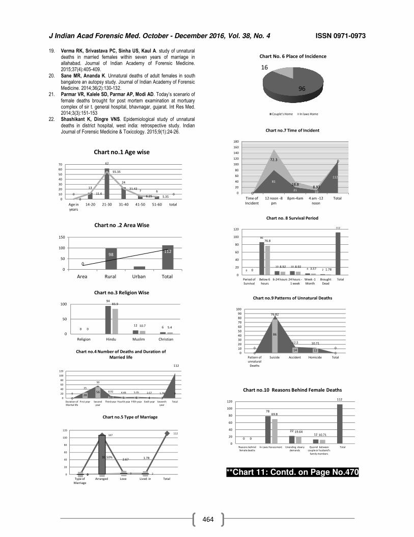
J Indian Acad Forensic Med. October - December 2016, Vol. 38, No. 4 ISSN 0971-0973
464
19. Verma RK, Srivastava PC, Sinha US, Kaul A. study of unnatural deaths in married females within seven years of marriage in allahabad. Journal of Indian Academy of Forensic Medicine. 2015;37(4):405-409.
20. Sane MR, Ananda K. Unnatural deaths of adult females in south bangalore an autopsy study. Journal of Indian Academy of Forensic Medicine. 2014;36(2):130-132.
21. Parmar VR, Kalele SD, Parmar AP, Modi AD. Today’s scenario of
female deaths brought for post mortem examination at mortuary complex of sir t. general hospital, bhavnagar, gujarat. Int Res Med. 2014;3(3):151-153
22. Shashikant K, Dingre VNS. Epidemiological study of unnatural
deaths in district hospital, west india: retrospective study. Indian Journal of Forensic Medicine & Toxicology. 2015;9(1):24-26.
**Chart 11: Contd. on Page No.470
0
13
62
24
7 60
0
11.6
55.35
21.42
6.25 5.350
10
20
30
40
50
60
70
Age in
years
14-20 21-30 31-40 41-50 51-60 total
Chart no.1 Age wise
0
98
14
112
0
50
100
150
Area Rural Urban Total
Chart no .2 Area Wise
0
94
126
0
83.9
10.75.4
0
50
100
Religion Hindu Muslim Christian
Chart no.3 Religion Wise
028
56
10 5 6 4 3
112
0
25
50
8.92 4.46 5.35 3.57 1.78 0
0
20
40
60
80
100
120
Duration of
Marital life
First year Second
year
Third year Fourth year Fifth year Sixth year Seventh
year
Total
Chart no.4 Number of Deaths and Duration of
Married life
0
107
3 2
112
0
95.50%2.67 1.78
0
20
40
60
80
100
120
Type of
Marriage
Arranged Love Lived -in Total
Chart no.5 Type of Marriage
96
16
Chart No. 6 Place of Incidence
Couple's Home In laws Home
0
81
2110
112
0
72.3
18.88.92
0
0
20
40
60
80
100
120
140
160
180
Time of
Incident
12 noon -8
pm
8pm-4am 4 am -12
noon
Total
Chart no.7 Time of Incident
0
86
10 104 2
112
0
76.8
8.92 8.923.57 1.78
0
20
40
60
80
100
120
Period of
Survival
Below 6
hours
6-24 hours 24 hours -
1 week
Week -1
Month
Brought
Dead
Total
Chart no. 8 Survival Period
0
86
14 120
0
76.82
12.5 10.71
0
0
10
20
30
40
50
60
70
80
90
100
Pattern of
unnatural
Deaths
Suicide Accident Homicide Total
Chart no.9 Patterns of Unnatural Deaths
0
78
2212
112
0
69.8
19.6410.71
0
20
40
60
80
100
120
Reasons behind
female deaths
In-Laws Harassment Unending dowry
demands
Quarrel between
couple or husband’s
family members
Total
Chart no.10 Reasons Behind Female Deaths
J Indian Acad Forensic Med. October - December 2016, Vol. 38, No. 4 ISSN 0971-0973
465
Original Research Paper
Epidemiological analysis of drowning deaths: A 10 year Study
1Sujan Kumar Mohanty,
2Virendra Kumar,
3Jaffar Hussain A.P,
4V. Bhuvan
Abstract Drowning is a significant global problem which causes a huge number of deaths worldwide. The
aim of this study is to analysedifferent epidemiological factors and explore the effect of such factors on mortality due to drowning deaths. There were 152 cases of drowning deaths during a ten-yearperiod at UMMC, Kuala Lumpur,consisting of 125 male and 27 female victims. The largest numbers of victims, (46%) were found in theage group of 20 to 34 years. Malays comprised the maximum proportion of victims (28.3%). Approximately61.8% of victims came from the unemployed group. A majority of victims were single (52.6%). Accidental falls (38.2%) were the commonly occurring incidents that lead to drowning death and most of drowning deaths(39.5%) occurred in the river. Accidental drowning deaths (71%)were the commonest.Majority (90.1%) of the victims died at the place of drowning. 10.5% of the deceased were testedpositive for alcohol, while 19.7% were confirmed as drug abusers. Decompositionchanges on the body were found in 52% cases. Externally, frothy or blood stained discharge at mouth and nostrils and "washer-women" appearance on hands and feet were the commonpostmortem findings; while internally, congestion and oedema of the lungsand presence of foreign bodies (FB) in airway,were the common findings.
Key Words: Accidental; Drowning deaths; Epidemiological aspects; Frothy discharge.
Introduction: Drowning is a significant global problem. The World Health Organization (WHO) estimates the annual worldwide incidence of death by drowning to be about 400,000.[1] Modell defined drowning as death by suffocation aftersubmersion in fluid medium, especially in water within 24 hours of submersion.[2] Meanwhile, FirstWorld Congress on drowning in Amsterdam, The Netherlands, in 2003 agreed upon a newdefinition of drowning as: “the process of experiencing respiratory impairment from submersion or immersion in a liquid.”[3,4] Indeveloped countries such as America, Australia and the United Kingdom, drowning ranks third, behind motor vehicles and fires as the leading cause of unintentional injury in children less than 15 years of age and adults.[1]
Corresponding Author:
1Associate Professor,
2Prof. & Head,
3Assistant Professor,
4Tutor,
Department of Forensic Medicine, Meenakshi Medical College Hospital & Research Institute, Kanchipuram, Tamilnadu Email:[email protected] DOR: 12/08/2016 DOA: 27/12 2016 DOI: 10.5958/0974-0848.2016.00116.0
The drowning death rate among children 0-19 year of age was 1.4 per 100,000, second only to transportation. In a developing country like India, nearly 19% of child (<14 y) injury deaths are due to drowning.[6] Rates of drowning vary with sex, age, socioeconomic status and race.[7,8] The sites ofdrowning are also varied, with the majority occurring in fresh water, including bath-tubs, swimmingpools, lakes, and rivers.[9,10] Certain medical conditions, notably epilepsy, physical or mental handicap and otherillnesses like myocardial infarction, cerebrovascular accident and autistic syndrome have beenassociated with an increased risk of drowning.[9,11-13] The use of alcohol and other substance-abuse, while near water, has been related to drowning, especially in young adults.[11,14] There are several specific autopsy findings for the positive diagnosis of drowning.[15,16] Themost common autopsy finding in cases of death by drowning is the presence of heavy andedematous lungs.[17] In macro-morphology finding, froth in the airways and over-inflated heavy lungsare of significance for confirmation of drowning.[15,16] The main objective of this present study is to find out the different epidemiological
J Indian Acad Forensic Med. October - December 2016, Vol. 38, No. 4 ISSN 0971-0973
466
aspects and specific postmortem findings in case of drowningdeaths.
Materials and methods: A total number of 152 cases of deathsdue to drowning, from the total number of5579 autopsies performed in the University of Malaya Medical Center (UMMC), from 1 Jan 1996to 31 Dec 2005, constituted the material of the present study.The autopsy reports of all the victims were traced and the relevant information extracted.All the epidemiological data like age, sex, ethnicity,socio-economic status, marital status and activity before death, place of event and death, manner of death, history of drug and alcohol abusewere collected. The external and internal postmortem findings were recorded. Then all the data was analysed to identify the various factors and explore the relationship of such factors with mortality and compared with other studies. However, we would like to note that there were certain limitations observed during the collection of data. Incomplete data in some files resulted in unknown categories for several parameters. Moreover, we were also unable to trace the victims’ next of kin to obtain missing information in certain cases.
Results: Drowning deaths constitutedabout 2.7% of all medico legal autopsies performed during the 10-year study period.The majority of the deceased (82.2%) were males (Fig-1) and 46.1% of the totalcases were in the 20 to 34 years of age group (Fig-2).Maximum number of the deceased were Malays (28.3%), followed by Indians (25%)and others (Fig-3).Most of the victims (61.8%) were unemployed (Table-1)and 52.6% were single (Fig-4). In 71.1% cases, the manner of drowning deaths wasclassified as accidental (Fig-5).Accidental falls (38.2%) were the commonly occurring incidents (Fig-6) and most of the drowning deaths (39.5%) occurred in the river (Fig-7). Death at the scene involved 90.1% of the total victims (Table-2). Most of the victims were not addicted but 10.5% of the deceased were tested positive for alcohol, while 19.7% of the deceasedwere confirmed as drug abuser (Table-3).In 52% victims, signs of decomposition were found on the body (Fig-8).Washer-woman appearance on hands and feet was found in 36.8% of the victims (Table 4), while frothy or bloodstained discharge at mouth and nostrils werepresent in 54.6% cases (Fig-9).Internally, congestion and oedema of the lungs (79%) andpresence of foreign bodies (FB)
in airway (54%) were the common findings (Table-5).
Discussion: In our study, drowning deaths contributed about 2.7% of the total cases. The incidents were more in males than that in the females, because the males aremore active, more exposed to aquatic environments and activity, and also because of their risk-taking behavior. Similar finding werereported in a study[11] in Australia.We observed in the study that people of all age groups were involved. Majority of the deceasedwere in the 20-34 years of age group (46%) whereas the least were elderly, more than 64 years ofage (3.3%). This result is consistent with a previous study.[9] This canbe explained by the fact that younger people are more involved in aquatic activities (out-dooractivities) such as swimming and fishing, while the elderly are usually more involved in indooractivities. Hence, they are not exposed to aquatic environments as much as younger people. A study in New Zealand[18] reported that 15–24 year olds drowned more than expected during Water Sport/ Recreation,while a high proportion of 30–39 year olds drowned during underwater activities. But men aged 55–64 had a high incidence of drowning while boating. In our study, thenumber of drowning deaths declined towards both extremes of age. Thirteen victims (8.55%)were found to be below the age of 4 years, most of their deaths were due inadequate supervisionby adults. Previous studies[19,20] also reported a similar finding. In this age group, only one case was due to physical abuse (thrown into river bykidnappers). Malaysia is a multiracial country. Malay, Chinese, and Indian are the main races, followed by others such as aborigines and immigrants. Malays contribute the highest incidence ofdrowning (28.3%) as they are the largest ethnic group in Malaysia. Since the population ofIndians is only 7.7% in Malaysia,[21] the ratio of drowning deaths to population of Indians is more predominant.It was not possible to list each victim's social status as such, but a rough estimation couldbe made from employment information. Deceased were classified into four socioeconomic groupsbased on their occupation: semiskilled-unskilled, clerical-skilled, professional-managerial andunemployed. Most of the victims came from the unemployed group, (61.8%) followed bysemiskilled-unskilled group (31.58%). Students, house-wives and pensioners were
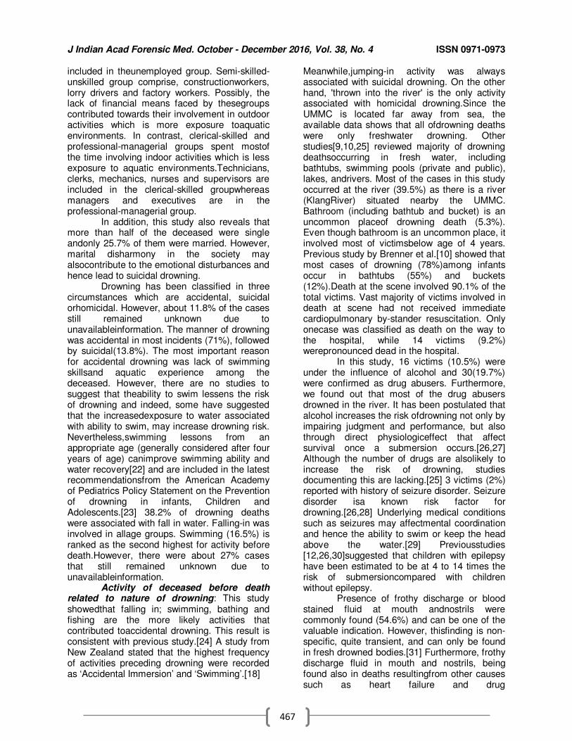
J Indian Acad Forensic Med. October - December 2016, Vol. 38, No. 4 ISSN 0971-0973
467
included in theunemployed group. Semi-skilled-unskilled group comprise, constructionworkers, lorry drivers and factory workers. Possibly, the lack of financial means faced by thesegroups contributed towards their involvement in outdoor activities which is more exposure toaquatic environments. In contrast, clerical-skilled and professional-managerial groups spent mostof the time involving indoor activities which is less exposure to aquatic environments.Technicians, clerks, mechanics, nurses and supervisors are included in the clerical-skilled groupwhereas managers and executives are in the professional-managerial group. In addition, this study also reveals that more than half of the deceased were single andonly 25.7% of them were married. However, marital disharmony in the society may alsocontribute to the emotional disturbances and hence lead to suicidal drowning. Drowning has been classified in three circumstances which are accidental, suicidal orhomicidal. However, about 11.8% of the cases still remained unknown due to unavailableinformation. The manner of drowning was accidental in most incidents (71%), followed by suicidal(13.8%). The most important reason for accidental drowning was lack of swimming skillsand aquatic experience among the deceased. However, there are no studies to suggest that theability to swim lessens the risk of drowning and indeed, some have suggested that the increasedexposure to water associated with ability to swim, may increase drowning risk. Nevertheless,swimming lessons from an appropriate age (generally considered after four years of age) canimprove swimming ability and water recovery[22] and are included in the latest recommendationsfrom the American Academy of Pediatrics Policy Statement on the Prevention of drowning in infants, Children and Adolescents.[23] 38.2% of drowning deaths were associated with fall in water. Falling-in was involved in allage groups. Swimming (16.5%) is ranked as the second highest for activity before death.However, there were about 27% cases that still remained unknown due to unavailableinformation. Activity of deceased before death related to nature of drowning: This study showedthat falling in; swimming, bathing and fishing are the more likely activities that contributed toaccidental drowning. This result is consistent with previous study.[24] A study from New Zealand stated that the highest frequency of activities preceding drowning were recorded as ‘Accidental Immersion’ and ‘Swimming’.[18]
Meanwhile,jumping-in activity was always associated with suicidal drowning. On the other hand, 'thrown into the river' is the only activity associated with homicidal drowning.Since the UMMC is located far away from sea, the available data shows that all ofdrowning deaths were only freshwater drowning. Other studies[9,10,25] reviewed majority of drowning deathsoccurring in fresh water, including bathtubs, swimming pools (private and public), lakes, andrivers. Most of the cases in this study occurred at the river (39.5%) as there is a river (KlangRiver) situated nearby the UMMC. Bathroom (including bathtub and bucket) is an uncommon placeof drowning death (5.3%). Even though bathroom is an uncommon place, it involved most of victimsbelow age of 4 years. Previous study by Brenner et al.[10] showed that most cases of drowning (78%)among infants occur in bathtubs (55%) and buckets (12%).Death at the scene involved 90.1% of the total victims. Vast majority of victims involved in death at scene had not received immediate cardiopulmonary by-stander resuscitation. Only onecase was classified as death on the way to the hospital, while 14 victims (9.2%) werepronounced dead in the hospital. In this study, 16 victims (10.5%) were under the influence of alcohol and 30(19.7%) were confirmed as drug abusers. Furthermore, we found out that most of the drug abusers drowned in the river. It has been postulated that alcohol increases the risk ofdrowning not only by impairing judgment and performance, but also through direct physiologiceffect that affect survival once a submersion occurs.[26,27] Although the number of drugs are alsolikely to increase the risk of drowning, studies documenting this are lacking.[25] 3 victims (2%) reported with history of seizure disorder. Seizure disorder isa known risk factor for drowning.[26,28] Underlying medical conditions such as seizures may affectmental coordination and hence the ability to swim or keep the head above the water.[29] Previousstudies [12,26,30]suggested that children with epilepsy have been estimated to be at 4 to 14 times the risk of submersioncompared with children without epilepsy. Presence of frothy discharge or blood stained fluid at mouth andnostrils were commonly found (54.6%) and can be one of the valuable indication. However, thisfinding is non-specific, quite transient, and can only be found in fresh drowned bodies.[31] Furthermore, frothy discharge fluid in mouth and nostrils, being found also in deaths resultingfrom other causes such as heart failure and drug
J Indian Acad Forensic Med. October - December 2016, Vol. 38, No. 4 ISSN 0971-0973
468
overdose.[32]‘Washer-woman’ appearance onhands and feet was found in 36.8% of the cases. This sign is not specific because relativelyprolonged immersion of the hands and feet, when washing clothes, produces similar changes.[33] In addition, 52% of the deceased were in decomposed state. The dead body cools in waterabout twice as fast as in air (i.e. about 5°F per hour) and reaches the temperature of the waterusually within 5 to 6 hours and nearly always within 12 hours. The normal changes ofdecomposition of a body are delayed in cold, deep water so that bodies may be surprisingly wellpreserved after a long period of immersion. These conditions also favour the formation ofadipocere preserving the body features. Once the bodyis removed from water, putrefactive changes advance with remarkable rapidity.In internal autopsy findings, congestion and oedema of the lungs (79%) were often seen,followed by presence of foreign bodies (FB) in the airway (54%) and voluminous lungs (39.5%). The lungs are voluminous, often to a degree which causes them to overlap thepericardium; rib markings maybe present. Presence of foreign material such as fluid in thestomach only comprised 29.6% of the cases. The absence of fluid in the stomach is noteworthy,because it may mean either that the death was rapid or that the victim was dead at the time ofsubmersion.[33]
Conclusion Drowning deaths were more common in males, among 20 to 34 years of age group.A majority of them were single and unemployed. Majority of drowning deaths were accidental in nature by falling in to the river. Almost all the deceased died at the scene.Frothy or blood stained discharge at mouth and nostrils, congested, edematous and voluminous lungs were common autopsy findings. As drowning is a major cause of mortality among teenagers after road traffic accidents, all over the world, more in developing countries compared to those ofdeveloped countries, this matter should not be overlooked. Preventive measures need to be taken, based on the risk factors that hadbeen identified. Therefore, a more comprehensive and strict approaches should be amended toprevent drowning death.
References: 1. World Health Organization. The World Health Report, 2002:
Reducing risks, promoting healthy life, Geneva, World Health Organization, 2002.Available on:http://www.who.int/whr/2002/ en/whr2002_annex2.pdf?ua=1
2. Modell JH. Drown versus near-drown: a discussion of definitions. Crit Care Med. 1981;9(4):351-2.
3. Van Dorp JCM, Knape JTA, Bierens JJLM. Recommendations: World Congress onDrowning, 2002. June 26-28; Amsterdam, The
Netherlands.Available on http://www.drowning.nl/. 2003. 4. Idris AH, Berg RA, Bierens J, Bossaert L, Branche CM, Gabrielli
A, et al. Recommended guidelines for uniform reporting of datafrom drowning: the “Utstein style”. Resuscitation 2003;59:45–
57. 5. CDC Childhood Injury Report: Patterns of Unintentional Injuries
among 0-19 year olds in the United States, 2000-2006 available on: https://www.cdc.gov/homeandrecreationalsafety/water-safety/pubs
html NCRB, 2012. India available on: ncrb.nic.in/CD-CII2012/Statistics2012.pdf
6. Fingerhut L, Kleinman JC. Trends and current status in childhood mortality, United States,1900-1985. In: Vital and Health statistics.
Series 3, No 26. Washington, DC: US PublicHealth Service; 1989. DHHS Publ. No PHS 89-1410
7. Brenner RA, Smith GS, Overpeck MD. Divergent trends in childhood drowning rates, 1971through 1988. JAMA.
1994;271:1606-8 8. Quan L, Cummings P. Characteristics of drowning by different age
groups. InjPrev. 2003;9:163–8. 9. Brenner RA, Trumble AC, Smith GS, Kessler EP, Overpeck MD.
Where children drown,United States. Pediatrics 2001;108:85–9 10. Mackie IJ. Patterns of drowning in Australia, 1992-1997. Med J
Aust 1999;171:587-90. 11. Kemp AM, Sibert JR. Epilepsy in children and the risk of drowning.
Arch Dis Child. 1993;68:684-5. 12. Sibert JR, Lyons RA, Smith BA, Cornall P, Summer V, Craven
MA,et al. Preventing deaths by drowning in children in theUnited Kingdom: have we made progress in 10 years? Population based
incidence study.Br Med J 2002;324:1070-1. 13. Cummings P, Quan L. Trends in unintentional drowning: the role of
alcohol and medicalcare. JAMA 1999;281:2198-202. 14. Zhu BL, Ishida K, Quan L, Li DR, Taniguchi M, Fujita MQ,
Maeda H, Tsuji T. Pulmonaryimmunohistochemistry and serum levels of a surfactant-associated protein A in fataldrowning. Legal Med 2002;4:1-6.
15. Morild I. Pleural effusion in drowning. Am J Forensic Med Pathol
1995;16:253-6. 16. Knight B, Saukko P. Immersion deaths. In: Knight B, Saukko P,
editors. Knight’s forensicpathology. 3rd ed. London: Edward Arnold; 2004:395-411.
17. Croft James L, Button Chris. Interacting factors associated with adult male drowning in new zealand. PLoS One. 2015;10(6): e0130545. Published online 2015 Jun 17
18. Rimsza ME, Schackner RA, Bowen KA, Marshall W. Can child
deaths be prevented? TheArizona Child Fatality Review program experience. Pediatrics 2002;110:e11.
19. Quan L, Gore EJ, Wentz K, Allen J, Novack AH. Ten-year study of pediatric drownings andnear-drownings in King County,
Washington: lessons in injury prevention. Pediatrics1989;83:1035–40.
20. Population distribution and Basic demographic characteristic report, Population andhousing Census 2000 [online]. 2000 [cited 2006
June 2]. Available on: 21. http://www.statistics.gov.my/english/census/pressdemo.htm 22. Asher KN, Rivara FP, Felix D, Vance L, Dunne R. Water safety
training as a potentialmeans of reducing risk of young children’s
drowning. InjPrev 1995;1:228–33. 23. Brenner RA. The Committee on Injury, Violence, and Poison
Prevention. Prevention of drowning in infants, children, and adolescents. Pediatrics 2003; 112:437-9.
24. Steensberg J. Epidemiology of accidental drowning in Denmark 1989-1993 Accidentanalysis and Prevention. 1998;30(6):755-762
25. Agran PF, Anderson C, Winn D, Trent R, Walton–Haynes L, Thayer S. Rates of pediatricinjuries by three-month intervals for
children 0 to 3 years of age. Pediatrics 2003;111:e683–92. 26. Smith GS, Brenner RA. The changing risks of drowning for
adolescents in the US andeffective control strategies.Adolesc Med. 1995;6:153-170
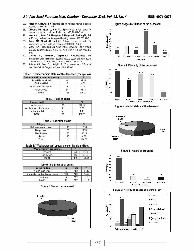
J Indian Acad Forensic Med. October - December 2016, Vol. 38, No. 4 ISSN 0971-0973
469
27. Hingson R, Howland J. Alcohol and non-traffic unintended injuries. Addiction. 1993;88:877-883
28. Diekema DS, Quan L, Holt VL. Epilepsy as a risk factor for submersion injury in children. Pediatrics. 1993;91:612–616
29. Howland J, Smith GS, Mangione T, Hingson R, DeJong W, Bell N. Missing the boat ondrinking and boating. JAMA 1993;270:91-2.
30. Kemp AM, Sibert JR, Holt VL. Epilepsy as a risk factor for submersion injury in children.Pediatrics 1993;91:612-616
31. Michel H.A. Piette and Els A. De Letter. Drowning: Still a difficult autopsy diagnosis.Forensic Sci Int. 2005 Dec 22; [Epub ahead of print]
32. Lunetta P., PenttiläA., SajantilaA. Circumstances and
macropathologic findings in 1590consecutive cases of bodies found in water, Am. J. Forensic Med. Pathol. 23 (2002) 371–376.
33. Polson CJ, Gee DJ, Knight B. The essentials of forensic Medicine.Oxford: PergamonPress; 1985: 421-48.
Table 1: Socioeconomic status of the deceased (occupation) Socioeconomic status (occupation) n %
Semiskilled-unskilled 48 31.58
Clerical-skilled 7 4.61
Professional-managerial 3 1.97
Unemployed 94 61.84
TOTAL 152 100.00
Table 2: Place of death Place of death n %
At the scene 137 90.13
On the way to the hospital 1 0.66
In the hospital 14 9.21
TOTAL 152 100.00
Table 3: Addiction status
Category n %
History of alcohol used 16 10.53
Drug abuser 30 19.74
No addiction 96 63.15
Unknown 10 6.58
Total 152 100.00
Table 4: "Washerwoman" appearance on hands and feet “Washerwoman” appearance N %
Present 56 36.84
Not present 96 63.26
TOTAL 152 100.00
Table 5: PM findings of Lungs
Internal finding n Total %
Voluminous lungs 60 152 39.47
Congestion and oedema of lungs 120 152 78.95
FB in airway 82 152 53.95
FB in GIT 45 152 29.61
Figure 1: Sex of the deceased
Figure 2: Age distribution of the deceased
Figure 3: Ethnicity of the deceased
Figure 4: Marital status of the deceased
Figure 5: Nature of drowning
Figure 6: Activity of deceased before death
J Indian Acad Forensic Med. October - December 2016, Vol. 38, No. 4 ISSN 0971-0973
470
Figure 7: Place of event
Figure 8: Body at autopsy
Fig-9: Frothy or blood stained discharge at mouth and nostrils
**Contd from Page No.464
0
51
1614
11
2 2
86
20
45.5
14.32
12.559.82
1.78 1.78
7.14 5.351.78
0
0.2
0.4
0.6
0.8
1
1.2
0
10
20
30
40
50
60
Cause of Female
Deaths
Poisoning Hanging Burns Drowning Electrocution Snake Bite RTA Assault Railway Trak
Injuries
Chart no.11 Cause of Female Deaths
J Indian Acad Forensic Med. October - December 2016, Vol. 38, No. 4 ISSN 0971-0973
471
Original Research Paper
Age estimation and eruption of Permanent Teeth between two Genders - A Cross sectional study
1Manjot Kaur,
2Chandrakanth HV,
3Dasari Harish
Abstract Identification is necessary in living persons, recently dead, decomposed bodies, mutilated bodies, skeletonised bodies etc. Age estimation constitutes one of the important tools for identification of a person, both in civil and criminal cases. Teeth are the most reliable material in identification and age estimation in the dead as they do not undergo mutilation by fire or decompositionA tooth is considered to be erupted when the crown or any part of the tooth becomes clinically visible in the oral cavity. The present study was conducted to collect the cross-sectional data for the time and sequence of eruption of permanent teeth among the school going children of north Indian population. The age group was limited to 6 to 14 years, which includes the age range at which all permanent teeth have been found to be erupted, except the third molars. Schools registered with Directorate of school education were considered for the study. A total of 1804 subjects were examined. The mean age of eruption of mandibular first molar has been found to be earlier than the permanent mandibular central incisor as well as earlier in females compared to males. Also the mean age of eruption of any tooth has been found to be earlier in mandible as compared to maxilla in both males and female. Permanent teeth erupted earlier in females than in males and also earlier in mandible compared to maxilla.
Key Words: Identification, Age estimation, Teeth eruption, Gender difference.
Introduction: Age estimation constitutes one of the important tools for identification of a person, both in civil and criminal cases. It can guide the investigators to the correct identity of an individual among a large number of possible matches as during natural calamities and mass disasters, where large numbers of lives are lost. Identification is necessary in living persons, recently dead, decomposed bodies, mutilated bodies, skeletonised bodies etc. Criminal cases include cases of assault, murder, rape, interchanging of newborn babies in hospital etc and Civil cases include marriage, inheritance, passport, insurance claim, disputed sex, etc.[1] The branch of Forensic Medicine that deals with the handling, examination, and
Corresponding Author:
1Lecturer,
Dr. H.S.J institute of dental sciences and hospital, Panjab University, Chandigarh, 2Prof. & Head, Dept. Forensic Medicine, JSS Medical
College, Jagadguru Shri Shivaratheeshwara University, SS Nagar, Bannimantap, Mysuru ,3Prof. & Head, Dept. Forensic Medicine & Toxicology,
Govt. Medical College & Hospital, Chandigarh Email: [email protected] DOR: 14/03/2016 DOA: 27/12/2016 DOI: 10.5958/0974-0848.2016.00117.2
presentation of dental evidence in court is known as Forensic Odontology. Forensic dentists are involved in assisting investigative agencies to identify recovered human remains in addition to the identification of whole or fragmented bodies; forensic dentists may also be asked to assist in determining age, race, occupation, previous dental history and socioeconomic status of unidentified human beings. Age estimation is done by examining the general physical characters, radiological examination and dental development in combination. Age can be determined from a variety of factors like the appearance of ossification centres and their fusions during skeletal development, from height and weight which is applicable in early periods of life, dental development and changes occurring at puberty like appearance of hair and their growth and colour changes, development of breast in females, starting of menarche and from menopause.[2]
Owing to a low variability of tooth formation and eruption, in relation to chronological age, methods based on stages of tooth formation and eruption can be used for assessment of chronological age. Teeth are the most reliable material in identification and age estimation in the dead as they do not undergo
J Indian Acad Forensic Med. October - December 2016, Vol. 38, No. 4 ISSN 0971-0973
472
mutilation by fire or decomposition. Eruption of teeth is one of the easily observable among the various dynamic changes that occur from formation till the final shedding of teeth. The process of gingival eruption of teeth can readily be recorded as having occurred or not, in any individual. This fact can be made use of in ascertaining the average age of eruption of the tooth.[3]
Tooth eruption is traditionally considered to be a developmental process whereby the tooth moves in an axial direction from its location within the alveolar crypt of the jaw into a functional position within the oral cavity. A tooth is considered to be erupted when the crown or any part of the tooth becomes clinically visible in the oral cavity.[4]
The most common sequence
of eruption of temporary teeth is the central incisor, lateral incisor, canine, first molar and the second molar and in the permanent teeth it is - first molar, central incisor, lateral incisor, first pre molar, second premolar, canine, second molar and the third molar. There are charts and tables for the assessment of age during development period, which shows the formation, eruption, and calcification of teeth. For this purpose table of Krefeld and Logan further modified by Kronfild and Schour is commonly used,
5 which has been
accepted standard for many years. Since the population of India is very large and its climate is different, a cross-sectional study was carried out to estimate the eruption time of primary and permanent teeth in the age group of six to fourteen years in Indian population. Age determination by eruption of teeth in children is a reliable method as it gives the age within 6 months to 1 year difference from the chronological age. Such a study has not been conducted till date in the Chandigarh zone which will provide a good database for future use and research.
Materials and Method: The present study was conducted to collect the cross-sectional data for the time and sequence of eruption of permanent teeth among the school going children of north Indian population. The age group was limited to 6 to 14 years, which includes the age range at which all permanent teeth have been found to be erupted, except the third molars. Schools registered with Directorate of school education were considered for the study. A single examiner conducted the examination with naked eye using a mouth mirror and explorer under natural light. The information regarding the age of the children was collected from the school registers
containing date of birth. The subjects were sub-divided into 14 groups according to age by 6 months intervals; months less than 6 rounded off to last year and exceeding 6 months were rounded off to the next year. For the purpose of understanding and application of the examination criteria, calibration was done under the guidance of the supervisor in order to limit the diagnostic variability. A group of 30 subjects in the age group of 6 to 14 years having mixed dentition were chosen from the school oral health programmes conducted in various schools. These subjects were examined in the Department and the observations were recorded in the self designed proforma under the supervision of the supervisors. Two supervisors were involved to evaluate the calibration of the examiner. The results so obtained were subjected to kappa statistics. A pilot study was undertaken to check the feasibility and practicality of the study, to have prior idea regarding the estimation of the time taken to examine each child and to plan survey accordingly. Modifications regarding excluding the irrelevant questions were made in the recording Performa to overcome the particular problem. The naked eye examination of the subjects was carried out in concerned school premises, under natural daylight conditions or using artificial illumination with subjects seated comfortably on ordinary chair with back rest and the examiner sitting in front of the child. The data was transformed from pre-coded survey form to computer. A master file was created for the purpose of data analysis. SPSS version 20.0 (SPSS Pvt Ltd, Chicago, IL, USA) was used for the statistical analyses.
Results: Table-1 shows the distribution of subjects according to age and gender with age ranging from 6 years to 14 years. The subjects were further sub-grouped based on 6 month interval. A total of 1804 subjects were examined, of which 924 were males and 880 were females. Maximum subjects were examined in the age group of 12-12.5 years with 76 males and 68 females. Table 2 shows the eruption of permanent teeth in the maxillary arch. The teeth to be erupted first were the first permanent molars, followed by the central incisors and then the lateral incisors. At 6 to 6.5 years of age, eruption of first permanent molars was more in males whereas central and lateral incisors were more in females. By the age of 8 years, 100 percent of the first molars had
J Indian Acad Forensic Med. October - December 2016, Vol. 38, No. 4 ISSN 0971-0973
473
erupted. However, in relation to central incisors, 100 percent eruption was seen by 9 years. In case of pre-molars, 100 percent eruption was seen by the age of 13.5 years. The canines were the last to erupt with 100 percent eruption taking place after 14 years. Table 3 shows the eruption of permanent teeth in the mandibular arch. The teeth to be erupted first were the first permanent molars, followed by the central incisors and then the lateral incisors. At 6 to 6.5 years of age, eruption of first molars was more in males whereas central and lateral incisors were more in females. By the age of 7 years 100 percent of the first permanent molars had erupted. However in relation to central incisors, 100 percent eruption was seen 7.5 years in males and 8.5 years in females. In case of pre-molars, 100 percent eruption was seen by the age of 14 years. Table 4 shows the mean age of eruption of permanent teeth that was analyzed according to sex and it was observed that there is no significant difference in the eruption of maxillary and mandibular dentition of males and females. Table5 shows the mean age of eruption of maxillary teeth in the left and right quadrant. No significant difference was seen, but in relation to right canines, there is significant difference in the eruption in relation to both the sexes.Table 6 shows the mean age of eruption of mandibular teeth in left and right quadrant in males and females. Left canines and left second premolars showed significant difference in eruption in relation to males and females.
Discussion: The tooth eruption is an important phenomenon which plays a significant role in the process of facial development. The appearance of permanent teeth shows variation both in age as well as sequence of eruption. The studies conducted in the past were mostly in the western population, thus the standards for age of eruption are based on those. This fails to provide relevant guidelines regarding Indian population, so the present study was carried amongst the school children of Chandigarh, to determine the age and sequence of eruption of permanent teeth. The age group was limited to 6 to 14 years, which includes the age range at which all permanent teeth have been found to be erupted except the third molars. The information regarding the age was tabulated and subjects were grouped in six months intervals of age. The error arising out rounding off the ages was minimized by following the same method in all calculations.
The subjects with no history of extraction of deciduous teeth due to caries were included in the study where as subjects undergoing orthodontic treatment and the ones with congenital abnormalities were excluded as it might have influenced the data obtained. Hundred percent of subjects had erupted mandibular first permanent molars by the age of seven years in both males and females but hundred percent eruption of mandibular central incisors was achieved by 7 years & 6 months in males and 8 years & 6 months in females, which does not confirm to the study carried by Sharma & Mittal.[6]
The
reason for this difference could be the sample size of the present study and also that the present study was carried out in North Indian children. The maxillary canine and second premolar reached hundred percent eruption status by 14 years 6 months in males as compared to 14 years in females and maxillary first premolars reached hundred percent eruption status at 14 years in males compared to 13 years 6 months in females. The mean age of eruption of mandibular first molar has been found to be earlier than the permanent mandibular central incisor as well as earlier in females compared to males. The mean age of eruption of a particular tooth has been found to be earlier on left side of the mouth compared to right side for most of the teeth in maxilla in both males and females, where as in mandible this difference was not found to be consistent. The mean age of eruption of a particular tooth on particular side of the mouth has been found to be earlier in females compared to males. This difference was probably due to early onset of puberty in females compared to males, as has been suggested in the study by Demirijien A and Levesque GF[7] and Agarwal, et al.[8] Among the females, the eruption sequence in the maxilla in right and left side was the same, except that the mean eruption ages of premolars and canines were identical. In mandibles, canine preceded the eruption of both the premolars. Among males, the eruption sequences in the maxilla in right and left side were different; on right side, second premolar erupted ahead of canine and on left side it was before first premolars. These findings were similar to another study by Singh K.[9]
Hence it could be inferred from the present study that permanent teeth erupted earlier in females than in males and also earlier in mandible compared to maxilla. The results however cannot be considered conclusive because of the small sample size in the present
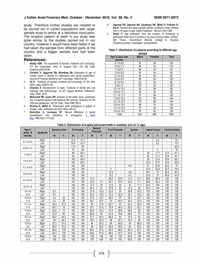
J Indian Acad Forensic Med. October - December 2016, Vol. 38, No. 4 ISSN 0971-0973
474
study. Therefore further studies are needed to be carried out in varied populations with larger sample sizes to arrive at a definitive conclusion. The eruption pattern of teeth in our study was quite similar to the studies carried out in our country, however it would have been better if we had taken the sample from different parts of the country and a bigger sample size had been taken.
References: 1. Reddy KSN. The essentials of forensic medicine and toxicology.
21st Ed. Hyderabad, India: K. Sugana Devi, Om Sai Laser Graphics;2002:49-84.
2. Chhokar V, Aggarwal SN, Bhardwaj DN. Estimation of age of sixteen years in female by radiological and dental examination.
Journal of Forensic Medicine and Toxicology. 1992;9:25-30. 3. Vij K. Textbook of forensic medicine and toxicology. 4th Ed. New
Delhi, India. 2008:51-53. 4. Chandra S. Development of teeth. Textbook of dental and oral
histology with embroyology. 1st Ed. Jaypee Brothers Publishers; India. 2004: 34-37.
5. McDonald RE, Avery DR. Eruption of the teeth: local, systematic and congenital factors that influence the process. Dentistry for the
child and adolescent. 5th Ed. Delhi, India.1988:190-3. 6. Sharma K, Mittal S. Permanent tooth emergence in gujjars of
Punjab, India. Anthropol Anz 2001;59(2):165-78. 7. Demirijien A, Levesque GY. Sexual difference in dental
development and prediction of emergence. J Dent Res. 1980;59(7):1110-22.
8. Agarwal DK, Agarwal KN, Upadhyay SK, Mittal R, Prakash R, Rai S. Physical and sexual growth pattern of affluent Indian children from 5-18 years of age. Indian Pediatrics. 1992;29:1203-1282.
9. Singh P. Age estimation from the eruption of temporary &
permanent teeth from 6 months to 25 years among Indian children. MD Thesis. Government Medical College & Hospital, PanjabUuniversity, Chandigarh. [unpublished]
Table 1: Distribution of subjects according to different age groups
Age in years and
months
Males Females Total
6.1 to 6.5 52 48 100
6.6 to 7.0 64 52 116
7.1to7.5 48 44 92
7.6 to 8.0 40 44 84
8.1 to 8.5 40 44 84
8.6 to 9.0 68 52 120
9.1 to 9.5 52 44 96
9.6 to 10 68 52 120
10.1 to 10.5 40 56 96
10.6 to 11.0 48 44 92
11.1 to 11.5 72 68 140
11.6 to 12.0 60 80 140
12.1 to 12.5 76 68 144
12.6 to 13.0 48 76 124
13.1 to 13.5 72 48 120
13.6 to 14.0 76 60 136
Total 924 880 1804
Table 2: Distribution of erupted permanent teeth in maxillary arch (in % age)
Age in years & months
Quadrant Second molar First molar
Second
Premolar First Premolar Canine Lateral Incisor Central Incisor
M F M F M F M F M F M F M F
6.1 to 6.5 Right - - 70.9 47.3 - - - - - - - 5.2 - 15.7
Left - - 76.9 47.3 - - - - - - - 5.2 - 15.7
6.6 to 7.0 Right - - 71.9 80 - - - - - - - - - 20.0
Left - - 75 80 - - - - - - - 4 - 16
7.1to7.5 Right - - 88 94.1 - - - - - - 24 11.7 15.4 41.1
Left - - 84 94.1 - - - - - - 24 11.7 15.4 41.1
7.6 to 8.0 Right - - 100 95.4 - - - - - - 38 31.8 18.8 68.1
Left - - 100 95.4 - - - - - - 33.3 27.2 18.8 63.6
8.1 to 8.5 Right - - 100 100 - - - - 4.5 - 41 52.9 52.0 70
Left - - 100 100 - - 9 - - - 34.9 52.8 52.0 76.4
8.6 to 9.0 Right - - 100 100 - - 15.3 - 4.8 - 49.1 75 90.4 83.3
Left - - 100 100 - - 19.2 - 4.2 - 43.5 75 85.8 91.6
9.1 to 9.5 Right - - 100 100 11.5 - 26.9 29.6 11.5 10.1 88.5 88.8 100 100
Left - - 100 100 22.5 3.7 26.7 33.3 22.5 11.1 88.5 92.5 100 100
9.6 to 10 Right - - 100 100 13.3 30 13.3 50 20 11.1 93.3 100 100 100
Left - - 100 100 26.3 30 57.9 50 13.3 20 93.3 100 100 100
10.1 to
10.5
Right 8.7 - 100 100 21.9 15.3 52.6 50 36.8 19.2 100 100 100 100
Left 8.7 - 100 100 26.3 19.2 72.3 52.3 42.1 19.2 100 100 100 100
10.6 to 11.0
Right 4.2 20 100 100 47.8 43.7 65.2 57 26.1 43.7 100 100 100 100
Left 4.2 20 100 100 39.1 43.7 75 64.5 30.4 37.5 100 100 100 100
11.1 to 11.5
Right 48.2 67.5 100 100 50 51.5 68.7 67 62.5 46.8 100 100 100 100
Left 48.1 75 100 100 43.7 51.5 75.4 68.3 62.5 42.1 100 100 100 100
11.6 to
12.0
Right 52.6 73 100 100 65.8 53.3 75.4 68.9 68.6 60 100 100 100 100
Left 52.3 80 100 100 65.5 60 81.6 79.8 68.6 73 100 100 100 100
12.1 to 12.5
Right 56.0 84.2 100 100 73.7 82.5 86.8 80 76.3 80 100 100 100 100
Left 60 84.2 100 100 78.9 82.5 88 87.2 76.3 85 100 100 100 100
12.6 to 13.0
Right 97.3 91.6 100 100 84 83.3 88 89.6 84 86.6 100 100 100 100
Left 84.5 91.6 100 100 84 84.2 95.6 93.8 84 93.3 100 100 100 100
13.1 to
13.5
Right 98.0 100 100 100 89.2 87.5 94.6 98.3 89.2 92.1 100 100 100 100
Left 83.3 100 100 100 94.6 91.6 98.3 97.3 91.9 100 100 100 100 100
13.6 to 14.0
Right 100 100 100 100 100 100 100 100 95.8 100 100 100 100 100
Left 100 100 100 100 100 100 100 100 95.8 100 100 100 100 100
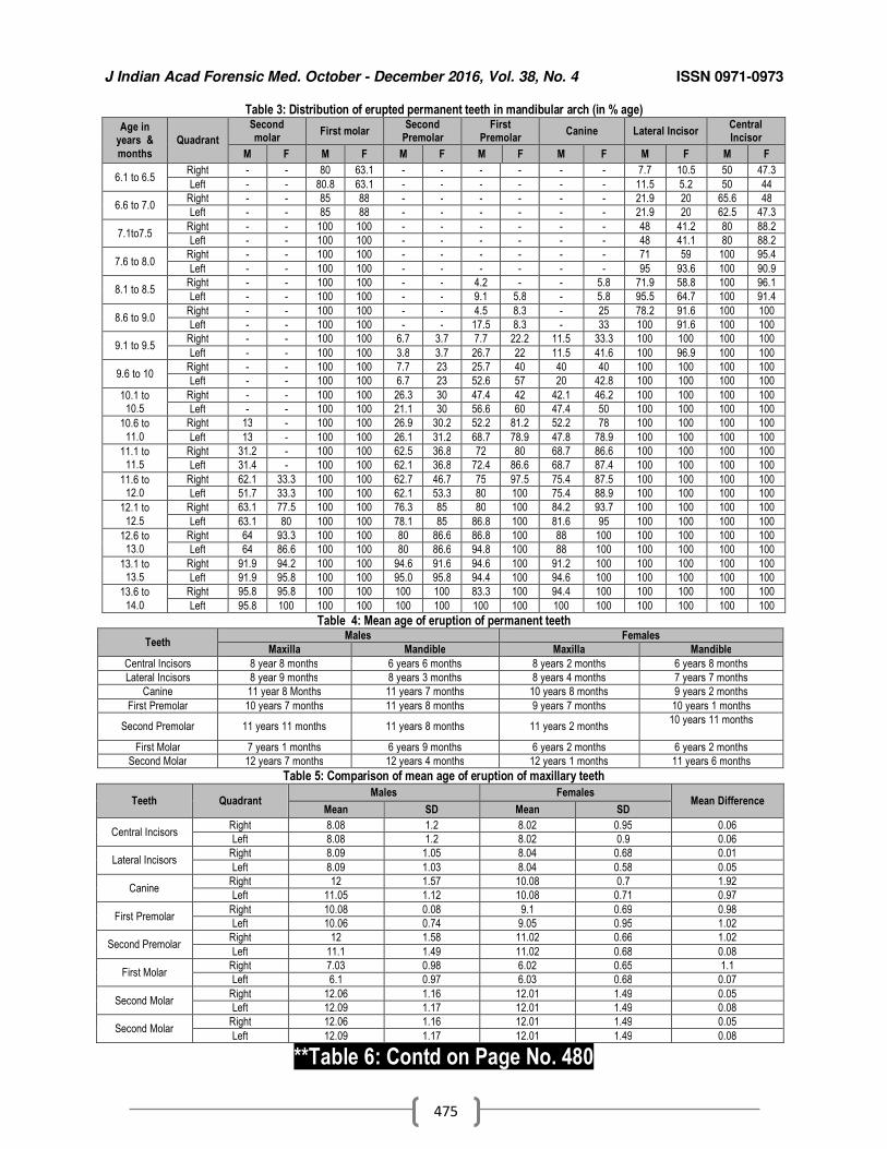
J Indian Acad Forensic Med. October - December 2016, Vol. 38, No. 4 ISSN 0971-0973
475
Table 3: Distribution of erupted permanent teeth in mandibular arch (in % age)
Age in years &
months
Quadrant
Second molar
First molar Second
Premolar First
Premolar Canine Lateral Incisor
Central Incisor
M F M F M F M F M F M F M F
6.1 to 6.5 Right - - 80 63.1 - - - - - - 7.7 10.5 50 47.3
Left - - 80.8 63.1 - - - - - - 11.5 5.2 50 44
6.6 to 7.0 Right - - 85 88 - - - - - - 21.9 20 65.6 48
Left - - 85 88 - - - - - - 21.9 20 62.5 47.3
7.1to7.5 Right - - 100 100 - - - - - - 48 41.2 80 88.2
Left - - 100 100 - - - - - - 48 41.1 80 88.2
7.6 to 8.0 Right - - 100 100 - - - - - - 71 59 100 95.4
Left - - 100 100 - - - - - - 95 93.6 100 90.9
8.1 to 8.5 Right - - 100 100 - - 4.2 - - 5.8 71.9 58.8 100 96.1
Left - - 100 100 - - 9.1 5.8 - 5.8 95.5 64.7 100 91.4
8.6 to 9.0 Right - - 100 100 - - 4.5 8.3 - 25 78.2 91.6 100 100
Left - - 100 100 - - 17.5 8.3 - 33 100 91.6 100 100
9.1 to 9.5 Right - - 100 100 6.7 3.7 7.7 22.2 11.5 33.3 100 100 100 100
Left - - 100 100 3.8 3.7 26.7 22 11.5 41.6 100 96.9 100 100
9.6 to 10 Right - - 100 100 7.7 23 25.7 40 40 40 100 100 100 100
Left - - 100 100 6.7 23 52.6 57 20 42.8 100 100 100 100
10.1 to 10.5
Right - - 100 100 26.3 30 47.4 42 42.1 46.2 100 100 100 100
Left - - 100 100 21.1 30 56.6 60 47.4 50 100 100 100 100
10.6 to
11.0
Right 13 - 100 100 26.9 30.2 52.2 81.2 52.2 78 100 100 100 100
Left 13 - 100 100 26.1 31.2 68.7 78.9 47.8 78.9 100 100 100 100
11.1 to 11.5
Right 31.2 - 100 100 62.5 36.8 72 80 68.7 86.6 100 100 100 100
Left 31.4 - 100 100 62.1 36.8 72.4 86.6 68.7 87.4 100 100 100 100
11.6 to 12.0
Right 62.1 33.3 100 100 62.7 46.7 75 97.5 75.4 87.5 100 100 100 100
Left 51.7 33.3 100 100 62.1 53.3 80 100 75.4 88.9 100 100 100 100
12.1 to
12.5
Right 63.1 77.5 100 100 76.3 85 80 100 84.2 93.7 100 100 100 100
Left 63.1 80 100 100 78.1 85 86.8 100 81.6 95 100 100 100 100
12.6 to 13.0
Right 64 93.3 100 100 80 86.6 86.8 100 88 100 100 100 100 100
Left 64 86.6 100 100 80 86.6 94.8 100 88 100 100 100 100 100
13.1 to 13.5
Right 91.9 94.2 100 100 94.6 91.6 94.6 100 91.2 100 100 100 100 100
Left 91.9 95.8 100 100 95.0 95.8 94.4 100 94.6 100 100 100 100 100
13.6 to
14.0
Right 95.8 95.8 100 100 100 100 83.3 100 94.4 100 100 100 100 100
Left 95.8 100 100 100 100 100 100 100 100 100 100 100 100 100
Table 4: Mean age of eruption of permanent teeth
Teeth Males Females
Maxilla Mandible Maxilla Mandible
Central Incisors 8 year 8 months 6 years 6 months 8 years 2 months 6 years 8 months
Lateral Incisors 8 year 9 months 8 years 3 months 8 years 4 months 7 years 7 months
Canine 11 year 8 Months 11 years 7 months 10 years 8 months 9 years 2 months
First Premolar 10 years 7 months 11 years 8 months 9 years 7 months 10 years 1 months
Second Premolar 11 years 11 months 11 years 8 months 11 years 2 months 10 years 11 months
First Molar 7 years 1 months 6 years 9 months 6 years 2 months 6 years 2 months
Second Molar 12 years 7 months 12 years 4 months 12 years 1 months 11 years 6 months
Table 5: Comparison of mean age of eruption of maxillary teeth
Teeth Quadrant Males Females
Mean Difference Mean SD Mean SD
Central Incisors Right 8.08 1.2 8.02 0.95 0.06
Left 8.08 1.2 8.02 0.9 0.06
Lateral Incisors Right 8.09 1.05 8.04 0.68 0.01
Left 8.09 1.03 8.04 0.58 0.05
Canine Right 12 1.57 10.08 0.7 1.92
Left 11.05 1.12 10.08 0.71 0.97
First Premolar Right 10.08 0.08 9.1 0.69 0.98
Left 10.06 0.74 9.05 0.95 1.02
Second Premolar Right 12 1.58 11.02 0.66 1.02
Left 11.1 1.49 11.02 0.68 0.08
First Molar Right 7.03 0.98 6.02 0.65 1.1
Left 6.1 0.97 6.03 0.68 0.07
Second Molar Right 12.06 1.16 12.01 1.49 0.05
Left 12.09 1.17 12.01 1.49 0.08
Second Molar Right 12.06 1.16 12.01 1.49 0.05
Left 12.09 1.17 12.01 1.49 0.08
**Table 6: Contd on Page No. 480
J Indian Acad Forensic Med. October - December 2016, Vol. 38, No. 4 ISSN 0971-0973
476
Original Research Paper
To Appraise the Medical Negligence Complaints and Liable Specialties with respect to CPA
1Megha Rani,
2Manushi Srivastava,
3Ratan K. Srivastava
Abstract Medical Profession is considered as a faith based service since antiquity and the relationship between doctor and patient is entirely based on altruistic approach. But with the changing era, this sacred service has also turned into a profitable business for many, which has changed the perceptions of patient and doctor’s community both in terms of quality health care services. The inclusion of medical services under the ambit of Consumer Protection Act has made it more difficult for medical professionals and institutions to deal with the legal complexities of the omission and commission of act, performed by them. The main Objective of this study was to identify the type of negligence & the specialties reported for medical negligence complaints and to study the patterns of filed medical negligence complaints under CPA in District Consumer Forum, Varanasi. Hence, this retrospective study is an effort to analyze the current pattern of such cases and its outcomes. For this study the Medical negligence complaints were retrieved from the Consumer Forum of District Varanasi from the period of 2001-2012. Total 32 cases were collected. Out of which only 30 cases related to medical negligence were studied and remaining 02 cases were left as they are related to medical insurance and policy. The study involves type of negligence, nature of hospital /clinic responsible for the treatment, status of filed complaints and specialty alleged for negligence. The maximum number of cases (33.33%) of medical negligence was filed against obstetrics and gynecology. The major part of negligence during the treatment was related to surgical procedure (56.6%). Negligence was proved in 33.33% cases and in 12 cases; the amount of compensation was sought for varied between 1 lakh to 5 lakh.
Key Words: Consumer Forum, Consumer Protection Act (CPA), Medical Negligence Complaints, Plaintiffs.
Introduction: Profiling of various dimensions of medical negligence complaints has become an integral aspect to avoid the preventable damages while rendering the medical services to the beneficiary. The number of medical negligence lawsuits is increasing day by day in India, and the threat of litigation has increased significantly after the introduction of Consumer Protection Act, 1986 in which Supreme Court of India brought the medical profession under the purview of CPA. On the other hand awareness among people regarding the fundamental rights guaranteed by the constitution of India has acquired great significance.
Corresponding Author:
1Research scholar,
2Assistant Professor,
3Professor, Department of Community Medicine,
Institute of Medical Sciences, Banaras Hindu University, Varanasi-221005 Email: [email protected] DOR: 18/07/2016 DOA: 27/12/2016 DOI: 10.5958/0974-0848.2016.00118.4
It has brought the medical profession under the scrutiny of the public and the courts. As a result, Indian Medical Professionals are facing medical negligence lawsuits, filed by the aggrieved patients on an unprecedented scale. The introduction of CPA changed the scenario dramatically. Aggrieved patients have started filing medical negligence cases at consumer forum throughout the country. Unfortunately, while the majority of medical professionals are unaware of medical negligence, very few of them have adequate knowledge about this subject for a safe and good practice.[1]
When it
comes to the point of providing compensations to the aggrieved party in such cases; it is also extremely difficult to decide the quantum of compensation. National Consumer Disputes Redressal Commission (NCDRC) pointed out the difficulty in calculation of compensation and further observed that the multiplier method which typically used in motor accident cases not often conclusive for just and adequate compensation. Hon’ble Supreme Court has held that there is no restriction that courts can award
J Indian Acad Forensic Med. October - December 2016, Vol. 38, No. 4 ISSN 0971-0973
477
compensation only up to what is demanded by the complainant.
2 Now a day almost every
medical professional, especially in the private sector, has taken insurance to prevent financial loss but very few really bother about the exact reason behind a lawsuit or the reason for losing it.[1]
The data taken from Confonet (a portal for Consumer Disputes Redressal), about the top 10 sector-wise filed cases between the year January 2001 to December 2012 in District Consumer Forum, Varanasi revealed that the complaints related to Medical is 76 (i.e.; 7.98% of the total filed complaints). Here it is pretty clear that number of medical complaints is 76 only in these 11 years of long time frame.[2] Though there are laws and redressal provisions for health service receivers and providers both. But only a hand full of persons/sufferers step in or come forward for taking legal actions against medical professionals. A large bunch of people don’t get bother to deal with legal actions and formalities against hospitals and doctors for their omission or commission of act for avoiding extra mental agony and financial crunch. However, CPA provides a legal platform for its consumers in medical field but it does not always include the medical experts for deciding such cases. Only in certain cases, fora include or approach the medical experts committee, wherever they do feel so. Else this is a big loophole in the system as it weakens the understanding and trust of medical professionals and their beliefs too. On the other hand, the forum provides compensations to the plaintiffs in terms of monetary value only. While negligence during the treatment arises into the emotional and physical pain and sufferings too that cannot be compensated by pecuniary compensation or by money at all. Rather there should be apologies from the side of consultants and hospitals/clinics as well, if allegations get proved. The communication between doctor and patient should be strong so that the unnecessary confusions and misunderstanding can be sorted out easily. This will surely help in strengthening the doctor-patient relationship. Moore, et al also opined that Positive physician communication behaviours increased patients' perceptions of physician competence and decreased malpractice claim intentions toward both the physician and the hospital. A more severe outcome increased only patients' intentions to sue the hospital.[3] The Data from the Confonet Dashboard also disclose about the pendency of cases in District Consumer forum in Varanasi from 2001 to 2012. Approximately 16,000 cases are there
between above mentioned period. Old pendency up to December’2000 is 17. Current pendency of cases is 1,180. The number of filed plus restored cases is 1,180. The number of disposed cases is 08 only. This data does not particularly show the pendency of cases from top 10 sector-wise cases. So, it is not easy to identify the cases of medical complaints in number.[4]
Keeping in mind the above mentioned scenario and to find out some of the most common medical specialties reported for medical negligence and type of medical error committed and how these cases were taken up under CPA and how many of them got proper justice as per their satisfaction had been analyzed in this article.
Materials and Methods: This is a retrospective study based on secondary database of medical negligence complaints filed in the consumer forum of Varanasi jurisdiction from 2001 to 2012. Information related to the type of Medical negligence complaints reported during this period, type of hospital/clinic where patient suffered from negligence during the treatment, specialty imparted the treatment, type of negligence faced by the patient, compensation sought by plaintiff, negligence proved or not, nature of negligence etc. were retrieved from the amassed cases. A total 30 complaints of medical negligence were studied. The collected data was analyzed and presented in tables by using various parameters. In present study an attempt is made to know the pattern and magnitude of medical negligence complaints with respect to types of negligence and specialty imparted the treatment.
Results: An effort was made to see the specialty against whom; various medico-legal complaints/cases were filed by plaintiffs in district consumer forum in Varanasi. The maximum number of cases (33.33%) of medical negligence were filed against obstetrics and gynecology followed by the cases against medicine (16.6%), Orthopedics (13.3%), Surgery (13.3%) & Ophthalmology (10%) (Table 1). The major part of negligence during the treatment was related to surgical procedure (56.6%). About 13.3 % of negligence was reported due to administration of wrong injection by Para-medical staffs. Some plaintiffs opined that the unnecessary treatment (10%) had been given by the doctor during the treatment procedures which were actually not required. While in 6.6%
J Indian Acad Forensic Med. October - December 2016, Vol. 38, No. 4 ISSN 0971-0973
478
cases, there were Tubectomy failures. This failure came in light when the beneficiaries became pregnant afterwards (Table 2). Negligence was proved in 33.33% cases, while on contrary in 16.66% cases; negligence was not proved due to lack of proper evidences and documentation of procedures or doctors were not actually negligent during the treatment. Consumer court dismissed 13.33% cases due to long absenteeism of plaintiffs. Approximately 7% cases were resolved by compromise with the both parties by the help of legal expert or court or by third party arbitration (Table 3). The data revealed that in maximum number of cases i.e., 12 cases, the compensation amount sought for was from 1 lakh to 5 lakh. However, in 04 cases less than 1 lakh amount was sought for compensation at the forum. The study also revealed that in few cases the amount of compensation sought for, were approved by the district consumer forum, Varanasi. In 01 case, the compensation amount was between 10 lakh to 15 lakh, which was approved by the forum. About 5 cases were granted with compensation amount that ranged between 1 lakh to 5 lakh. While in 3 cases <1 lakh compensation amount was approved by the respective forum (Table 4).
Discussion: In an egalitarian country like India, the execution and implementation of legislative measures are for the welfare and betterment of its citizen and society as well. The constitutional rights of health and patient safety are there to provide its citizen a safer environment of utilizing the medical facilities in a more better and secured way. The inclusion of medical services in Consumer Protection Act has brought up immediate changes in the awareness level of patient and doctors both. Earlier the cases of medical negligence under Law of Tort led to lot of discussions by the legal and medical experts due to complexities of medicine and laws in a same frame. But afterwards this radical inclusion of medical services in CPA brought up a dynamic change in the history of legislation. Now days, number of cases related to medical and its related field are piled up in consumer dispute redressal agencies. Frequent amendments and appeal in the Hon’ble Supreme Court of India has also upgraded the situation by accomplishing the landmark judgments. The Hon’ble Supreme Court in 1995 in the case of IMA V/s VP Shanta and others (Civil appeal no.688 of 1993) wherein the Hon’ble
Court gave the decision that services rendered to a patient by medical practitioner (except where the doctors gives services free of charge to every patient) by way of consultation, diagnosis or treatment (both medical and surgical) will fall within the ambit of service. The fact that medical practitioners belong to a profession and are regulated by Medical Council of India, do not exclude them from this act.[5]
In
another landmark judgment of Martin F. Douseza V/s Mohammed Ishfaq, the Hon’ble Supreme Court directed that before issuing notice to the doctor or hospital accused of negligence, the matter must be referred to a competent doctor or committee of doctors to establish the prima facie or medical negligence, only then the notice be issued.[5]
Hon’ble
Supreme Court of India in Jacob Mathew vs. State of Punjab & Anr., 2005 observed that with the awareness in the society and the people in general gathering consciousness about their rights, actions for damages in tort are on the increase.[5]
As far as it is concerned to the present study, the cases of medical negligence revealed that certain specialties are more at risk of claims and litigation as such maximum numbers of medical negligence complaints (33.33%) were against of Obstetrics & Gynaecology followed by Medicine, Surgery Orthopedics & Ophthalmology. In 33.33% cases, negligence was proved. In consistency with these findings Yadav M, et al in their study revealed the fact that Obstetrics & Gynaecology and orthopedics specialty imparted treatment faced with allegations of medical negligence in 14.29% cases each; followed by Ophthalmology and cardiology. Surgery and Allied Specialty are also at the risk of medical negligence in 59.18% cases and 24.49% cases of medical negligence belong to Medicine and Allied Specialty.[6] While on contrary Sharma, et al in their study found that Orthopedics ranked highest (approx. 30%) and Obstetrics & Gynaecology & General Surgery being the other two ( 20% cases each).[5] Regarding the type of hospitals and clinics alleged for medical negligence, the data revealed that maximum number of medical negligence cases/complaints were against the private settings i.e., 83.3% that is followed by 16.6% cases from public settings. The cases of private setting is large in number because private medical services come under the ambit of Consumer Protection Act and one who is spending money on medical care and facilities, know well to access their rights. That is why if patient/attendant is not satisfied then they
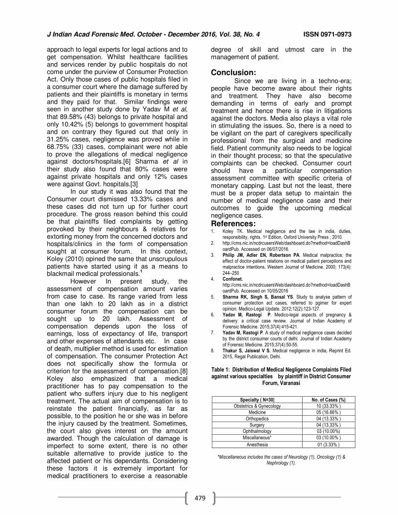
J Indian Acad Forensic Med. October - December 2016, Vol. 38, No. 4 ISSN 0971-0973
479
approach to legal experts for legal actions and to get compensation. Whilst healthcare facilities and services render by public hospitals do not come under the purview of Consumer Protection Act. Only those cases of public hospitals filed in a consumer court where the damage suffered by patients and their plaintiffs is monetary in terms and they paid for that. Similar findings were seen in another study done by Yadav M et al, that 89.58% (43) belongs to private hospital and only 10.42% (5) belongs to government hospital and on contrary they figured out that only in 31.25% cases, negligence was proved while in 68.75% (33) cases, complainant were not able to prove the allegations of medical negligence against doctors/hospitals.[6] Sharma et al in their study also found that 80% cases were against private hospitals and only 12% cases were against Govt. hospitals.[3] In our study it was also found that the Consumer court dismissed 13.33% cases and these cases did not turn up for further court procedure. The gross reason behind this could be that plaintiffs filed complaints by getting provoked by their neighbours & relatives for extorting money from the concerned doctors and hospitals/clinics in the form of compensation sought at consumer forum. In this context, Koley (2010) opined the same that unscrupulous patients have started using it as a means to blackmail medical professionals.
1
However In present study, the assessment of compensation amount varies from case to case. Its range varied from less than one lakh to 20 lakh as in a district consumer forum the compensation can be sought up to 20 lakh. Assessment of compensation depends upon the loss of earnings, loss of expectancy of life, transport and other expenses of attendants etc. In case of death, multiplier method is used for estimation of compensation. The consumer Protection Act does not specifically show the formula or criterion for the assessment of compensation.[8] Koley also emphasized that a medical practitioner has to pay compensation to the patient who suffers injury due to his negligent treatment. The actual aim of compensation is to reinstate the patient financially, as far as possible, to the position he or she was in before the injury caused by the treatment. Sometimes, the court also gives interest on the amount awarded. Though the calculation of damage is imperfect to some extent, there is no other suitable alternative to provide justice to the affected patient or his dependants. Considering these factors it is extremely important for medical practitioners to exercise a reasonable
degree of skill and utmost care in the management of patient.
Conclusion: Since we are living in a techno-era; people have become aware about their rights and treatment. They have also become demanding in terms of early and prompt treatment and hence there is rise in litigations against the doctors. Media also plays a vital role in stimulating the issues. So, there is a need to be vigilant on the part of caregivers specifically professional from the surgical and medicine field. Patient community also needs to be logical in their thought process; so that the speculative complaints can be checked. Consumer court should have a particular compensation assessment committee with specific criteria of monetary capping. Last but not the least, there must be a proper data setup to maintain the number of medical negligence case and their outcomes to guide the upcoming medical negligence cases.
References: 1. Koley TK. Medical negligence and the law in india, duties,
responsibility, rights. 1st Edition, Oxford University Press ; 2010. 2. http://cms.nic.in/ncdrcusersWeb/dashboard.do?method=loadDashB
oardPub. Accessed on 06/07/2016. 3. Philip JM, Adler EN, Robertson PA. Medical malpractice: the
effect of doctor-patient relations on medical patient perceptions and malpractice intentions, Western Journal of Medicine. 2000; 173(4):
244–250 4. Confonet.
http://cms.nic.in/ncdrcusersWeb/dashboard.do?method=loadDashBoardPub. Accessed on 10/05/2016
5. Sharma RK, Singh S, Bansal YS. Study to analyse pattern of consumer protection act cases, referred to pgimer for expert opinion. Medico-Legal Update. 2012;12(2):123-127.
6. Yadav M, Rastogi P. Medico-legal aspects of pregnancy &
delivery: a critical case review. Journal of Indian Academy of Forensic Medicine. 2015;37(4):415-421.
7. Yadav M, Rastogi P. A study of medical negligence cases decided by the district consumer courts of delhi. Journal of Indian Academy
of Forensic Medicine. 2015;37(4):50-55. 8. Thakur S, Jaiswal V S. Medical negligence in india, Reprint Ed.
2015, Regal Publication, Delhi.
Table 1: Distribution of Medical Negligence Complaints Filed against various specialties by plaintiff in District Consumer
Forum, Varanasi
*Miscellaneous includes the cases of Neurology (1), Oncology (1) &
Nephrology (1).
Specialty ( N=30) No. of Cases (%)
Obstetrics & Gynecology 10 (33.33% )
Medicine 05 (16.66% )
Orthopedics 04 (13.33% )
Surgery 04 (13.33% )
Ophthalmology 03 (10.00%)
Miscellaneous* 03 (10.00% )
Anesthesia 01 (3.33% )
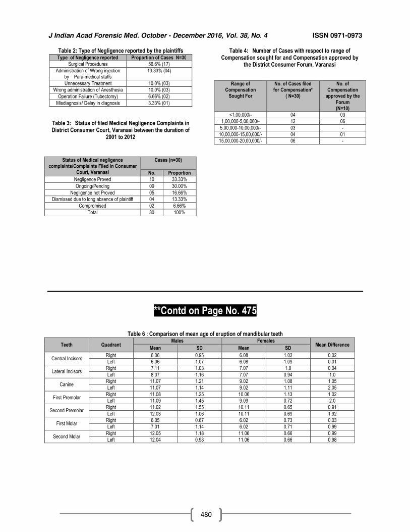
J Indian Acad Forensic Med. October - December 2016, Vol. 38, No. 4 ISSN 0971-0973
480
Table 2: Type of Negligence reported by the plaintiffs Type of Negligence reported Proportion of Cases N=30
Surgical Procedures 56.6% (17)
Administration of Wrong injection by Para-medical staffs
13.33% (04)
Unnecessary Treatment 10.0% (03)
Wrong administration of Anesthesia 10.0% (03)
Operation Failure (Tubectomy) 6.66% (02)
Misdiagnosis/ Delay in diagnosis 3.33% (01)
Table 3: Status of filed Medical Negligence Complaints in District Consumer Court, Varanasi between the duration of
2001 to 2012
Table 4: Number of Cases with respect to range of Compensation sought for and Compensation approved by
the District Consumer Forum, Varanasi
**Contd on Page No. 475
Table 6 : Comparison of mean age of eruption of mandibular teeth
Teeth Quadrant Males Females
Mean Difference Mean SD Mean SD
Central Incisors Right 6.06 0.95 6.08 1.02 0.02
Left 6.06 1.07 6.08 1.09 0.01
Lateral Incisors Right 7.11 1.03 7.07 1.0 0.04
Left 8.07 1.16 7.07 0.94 1.0
Canine Right 11.07 1.21 9.02 1.08 1.05
Left 11.07 1.14 9.02 1.11 2.05
First Premolar Right 11.08 1.25 10.06 1.13 1.02
Left 11.09 1.45 9.09 0.72 2.0
Second Premolar Right 11.02 1.55 10.11 0.65 0.91
Left 12.03 1.06 10.11 0.69 1.92
First Molar Right 6.05 0.67 6.02 0.73 0.03
Left 7.01 1.14 6.02 0.71 0.99
Second Molar Right 12.05 1.18 11.06 0.66 0.99
Left 12.04 0.98 11.06 0.66 0.98
Status of Medical negligence complaints/Complaints Filed in Consumer
Court, Varanasi
Cases (n=30)
No. Proportion
Negligence Proved 10 33.33%
Ongoing/Pending 09 30.00%
Negligence not Proved 05 16.66%
Dismissed due to long absence of plaintiff 04 13.33%
Compromised 02 6.66%
Total 30 100%
Range of Compensation
Sought For
No. of Cases filed for Compensation*
( N=30)
No. of Compensation
approved by the
Forum (N=10)
<1,00,000/- 04 03
1,00,000-5,00,000/- 12 06
5,00,000-10,00,000/- 03 -
10,00,000-15,00,000/- 04 01
15,00,000-20,00,000/- 06 -
J Indian Acad Forensic Med. October - December 2016, Vol. 38, No. 4 ISSN 0971-0973
481
Review Research Paper
Postmortem interval (PMI) estimations from vitreous humor: a review of current status, limitations and probabilities
1Nikita Gupta,
2JS Sehrawat,
3Dasari Harish
Abstract Estimation of time since death is an essential, though complicated, component of forensic death investigations. Physical, chemical and biological changes occurring in human body after death start this clock whose estimation may prove vital for criminal legal system in deciding various cases. Commonly used body fluids like vitreous humor, pericardial, cerebrospinal or synovial fluids etc., remain well protected within anatomical structures of body, undergo various biochemical changes in well defined way after death. Factors like age, sex, nutrition, health condition of deceased, climatic conditions of the area, time of collection of body fluid et.c., severely affect such estimations. This review will present current scenario of Postmortem interval estimations from vitreous humor, its limiting factors and future possible improvements, if any.
Key Words: Postmortem Interval, Body-Fluids, Vitreous Humor, Factors Affecting Postmortem Interval
Introduction: Accurate PMI estimations have
remained one of the important but controversial issues in forensic practice. Post-mortem interval is the time period between death of a person and examination of his body by a forensic pathologist during autopsy.[1] Human corpse undergoes various physical, metabolic, physiochemical, biochemical and autolytic changes in an orderly manner until it disintegrates to skelet.al form. Such patterned changes can help deduce appropriate PMI ranges.[2] Cadaveric physical changes are sometimes influenced by a number of extraneous factors, making such estimations unrealistic for forensic purposes. Body fluids like vitreous humor, pericardial fluid, cerebrospinal fluid, synovial fluid etc., remain stable for a longer period of time and don’t get contaminate or disintegrate quickly after death.[3]
Corresponding Author:
2Assistant Professor,
Department of Anthropology, Panjab University, Chandigarh 1UGC-JRF, Institute of Forensic Science and
Criminology, Panjab University, Chandigarh 3Professor and Head,
Department of Forensic Medicine and Toxicology, Govt Medical College &Hospital-32,Chandigarh. Email: [email protected] DOR: 16/04/2016 DOA: 27/12/2016 DOI: 10.5958/0974-0848.2016.00119.6
Of all these fluids vitreous humor is the most investigated body fluid for estimation of PMI as the same can be collected and isolated very easily while it remains well protected anatomically from contaminations and putrefactions. It can be found well preserved even in cases of severe head trauma.[2,4-6] Changes in chemical components or analytes of vitreous humor - The levels of various chemical components get. changed in human vitreous humor in a sequential manner which facilitates PMI estimations. Potassium, glucose, urea, creatnine, calcium, sodium, chloride, lactate, creatine, urea and hypoxanthine are some of the investigated vitreous analytes that vary in their concentration levels with varying extents of PMI.[2,5,6] During life-time, the concentration of potassium is very low in vitreous humor, but much higher in peripheral eye tissue like choroid, vitreous layer and retina. There is abrupt increase in the concentration of potassium in vitreous fluid after death as sodium potassium- ATPase pump gets broken down to allow potassium ions to leak through the membrane to approach the equilibrium.[2] Various researchers have found a statistically significant correlation between potassium concentration and PMI in vitreous humor. Some workers have found a linear increase in potassium level with increase in death duration,[7-11] however few others have either denied such a linear relationship,[4,6] or have found no significant relation at all between
J Indian Acad Forensic Med. October - December 2016, Vol. 38, No. 4 ISSN 0971-0973
482
vitreous potassium levels and time durations since death.[12] A weak correlation between vitreous potassium and death interval in first 12 hours has been found which becomes significantly stronger afterwards.[13] Hypoxanthine (Hx) is another analytic constituent of vitreous fluid formed as a degradation product of adenosine monophosphate (AMP). Hx level increases with postmortem in a linear fashion under hypoxic conditions and thus, has been widely employed for time since death estimations in forensics.[9,14-17] The linear correlation between levels of potassium and hypoxanthine with PMI intervals is temperature dependent. [14] Glucose level has also been found to decrease with PMI, though it was not found to be a reliable indicator of PMI as its level may depend upon ante-mortem metabolic and physiological state of the deceased.[14, 18] The concentration of sodium, chloride, urea, creatnine, creatine, calcium etc., has an ambiguous relationship with time since death. It is found that sodium; calcium, creatinine and urea levels don’t change significantly after death and thus, reflect no relation with PMI estimations.[19] No significant correlation has been established between chloride concentration and time since death, though its concentration in vitreous humor has been reported to decrease or remain stable after death.[14,20] Glucose and sodium levels in human vitreous humor decreases, while that of urea, creatinine, potassium and calcium increases after death.[21] A statistically significant correlation between sodium conc. and PMI has been reported but significant correlation coefficient with increasing PMI was not observed. [11] Gregora, et.al [22] observed a linear increase in calcium concentration with PMI is which is contrary to observations Coe, et.al.[23] Similarly, Chandrakanth, et.al[16] and Jashnani, et.al[1] didn’t find any correlation between postmortem interval and conc. of sodium, chloride or even potassium. A highly significant double logarithmic inverse relationship has been observed between vitreous sodium/potassium concentration ratio and time since death.[9] Piette[23] reported a satisfactory correlation between PMI and the creatine concentration in vitreous humor but Takata, et.al[24] reported such a relationship very weak, though both researchers were unanimous that creatine concentration varies between natural and unnatural deaths. Various formulas and equations suggested by various researchers to estimate PMI using vitreous potassium, hypoxanthine and glucose
concentrations have been shown in Table 1, 2 & 3 Effect of temperature, sex, age and eye difference on analytes used for PMI estimations Statistically significant sex differences have not been observed in levels of vitreous electrolytes or sodium/potassium ratio.[1,7,10,11,27] Contradictory findings have been reported with respect to the effect of temperature, age of deceased and side difference of eyes on concentration of postmortem analytes. Some studies have found no difference in concentration of electrolytes between right and left eyes,[11,28,29,30] whereas Pounder, et.al[25] have observed it. Rognum, et.al[17], Zilg, et.a [26] reported that conc. of various analytes vary significantly with temperature while others have denied the same.[11,27,28,30] Zilg, et.al[26] reported that age of deceased have a decisive role in electrolyte levels in body fluids after death, whereas others didn’t confirm it.[1,7,8,11,28] Approximately 16% of variation observed in potassium concentration is due to age differences.[26] In a recent study from India, it has been concluded that age, sex, cause of death, season of death and refrigeration of sample does not influence vitreous chemistry or its potassium levels.[1] Thus, conflicting results of vitreous chemistry and its relation with postmortem interval among different studies may be attributed to discrepancy in vitreous humor aspiration techniques, selection of cases, age and health status of deceased, variations in methodology including sample storage, pretreatment methods, analytical procedures and instruments used.[11,29,31,32,33] Estimation errors and efficacy of PMI estimations from vitreous Humor Tumram, et. al[12] in 2014 analyzed potassium content in the vitreous humor collected from 308 autopsied cadavers with known time of death and found that vitreous potassium content was significantly correlated with death interval (with standard error of estimate of ±2.9 hours). The mean overestimation of TSD varied from 18.6 ± 9.5 hours to 30.5±21.4 h as studied by various workers.[ 2,3,10,34] Swain et.al[10] examined sodium, potassium and chloride levels in vitreous humor of 100 autopsied subjects for estimation of time since death during the first 36 h after death. Chandrakanth et.al[11] analyzed 114 vitreous samples collected from 76 male and 38 female postmortem cases with a mean age of 36.4 ± 15.6 years of the cadavers.
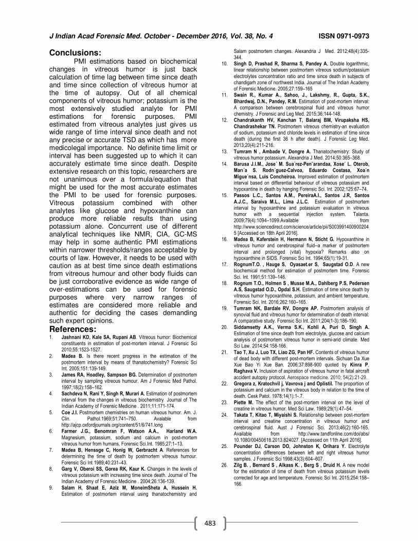
J Indian Acad Forensic Med. October - December 2016, Vol. 38, No. 4 ISSN 0971-0973
483
Conclusions: PMI estimations based on biochemical changes in vitreous humor is just back calculation of time lag between time since death and time since collection of vitreous humor at the time of autopsy. Out of all chemical components of vitreous humor; potassium is the most extensively studied analyte for PMI estimations for forensic purposes. PMI estimated from vitreous analytes just gives us wide range of time interval since death and not any precise or accurate TSD as which has more medicolegal importance. No definite time limit or interval has been suggested up to which it can accurately estimate time since death. Despite extensive research on this topic, researchers are not unanimous over a formula/equation that might be used for the most accurate estimates the PMI to be used for forensic purposes. Vitreous potassium combined with other analytes like glucose and hypoxanthine can produce more reliable results than using potassium alone. Concurrent use of different analytical techniques like NMR, CIA, GC-MS may help in some authentic PMI estimations within narrower thresholds/ranges acceptable by courts of law. However, it needs to be used with caution as at best time since death estimations from vitreous humour and other body fluids can be just corroborative evidence as wide range of over-estimations can be used for forensic purposes where very narrow ranges of estimates are considered more reliable and authentic for deciding the cases demanding such expert opinions.
References: 1. Jashnani KD, Kale SA, Rupani AB. Vitreous humor: Biochemical
constituents in estimation of post-mortem interval. J Forensic Sci 2010;55:1523-1527.
2. Madea B. Is there recent progress in the estimation of the postmortem interval by means of thanatochemistry? Forensic Sci Int. 2005;151:139-149.
3. James RA, Hoadley, Sampson BG. Determination of postmortem interval by sampling vitreous humour. Am J Forensic Med Pathol.
1997;18(2):158–162. 4. Sachdeva N, Rani Y, Singh R, Murari A. Estimation of postmortem
interval from the changes in vitreous biochemistry. Journal of The Indian Academy of Forensic Medicine . 2011;11:171-174.
5. Coe J.I. Postmortem chemistries on human vitreous humor. Am. J. Clin. Pathol.1969;51:741–750. Avaiable from http://ajcp.oxfordjournals.org/content/51/6/741.long
6. Farmer J.G., Benomran F, Watson A.A., Harland W.A.
Magnesium, potassium, sodium and calcium in post-mortem vitreous humor from humans, Forensic Sci.Int. 1985;27:1–13.
7. Madea B, Henssge C, Honig W, Gerbracht A. References for determining the time of death by postmortem vitreous humour.
Forensic Sci Int 1989;40:231–43. 8. Garg V, Oberoi SS, Gorea RK, Kaur K. Changes in the levels of
vitreous potassium with increasing time since death. Journal of The Indian Academy of Forensic Medicine . 2004;26:136-139.
9. Salam H, Shaat E, Aziz M, MoneimSheta A, Hussein H. Estimation of postmortem interval using thanatochemistry and
Salam postmortem changes. Alexandria J Med. 2012;48(4):335-344.
10. Singh D, Prashad R, Sharma S, Pandey A. Double logarithmic, linear relationship between postmortem vitreous sodium/potassium
electrolytes concentration ratio and time since death in subjects of chandigarh zone of northwest India. Journal of The Indian Academy of Forensic Medicine. 2005;27:159–165
11. Swain R., Kumar A., Sahoo, J., Lakshmy, R., Gupta, S.K.,
Bhardwaj, D.N., Pandey, R.M. Estimation of post-mortem interval: A comparison between cerebrospinal fluid and vitreous humor chemistry. J Forensic and Leg Med. 2015;36:144-148.
12. Chandrakanth HV, Kanchan T, Balaraj BM, Virupaksha HS,
Chandrashekar TN. Postmortem vitreous chemistry-an evaluation of sodium, potassium and chloride levels in estimation of time since death (during the first 36 h after death). J Forensic Leg Med. 2013;20(4):211-216.
13. Tumram N , Ambade V, Dongre A. Thanatochemistry: Study of vitreous humor potassium. Alexandria J Med. 2014;50:365–368.
14. Barusa J.I.M., Jose´ M. Sua´rez-Pen˜arandaa, Xose´ L. Oterob, Marı´a S. Rodrı´guez-Calvoa, Eduardo Costasa, Xoa´n
Migue´nsa, Luis Concheiroa. Improved estimation of postmortem interval based on differential behaviour of vitreous potassium and hypoxantine in death by hanging Forensic Sci. Int. 2002;125:67–74.
15. Passos L.C., Santos A.M., PereiraA.I., Santos J.R, Santos
A.J.C., Saraiva M.L., Lima J.L.C. Estimation of postmortem interval by hypoxanthine and potassium evaluation in vitreous humor with a sequential injection system. Talanta. 2009;79(4):1094–1099.Available from
http://www.sciencedirect.com/science/article/pii/S0039914009002045 [Accessed on 18th April 2016].
16. Madea B, Kaferstein H, Hermann N, Sticht G. Hypoxanthine in vitreous humor and cerebrospinal fluid--a marker of postmortem
interval and prolonged (vital) hypoxia? Remarks also on hypoxanthine in SIDS. Forensic Sci Int. 1994;65(1):19-31.
17. RognumT.O. , Hauge S, Oyasaet.er S, Saugstad O.D. A new biochemical method for estimation of postmortem time. Forensic
Sci. Int. 1991;51:139–146. 18. Rognum T.O., Holmen S , Musse M.A., Dahlberg P.S, Pedersen
A.S, Saugstad O.D., Opdal S.H. Estimation of time since death by vitreous humor hypoxanthine, potassium, and ambient temperature.
Forensic Sci. Int. 2016;262:160–165. 19. Tumram NK, Bardale RV, Dongre AP. Postmortem analysis of
synovial fluid and vitreous humor for determination of death interval: A comparative study. Forensic Sci Int. 2011;204(1-3):186-190.
20. Siddamsetty A.K., Verma S.K., Kohli A, Puri D, Singh A. Estimation of time since death from electrolyte, glucose and calcium analysis of postmortem vitreous humor in semi-arid climate. Med Sci Law. 2014;54:158-166.
21. Tao T, Xu J, Luo TX, Liao ZG, Pan HF. Contents of vitreous humor of dead body with different post-mortem intervals. Sichuan Da Xue Xue Bao Yi Xue Ban. 2006;37:898-900 quoted by Kinra P, Raghava V. Inclusion of aspiration of vitreous humor in fatal aircraft
accident autopsy protocol. Aerospace medicine. 2010; 54(2):21-29. 22. Gregora z, Kratochvil j, Vavrova j and Oplistil. The proportion of
potassium and calcium in the vitreous body in relation to the time of death. Cesk Patol. 1978;14(1):1-.7.
23. Piette M. The effect of the post-mortem interval on the level of creatine in vitreous humor. Med Sci Law. 1989;29(1):47–54.
24. Takata T, Kitao T, Miyaishi S. Relationship between post-mortem interval and creatine concentration in vitreous humor and
cerebrospinal fluid. Aust J Forensic Sci. 2013;46(2):160-165. Available from http://www.tandfonline.com/doi/abs/ 10.1080/00450618.2013.824027. [Accessed on 11th April 2016].
25. Pounder DJ, Carson DO, Johnston K, Orihara Y. Electrolyte
concentration differences between left and right vitreous humor samples. J Forensic Sci 1998;43(3):604–607.
26. Zilg B. , Bernard S , Alkass K. , Berg S , Druid H. A new model for the estimation of time of death from vitreous potassium levels
corrected for age and temperature. Forensic Sci Int. 2015;254:158–166.
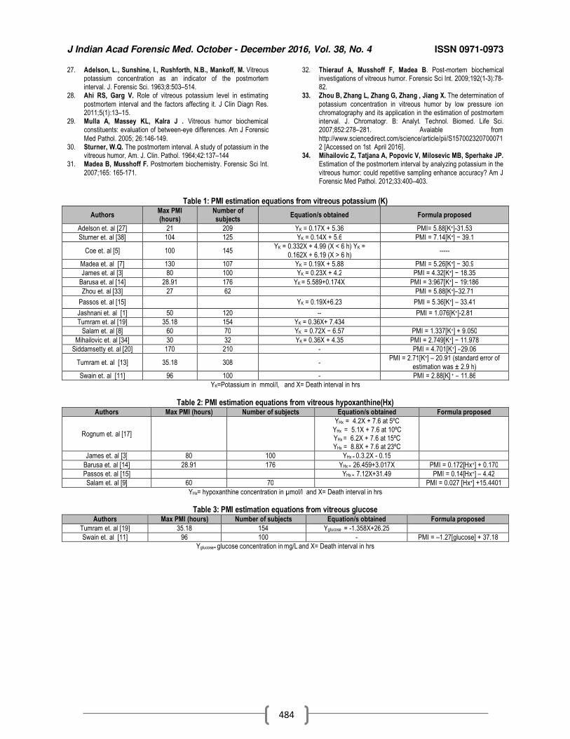
J Indian Acad Forensic Med. October - December 2016, Vol. 38, No. 4 ISSN 0971-0973
484
27. Adelson, L., Sunshine, I., Rushforth, N.B., Mankoff, M. Vitreous potassium concentration as an indicator of the postmortem interval. J. Forensic Sci. 1963;8:503–514.
28. Ahi RS, Garg V. Role of vitreous potassium level in estimating
postmortem interval and the factors affecting it. J Clin Diagn Res. 2011;5(1):13–15.
29. Mulla A, Massey KL, Kalra J . Vitreous humor biochemical constituents: evaluation of between-eye differences. Am J Forensic
Med Pathol. 2005; 26:146-149. 30. Sturner, W.Q. The postmortem interval. A study of potassium in the
vitreous humor, Am. J. Clin. Pathol. 1964;42:137–144 31. Madea B, Musshoff F. Postmortem biochemistry. Forensic Sci Int.
2007;165: 165-171.
32. Thierauf A, Musshoff F, Madea B. Post-mortem biochemical investigations of vitreous humor. Forensic Sci Int. 2009;192(1-3):78-82.
33. Zhou B, Zhang L, Zhang G, Zhang , Jiang X. The determination of
potassium concentration in vitreous humor by low pressure ion chromatography and its application in the estimation of postmortem interval. J. Chromatogr. B: Analyt. Technol. Biomed. Life Sci. 2007;852:278–281. Avaiable from
http://www.sciencedirect.com/science/article/pii/S1570023207000712 [Accessed on 1st April 2016].
34. Mihailovic Z, Tatjana A, Popovic V, Milosevic MB, Sperhake JP. Estimation of the postmortem interval by analyzing potassium in the
vitreous humor: could repetitive sampling enhance accuracy? Am J Forensic Med Pathol. 2012;33:400–403.
Table 1: PMI estimation equations from vitreous potassium (K)
Authors Max PMI (hours)
Number of subjects
Equation/s obtained Formula proposed
Adelson et. al [27] 21 209 YK = 0.17X + 5.36 PMI= 5.88[K+]-31.53
Sturner et. al [38] 104 125 YK = 0.14X + 5.6 PMI = 7.14[K+] − 39.1
Coe et. al [5] 100 145 YK = 0.332X + 4.99 (X < 6 h) YK =
0.162X + 6.19 (X > 6 h) -----
Madea et. al [7] 130 107 YK = 0.19X + 5.88 PMI = 5.26[K+] − 30.9
James et. al [3] 80 100 YK = 0.23X + 4.2 PMI = 4.32[K+] − 18.35
Barusa et. al [14] 28.91 176 YK = 5.589+0.174X PMI = 3:967[K+] – 19:186
Zhou et. al [33] 27 62 PMI = 5.88[K+]–32.71
Passos et. al [15] YK = 0.19X+6.23 PMI = 5.36[K+] – 33.41
Jashnani et. al [1] 50 120 -- PMI = 1.076[K+]-2.81
Tumram et. al [19] 35.18 154 YK = 0.36X+ 7.434
Salam et. al [8] 60 70 YK = 0.72X − 6.57 PMI = 1.337[K+] + 9.050
Mihailovic et. al [34] 30 32 YK = 0.36X + 4.35 PMI = 2.749[K+] − 11.978
Siddamsetty et. al [20] 170 210 - PMI = 4.701[K+] –29.06
Tumram et. al [13] 35.18 308 - PMI = 2.71[K+] – 20.91 (standard error of
estimation was ± 2.9 h)
Swain et. al [11] 96 100 - PMI = 2.88[K] + – 11.86
YK=Potassium in mmol/l, and X= Death interval in hrs
Table 2: PMI estimation equations from vitreous hypoxanthine(Hx) Authors Max PMI (hours) Number of subjects Equation/s obtained Formula proposed
Rognum et. al [17]
YHx = 4.2X + 7.6 at 5ºC
YHx = 5.1X + 7.6 at 10ºC YHx = 6.2X + 7.6 at 15ºC YHx = 8.8X + 7.6 at 23ºC
James et. al [3] 80 100 YHx = 0.3.2X - 0.15
Barusa et. al [14] 28.91 176 YHx = 26.459+3.017X PMI = 0.172[Hx+] + 0.170
Passos et. al [15] YHx = 7.12X+31.49 PMI = 0.14[Hx+] – 4.42
Salam et. al [9] 60 70 PMI = 0.027 [Hx+] +15.4401
YHx= hypoxanthine concentration in µmol/l and X= Death interval in hrs
Table 3: PMI estimation equations from vitreous glucose Authors Max PMI (hours) Number of subjects Equation/s obtained Formula proposed
Tumram et. al [19] 35.18 154 Yglucose = -1.358X+26.25
Swain et. al [11] 96 100 - PMI = –1.27[glucose] + 37.18
Yglucose= glucose concentration in mg/L and X= Death interval in hrs
J Indian Acad Forensic Med. October - December 2016, Vol. 38, No. 4 ISSN 0971-0973
485
Review Research Paper
RAJ – TAJ and MODI The life & times of Dr. J. P. Modi (1875 - 1954) (A leaf in the
history of forensic medicine in India) 1A.M.M.Patnaik,
2M.Jagadeesh Naik,
3Ch.Lakshmi Kumar
Abstract Narrating the history of a bygone era from such reluctant material as is available is difficult and to
perceive the events, institutions, personalities and the powers that be, would even be more difficult. This article aims to reconstruct the history that is 100 years old. The content may not be exhaustive due to limited scope of the article to be published in a journal. Per say, this is an effort to reconstruct the same, highlighting the events that have occurred during the times of late Rai Bahadur Dr. J. P. Modi a great teacher, much revered medico legist and pioneering author of a text book, “Modi’s Medical Jurisprudence and Toxicology”, published in 1920; nearly hundred years ago. Through this text book, he has immortalized himself. His legacy is the real life stories of crime and passion that represent the episodes of a bygone era. Modi appears to have joined Government service in the earls-while United Provinces, around 1906 at Agra, the Seat of the Taj. His first case was reported from Hardoi in 1906. He retired from service in1934.[1] This period was the zenith of the British Raj. Aims and objectives: 1. To remember and pay homage to Dr. Modi; 2. Narrating history of forensic medicine of his time; 3. To connect the past with the present Material and methods: 1. 1st and 16nth editions of his text book-1920, 1967; 2. Material available on the internet; 3. standing orders for the year 1896 of the civil medical department, Government of Madras.
.
Key Words: Modi, Raj, Taj, Historical, Medico-Legal
Introduction: Forensic medicine is about the doctors, the police, the bar and the bench. The times and conditions under which Modi worked were alien to Indians. British police officers, civil surgeons, judges and civil servants worked at district and state levels. Dr. Modi in their midst was one of the few Indians entrusted with medico-legal duties. To understand the medico-legal history of Modi’s time we have to study the socio-political and the medico-legal systems of those days. The Socio-political conditions in India during the first 5 decades of the 20
th century:[2]
The territory of India was divided into British ruled provinces like Madras, Bengal, Bombay, Central provinces, united provinces,
Corresponding Author: 1Associate Professor
2Associate Professor
3Professor & Principal;
Dept. of Forensic Medicine & Toxicology, Maharajahs Institute of Medical Sciences, Nellimarla (A. P.) Email: [email protected]
DOR: 08/02/2016 DOA: 29/12/2016 DOI: 10.5958/0974-0848.2016.00120.2
Punjab, Burma etc…, that constituted the bulk of the country and native princely states, numbering around 565, ruled by Indian Rajas on the principle of British paramountcy. The territories of Goa, Diu, Daman and Pondicherry were under the control of the Portuguese and the French, respectively. Indians were de-armed across the length and breadth of the country after the 1857 uprising. The society was largely feudal in nature. Peace and tranquility prevailed everywhere. The British raj came into existence in June1858. The administration of the country changed hands from the East India Company to the British crown. The British had their capital at Calcutta till 1912 which was then shifted to New Delhi. Best British Indian institutions: The viceroy and the Governor General administered India on behalf of the British crown. The steel framework of Indian Civil Service (I.C.S), the Indian Police Service (I.P.S), the Indian judicial service, the Indian Medical Service (IMS) and the Indian railways were the seat anchor of civil administration in India. The district magistrate of ICS played a pivotal role even in medico-legal cases.
J Indian Acad Forensic Med. October - December 2016, Vol. 38, No. 4 ISSN 0971-0973
486
The judicial system:[3] British judicial procedures were replicated in India. The Indian Penal Code (I.P.C), the Criminal Procedure Code (Cr.P.C) and the Indian Evidence Act (I.E.A) were enacted in 1860, 1861 & 1872, respectively. In the British ruled provinces, the High courts, the Session’s courts and the courts of the Magistrates were established with successive line of appeals. In the presidency towns of Calcutta, Madras and Bombay, the magistrates directly committed their cases to the High court without the via media of the Session’s courts. In the districts, however, the magistrates had no power to commit their cases directly to the High Court. The magistrates in the presidency towns have been re-designated as metropolitan magistrates after independence. Trial with the help of jury was in vogue in the high courts. The session’s judge tried cases with the help of assessors except in some districts of Bombay and Bengal where a jury assisted the sessions judge.[4] In the provinces, the European accused were tried by European magistrates only. There are claims by researchers that the predominantly European jury favored the European accused. Trial by jury was abolished in independent India in1959[5] after an emotional failure of jury in the sensational Commander Nanavathi case where in the jury; this time Indian, acquitted the accused naval commander in the sensational murder case of Ahuja, his own friend. In the presidency towns of Bombay and Calcutta, the “Coroner’s System”[6] was introduced in 1871. By 1999 the system was abolished. The rest of the Indian princely states more or less followed the British system. Even in those remote days, Indians were posted as magistrates and judges. With in two years; the sentence of death would be confirmed by the high court. There was no Supreme Court. Appeals from high courts were to be made to the judicial commission of the Privy Council[7] located in London, which was established in1833 by an Act of the British parliament. This was the highest court of appeal for the British overseas territories. A federal court was established in 1937 which appears to be nonfunctional. The jurisdiction of Privy Council over India was abolished by an Act of the interim government in Delhi in 1949.[8] The Supreme Court of India was established in January 1950[9] and started functioning two days after the first Republic day.
The police system:[10] The police system of those days was more or less the same as the present day system. The Inspector General of Police headed the police department in a state. In the districts, higher police officials like the S.P and the D.S.P were exclusively Europeans. Officers below the rank of DSP were manned by Indians. In the presidency towns, European sergeants in the non gazetted rank were posted to police stations and district headquarters. Officers at station level indulged in ruthless third degree methods as a means to detect crime, which practice continues to this day. The so called lockup deaths occasionally invited official action against the errant officers. The medico-legal system:[11] The Surgeon General to Government (equivalent to the Director General of Health Services) was both the Head of the Department and secretary to Government at the state level. He drew a salary of Rs 2500/per month. Civil surgeons, from the IMS,[12] mostly Europeans and few Indians, presided over the districts and were the only officers authorized to conduct medico-legal autopsies and other important medico - legal works. Some acts of his time:[13] The Indian lunacy Act of 1912, which replaced the Mental Asylum Act of 1858, is a classic piece of legislation with a European content. The MH Act of 1987, which replaced this Act with additional safeguards, is a mere replication of the Act of 1912 minus the European content. Incidentally both Acts run into hundred sections. The medical degrees Act of 1916, the first of its kind, regulated the Western medical degrees granted by medical colleges in India. The workmen’s compensation Act of 1923 is a social security Act meant for the disabled workers of the factories at work place. The Act opened flood gates in awarding compensation in independent India.
Discussion: Highlights of Text Book (1
st edition):[4]
The contents of the First edition are more or less same as the present text books except for addition of one chapter on life assurance. Such techniques as DNA finger printing, lie detector, truth serum tests etc… were not available. In the appendices section, a whole lot of information on medico-legal certificates, rules of medical evidence, likely questions asked to medical witness, medico-legal formats for sending viscera and
J Indian Acad Forensic Med. October - December 2016, Vol. 38, No. 4 ISSN 0971-0973
487
incriminating material to the labs, forms of Indian lunacy Act, poisonous Acts of that time and several Sections of I.P.C, C.P.C & I.E.A were incorporated. European accused were to be tried by European judges. Not many of his cases were mentioned in this text book, nor were there any figures included. The ecstasy of reading such an antique material cannot be put on pen and paper. That can only be experienced. Contents of 16 nth edition:[13] Punishments authorized by law. In addition to the punishments of the present day; medieval punishments like whipping and transportation for solitary confinement to the cellular jail at Andaman & Nicobar, were authorized by law. The cellular jail was closed soon after independence and whipping, as punishment, was abolished by an Act of Parliament in 1955. Volunteering a statement. Though the witness was not supposed to volunteer a statement in a court of law, Modi used to volunteer statements in the courts to elicit an important point which was missed during cross examination and was highly appreciated by the judges. Such a practice is almost unthinkable now. The Bhowal Sanyasi case (disputed case of identification) - A sensational case of disputed identity of Kumar Ramendra Narayan Roy (1880-1946) In this case, the above Kumara Raja (second son of Rajendra Narayana Roy of Bhowal estates in Dacca), was alleged to have been cremated in 1909 at Darjeeling after his death due to biliary colic. In 1921 a Naga sanyasi by the name of Sunder Das from Punjab appeared in the estate and claimed himself to be the Kumara Raja, and demanded his share of the estate. His contention was that the funeral pyre was never lit due to a heavy storm on that night and that he was subsequently rescued and revived by some passing Naga Sadhus. After recovery he wandered here and there due to retrograde amnesia. The Kumara Raja’s wife did not accept this version and declared him an imposter and denied to any share in the property. Being denied of his rights he filed a case in the subordinate court of Dhaka in 1930. In this phenomenal case the medico-legal points which strongly went in favour of the sanyasi were several anthropometric features including the size of the shoes, claw marks on the right arm, syphilitic marks on the penis, operation scar near the groin and most importantly, a minute mole on the dorsum of the penis. Also his voice, gait and expression tallied
with the features of the Kumar Raja. The judge gave a decree in favour of the plaintiff. His wife Bibhabati Devi contested the case in the High court of Calcutta, which upheld the verdict of the district court of Dhaka. After this, the courts of wards (District Collector of Dhaka) filed an appeal in the Privy Council at London against the judgment of the High court of Calcutta. The Privy Council after a long delay decided the case in favour of the Kumar Raja in July 1946. Two days after this landmark judgment the Kumar Raja collapsed and died while at prayer; a dramatic end to a sensational case. This case was one of the few cases where justice was inordinately delayed and thus justice was denied. Dactylography: This system was first used in India in 1858 by Sir William Herschel, ICS to prevent impersonation. Sir Francis Galton systemized it for identification of criminals. Dactylography was officially adopted in England in 1894 and in India in1899. To this day the system is a flawless method of personal identification. On exhumation. Modi reports a notable case of exhumation on the body of the late Mr. Fulham, conducted at Agra. In this case, orders for exhumation were issued by the district magistrate of Agra on 6/12/1912. The superintendent of police and few others connected with this case were present at the exhumation site. This case is a role model for generations of medical officers to follow the rules on exhumation in the proper context. Experiments on cadaveric rigidity and flaccidity. Modi mentions the investigations of Mackenzie in Calcutta in 1889 regarding the onset and duration of rigor mortis. On preternatural combustibility. Recently, doctors have been investigating spontaneous combustion that occurred in a child near Chennai. Modi mentions that inflammable gases can form in elementary canal, and such gases, when belched may be ignited on the application of flame. On accidents. In those days, few accidental deaths occured due to run over by tonga, carts and cycles, unlike lakhs of people dying due to road traffic accidents in these days. Accidental deaths included cases of drowning, poisoning and fall from heights. Fire arm injuries. We don’t find mention of firearm injuries of an earlier period. Burn injuries. Practices like bride burning, suicidal and accidental burning were common.
J Indian Acad Forensic Med. October - December 2016, Vol. 38, No. 4 ISSN 0971-0973
488
Spontaneous rupture of spleen. In those days, due to such infestations as malaria, black fever, sickle cell anemia, Leshmaniasis etc… the spleen used to be enlarged considerably and was the cause for spontaneous rupture at the slightest or no provocation. On sexual crimes. Rape cases were as common as today. Gang rape cases were few in number. Cases of rape by impersonation were frequently reported. Modi mentions Sec.497 casually with no reported cases. Age of the male accused in cases of rape. In accordance with Sec.83 I.P.C. an Indian boy aged 10 years was found guilty of raping an European girl aged 7 years and was sentenced to 2 years rigorous imprisonment. In the present day, Sec.83 has no relevance. And as per Juvenile Justice Act, a juvenile under 18 years cannot be sent to jail whatever crime he perpetuates. Bestiality: Modi reports examination of half a dozen cases of bestiality during an eleven year period. In the present day, cases of bestiality are not reported. On toxicology: Arsenic was the preferred poison for homicidal causes in those days. Arsenic is now replaced by such drugs as barbiturates and other narcotics. In the Fulham (exhumation) case, arsenic was detected in the bones of the victim. For suicidal poisoning, oleander was preferred. Children were killed by administering such poisons as opium. Collection, preservation, packing and transportation of viscera was being done meticulously, as per procedure. Bottles for preservation of viscera, a cardboard boxes and wooden boxes with lock and key along with preservative material were supplied by the chemical examiner’s office to the civil surgeon’s office. This beauty of information should be kept in mind. No such facilities are available now. A rare case of murder by injecting plague bacillus at Howrah was reported by Modi which occurred in 1933. This has come to be known as “the curse of kalimatha” as the victim Amrendra developed pustules and fever
similar to the pustules of smallpox before his death.
Conclusion: Because of his teachings, writings and his revered position in the medico-legal and legal circles; he was awarded the title of Rai Bahadur by the British. After his departure in 1934 from King George Medical College, the Department of Medical Jurisprudence remained headless till 1982. Also, there were no regular professors in the majority of medical colleges in India till the 60’s. Undoubtedly, Dr. Modi is the pioneer Indian in the field of Forensic Medicine in India. Modi lived in the momentous times of freedom struggle along with stalwarts like Mahatma Gandhi, Pandit Nehru and others. Another great Indian, freedom fighter and the Iron Man of India, Sardar Vallabhabai Patel, a fellow Gujarati was born in the same year as Modi. What more tribute can we pay to this legend?
References: 1. Foreword by Singh CB. In Modi’s medical jurisprudence &
toxicology. 16th Ed. NM Tripathy pvt. Ltd – 1967: 2. https://en.wikipedia.org/wiki/British_Raj,accessed-
January2016&17/01/2017.
3. https://en.wikipedia.org/wiki/Judiciary_of_India. accessed -January2016&17/01/2017.
4. Modi JP. A text book of medical jurisprudence & toxicology. 1st Ed. Butter worth & co. (India) Ltd. 1920: 8; appendix: I, III, IV, VII, VIII,
IX, X & XII, page no: i – xci, 5. https://en.wikipedia.org/wiki/K._M._Nanavati_v._State_of_Maharash
tra.accessed-January2016&17/01/2017 6. http://indiankanoon.org/doc/792853/ accessed -
January2016&17/01/2017. 7. https:// www.jcpc.uk/ accessed -January2016&17/01/2017. 8. http://bombayhighcourt.nic.in/libweb/actc/1949.05.pdf accessed -
January2016&17/01/2017.
9. http://supremecourtofindia.nic.in/history.htm accessed -January2016&17/01/2017.
10. https://en.wikipedia.org/wiki/Indian_Police_Service accessed -January2016&17/01/2017.
11. Standing orders of the surgeon – general with the govt. of Madras, printed book by: the superintendent, Govt. press Madras, dt: 1897; of standing order (SO) no: 58, page no: 12.
12. https://en.wikipedia.org/wiki/Indian_Medical_Service accessed -
January2016&17/01/2017. 13. Modi NJ. In Modi’s medical jurisprudence & toxicology, 16th edition
– 1967, Ed. NM Tripathy pvt. Ltd. Princess Street, Bombay – 2. page no: 404, 428, 447; 8(3), 17(3), 43(4), 44, 48(2), 49, 89, 90,
135(9), 136(1), 206(2), 285(6), 295(5,6,7), 332(5), 344(5), 70, 71, 462(3).
J Indian Acad Forensic Med. October - December 2016, Vol. 38, No. 4 ISSN 0971-0973
489
Review Research Paper
Child Sexual Abuse (CSA): India & the World 1Raghvendra Kumar Vidua,
2Sweta Patel,
3B.L.Chaudhary,
4Alok K. Mishra,
5Arneet Arora
Abstract Child Sexual abuse (CSA) is a mostly hidden but widely prevalent problem in the society. It is
very difficult to understand the exact aetiology but certain factors keep children on a constant risk of falling prey to it. The magnitude of problem is quite high and scars the child’s life with undesired consequences. The professionals who deal with such cases are supposed to tackle them in different ways and manner than routine cases. It requires a unique way of forensic investigation, as often these children are hesitant to tell the true story and put a challenge before us to diagnose with the available methods and techniques. The appropriate preventive, curative, supportive and legal remedies are required to pull them out from this dreaded situation and allowing them to lead a normal life. There is a need to frame uniform standard operating procedures (SOPs) to guide the medical, police, law, social service and other personnel to effectively prosecute the abusers and stopping the re-victimisation of the children.
Key Words: Child sexual abuse (CSA), vulnerable child, Abuser, Standard operating procedures
Worldwide prominent cases of Child Sexual abuse:
Most recently, in a shocking instance of minor rapes, a tailor has confessed to sexually abusing around 600 children over 12 years.[1] Recently, a video became viral showing sexual abuse of a toddler in the joy rides area in a mall.[2] In another instance, Pakistan was horrified by revelations of a blackmailing gang, which sexually abused some 270 children.[3. In a recent case in India, a 16 year old girl has alleged sexual exploitation by one 72-year-old Mr. Asumal Sirumalanai, or Asaram Bapu in a Jodhpur ashram.[4]. The Noida serial murders (Nithari Kand) case,[5] the molestation of 14-year-old budding tennis player by the Inspector General (IG) of Police [6] and of a 14 year old Gujarati girl in south Kolkata by the apartment
Corresponding Author: 1Assistant professor,
2Senior Resident,
5Professor & Head,
Dept. of Forensic Medicine & Toxicology, AIIMS Bhopal, MP 462020
3Associate Professor,
Dept. of Forensic Medicine & Toxicology, LHMC, New Delhi 4Chairman,
Brain Behaviour Research Foundation of India(BBRFI), New Delhi DOR: 07/04/2016 DOA: 29/12/2016 DOI: 10.5958/0974-0848.2016.00121.4
guard,[7] again raised the concern on child sexual abuse. On various occasions, many famous celebrities like Pop singer Lady Gaga, actress Kalki Koechlin, former actress Somi Ali, film director Anurag Kashyap, Indian sitar player Anoushka Shankar, Hollywood actresses Pamela Anderson, Oprah Winfrey, Teri Hatcher, Sophia Hayat and Ashley Judd have accepted sexual abuse during their childhood. [8]
Definitions Child abuse is the physical, sexual or emotional ill-treatment or neglect of a child, especially by those responsible for its welfare.[9] In the USA, the CDC and the DCF define child maltreatment as any act or series of acts of commission or omission by a parent or other caregiver that results in harm, potential for harm, or threat of harm to a child.[10] Intrafamilial child sexual abuse (IFCSA) is defined as child sexual abuse perpetrated by a family member or one that takes place within a family context or environment, whether or not by a family member.[11]
Types There may be two different broad categories of sexual abuse possible: contact abuse with actual physical contact and non-contact abuse with no physical contact with the child. Re-victimisation When after the first episode, child may be continuously exploited by the abusers in form
J Indian Acad Forensic Med. October - December 2016, Vol. 38, No. 4 ISSN 0971-0973
490
of threat to send images, video or copies of conversations to the friends and family of victim unless they take part in sexual activity again. The ‘victim-to-victimiser’ cycle Sometimes the progression from victim to victimiser occurs, as child sexual abusers often report a history of sexual victimisation.[12] It means that victims, when become adults themselves, turn as abusers. This is particularly common in paedophiles whose preferred targets are boys.[13] Child Sexual Abuse Online The virtual world is also responsible for child sexual abuse (e-crime). Children are often pressurised by the abusers to perform sexual acts on themselves or others and images of their abuse often remain available online.[14] Facts about child in India In India, the legal definition of child after the introduction of the Juvenile Justice Care and Protection of Children Act is considered as a person below 18 years[15] and every second child is being exposed to one or the other form of sexual abuse and every fifth child faces critical forms of it.[16] Further, India has the world's largest number of cases and for every 155
th minute, a child less than 16 years is raped,
for every 13th hour a child under 10.[15]
Aetiology It is very difficult to pinpoint the exact cause behind the sexually abusive behaviour towards children but some biological and psychological factors certainly play an important role. Contrary to the common belief, only some sexual abusers are paedophiles, having a primary sexual interest in children. Various factors, often in combination, may predispose child for sexual abuse such as poverty, illiteracy, unemployment, troubled family environment, domestic abuse, geographical and social isolation, inadequate housing, adultification of kids, easy access to modern form of communication such as internet, mobile phone, etc. [17] Child Vs adult commercial sexual abuse Currently, there are 10 million children in the Sex Industry. This includes forced child prostitution, pornography & sex trade tourism [9] At present this is a well organised and one of the most profitable businesses in the world.[18] Children, in contrast to adults, are clearly much more vulnerable and helpless against the established structures and vested interests in the sex sector.[19] Models of Abuser-victim relationship It may be inappropriate relationship, where an abuser has some kind of power over
their victim ,the boyfriend relationship, where abuser and victim enter into an almost conventional relationship and the organised exploitation and trafficking, where children are abused by more than one adult as part of a network.[20,21] Dimensions & Circumstances Although child sexual abuse is generally referred to as a distinct or singular phenomenon but actually, wide variations have been observed in the characteristics, circumstances and outcomes for victims and in the physical and social settings of child sexual abuse.[22] It has a very wide dimension and comprised of any activity with a child, before the age of legal consent, for the sexual gratification of an adult or a significantly older child. It includes oral-genital, genital-genital, genital-rectal, hand-genital, hand-rectal, or hand- breast contact, exposure of sexual anatomy, forced view of sexual anatomy; and a spectrum of activities that does not involve contact such as exhibitionism, and voyeurism apart from encouraging a child into prostitution or pornography.[23]
Incidence and Prevalence Child sexual abuse is largely a hidden crime, so it is difficult to accurately estimate the incidence and prevalence. [24] Experts now agree that child sexual abuse exists in all socioeconomic groups and is vastly under-reported. Figures from USA show that 1 in 4 girls and 1 in 6 boys is sexually abused before the age of 18.The exact magnitude of the problem in other areas in Asia and Africa is not known but it is probably even greater.[25] A meta-analysis estimated 7.9% of males and 19.7% of females universally faced sexual abuse before the age of 18 years and the highest prevalence rate was seen in Africa (34.4%). Child sexual abuse is an extensive problem and even the lowest prevalence includes a huge number of victims.[26] In 1998, an Indian NGO, Recovery and Healing from Incest (RAHI) found a prevalence of 40 percent. In 2006,Tulir-CPHCSA`s study, indicated a child sexual abuse prevalence rate of 42%. A survey by United Nations International Children Education Fund reported that 10% of Indian girls might have experienced sexual violence when they were 10–14 years of age and 30% during 15–19 years of age.[27] IB Times reported UK as one of the countries in the world with the highest rate of Child Sexual Abuse, others are South Africa, India Zimbabwe, United Kingdom and United
J Indian Acad Forensic Med. October - December 2016, Vol. 38, No. 4 ISSN 0971-0973
491
States.[28] The CDC found a prevalence of sexual abuse ranging from 4.4% among females in Cambodia to 37.6% among females in Swaziland, with prevalence in most countries greater than 25.0%.[29] Sexual abuse is the highest in Madhya Pradesh,. followed by Uttar Pradesh and Maharashtra. In 2012, the total reported crimes against children in India was 38172. In 2013, the graph of total number of reported crime against children has been increased to 58, 224.[30] It is a major issue which affects more than one out of five females and one in 10 males globally. It is accountable for about one per cent of the global burden of disease.[31] Three main issues viz. definition of child, underrating of number of victim cases reported by the official organizations as many cases never get reported to them and reporting prevalence for different time periods by different studies make it difficult to estimate exact number of victims.[32] Vulnerable child against sexual abuse Though all children are at risk but a combination of individual, relational, community and societal factors make some child more vulnerable to sexual abuse than others. These are only the contributory factors and not the direct causes.[33] Vulnerable group includes street children, child labourers, neglected children, children of poor parents or with physical, mental or terminal illness, children of refugees, migrants, construction workers prostitutes, and of rape victims.[34] Indicators They are certain features viz.behavioural and physical in a victim of a sexual abuse may be regarded as indicators of sexual abuse. These are only indicators and not the specific signs. However, these indicators must be used with caution, especially in the absence of a disclosure or a diagnostic physical finding. In practice, clear physical findings of sexual abuse are seldom seen in children.[34] Consequences Childhood is considered a development period and sexual abuse in this period predisposes a child to physical, emotional social and psychosocial risks. When sexual abuse occurs, a child can develop both short-term and long-term distressing feelings, thoughts, and behaviours apart from the physical injuries or in some cases death of the child. In most cases, the fundamental damage inflicted by child sexual abuse is to the child's developing capacities for trust, intimacy, agency
and sexuality, and many of the mental health problems of adult life.[12] Abusers The abusers may be men, women, teenagers, and other children or the person belongs to any profession. They come from all the sections of society and all types of backgrounds. Most often, friends, relatives, co-workers and other closely associated persons or a person in a position of trust, rather than a stranger are the common abusers of a child. The majority of child sexual abuse is committed by male abusers.[35]Abuse by females is almost certainly under-reported.[36] Boys and girls are equally likely to be abused by men and women.[37] Abusers look for weak spots in a family, a community or an organisation to gain unsupervised access to children.
Place of sexual abuse It is commonly believed that child abuse is a problem of lower socio-economic class and happens to vulnerable children staying in unsafe places but the truth is that most of the abuse occurs to normal children in regular homes. Child abuse usually takes place in the home with someone that the child knows rather than with strangers. [34] Doctor’s response The response of a doctor to a child sexual abuse case can be broadly classified into Immediate or emergency response. The child will need emergency care and information to police and child social Service. Admission to the hospital is needed in all cases of serious injuries or persisting threat to his safety at home. Another is planned or graded response in which a proper forensic investigation and rehabilitation of child must be ensured in a child friendly atmosphere. Medical management of a case of child sexual abuse is a medico legal emergency. Management should be child friendly and aim at achieving the short term and long term goals. If necessary referrals to appropriate specialties must be made. Consent During examination of a case of sexual abuse, the police need to be informed and consent is a must. Consent for medical examination may be obtained from the child if he/she is more than 12 years of age but if it is not so then from parents or relatives or local guardian such as school teacher. Consent is also required for evidence collection, treatment, photography, police intimation apart from examination, No Court or police permission is required for medical
J Indian Acad Forensic Med. October - December 2016, Vol. 38, No. 4 ISSN 0971-0973
492
examination of a child sexual abuse victim. The Police intimation also requires consent as per section 164 A CrPC (S.19 POCSO says mandatory reporting even when not consented). In such cases an informed refusal should be noted as per MoHFW Guidelines. Investigation of a child sexual abuse case It is difficult to successfully prosecute an abuser because victims are young and have a difficult time giving testimony about sensitive and traumatic incidents in the unfamiliar and intimidating setting of the courtroom and also because of some kind trust relationship between victim and abuser. Many times a child victim is interviewed repeatedly by investigators who do not have adequate training and experience may revictimize the child by emotional trauma. The success of investigation in such cases depends upon Individual characteristics of the interviewer (i.e., gender), the child or adolescent (i.e., age), and the interview itself.[38] Approximately 90% of child victims of abuse do not show evidence of physical damage. Physical signs of abuse often are difficult to recognize and should not be the only indicators.[39]The one session forensic interview, a commonly used investigative approach, is sometimes inadequate. Specific models can be developed considering different variables.[40] History should be taken with a sensitive, empathic and nonjudgmental attitude and recorded verbatim. The child and the parents are to be treated with respect and dignity without making accusations. If possible, photographic evidence is to be recorded. Examination of clothes of victim for semen stains, struggle tears, trace material etc. should be done.[34] It is also important to record about any kind of resistance to examination, tip-to-toe general physical examination with special attention to the mouth, breasts, genitals, perineal region, buttocks and anus.[25]Forensic samples maintaining the chain of evidence are preserved and sent to Forensic Science laboratory for further evaluation alongwith the brief assessment of the developmental, behavioural, mental and emotional status. Documentation and Reporting All consultations with the patient should be in hand written notes, with diagrams, body charts and ensure that the important details are not omitted. Patient’s records have to be kept strictly confidential and stored securely. The documentation should be confined to areas of health care expertise only.[34] Legal remedies
Child sexual abuse is outlawed nearly everywhere in the world, generally with severe criminal penalties. The Constitution of India recognizes the vulnerable position of children and guarantees their right to protection under various articles like 14, 15, 15(3), 19(1) (a), 21, 21(A), 23, 24, 39(e) 39(f). Various provisions of Indian Penal Codes (S.317, S.366-A S.372, S.373, S.376, S.377 ,S.354), CrPC 1973( S.53A,160, 164,198,273) and IEA 1872(e.g.IEA-S.45,114,146,155) also deal with the sexual abuse against children
Prevention and Protection Sexual abuse of children remains a taboo but it’s a very real problem in India, and the situation is aided by the absence of effective legislation and the silence that surrounds the offence. Disbelief, denial and cover up to preserve family reputation has made child sexual abuse an invisible crime in India.[23] Various IEC (information, education and communication) techniques are required to educate the children about the personal safety skills and their parents to impart more awareness against child sexual abuse. India does not have a law that protects children against abuse in the home.[15] Policy, legislation, guidance and qualified social worker must work together to safeguard and promote the welfare of children. Strengthening families guidance about protective factors, public awareness, community activities, positive parenting, prevention programs, evidence-based practice and strategies supported by scientific research[41] are some other important aspects in prevention and further evaluation is needed to test efficacy and determine which approaches are most effective.[22] It is essential to understand that sexual abuse affects children differently and not to treat them just only as victims. Instead, there is need to know about similar and different needs of children and using victims as mentor for other young people after completion of therapy.[42]
Scope of a Standard operating procedure format At present, there is no uniformity in report writing, method of examination or prescribed format for documentation. Therefore, there is high scope for devising a manual of standard operating procedures (SOPs) to guide the medical practitioners as well as police law and social service personnel in dealing with such cases.
J Indian Acad Forensic Med. October - December 2016, Vol. 38, No. 4 ISSN 0971-0973
493
Conclusion Child Sexual abuse is global public health problem and a potential harm to the child’s health, survival, development or dignity with many lifelong consequences. It is a serious infringement of a child’s rights to health and protection. There is need to gear up the pace of delivery of child sexual abuse prevention and protection services for making children educated, healthy, happy and access to opportunities and developing them as the country's greatest human resource. It is also essential to have better implementation of different policy and legislations at the grassroots level and ensuring a level of sensitivity in dealing with such cases. Also there is ample scope of devising the standard operating procedures (SOPs) in such cases along with the children friendly procedures and environment to deal with such cases effectively in a better way
References: 1. Paedophile-arrested-in-delhi-accused-of-molesting-kids-for-over-12-
years. http://www.Naradanews.com/2017/01/ (accessed 27 Jan
2017). 2. Rajendran D. The bane of vigilantism: video of a toddler being
kissed forcibly was from saudi arabia. http://www.thenewsminute.com/article/bane-vigilantism-video-
toddler-being-kissed-forcibly-was-saudi-arabia-34114 (accessed 30 July 2015).
3. Khan MZ. Pakistan horrified over child abuse ring revelations. http://www.telegraph.co.uk/news/ world news/ asia/
pakistan/11795388/Pakistan-horrified-over-child-abuse-ring-revelations.html. (accessed 18 August 2015).
4. PTI. Sex abuse case: Jodhpur police team reaches asaram bapu's indore ashram. http://timesofindia.indiatimes.com/india/Sex-abuse-
case-Jodhpur-police-team-reaches-Asaram-Bapus-Indore-ashram/articleshow/ 22093271.cms (accessed 16 August 2015).
5. Kumar L. Noida serial killer confesses to raping, strangling eight kids. http://timesofindia.indiatimes.com/india/Noida-serial-killer-
confesses-to-raping-strangling-eight-kids/articleshow/987337.cms. (accessed 18 August 2015).
6. Sura A. Ruchika Girhotra case: Family gives up fight against ex-haryana dgp. http://timesofindia.indiatimes.com/city/chandigarh
/Ruchika-Girhotra-case-Family-gives-up-fight-against-ex-Haryana-DGP/ articleshow/13733415.cms. (accessed 18 August 2015).
7. India tv crime desk. Know Dhananjoy Chatterjee, the rapist who was hanged in Bengal. http://www.indiatvnews.com/crime/news/know-
dhananjoy-chatterjee-the-rapist-who-was-hanged-in-bengal-3845.htm (accessed 10 August 2015).
8. Shahi P. Celebrities who were sexually abused during childhood.. http://timesofindia.indiatimes.com/ entertainment/ hindi/ bollywood/
Celebrities-who-were-sexually -abused-as-children/photostory/ 45357993. Cms (accessed 14 August 2015).
9. WordNet 3.0, Farlex clipart collection. Child abuse. http://www.thefreedictionary.com/child+abuse (accessed 8 July
2015). 10. Leeb RT, Paulozzi LJ, Melanson C. Simon TR, Arias. Child
maltreatment surveillance: uniform definitions for public health and recommended data elements. http://www.cdc.gov
/violenceprevention/ childmaltreatment/definitions.html. (accessed 10 July 2015).
11. Miranda AH, Davidson J, Gekoski A. It's a lonely journey: A rapid evidence assessment on infrafamilial child sexual abuse: Technical
report.. https://www.researchgate.net/profile/Anna_Gekoski. (accessed 02 August, 2015).
12. Hilton MR & Mezey GC. Victims and perpetrators of child sexual abuse. British Journal of Psychiatry. 1996;169(4):408-415.
13. Knopp FH. Retraining Adult Sex Offenders - Methods and Models.
https://www.ncjrs.gov/App/publications/abstract. aspx? ID= 96499 (accessed 8 August 2015).
14. Leonard MM. I did what i was directed to do but he didn’t touch me: The impact of being a victim of internet offending. Journal of Sexual
Aggression 2010;16(2):249. 15. Prasad C. Childlineindiaorgin. http://www.childlineindia.org.in/CSA-
Awareness-Model.htm (accessed 11 August 2015). 16. Behere PB, Sathyanarayana Rao TS, Mulmule AN. Sexual
abuse in women with special reference to children: barriers, boundaries and beyond. Indian J Psychiatry 2013;55(4):316-319.
17. State government of victoria. What are the causes of child abuse? http://www.dhs.vic.gov.au/for-individuals/children,-families-and-
young-people/child-protection/about-child-abuse/what-is-child-abuse/what-are-the-causes-of-child-abuse (accessed 2 July 2015).
18. Belsher P. Forced labour and trafficking: estimating the profits. http://www.ilo.org/wcmsp5/groups/public/@ed_norm/@declaration/d
ocuments/ publication/wcms_081971.pdf (accessed 10 August 2015)
19. ILO. Sex industry assuming massive proportions in Southeast Asia. http://www.ilo.org/global/about-the-ilo/media-centre/press-
release/WCMS_007994/ lang--en/index. htm. (accessed 18 July 2015).
20. Dagon D. Preventing sexual exploitation. http://www.nspcc.org.uk/globalassets/ documents/information-
service/research-briefing-child-sexual-abuse.pdf. (accessed 25 July 2015).
21. Pemberton C. Disturbing signs. http://www.nspcc.org.uk/globalassets/documents/ information-
service/research-briefing-child-sexual-abuse.pdf (accessed 1 August 2015).
22. Zwi K, Woolfenden S, Wheeler DM, O’Brien T, Tait P, Williams KJ. School-based education programmes for the prevention of child
sexual abuse. http://onlinelibrary.wiley.com/doi/10.1002/14651858.CD004380.pub2/otherversions (accessed 1 August 2015).
23. Kacker L. Study on child abuse. ministry of women and child
development, goi. http://wcd.nic.in/childabuse.pdf (accessed 1 August 2015).
24. Tembhekar C. More than 53% children in India have faced sexual abuse.http://timesofindia.indiatimes.com/india/More-than-53-
children-in-India-have-faced-sexual-abuse/articleshow /40072327.cms. (accessed 18 July 2015).
25. Sakelliadis EI, Spiliopoulou CA, Papadodima SA. Forensic Investigation of Child Victim with Sexual Abuse. Indian paediatrics
2009;46:144-151. 26. Wihbey J. Global prevalence of child sexual abuse.
Journalistsresource.org/studies/./global-prevalence-child-sexual-abuse (accessed 1 August 2015).
27. Ray A. 42% of Indian girls are sexually abused before 19: Unicef. http://timesofindia.indiatimes.com/india/42-of-Indian-girls-are-sexually-abused-before-19-Unicef/articleshow/42306348.cms (accessed 16 August 2015).
28. Iaccino L. Child Sexual Abuse: Top 5 Countries with the Highest
Rates.. http://www.ibtimes.co.uk/child-sexual-abuse-top-5-countries-highest-rates-1436162 (accessed 10 August 2015).
29. Steven A. Sumner, James A. Mercy, Janet Saul, Nozipho Motsa-Nzuza; Gideon Kwesigabo, Robert Buluma, et al.
Prevalence of sexual violence against children and use of social services — seven countries, 2007–2013. morbidity and mortality weekly report (MMWR).Centres for disease control and prevention. www.cdc.gov (accessed 4 August 2015).
30. Ministry of home affairs, Govt.of india. Crime against children 2011. www.ncrb.nic.in (accessed 10 August 2015).
31. Bassani DG, Palazzo LS, Beria JU, Gigante LP, Figueiredo AC, Aerts DR, et al. Child sexual abuse in southern Brazil and
associated factors: A population based study. http://bmcpublichealth.biomedcentral.com/articles/10.1186/1471-2458-9-133 (accessed 7 August 2015).
J Indian Acad Forensic Med. October - December 2016, Vol. 38, No. 4 ISSN 0971-0973
494
32. Singh, M.M, Parsekar, S.S & Nair, S.N. An Epidemiological Overview of Child Sexual Abuse. J Family Med Prim Care. 2014;3(4):430-435.
33. Psychologytoday. Child abuse; causes. https://www.psychologytoday.com/conditions/child-abuse. (accessed 4 August 2015).
34. WHO. Child Sexual abuse, 1998.http://www.who.int/violence
_injury_prevention/resources/publications/en/guidelines_chap7.pdf (accessed 8 July 2015).
35. Radford, L., Susana Corral, Christine Bradley, Helen Fisher, Claire Bassett, Nick Howat, et al. Child abuse and neglect in the
UK today. http://www.nspcc.org.uk/globalassets /documents/research-reports/child-abuse-neglect-uk-today-research-report.pdf. (accessed 10 August 2015).
36. Ford, H. Women who sexually abuse children., 1st ed. England:
John Wiley & Sons; 2006. 37. Mariathasan, J. Children talking to Childline about sexual abuse.
London: NSPCC; 2009.
38. Lindsay E. Cronch, Jodi L. Viljoen and David J. Hansen. Forensic interviewing in child sexual abuse cases: Current techniques and future directions. Aggression and Violent Behavior.
2006;11(3):195–207. 39. Manoel E.S. Modelli,Malthus Fonseca Galvão,Riccardo Pratesi.
Child sexual abuse. Forensic Science International.2012;217(1-3): 1-4.
40. Tishelman, AC & Geffner, R. Forensic, cultural, and systems issues in child sexual abuse cases-part 1: an introduction. J Child Sex Abuse. 2010;19(5): 485-490.
41. Gupta N, Aggarwal NK. Child abuse. Delhi Psychiatry Journal.
2012;15(2):416-419. 42. Theme Lead, Jon Brown, Trish O’Donnell Theme Advisor,
Marcus Erooga. Sexual abuse: a public health change. http://www.nspcc.org.uk/services-and-resources/research-and-
resources/sexual-abuse-public-health (accessed 14 August 2015).
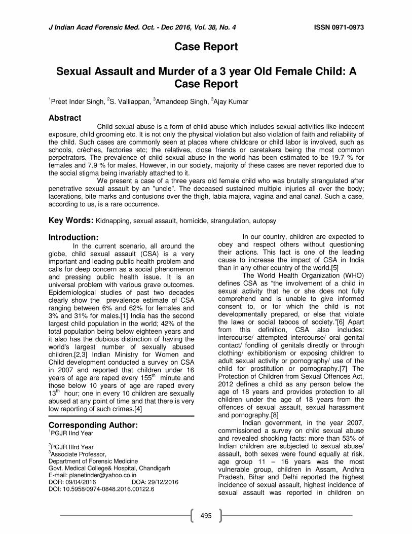
J Indian Acad Forensic Med. Oct. - Dec 2016, Vol. 38, No. 4 ISSN 0971-0973
495
Case Report
Sexual Assault and Murder of a 3 year Old Female Child: A Case Report
1Preet Inder Singh,
2S. Valliappan,
3Amandeep Singh,
3Ajay Kumar
Abstract Child sexual abuse is a form of child abuse which includes sexual activities like indecent
exposure, child grooming etc. It is not only the physical violation but also violation of faith and reliability of the child. Such cases are commonly seen at places where childcare or child labor is involved, such as schools, crèches, factories etc; the relatives, close friends or caretakers being the most common perpetrators. The prevalence of child sexual abuse in the world has been estimated to be 19.7 % for females and 7.9 % for males. However, in our society, majority of these cases are never reported due to the social stigma being invariably attached to it.
We present a case of a three years old female child who was brutally strangulated after penetrative sexual assault by an "uncle". The deceased sustained multiple injuries all over the body; lacerations, bite marks and contusions over the thigh, labia majora, vagina and anal canal. Such a case, according to us, is a rare occurrence.
Key Words: Kidnapping, sexual assault, homicide, strangulation, autopsy
Introduction: In the current scenario, all around the
globe, child sexual assault (CSA) is a very important and leading public health problem and calls for deep concern as a social phenomenon and pressing public health issue. It is an universal problem with various grave outcomes. Epidemiological studies of past two decades clearly show the prevalence estimate of CSA ranging between 6% and 62% for females and 3% and 31% for males.[1] India has the second largest child population in the world; 42% of the total population being below eighteen years and it also has the dubious distinction of having the world's largest number of sexually abused children.[2,3] Indian Ministry for Women and Child development conducted a survey on CSA in 2007 and reported that children under 16 years of age are raped every 155
th minute and
those below 10 years of age are raped every 13
th hour; one in every 10 children are sexually
abused at any point of time and that there is very low reporting of such crimes.[4]
Corresponding Author: 1PGJR IInd Year
2PGJR IIIrd Year
3Associate Professor,
Department of Forensic Medicine Govt. Medical College& Hospital, Chandigarh E-mail: [email protected]
DOR: 09/04/2016 DOA: 29/12/2016 DOI: 10.5958/0974-0848.2016.00122.6
In our country, children are expected to obey and respect others without questioning their actions. This fact is one of the leading cause to increase the impact of CSA in India than in any other country of the world.[5] The World Health Organization (WHO) defines CSA as “the involvement of a child in sexual activity that he or she does not fully comprehend and is unable to give informed consent to, or for which the child is not developmentally prepared, or else that violate the laws or social taboos of society.”[6] Apart from this definition, CSA also includes: intercourse/ attempted intercourse/ oral genital contact/ fondling of genitals directly or through clothing/ exhibitionism or exposing children to adult sexual activity or pornography/ use of the child for prostitution or pornography.[7] The Protection of Children from Sexual Offences Act, 2012 defines a child as any person below the age of 18 years and provides protection to all children under the age of 18 years from the offences of sexual assault, sexual harassment and pornography.[8] Indian government, in the year 2007, commissioned a survey on child sexual abuse and revealed shocking facts: more than 53% of Indian children are subjected to sexual abuse/ assault, both sexes were found equally at risk, age group 11 – 16 years was the most vulnerable group, children in Assam, Andhra Pradesh, Bihar and Delhi reported the highest incidence of sexual assault, highest incidence of sexual assault was reported in children on
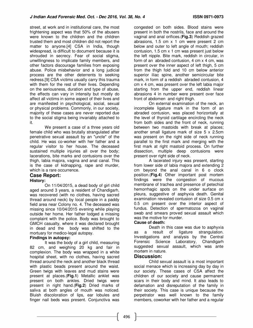
J Indian Acad Forensic Med. Oct. - Dec 2016, Vol. 38, No. 4 ISSN 0971-0973
496
street, at work and in institutional care, the most frightening aspect was that 50% of the abusers were known to the children and the children trusted them and most children did not report the matter to anyone.[4] CSA in India, though widespread, is difficult to document because it is shrouded in secrecy. Fear of social stigma, unwillingness to implicate family members, and other factors discourage families from exposing abuse. Police misbehavior and a long judicial process are the other deterrents to seeking redress.[9] CSA victims usually carry this trauma with them for the rest of their lives. Depending on the seriousness, duration and type of abuse, the effects can vary in intensity but mostly do affect all victims in some aspect of their lives and are manifested in psychological, social, sexual or physical problems. Commonly, in our society, majority of these cases are never reported due to the social stigma being invariably attached to it. We present a case of a three years old female child who was brutally strangulated after penetrative sexual assault by an "uncle" of the child. He was co-worker with her father and a regular visitor to her house. The deceased sustained multiple injuries all over the body; lacerations, bite marks and contusions over the thigh, labia majora, vagina and anal canal. This is the case of kidnapping, rape and murder, which is a rare occurrence.
Case Report: History: On 11/04/2015, a dead body of girl child aged around 3 years, a resident of Chandigarh, was recovered (with no clothes, having sacred thread around neck) by local people in a paddy field area near Colony no. 4. The deceased was missing since 10/04/2015 evening while playing outside her home. Her father lodged a missing complaint with the police. Body was brought to GMCH casualty, where it was declared brought in dead and the body was shifted to the mortuary for medico-legal autopsy. Findings in autopsy: It was the body of a girl child, measuring 82 cm, and weighing 20 kg and fair in complexion. The body was wrapped in a white hospital sheet, with no clothes, having sacred thread around the neck and another black thread with plastic beads present around the waist. Green twigs with leaves and mud stains were present at places.(Fig.1) Metallic anklet was present on both ankles. Dried twigs were present in right hand.(Fig.2) Dried marks of saliva at both angles of mouth was noticed. Bluish discoloration of lips, ear lobules and finger nail beds was present. Conjunctiva was
congested on both sides. Blood stains were present in both the nostrils, face and around the vaginal and anal orifices.(Fig.3) Reddish grazed abrasions, 1.5 cm x 1 cm were present 2 cm below and outer to left angle of mouth; reddish contusion, 1.5 cm x 1 cm was present just below the left nipple. Bite mark, reddish in circular, in form of an abraded contusion, 4 cm x 4 cm, was present over the inner aspect of left thigh, 5 cm from the thigh fold and 10 cm below anterior superior iliac spine, another semicircular bite mark, in form of a reddish abraded contusion, 4 cm x 4 cm, was present over the left labia major starting from the upper end, reddish linear abrasions 4 in number were present over face front of abdomen and right thigh. On external examination of the neck, an incomplete ligature mark in the form of an abraded contusion, was placed horizontally at the level of thyroid cartilage encircling the neck from both sides and the front of neck, running between two mastoids with break at places; another small ligature mark of size 5 x 2.5cm was present on the right side of neck running parallel to the first mark and merging with the first mark at right mastoid process. On further dissection, multiple deep contusions were present over right side of neck. A lacerated injury was present, starting from lower side of labia majora and extending 2 cm beyond the anal canal in 6 o clock position.(Fig.4) Other important post mortem findings were the congestion of mucous membrane of trachea and presence of petechial hemorrhagic spots on the under surface on pleura, suggestive of asphyxia death. Genital examination revealed contusion of size 0.5 cm x 0.5 cm present over the interior aspect of fundus. Detection of spermatozoa on vaginal swab and smears proved sexual assault which was the motive for murder. Cause of death: Death in this case was due to asphyxia as a result of ligature strangulation. Investigations and analysis by the Central Forensic Science Laboratory, Chandigarh suggested sexual assault, which was ante mortem in nature.
Discussion: Child sexual assault is a most important social menace which is increasing day by day in our society. These cases of CSA affect the children of our society and cause permanent scars in their body and mind. It also leads to defamation and disreputation of the family in their society. This case is unique because the perpetrator was well known to the family members, coworker with her father and a regular
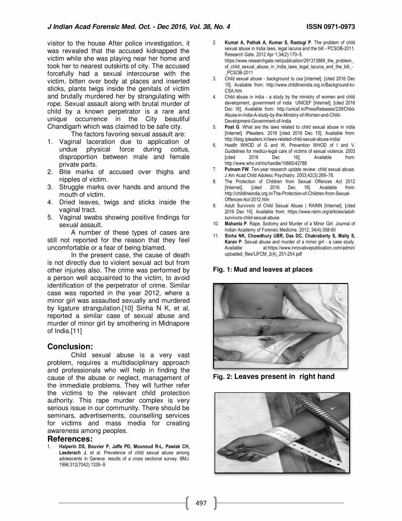
J Indian Acad Forensic Med. Oct. - Dec 2016, Vol. 38, No. 4 ISSN 0971-0973
497
visitor to the house After police investigation, it was revealed that the accused kidnapped the victim while she was playing near her home and took her to nearest outskirts of city. The accused forcefully had a sexual intercourse with the victim, bitten over body at places and inserted sticks, plants twigs inside the genitals of victim and brutally murdered her by strangulating with rope. Sexual assault along with brutal murder of child by a known perpetrator is a rare and unique occurrence in the City beautiful Chandigarh which was claimed to be safe city. The factors favoring sexual assault are: 1. Vaginal laceration due to application of
undue physical force during coitus, disproportion between male and female private parts.
2. Bite marks of accused over thighs and nipples of victim.
3. Struggle marks over hands and around the mouth of victim.
4. Dried leaves, twigs and sticks inside the vaginal tract.
5. Vaginal swabs showing positive findings for sexual assault.
A number of these types of cases are still not reported for the reason that they feel uncomfortable or a fear of being blamed. In the present case, the cause of death is not directly due to violent sexual act but from other injuries also. The crime was performed by a person well acquainted to the victim, to avoid identification of the perpetrator of crime. Similar case was reported in the year 2012, where a minor girl was assaulted sexually and murdered by ligature strangulation.[10] Sinha N K, et al, reported a similar case of sexual abuse and murder of minor girl by smothering in Midnapore of India.[11]
Conclusion: Child sexual abuse is a very vast problem, requires a multidisciplinary approach and professionals who will help in finding the cause of the abuse or neglect, management of the immediate problems. They will further refer the victims to the relevant child protection authority. This rape murder complex is very serious issue in our community. There should be seminars, advertisements, counselling services for victims and mass media for creating awareness among peoples.
References: 1. Halperin DS, Bouvier P, Jaffe PD, Mounoud R-L, Pawlak CH,
Laederach J, et al. Prevalence of child sexual abuse among adolescents in Geneva: results of a cross sectional survey. BMJ. 1996;312(7042):1326–9.
2. Kumar A, Pathak A, Kumar S, Rastogi P. The problem of child sexual abuse in India laws, legal lacuna and the bill - PCSOB-2011. Research Gate. 2012 Apr 1;34(2):170–5.
https://www.researchgate.net/publication/291313869_the_problem_of_child_sexual_abuse_in_India_laws_legal_lacuna_and_the_bill_-_PCSOB-2011
3. Child sexual abuse - background to csa [internet]. [cited 2016 Dec
15]. Available from: http://www.childlineindia.org.in/Background-to-CSA.htm
4. Child abuse in india - a study by the ministry of women and child development, government of india UNICEF [Internet]. [cited 2016
Dec 16]. Available from: http://unicef.in/PressReleases/239/Child-Abuse-in-India-A-study-by-the-Ministry-of-Women-and-Child-Development-Government-of-India
5. Post G. What are the laws related to child sexual abuse in india
[Internet]. iPleaders. 2016 [cited 2016 Dec 15]. Available from: http://blog.ipleaders.in/laws-related-child-sexual-abuse-india/
6. Health WHOD of G and W, Prevention WHOD of I and V. Guidelines for medico-legal care of victims of sexual violence. 2003
[cited 2016 Dec 16]; Available from: http://www.who.int/iris/handle/10665/42788
7. Putnam FW. Ten-year research update review: child sexual abuse. J Am Acad Child Adolesc Psychiatry. 2003;42(3):269–78.
8. The Protection of Children from Sexual Offences Act 2012 [Internet]. [cited 2016 Dec 16]. Available from: http://childlineindia.org.in/The-Protection-of-Children-from-Sexual-Offences-Act-2012.htm
9. Adult Survivors of Child Sexual Abuse | RAINN [Internet]. [cited 2016 Dec 15]. Available from: https://www.rainn.org/articles/adult-survivors-child-sexual-abuse
10. Mahanta P. Rape, Sodomy and Murder of a Minor Girl. Journal of
Indian Academy of Forensic Medicine. 2012; 34(4):358-60 11. Sinha NK, Chowdhury UBR, Das DC, Chakraborty S, Maity S,
Karan P. Sexual abuse and murder of a minor girl - a case study. Available at:https://www.innovativepublication.com/admin/
uploaded_files/IJFCM_2(4)_251-254.pdf
Fig. 1: Mud and leaves at places
Fig. 2: Leaves present in right hand
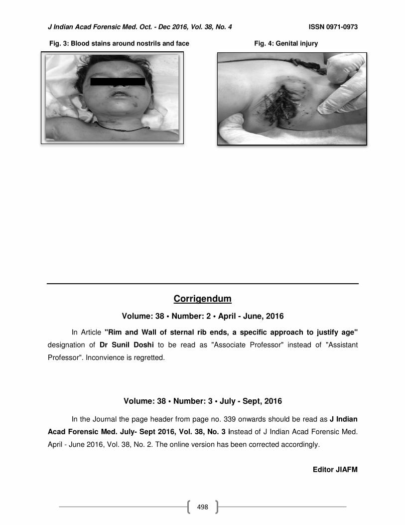
J Indian Acad Forensic Med. Oct. - Dec 2016, Vol. 38, No. 4 ISSN 0971-0973
498
Fig. 3: Blood stains around nostrils and face Fig. 4: Genital injury
Corrigendum
Volume: 38 • Number: 2 • April - June, 2016
In Article "Rim and Wall of sternal rib ends, a specific approach to justify age"
designation of Dr Sunil Doshi to be read as "Associate Professor" instead of "Assistant
Professor". Inconvience is regretted.
Volume: 38 • Number: 3 • July - Sept, 2016
In the Journal the page header from page no. 339 onwards should be read as J Indian
Acad Forensic Med. July- Sept 2016, Vol. 38, No. 3 instead of J Indian Acad Forensic Med.
April - June 2016, Vol. 38, No. 2. The online version has been corrected accordingly.
Editor JIAFM
J Indian Acad Forensic Med. Oct. - Dec 2016, Vol. 38, No. 4 ISSN 0971-0973
499
Case Report
Revising the current MTP Act: Hasn’t the Time Come Yet?? 1Jyoti Barwa,
2Amandeep Singh,
2Ajay Kumar,
3Dasari Harish
Abstract The Medical Termination of Pregnancy (MTP) Act puts a ceiling of 20 weeks for termination of pregnancy, except in cases which are immediately necessary to save the life of the pregnant woman. However, in some special cases, judgments have been made in favour of termination even beyond this specified time limit. Unwanted pregnancy, especially if occurring as an outcome of rape, is considered as a social stigma, all over the world but the physical trauma and mental agony of the woman in continuing that pregnancy is often overlooked. Relief from such a burden is possible to some extent if termination of pregnancy is performed in all those cases where continuation is not desired or intended by the pregnant woman. We report a case of an unmarried 19 yrs old pregnant girl, who presented to the court in advanced stage with a plea for termination of pregnancy; a board of doctors was constituted to opine for the same. The question as to whether such upper limit or ceiling is truly required or does it need to be extended, along with the requisite amendment in the current MTP act is being addressed.
Key Words: Termination, Pregnancy, Judgement, Amendment
Introduction: The Medical Termination of Pregnancy (MTP) Act, 1971 was enacted to save the pregnant women’s health, strength and sometimes life along with an effort to regulate criminal abortion. Hence, as per the Act, there is a ceiling of 20 weeks for termination of pregnancy, except in cases which are immediately necessary to save the life of the pregnant woman (S 5 of MTP act)[1]
In the Indian scenario, abortion has always been a burning issue, which is often associated with sex selection and female foeticide. Strict implementation of the MTP Act, therefore, becomes the need of the hour; but should it necessarily be done at the cost of subjecting the pregnant woman to mental agony and pain?? Forcing a woman to go through an unwanted pregnancy is definitely a violation of her right to dignity, and sexual and reproductive freedom, as guaranteed by the Constitution.
Corresponding Author: 1Assistant Professor
2 Associate Professor
3Professor & Head
Department of Forensic Medicine, Govt. Medical College & Hospital, Chandigarh E-mail: [email protected] DOR: 22/01/2017 DOA: 25/01/2017 DOI: 10.5958/0974-0848.2016.00123.8
The anguish caused by pregnancy occurring as an outcome of rape is presumed to constitute grave injury to the mental health of the pregnant woman.[1] Most of the congenital cardiac abnormalities/ malformations are detected usually after 18 weeks and since only 5-10 per cent of these foetuses are found to be chromosomically abnormal, foreseabilty of such cardiac anomalies is not feasible.[2,3] Hence, even if found at a much later stage, it mandates immediate termination as continuation of such pregnancy would result in the baby being born with severe disabilities and the baby along with parents will have to face continuous ordeal in their life thereafter.
Case report: An unmarried 19 yrs old girl along with
her distressed mother approached the district court in Chandigarh in September 2016 with a plea for termination of pregnancy. The girl had been repeatedly sexually abused by the accused, who being the neighbour - was in fact one of her acquaintances, for the past two years. He had threatened to use some of her objectionable pictures taken by him, which was the reason that she never raised her voice against him and would have probably never disclosed the same to anyone had this particular incident not resulted in pregnancy. As she had presented in an advanced stage of pregnancy; on court orders on 07-09-16, a Board of doctors comprising of members from the department of Obstetrics, Forensic Medicine and Psychiatry, was constituted to estimate the period of
J Indian Acad Forensic Med. Oct. - Dec 2016, Vol. 38, No. 4 ISSN 0971-0973
500
gestation and to opine regarding the feasibility of termination at this stage.
After complete physical, mental and sonographic evaluation of the pregnant girl, the Board opined the period of gestation of foetus to be 23 weeks 3 days and that the ongoing pregnancy was likely to affect the mental health of the girl adversely. The Board also clarified that as far as the risk of life of the pregnant girl and injury to the physical and mental health is concerned, the effect of termination of pregnancy is the same at 20 or 24 weeks. Following this, after an interval of 5 days, i.e on 13-09-16, another court order was passed by the ASJ, Chandigarh, as a revision against the previous order passed on 09-09-16 by the JMIC, Chandigarh, who had denied permission for termination of this unwanted pregnancy. The fresh order, thus directed the Board to terminate the pregnancy as soon as possible and to preserve DNA samples of the same.
In compliance to the aforementioned order, prompt admission of the girl was done in the Gynaecology ward but since, haemoglobin level was low and she was also found to be suffering from urinary tract infection, multiple blood transfusions and a course of antibiotics was initiated. It took another 2 days to stabilize her condition, following which labour was induced and she progressed fairly well. Finally, on 17-09-2016, the foetus was expelled out. However, to the surprise of everyone, the foetus which was to be aborted, cried out aloud to prove its existence.[Fig 1] This female baby, weighing about 740 grams, was immediately handed over to the neonatologist and shifted to NICU for due care. The baby unfortunately could not see the light of the day and expired due to extreme prematurity after 6 hrs.
The blood samples of mother and baby (preserved in plain and EDTA vials) along with the whole baby itself were sealed and sent to CFSL for DNA analysis after ensuring appropriate arrangements of temperature requirement for transport of these samples.
Discussion: The MTP Act was initially enacted in the
year 1971 to save the pregnant women’s health, strength and sometimes, life. Back then, equipments for diagnosis and medical facilities were not efficient to determine the health status of the unborn child or mother. Thereby, a ceiling of 20 weeks was established, beyond which termination of pregnancy was deemed likely to affect the health of pregnant woman adversely. But now, the infrastructure, facilities and advancements in the field of medicine have prospered considerably high; barring the few
risks involved, if facilities are available, it is possible to manage foetal and obstetrical emergencies effectively at any stage of pregnancy.[4-6]
Recently, there has been a surge in the cases seeking termination of pregnancy beyond this specified time limit. Though, most have been turned down at the district court level itself, a few of them managed to obtain permission from the apex court after an appeal as an exception under section 5 of the MTP act in which abortion is allowed if it “is immediately necessary to save the life of the pregnant woman”.
These series of cases began in the year 2008, when a couple petitioned Bombay High Court to allow them to abort their 26-week-old foetus, diagnosed with a congenital heart defect which was revealed in the USG done at 24 weeks. The couples’ plea was turned down on expert advice, which in fact acted as a spark and fuelled the process of this dire need to re-evaluate provisions of the Medical Termination of Pregnancy Act, 1971.[7] The pregnant lady, incidentally, had a miscarriage soon after the verdict.[8]
In July 2015, a 14-year-old rape victim from Gujarat sought and received permission from the Supreme Court after the Gujarat High court had turned down her plea to abort after the 20 weeks deadline had passed. This particular petition was treated as a “special case” and was not to be considered as precedence in any other case.[9] Similarly, in the present case permission was granted for termination of pregnancy irrespective of the foetus being sound and healthy. However, in July 2016 and recently in January 2017, women were allowed to abort their foetus in 24th week of gestation period only after the Supreme Court observed that the baby suffered from anencephaly, where life is not sustainable after birth.[10,11]
In a unique case, ironically permission was granted initially by the High Court of Punjab and Haryana wherein a mentally retarded woman had become pregnant as a result of an alleged rape that took place while she was an inmate at a government-run welfare institution located in Chandigarh. This order was later challenged in the SC by some social activists on account of lack of consent of the pregnant woman for the same, resulting in subsequent quashing of the previous order.[12]
In May 2016, a gynecologist in Pune was penalized for performing MTP on 21 weeks and 4 days pregnant woman. Though there was no threat to the life of the patient, the doctor pointed that foetus had cardiac and neural defects which were picked up late by USG.[13]
J Indian Acad Forensic Med. Oct. - Dec 2016, Vol. 38, No. 4 ISSN 0971-0973
501
The question here arises, why termination of pregnancy beyond a specified limit is considered as a violation of law, even if done in good faith, in order to ensure better quality of life to both mother and the child? In our society, the apprehension and anxiety regarding the consequences of unwanted pregnancy especially in an unmarried state is such that woman would be compelled to take extreme steps.
As a matter of fact, if the parents are aware that unborn child is likely to be born with features incompatible with life or with severe disabilities then it should completely be their decision to opt for its outcome. Either of the choices i.e termination or continuation of pregnancy, would anyway result in a traumatizing experience, especially for the pregnant woman. Even certain studies reveal that as opposed to the chromosomal defects detected earlier, some cardiac and neural anomalies are only diagnosed after a normal 20 -24 weeks scan; thus these foetuses having such anomaly needs to be aborted to prevent unnecessary burden on parents and society later on.[2,14,15]
Various speculations regarding the age of separate existence of the foetus from its mother are still being carried out. Studies reveal that survival of foetus below 23 weeks of gestation and that below 500 gm is most unlikely. Only those more than or equal to 25 weeks' gestation and with a birth weight of more than or equal to 600 gms are mature enough to warrant initiation of intensive care; more than 90% of them born at 26 to 27 weeks, survive without severe long-term disabilities.[16-18] The limit of viability of foetus varies considerably worldwide, ranging from 24 – 28 weeks.[19,20] In India, this period of viability is 28 weeks of gestation, hence ceiling of 20 weeks for termination of pregnancy appears to be inconsistent with the existing scenario.[20] However, unregulated extension to the upper limit would inadvertently invite another issue requiring further redressal, as then the odds against the foetus being born alive would become high; raising a question on the legal status and provision of facilities of childcare for this unfortunate child who survives the procedure of abortion especially in cases of unwanted pregnancy.
Conclusion & Suggestions: Looking into the various prospects of the
current MTP Act, there is an imperative need to incorporate amendments in terms of excluding the time limits all together where doctors have detected substantial foetal abnormalities or if
termination is to be carried out on humanitarian grounds. Also, the upper limit of termination need to be raised considering that congenital anomalies particularly cardiac and nueral defects are detected at a much later stage; provision of a window period of about two weeks for the couples should be made admissible in order to formulate any decision regarding termination/ continuation of pregnancy thereafter. It is also important that in cases where pregnancy is outcome of rape, due consideration should be given to the petitioner irrespective of the period of gestation. These measures, when implemented, will not only regulate unsafe abortion and safeguard health of the pregnant woman but also empower her with the right to make reproductive choices and live with dignity.
References: 1. MTP Act, 1971. [online]. [cited 2016 Dec 28] Available from:
http://www.mohfw.nic.in/index1.php?lang=1&level=4&sublinkid=3615&lid=2598
2. Todros T, Faggiano F, Chiappa E, Gaglioti P, Mitola B, Sciarrone A. Accuracy of routine ultrasonography in screening heart disease prenatally. Gruppo Piemontese for Prenatal Screening of Congenital Heart Disease. Prenat
Diagn. 1997;17(10):901-6. 3. Todros T, Capuzzo E, Gaglioti P. Prenatal diagnosis of congenital
anomalies. Images Paediatr Cardiol. 2001;3(2):3-18. 4. Sim WH, Araujo JE, Da Silva Costa F, Sheehan PM. Maternal
and neonatal outcomes following expectant management of preterm prelabour rupture of membranes before viability. J Perinat Med. 20171;45(1):29-44.
5. Chavan R, Latoo MY. Recent advances in the management of
major obstetric haemorrhage. BJMP 2013;6(1):a604 6. Pacheco LD, Saade GR, Gei AF, Hankins GD. Cutting-edge
advances in the medical management of obstetrical hemorrhage. American Journal of Obstetrics & Gynecology. December
2011;32(4):526-32. 7. High Court rejects Niketa’s plea to terminate pregnancy. [online].
2008 Aug 5. [cited 2016 Dec 26]. Available from: http://www.thehindu.com/todays-paper/High-Court-rejects-
Niketarsquos-plea-to-terminate-pregnancy/article15273010.ece 8. Mumbai abortion case: Niketa Mehta suffers miscarriage. [online].
2008 Aug 14. [cited 2016 Dec 20]. Available from: http://timesofindia.indiatimes.com/city/mumbai/Mumbai-abortion-
case-Niketa-Mehta-suffers-miscarriage/articleshow/3363293.cms 9. Supreme Court: A 24-week pregnant teen can abort if
doctors permit. [online] 2015 Jul 30. [cited 2016 Dec 31]. Available from: http://indianexpress.com/article/india/india-others/sc-quashes-
hc-order-allows-minor-rape-victim-to-abort-foetus-after-medical-permissions/
10. Supreme Court: A 24-week pregnant teen can abort if doctors permit. [online] 2016 Jul 25. [cited 2016 Dec 31]. Available
from: http://timesofindia.indiatimes.com/india/Abnormal-fetus-Supreme-Court-permits-woman-to-terminate-24-week-old-pregnancy/articleshow/53378678.cms
11. SC allows 24-week oregnant wman to abort foetus with
undeveloped skull. [online]. 2017 Jan 17. [cited 2017 Jan 22]. Available from:http://indianexpress.com/article/india/sc-allows-24-week-pregnant-woman-to-abort-foetus-with-undeveloped-skull/
12. Suchita Srivastava & Anr vs Chandigarh Administration. Bench:
K.G. Balakrishnan, P. Sathasivam, B.S. Chauhan. Available at: http://indiankanoon.org/doc/1500783/. [cited: 2017 Jan 22]
13. Gynaecologist booked for illegal abortion. [online]. 2016 May 5. [cited 2017 Jan 2]. Available from:
http://timesofindia.indiatimes.com/city/pune/Gynaecologist-booked-for-illegal-abortion/articleshow/52116339.cms
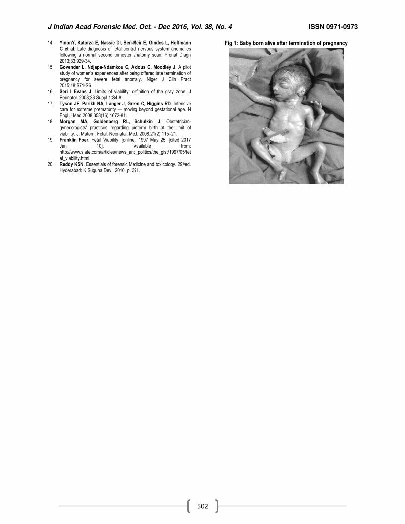
J Indian Acad Forensic Med. Oct. - Dec 2016, Vol. 38, No. 4 ISSN 0971-0973
502
14. YinonY, Katorza E, Nassie DI, Ben-Meir E, Gindes L, Hoffmann C et al. Late diagnosis of fetal central nervous system anomalies following a normal second trimester anatomy scan. Prenat Diagn
2013;33:929-34. 15. Govender L, Ndjapa-Ndamkou C, Aldous C, Moodley J. A pilot
study of women's experiences after being offered late termination of pregnancy for severe fetal anomaly. Niger J Clin Pract
2015;18:S71-S6. 16. Seri I, Evans J. Limits of viability: definition of the gray zone. J
Perinatol. 2008;28 Suppl 1:S4-8. 17. Tyson JE, Parikh NA, Langer J, Green C, Higgins RD. Intensive
care for extreme prematurity — moving beyond gestational age. N Engl J Med 2008;358(16):1672-81.
18. Morgan MA, Goldenberg RL, Schulkin J. Obstetrician-gynecologists' practices regarding preterm birth at the limit of
viability. J. Matern. Fetal. Neonatal. Med. 2008;21(2):115–21. 19. Franklin Foer. Fetal Viability. [online]. 1997 May 25. [cited 2017
Jan 10]. Available from: http://www.slate.com/articles/news_and_politics/the_gist/1997/05/fet
al_viability.html. 20. Reddy KSN. Essentials of forensic Medicine and toxicology. 29thed.
Hyderabad: K Suguna Devi; 2010. p. 391.
Fig 1: Baby born alive after termination of pregnancy
J Indian Acad Forensic Med. Oct. - Dec 2016, Vol. 38, No. 4 ISSN 0971-0973
503
Case Report
Fatal Paint Thinner Ingestion- A Case Report 1Jitendra Singh Tomar,
2Pradeep Kr Mishra,
3Mandar Ramchandra Sane,
4Divyesh Saxena,
4Manish
Kumar
Abstract Household poisons can cause serious damage to the health of children. Turpentine is a volatile hydrocarbon used in polishes, solvents, paints and textile industry. It is capable of causing serious toxicity, whether ingested or inhaled. When hydrocarbons are aspirated into the lung, they cause chemical pneumonitis, acute respiratory distress syndrome (ARDS), and rarely pneumatoceles and pneumothorax. We report a case of accidental ingestion of turpentine oil by a 13 year old female child brought to SAMC and PGI, Indore, leading to aspiration pneumonitis and ultimately causing death after 6 days of hospitalization.
Key Words: Turpentine oil, Aspiration potential, Aspiration pneumonitis, Consolidations
Introduction: Turpentine oil is a distillate of pine gum.
Along with pine oil, it belongs to a class of hydrocarbons called cyclic turpenes or turpene derivatives. It is used in shoe polishes, waxes, paints, varnishes, and insect repellents. Largely replaced in the West with white spirit and turpentine substitute, it is still widely used in the developing world. Kerosene is, by far, the most common hydrocarbon responsible for poisoning in children in studies published from developing countries.[1,2]
Case History On 14/5/2014 , a 13 yr old female child
was brought to casualty with the alleged h/o accidental paint thinner ingestion 2 days back in the afternoon when she woke up and took few sips of a liquid kept in the refrigerator, which later on turned out to be turpentine oil.
Corresponding Author: 1Assistant Professor,
FMT, Index Medical College & Research centre, Indore, M.P
4 Assistant Professor,
FMT , Sukhsagar Medical College, Jabalpur 2Professor,
3Assisstant Professor,
5Research Assisstant,
Department of Forensic Medicine & Toxicology, Sri Aurobindo Medical College & P G Institute, Indore. Email: drjitendersinghtomar @gmail.com DOR: 14/08/2016 DOA: 31/12/2016 DOI: 0.5958/0974-0848.2016.00124.X
After 2 hours, she experienced acute spasmodic abdominal pain- non radiating & generalized. She developed difficulty in breathing within 6 hours of ingestion. Chief complaints at the time of admission were 2-3 episodes of vomiting, pain abdomen and fast breathing. She was taken to a local private hospital where she was put on ventilator & after two days, referred to SAMC & PGI with BP-130/90 mm of Hg, HR- 110/min, RR- 66/min, SPO2- on ventilator- 90%, Pallor
+/jaundice
+/
cyanosis-/ edema/ lymphadenopathy. Endo-
tracheal Tube was in situ and Ryle's Tube was also in situ. Bilateral ronchi with crepts were heard and air entry was decreased (R>>L). She was admitted in view of chemical pneumonitis which she developed due to aspiration and was treated for the same. During the course of hospitalization patient was intubated and was put on ventillator. Continuously she was having fever which was not associated with chills or rigor. She had spasmodic abdominal pain throughout. She was treated with I/V fluids, antibiotics, relaxants and sedatives. Despite of all the treatment patients condition worsened and she expired on 17/5/14 at 8:30pm.The deceased was shifted to mortuary for autopsy. Investigations-
• CBC, S. Creatinine & S. Electrolytes were within normal limits.
• LFT - Bilirubin (T/D/I)- 5.76/1.79/3.97 mg/dL and SGOT/SGPT- 35/99 IU/L
• Total protein/ Albumin/ Globulin – 5.37 / 2.62 / 2.75gm/dL, INR – 1.9 IU
• Urine R/M - Alb. ++,RBC – 20-25/hpf, Pus cells – 10-12/hpf and Epithelial Cells – 10-12/hpf
J Indian Acad Forensic Med. Oct. - Dec 2016, Vol. 38, No. 4 ISSN 0971-0973
504
• RT aspirate was sent for toxicological analysis.
Test for turpentine- 2 ml. of ether extract is evaporated to dryness on a spotting tile. 1 drop of conc. sulphuric acid is added. A deep reddish brown color is produced which confirms the presence of Turpentine. (Picture 1 and 2)
Autopsy findings Average built female child aged about 13 yrs. Greenish color fluid coming out of both nostrils. Rigor mortis was present all over the body. Postmortem lividity was present over back and fixed. On internal examination, trachea contained little amount of greenish fluid material, similar to gastric content (picture 5). Mucosa was congested. Right lung was collapsed with pus present at the base (picture 3). Pus was present on the base of left lung (picture 4). Consolidations with signs of pneumonitis were present in both the lungs. Stomach contained about 50 cc of dark greenish fluid; mucosa was congested with patchy hemorrhages at places (picture 6). Liver was enlarged and pale. Spleen and both the kidneys were congested. Opinion was given as death due to cardio-respiratory failure as a result of suspected poisoning and its complications and viscera was preserved. Lung and liver were sent for histopathological examination. Histopathology of both the lungs revealed lobar pneumonia and histopathology of liver showed signs of necrosis (picture 7 and 8).
Discussion Turpentine is a fluid obtained by the distillation of resin obtained from live pine trees.[3] There are two forms - Gum form (the pitch obtained from living pine trees; a sticky viscous liquid) and Oil form (a volatile liquid obtained by steam distillation of gum turpentine). Turpentine is composed of terpenes, mainly the monoterpenes alpha-pinene and beta-pinene with lesser amounts of carene, camphene, dipentene, and terpinolene. Since turpentine is a lipophilic substance, it accumulates in fatty tissues.[3] It is used in perfumes, sprays, insecticides, disinfectants, human and veterinary medicines, preparation of shoe and furniture polish, manufacturing of synthetic camphor and menthol, thinner for paint and varnishes and removing paint stains. Mechanism of Toxicity[3]- Turpentine is readily absorbed through the gastrointestinal and respiratory tracts and skin. Terpenes are oxidized by cytochrome P450, conjugated principally with glucuronic acid in the liver, and are excreted by the kidney.Liver microsomal
epoxide hydrase and uridine diphosphoglucuronosyl transferase activities were elevated during chronic turpentine exposures. Turpentine may be eliminated unchanged through the lungs. Most turpentine and its metabolites are eliminated through the urinary tract as glucuronides. The excretory product of turpentine has a characteristic odor of violet. Exposure routes are throughinhalational, ingestional, and dermal contact. Aspiration potential of a hydrocarbon[4]- It depends on 3 properties-viscosity, surface tension, and volatility. Viscosity is the tendency of a substance to resist flow (the ability to resist stirring) which is measured in Saybolt Seconds Universal (SSU). The lower the viscosity (i.e. below 60 SSU), the higher the tendency for aspiration.Surface tension refers to the adherence of a liquid compound along its surface (the ability to creep). It is the result of cohesive forces generated by the attraction between molecules (van der Waals forces). The lower the surface tension, the higher the tendency for aspiration. Volatility refers to the ability of a liquid to become a gas. The higher the volatility, the higher the tendency for aspiration.Ingestion of turpentine products produces a significantly lower incidence of pneumonitis compared with petroleum distillates.[5] Turpentine produces more GI and CNS symptoms than a similar ingestion of a petroleum distillate.[6] Acute and short-term toxicity[3,7]- Mostly ingestion of turpentine seen in children are accidental. Exposure to a 75 ppm -irritates the nose and throat, 175 ppm - irritates the eyes. The lowest estimated oral dose reported to be lethal in humans is 441 mg/ kg. Signs and symptoms - Nausea, vomiting, diarrhoea, abdominal pain, can also lead to haematemesis. Aspiration lead to coughing, choking and gasping. Bronchospasm may develop, resulting in mismatch of ventillation and perfusion resulting in hypoxia and CNS depression. Hemoptysis may also occur. Acute effects include coma and seizures. It may produce initial euphoria, agitation, hallucinations. Hepatic and renal damage may be seen leading to hematuria, albuminuria, oliguria, and dysuria and DIC can also be seen. Myocardial injury, cardiac arrhythmias and myoglobinuria can be seen. Parenteral injection causes hypoxemia and non cardiogenic pulmonary edema, cellulitis and a sterile abscess at the injection site.[8] Treatment[4,7,9]- Removal of unabsorbed poison from the system, administration of antidotes, elimination of
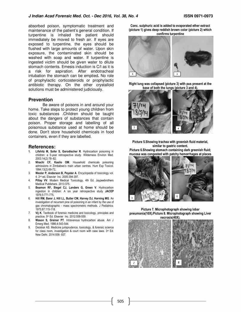
J Indian Acad Forensic Med. Oct. - Dec 2016, Vol. 38, No. 4 ISSN 0971-0973
505
absorbed poison, symptomatic treatment and maintenance of the patient’s general condition. If turpentine is inhaled the patient should immediately be moved to fresh air. If eyes are exposed to turpentine, the eyes should be flushed with large amounts of water. Upon skin exposure, the contaminated skin should be washed with soap and water. If turpentine is ingested victim should be given water to dilute stomach contents. Emesis induction is C/I as it is a risk for aspiration. After endotracheal intubation the stomach can be emptied. No role of prophylactic corticosteroids or prophylactic antibiotic therapy. On the other crystalloid solutions must be administered judiciously.
Prevention Be aware of poisons in and around your home. Take steps to protect young children from toxic substances .Children should be taught about the dangers of substances that contain poison. Proper storage and labelling of all poisonous substance used at home should be done. Don't store household chemicals in food containers, even if they are labelled.
References: 1. Lifshitz M, Sofer S, Gorodischer R. Hydrocarbon poisoning in
children: a 5-year retrospective study. Wilderness Environ Med. 2003;14(2):78–82.
2. Nhachi CF, Kasilo OM. Household chemicals poisoning admissions in Zimbabwe’s main urban centres. Hum Exp Toxicol. 1994;13(2):69-72.
3. Wexler P, Anderson B, Peyster A. Encyclopedia of toxicology vol.
4. 2nd ed. Elsevier Inc. 2005:394-397. 4. Pillay VV. Modern Medical Toxicology, 4th Ed. Jaypeebrothers
Medical Publishers. 2013:375. 5. Beamon RF, Siegel CJ, Landers G, Green V. Hydrocarbon
ingestion in children: A six year retrospective study JACEP 1976;5:771-775.
6. Hill RM, Barer J, Hill LL, Butler CM, Harvey DJ, Horning MG. An investigation of recurrent pine oil poisoning in an infant by the use of
gas chromatographic - mass spectrometric methods. J Pediatrics. 1975;87:115-118.
7. Vij K. Textbook of forensic medicine and toxicology, principles and practice. 5th Ed. Elsevier Inc. 2012:558-559.
8. Wason S, Greiner PT. Intravenous hydrocarbon abuse. Am J Emerg Med. 1986;4:543-544.
9. Deoskar AS. Medicine jurisprudence, toxicology, & forensic science for class room, investigation & court room with case laws. 3rd Ed.
New Delhi. 2014:936- 937.
Conc. sulphuric acid is added to evaporated ether extract (picture 1) gives deep reddish brown color (picture 2) which
confirms turpentine
Right lung was collapsed (picture 3) with pus present at the base of both the lungs (picture 3 and 4).
Picture 5.Showing trachea with greenish fluid material, similar to gastric content.
Picture 6.Showing stomach containing dark greenish fluid; mucosa was congested with patchy hemorrhages at places
Picture 7. Microphotograph showing lobar pneumonia(10X).Picture 8. Microphotograph showing Liver
necrosis(40X).
J Indian Acad Forensic Med. Oct. - Dec 2016, Vol. 38, No. 4 ISSN 0971-0973
506
Case Report
Breast Feeding Pulmonary Aspiration Death: A Case Report
1Bhullar DS,
2Aggarwal KK,
3Mehta N
Abstract Accidental aspiration of solid or liquid gastric contents into the respiratory passages is a common
universal phenomenon and a medical emergency, both in the children and in the adults. It can prove fatal
in the absence of timely intervention, leading to death due to asphyxia. One such case of death of a child
from asphyxia due to choking from pulmonary aspiration of stomach contents is being discussed with
stress on first aid training to all to tackle such easily treatable medical emergencies.
Key Words: Aspiration, Accidental, Breast feeding, Asphyxia
Introduction: Pulmonary aspiration is the entry of material (such as pharyngeal secretions, food or drink, or stomach contents) from the oropharynx or gastrointestinal tract into the larynx (voice box) and lower respiratory tract. A person may either inhale the material, or it may be delivered into the tracheo-bronchial tree during positive pressure ventilation. When pulmonary aspiration occurs during eating and drinking, the aspirated material is often colloquially referred to as "going down the wrong pipe." Consequences of pulmonary aspiration range from no injury at all, to chemical pneumonitis or pneumonia, to death within minutes from asphyxiation.[1] Death from asphyxiation from aspiration may be referred to as choking. Death from choking can be the result of pure hypoxia from occlusion of the airway, when all the attendant signs of congestion, cyanosis and perhaps petechiae may be present, usually where the victim struggles to breathe for an appreciable period. However, a large proportion of deaths occur suddenly before any possible hypoxic manifestations have time to take effect; these fatalities must be caused by neurogenic cardiac arrest, either purely neurogenic or accelerated by excess catecholamine release from the adrenaline response. [2]
Corresponding Author: 1Associate Professor (D),
2Prof. & Head,
3Junior Resident,
Department of Forensic Medicine & Toxicology, Government Medical College Patiala E-mail: [email protected] DOR: 16/07/2016 DOA: 31/12/2016 DOI: 10.5958/0974-0848.2016.00125.1
Case History: A male baby, aged two years, was referred from Central Jail Patiala along with the convict mother to Government Medical College & Rajindra Hospital, Patiala at 7-20 A.M. on 16.04.2016, in a gasping state and on reaching emergency ward of pediatrics ward of the hospital, it was pronounced dead at 7. 40 A.M. As per medical records of the jail, the child was suffering from asthmatic bronchitis. As per the statement of the Jail Medical Officer, the probable cause of death was aspiration of milk, as the mother was continuously feeding the child and did not inform anyone else of the illness. As per record, the mother of deceased child - a convict in the jail, was getting treatment from psychiatric department of Rajindra Hospital (Tablet Qutan 50 X 30 days).
Postmortem Examination:- On external examination, it was dead body of an average built male baby with rigor mortis not fully developed, and with postmortem staining fixed and present on the back and left side on the face, except parts of contact flattening. Bluish discoloration was present on lips, nails and ear lobule. Frothy secretions were present in the nostrils. On internal examination, organs were with generalized congestion, milky white frothy secretions were present in the mouth, pharynx & esophagus, larynx and trachea were full of milky white fluid and frothy secretions, stomach was full of milky white and greenish colored liquid and semi digested food material. Required viscera were preserved for chemical and histopathological examination.
Cause of Death:-
J Indian Acad Forensic Med. Oct. - Dec 2016, Vol. 38, No. 4 ISSN 0971-0973
507
The provisional cause of death was given as “Postmortem findings suggest death due to asphyxia resulting from choking from aspiration of gastric contents into the respiratory passage. However, final opinion will be given on receiving the reports of chemical and histopathological examination of viscera”. On receiving the reports of chemical and histopathological examination of viscera, the final opinion on cause of death was given as “From post-mortem findings & the reports of Chemcal and Histopathological examination of viscera, the cause of death in this case is asphyxia resulting from choking from aspiration of gastric contents into the respiratory passage, sufficient to cause death in the ordinary course of nature.
Discussion:- The case under study is a typical example of death due to accidental choking from aspiration of gastric contents wherein the mother with perhaps unsound mind went on feeding the baby without taking note of the diseased lung condition of her son and the extent to which it could be given the feed. Choking is almost exclusively accidental in nature. Homicidal choking is argumentatively possible with infant victims. In mental unsound persons suicidal choking can only be thought of.[3] Different causes of death in choking are asphyxia, laryngeal spasm, vagal inhibition and reflex neurogenic cardiovascular failure.[4] It is not necessary that a foreign body should be of such a size as to block the air passages completely. Even a small object blocking the lumen partially may cause death by laryngeal spasm.[5] In adults, choking virtually always involves food. Here it is commonly associated with acute alcoholic intoxication, bad fitting dentures, neurological injury or senility. Complications may develop after a latent interval, if the person survives.[6] About 3.6 million cases of pulmonary aspiration or foreign body in the airway occurred in 2013.[7] Aspiration can cause signs and symptoms in a baby such as:
• Weak sucking
• Choking or coughing while feeding • Other signs of feeding trouble, like a red
face, watery eyes, or facial grimaces
• Stopping breathing while feeding
• Faster breathing while feeding
• Voice or breathing that sounds wet after feeding
• Slight fever after feedings
• Wheezing and other breathing problems
• Repeated lung or airway infections.[8]
Usually, any object in the air passages can excite violent coughing out, but if this is not successful in expelling the object out, choking results.[9] Like in a case study by Dalal. D et al., where the person died within 24 hours of the onset of the terminal symptoms, and which could not be called as sudden death by the authors; the death in this case too cannot be labeled as sudden because the baby was referred from the jail hospital in a gasping state and declared dead on arrival.[10] The universal sign for choking is hands clutched to the throat. If the person doesn't give the signal, look for these indications:
• Inability to talk • Difficulty breathing or noisy breathing
• Inability to cough forcefully
• Skin, lips and nails turning blue or dusky
• Loss of consciousness If choking is occurring, the Red Cross recommends a "five-and-five" approach to delivering first aid. [11]
• Give 5 back blows. First, deliver five back blows between the person's shoulder blades with the heel of your hand.
• Give 5 abdominal thrusts. Perform five abdominal thrusts (also known as the Heimlich maneuver).
• Alternate between 5 blows and 5 thrusts until the blockage is dislodged.
The American Heart Association doesn't teach the back blow technique, only the abdominal thrust procedures. [11] It's OK not to use back blows, if you haven't learned the technique. Both approaches are acceptable. To clear the airway of an unconscious person:
• Lower the person on his or her back onto the floor.
• Clear the airway. If a blockage is visible at the back of the throat or high in the throat, reach a finger into the mouth and sweep out the cause of the blockage. Be careful not to push the food or object deeper into the airway, which can happen easily in young children.
• Begin cardiopulmonary resuscitation (CPR) if the object remains lodged and the person doesn't respond after you take the above measures. The chest compressions used in CPR may dislodge the object. Remember to recheck the mouth periodically.
To clear the airway of a choking infant younger than age 1:
J Indian Acad Forensic Med. Oct. - Dec 2016, Vol. 38, No. 4 ISSN 0971-0973
508
• Assume a seated position and hold the infant face down on your forearm, which is resting on your thigh.
• Thump the infant gently but firmly five times on the middle of the back using the heel of your hand. The combination of gravity and the back blows should release the blocking object.
• Hold the infant face up on your forearm with the head lower than the trunk if the above doesn't work. Using two fingers placed at the center of the infant's breastbone, give five quick chest compressions.
• Repeat the back blows and chest thrusts if breathing doesn't resume. Call for emergency medical help.
• Begin infant CPR if one of these techniques opens the airway but the infant doesn't resume breathing.
• If the child is older than age 1, give abdominal thrusts only.
• To prepare yourself for these situations, learn the Heimlich maneuver and CPR in a certified first-aid training course. [11]
Conclusion:- Choking of the respiratory passages from aspiration can be said to be a common medical emergency both in children as well as adults, requires medical intervention as early as possible and can be treated successfully with timely starting the emergency treatment. Apart
from medical professionals dealing with such cases that may not be available in time for more than one reason, it is all important for the general public to know how to tackle the situation by emergency first aid and all must have such first aid training to save valuable human life.
References: 1. https://en.wikipedia.org/wiki/Pulmonary aspiration. [June 2010] 2. Saukko P, Knight B. Knight’s Forensic Pathology, 3rd Ed. London:
Arnold 2004.p. 361.
3. Nandy A. Principles of Forensic Medicine & Toxicology, 3rd ed. New Central Book Agency (P) Ltd: 2010. P. 560.
4. Karmakar RN. Forensic Medicine and Toxicology, 4th Ed. Academic Publishers; 2013. P. 213.
5. Modi JP. A Textbook of Medical Jurisprudence and Toxicology, 24th Edition; Nagpur, India: Lexis Nexis Butterworths Wadhwa; 2012. P. 464
6. Reddy KSN. The Essentials of Forensic Medicine and Toxicology.
33rd Edition; India: Jaypee Brothers Medical Publishers (p) Ltd. ; 2014. P. 362.
7. Global Burden of Disease Study 2013, Collaborators (22 August 2015). "Global, regional, and national incidence, prevalence, and
years lived with disability for 301 acute and chronic diseases and injuries in 188 countries, 1990-2013: a systematic analysis for the Global Burden of Disease Study 2013." Lancet (London, England). 386 (9995):743–800.
8. Aspiration in Babies and Children: Saint Luke’s Health System; http://www.saintlukeshealthsystem.org/health-library/aspiration-babies-and-children.
9. Rao NG. Textbook of Forensic Medicine and Toxicology. 2nd
Edition. India. Jaypee Brothers Medical Publishers (P) Ltd. 2010. P. 210.
10. Dadal. D.; Day A.; Bhattacharya C.R.; Roy A.; Mazumder A. Choking with food bolus leading to violent asphyxia death in a
healthy young male – a case study; Journal of Punjab Academy of Forensic Medicine & Toxicology; Vol. 15. No. 02. P. 101.
11. http://www.mayoclinic.org/first-aid/first-aid-choking/basics/art-20056637: Choking: First Aid [Date of Sept. 19, 2014]
J Indian Acad Forensic Med. Oct. - Dec 2016, Vol. 38, No. 4 ISSN 0971-0973
509
Case Report
Bicuspid Aortic Valve: Cause for Sudden Cardiac Death - A Case Report
1Bhagavan.S,
2Punitha.R,
3Pradeep Kumar,
4Jagadeesh.N.H.
Abstract Sudden death in young adults is always a challenge for the autopsy surgeon. Most of the cases
of sudden death are related to the cardiovascular system. The other major contributions include infection, epilepsy, intracranial haemorrhage and asthma. Most often, unrecognised structural heart diseases such as hypertrophic cardiomyopathy, dilated cardiomyopathy, arrthymogenic right ventricular dysplasia and premature coronary artery disease are the causes for sudden cardiac death in the young. There are some other rare causes of sudden cardiac death which one has to rule out before arriving at the cause of death. Here we are presenting a rare case of sudden cardiac death following bicuspid aortic valve in a middle aged male.
Key Words: Sudden Death, Bicuspid Aortic Valve
Introduction: A bicuspid aortic valve (BAV) is most commonly a congenital condition of the aortic valve where two of the aortic valvular leaflets fuse during development, resulting in a valve that is bicuspid instead of the normal tricuspid configuration. The normal aortic valve has 3 equal-sized leaflets or cusps with 3 lines of coaptation. Bicuspid aortic valves are the most common cardiac valvular anomaly, occurring in 1–2% of the general population. It is twice as common in males as in females.[1] In adulthood, complications are common, and therefore, the burden of disease from BAV disease is more significant than any other congenital cardiac lesion.[2] Although inheritance is variable, it is a familial disease with an estimated 10% chance of a first degree relative being affected. Additionally, certain gene mutations (NOTCH1) have been associated.[3,4] Despite its importance, our understanding of BAV disease is incomplete and questions remain unanswered about this common condition.
Corresponding Author: 1Post Graduate Student,
3Prof and HOD,
2,4Assistant Professor.
Department of Forensic Medicine & Toxicology, Rajarajeswari Medical College and Hospital, Bangalore. Email:[email protected] DOR: 07/07/2016 DOA: 1/1/2017 DOI: 10.5958/0974-0848.2016.00126.3
Although much of the original focus centred on the abnormal bi-leaflet valve, the disease is significantly more complex. In many cases, a bicuspid aortic valve will cause no problems. However BAV may become calcified later in life, which may lead to varying degrees of severity of aortic stenosis.
Case Report: A 38years old male, a BMTC bus driver by occupation, fell unconscious over the steering wheel when he was about to start the vehicle. Immediately, he was rushed to the hospital where he was declared brought dead in the early hours of the day. On interrogation with the family members there was no relevant past medical or family history. The deceased was a known alcoholic and chronic smoker. On external examination, the body measured 167 cm in length and weighed 81 kg. The clothes present over the body were intact. Rigor mortis was present throughout the body. Lividity seen over back surface of the body and not fixed. No demonstrable external injuries noted over the body. On internal examination, heart weighed 500gm. A hard calcified lesion involving the Aortic valves, partially blocking the outflow was noted. Mild calcification of mitral valve was present. Pulmonary and Tricuspid valves were unremarkable. Atherosclerotic changes were present in the Left Anterior Descending artery (LAD). (Fig 1 & 2) Right lung was adherent to the chest wall, left lung was intact. All the other organs were intact and unremarkable. Organs were sent for Histopathological examination. Provisional
J Indian Acad Forensic Med. Oct. - Dec 2016, Vol. 38, No. 4 ISSN 0971-0973
510
opinion was given as Respiratory & Circulatory failure as a result of aortic stenosis. However, final opinion was reserved, pending HPE report. HPE revealed Bicuspid aortic valve with marked calcification. Left ventricular wall and left papillary muscle hypertrophy was present. Atherosclerosis and significant degree of luminal obstruction were seen in the right coronary artery, left main coronary artery and anterior descending artery. Atherosclerotic lesion was seen in the aorta. (Fig 3)
Discussion: Sir William Osler was one of the first to recognize the bicuspid aortic valve as a common congenital anomaly of the heart.[5] Bicuspid aortic valves are the most common cardiac valvular anomaly, occurring in 1–2% of the general population. It is twice as common in males as in females.[1] Most cases of bicuspid aortic valve are sporadic, familial clusters have been identified, with incidence as high as 10-17% in first-degree relatives of probands.[6] Increasing evidence suggests an autosomal-dominant inheritance pattern with variable penetrance, encompassing the entire spectrum of left heart obstruction.[7] The definitive foetal cardiac structure is developed by 8 weeks. The semilunar valves form the division of the truncus arteriosus into two separate channels which form the aortic and pulmonary trunks.In the normal aortic valve the left and right leaflets of the adult valve are formed from the respective swellings while the posterior leaflet is formed from a swelling in the aortic trunk.[8] The posterior leaflet normally forms from additional conotruncal channel tissue. Abnormalities in this area lead to the development of a bicuspid valve, often through incomplete separation (or fusion) of valve tissue.[9] Mutations in the signalling and transcriptional regulators NOTCH1 (gene map locus 9q34.3) result in abnormal aortic valve development (BAV) and later to de-repression of calcium deposition.[3] However, exact pathogenesis of congenital aortic valve malformations is unknown. Proponents of environmental causes believe that abnormal blood flow through the aortic valve during valvulogenesis results in a failure of cusp separation.[4] However, there is no convincing evidence to support this claim. Bicuspid aortic valve is often observed with other left-sided obstructive lesions such as coarctation of the aorta or interrupted aortic arch, suggesting a common developmental mechanism.[10] With degeneration of aging valves, aortic sclerosis
and calcification can occur.[11] About 30% of individuals with a bicuspid aortic valve develop complications.[12] The main complications identified in patients with BAV are aortic stenosis, aortic incompetence, aortopathy/dissection, endocarditis, and sudden death. The symptoms of the BAV tend to worsen with increasing stenosis severity, and measurements of the valve orifice. Complications like Critical aortic stenosis and infective endocarditis may be considered relatively early sources of morbidity and mortality.[13] Patients with bicuspid aortic valves may be completely asymptomatic. It may be only an incidental finding at autopsy. Bicuspid aortic valve may remain silent and be discovered as an incidental finding on echocardiographic examination of the heart. Hence a thorough examination of heart is required in all cases of sudden death brought for post-mortem examination.
Conclusion: Bicuspid aortic valve disease is the commonest congenital cardiac abnormality. BAV disease must be considered as a possible cause of sudden death in bodies brought for autopsy where internal findings show aortic stenosis. Autopsy and dissection of the heart is vital to diagnosis, particularly when deaths are unexpected in nature. This is vital both for the family to understand the cause for their loved ones demise and also for any legal or insurance purposes that may follow.
References: 1. Tzemos N, Therrien J, Yip J, Thanassoulis G, Tremblay S, Jamorski
MT, Webb GD, Siu SC. Outcomes in adults with bicuspid aortic
valves. JAMA .2008;300(11):1317-25. 2. Michelena HI, Desjardins VA, Avierinos JF. Natural history of
asymptomatic patients with normally functioning or minimally dysfunctional bicuspid aortic valve in the community. Circulation.
2008;11(21):2776-84 3. Garg V, Muth AN, Ransom JF, Schluterman MK, Barnes R, King
IN, Grossfeld PD. Mutations in NOTCH1 cause aortic valve disease. Nature. 2005;437(7056):270-4.
4. Cripe L, Andelfinger G, Martin LJ, Shooner K, Benson DW. Bicuspid aortic valve is heritable. J Am Coll Cardiol. 2004;44(1):138-43.8
5. Keith JD. Chapter: Bicuspid aortic valve. heart disease in
infancy and childhood. Keith JD, Rowe RD, Vlad P. Eds. New York: MacMillan.1978:728-35.
6. Cripe L, Andelfinger G, Martin LJ, Shooner K, Benson DW. Bicuspid aortic valve is heritable. J Am Coll Cardiol. 2004;44(1):138-
43. 7. Wessels MW, Berger RM, Frohn-Mulder IM, Roos-Hesselink JW,
Hoozeboom JJ, Mancini GS et al. Autosomal dominant inheritance of left ventricular outflow tract obstruction. Am J Med Genet A.
2005;134(2):171-9 8. Anderson R.H, Webb S, Brown N.A, Lamers W, Moorman A.
Development of the heart: formation of the ventricular outflow tracts, arterial valves, and intrapericardial arterial trunks.
Heart.2003;89(9):1110-8. 9. Titus JL, Edwards JE. The aortic root and valve: anatomy and
congenital anomalies. Emery RW, Arom KV. The Aortic Valve. Philadelphia: Hanley &Belfus, Inc; 1991:1-8.
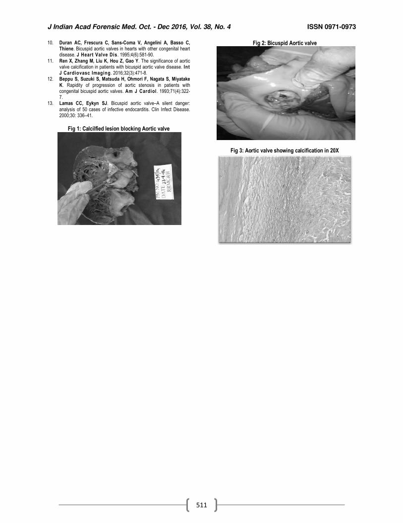
J Indian Acad Forensic Med. Oct. - Dec 2016, Vol. 38, No. 4 ISSN 0971-0973
511
10. Duran AC, Frescura C, Sans-Coma V, Angelini A, Basso C, Thiene. Bicuspid aortic valves in hearts with other congenital heart disease. J Heart Valve Dis. 1995;4(6):581-90.
11. Ren X, Zhang M, Liu K, Hou Z, Gao Y. The significance of aortic valve calcification in patients with bicuspid aortic valve disease. Int J Cardiovasc Imaging . 2016;32(3):471-8.
12. Beppu S, Suzuki S, Matsuda H, Ohmori F, Nagata S, Miyatake
K. Rapidity of progression of aortic stenosis in patients with congenital bicuspid aortic valves. Am J Cardiol . 1993;71(4):322-7.
13. Lamas CC, Eykyn SJ. Bicuspid aortic valve–A silent danger:
analysis of 50 cases of infective endocarditis. Clin Infect Disease. 2000;30: 336–41.
Fig 1: Calcilfied lesion blocking Aortic valve
Fig 2: Bicuspid Aortic valve
Fig 3: Aortic valve showing calcification in 20X
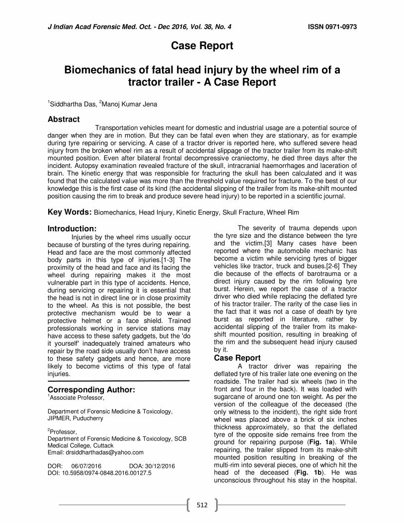
J Indian Acad Forensic Med. Oct. - Dec 2016, Vol. 38, No. 4 ISSN 0971-0973
512
Case Report
Biomechanics of fatal head injury by the wheel rim of a tractor trailer - A Case Report
1Siddhartha Das,
2Manoj Kumar Jena
Abstract Transportation vehicles meant for domestic and industrial usage are a potential source of
danger when they are in motion. But they can be fatal even when they are stationary, as for example during tyre repairing or servicing. A case of a tractor driver is reported here, who suffered severe head injury from the broken wheel rim as a result of accidental slippage of the tractor trailer from its make-shift mounted position. Even after bilateral frontal decompressive craniectomy, he died three days after the incident. Autopsy examination revealed fracture of the skull, intracranial haemorrhages and laceration of brain. The kinetic energy that was responsible for fracturing the skull has been calculated and it was found that the calculated value was more than the threshold value required for fracture. To the best of our knowledge this is the first case of its kind (the accidental slipping of the trailer from its make-shift mounted position causing the rim to break and produce severe head injury) to be reported in a scientific journal.
Key Words: Biomechanics, Head Injury, Kinetic Energy, Skull Fracture, Wheel Rim
Introduction: Injuries by the wheel rims usually occur because of bursting of the tyres during repairing. Head and face are the most commonly affected body parts in this type of injuries.[1-3] The proximity of the head and face and its facing the wheel during repairing makes it the most vulnerable part in this type of accidents. Hence, during servicing or repairing it is essential that the head is not in direct line or in close proximity to the wheel. As this is not possible, the best protective mechanism would be to wear a protective helmet or a face shield. Trained professionals working in service stations may have access to these safety gadgets, but the 'do it yourself' inadequately trained amateurs who repair by the road side usually don’t have access to these safety gadgets and hence, are more likely to become victims of this type of fatal injuries.
Corresponding Author: 1Associate Professor,
Department of Forensic Medicine & Toxicology, JIPMER, Puducherry 2Professor,
Department of Forensic Medicine & Toxicology, SCB Medical College, Cuttack Email: [email protected] DOR: 06/07/2016 DOA: 30/12/2016 DOI: 10.5958/0974-0848.2016.00127.5
The severity of trauma depends upon the tyre size and the distance between the tyre and the victim.[3] Many cases have been reported where the automobile mechanic has become a victim while servicing tyres of bigger vehicles like tractor, truck and buses.[2-6] They die because of the effects of barotrauma or a direct injury caused by the rim following tyre burst. Herein, we report the case of a tractor driver who died while replacing the deflated tyre of his tractor trailer. The rarity of the case lies in the fact that it was not a case of death by tyre burst as reported in literature, rather by accidental slipping of the trailer from its make-shift mounted position, resulting in breaking of the rim and the subsequent head injury caused by it.
Case Report A tractor driver was repairing the
deflated tyre of his trailer late one evening on the roadside. The trailer had six wheels (two in the front and four in the back). It was loaded with sugarcane of around one ton weight. As per the version of the colleague of the deceased (the only witness to the incident), the right side front wheel was placed above a brick of six inches thickness approximately, so that the deflated tyre of the opposite side remains free from the ground for repairing purpose (Fig. 1a). While repairing, the trailer slipped from its make-shift mounted position resulting in breaking of the multi-rim into several pieces, one of which hit the head of the deceased (Fig. 1b). He was unconscious throughout his stay in the hospital.
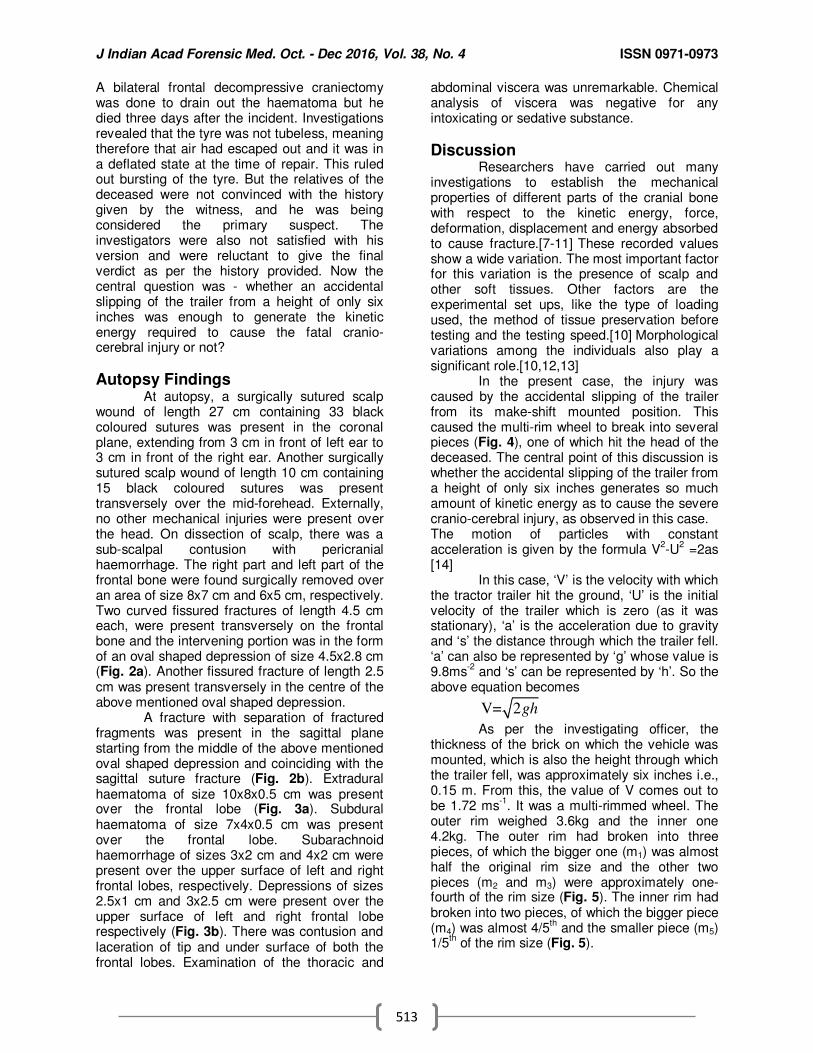
J Indian Acad Forensic Med. Oct. - Dec 2016, Vol. 38, No. 4 ISSN 0971-0973
513
A bilateral frontal decompressive craniectomy was done to drain out the haematoma but he died three days after the incident. Investigations revealed that the tyre was not tubeless, meaning therefore that air had escaped out and it was in a deflated state at the time of repair. This ruled out bursting of the tyre. But the relatives of the deceased were not convinced with the history given by the witness, and he was being considered the primary suspect. The investigators were also not satisfied with his version and were reluctant to give the final verdict as per the history provided. Now the central question was - whether an accidental slipping of the trailer from a height of only six inches was enough to generate the kinetic energy required to cause the fatal cranio-cerebral injury or not?
Autopsy Findings At autopsy, a surgically sutured scalp
wound of length 27 cm containing 33 black coloured sutures was present in the coronal plane, extending from 3 cm in front of left ear to 3 cm in front of the right ear. Another surgically sutured scalp wound of length 10 cm containing 15 black coloured sutures was present transversely over the mid-forehead. Externally, no other mechanical injuries were present over the head. On dissection of scalp, there was a sub-scalpal contusion with pericranial haemorrhage. The right part and left part of the frontal bone were found surgically removed over an area of size 8x7 cm and 6x5 cm, respectively. Two curved fissured fractures of length 4.5 cm each, were present transversely on the frontal bone and the intervening portion was in the form of an oval shaped depression of size 4.5x2.8 cm (Fig. 2a). Another fissured fracture of length 2.5 cm was present transversely in the centre of the above mentioned oval shaped depression.
A fracture with separation of fractured fragments was present in the sagittal plane starting from the middle of the above mentioned oval shaped depression and coinciding with the sagittal suture fracture (Fig. 2b). Extradural haematoma of size 10x8x0.5 cm was present over the frontal lobe (Fig. 3a). Subdural haematoma of size 7x4x0.5 cm was present over the frontal lobe. Subarachnoid haemorrhage of sizes 3x2 cm and 4x2 cm were present over the upper surface of left and right frontal lobes, respectively. Depressions of sizes 2.5x1 cm and 3x2.5 cm were present over the upper surface of left and right frontal lobe respectively (Fig. 3b). There was contusion and laceration of tip and under surface of both the frontal lobes. Examination of the thoracic and
abdominal viscera was unremarkable. Chemical analysis of viscera was negative for any intoxicating or sedative substance.
Discussion Researchers have carried out many
investigations to establish the mechanical properties of different parts of the cranial bone with respect to the kinetic energy, force, deformation, displacement and energy absorbed to cause fracture.[7-11]
These recorded values
show a wide variation. The most important factor for this variation is the presence of scalp and other soft tissues. Other factors are the experimental set ups, like the type of loading used, the method of tissue preservation before testing and the testing speed.[10]
Morphological
variations among the individuals also play a significant role.[10,12,13]
In the present case, the injury was
caused by the accidental slipping of the trailer from its make-shift mounted position. This caused the multi-rim wheel to break into several pieces (Fig. 4), one of which hit the head of the deceased. The central point of this discussion is whether the accidental slipping of the trailer from a height of only six inches generates so much amount of kinetic energy as to cause the severe cranio-cerebral injury, as observed in this case. The motion of particles with constant acceleration is given by the formula V
2-U
2 =2as
[14]
In this case, ‘V’ is the velocity with which the tractor trailer hit the ground, ‘U’ is the initial velocity of the trailer which is zero (as it was stationary), ‘a’ is the acceleration due to gravity and ‘s’ the distance through which the trailer fell. ‘a’ can also be represented by ‘g’ whose value is 9.8ms
-2 and ‘s’ can be represented by ‘h’. So the
above equation becomes
V= 2gh
As per the investigating officer, the
thickness of the brick on which the vehicle was mounted, which is also the height through which the trailer fell, was approximately six inches i.e., 0.15 m. From this, the value of V comes out to be 1.72 ms
-1. It was a multi-rimmed wheel. The
outer rim weighed 3.6kg and the inner one 4.2kg. The outer rim had broken into three pieces, of which the bigger one (m1) was almost half the original rim size and the other two pieces (m2 and m3) were approximately one-fourth of the rim size (Fig. 5). The inner rim had broken into two pieces, of which the bigger piece (m4) was almost 4/5
th and the smaller piece (m5)
1/5th of the rim size (Fig. 5).
J Indian Acad Forensic Med. Oct. - Dec 2016, Vol. 38, No. 4 ISSN 0971-0973
514
Applying the law of Conservation of Momentum in this case,[15]
the momentum
produced by that portion of the trailer hitting the ground and coming to still, is equal to the sum of the momentum of the broken rim parts that came out of the wheel. But two other factors also need consideration here. When the trailer hit the ground, part of the energy would have been absorbed by the ground (K1) and another part would have been utilised in breaking the rims (K2). As per the investigating officer, the broken rim parts were lying close to the wheel on one side. Assuming the broken rim parts to move with an average velocity ‘v’, the equation becomes MV = (m1+m2+m3+m4+m5)v + K1 + K2
The thrust of fall was borne by the two front wheels. Hence the above equation becomes MV = 2{(m1+m2+m3+m4+m5)v + K1 + K2}
As per the specifications of the manufacturer, weight of the trailer was 2 tonnes.
Now we have to calculate the value of ‘M’, which is the mass of the trailer and sugarcane that actually participated in the fall. M = ½ (weight of portion of trailer + weight of sugarcane) + weight of 2 front wheels.
We have considered ½ i.e., 50% because the rear portion of the trailer was grounded and slipping of the trailer took place in the front portion only. Weight of portion of trailer = 2000 kg – weight of 6 wheels = 1140 kg. (150 kg is the weight of the tyre with air and 110 kg is the weight of the tyre in a deflated state) In other words, M = ½ (1140 + 1000) +260 = 1330 kg. So the above equation becomes
2v = MV
2(m1+m2+m3+m4+m5) and the value for ‘v’ comes out to be 73.32 ms
-1.
As no blood stains were found on the rims, the question that arose was which one of the broken rim part had hit the deceased. The outer rim was more likely to fracture first as less energy was required to break it and the smaller piece would have travelled a bit faster than the bigger one, hence it is assumed that either m2 or m3 would have hit the head of the deceased. The kinetic energy with which it hit is given by the formula
KE=
1
2 mv2
the value of which comes out to be ½x0.9x73.32x73.32 = 2419 J. Calculating the exact value of K1 and K2 is not necessary here as we are trying to estimate the approximate, not the exact energy of impact. But we presume the value of ‘K2’ to be not so high as the rims were very old and rusted (Fig. 6). Similarly, value of K1 would have been
less considering impact on a hard stony soil. When the trailer hit the ground, the impact was borne by the rims, causing them to break as the tyre was deflated. Had the tyre been in an inflated state, it would have acted as a cushion and borne the impact instead of the rims. The absorbed energy required to fracture the skull bone has been reported to be between 2.77 – 68.5 J.[8,9]
Both these studies
do not speak of the energy of impact that is required to fracture the skull. Moreover, calculating the absorbed energy for skull fracture may require a laboratory infrastructure which may not be present in many forensic departments. Further, tedious experimental procedures are required which many forensic pathologists working in busy autopsy centres may not like to do for a single case only. In that case, calculating the approximate energy of impact from simple formulas is practically more helpful for forensic use. Because of few constraints we could not calculate the exact energy of impact. The calculated value is an approximate value which may be close to the actual impact energy and definitely more than the threshold required to cause fracture of the frontal bone. Thus, it could be concluded that the trailer falling through a height of six inches only, could generate the required amount of energy to not only cause fracture of the skull bone, but also, the underlying cerebral injury.
Acknowledgements The authors would like to thank
Mr.Subrato Das, an engineering background IT executive in a reputed multinational company. He helped with the physics part of the manuscript. The authors also express their vote of appreciation to Mr. Y Kannan, Artist, Medical Illustration Division, JIPMER for preparing the illustrations used in the manuscript and all those residents of the department of Forensic Medicine & Toxicology, JIPMER who were involved in this work.
References: 1. Suruda A, Floccare D, Smith G. Injuries from tire and wheel
explosions during servicing. Ann Emerg Med. 1991;20:848–51. 2. Hefny AF, Eid HO, Al-Bashir M, Abu-Zidan FM. Blast injuries of
large tyres: Case series. Int J Surg. 2010;8:151–4. 3. Hefny AF, Eid HO, Abu-Zidan FM. Severe tyre blast injuries
during servicing. Injury. 2009;40:484–7. 4. Kumral B, Avşar A, Büyük Y, Göktürk C, Şam B. An unusual
fatal injury due to tyre blast an autopsy case. Rom J Leg Med. 2014;22:5–7.
5. Pomara C, D’Errico S, Riezzo I, Perilli G, Volpe U, Fineschi V. Blast overpressure after tire explosion a fatal case. Am J Forensic Med Pathol. 2013;34:306–10.
6. Rautji R, Rudra A, Dogra TD. An unusual fatal injury due to tyre burst: a case report. Forensic Sci Int. 2003;138:111–3.
7. Glaser N, Kneubuehl BP, Zuber S, Axmann S, Ketterer T, Thali MJ, et al. Biomechanical examination of blunt trauma due to baseball bat blows to the head. J Forensic Biomech. 2011;2:1-5.
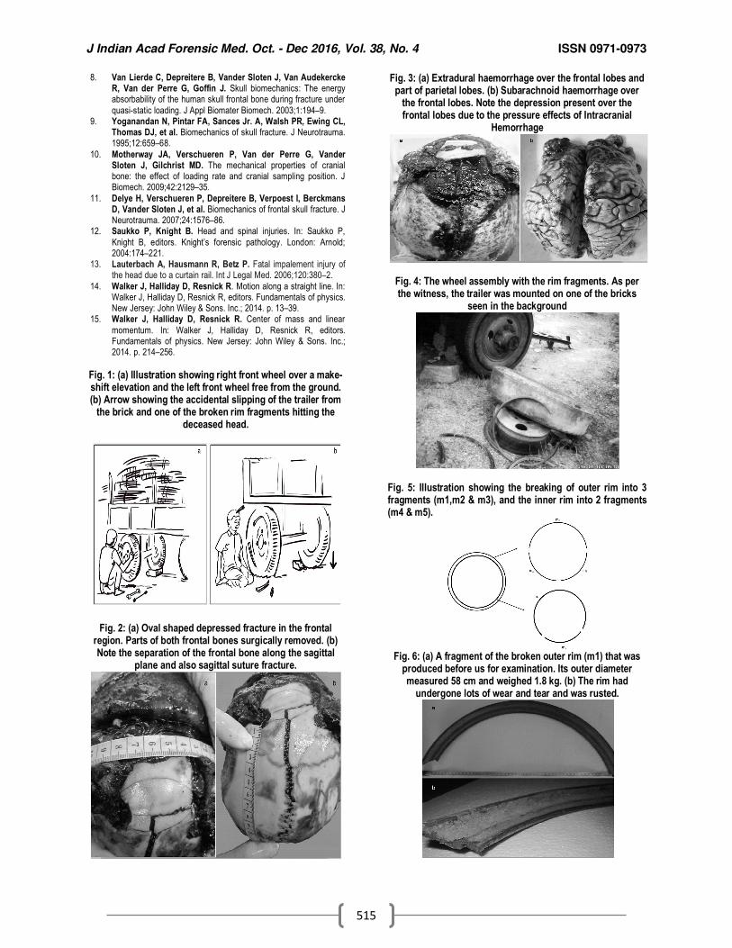
J Indian Acad Forensic Med. Oct. - Dec 2016, Vol. 38, No. 4 ISSN 0971-0973
515
8. Van Lierde C, Depreitere B, Vander Sloten J, Van Audekercke R, Van der Perre G, Goffin J. Skull biomechanics: The energy absorbability of the human skull frontal bone during fracture under
quasi-static loading. J Appl Biomater Biomech. 2003;1:194–9. 9. Yoganandan N, Pintar FA, Sances Jr. A, Walsh PR, Ewing CL,
Thomas DJ, et al. Biomechanics of skull fracture. J Neurotrauma. 1995;12:659–68.
10. Motherway JA, Verschueren P, Van der Perre G, Vander Sloten J, Gilchrist MD. The mechanical properties of cranial bone: the effect of loading rate and cranial sampling position. J Biomech. 2009;42:2129–35.
11. Delye H, Verschueren P, Depreitere B, Verpoest I, Berckmans D, Vander Sloten J, et al. Biomechanics of frontal skull fracture. J Neurotrauma. 2007;24:1576–86.
12. Saukko P, Knight B. Head and spinal injuries. In: Saukko P,
Knight B, editors. Knight’s forensic pathology. London: Arnold; 2004:174–221.
13. Lauterbach A, Hausmann R, Betz P. Fatal impalement injury of the head due to a curtain rail. Int J Legal Med. 2006;120:380–2.
14. Walker J, Halliday D, Resnick R. Motion along a straight line. In: Walker J, Halliday D, Resnick R, editors. Fundamentals of physics. New Jersey: John Wiley & Sons. Inc.; 2014. p. 13–39.
15. Walker J, Halliday D, Resnick R. Center of mass and linear
momentum. In: Walker J, Halliday D, Resnick R, editors. Fundamentals of physics. New Jersey: John Wiley & Sons. Inc.; 2014. p. 214–256.
Fig. 1: (a) Illustration showing right front wheel over a make-shift elevation and the left front wheel free from the ground. (b) Arrow showing the accidental slipping of the trailer from
the brick and one of the broken rim fragments hitting the deceased head.
Fig. 2: (a) Oval shaped depressed fracture in the frontal region. Parts of both frontal bones surgically removed. (b) Note the separation of the frontal bone along the sagittal
plane and also sagittal suture fracture.
Fig. 3: (a) Extradural haemorrhage over the frontal lobes and part of parietal lobes. (b) Subarachnoid haemorrhage over
the frontal lobes. Note the depression present over the frontal lobes due to the pressure effects of Intracranial
Hemorrhage
Fig. 4: The wheel assembly with the rim fragments. As per the witness, the trailer was mounted on one of the bricks
seen in the background
Fig. 5: Illustration showing the breaking of outer rim into 3 fragments (m1,m2 & m3), and the inner rim into 2 fragments (m4 & m5).
Fig. 6: (a) A fragment of the broken outer rim (m1) that was
produced before us for examination. Its outer diameter measured 58 cm and weighed 1.8 kg. (b) The rim had
undergone lots of wear and tear and was rusted.
rI
t.......uuuruttr,
I..,,,,,,,,,,,,,-.
Th*uiaers of $e JIAFil
AbhisHfDemorseaff" lrldild CUIES t tteilnKolkataYV klgEdAbhisHtErAssisffi h*ssu. Alfrs- t{er DehiAjay !(rnnaAssocide ffiss- GHCI{ ChanOgarhAkashdeep fggnrdAssodde Mssoe Gf,rc. Pariah, PuniabAmandeep SiinghAssociate Profiessor. GircH, ChandigarhAntara lleb BarmaSenior Resi<Jent, AllMS. New DelhiAnand P RayramneAssistant Professor, Mysore Medical College&Research lnstitute, MysoreArneet Arora
;, Prof & Head , AIIMS BhopalAshok ChananaAssociate Professor,Government Medical College, AmritsarB Suresh Kumar ShettyProf.& Head, State Medicolegal Consultant,Govt of Karnataka,Kasturba Medical College, MangaluruC B JaniProf. & Head, GCS Medical CollegeAhmedabadChittranjan BeheraAssistant Professor, AllMS, New DelhiCyriac JobVice Principal & Head, Govt. MedicalCollege,Manjeri, Malappuram, KeralaDeepa Durga RoySenior Resident, V.M.M.C & SafdarjungHospital, New DelhiHemant Bhardwaj [Lt. Colonel]Army Medical CorpsKewal Krishan,Associate Professor, Department ofAnthropology, Punjab U niversity, ChandigarhLuv SharmaProfessor, PGIMS, Rohtak, HaryanaMandar Ramchandra SaneAssistant Professor,Sri Aurobindo Medical College & PG lnstitute,lndoreManish KumatProfessor, V.M.M.C & Safdarjung Hospilal,New Delhi
knish tligilnProf. & Head, Sd A.rrobhdo ft|edcal Coilqe &PG lnstiMe,lndorellanoi Kumar llottantyProf. & Head, AllMS, BhubanesmrMukesh SharmaI n-Charge, Assistant D irector Regkmal ForensicScience Laboratory RajasthanN K AgganralDir. Prof & Head, UCMS & GTB Hospital,New DelhiN. SrinivasaragavanProf & Head , ESIC MedicalCollege & PGIMSR,KK Nagar, ChennaiPankaj GuptaProfessor, SGHD Medical College, Amritsar,PunjabPrateek RastogiAssociate Professor, Kasturba Medical College,MangalorePratikVijayTarvadiAssociate Professor, Pacific lnstitute of MedicalSciences, Umarda, UdaipurPutu! MahantaAssociate Professor, Tezpur Medical College,Tezpur, AssamRajiv JoshiProf. & Head, Guru Gobind Singh MedicalCollege, Faridkot, PunjabRambarapu SudhaAssociate Professor, Osmania Medical College& General Hospital, Koti, Hyderabad,TelanganaRashmi KulkarniSr. Research Associate, SAIMS lndoreSatinder Pal SinghAssistant Professor, GMCH, ChandigarhShilekh MittalAssociate Professor, Guru Gobind SinghMedical College, Faridkot, PunjabTanuj KanchanAssociate Professor, AllMS, JodhpurTapas Kumar BoseProf. & Head, N R S Medical College, KolkalaVirendar Pal SinghAssociate Professor,Dayanand Medical College, LudhianaDasari HarishProf. & HeadGovernment Medical College & Hospital,'Chandigarh
The EditorialTeam also wishes to whole heartedly thank Prof B D Gupta for goilng
through and giving ltis Valuable inputs to the "lnstructions to the AfthorS1,