japanese viral encephalitis
TRANSCRIPT

REVIEW
Japanese viral encephalitisS V Tiroumourougane, P Raghava, S Srinivasan. . . . . . . . . . . . . . . . . . . . . . . . . . . . . . . . . . . . . . . . . . . . . . . . . . . . . . . . . . . . . . . . . . . . . . . . . . . . . . . . . . . . . . . . . . . . . . . . . . . . . . . . . . . . . . . . . . . . . . . . . . . . .
Postgrad Med J 2002;78:205–215
One of the leading causes of acute encephalopathy inchildren in the tropics is Japanese encephalitis (JE).Transmitted by the culex mosquito, this neurotropic viruspredominately affects the thalamus, anterior horns of thespinal cord, cerebral cortex, and cerebellum. It mainlyaffects children <15 years and is mostly asymptomatic.The occasional symptomatic child typically presents witha neurological syndrome characterised by alteredsensorium, seizures, and features of intracranialhypertension. Aetiological diagnosis is based on virusisolation or demonstration of virus specific antigen orantibodies in the cerebrospinal fluid/blood. Though noantiviral drug is available against JE, effectivesupportive management can improve the outcome.Control of JE involves efficient vector control andappropriate use of vaccines.. . . . . . . . . . . . . . . . . . . . . . . . . . . . . . . . . . . . . . . . . . . . . . . . . . . . . . . . . . . . . . . . . . . . . . . . . .
Japanese encephalitis (JE) is one of the leading
causes of acute encephalopathy affecting
children1 and adolescents in the tropics. Nearly
50 000 cases of JE occur worldwide and 15 000
of them die.2 3 Considerable information on
epidemiology and clinical features of this dreaded
disease is available, yet much more needs to be
understood in terms of pathophysiology, clinical
management, and prognostication.
THE PASTThough outbreaks of encephalitis attributed to JE
virus (JEV) were reported in Japan as early as
1871, it wasn’t until 1924 that JEV was isolated
from a clinical case in the first recorded epidemic
in Japan (see box 1).4 The Nakayama strain of
JEV, used in development of mouse brain
inactivated virus vaccine was first isolated in
1935. The mode of transmission by mosquito vec-
tor was elucidated only 25 years after recognition
of JEV. Until 1970, the temperate zone of Asia was
the principal site of JE transmission. In the last
three decades, the focus of viral epidemics has
switched over to South and Southeast Asia.
EPIDEMIOLOGYEpidemics and sporadic cases of JE occur in many
Asian countries (see fig 1), including Cambodia,
China, Indonesia, India, Japan, Malaysia, Myan-
mar, Nepal, Pakistan, Philippines, Republic of
Korea, Sri Lanka, Thailand, Vietnam, and the
south eastern Russian federation. Gradual spread
to other non-Asian regions—for example, Torres
strait of Australian mainland has been reported
recently.5
Patterns of JE transmission vary within indi-
vidual countries and from year to year (table 1).6
In endemic areas, the annual incidence of disease
ranges from 10–100 per 100 000 population. An
endemic situation, with occurrence of sporadic
cases throughout the year, is present in tropical
zones. In temperate regions of Asia and the
northern tropical region, JEV is transmitted
seasonally. A probable explanation could be the
prolonged mosquito larval development time and
longer extrinsic period of JEV at cooler tempera-
tures in temperate regions, which can reduce the
viral transmission.7 8 In some instances, outbreaks
have been associated with rainfall, floods, or irri-
gation of rice fields.
The risk of travellers acquiring JE is very low
(monthly incidence is less than one per million
travellers among short term and urban travellers,
0.25 to 1 per 5000 travellers among rural travellers
to endemic regions).9–11 Travellers living for
prolonged periods in rural areas where JE is
endemic or epidemic are at greatest risk. Travel-
lers with extensive unprotected outdoor, evening,
and night-time exposure in rural areas might be
at high risk even if their trip is brief.
JEV is transmitted in a zoonotic cycle among
mosquitoes and vertebrate-amplifying hosts,
chiefly pigs and wading birds. The presence of
prolonged and high titres of viraemia without
clinical symptoms, the ability to produce many
uninfected offspring, and amplification of the
virus by multiplication makes pigs the most
important natural hosts for human
transmission.12 The mosquito vector of JE differs
in different regions. The major mosquito vector of
JE in South East Asia is Culex tritaeniorhynchus.Culex vishnui complex is also incriminated as a
vector in India.13 JEV has been isolated from 10
different species of culex, four species of anoph-
eles, and three species of mansonia
mosquitoes.14 15 Humans are considered as the
. . . . . . . . . . . . . . . . . . . . . . . . . . . . . . . . . . . . . . . . . . . . . . . . .
Abbreviations: CNS, central nervous system; CSF,cerebrospinal fluid; EEG, electroencephalogram; JE,Japanese encephalitis; JEV, Japanese encephalitis virus;Mac-ELISA, IgM antibody capture ELISA; M-IGSS,monoclonal antibody/immunogold/silver staining; PCO2,carbon dioxide tension
Box 1: History
1871: First outbreak of JE attributable to JE.1924: Isolation of JEV.1934: Isolation of Nakayama strain.1950: Elucidation of the route of transmission.1970: Change in geographical location of viral
transmission.
See end of article forauthors’ affiliations. . . . . . . . . . . . . . . . . . . . . . .
Correspondence to:Dr S Srinivasan,Department of Paediatrics,Jawaharlal Institute ofPostgraduate MedicalEducation and Research(JIPMER),Pondicherry–605006,India; [email protected] [email protected]
Submitted 5 March 2001Accepted 14 September2001. . . . . . . . . . . . . . . . . . . . . . .
205
www.postgradmedj.com

dead end host, as the brief periods of viraemia and low titres
of virus do not facilitate transmission.
THE PATHOGENJEV is a member of the flaviviridae. The cellular characteristics
of JEV are given in box 2. The M protein containing
hydrophobic domains presumably serves as a transmission
anchor.16 E protein constitutes the major immunogen and is
also expressed on the plasma membrane of infected
neurons.17 It is thought to be the cell receptor binding protein,
and a mediator of membrane fusion and cell entry.18 This pro-
tein is the major target of the host antiviral immune
response.19 20 JEV is related antigenically to St Louis, Murray
Valley, West Nile encephalitis viruses, and dengue fever virus.
PATHOLOGYGrossly, the brain appears oedematous with changes mainly
involving grey matter. The areas most commonly affected are
the thalamus, substantia nigra, anterior horns of the spinal
cord, cerebral cortex, and cerebellum. Microscopy reveals pan-
encephalitis with abundant glial nodules, perivascular cuffing,
and necrosis with or without characteristic circumscribed
necrotic foci.21–23 Neuronal inflammation is typically associated
with mononuclear cell infiltration. Diffuse microglial prolif-
eration and formation of gliomesenchymal nodules in brain
parenchyma dominate the histological picture in acute
encephalitis. In the post-encephalitic phase, lesions become
linear and tend to localise in thalamus, substantia nigra, and
Ammon’s horn. Histopathological examination of these focal
lesions show rarefied areas with few cellular and fibrous
elements surrounded by dense gliomesenchymal scarring.24 25
Pathological changes described in extraneural tissues
include hyperplasia of germinal centres of lymph nodes,
enlargement of malpighian bodies in spleen, interstitial myo-
carditis, swelling and hyaline changes in hepatic Kuffer’s cells,
pulmonary interalveolitis, and focal haemorrhages in the kid-
ney.
PATHOGENESISAfter transmission to man by an infected vector mosquito, JEV
multiplies locally and in regional nodes.26 After a phase of
transient viraemia, invasion of the central nervous system
(CNS) occurs. JEV is thought to invade brain via vascular
endothelial cells by endocytosis.27 In the neurons, JEV
replicates and matures in the neuronal secretory system,
mainly the rough endoplasmic reticulum and Golgi apparatus,
eventually destroying them.28 Experimental studies in mam-
malian hosts have shown JEV tropism to neurons in the CNS,
Figure 1 Map showing thedistribution of Japanese encephalitis.
Areas where Japanese encephalitishas been reported
Pakistan
India Myanmar
China
Philippines
Indonesia
North Korea
South Korea Japan
Japan
Vietnam
Malaysia
Bangladesh
Thailand
Nepal
Sri Lanka
Box 2: Cellular characteristics
• Single positive stranded RNA genome: length 11 kb.120
• Spherical virion: 30 nm core with a surrounding lipid enve-lope.• RNA genome encodes a single polypeptide that is cleaved
into:1. Structural capsid, member (M) and envelope (E) proteins.2. Non-structural proteins (NSI, 2A, 2B, 3, 4A, 4B, and 5).121
Table 1 Transmission patterns
Transmission pattern Transmission season Countries
Sporadic Year round Brunei, Malaysia, SingaporeJune to September JapanJuly to October South Korea
Endemic/hyperendemic July to December Bangladesh, Northern India, NepalMay to October Myanmar, Cambodia, Vietnam, Thailand, southern India,
Laos, northern China
206 Tiroumourougane, Raghava, Srinivasan
www.postgradmedj.com

indirectly indicating the presence of specific receptors with
strong affinity for the virus.29
IMMUNOLOGYBoth humoral and cellular arms of immune system are
involved in immunity to JEV. But the relative contribution of
individual components has not been well understood. After
primary infection with JEV, a rapid and potent monotypic IgM
response occurs in serum and cerebrospinal fluid (CSF), usu-
ally within seven days.30 The role of antibodies in protection is
not yet clearly understood. Passive administration of anti-JEV
antibodies has shown to confer protection in mice.31 Disap-
pearance of neurological signs has been noted in the presence
of IgM antibodies.32 Presence of CSF IgM antibodies has been
correlated with a favourable outcome in JE.33 However, the
significance of this protection remains unknown, since neuro-
virulence of JEV has been enhanced by administration of virus
specific antibodies.34 An anamnestic antibody response with
an early rapid IgG and delayed slow IgM response has been
noted in patients previously infected with antigenically
related flaviviruses.
The importance of cell mediated immunity is being
recognised of late. In vitro studies using macrophage and leu-
cocyte migration inhibition tests have suggested development
of cell mediated immunity in mice.35 36 Though, cytotoxic T
lymphocyte response to flaviviral infection has been noted in
man and mice, their role in JE is not very clear.37 38 Life long cell
mediated immunity could be conferred by passive transfer of
immune spleen T cells in mice.39 Immunisation with
inactivated JE vaccine induces T cell activation in vivo.40 These
studies indicate that cell mediated immunity probably has a
greater role than has been reflected so far.
CLINICAL FEATURESChildren under 15 years of age are principally affected in
endemic areas. When JEV first affects a nascent population,
adults are also affected.41 Seroprevalence studies in endemic
areas indicate nearly universal exposure by adulthood.
Approximately 10% of the susceptible population is infected
every year. Infection with JEV is often asymptomatic. The ratio
of asymptomatic to symptomatic infection varies between
25:1 and 1000:1.9 42 Man to man transmission in JE has not
been reported. However, the risk of acquiring JE in laboratory
settings does exist and laboratory acquired cases have been
reported.43 The exact risk of acquiring JE in the laboratory,
particularly in research settings, is not known. The incubation
period in man, after a mosquito bite, is not exactly known. In
general, it varies from 1–6 days. However, it can be as long as
14 days.
Onset of the illness can be abrupt, acute, subacute, or
gradual. The course of the disease can be conveniently divided
into three stages: (i) a prodromal stage preceding CNS
features, (ii) an encephalitis stage marked by CNS symptoma-
tology, and (iii) a late stage noticeable by recovery or
persistence of signs of CNS injury.44
The prodromal stage is characterised by high grade fever
with or without rigors, headache, general malaise, nausea, and
vomiting. During this stage, a definitive clinical diagnosis is
not possible. This is followed by the encephalitis stage (third to
fifth day), which manifests with altered sensorium, convul-
sions, neck stiffness, muscular rigidity, mask-like facies, and
abnormal movements. The relative frequency of various
symptoms in different studies is given in table 2.45–49
Abnormal oculocephalic reflex, acute onset hemiparesis
with hypertonia and decorticate and decerebrate posturing are
important CNS signs, which help in early clinical identifica-
tion of intracranial hypertension. Features of extraneural
involvement reported in JE include gastric haemorrhage in
absence of bleeding diathesis and pulmonary oedema. Death
usually occurs due to neurological illness in the first week. The
reported mortality rate varies between 8.5% and 72% (table 2).
Children who survive slowly regain neurological function
over several weeks. Residual neurological impairment in-
cludes thick, slow speech, aphasia, and paresis. Only one third
of cases recover normal neurological function. Intellectual
involvement may be noted in 30% of cases, speech disturbance
in 34%, and motor deficits in 49%. Secondary infections, espe-
cially pneumonia, urinary tract infection, and stasis ulcers are
frequent complications during recovery period.
Apart from the classical presentation described above, other
atypical presentations of JE have been reported. In a few chil-
dren, sensorium may recover rapidly after initial an episode of
seizure leading to a false clinical diagnosis of “atypical/ typical
febrile seizures” or “fever associated seizure disorder”.
Isolated acute onset abnormal behaviour can be the initial
presentation and is seen in adolescents. A small proportion of
children may present with features of aseptic meningitis with
no other clinical features of encephalopathy.50 An acute flaccid
paralysis-like illness has been recently reported as the initial
presenting feature.51
LABORATORY DIAGNOSISClinical diagnostic studiesIn JE, the leucocyte count is often raised (total counts 10–34 ×109/l). Differential counts often reveal neutrophilia ranging
between 51% and 90%. CSF examination shows a raised open-
ing pressure, cell count of 10–980 × 106/l, protein <900 mg/l,
and normal glucose concentration. Features on an electroen-
cephalogram (EEG) are non-specific and include diffuse theta
and delta waves, burst suppression, epileptiform activity, and
alpha coma.52 53 The generalised changes in an EEG may help
in differentiating JE from herpes encephalitis.54
Computed tomography show non-enhancing low density
areas in the thalamus, basal ganglia, midbrain, pons, and
medulla. Cortical atrophy has been noted in some children in
the post-encephalitic phase. Magnetic resonance imaging is
more sensitive than computed tomography in revealing neural
lesions. Magnetic resonance imaging on T2-weighted images
shows extensive hyperintense lesions of the thalamus,
cerebrum, and cerebellum.55 56 These neuroimaging techniques
Table 2 Clinical features
Feature No of casesAlteredsensorium (%)
Convulsion(%)
Meningealsigns (%) Fever (%)
Headache(%)
Mortality(%)
Lincoln and Silverstone (1952)45 201 Majority – – 100 100 8.5Webb and Perriera (1956)44 16 100 87.5 75 100 56.7 –Sengupta et al (1976)46 340 100 50.3 80 100 42.3 45.2–72Mohan Rao et al (1983)47 26 84.6 27.3 70 96.2 37.3 26.8Gourie Devi (1984)48 133 85 67.3 35.2 94.7 24.0 50.0Poneprasert et al (1989)122 59 100 59 61 96 76 17Kumar et al (1990)49 92 100 84.7 13 94.5 20.6 36.9Desai et al (1994)123 72 77.6 59.7 – 98.5 62.7 32
Japanese viral encephalitis 207
www.postgradmedj.com
may be useful in distinguishing JE from herpes encephalitis.
In JE, the regions mainly affected are the diencephalon and
basal ganglia, whereas in herpes encephalitis, the changes are
characteristically frontotemporal. Single photon emission
tomography in acute encephalitic phase reveals hyperper-
fusion of thalamus and putamen in some cases.57 In the post-
encephalitic phase, hypoperfusion of the thalamus, frontal
cortex, and lentiform area has been demonstrated.58 Though
these neuroimaging techniques demonstrate significant
changes, they are too non-specific to be used for aetiological
diagnosis.
Aetiological diagnosisAetiological diagnosis of JE is based on virus isolation or
demonstration of virus specific antigen or antibodies in
CSF/blood (see box 3). The laboratory diagnosis of a confirmed
case of Japanese encephalitis is based on one of the following.
1. Fourfold or greater rise in serum antibody titre, or
2. Isolation of virus from or demonstration of viral antigen or
genomic sequences in tissue, blood, CSF, or other body fluid, or
3. Specific IgM antibody by enzyme immunoassay antibody
captured in CSF or serum.
1. CultureIsolation of JEV was conventionally carried out by intracer-
ebral inoculation of clinical specimens in suckling mouse
brain. Various cell cultures that are being used more recently
include primary chick, duck embryo cells, and lines of Vero,
LLCMK2, C6/36, and AP61 cells. Virus can be isolated from the
blood of patients in preneuroinvasive and neuroinvasive
phases of the illness, usually not later than six or seven days
after the onset of symptoms.59 60 However, isolation of virus
from clinical specimens is generally considered a rare
occurrence61 probably because of low viral titres, rapid produc-
tion of neutralising antibodies, and the logistic difficulty in
transportation of specimens in developing countries and
frequent freezing and thawing of clinical material.62 63 Recently
sensitive mosquito inoculation techniques have been de-
scribed for isolation of JEV.64 Identification of JEV in culture
substrates was traditionally carried out by the complement
fixation test and agar gel diffusion. The neutralisation test,
monoclonal based immunofluorescence technique, and en-
zyme immunoassay are presently being used.65
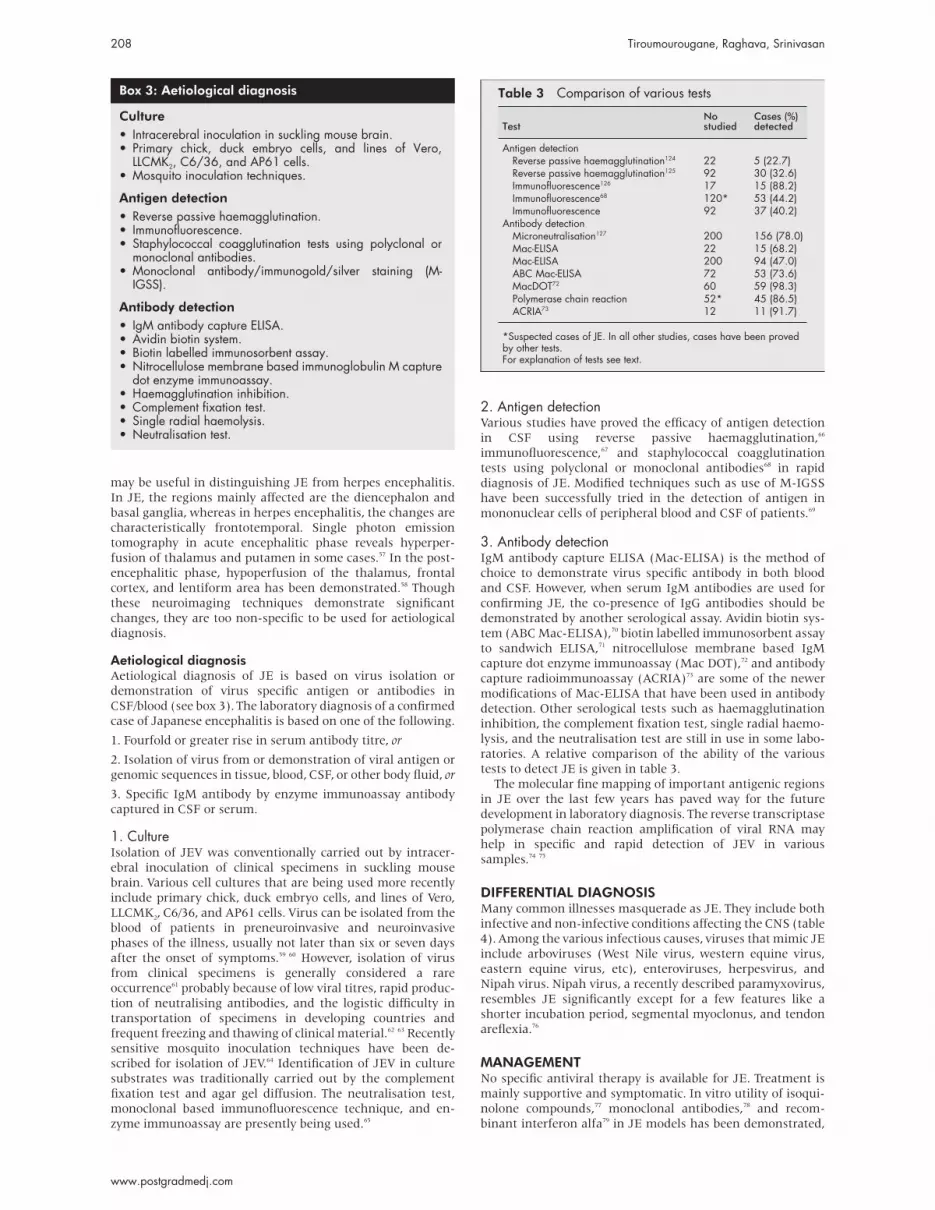
2. Antigen detectionVarious studies have proved the efficacy of antigen detection
in CSF using reverse passive haemagglutination,66
immunofluorescence,67 and staphylococcal coagglutination
tests using polyclonal or monoclonal antibodies68 in rapid
diagnosis of JE. Modified techniques such as use of M-IGSS
have been successfully tried in the detection of antigen in
mononuclear cells of peripheral blood and CSF of patients.69
3. Antibody detectionIgM antibody capture ELISA (Mac-ELISA) is the method of
choice to demonstrate virus specific antibody in both blood
and CSF. However, when serum IgM antibodies are used for
confirming JE, the co-presence of IgG antibodies should be
demonstrated by another serological assay. Avidin biotin sys-
tem (ABC Mac-ELISA),70 biotin labelled immunosorbent assay
to sandwich ELISA,71 nitrocellulose membrane based IgM
capture dot enzyme immunoassay (Mac DOT),72 and antibody
capture radioimmunoassay (ACRIA)73 are some of the newer
modifications of Mac-ELISA that have been used in antibody
detection. Other serological tests such as haemagglutination
inhibition, the complement fixation test, single radial haemo-
lysis, and the neutralisation test are still in use in some labo-
ratories. A relative comparison of the ability of the various
tests to detect JE is given in table 3.
The molecular fine mapping of important antigenic regions
in JE over the last few years has paved way for the future
development in laboratory diagnosis. The reverse transcriptase
polymerase chain reaction amplification of viral RNA may
help in specific and rapid detection of JEV in various
samples.74 75
DIFFERENTIAL DIAGNOSISMany common illnesses masquerade as JE. They include both
infective and non-infective conditions affecting the CNS (table
4). Among the various infectious causes, viruses that mimic JE
include arboviruses (West Nile virus, western equine virus,
eastern equine virus, etc), enteroviruses, herpesvirus, and
Nipah virus. Nipah virus, a recently described paramyxovirus,
resembles JE significantly except for a few features like a
shorter incubation period, segmental myoclonus, and tendon
areflexia.76
MANAGEMENTNo specific antiviral therapy is available for JE. Treatment is
mainly supportive and symptomatic. In vitro utility of isoqui-
nolone compounds,77 monoclonal antibodies,78 and recom-
binant interferon alfa79 in JE models has been demonstrated,
Box 3: Aetiological diagnosis
Culture• Intracerebral inoculation in suckling mouse brain.• Primary chick, duck embryo cells, and lines of Vero,
LLCMK2, C6/36, and AP61 cells.• Mosquito inoculation techniques.
Antigen detection• Reverse passive haemagglutination.• Immunofluorescence.• Staphylococcal coagglutination tests using polyclonal or
monoclonal antibodies.• Monoclonal antibody/immunogold/silver staining (M-
IGSS).
Antibody detection• IgM antibody capture ELISA.• Avidin biotin system.• Biotin labelled immunosorbent assay.• Nitrocellulose membrane based immunoglobulin M capture
dot enzyme immunoassay.• Haemagglutination inhibition.• Complement fixation test.• Single radial haemolysis.• Neutralisation test.
Table 3 Comparison of various tests
TestNostudied
Cases (%)detected
Antigen detectionReverse passive haemagglutination124 22 5 (22.7)Reverse passive haemagglutination125 92 30 (32.6)Immunofluorescence126 17 15 (88.2)Immunofluorescence68 120* 53 (44.2)Immunofluorescence 92 37 (40.2)
Antibody detectionMicroneutralisation127 200 156 (78.0)Mac-ELISA 22 15 (68.2)Mac-ELISA 200 94 (47.0)ABC Mac-ELISA 72 53 (73.6)MacDOT72 60 59 (98.3)Polymerase chain reaction 52* 45 (86.5)ACRIA73 12 11 (91.7)
*Suspected cases of JE. In all other studies, cases have been provedby other tests.For explanation of tests see text.
208 Tiroumourougane, Raghava, Srinivasan
www.postgradmedj.com

but they should not be used in routine practice without
further clinical studies. Corticosteroids have been used
empirically, but a recent double blind controlled placebo trial
failed to demonstrate any benefit.80
Though we are handicapped by the non-availability of aspecific drug against JE, mortality and morbidity can bedecreased appreciably by control and treatment of factorscausing secondary deterioration such as raised intracranialpressure and convulsions. The value of this approach has beendocumented in traumatic coma,81 and has been effectivelyapplied in our centre in management of JE (table 5 and boxes
4 and 5).
Reduction of intracranial pressure, optimisation of systemic
arterial pressure so as to maintain adequate cerebral perfusion
pressure, and prevention of secondary complications are the
main objectives of treatment. The concept of cerebral
perfusion pressure is an important practical guide to manage-
ment of intracranial hypertension (which often accompanies
JE). Cerebral perfusion pressure is defined as the difference
between mean arterial pressure and cerebral venous pressure.
As cerebral venous pressure is equal to intracranial pressure in
most circumstances, this equation may be rewritten as
follows:
Cerebral perfusion pressure = mean arterial pressure –
intracranial pressure82
In presence of intracranial hypertension, autoregulation of
cerebral blood flow is impaired, if cerebral perfusion pressure
is less than 40 mm Hg.83 When autoregulation is impaired, the
degree of cerebral ischaemia is significantly worse due to lack
of functional integrity of cerebral blood vessels, especially in
conditions like viral encephalitis. Even minor degrees of hypo-
tension or intracranial hypertension may markedly aggravate
cerebral ischaemia. Focal cerebral lesions may impair au-
toregulation in affected areas alone. Other uninvolved areas
with intact autoregulation may be affected during treatment,
due to the intracranial steal phenomenon, as the induced
vasoconstriction will result in shunting of blood away from
these areas.
Maintenance of cerebral perfusion is essential to prevent
secondary cerebral ischaemia. In conditions where cerebral
autoregulation is impaired, cerebral perfusion depends exclu-
sively on cerebral perfusion pressure. The normal cerebral per-
fusion pressure in infants is in the range of 30 mm Hg.84 Hence
it is important that cerebral perfusion pressure should be
maintained above 30 mm Hg in infants less than 6 months,
above 40 mm Hg in older children,85 and above 60 mm Hg in
adolescents.86 Considering the fact that clinical signs appear
when intracranial pressure is usually above 15–20 mm Hg,
mean arterial pressure should be maintained above 75 mm Hg
in mild and moderate grade coma and more than 85 mm Hg
in severe grade coma.
The most important aspect of managing intracranial hyper-
tension is early identification and initiation of appropriate
therapeutic measures. Most therapeutic measures fail if insti-
tuted late, as irreversible cerebral damage often occurs before
these agents start their action. The control of intracranial
hypertension involves a two pronged approach: (1) careful
avoidance and control of situations that exacerbate intracra-
nial pressure and (2) therapeutic measures to decrease intrac-
ranial hypertension, if present.
(1) Control of factors aggravating intracranial pressure(A) Positioning of patientA 15–30° head up tilt with head in midline position decreases
intracranial hypertension and improves cerebral perfusion
pressure.87 Elevation enhances CSF drainage and maximises
cerebral venous output. Midline position prevents any
obstruction of jugular venous drainage.
(B) Temperature controlAggressive treatment of fever is essential as fever worsens
intracranial hypertension by increasing cerebral metabolism,
cerebral blood flow, and cerebral oedema.88 Antipyretics and
other physical measures like tepid sponging and cooling blan-
kets should be employed to reduce temperature effectively. As
physical measures cause shivering, which can aggravate
intracranial hypertension, they should always be used in con-
junction with antipyretics.
(C) Role of sedationPain and arousal cause raised intracranial pressure by increas-
ing cerebral blood flow. Sedatives have a huge role in preven-
tion of the worsening of intracranial hypertension by this
mechanism.89 Conventionally, sedatives were avoided, due to
fear of “clouding the neurological examination”. Such an ill
supported prejudice should not be allowed to supersede
proper control of intracranial pressure through sedation. In a
clinical study conducted to identify the effect of management
parameters on outcome in JE, it was found that sedation posi-
tively influenced immediate survival irrespective of severity of
coma (unpublished data).
(D) Seizure controlSeizures increase intracranial pressure by increasing cerebral
metabolism and cerebral blood flow and by Valsalva. Hence
anticonvulsants should be administered prophylactically or
therapeutically.
Table 4 Differential diagnosis of viral encephalitis
(A) Bacterial infectionsPyogenic meningitis LeptospirosisSuppurative brain lesions TuberculosisTyphoid encephalopathy Mycoplasma encephalitis
(B) Viral infectionsArboviruses (West Nile, eastern equine, western equine) Enterovirus (Coxsackie virus, echovirus)Herpesvirus Nipah virusAcute disseminated encephalomyelitis
(C) Parasitic infectionsCerebral malaria Toxoplasmosis (HIV patients)Amoebic encephalitis Neurocysticercosis
(D) Rickettsial disordersRocky Mountain spotted fever Ehrlichiosis
(E) Fungal meningitis(F) Inflammatory conditions
Systemic lupus erythematosis Beçhet’s disease(G) Primary CNS tumours: non-Hodgkin’s lymphoma in HIV infected patients(H) Cerebral infarction
Japanese viral encephalitis 209
www.postgradmedj.com

(E) Fluid managementTraditionally, fluid and sodium restriction have been advo-
cated in hope of preventing cerebral oedema. The value of such
an approach has been seriously questioned in traumatic
coma.90 Dehydration has been found to increase risk of
cerebral infarction in patients with subarachnoid
haemorrhage.91 Hypovolaemia often accompanies viral en-
cephalitis due to decreased intake and increased loss (vomit-
ing, sweating). In our centre, fluid restriction is used only for
those children with mild and moderate grade coma. For those
with severe grade coma, central venous pressure is used to
guide fluid therapy.
(F) Electrolyte managementFull maintenance of sodium should be given as hypo-
natraemia impairs cerebrovascular reactivity. In our centre, we
provide normal sodium maintenance and vigilantly treat any
Table 5 Management protocol
Mild gradeIntravenous fluids Three quarters of maintenance (100 ml/100 kcal/day), with allowances for temperature,
hyperventilation, and excess urine outputElectrolytes Sodium: 3 mmol/100 kcal/day; potassium: 2 mmol/100 kcal/dayTemperature control Paracetamol (60 mg/kg/day: divided doses every 4–6 hours)
Physical measures: tepid sponging, cooling blanketsSedation Diazepam 0.1–0.3 mg/kg/dose as and when requiredAnticonvulsants
Moderate gradeIntravenous fluids, electrolytes, temperature control Similar to management of mild gradeSedation Diazepam every 6–8 hoursHead end elevation by 15°–30°
Severe gradeIntravenous fluids Three quarters of maintenance: initial and when CVP is not available
Full maintenance: if hypovolemia is present or if diuretic therapy is employed or after 48–72hours, if no signs of ICT
Sedation Intravenous diazepam every 6–8 hoursPhenobarbitone 3–5 mg/kg/day in two divided doses
Temperature control, head end elevation, electrolytes Similar to management of moderate gradeSeizure control Step 1: Intravenous diazepam; followed by intravenous phenytoin
Step 2: Phenytoin: loading dose 15–20 mg/kg infused at 0.5–1.5 mg/kg/min. Maintenancedose 5–8 mg/kg/day in two divided dosesStep 3: Phenobarbitone: for uncontrolled seizures, 15–20 mg/kg at a rate not exceeding 1mg/kg/min. Maintenance dose: 5–8 mg/kg/dayStep 4: Diazepam infusion: if seizures still persist, then diazepam infusion at a rate of 0.1–0.4mg/kg/hour or midazolam infusion: loading dose of 0.05–0.2 mg/kg followed by infusion at1–5 µg/kg/min. Diluents to be used: sterile water, normal salineStep 5: Thiopentone infusion as mentioned below
Management of ICTStabilisation of vital functions and control of factors aggravating ICT should be instituted before the following medications are used
Mannitol (only if serum osmolality is <300 mmol/kg) 0.2–1 g/kg given over 30 min.128 Repeat doses (0.25–0.5 g/kg) can be given every 4–6hours, after reassessing ICP status
Frusemide (furosemide) Persistent ICT after mannitol therapy: 1 mg/kg/dose every 12 hours (potassium levels to bemonitored)
Hyperventilation Bag and mask ventilation or after endotracheal intubation. 4% lignocaine as local spray orintravenous lignocaine at 1 mg/kg/dose (slow intravenous) to be used to avoid further rise inICP during intubation
Thiopentone Used when above measures fail. Loading dose of 5 mg/kg over 30 min with maintenancedose of 1 mg/kg/hour as infusion. Maximum maintenance dose: 5 mg/kg/hour. Whenevermaintenance dose is increased by 1 mg/kg/hour, a loading dose of 5 mg/kg to be given
Paralysis and ventilation Pancuronium: 0.05–0.1 mg/kg intravenously to be used along with a loading dose ofthiopentone 5 mg/kg. Succinyl choline and ketamine should be avoided. While trachealsuctioning, boluses of intravenous lignocaine 0.5–1 mg/kg may be used to prevent raise in ICP
Indications for antibiotics:1. When lumbar puncture is with held or traumatic, then crystalline penicillin at a dose of 400000 unit/kg/day and chloramphenicol 100 mg/kg/day
in divided doses to be given till CSF findings are available.2. For aspiration pneumonia, crystalline penicillin at a dose of 200000 units/kg/day to be given.
Mild: 13–15, moderate: 9–12, severe: <8. CSF, cerebrospinal fluid; CVP, central venous pressure; ICP, intracranial pressure; ICT, intracranialhypertension.
Box 4: Monitoring
• Coma scale score.• Seizure type and frequency.• Clinical signs of intracranial hypertension.• Blood pressure (continuous/hourly): mean arterial pressure
>75 mm Hg in mild and moderate grade and >85 mm Hgin severe grade.
• Urine output every four hours: maintained at 0.5ml/kg/hour.
• Calculated serum osmolality: every 24 hours (preferablyevery 12 hours).
• Continuous oxygen saturation monitoring, if not possible atleast hourly monitoring.
• Central venous pressure for severe coma.
Box 5: Management of intracranial hypertension
Control of factors aggravating intracranial pressure• Positioning of head.• Temperature control.• Sedation.• Seizure control.• Appropriate fluid therapy.• Appropriate electrolyte therapy.
Therapeutic measures to decrease intracranialpressure• Hyperventilation.• Mannitol.• Frusemide (furosemide).• Barbiturates.
210 Tiroumourougane, Raghava, Srinivasan
www.postgradmedj.com

degree of hyponatraemia, as and when it arises and maintain
serum sodium above 140 mmol/l.
(2) Therapeutic measures to decrease intracranialpressure(A) HyperventilationReduction in intracranial pressure induced by hyperventila-
tion is the result of a decrease in cerebral blood flow secondary
to cerebral vasoconstriction caused by carbon dioxide wash-
out. In normal subjects, cerebral blood flow decreases by 4%
per mm Hg decrease in carbon dioxide tension (PCO2). The
ceiling limit of cerebral blood flow reduction is 40%, which
corresponds to a PCO2 2.67–3.33 of kPa (20–25 mm Hg). Fur-
ther reduction in PCO2 level has no beneficial effect on cerebral
blood flow. Intracranial pressure begins to diminish 10–30
seconds after inception of hyperventilation, reaches a nadir in
30 minutes, and returns to the original value in less than
hour.92 The potential deleterious effects of hyperventilation
include an elevation of mean airway pressure and diminished
cardiac filling pressures with resultant barotrauma and hypo-
tension respectively. Although hyperventilation is useful in
acutely decreasing intracranial pressure, prolonged hyperven-
tilation, in fact, worsens the outcome. This could be probably
due to production of oligaemia in marginally perfused brain
tissue by chronic aggressive hyperventilation.93 Hence, hyper-
ventilation should be gradually withdrawn (rapid withdrawal
causes rebound intracranial hypertension) after a period of
30–60 minutes so as to raise PCO2 level by 0.2–0.29 kPa/hour,
with institution of other modes of therapy simultaneously.
(B) MannitolMannitol is the most commonly used agent for control of
intracranial hypertension. Response to mannitol depends on
original intracranial pressure, dose given over the previous
three hours (the lesser, the better effect),94 and rate of admin-
istration. Rapid administration of mannitol is more effective in
reducing intracranial pressure, but the action has a much
shorter duration. A slower infusion rate produces a lesser
degree of decrease that lasts longer.95 96 The protracted effect of
mannitol is due to water reduction from the intravascular
compartment (diuresis), whereas vascular mechanisms ex-
plain the acute effect of mannitol. Intravenous administration
of mannitol decreases blood viscosity and cerebral blood flow
increases transiently.97 In the presence of autoregulation, this
causes a reflex vasoconstriction and decrease in cerebral blood
flow and intracranial pressure. If autoregulation is impaired,
as in the case of diffuse neuronal injury in encephalitis, then
the effect of mannitol is not maximal.98 Though mannitol is a
very useful drug, significant side effects can occur if used
inappropriately. Mannitol tends to accumulate in oedematous
white matter and cause a hyperosmolar state, which is injuri-
ous to the brain.99 Hence overdosing should be avoided to pre-
vent formation of this hyperosmolar state. When prescribing
mannitol, both standing and prophylactic orders should be
avoided. Use of mannitol should be limited to patients with a
serum osmolality of less than 300 mmol/l.100 This is because
administration of mannitol at a dose of 1 g/kg over a period of
20 minutes increases serum osmolality by 11%–15%,101 and
there is a high risk of tubular damage and renal failure when
serum osmolality exceeds 330 mmol/l. Chronic continuous
therapy should be avoided as the brain adapts to the sustained
hyperosmolality of plasma with an increase in intracellular
hyperosmolarity by increased concentrations of cations,
sodium and potassium and free amino acids, which contribute
to “idiogenic” osmoles (that appear in brain in its adaptation
to hyperosmolality and contribute to rebound cerebral oedema
after withdrawal of anticerebral oedema measures).102
(C) Frusemide (furosemide)It acts by interfering with the formation of CSF and preferen-
tial excretion of water over solute in the distal renal tubule.103
Frusemide (furosemide) alone causes a slow reduction in
intracranial pressure, but when combined with mannitol, the
fall in intracranial pressure is rapid and is sustained for a con-
siderably longer period than when either agent is used
alone.104
(D) BarbituratesShort acting barbiturates like thiopentone, as a continuous
infusion, evoke a decrease in intracranial pressure by decreas-
ing cerebral blood flow and cerebral metabolic oxygen
demand.105 The therapeutic objective is either to elicit a satis-
factory decrease in intracranial pressure or to produce burst
suppression in a continuously monitored EEG. Barbiturate
therapy has the added advantage of causing sedation, which
by itself can contribute to reduction of intracranial hyper-
tension. Unfortunately, barbiturate therapy is associated with
major adverse effects, including precipitate hypotension,106
and hence should be reserved only for cases in which other
measures have failed.
PROGNOSTIC FACTORS IN JECertain factors have been identified which can be used to pre-
dict outcome in terms of mortality and morbidity. These are
listed in table 6 (unpublished data).107 108
CONTROL OF JE (SEE BOX 6)Vector controlInformation regarding prevalence, density, and insecticide
susceptibility of known and potential vectors of JE is essential
for control of vectors. Surveillance of the adult mosquito
Table 6 Prognostic factors
Poor prognostic factors Good prognostic factors
Age less than 10 years High levels of neutralising antibodies in CSFLow Glasgow coma scale High levels of JEV IgG in CSFHyponatraemiaShockPresence of immune complexes in CSFPresence of antiNFP or antiMBP antibodiesIncreased levels of tumour necrosis factorCoexisting evidence of neurocysticercosis129
CSF, cerebrospinal fliud; JEV, Japanese viral encephalitis; MBP, myelin basic protein; NFP, neurofilamentprotein.
Box 6: Prevention
• Control of mosquito vectors.• Vaccination of humans.• Prevention of mosquitoes from biting humans.• Measures against reservoirs.
Japanese viral encephalitis 211
www.postgradmedj.com

population should be carried out throughout the year. Spray-
ing of an appropriate insecticide should be carried out in the
resting places of mosquitoes. Thermal fogging with ultra low
volume insecticides such as pyrethrum or malathion has been
recommended for the prevention of local transmission during
epidemics, particularly in periurban areas with marshes. The
vastness of breeding places make larvicidal measures cur-
rently impracticable. Effective measures undertaken in some
countries to prevent or inhibit larval development include
novel water management and irrigation practices such as
periodic lowering of the water level, intermittent irrigation,
and constant flow systems. Vector control alone cannot be
relied upon to prevent JE.
VaccineVaccination of the population at risk is the method of choice
for prevention of JE. The earliest controlled field trials with a
vaccine against JE were carried out in Japanese schoolchil-
dren in the latter half of the 1940s. Three JE vaccines are in use
worldwide (table 7).Inactivated mouse brain vaccine had been used in Japan for
30 years and Asian field trials have shown that two doses (oneto weeks apart) provide an efficiency of 91%. However threedoses (0, 7, and 14/30 days) appear to be necessary to producea protective immune response in persons living in non-endemic regions. In China, 75 million doses of inactivated pri-mary hamster kidney cell derived vaccine are distributed eachyear. The vaccine’s efficacy is only 85% and a live attenuatedvaccine, made from the SA-14-14-2 strain, has been developedas a potentially more effective alternative. This vaccine ischeap (60 cents). Extensive efficacy trials have demonstrated>95% protection with two doses.109 The vaccine appears to besafe and neuroattenuation evidently is stable.110
The decision on the population to be vaccinated should bedecided by local epidemiological factors. The use of JE vaccinein travellers should be decided after adequate consideration ofthe risk-benefit ratio for the various factors on the risks forexposure to the virus and for developing illness, the availabil-ity and acceptability of repellents and other alternativeprotective measures, and the side effects of vaccination.111
However, it is better to individualise the considerationsregarding vaccination, particularly in travellers who are likelyto spend less than 30 days in an endemic area, as it requiresjust a single bite by an infected mosquito to cause JE in a par-ticular individual. Risk assessments should be interpretedcautiously since risk can vary within areas and from year to
year and available data are incomplete. Infants need not be
vaccinated, though as a precautionary measure they should be
protected from mosquito bites. In general, vaccination is indi-
cated in following groups.
1. People living in endemic areas.
2. Travellers spending 30 days or more in an endemic area.
3. Travellers spending less than 30 days during epidemics or
extensive outdoor activity in rural areas is expected.
4. Laboratory workers with potential risk of exposure to JEV.
JE vaccination is associated with local side effects (20%:
tenderness, redness, swelling), sometimes with systemic
adverse reactions (10%: fever, headache, malaise, rash), occa-
sionally with hypersensitivity reactions (0.5%: generalised
urticaria, angio-oedema, respiratory distress, and anaphy-
laxis), and very rarely major neurological side effects (1–2.3
per million recipients: encephalitis, seizures, and peripheral
neuropathy).112–114 However, a report from Europe (Denmark)
has suggested that the incidence of major neurological side
effects might be higher. It is possible that the difference in the
incidence of neurological side effects may due to differences in
surveillance or ethnic or geographical difference.115 Type I
hypersensitivity reactions have been recognised among Euro-
pean, American, and Australian vaccine recipients in the last
decade.116
Second generation recombinant vaccines are being devel-
oped with the aim of improving immunogenicity and decreas-
ing adverse reactions seen with current vaccines. In these vac-
cines, signal sequences of PrM along with genes encoding PrM
and E proteins are packaged into viral vector like Escherichiacoli and vaccinia.117–119 The immunogenicity and protective effi-
cacy of these expression systems has been demonstrated in
animal models.
Prevention of mosquito biteUse of nets and mosquito repellents by the population at risk
and avoidance of outdoor sleeping in the tropics in evening
hours, staying in screened houses, and wearing long sleeved
shirts and long trousers111 reduces the risk of exposure to vec-
tor mosquitoes.
Protection of reservoirsBuilding of piggeries away from human dwellings in countries
where pigs are reared near human settlement and making
them mosquito proof would be desirable. Spraying of piggeries
and mixed dwelling with residual insecticides, wherever there
is an alarming rise in vector species, should be carried out
promptly. Vaccination of pigs has also shown encouraging
results.
MULTIPLE CHOICE QUESTIONS (ANSWERS AT ENDOF PAPER)Q1: From which of the following species of mosquitoes,has JEV not been isolated?(A) Culex
(B) Anopheles
(C) Mansonia
(D) Aedes
Q2: The major immunogen in JEV is(A) Capsid protein
(B) Member protein
(C) Envelope protein
(D) Non-structural proteins
Table 7 Vaccines against Japanese encephalitis
Vaccine Strain Efficacy
Inactivated mouse brain Nakayama,Beijing-1
91%130
Inactivated primary hamster kidney cell P3 85%41
Live attenuated primary hamster kidney cell SA 14-14-2 >95%109
Box 7: Key references
1. Solomon T, Dung NM, Kneen R, et al. Japanese encepha-litis. J Neurol Neurosurg Psychiatry 2000;68:405–15.2. Monath TP, Heinz FX. Flaviviruses. In: Fields BN, KnipeDM, Howley M, eds. Fields virology. 3rd Ed. Philadelphia:Lippincot-Raven, 1996: 961–1034.3. Pickard JD, Czosnyka M. Management of raised intracra-nial pressure. J Neurol Neurosurg Neuropsychiatry1993;56:845–58.4. Japanese encephalitis vaccines. WHO position paper.Wkly Epidemiol Rec 1998;73:337–344.5. Inactivated Japanese encephalitis virus vaccine. Recom-mendations of the Advisory Committee on Immunization Prac-tices (ACIP). MMWR Morb Mortal Wkly Rep 1993;42:1–14.
212 Tiroumourougane, Raghava, Srinivasan
www.postgradmedj.com

Q3: In which of the following organelles does JEVmultiply intracellularly—I, Golgi apparatus; II, roughendoplasmic reticulum; III, mitochondria; IV, lysozyme?(A) I and II
(B) I and III
(C) II and III
(D) II and IV
Q4: What is the method of choice for detection of IgMantibodies in blood and CSF?(A) Reverse passive haemagglutination
(B) Immunofluorescence
(C) Staphylococcal coagglutination tests
(D) Mac-ELISA
Q5: Who among the following do not require JEvaccine?(A) People living in endemic areas
(B) Travellers spending 30 days or less in an endemic area
(C) Travellers spending less than 30 days during epidemics
(D) Laboratory workers with potential risk of exposure to JEV
Q6: One of the following statements regarding JE istrue(A) Sedation of children with JE should be avoided
(B) Mannitol can be given irrespective of serum osmolarity
(C) Chronic aggressive hyperventilation an be used in the
management of intracranial hypertension in JE
(D) A 15–30° head up tilt with the head in midline position
decreases intracranial pressure
. . . . . . . . . . . . . . . . . . . . .Authors’ affiliationsS V Tiroumourougane, S Srinivasan , Department of Paediatrics,Jawaharlal Institute of Postgraduate Medical Education and Research,Pondicherry, IndiaP Raghava, Department of Microbiology, Jawaharlal Institute ofPostgraduate Medical Education and Research, Pondicherry, India
REFERENCES1 Kumar R, Mathur A, Kumar A, et al. Japanese encephalitis—an
important cause of acute childhood encephalopathy in Lucknow, India.Postgrad Med J 1988;64:18–22.
2 Tsai TF. Factors in the changing epidemiology of JE and west Nile fever.In: Saluzzo JF, Dodet B, eds. Factors in emergence of arbovirus diseases.Paris: Elselvier, 1997: 179–89.
3 Solomon T. Viral encephalitis in Southeast Asia. Neurological Infectionsand Epidemiology 1997;2:191–9.
4 Miyake M. The pathology of Japanese encephalitis. Bull World HealthOrgan 1964;30:153–60.
5 Hanna JN, Ritchie SA, Phillips DA, et al. An outbreak of JE in the TorresStrait, Australia, 1995. Med J Aust 1996;165:256–60.
6 Inactivated Japanese encephalitis virus vaccine. Recommendationsof the Advisory Committee on Immunization Practices (ACIP). MMWRMorb Mortal Wkly Rep 1993;42:1–14.
7 Solomon T, Dung NM, Kneen R, et al. Japanese encephalitis. J NeurolNeurosurg Psychiatry 2000;68:405–15.
8 Mogi M. Relationship between number of human Japanese encephalitiscases and summer meteorological conditions in Nagasaki, Japan. Am JTrop Med Hyg 1983;32:470–4.
9 Halstead SB, Grosz CR. Subclinical Japanese encephalitis. Infections ofAmericans with limited residence in Korea. Am J Hyg 1962;75:190–201.
10 Sabin AB, Schlesinger RW, Ginder DR, et al. Japanese B encephalitis inAmerican soldiers in Korea. Am J Hyg 1947;46:356–75.
11 Benenson MW, Top FH, Gresso W, et al. The virulence to man ofJapanese encephalitis virus in Thailand. Am J Trop Med Hyg1975;24:974–80.
12 Gubler DJ, Rochrig JT. Arboviruses (togaviridae and flavivirdae). In:Collier L, Balows A, Sussman M, eds. Topley & Wilson’s microbiologyand microbial infection. 9th Ed. London: Arnold, 1998: 579–600.
13 Mishra AC. Monitoring of vectors of Japanese encephalitis. Proceedingsof the national conference on Japanese encephalitis, 1984. New Delhi:Indian Council of Medical Research, 1984: 62–9.
14 Bhattacharya S. Vector diversity in Japanese encephalitis epidemiologywith special reference to West Bengal. Proceedings of the secondsymposium on vector and vector borne diseases. 1997 March: 109–15.
15 Reuben R, Gajanana A. Japanese encephalitis in India. Indian J Pediatr1997;64:243–51.
16 McAda PC, Manson PW, Schmaljohm CS, et al. Partial sequence of theJapanese encephalitis virus genome. Virology 1987;58:348–60.
17 Russell PK, Brandt WA, Dalrymple JM. Chemical and antigenic structureof flaviviruses. In: Schlesinger RW, ed. The toga viruses. New York:Academic Press, 1980: 503–29.
18 Monath TP, Heinz FX. Flaviviruses. In: Fields BN, Knipe DM, Howley M,eds. Fields virology. 3rd Ed. Philadelphia: Lippincott-Raven, 1996:961–1034.
19 Heinz FX. Epitope mapping of flavivirus glycoproteins. Adv Virus Res1986;31:103–68.
20 Mason PW, Pincus S, Fournier MJ, et al. Japanese encephalitisvirus-vaccinia recombinants produce particulate forms of structuralproteins and induce high levels of protection against lethal Japaneseencephalitis virus infection. Virology 1991;180:294–305.
21 Hayashi M. Encephalitis epidemica Japonica [abstract]. Allg Z Psychiat1931;95:55–8.
22 Zimmerman HM. The pathology of Japanese B encephalitis. Am JPathol 1946;22:965–75.
23 Shiraki H, Goto A, Narabayashi H. Etat passe et present del’encephalite Japonaise an Japon [abstract]. Rev Neurol1963;108:633–99.
24 Kunimoto G. Histopathological examination of central nervous system inprotracted cases of encephalitis Japonica [abstract]. KyoshuNeuropsychiatr 1960;8:63–70.
25 Shiraki H, Fujisawa K. A case of protracted (151 days) Japanese Bencephalitis. Recent Adv Res Nerv Sys 1969;13:309–17.
26 Ahmed A. Japanese encephalitis. In: Chopra JS, Sawhney IMS, eds.Neurology in the tropics. 1st Ed. New Delhi: BI Churchill Livingstone,1999: 176–90.
27 Liou M-L, Hsu C-Y. Japanese encephalitis virus is transported across thecerebral blood vessels by endocytosis in mouse brain. Cell Tissue Res1998;293:389–94.
28 Hase T, Summers PL, Dubois DR. Ultra structure changes of mouse brainneurons infected with Japanese encephalitis virus. Int J Exp Pathol1990;71:493–505.
29 Oyanagi S, Ikuta F, Ross ER. Electron microscopic observation in miceinfected with Japanese encephalitis. Acta Neuropathol (Berl)1969;13:169–81.
30 Burke DS, Nisalak A, Ussery MA, et al. Kinetics of IgM and IgGresponse to Japanese encephalitis virus in human serum andcerebrospinal fluid. J Infect Dis 1985;151:1093–9.
31 Kimura-Kuroda J, Yasui K. Protection of mice against Japaneseencephalitis virus by passive administration with monoclonal antibodies. JImmunol 1988;191:3606–10.
32 Edelman R, Schneider RJ, Vejjajiva A, et al. Persistence of virus specificIgM and clinical recovery after Japanese encephalitis. Am J Trop MedHyg 1976;23:733–8.
33 Burke DS, Lorsomrudee W, Leake CJ, et al. Fatal outcome in Japaneseencephalitis. Am J Trop Med Hyg 1985;34:1203–10.
34 Gould EA, Buckley A. Antibody-dependent enhancement of yellow feverand Japanese encephalitis neurovirulence. J Gen Virol1989;70:1600–8.
35 Huang CH. Studies of Japanese encephalitis in China. Adv Virus Res1982;27:71–101.
36 Kelkar SD, Banerjee K. Cross reactions among flaviviruses inmacrophage migration inhibition assay. Acta Virol 1978;22:337–40.
37 Hill AB, Mulbacher A, Parhish C, et al. Broad cross-reactivity withmarked fine specificity in the cytotoxic T cell response to flaviviruses. JGen Virol 1992;73:1115–23.
38 Kulkarni AB, Mullbacher A, Blanden RV. In vitro T-cell proliferativeresponse to the flavivirus, West Nile. Virol Immunol 1991;4:78–82.
39 Jia FL, Huang ZX. Cell-mediated immunity in mice infected with JE virus.II. Passive transfer of immune spleen T cells for life protection in infectedmice [abstract]. Chung Kuo I Hsueh Ko Hsueh Yuan Hsueh Pao1983;5:68–9.
40 Aihara H, Takasaki T, Toyosaki-Maeda T, et al. T-cell activation andinduction of antibodies and memory T cells by immunization withinactivated JE vaccine. Viral Immunol 2000;13:179–86.
41 Japanese encephalitis vaccines. WHO position paper. WklyEpidemiol Rec 1998;73:337–44.
42 Thongcharoen P. Japanese encephalitis-virus encephalitis: an overview.Southeast Asian J Trop Med Public Health 1989;20:59–73.
43 Steffen R. Vaccinating against Japanese encephalitis [letter]. Lancet1987;ii:511.
44 Webb JKG, Perriera S. Clinical diagnosis of an arthropod borne typevirus encephalitis in children in North Arcot District, Madras State, India.Indian J Med Sci 1956;10:573–81.
45 Lincoln AF, Silverstone SE. Acute phase of Japanese B encephalitis.JAMA 1952;150:268–73.
46 Sengupta SN, Sen MK, Das PK, et al. Clinical profile of epidemic ofJapanese encephalitis. Indian J Med Res 1976;54:1393–402.
47 Mohan Rao CVR, Prasad SR, Rodrigues JJ, et al. The first laboratoryproven outbreak of Japanese encephalitis in Goa. Indian J Med Res1983;78:745–50.
48 Gourie-Devi M. Clinical aspects and experience in the management ofJapanese encephalitis patients. Proceedings of the national conference onJapanese encephalitis, 1984. New Delhi: Indian Council of MedicalResearch, 1984: 25–9.
Japanese viral encephalitis 213
www.postgradmedj.com

49 Kumar R, Mathur A, Kumar A, et al. Clinical features and prognosticindicators of Japanese encephalitis in children in Lucknow (India). IndianJ Med Res 1990;91:321–7.
50 Solomon T, Thao LTT, Dung NM, et al. Clinical features of Japaneseencephalitis: prognostic and pathophysiological significance in 50patients. 7th International congress for infectious diseases. Hong Kong:International Society for Infectious Diseases, 1996: 132.
51 Solomon T, Kneen R, Dung NM, et al. Poliomyelitis like illness due toJapanese encephalitis virus. Lancet 1998;351:1094–7.
52 Misra UK, Kalita J, Jain SK, et al. Radiological and neurophysiologicalchanges in Japanese encephalitis. J Neurol Neurosurg Psychiatry1994;57:1484–7.
53 Kalita K, Misra UK. EEG in Japanese encephalitis: a clinico-radiologicalcorrelation. Electroencephalogr Clin Neurophysiol 1998;106:238–43.
54 Misra UK, Kalita J. A comparative study of Japanese and Herpessimplex encephalitis. Electromyogr Clin Neurophysiol 1998;38:41–6.
55 Abe TK, Kojima K, Shoji N, et al. Japanese encephalitis. J Magn ResonImaging 1998;8:755–61.
56 Kalita J, Misra UK. Comparison of CT scan and MRI findings in thediagnosis of Japanese encephalitis. J Neurol Sci 2000;174:3–8.
57 Kimura K, Dosaka A, Hashimoto Y, et al. Single photon emission CTfindings in acute Japanese. Am J Neuroradiol 1997;18:465–9.
58 Kalita J, Das BK, Misra UK. SPECT studies of regional cerebral bloodflow in 8 patients with Japanese encephalitis in subacute and chronicstage. Acta Neurol Scand 1999;99:213–8.
59 Kim-Thoa MT, Lam-Thai CT, Ngo-TV, et al. Isolation of a strain ofJapanese encephalitis from the blood of a young patient suffering fromcardiovascular collapse. Bull Soc Pathol Exot 1974;67:341.
60 Kedarnath N, Prasad SR, Dandawate CN, et al. Isolation of Japaneseencephalitis and West Nile viruses from peripheral blood of encephalitispatients. Indian J Med Res 1984;79:1–7.
61 Shope RE, Sather GE. Arboviruses. In: Lennette EH, Schmidt NJ, eds.Diagnostic procedures for viral, rickettsial and chlamydial infections.Washington, DC: American Public Heath Association, 1979: 778–80.
62 Mohan Rao CVR, Risbud AR, Rodrigues FM, et al. The epidemic ofJapanese encephalitis in Tamil Nadu and Pondicherry. Indian J Med Res1988;87:417–27.
63 Leake CJ, Burki DS, Nisalak K, et al. Isolation of Japanese encephalitisvirus in clinical specimens using continuous mosquito cell line. Am J TropMed Hyg 1996;35:1045–50.
64 Gajanana A, Rajendran R, Thenmozhi V, et al. Comparative evaluationof bioassay and ELISA for detection of Japanese encephalitis virus in fieldcollected mosquitoes. Southeast Asian J Trop Med Public Health1995;26:91–7.
65 Zhang YH, Yu WF, Tian ZW, et al. A simplified new method for theidentification of arboviruses: virus detection using enzyme immunoassayon cultured cells. J Virol Methods 1984;9:45–51.
66 Ravi V, Premkumar S, Chandramuki A, et al. A reverse passivehemagglutination test for detection of JE virus antigen in cerebrospinalfluid. J Virol Methods 1989;23:291–3.
67 Zhang YH, Yu WF, Cai J, et al. A rapid method for detection offlavivirus antigens—staphylococcal co-agglutination test usingmonoclonal antibodies to Japanese encephalitis virus. Acta Virol1989;33:24–31.
68 Raghava PV, Badrinath S. Detection of Japanese encephalitis cellassociated antigen in CSF by indirect immunofluorescence. Ann NatlAcad Med Sci (India) 1998;34:207–11.
69 Deng YC, Su XC, Feng YQ. Immunocytochemical study of mononuclearcells in peripheral blood and cerebrospinal fluid of patients withJapanese B encephalitis [abstract]. Chung Hna Ping Li Hsueh Tsa Chin1994;23:20–2.
70 Chow L, Sun HC, Chen HY, et al. Detection and differentiation ofdengue-1 from Japanese encephalitis virus infection by ABC MAC-ELISA[abstract]. Chung Hua Min Kuo Wei Sheng Wu Chi Mien I Hsueh TsaChin 1992;25:172–80.
71 Chang HC, Takashima I, Arikawa J, et al. Biotin-labelled antigensandwich enzyme-linked immunosorbent assay (BLA-S-ELISA) for thedetection of Japanese encephalitis antibody in human and a variety ofanimal sera. J Immunol Methods 1984;72:401–9.
72 Solomon T, Thao LT, Dung NM, et al. Rapid diagnosis of Japaneseencephalitis by using an immunoglobulin M dot enzyme immunoassay. JClin Microbiol 1998;36:2030–4.
73 Burke DS, Nisalak A, Ussery MA. ACRIA—antibody captureimmunoassay detection of Japanese encephalitis virus Immunoglobulin Mand G antibodies in cerebrospinal fluid. J Clin Microbiol1982;16:1034–42.
74 Meiyu F, Hnosheng C, Cuihua C, et al. Detection of flaviviruses byreverse transcriptase-polymerase chain reaction with the universal primerset. Microbiol Immunol 1997;41:209–13.
75 Paranjpe S, Banerjee K. Detection of Japanese encephalitis virus byreverse transcriptase/polymerase chain reaction. Acta Virol1998;42:1–5.
76 Goh KJ, Tan CT, Chew NK, et al. Clinical features of Nipah virusencephalitis among pig farmers in Malaysia. N Engl J Med2000;342:1229–35.
77 Takagami T, Simamura E, Hirari K, et al. Inhibitory effect offuranonaphthoquinone derivatives on the replication of Japaneseencephalitis virus. Antiviral Res 1998;37:37–45.
78 Zhang M, Wang M, Jiang S, et al. Passive protection of mice, goats andmonkeys against Japanese encephalitis virus with monoclonal antibodies.J Med Virol 1989;29:133–8.
79 Harinasatu C, Wasi C, Vithanomstat S. The effect of interferon onJapanese encephalitis virus in vitro. Southeast Asian J Trop Med PublicHealth 1984;15:564–8.
80 Hoke CH Jr, Vaughn DW, Nisalak A, et al. Effect of high dosedexamethasone on the outcome of acute encephalitis due to Japaneseencephalitis virus. J Infect Dis 1992;165:631–7.
81 Ghajar J, Hariri RJ. Management of pediatric head injury. Pediatr ClinNorth Am 1992;39:1093–12.
82 Bingaman WE, Frank JI. Malignant cerebral edema and intracranialhypertension. Neurol Clin 1995;13:479–509.
83 Jennet WB, Harper AM, Miller JD, et al. Relation between cerebralblood and cerebral perfusion pressure [abstract]. Br J Surg1970;57:390.
84 Raju TNK, Doshi UV, Vidyasagar D. Cerebral perfusion in healthypreterm and term newborn infants. J Pediatr 1982;100:139–42.
85 Goldstein KJ, Tamor I. Cerebral perfusion pressure in central nervoussystem infections of infancy and childhood. J Pediatr 1983;103:40–3.
86 Shaywitz BA, Rothestein P, Venes JL. Monitoring and management ofincreased intracranial pressure in Reye syndrome: results in 29 children.Pediatrics 1980;66:198–204.
87 Grant IS, Andrews PJD. Neurologic support. BMJ 199;319:110–388 Clasen RA, Pandolfi S, Laing I, et al. Experimental study of relation of
fever to cerebral edema. J Neurosurg 1974;41:576–81.89 Poss BW, Brockmeyer D, Clay B, et al. Pathophysiology and
management of the intracranial vault. In: Rogers MC, Nichols D, eds.Textbook of pediatric intensive care. 1st Ed. Philadelphia: Williams &Wilkins, 1995: 645–65.
90 Shenkin HA, Beizier HS, Bouzarth WF. Restricted fluid intake: rationalmanagement of the neurosurgical patient. J Neurosurg 1976;45:432–7.
91 Pickard JD, Czosnyka M. Management of raised intracranial pressure. JNeurol Neurosurg Neuropsychiatry 1993;56:845–58.
92 Miller JD, Dearden NM. Measurement, analysis and the management ofraised intracranial pressure. In: Teasdale GM, Miller JD, eds. Currentneurosurgery. Edinburgh: Churchill Livingstone, 1992: 119–58.
93 Muizelaar JP, Marmarou A, Ward JD, et al. Adverse effects ofprolonged hyperventilation in patients with severe head injury: arandomized clinical trial. J Neurosurg 1991;75:731–9.
94 McGraw CP, Alexander E Jr, Howard G. Effect of dose and doseschedule on the response of intracranial pressure to mannitol. SurgNeurol 1978;10:127–30.
95 Roberts PA, Pollay M, Engles C, et al. Effect on intracranial pressure offurosemide combined with varying doses and administration rates ofmannitol. J Neurosurg 1987;66:440–6.
96 Node Y, Nakazawa S. Clinical study of mannitol and glycerol on raisedintracranial pressure and on their rebound phenomenon. Adv Neurol1990;52:359–63.
97 Muizelaar JP, Wei ED, Kontos HA, et al. Mannitol causes compensatorycerebral vasoconstriction and vasodilation in response to blood viscositychanges. J Neurosurg 1983;59:822–6.
98 Muizelaar JP, Lutz HA, Becker DP. Effect of mannitol on intracranialpressure and cerebral blood flow and correlation with pressure autoregulation in severely head-injured patients. J Neurosurg1984;61:700–6.
99 Kaufman AM, Cardosa ER. Aggravation of vasogenic cerebral edemaby multiple dose mannitol. J Neurosurg 1998;77:584–9.
100 Greenwald BM, Ghajar J, Notterman DA. Critical care of children withacute brain injury. Adv Pediatr 1995;42:47–89.
101 Czernicki Z, Kunicki A, Rycembel Z, et al. Osmotic serum pressure inpatients treated with furosemide and hypertonic mannitol solution[abstract]. Neurol Neurochir Pol 1976;10:787–91.
102 Fishman RA. Brain edema. N Engl J Med 1975;293:706–11.103 Pollay M, Fullenwider C, Roberts PA, et al. Effect of mannitol and
furosemide on blood-brain osmotic gradient and intracranial pressure. JNeurosurg 1983;59:945–50.
104 Wilkinson HA, Rosenfeld SR. Furosemide and mannitol in the treatmentof acute experimental intracranial hypertension. Neurosurgery1983;12:405–10.
105 Nordstrom CH, Messeter K, Sundbarg G, et al. Cerebral blood flow,vasoreactivity and oxygen consumption during barbiturate therapy insevere traumatic brain lesions. J Neurosurg 1988;68:424–31.
106 Ward JD, Becker DP, Miller JD, et al. Failure of prophylactic barbituratecoma in the treatment of severe head injury. J Neurosurg1985;62:383–8.
107 Misra UK, Kalita J, Srivastava M. Prognosis of Japanese encephalitis: amultivariate analysis. J Neurol Sci 1998;162:143–7.
108 Gourie-Devi M. Neuroinfection. Dr S G Desai Oration. Proceedings ofthe Karnataka Pedicon, 2000 Dec 1–3. Bangalore, India: IndianAcademy of Pediatrics, 2000.
109 Hennessy S, Liu ZL, Tsai TF, et al. Effectiveness of live-attenuatedJapanese encephalitis vaccine (SA 14–14–2): a case control study.Lancet 1996;347:1583–6.
110 Liu ZL, Hennessy S, Strom BL, et al. Short term safety of live attenuatedJapanese encephalitis vaccine (SA 14-14-2): results of a randomisedcontrolled with 26239 subjects. J Infect Dis 1997;176:1366–9.
111 Tsai TF. Arboviral infections—general considerations for prevention,diagnosis and treatment in travelers. Semin Pediatr Infect Dis1992;3:62–9.
112 Anderson MM, Ronne T. Side effects with Japanese encephalitis vaccine[letter]. Lancet 1991;337:1044.
113 Ohtaki E, Matsuishi T, Hirano Y, et al. Acute disseminatingencephalomyelitis after treatment with Japanese B encephalitis vaccine(Nakayama-Yoken and Beijing strain). J Neurol Neurosurg Psychiatry1995;59:316–7.
114 Plesner AM, Ronne T. Allergic mucocutaneous reactions to Japaneseencephalitis vaccine. Vaccine 1997;15:1239–43.
214 Tiroumourougane, Raghava, Srinivasan
www.postgradmedj.com
115 Plesner AM, Soborg PA, Herning M. Neurological complications tovaccination against Japanese encephalitis. Eur J Neurol1998;5:479–85.
116 Ruff TA, Eisen D, Fuller A, et al. Adverse reactions to Japaneseencephalitis vaccine. Lancet 1991;338:881–2.
117 Srivastava AK, Morita K, Matsuo S, et al. Japanese encephalitis virusfusion protein with a protein A expressed in Escherichia coli confersprotective immunity in mice. Microbiol Immunol 1991;13:1515–21.
118 Seif SA, Morita K, Igarashi A. A 27-aminoacid coding region of JE virusE-protein expressed in E coli as fusion protein with glutathione-S-transferase elicit neutralizing antibody in mice. Virus Res1996;43:91–6.
119 Sato T, Takamura C, Yasuda A, et al. High-level expression of JE virus Eprotein by recombinant vaccinia virus and enhancement of itsextracellular release by the NS3 gene product. Virology1993;192:483–90.
120 Westaway EG, Brinton MA, Gaidamovich SYA, et al. Flaviviridae.Intervirology 1985;24:183–92.
121 Thomas JC, Chang SH, Ricardo G, et al. Flaviviruses genomeorganization, expression and replication. Annu Rev Microbiol1990;44:649–88.
122 Ponephrasert B. Japanese encephalitis in children in northern Thailand.Southeast Asian J Trop Med Public Heallth 1989;20:599–603.
123 Desai A, Ravi V, Guru SC, et al. Detection of autoantibodies to neuralantigen in the CSF of Japanese encephalitis patients and correlation offindings with the outcome. J Neurol Sci 1994;122:109–16.
124 Ravi V, Vanajakshi S, Gowda A, et al. Laboratory diagnosis of Japaneseencephalitis using monoclonal antibodies and correlation of findings withoutcome. J Med Virol 1989;29:221–3.
125 Desai A, Chandramuki A, Gourie-Devi M, et al. Detection of Japaneseencephalitis virus antigens in the cerebrospinal fluid using monoclonalantibodies. Clin Diagn Virol 1994;2:191–9.
126 Mathur A, Kumar R, Sharma S, et al. Rapid diagnosis of Japaneseencephalitis by immunofluorescent examination of cerebrospinal fluid.Indian J Med Res 1990;91:1–4.
127 Desai A, Ravi V, Chandramuki A, et al. Detection of neutralizingantibodies to Japanese encephalitis virus in the cerebrospinal fluid usingrapid microneutraliztion test. Serodiagn Immunother Infect Dis1994;6:1–5.
128 Bullock R. Mannitol and other diuretics in severe neurotrauma. NewHorizons 1995;3:448–52.
129 Desai A, Shankar SK, Jayakumar PN, et al. Co-existence of cerebralcysticercosis with Japanese encephalitis: a prognostic modulator.Epidemiol Infect 1997;118:165–71.
130 Hoke CH, Nisalak A, Sangawhipa N, et al. Protection against Japaneseencephalitis by inactivated vaccines. N Engl J Med 1988;319:608–14.
ANSWERS1: D; 2: C; 3: A; 4: D; 5: B; 6: D.
www.postgradmedj.com
If you wish to comment on any article published in the Postgraduate Medical Journal you can send an
eLetter using the eLetters link at the beginning of each article. Your response will be posted on
Postgraduate Medical Journal online within a few days of receipt (subject to editorial screening).
eLetters
Have your say
Japanese viral encephalitis 215
www.postgradmedj.com