exacerbated pathology of viral encephalitis in mice with central nervous system-specific...
TRANSCRIPT

Immunopathology and Infectious Disease
Exacerbated Pathology of Viral Encephalitis in Micewith Central Nervous System-Specific Autoantibodies
Renaud Burrer,* Michael J. Buchmeier,*Tom Wolfe,† Joey P.C. Ting,* Ralph Feuer,*Antonio Iglesias,‡ and Matthias G. von Herrath†
From the Molecular and Integrative Neurosciences Department,*
The Scripps Research Institute, La Jolla, California; the La Jolla
Institute of Allergy and Immunology,† Immune Regulation Lab,
La Jolla, California; and the Roche Center for Medical
Genomics,‡ Basel, Switzerland
We examine here the outcome of viral encephalomy-elitis [mouse hepatitis virus (MHV) A59, Theiler’s en-cephalomyelitis virus, and Coxsackievirus B3] in micewith autoantibodies to a central nervous system(CNS)-specific antigen, myelin oligodendrocyte glyco-protein, that usually develop no clinical disease. Mor-bidity and mortality of the acute viral CNS disease wasaugmented by the presence of the autoantibodies inall three viral infections. Transfer of serum contain-ing the autoantibodies at the time of infection withMHV was sufficient to reproduce the exacerbated dis-ease. The presence of the autoantibodies was found toresult in increased infiltration of mononuclear cellsinto the brain. Early demyelination was severely aug-mented in brains and spinal cords of MHV-infectedmice with CNS-specific autoantibodies. The antibody-mediated exacerbation was shown to be independent ofthe complement system but to require expression of Fcreceptors, because it was observed in C�-3-deficient butnot in Fc receptor-deficient mice. Our study illustratesthe possibility that infections can lead to much moreprofound immunopathology in the presence of an oth-erwise latent autoimmune condition. (Am J Pathol 2007,170:557–566; DOI: 10.2353/ajpath.2007.060893)
It is now well established in animal models that infectiousorganisms can trigger or augment immune responses toself-antigens, by mechanisms such as molecular mimicryor bystander activation (see review1). However, autoim-munity does not always result in clinically apparent dis-ease, and cumulative infections by the same or unrelatedagents may be required to unmask a pre-existing auto-immune process.1 Conversely, a viral infection could take
a worsened course in autoimmune-prone individuals. Ithas been shown that infections can provoke relapses orworsen autoimmune conditions in animal models.2–4 Thisis consistent with reports showing that common infectionsaugment the risk for or the severity of relapses in multiplesclerosis (MS) patients.5,6
The aforementioned animal reports all studied T-cell-mediated autoimmune responses, but autoantibodiesalso have an important pathogenic potential (see re-view7). In the central nervous system (CNS), autoantibod-ies to myelin components and to myelin oligodendrocyteglycoprotein (MOG) in particular can play a damagingrole. MOG-specific antibodies have been shown to play amajor role in the murine model of MS, experimental au-toimmune encephalomyelitis (EAE),8 and have beenclearly associated with demyelinating lesions in MS pa-tients.9 They are thus likely to mediate some of the pa-thology, at least in the subset of patients with lesionspresenting antibody depositions.10 However, the pres-ence of antibodies specific for MOG or other CNS anti-gens is detected not only in MS patients but also inhealthy subjects, albeit with a lower frequency,11 and it isthus likely that additional triggers may be required fordisease development.
Litzenburger et al12 previously demonstrated thattransgenic mice, in which the rearranged VDJ region of aMOG-specific monoclonal antibody H chain replaced thegermline JH locus [anti-MOG immunoglobulin (Ig)“knock-in” mice, also referred to as THMOG mice in thepresent report], exhibited an exacerbated form of EAE.However, without additional autoantigenic immunization,the THMOG mice do not develop spontaneous clinicaldisease, despite the presence of autoreactive B cells andthe release of MOG-specific IgG and IgM in the serum.12
Because the transgenic B cells in THMOG can undergonormal differentiation and maturation, autoantibodies ofdifferent classes and subclasses, membrane-associated
Supported by National Institutes of Health grants U19 AI51973, P01AI058105, DK51091, AI44451, and JDRF 1-2002-726 to M.G.V.H. andAI25913 and AI43103 to M.J.B.
Accepted for publication October 31, 2006.
Address reprint requests to Matthias G. von Herrath, La Jolla Institute ofAllergy and Immunology, Immune Regulation Lab, 9420 Athena Circle, LaJolla, CA 92037. E-mail: [email protected].
Related Commentary on page 436The American Journal of Pathology, Vol. 170, No. 2, February 2007
Copyright © American Society for Investigative Pathology
DOI: 10.2353/ajpath.2007.060893
557
or secreted, are generated. Thus, all of the possibleinteractions of the autoreactive Igs with the other effec-tors of the immune system can occur in these mice.Therefore, they constituted a good model to evaluate thehypothesis that a viral infection of the CNS would lead toworsened immunopathology and disease, if it were to besuperimposed on an underlying humoral autoimmunecondition.
The neurotropic strains of murine coronaviruses mousehepatitis virus (MHV) A59, MHV JHM, and related mu-tants have provided an informative model for the patho-genesis of virus-induced encephalitis and demyelination(see reviews13,14). Mice infected with MHV A59 typicallyundergo a brief period of acute encephalomyelitis fol-lowed by an extended period of chronic demyelination.During the acute phase of the infection, the virus can befound in astrocytes, oligodendrocytes, and neurons.15 Tcells are responsible for the initial control of the infec-tion,16 but antibodies are required for complete clear-ance of the virus and prevention of re-emergence.17,18
During the second phase of the disease, macrophages19
but also CD4�20,21 and CD8�21 T cells have been shownto mediate the demyelination.
We report here that intracranial infection of THMOG
mice with MHV A59 results in accelerated kinetics ofonset, severity of clinical disease, and increased death inTHMOG compared with wild-type mice. The exacerbationof the CNS disease was shown to be transferable by theautoantibodies and was observed in other models of viralencephalitis as well. Immunohistochemical examinationof brain sections and fluorescence-activated cell sortinganalysis of infiltrating cells in brains showed an overallincrease of the number of mononuclear cells. Demyeli-nation was found to be augmented in brains and spinalcords of mice with anti-MOG antibodies. Fc receptor-deficient mice were shown to be protected from the au-toantibody-mediated enhanced pathology, indicatingthat the mechanism for the exacerbation involved Fc-mediated effects. Thus, autoantibodies specific for anti-gens in the immunologically privileged CNS in combina-tion with local, virally induced inflammation and tissuedestruction can lead to increased sensitivity of disease.
Materials and Methods
Mice
THMOG mice encode the rearranged cDNA of the patho-genic MOG-specific monoclonal antibody 8.18C5 inplace of the germline JH locus.12 This results in MOG-binding Ig surface expression on about one-third of the Bcells that can undergo normal class-switching and secre-tion and affinity maturation after encountering the anti-gen.12 C57Bl/6 control mice were obtained from TheScripps Research Institute breeding facility. CD4-KO andCD8-KO mice obtained from Dan Littman (Sloan-Ketter-ing Institute, New York, NY) and C�-3-KO and inducibleNO synthase (iNOS)-KO mice purchased from the Jack-son Laboratory (Bar Harbor, ME) (all on a C57Bl/6 back-ground) were bred at Scripps. Fcer1-KO mice (583-M,
C57Bl/6 background) were purchased from TaconicsTransgenics (Germantown, NY). All experiments wereconducted in accordance with the Scripps Research In-stitute Institutional Animal Care and Use Committeeguidelines.
Viruses
Mouse hepatitis virus-attenuated neurotropic isolate A-59was grown on delayed brain tumor (DBT) cells. Theiler’smurine encephalomyelitis virus (TMEV) Daniel’s strain ob-tained from Dr. D. McGavern (The Scripps ResearchInstitute) was grown on BHK21 cells. Viral titers weredetermined by plaque assay on DBT cells. Coxsackievi-rus B3 was grown and titered as previously described.22
Pichinde virus stocks were grown and titrated on Verocells.
Induction of CNS Immune Pathology
Isoflurane-anesthetized mice were inoculated with 30 �lcontaining the virus diluted in sterile phosphate-bufferedsaline (PBS), using a 27-gauge needle with no more than2-mm penetration of the cranium just lateral of the mid-line. Signs of clinical CNS disease were monitored dailyand scored using standards as described in Figure 1.Serum from THMOG mice was obtained by retroorbitalbleeding or cardiac puncture and pooled to be used fortransfer experiments. Recipient animals received 500 �lof the serum pool intraperitoneally at the time of the viralinfection, whereas controls received the same volume ofnormal mouse serum or in later repeat experiments, PBS,because normal serum was found to have no measurableeffect on the course of the infection.
Histology
Mice were euthanized with an overdose of chloral hydratediluted in saline and perfused with 10 ml of 10% formalin.After fixation overnight, brains and spinal cord paraffin-embedded sections were stained by hematoxylin andeosin (H&E) (to evaluate infiltration) and luxol fast blue(for demyelination).
Flow Cytometry
Brains from PBS-perfused mice were mechanically dis-rupted between frosted slides. The lipid components anddebris were removed after collagenase (Sigma C-6885)treatment and centrifugation through Percoll (Sigma).Brains of groups of five or six mice were pooled to obtainenough mononuclear cells to assay. Fc receptors wereblocked (14-0161; eBiosciences, San Diego, CA), andcells were stained with antibodies directly coupled tofluorochromes. Data were acquired on a BD FACSCali-bur. CD11b� (for macrophages/microglia) or CD45� (forthe other cell types) cells were gated to analyze theexpression of specific markers. All antibodies were ob-tained from eBiosciences.
558 Burrer et alAJP February 2007, Vol. 170, No. 2

Statistical Analysis
Significance was evaluated using Mann-Whitney (forday-by-day differences between clinical scores, viraltiters or ratio of areas affected by demyelination), Fish-er’s exact test (for incidence of disease in the autoim-mune mice when there was none in the control group),or log-rank survival (for differences in mortality overentire experiments) tests using the GraphPad/Instatsoftware.
Results
MHV-Induced Acute CNS Disease IsExacerbated in THMOG Transgenic Mice
THMOG and C57Bl/6 mice were infected with threeincreasing doses of MHV, strain A59. Typical symp-toms of encephalitis were observed in both groups andfor each of the viral doses. However, clinical signs ofCNS disease occurred earlier and were stronger in theTHMOG mice compared with Bl6 controls (Figure 1,top). The three viral doses also resulted in an in-creased death rate in THMOG mice in comparison withcontrols (Figure 1, bottom).
The Enhanced Susceptibility to CNS Diseaseafter MHV Infection Can Be Transferred by Anti-MOG Autoantibodies
To rule out that the enhanced disease observed in thetransgenic mice could have been caused by the absenceof a functional antibody repertoire or any other possiblegenetic defect, we first examined the outcome of the CNSinfection by MHV A59 in heterozygous THMOG mice ob-tained by backcrossing the transgenic mice with theparental C57Bl/6 strain. The acute encephalitis was alsoexacerbated in these mice compared with controls (Fig-ure 2A), indicating that the aggravation of disease wasprobably due to the presence of CNS-specific B cells orautoantibodies.
We then examined whether the transgenic B cells, theautoantibodies, or both were responsible for the in-creased CNS disease. We first transferred purifiedTHMOG B cells to WT mice and observed some exacer-bation of the CNS disease compared with controls receiv-ing wild-type B cells, although to a much lesser extentthan in the original THMOG animals (data not shown).However, when serum from uninfected THMOG mice wastransferred to wild-type recipients at the time of infectionwith MHV, an exacerbation of CNS disease similar to thelevels found in MHV-infected THMOG mice was observed(Figure 2B). In addition, transfer of Protein-A purified
Figure 1. The outcome of MHV A59 acute infection of the CNS is exacerbated in THMOG knock-in mice. Mice were observed and given a numerical score (toppanels) for behavioral signs of encephalitis (0, no detectable sign of disease; 1, ruffled fur; 2, slightly hunched back and ruffled fur; 3, very hunched back andlethargy; and 4, death) daily during the acute infection phase in mice infected with increasing doses of virus (left to right). Data represent the average and standarderrors for each group. The bottom panels show the survival of the animals in the same experiments. *P � 0.05; **P � 0.01; ***P � 0.001.
CNS Autoantibodies and Viral Encephalitis 559AJP February 2007, Vol. 170, No. 2

antibodies from the serum of THMOG resulted in a com-parable exacerbation of MHV-induced encephalitis asthe serum (data not shown). These studies clearlymapped the cause for enhancement of the virally in-duced CNS disease in THMOG mice to the anti-MOGautoantibodies.
Anti-MOG Antibodies Exacerbate ViralEncephalitis in Other Viral ModelsWe then tested the effects of the presence of the anti-MOGantibodies in other nonlethal viral models of CNS infection todetermine whether viruses from different families and withdifferent degrees of pathogenicity could be exacerbated bythe presence of anti-MOG autoantibodies. Although no dif-ference was observed in the clinically silent encephalitisinduced by 105 plaque-forming units (PFU) of Pichindevirus, an exacerbation of the CNS disease by the autoanti-bodies was observed in mice infected with both TMEV andCV B3 (Table 1). C57Bl/6 are genetically resistant to TMEVinfection, ie, the virus replicates but does not cause detect-able inflammation or demyelination,23 and as expected, nobehavioral signs of acute CNS disease were seen in thecontrol mice. However, 44% of the THMOG heterozygous
animals developed a clinically evident disease, and one-half of the sick animals died within the 1st week (Table 1).Likewise, no signs of disease could be observed in any ofthe 2-week-old control pups infected with 103 or 104 PFUCV B3, whereas 20 or 100% of the heterozygous autoim-mune mice became sick, respectively (Table 1). This wasreflected by a profound difference in the weights of the twogroups when injected with 104 PFU CV B3: all of the pupsinitially gained weight at an equal rate (111 � 3% inTHMOG� versus 108 � 4% in controls at 5 days after infec-tion). The transgenic animals then started to grow at aslower rate and eventually lost weight, resulting in an aver-age of 109 � 2% of their starting weight at day 10 afterinfection, versus 137 � 1% in the controls (P � 0.01, Mann-Whitney test). These experiments show that the adverseeffects of pre-existing autoantibodies can have an impacton the outcome of a variety of viral encephalitis. They alsoshow that exacerbation can occur over a large spectrum ofclinical manifestations of encephalitis: Autoantibodies cantransform a clinically silent infection into a symptomaticdisease (TMEV and CV B3), transform a mild CNS pathol-ogy into a lethal one (MHV A59, 10 PFU), or accelerate theclinical course (MHV A59, 1000 PFU).
Table 1. Outcome of Other Viral CNS Infections in Control and THMOG Heterozygous Mice
VirusDose(PFU)
C57Bl/6 controls THMOG �/�
Clinical signs
Mortality
Clinical signs
Mortality (days)‡Incidence Score Incidence* Score (days)†
Pichinde 10E5 0/4 N.A. 0/4 0/4 N.A. 0/4TMEV 10E5 0/5 N.A. 0/5 4/9 0.83 � 0.59 (5) 2/9 (5, 7)
CV B3 10E3 0/5 N.A. 0/5 2/5 0.70 � 0.19 (15) 1/5 (16)
10E4 0/5 N.A. 0/5 7/7§ 1.5 � 0.14 (9) 0/7
The viruses were injected i.c. at the indicated doses in 2-week-old (CV B3) or 6-week-old (Pichinde and TMEV) mice. The table summarizes thefollow-up of the clinical status and mortality in control (C57Bl/6) and autoimmune (heterozygous THMOG) animals. N.A., not applicable.
*Number of mice with detectable clinical disease over total number in each group.†Highest average clinical score for each group, and the day after infection when it was observed.‡Number of deaths over total number of animals studied and timing of death.§P � 0.01, Fisher’s exact test.
Figure 2. Autoantibodies account for the exacerbated viral encephalitis. A: Clinical disease and survival after intracranial (i.c.) inoculation of 10 PFU of MHV A59in C57Bl/6 controls (black squares, plain lines) and THMOG heterozygous mice (open diamonds, dashed lines). B: Comparison of the viral encephalitis afterinjection of 10 PFU of MHV A59 in C57Bl/6 that received serum of THMOG mice intraperitoneally at the time of infection (open diamonds, dashed lines) ormock-treated controls (black squares, plain lines). *P � 0.05; **P � 0.01; ***P � 0.001.
560 Burrer et alAJP February 2007, Vol. 170, No. 2

Viral Replication of MHV in Mice with MOG-Specific Antibodies
To test whether viral replication was required to triggerdisease in mice with CNS autoantibodies, we injected5 � 105 PFU equivalent of UV-inactivated MHV A59,which did not cause any detectable clinical disease inhomozygous THMOG mice over a 21-day period (n � 2;data not shown). Likewise, no sign of clinical disease wasdetected in such mice on infection by 100 PFU of MHVA59 via the intraperitoneal route (n � 2; data not shown),a dose that results in a clinically silent hepatitis with activeviral replication for over a week in C57Bl/6 mice (R.B. andM.J.B., personal observations). Thus, neither the disrup-tion of the blood brain barrier, viral antigens in the CNS,nor infection of a peripheral organ elicited enhancedimmunopathology in THMOG mice, showing that activeviral replication in the CNS was required.
Viral titers in the brains of control C57Bl/6 mice andmice with autoantibodies (THMOG homo- and heterozy-gous mice and C57Bl/6 recipients of anti-MOG serum)were then compared at different time points. No signifi-cant difference was found in the titers in mice with auto-antibodies compared with the controls from day 3 (datanot shown) up to days 5 and 6 (Figure 3) after the infec-tion, which indicates that the virus replicates initially atsimilar rates in all of the animals. However, although thetiters started to decrease in both groups at day 7, thereduction of the viral load was less pronounced in micewith anti-MOG autoantibodies compared with controls(Figure 3). Thus, the ability to control the viral CNS infec-tion is slightly impaired in the presence of the CNSautoantibodies.
To exclude any systemic defect on the cytotoxic im-mune response to MHV in the MOG-Ig mice, we com-pared the frequencies of virus-specific CD8� T cells inthe spleen of controls and recipients of anti-MOG serumby enzyme-linked immunospot (ELISPOT) assays, whichwere performed as described previously.24 We found nosignificant difference in the frequencies of purified CD8�
cells that responded to the immunodominant MHV A59epitope S598-605, which is located in the viral envelopeprotein, at day 5 or 7 after infection (data not shown).Thus, the autoantibodies did not alter the systemic anti-viral cytotoxic immune response.
Infiltration Is Augmented in Mice with Anti-MOGAutoantibodies
To study the mechanisms responsible for the autoanti-body-mediated augmentation of viral encephalitis, wechose a model that results in severe outcome in a major-ity of the autoimmune mice while sparing most of thecontrol animals, and the following experiments have thusall been performed with 10 PFU of MHV A59.
H&E-stained brain and spinal cord sections obtainedfrom controls and mice with autoantibodies were exam-ined for the presence of infiltrating cells. The degree ofinfiltration appeared to be augmented in mice with auto-antibodies compared with control animals at days 7 to 9after infection. Both the frequency of perivascular cuffingand the number of infiltrating cells within inflammationareas were higher in mice with autoantibodies comparedwith controls (Figure 4, A–E). Morphologically, the infil-trates appeared to be composed of mixed inflammatorycell populations, with numerous neutrophils being seenboth in the controls and in the mice with autoantibodies(Figure 4, F–G). To determine more precisely the natureof the infiltrating cells, we performed flow cytometry ex-periments on mononuclear cells purified from infectedbrains. Enumeration of the purified cells from individualbrains confirmed the increased cellularity in the brains ofmice with autoantibodies (Figure 4H). The presence ofthe autoantibodies resulted in no significant differencesin the numbers of T or B lymphocytes (Figure 4I). On thecontrary, the frequency of NK cells and blood-bornemacrophages was somewhat more elevated in mice withautoantibodies (Figure 4I). Altogether, these results showthat brain infiltration of lymphocytes, and in particularcells bearing Fc receptors, is augmented by the pres-ence of anti-MOG antibodies during MHV A59encephalitis.
Anti-MOG Antibodies Augment Demyelination inMHV Infection
Both MHV A59 and anti-MOG antibodies (in EAE models)can provoke demyelination on their own. To evaluate theeventual additive effects of their joint presence in theCNS, we examined sections from brains and spinal cordsof MHV-infected autoimmune and control mice for thepresence of demyelinating lesions at 7 to 9 days afterinfection. The vast majority of the demyelinating areaswere found in the central white matter of the cerebellum,in the brainstem, and in the region surrounding the thirdventricle, both in controls and in mice with autoantibod-ies, but the extent of the lesions was found to be aug-mented in the latter group. In wild-type mice brains, smalllesions were detected in a majority of the animals, but the
Figure 3. Infectious virus levels in the brain. Brains obtained from controlmice (black diamonds) or animals with autoantibodies (white squares,homozygous THMOG; white triangles, heterozygous THMOG; white cir-cles, wild-type recipients of THMOG serum) infected with 10 PFU of MHVA59 were obtained at various time points after inoculation. Viral titers fromhomogenized half-brains were determined by plaque assay. Data are pooledfrom six independent experiments. Each point represents an individualanimal, and black lines represent the average for each group. *P � 0.05.
CNS Autoantibodies and Viral Encephalitis 561AJP February 2007, Vol. 170, No. 2

mice with autoantibodies displayed lesions that wereboth more widespread in the tissue and larger in size(Figure 5A, left panel). This difference was even morepronounced in spinal cords, with only a small subset of
the control mice displaying lesions at 7 to 10 days afterinfection [Figure 5, A (right panel) and B], whereas mostanimals with autoantibodies showed heavy demyelinationin coronal (Figure 5A, right panel) and longitudinal (Fig-
562 Burrer et alAJP February 2007, Vol. 170, No. 2

ure 5, C and D) sections. These results show that demy-elination plays a role in the enhancement of the CNSdisease in our model.
The Autoantibody-Mediated Exacerbation of theCNS Disease Is T-Cell-Complement- andiNOS-Independent
To investigate the possible involvement of differenteffectors that have been associated with demyelination
and CNS pathology in other models, we compared theoutcome of CNS infection by MHV A59 in the presenceor absence of anti-MOG antibodies in different KOmouse models. In the absence of either the CD4� orthe CD8� T cells, control of the MHV A59 infection ofthe CNS is impaired. The mortality was found to bemuch higher for the control group in CD8 KO mice thanwhat we had observed in WT C57Bl/6 mice. However,an exacerbation of the disease in the presence ofautoantibodies was still observed in the absence of
Figure 4. Infiltration is augmented in the presence of anti-MOG antibodies. A–E: Representative H&E-stained sections from brains of MHV A59-infected mice.Moderate inflammation, with some perivascular cuffing (black arrows), was detected in the brains of control mice (A and B, both obtained from differentmoribund animals 8 days after the infection). The degree of infiltration was augmented in all mice with autoantibodies (E, recipient of anti-MOG serum at day8, clinical score 2) or earlier in the course of infection (C and D, obtained from moribund mice at day 7 after infection). Black bar � 0.2 mm. F and G: Close-uppictures from sections A and D, respectively, showing mixed inflammatory cell infiltrates. White arrowheads indicate some individual polymorphonuclear cells,which also can be found in large cluster in G. Some cells with lymphocyte morphology (black arrows) can also be seen in infiltrates in both groups of mice.Black bar � 0.05 mm. H: Total numbers of mononuclear cells purified from individual brains collected from control (black bars) or recipient of anti-MOG serum(open bars) mice 5 or 6 days after infection with MHV A59. Average and SE, n � 6 (day 50) or 13/14 (day 6) per group. I: Brains from control mice (averageclinical score, 0.56 � 0.12, n � 8) and recipients of anti-MOG serum (1.78 � 0.18, n � 7) were collected 6 days after MHV A59 infection. Mononuclear cells werepurified from each individual brain, counted, pooled by group, and stained for flow cytometry analysis. The graph shows the average number of T lymphocytes(CD4�CD8-cd45� and CD4-CD8�cd45�), B lymphocytes (B220�CD19�CD45�), NK cells (NK1.1�CD45�), neutrophils (Ly6G�CD45�), blood-borne macro-phages (CD11b�F4/80�CD45HIGH), and resident microglia (CD11b�F4/80�CD45LOW) per animal. Similar results were obtained in additional independentexperiments performed at days 5, 6, and 7 after infection.
Figure 5. Demyelinating lesions in the brains and spinal cords of MHV-A59-infected mice. Luxol Fast Blue-stained tissue sections from control and autoimmunemice were examined for demyelination. A: Quantification of demyelinating lesions in control (black diamonds) and autoimmune (empty symbols) mice. Asquare grid was applied to sagittal brain sections (left) and coronal spinal cord sections (right), and results are expressed as the percentage of the total quadrantsdisplaying lesions in each individual animal. Differences were statistically significant at day 7 for both organs. *P � 0.05. **P � 0.01. B and C: Representativepictures of spinal cords (longitudinal sections) from a control animal (B) and a C57Bl/6 recipient of anti-MOG serum (C) both obtained at day 8 after infectionwith 10 PFU of MHV A59. Note the massive infiltration in the lesion in C. Black bars � 0.2 mm. D: The reconstituted picture shows demyelinating lesions(arrowheads) spreading over the entire length of a spinal cord longitudinal section, obtained from the same animal as in C. It is representative of mice withautoantibodies at 7 to 9 days after infection, whereas all of the control mice in this study had little or no lesions in the spinal cord. Black bar � 1 mm.
CNS Autoantibodies and Viral Encephalitis 563AJP February 2007, Vol. 170, No. 2
CD4 or CD8, both at the clinical (data not shown) andsurvival (Table 2) levels. Likewise, the exacerbation ofCNS disease was still evident in the absence of afunctional complement system in C�-3 KO mice (Table2). Last, in mice deficient in iNOS, all of the controlssurvived, whereas all of the mice transferred with anti-MOG serum succumbed to the infection (Table 2).These results show that neither CD4 T cells, CD8 Tcells, complement, nor iNOS alone is responsible forthe autoantibody-mediated pathology.
Fc Receptors Are Required for Enhanced CNSDisease in MHV-Infected MOG-Ig Mice
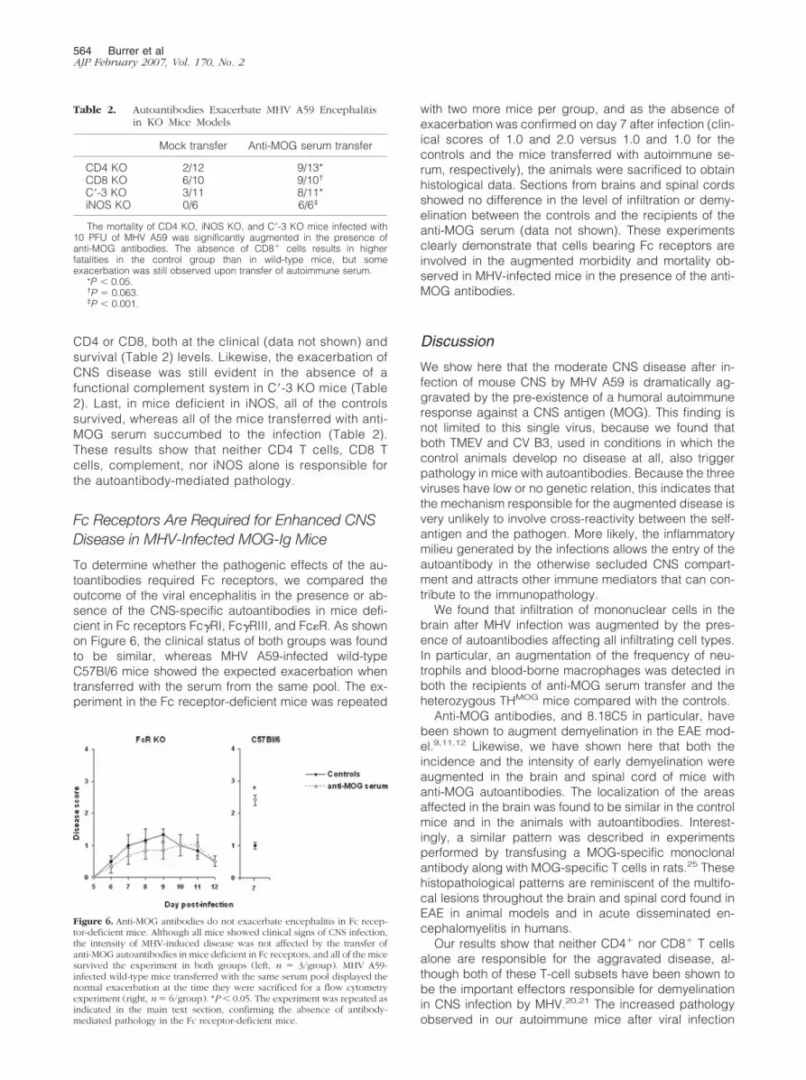
To determine whether the pathogenic effects of the au-toantibodies required Fc receptors, we compared theoutcome of the viral encephalitis in the presence or ab-sence of the CNS-specific autoantibodies in mice defi-cient in Fc receptors Fc�RI, Fc�RIII, and Fc�R. As shownon Figure 6, the clinical status of both groups was foundto be similar, whereas MHV A59-infected wild-typeC57Bl/6 mice showed the expected exacerbation whentransferred with the serum from the same pool. The ex-periment in the Fc receptor-deficient mice was repeated
with two more mice per group, and as the absence ofexacerbation was confirmed on day 7 after infection (clin-ical scores of 1.0 and 2.0 versus 1.0 and 1.0 for thecontrols and the mice transferred with autoimmune se-rum, respectively), the animals were sacrificed to obtainhistological data. Sections from brains and spinal cordsshowed no difference in the level of infiltration or demy-elination between the controls and the recipients of theanti-MOG serum (data not shown). These experimentsclearly demonstrate that cells bearing Fc receptors areinvolved in the augmented morbidity and mortality ob-served in MHV-infected mice in the presence of the anti-MOG antibodies.
Discussion
We show here that the moderate CNS disease after in-fection of mouse CNS by MHV A59 is dramatically ag-gravated by the pre-existence of a humoral autoimmuneresponse against a CNS antigen (MOG). This finding isnot limited to this single virus, because we found thatboth TMEV and CV B3, used in conditions in which thecontrol animals develop no disease at all, also triggerpathology in mice with autoantibodies. Because the threeviruses have low or no genetic relation, this indicates thatthe mechanism responsible for the augmented disease isvery unlikely to involve cross-reactivity between the self-antigen and the pathogen. More likely, the inflammatorymilieu generated by the infections allows the entry of theautoantibody in the otherwise secluded CNS compart-ment and attracts other immune mediators that can con-tribute to the immunopathology.
We found that infiltration of mononuclear cells in thebrain after MHV infection was augmented by the pres-ence of autoantibodies affecting all infiltrating cell types.In particular, an augmentation of the frequency of neu-trophils and blood-borne macrophages was detected inboth the recipients of anti-MOG serum transfer and theheterozygous THMOG mice compared with the controls.
Anti-MOG antibodies, and 8.18C5 in particular, havebeen shown to augment demyelination in the EAE mod-el.9,11,12 Likewise, we have shown here that both theincidence and the intensity of early demyelination wereaugmented in the brain and spinal cord of mice withanti-MOG autoantibodies. The localization of the areasaffected in the brain was found to be similar in the controlmice and in the animals with autoantibodies. Interest-ingly, a similar pattern was described in experimentsperformed by transfusing a MOG-specific monoclonalantibody along with MOG-specific T cells in rats.25 Thesehistopathological patterns are reminiscent of the multifo-cal lesions throughout the brain and spinal cord found inEAE in animal models and in acute disseminated en-cephalomyelitis in humans.
Our results show that neither CD4� nor CD8� T cellsalone are responsible for the aggravated disease, al-though both of these T-cell subsets have been shown tobe the important effectors responsible for demyelinationin CNS infection by MHV.20,21 The increased pathologyobserved in our autoimmune mice after viral infection
Figure 6. Anti-MOG antibodies do not exacerbate encephalitis in Fc recep-tor-deficient mice. Although all mice showed clinical signs of CNS infection,the intensity of MHV-induced disease was not affected by the transfer ofanti-MOG autoantibodies in mice deficient in Fc receptors, and all of the micesurvived the experiment in both groups (left, n � 3/group). MHV A59-infected wild-type mice transferred with the same serum pool displayed thenormal exacerbation at the time they were sacrificed for a flow cytometryexperiment (right, n � 6/group). *P � 0.05. The experiment was repeated asindicated in the main text section, confirming the absence of antibody-mediated pathology in the Fc receptor-deficient mice.
Table 2. Autoantibodies Exacerbate MHV A59 Encephalitisin KO Mice Models
Mock transfer Anti-MOG serum transfer
CD4 KO 2/12 9/13*CD8 KO 6/10 9/10†
C�-3 KO 3/11 8/11*iNOS KO 0/6 6/6‡
The mortality of CD4 KO, iNOS KO, and C�-3 KO mice infected with10 PFU of MHV A59 was significantly augmented in the presence ofanti-MOG antibodies. The absence of CD8� cells results in higherfatalities in the control group than in wild-type mice, but someexacerbation was still observed upon transfer of autoimmune serum.
*P � 0.05.†P � 0.063.‡P � 0.001.
564 Burrer et alAJP February 2007, Vol. 170, No. 2

thus results from a distinct autoantibody-mediated mech-anism rather than from exacerbation of the damagecaused directly by the virus or by the antiviral immuneresponse. The augmented disease is transferred by se-rum containing the autoantibodies and is Fc receptor-mediated. In further support of this hypothesis, viralreplication was similar in controls and mice with autoan-tibodies during the first 6 days of the infection and wasonly significantly higher in the autoimmune mice on day 7after infection. ELISPOT and intracellular cytokine dataindicated, however, that the viral-specific cytotoxic re-sponse was not significantly impaired in those mice, nei-ther at the systemic nor at the local level.
Complement deposition has been found in lesions inMS patients,10 and the demyelination potential of myelin-specific antibodies has been shown to correlate withcomplement-binding ability.26 The experiments per-formed in C�-3 KO mice indicate that in our model, com-plement has little or no involvement in the autoantibody-mediated pathology. Another possible source of CNSdamage can be NO,27 which has been shown to beinvolved in MS lesions.28 However, NO plays little role inMHV-induced demyelination, even though it can damageneurons during the infection,29,30 and iNOS-deficientmice have been reported to have a better survival ratethan wild-type animals.29 In accordance with this litera-ture, we found control iNOS KO mice to survive the in-fection better than the C57Bl/6 animals. However, ourexperiments showed that iNOS was not involved in theantibody-mediated pathology, because all of the KOmice that were transfused with autoimmune serum suc-cumbed to the infection. On the contrary, we found that adeficiency in cellular Fc receptors Fc�RI and Fc�RIII wassufficient to abrogate completely the autoantibody-medi-ated pathology at both clinical and histological levels.This indicates that neutrophils, macrophages, or NK cellsare responsible for the autoantibody-mediated pathol-ogy. Based on these results and on the characteristics ofthe mononuclear infiltration in the autoimmune mice, wepropose that cells of the monocyte/macrophage/micro-glia lineage, neutrophils, or NK cells are responsible forthe autoantibody generated damage, through a mecha-nism that does not involve complement or iNOS release.
The existence of cross-reactivity between antibodiesbinding to viral and self-antigens has been extensivelyinvestigated, but only a few reports, to our knowledge,have focused on the in vivo interplay of noncross-reactiveautoantibodies and viral infections. The �-herpesvirus�HV-68 has been shown to provoke relapses of experi-mental autoimmune arthritis in mice.31 Lactate dehydro-genase-elevating virus and MHV each resulted in dra-matic exacerbation of the pathology and mortality in amouse model of antibody-triggered autoimmune hemo-lytic anemia.32 Likewise, both lactate dehydrogenase-elevating virus and MHV infections also provoke a severethrombocytopenia in mice treated with anti-platelet anti-bodies at otherwise nonpathogenic doses.33 To ourknowledge, the present report constitutes the first de-scription of a similar phenomenon in the CNS compart-ment in an animal model. Our findings are consistent withprevious studies performed with human patients, which
suggest a correlation between the presence of autoanti-bodies in the cerebrospinal fluid (CSF) and the outcomeof encephalitis. Desai et al34 reported that fatal outcomein a cohort of 72 patients with Japanese encephalitis virusinfection was significantly associated to the presence ofCSF autoantibodies, against neurofilament proteins inparticular. Likewise, Matsui et al35 found that in a group of14 patients with viral encephalitis of undefined etiology,three of the four patients with CNS autoantibodies wereamong the total of five individuals with an unfavorableoutcome. Both of these studies were performed with pa-tients enrolled at the time that pathology was declared,making it impossible to determine whether the autoanti-bodies existed before the CNS infection or if they ap-peared as a consequence of the encephalitis. Likewise,there was no way to determine whether the autoantibod-ies had a direct role in the unfavorable outcomes or weremerely an associated marker. However, in view of theresults obtained in the present study, it is very possiblethat the autoimmune status of those patients had anadverse effect on the evolution of their CNS disease.
In conclusion, our study strengthens the hypothesisthat in individuals with pre-existing autoantibodies, withor without clinical autoimmune disease, certain virusescan have a heightened pathogenic potential. It reinforcesthe concept that pathogens that are unrelated to the initialdevelopment of the autoimmune condition may causeexacerbation episodes by modifying the local or systemicenvironment. The model of an autoimmune “fertile field”proposes that autoreactive T cells are first generated bysome infections through molecular mimicry or bystandermechanisms but may remain nonpathogenic for ex-tended periods and that unrelated viruses or bacteriamay be responsible for triggering the actual autoimmunepathology.1,4 In this model, Koch’s postulates (one dis-ease/one pathogen) are not strictly respected, which mayexplain why it has been so difficult to associate a singleorganism to a given autoimmune disease.
Acknowledgments
We are very grateful to Dr. Kent Osborn for his help withthe analysis of the morphological analysis of the infil-trates. We also thank Dr. Jason Botten for advice indesigning the ELISPOT assays and for useful discussion.
References
1. von Herrath MG, Fujinami RS, Whitton JL: Microorganisms andautoimmunity: making the barren field fertile? Nat Rev Microbiol 2003,1:151–157
2. Christen U, Edelmann KH, McGavern DB, Wolfe T, Coon B, TeagueMK, Miller SD, Oldstone MB, von Herrath MG: A viral epitope thatmimics a self antigen can accelerate but not initiate autoimmunediabetes. J Clin Invest 2004, 114:1290–1298
3. Panoutsakopoulou V, Sanchirico ME, Huster KM, Jansson M,Granucci F, Shim DJ, Wucherpfennig KW, Cantor H: Analysis of therelationship between viral infection and autoimmune disease. Immu-nity 2001, 15:137–147
4. McCoy L, Tsunoda I, Fujinami RS: Multiple sclerosis and virus in-duced immune responses: autoimmunity can be primed by molecular
CNS Autoantibodies and Viral Encephalitis 565AJP February 2007, Vol. 170, No. 2

mimicry and augmented by bystander activation. Autoimmunity 2006,39:9–19
5. Buljevac D, Flach HZ, Hop WC, Hijdra D, Laman JD, Savelkoul HF,van Der Meche FG, van Doorn PA, Hintzen RQ: Prospective study onthe relationship between infections and multiple sclerosis exacerba-tions. Brain 2002, 125:952–960
6. Andersen O, Lygner PE, Bergstrom T, Andersson M, Vahlne A: Viralinfections trigger multiple sclerosis relapses: a prospective seroepi-demiological study. J Neurol 1993, 240:417–422
7. Martin F, Chan AC: B cell immunobiology in disease: evolving con-cepts from the clinic. Annu Rev Immunol 2006, 24:467–496
8. Lyons JA, Ramsbottom MJ, Cross AH: Critical role of antigen-specificantibody in experimental autoimmune encephalomyelitis induced byrecombinant myelin oligodendrocyte glycoprotein. Eur J Immunol2002, 32:1905–1913
9. Genain CP, Cannella B, Hauser SL, Raine CS: Identification of auto-antibodies associated with myelin damage in multiple sclerosis. NatMed 1999, 5:170–175
10. Lucchinetti C, Bruck W, Parisi J, Scheithauer B, Rodriguez M, Lass-mann H: Heterogeneity of multiple sclerosis lesions: implications forthe pathogenesis of demyelination. Ann Neurol 2000, 47:707–717
11. Cross AH, Trotter JL, Lyons J: B cells and antibodies in CNS demy-elinating disease. J Neuroimmunol 2001, 112:1–14
12. Litzenburger T, Fassler R, Bauer J, Lassmann H, Linington C,Wekerle H, Iglesias A: B lymphocytes producing demyelinatingautoantibodies: development and function in gene-targeted trans-genic mice. J Exp Med 1998, 188:169–180
13. Lane TE, Buchmeier MJ: Murine coronavirus infection: a paradigm forvirus-induced demyelinating disease. Trends Microbiol 1997, 5:9–14
14. Matthews AE, Weiss SR, Paterson Y: Murine hepatitis virus: a modelfor virus-induced CNS demyelination. J Neurovirol 2002, 8:76–85
15. Sun N, Perlman S: Spread of a neurotropic coronavirus to spinal cordwhite matter via neurons and astrocytes. J Virol 1995, 69:633–641
16. Gombold JL, Sutherland RM, Lavi E, Paterson Y, Weiss SR: Mousehepatitis virus A59-induced demyelination can occur in the absenceof CD8� T cells. Microb Pathog 1995, 18:211–221
17. Bergmann CC, Ramakrishna C, Kornacki M, Stohlman SA: Impaired Tcell immunity in B cell-deficient mice following viral central nervoussystem infection. J Immunol 2001, 167:1575–1583
18. Dandekar AA, Jacobsen G, Waldschmidt TJ, Perlman S: Antibody-mediated protection against cytotoxic T-cell escape in coronavirus-induced demyelination. J Virol 2003, 77:11867–11874
19. Wu GF, Perlman S: Macrophage infiltration, but not apoptosis, iscorrelated with immune-mediated demyelination following murine in-fection with a neurotropic coronavirus. J Virol 1999, 73:8771–8780
20. Lane TE, Liu MT, Chen BP, Asensio VC, Samawi RM, Paoletti AD,Campbell IL, Kunkel SL, Fox HS, Buchmeier MJ: A central role forCD4(�) T cells and RANTES in virus-induced central nervous systeminflammation and demyelination. J Virol 2000, 74:1415–1424
21. Wu GF, Dandekar AA, Pewe L, Perlman S: CD4 and CD8 T cells have
redundant but not identical roles in virus-induced demyelination.J Immunol 2000, 165:2278–2286
22. Feuer R, Mena I, Pagarigan RR, Harkins S, Hassett DE, Whitton JL:Coxsackievirus B3 and the neonatal CNS: the roles of stem cells,developing neurons, and apoptosis in infection, viral dissemination,and disease. Am J Pathol 2003, 163:1379–1393
23. Rodriguez M, Leibowitz J, David CS: Susceptibility to Theiler’s virus-induced demyelination: mapping of the gene within the H-2D region.J Exp Med 1986, 163:620–631
24. Botten J, Alexander J, Pasquetto V, Barrowman P, Ting J, Peters B,Southwood S, Stewart B, Rodriguez-Carreno MP, Mothe B, WhittonJL, Sette A, Buchmeier MJ: Identification of protective Lassa virusepitopes that are restricted by HLA-A2. J Virol 2006, 80:8351–8361
25. Meeson AP, Piddlesden S, Morgan BP, Reynolds R: The distributionof inflammatory demyelinated lesions in the central nervous system ofrats with antibody-augmented demyelinating experimental allergicencephalomyelitis. Exp Neurol 1994, 129:299–310
26. Piddlesden SJ, Lassmann H, Zimprich F, Morgan BP, Linington C:The demyelinating potential of antibodies to myelin oligodendrocyteglycoprotein is related to their ability to fix complement. Am J Pathol1993, 143:555–564
27. Brown GC, Bal-Price A: Inflammatory neurodegeneration mediatedby nitric oxide, glutamate, and mitochondria. Mol Neurobiol 2003,27:325–355
28. Liu JS, Zhao ML, Brosnan CF, Lee SC: Expression of inducible nitricoxide synthase and nitrotyrosine in multiple sclerosis lesions. Am JPathol 2001, 158:2057–2066
29. Chen BP, Lane TE: Lack of nitric oxide synthase type 2 (NOS2) resultsin reduced neuronal apoptosis and mortality following mouse hepa-titis virus infection of the central nervous system. J Neurovirol 2002,8:58–63
30. Wu GF, Pewe L, Perlman S: Coronavirus-induced demyelination oc-curs in the absence of inducible nitric oxide synthase. J Virol 2000,74:7683–7686
31. Yarilin DA, Valiando J, Posnett DN: A mouse herpesvirus inducesrelapse of experimental autoimmune arthritis by infection of the in-flammatory target tissue. J Immunol 2004, 173:5238–5246
32. Meite M, Leonard S, Idrissi ME, Izui S, Masson PL, Coutelier JP:Exacerbation of autoantibody-mediated hemolytic anemia by viralinfection. J Virol 2000, 74:6045–6049
33. Musaji A, Cormont F, Thirion G, Cambiaso CL, Coutelier JP: Exacer-bation of autoantibody-mediated thrombocytopenic purpura by infec-tion with mouse viruses. Blood 2004, 104:2102–2106
34. Desai A, Ravi V, Guru SC, Shankar SK, Kaliaperumal VG, Chan-dramuki A, Gourie-Devi M: Detection of autoantibodies to neuralantigens in the CSF of Japanese encephalitis patients and correlationof findings with the outcome. J Neurol Sci 1994, 122:109–116
35. Matsui M, Tanaka K, Nagumo F, Kuroda Y: Central nervous systemimmunity associated with clinical outcome in acute encephalitis.J Neurol Sci 2004, 227:139–147
566 Burrer et alAJP February 2007, Vol. 170, No. 2