identification of autoantibodies to rna polymerase ii. occurrence in systemic sclerosis and...
TRANSCRIPT
Identification of Autoantibodies to RNA Polymerase 11Occurrence in Systemic Sclerosis and Association with Autoantibodies to RNA Polymerases I and III
Michito Hirakata, * Yutaka Okano,t Uttam Pati, Akira Suwa, * Thomas A. Medsger, Jr.,t John A. Hardin, and Joe Craft**Section ofRheumatology, Department ofInternal Medicine, Yale University School ofMedicine, New Haven, Connecticut 06510;tDivision ofRheumatology and Clinical Immunology, Department ofMedicine, University ofPittsburgh School ofMedicine,Pittsburgh, Pennsylvania 15261; and ODepartment ofMedicine, Medical College ofGeorgia, Augusta, Georgia 30912-3100
Abstract
In this study, autoantibodies to RNA polymerase II from seraof patients with systemic sclerosis have been identified andcharacterized. These antibodies immunoprecipitated polypep-tides of 220 kD (IIA) and 145 kD (IIC), the two largest sub-units ofRNA polymerase II, and bound both subunits in immu-noblots. These polypeptides were immunoprecipitated by theanti-RNA polymerase II monoclonal antibody 8WG16, whichrecognizes the carboxyl-terminal domain of the 220-kD sub-unit, and their identity to the proteins bound by human sera wasconfirmed in immunodepletion studies. Sera with anti-RNApolymerase II antibodies also immunoprecipitated proteinsthat were consistent with components of RNA polymerases Iand III. In vitro transcription experiments showed that the hu-man antibodies were an effective inhibitor ofRNA polymeraseII activity. In indirect immunofluorescence studies, anti-RNApolymerase II autoantibodies stained the nucleoplasm, as ex-pected from the known location of RNA polymerase II, andcolocalized with the anti-RNA polymerase II monoclonal anti-body. The human sera also stained the nucleolus, the location ofRNA polymerase I. From a clinical perspective, these antibod-ies were found in 13 of 278 patients with systemic sclerosis,including 10 with diffuse and three with limited cutaneous dis-ease, but were not detected in sera from patients with otherconnective tissue diseases and from normal controls. We con-clude that anti-RNA polymerase II antibodies are specific topatients with systemic sclerosis, and that they are apparentlyassociated with antibodies to RNA polymerases I and III.These autoantibodies may be useful diagnostically and as aprobe for further studies ofthe biological function ofRNA poly-merases. (J. Clin. Invest. 1993. 91:2665-2672.) Key words:RNA polymerases - autoantibodies * antinuclear antibodies.systemic sclerosis * autoimmunity
Introduction
The three RNA polymerases (RNAP)' catalyze the transcrip-tion of different sets of genes into RNA. RNAP I synthesizes
Address correspondence to Joe Craft, M.D., Section ofRheumatology,610 LCI, Yale University School ofMedicine, Box 3333, 333 Cedar St.,New Haven, CT 06510.
Receivedfor publication 30 July 1993 and in revisedform 28 De-cember 1992.
ribosomal RNA precursors in nucleoli. RNAP II synthesizesthe precursors ofmRNAs and most ofthe small nuclear RNAswhich are found in ribonucleoprotein (RNP) particles that me-diate pre-mRNA splicing. RNAP III synthesizes small RNAs,including 5S ribosomal RNAs and transfer RNAs. These mul-timeric enzymes are comprised of 8-14 polypeptide subunits,depending upon the species from which they are isolated ( 1, 2);however, the details of their complex structures are still beingdefined.
In mammalian cells, RNAP II contains two large polypep-tides (220 kD and 145 kD) that are highly conserved acrossspecies, and at least six smaller subunits ( 1, 2). Several of thelatter may be shared among RNAP I, II, and III, whereas thelarger two subunits are unique to RNAP 11 ( 1-5). The largest220-kD polypeptide ofRNAP II migrates as a phosphorylatedsubunit of 240 kD, a nonphosphorylated subunit of 220 kD,and a proteolytic derivative of 180 kD, referred to as IIO, IIA,and IIB, respectively, when analyzed in SDS-polyacrylamidegels (4, 6-8). Recent studies have revealed that the 220-kDsubunit contains a unique carboxyl-terminal domain (CTD)consisting of a Tyr-Ser-Pro-Thr-Ser-Pro-Ser heptapeptide thatis repeated 52 times in mammalian cells (2, 4, 7, 9). The CTDis released by proteolysis generating the 1 80-kD subunit (10,11). This sequence is highly phosphorylated, required for cellviability, and thought to mediate promoter specific transcrip-tion (12, 13). Thompson and colleagues have developed amonoclonal antibody referred to as 8WG16 (anti-CTD mAb)that reacts with this highly conserved carboxyl-terminal repeatof the 220-kD subunit ( 14, 15).
Some patients with the connective tissue disease systemicsclerosis (scleroderma) have been shown to produce autoanti-bodies that bind RNA polymerase I ( 16, 17). Correspondingautoantibodies to RNAP II and III have not yet been identified.In the present study, we have identified and characterized auto-antibodies from sera of patients with systemic sclerosis thatrecognize RNAP II subunits.
Methods
Human autoimmune sera and RNAP II monoclonal antibody. Serawere obtained from 84 patients with systemic sclerosis (diffuse cutane-ous systemic sclerosis, limited cutaneous systemic sclerosis, and sys-temic sclerosis overlap syndromes) who were identified from a reviewof patient records of the Section of Rheumatology, Yale University
1. Abbreviations used in this paper: CMV, cytomegalovirus; CTD, car-boxyl-terminal domain; MCTD, mixed connective tissue disease; PM/DM, polymyositis or dermatomyositis; RNAP, RNA polymerase;RNP, ribonucleoprotein; snRNP, small nuclear RNP; TBS, Tris-buff-ered saline.
Autoantibodies to RNA Polymerase II 2665
J. Clin. Invest.© The American Society for Clinical Investigation, Inc.0021-9738/93/06/2665/08 $2.00Volume 91, June 1993, 2665-2672
School of Medicine, and from 194 patients with systemic sclerosis whowere seen at the University of Pittsburgh School of Medicine during1986-1988. All patients seen at Yale University and 167 of the 194patients seen at University of Pittsburgh fulfilled the preliminary crite-ria of the American College of Rheumatology for classification as defi-nite systemic sclerosis ( 18). Sera from 217 patients with other systemicautoimmune diseases, including SLE (n = 126) (19), polymyositis/dermatomyositis (PM/DM) (n = 64) (20), and mixed connectivetissue disease (MCTD) (n = 27) (2 1 ), and sera from 30 normal individ-uals were used as controls. A murine monoclonal antibody (8WG16;anti-CTD mAb) ( 14), raised against wheat germ RNAP II, whichreacts with the carboxyl-terminal repeat domain of the largest subunitof RNAP II, was a kind gift from Dr. Nancy E. Thompson (McArdleLaboratory for Cancer Research, University ofWisconsin). This mAbbinds the 220-kD large subunit of RNAP II in immunoblots and asynthetic heptapeptide repeat corresponding to the CTD region of thispolypeptide. It also inhibits promoter-specific transcription by RNAPII (14, 15).
Indirect immunofluorescence. Human sera were diluted 1:40 inTris-buffered saline (TBS) ( 150 mM NaCl, 10mM Tris HCl, pH 7.4),and were screened by indirect immunofluorescence (22) using HEp-2cells (Immunoconcepts, Sacramento, CA) with goat anti-human IgGfluorescein isothiocyanate conjugate (Sigma, St. Louis, MO) and goatanti-mouse IgG Texas red conjugate (Tago, Inc., Burlingame, CA).Each well was viewed at a magnification of 1,000 on an immunofluores-cence microscope (Carl Zeiss, Oberkochen, Germany) to detect cyto-plasmic, nuclear, and nucleolar staining.
Preparation of radiolabeled cell extracts. HeLa cells (2-2.5 X 10⁵cells/ml) were radiolabeled for 14 h in deficient media, as previouslydescribed, with [35S]methionine ( 10 qCi/ml cells) (ICN BiomedicalInc., Irvine, CA) for analysis of proteins and with [32P]orthophosphate( 10 pCi/ml cells) (ICN Biomedical Inc.) for RNA analysis (22, 23).Cells were collected by centrifugation, washed in TBS ( 10 mM Tris Cl,pH 7.5, 150 mM NaCI), and sonicated three times each for 40 s inNET-2 buffer (50 mM Tris Cl, pH 7.5, 150 mM NaCl, 0.05% NonidetP-40) with a sonifier (Branson Sonic, Danbury, CT) at setting 3. Thesonicated lysate was centrifuged at 15,000 g for 10 min to removecellular debris.
Immunoprecipitation ofradiolabeled cell extracts. Immunoprecipi-tation of radiolabeled cell extracts was performed as previously de-scribed (22-24). Briefly, 10 ,ul of patient sera was mixed with 2 mg ofprotein A-sepharose CL-4B (Pharmacia Inc., Piscataway, NJ) in 500 /IIofimmunoprecipitation buffer (IPP) ( 10 mM Tris Cl, pH 8.0, 500mMNaCI, 0.1% Nonidet P-40) and incubated with end-over-end rotation(Labquake shaker; Lab Industries, Berkeley, CA) for 2 h at 4°C. Thesepharose particles with adsorbed IgG were washed four times in 500 1AIof IPP buffer using 10-s spins in a microfuge tube, and resuspended in400 ,ul ofNET-2 buffer. For protein studies, antibody-coated sepharosebeads were mixed with 400 ,l of [35S]methionine-labeled extracts (8X 10⁶ cells) and rotated at 4°C for 2 h. After four washes with NET-2,the sepharose beads were resuspended in SDS sample buffer (2% SDS,10% glycerol, 62.5 mM Tris Cl, pH 6.8, 0.005% bromophenol blue).After heating (90°C for 5 min), the proteins were fractionated on SDS-10% polyacrylamide gels, enhanced with 0.5 M sodium salicylate, anddried; labeled proteins were analyzed by autoradiography. Immuno-precipitated RNAs were analyzed as previously described (22, 23).
Antigen depletion. [ 35S ] methionine-labeled HeLa cell extracts wereincubated with 20-160 tg ofanti-CTD mAb bound to protein A Sepha-rose CL-4B for 2 h at 4°C to deplete the extracts of RNAP II. Thedepleted supernatant was preincubated with protein A-sepharose toabsorb any antibody excess, and then used in immunoprecipitation asthe antigen source for patient anti-RNAP II sera. In the reciprocalexperiment, [35S ] methionine-labeled HeLa cell extracts were absorbedwith 40 gig of IgG isolated from an anti-RNAP II human serum asdescribed and then probed with the anti-CTD mAb. The same amountof IgG from a normal human serum and from an SLE serum with theanti-Sm specificity (25) were included as controls.
In vitro transcription from the adenovirus major late promoter andfrom the cytomegalovirus (CMV) immediate early promoter. Dignamextracts from HeLa cells were used for studying transcription fromboth these promoters (26). For transcription from the adenovirus ma-jor late promoter, the plasmid vector pBR 322 containing the 877-bpfragment with the major late promoter region (from position -680 toposition + 197) was prepared as previously described (27). The gel-purified fragment was used as a template in a transcription assay togenerate a single 197-nucleotide transcript. Transcription from theCMV immediate early promoter (Promega, Madison, WI) was carriedout according to the protocol of the manufacturer; the CMV run-offproduct was 363 nucleotides.
Purification ofRNAP II subunits. Purification ofRNA polymeraseII from calfthymus was carried out by a modification ofthe method ofHodo and Blatti (28), except that all buffers contained 0.1% NonidetP-40. Fractions from the phosphocellulose column containing RNAPII activity were pooled and applied to a heparin-agarose column equili-brated with buffer containing 60 mM ammonium sulfate. Active frac-tions were eluted with buffer containing 500 mM ammonium sulfate.The active fractions were pooled and passed through a Sephadex A-25column. RNAP II was further purified by glycerol gradient centrifuga-tion. RNAP II polypeptides were analyzed on gradient (7-15%) poly-acrylamide gels and the 145-kD polypeptide ofRNAP II was separatedfrom the gel by an electroeluter (Bio Rad, Richmond, CA). Purifica-tion ofthe 220-kD polypeptide ofRNAP II from HeLa cells was carriedout by immunoaffinity chromatography with the anti-CTD mAb8WG 16 (14, 15). The purified 145-kD and 220-kD subunits were usedas substrates for immunoblots.
Immunoblots. Immunoblots were performed by a modification ofthe Towbin et al. procedure (29).
Results
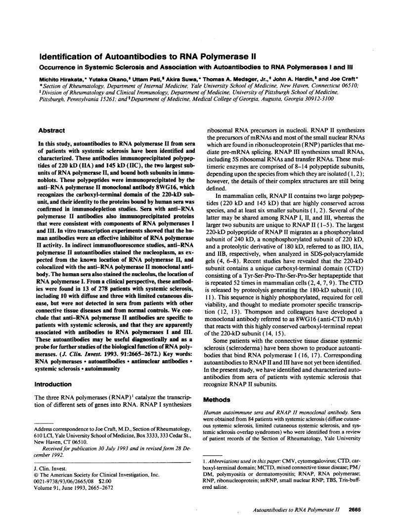
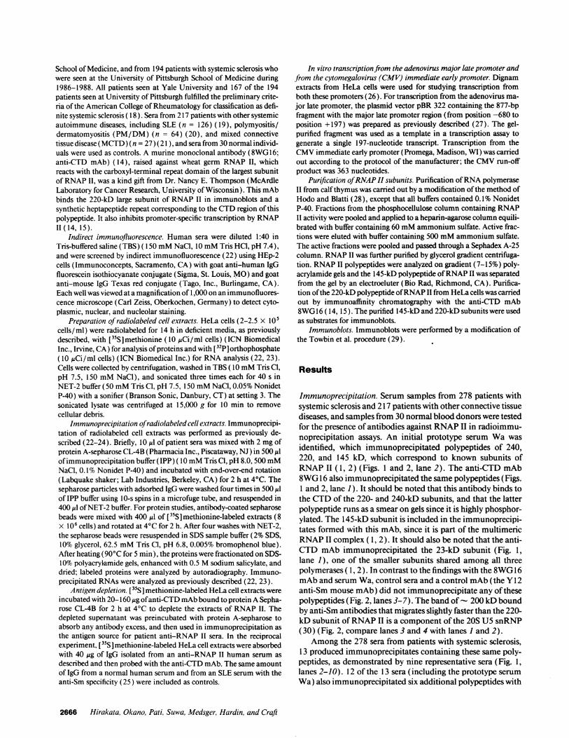
Immunoprecipitation. Serum samples from 278 patients withsystemic sclerosis and 217 patients with other connective tissuediseases, and samples from 30 normal blood donors were testedfor the presence of antibodies against RNAP II in radioimmu-noprecipitation assays. An initial prototype serum Wa wasidentified, which immunoprecipitated polypeptides of 240,220, and 145 kD, which correspond to known subunits ofRNAP 11 (1, 2) (Figs. 1 and 2, lane 2). The anti-CTD mAb8WG 16 also immunoprecipitated the same polypeptides (Figs.1 and 2, lane 1). It should be noted that this antibody binds tothe CTD of the 220- and 240-kD subunits, and that the latterpolypeptide runs as a smear on gels since it is highly phosphor-ylated. The 145-kD subunit is included in the immunoprecipi-tates formed with this mAb, since it is part of the multimericRNAP II complex ( 1, 2). It should also be noted that the anti-CTD mAb immunoprecipitated the 23-kD subunit (Fig. 1,lane 1), one of the smaller subunits shared among all threepolymerases ( 1, 2). In contrast to the findings with the 8WG16mAb and serum Wa, control sera and a control mAb (the Y12anti-Sm mouse mAb) did not immunoprecipitate any of thesepolypeptides (Fig. 2, lanes 3-7). The band of- 200 kD boundby anti-Sm antibodies that migrates slightly faster than the 220-kD subunit ofRNAP II is a component of the 20S U5 snRNP(30) (Fig. 2, compare lanes 3 and 4 with lanes 1 and 2).
Among the 278 sera from patients with systemic sclerosis,13 produced immunoprecipitates containing these same poly-peptides, as demonstrated by nine representative sera (Fig. 1,lanes 2-10). 12 of the 13 sera (including the prototype serumWa) also immunoprecipitated six additional polypeptides with
2666 Hirakata, Okano, Pati, Suwa, Medsger, Hardin, and Craft
-Q
C)
Mr 'ac
E
j FCf)I
Human sera
kD
200-- - * ;
~~~~~~0 mv .W * ONtE~"" *W.~_
97.4--#
69- ii
46- - --
30--- e - -
1 2 3 4 5 6 7 8 9 10 11 12 13 14
Figure 1. ["S ]methionine-labeled HeLacell proteins immunoprecipitated with hu-man sera and the anti-CTD mAb, followedby resolution in an SDS-7% polyacrylamidegel (24). The anti-CTD mAb immunopre-cipitated RNAP II polypeptides of 240,220, and 145 kD (lane 1). The anti-RNAPII prototype serum Wa (lane 2) and otherrepresentative sera from patients with sys-temic sclerosis immunoprecipitated theidentical polypeptides (lanes 3-10). Thesesera also immunoprecipitated polypeptidesof 190, 155, 138, 126, 44, and 23 kD (lanes2-8 and 10), except for one serum (lane9), which did not react with the 190-, 126-,and 44-kD polypeptides. Sera presumablycontaining anti-RNAP I/RNAP III anti-bodies immunoprecipitated the same sixpolypeptides, but not those of 240, 220,and 145 kD (lanes 11-14). M, shows mo-lecular weight markers; asterisks and arrowsdenote the 240-, 220-, and 145-kD subunitsofRNAP II.
-o
I- m
Mr icz
-o
Ec\1J
-j
C/) cn YIcul'CIO I SCI -M
200- EM__
97.4-
69- _
46- _ 2 -
1 2 3 4 5 6 7Figure 2. [35S]methionine-labeled HeLa cell proteins immunoprecipi-tated with the anti-CTD mAb, human sera, and the Y12 anti-SmmAb, followed by resolution in an SDS-7% polyacrylamide gel (24).The anti-CTD mAb immunoprecipitated RNAP II polypeptides of240, 220, and 145 kD (lane 1); the bands at - 100 and 70 kD rep-
resent degradation products that were not reproducibly seen in otherimmunoprecipitates formed with the anti-CTD mAb. The anti-RNAP II prototype serum Wa immunoprecipitated the identicalpolypeptides as the CTD mAb (lane 2). The Y12 anti-Sm mAb andan anti-Sm patient serum immunoprecipitated the U5-specific dou-blet of 200 kD (30) and other snRNP proteins (lanes 3 and 4). Ananti-La patient serum immunoprecipitated the 48-kD La protein(lane 5), an anti-Ku patient serum immunoprecipitated the 70- and80-kD Ku polypeptides (lane 6), and a normal human serum (NHS)did not immunoprecipitate any specific polypeptides. M, shows mo-lecular weight markers.
molecular masses of 190, 155, 138, 126, 44, and 23 kD, whichcorrespond to known subunits of RNAP I and RNAP III (1)(Fig. 1, examples in lanes 2-8 and 10). In contrast, the serumshown in lane 9 immunoprecipitated only the 155-, 138-, and23-kD polypeptides, along with the 240-, 220-, and 145-kDsubunits of RNAP II.
Sera from another 54 patients with systemic sclerosis im-munoprecipitated the six smaller proteins (190, 155, 138, 126,44, and 23 kD), without immunoprecipitation of the 240-,220-, and 145-kD subunits of RNAP II (Fig. 1, examples inlanes 11-14). None of the control specimens, consisting of 30normal human sera and 217 autoimmune sera from patientswith SLE, MCTD, and PM/DM were able to immunoprecipi-tate the RNAP II profile, or the 190-, 155-, 138-, 126-, 44-, and23-kD proteins which, by their size, are consistent with compo-nents ofRNAP I and III. When studies were carried out withHeLa cells labeled with [32P]orthophosphate (22, 23), nosmall RNAs were specifically immunoprecipitated by 13 anti-RNAP II sera, in contrast to their ready immunoprecipitationby positive control sera (anti-Sm, anti-U 1 RNP, anti-Ro, andanti-La; data not shown).
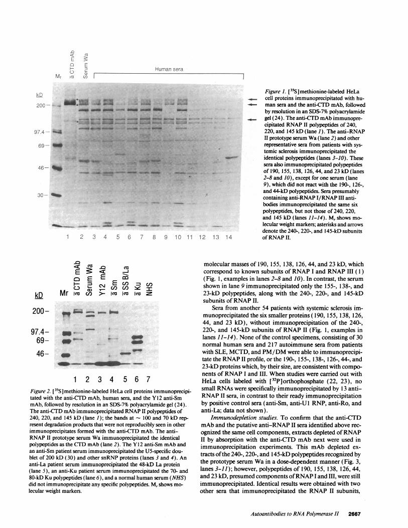
Immunodepletion studies. To confirm that the anti-CTDmAb and the putative anti-RNAP II sera identified above rec-ognized the same cell components, extracts depleted ofRNAPII by absorption with the anti-CTD mAb next were used inimmunoprecipitation experiments. This mAb depleted ex-tracts ofthe 240-, 220-, and 145-kD polypeptides recognized bythe prototype serum Wa in a dose-dependent manner (Fig. 3,lanes 3-11); however, polypeptides of 190, 155, 138, 126, 44,and 23 kD, presumed components ofRNAP-I and III, were stillimmunoprecipitated. Identical results were obtained with twoother sera that immunoprecipitated the RNAP II subunits,
Autoantibodies to RNA Polymerase II 2667
kD
DC) <1c E
.9_cO0 C
Z .9CL a)a)
E -0U) co
Mr Imka
£2
E
HC)
X 2X 4X 8X
: : :::.kD
200- w
97.4- bI
69-
46-
30- 4IM
1 2 3 4 5 6 7 8 9 10 11
along with the putative polypeptides of RNAP I and III. In areciprocal fashion, when HeLa cell extracts were absorbed withserum Wa and then probed with the anti-CTD mAb, theRNAP II subunits of 240, 220, and 145 kD were not immuno-precipitated (Fig. 3, lane 2). In contrast, depletion of radiola-beled HeLa cell extracts using autoantibodies of different im-munologic specificities (e.g., anti-Sm antibodies) did not affectspecific antigen recognition by the anti-CTD mAb (Fig. 3, lane1). These findings indicate that the epitope bound by the mono-clonal antibody to RNAP II resides on the same macromolecu-lar structure recognized by the autoantibodies from sera of pa-tients with systemic sclerosis.
Similar immunodepletion studies using human sera con-firmed that the six smaller polypeptides (190, 155, 138, 126,44, and 23 kD) immunoprecipitated by the anti-RNAP II serawere identical to those bound by the additional 54 sera fromsystemic sclerosis patients that did not bind RNAP II (data notshown).
Immunofluorescence studies. Indirect immunofluores-cence was next used to identify the cellular location of theantigen(s) bound by the sera studied here. As shown by a repre-sentative example, all 13 systemic sclerosis sera that immuno-precipitated RNAP II subunits produced nucleoplasmic andnucleolar staining that was indistinguishable from that ob-served with a control antibody to RNAP I (Fig. 4, A and B,respectively). In contrast, staining produced with the anti-CTD mAb was limited to the nucleoplasm (Fig. 4, C). There-fore, to further define the site of binding of the human anti-RNAP II antibodies, double immunostaining was performedusing the patient sera and the anti-CTD mAb. The location ofthe both antibodies was then determined with goat anti-hu-man IgG conjugated to fluorescein isothiocyanate and goat
£2< C- CZ
C E°0
E~~~~IE5e Co
1EC/) '0 Amoin:
0 X 2X 4X 8X 0 -a CTDrAD(x = 20 Lig
~~~~~~~~~~~~~~~~~3SFigure 3. Depletion of RNAP II from [355J-methionine-labeled HeLa cell extracts by the anti-CTD mAb and human anti-RNAP II antibodies. Theanti-CTD mAb immunoprecipitated the RNAP IIlarge subunits (240-, 220-, and 145-kD polypeptides;denoted by asterisks and arrows) in a dose-dependentmanner (lanes 3-6), and effectively depleted theseproteins from solution before immunoprecipitationwith the prototype human serum Wa (lanes 7-11).Immunoprecipitation studies shown in lanes 8-11were performed with the serum Wa and the superna-tants were derived from the immunoprecipitatesshown in lanes 3-6. A control immunoprecipitationperformed with an anti-Sm serum from a patient withSLE is shown in lane 12. The supernatant ofthe latterimmunoprecipitation still contained RNAP II largesubunits (240-, 220-, and 145-kD polypeptides; de-noted by asterisks), which could be immunoprecipi-tated with the anti-CTD mAb (lane 1), whereas thesupernatant absorbed with serum Wa from lane 7 did
12 not contain these polypeptides (lane 2).
anti-mouse IgG conjugated to Texas red. As shown by theorange fluorescence, the nucleoplasm was stained by both theanti-CTD mAb and anti-RNAP II sera (Fig. 4, D). In contrast,cells stained with normal human sera did not produceimmuno-fluorescence, while those stained with anti-Sm antibodies(from human sera and the Y12 mAb) produced only finelyspeckled nuclear fluorescence (data not shown).
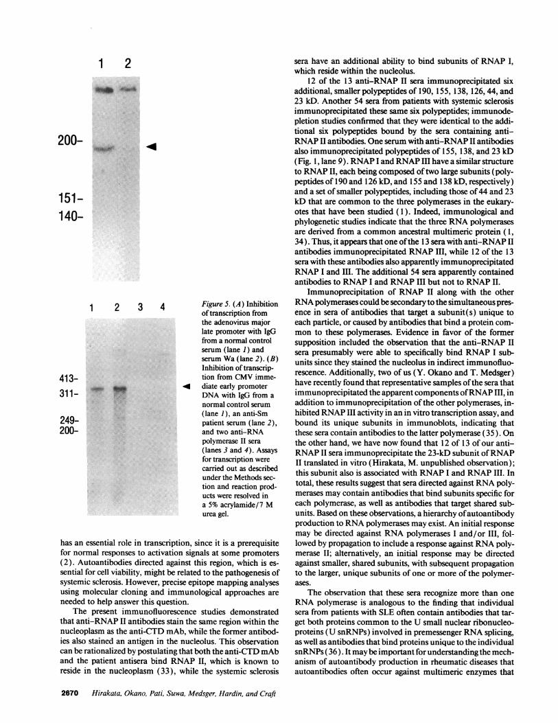
Inhibition of transcription from the adenovirus major latepromoter andfrom the CMV immediate early promoter withIgG from systemic sclerosis sera. These studies were designedto test the ability of anti-RNAP II antibodies from patients toinhibit promoter-directed transcription in vitro. In these tran-scription assays, 10 Ail ofa Dignam extract was incubated for 30min with IgG (from 50 ,l of patient serum) bound to proteinA-sepharose beads, before the addition ofNTPs and the DNAtemplate. The beads then were removed with centrifugationand the supernatant was used for the transcription from theDNA segments containing the adenovirus major late promoterand the CMV immediate early promoter. RNA was purifiedand analyzed on a 5% polyacrylamide/7 M urea gel (26). IgGfrom serum Wa and from a second serum that immunoprecipi-tated RNAP II subunits significantly inhibited RNAP II activ-ity (Fig. 5 A, lane 2; Fig. 5 B, lanes 3 and 4). In contrast,normal human IgG (Fig. 5 A and B, lane 1) and IgG from ananti-Sm patient serum did not produce any inhibition (Fig. 5B, lane 2). In this assay system, no transcription was detectedwhen a-amanitin was used at a concentration of 1 gg/ml,which inhibits RNAP II activity, but not RNAP I activity (datanot shown) (27).
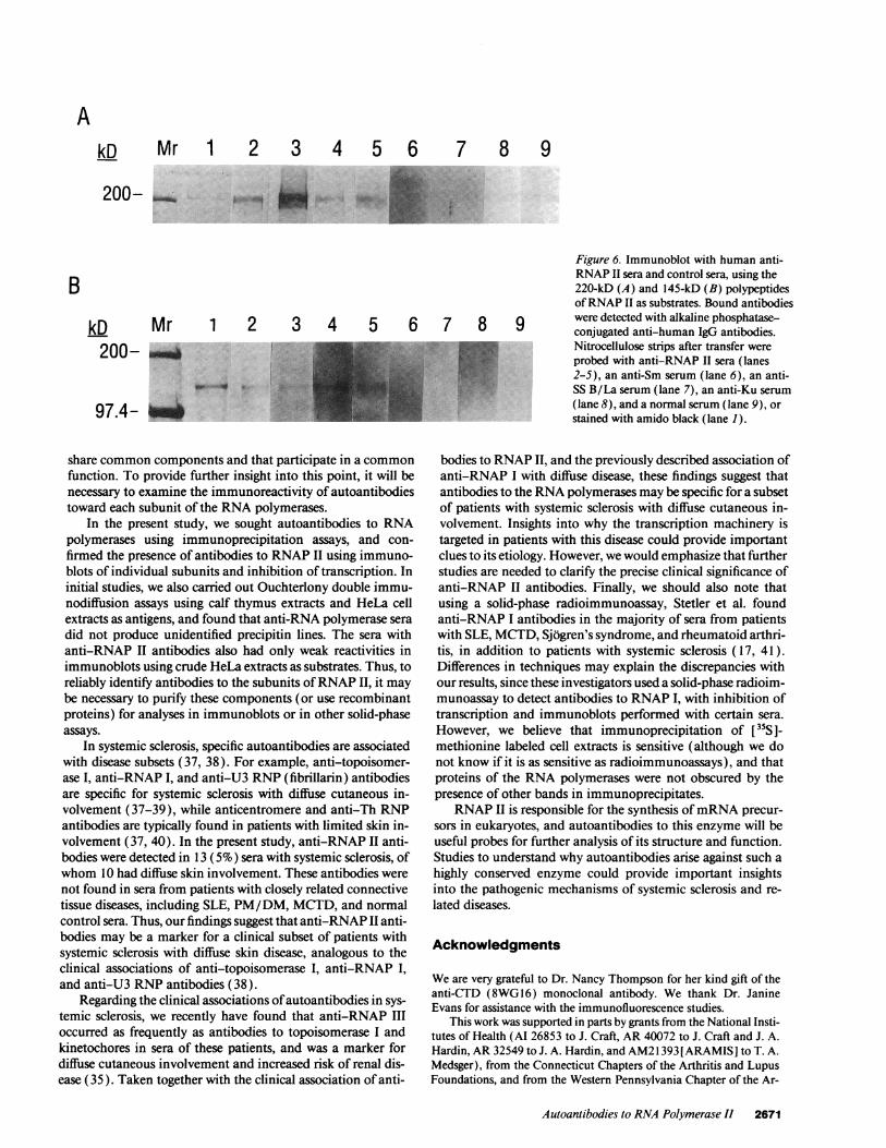
Immunoreactivity ofpatient anti-RNAP II antibodies withthe 220- and 145-kD polypeptides ofRNA polymerase I. Inimmunoblots, 9 of 13 anti-RNAP II sera bound the 220-kD
2668 Hirakata, Okano, Pati, Suwa, Medsger, Hardin, and Craft
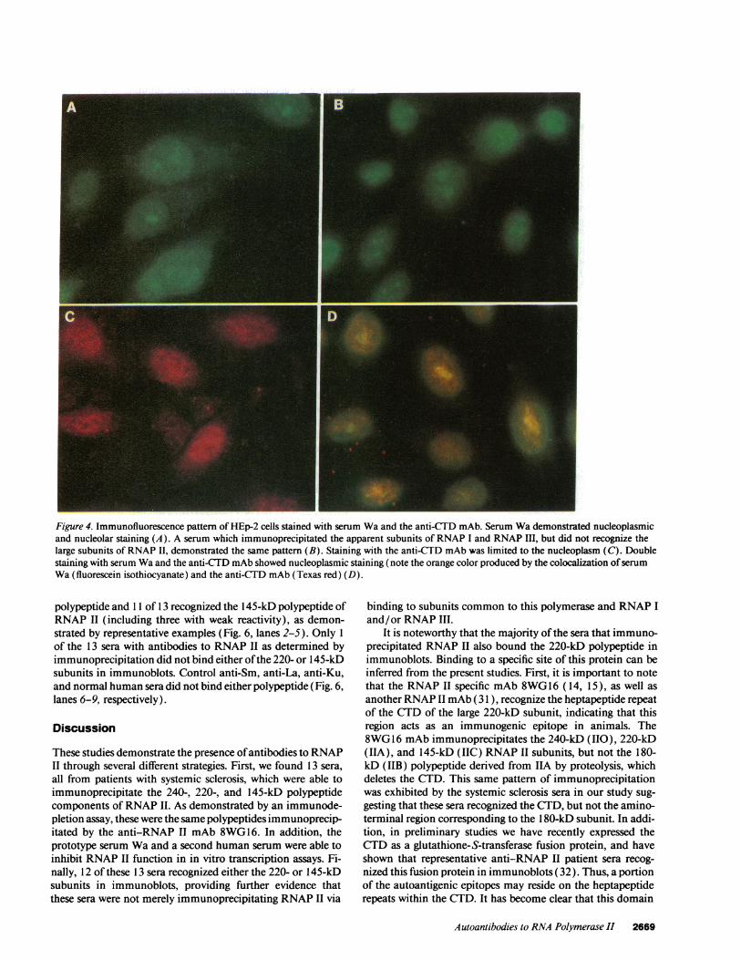
Figure 4. Immunofluorescence pattern of HEp-2 cells stained with serum Wa and the anti-CTD mAb. Serum Wa demonstrated nucleoplasmicand nucleolar staining (A). A serum which immunoprecipitated the apparent subunits of RNAP I and RNAP III, but did not recognize thelarge subunits of RNAP II, demonstrated the same pattern (B). Staining with the anti-CTD mAb was limited to the nucleoplasm (C). Doublestaining with serum Wa and the anti-CTD mAb showed nucleoplasmic staining (note the orange color produced by the colocalization ofserumWa (fluorescein isothiocyanate) and the anti-CTD mAb (Texas red) (D).
polypeptide and 11 of 13 recognized the 145-kD polypeptide ofRNAP II (including three with weak reactivity), as demon-strated by representative examples (Fig. 6, lanes 2-5). Only 1of the 13 sera with antibodies to RNAP II as determined byimmunoprecipitation did not bind either ofthe 220- or 145-kDsubunits in immunoblots. Control anti-Sm, anti-La, anti-Ku,and normal human sera did not bind either polypeptide (Fig. 6,lanes 6-9, respectively).
Discussion
These studies demonstrate the presence ofantibodies to RNAPII through several different strategies. First, we found 13 sera,all from patients with systemic sclerosis, which were able toimmunoprecipitate the 240-, 220-, and 145-kD polypeptidecomponents ofRNAP II. As demonstrated by an immunode-pletion assay, these were the same polypeptides immunoprecip-itated by the anti-RNAP II mAb 8WG 16. In addition, theprototype serum Wa and a second human serum were able toinhibit RNAP II function in in vitro transcription assays. Fi-nally, 12 of these 13 sera recognized either the 220- or 145-kDsubunits in immunoblots, providing further evidence thatthese sera were not merely immunoprecipitating RNAP II via
binding to subunits common to this polymerase and RNAP Iand/or RNAP III.
It is noteworthy that the majority ofthe sera that immuno-precipitated RNAP II also bound the 220-kD polypeptide inimmunoblots. Binding to a specific site of this protein can beinferred from the present studies. First, it is important to notethat the RNAP II specific mAb 8WG16 (14, 15), as well asanother RNAP II mAb (31 ), recognize the heptapeptide repeatof the CTD of the large 220-kD subunit, indicating that thisregion acts as an immunogenic epitope in animals. The8WG16 mAb immunoprecipitates the 240-kD (110), 220-kD(IIA), and 145-kD (IIC) RNAP II subunits, but not the 180-kD (IIB) polypeptide derived from IIA by proteolysis, whichdeletes the CTD. This same pattern of immunoprecipitationwas exhibited by the systemic sclerosis sera in our study sug-gesting that these sera recognized the CTD, but not the amino-terminal region corresponding to the 1 80-kD subunit. In addi-tion, in preliminary studies we have recently expressed theCTD as a glutathione-S-transferase fusion protein, and haveshown that representative anti-RNAP II patient sera recog-nized this fusion protein in immunoblots (32). Thus, a portionof the autoantigenic epitopes may reside on the heptapeptiderepeats within the CTD. It has become clear that this domain
Autoantibodies to RNA Polymerase II 2669
1 2
200-
151-140-
1 2 3 4 Figure 5. (A) Inhibitionof transcription fromthe adenovirus majorlate promoter with IgGfrom a normal controlserum (lane 1) andserum Wa (lane 2). (B)Inhibition oftranscrip-
413- tion from CMV imme-311-~ 4 diate early promoter311- r DNA with IgG from a
normal control serum(lane 1), an anti-Sm
249- patient serum (lane 2),200- and two anti-RNA
polymerase II sera(lanes 3 and 4). Assaysfor transcription werecarried out as describedunder the Methods sec-tion and reaction prod-ucts were resolved ina 5% acrylamide/7 Murea gel.
has an essential role in transcription, since it is a prerequisitefor normal responses to activation signals at some promoters(2). Autoantibodies directed against this region, which is es-sential for cell viability, might be related to the pathogenesis ofsystemic sclerosis. However, precise epitope mapping analysesusing molecular cloning and immunological approaches areneeded to help answer this question.
The present immunofluorescence studies demonstratedthat anti-RNAP II antibodies stain the same region within thenucleoplasm as the anti-CTD mAb, while the former antibod-ies also stained an antigen in the nucleolus. This observationcan be rationalized by postulating that both the anti-CTDmAband the patient antisera bind RNAP II, which is known toreside in the nucleoplasm (33), while the systemic sclerosis
sera have an additional ability to bind subunits of RNAP I,which reside within the nucleolus.
12 of the 13 anti-RNAP II sera immunoprecipitated sixadditional, smaller polypeptides of 190, 155, 138, 126,44, and23 kD. Another 54 sera from patients with systemic sclerosisimmunoprecipitated these same six polypeptides; immunode-pletion studies confirmed that they were identical to the addi-tional six polypeptides bound by the sera containing anti-RNAP II antibodies. One serum with anti-RNAP II antibodiesalso immunoprecipitated polypeptides of 155, 138, and 23 kD(Fig. 1, lane 9). RNAP I and RNAP III have a similar structureto RNAP II, each being composed oftwo large subunits (poly-peptides of 190 and 126 kD, and 155 and 138 kD, respectively)and a set of smaller polypeptides, including those of44 and 23kD that are common to the three polymerases in the eukary-otes that have been studied (1). Indeed, immunological andphylogenetic studies indicate that the three RNA polymerasesare derived from a common ancestral multimeric protein ( 1,34). Thus, it appears that one ofthe 13 sera with anti-RNAP IIantibodies immunoprecipitated RNAP III, while 12 of the 13sera with these antibodies also apparently immunoprecipitatedRNAP I and III. The additional 54 sera apparently containedantibodies to RNAP I and RNAP III but not to RNAP II.
Immunoprecipitation of RNAP II along with the otherRNA polymerases could be secondary to the simultaneous pres-ence in sera of antibodies that target a subunit(s) unique toeach particle, or caused by antibodies that bind a protein com-mon to these polymerases. Evidence in favor of the formersupposition included the observation that the anti-RNAP IIsera presumably were able to specifically bind RNAP I sub-units since they stained the nucleolus in indirect immunofluo-rescence. Additionally, two of us (Y. Okano and T. Medsger)have recently found that representative samples ofthe sera thatimmunoprecipitated the apparent components ofRNAP III, inaddition to immunoprecipitation ofthe other polymerases, in-hibited RNAP III activity in an in vitro transcription assay, andbound its unique subunits in immunoblots, indicating thatthese sera contain antibodies to the latter polymerase (35). Onthe other hand, we have now found that 12 of 13 of our anti-RNAP II sera immunoprecipitate the 23-kD subunit ofRNAPII translated in vitro (Hirakata, M. unpublished observation);this subunit also is associated with RNAP I and RNAP III. Intotal, these results suggest that sera directed against RNA poly-merases may contain antibodies that bind subunits specific foreach polymerase, as well as antibodies that target shared sub-units. Based on these observations, a hierarchy ofautoantibodyproduction to RNA polymerases may exist. An initial responsemay be directed against RNA polymerases I and/or III, fol-lowed by propagation to include a response against RNA poly-merase II; alternatively, an initial response may be directedagainst smaller, shared subunits, with subsequent propagationto the larger, unique subunits of one or more of the polymer-ases.
The observation that these sera recognize more than oneRNA polymerase is analogous to the finding that individualsera from patients with SLE often contain antibodies that tar-get both proteins common to the U small nuclear ribonucleo-proteins (U snRNPs) involved in premessenger RNA splicing,as well as antibodies that bind proteins unique to the individualsnRNPs (36). It may be important for understanding the mech-anism of autoantibody production in rheumatic diseases thatautoantibodies often occur against multimeric enzymes that
2670 Hirakata, Okano, Pati, Suwa, Medsger, Hardin, and Craft
AkD Mr 1 2 3 4 5 6 7 8 9
200- _
kD Mr200- _mi
97.4- X
Figure 6. Immunoblot with human anti-RNAP II sera and control sera, using the220-kD (A) and 145-kD (B) polypeptidesofRNAP II as substrates. Bound antibodies
2 3 4 5 6 7 8 9 were detected with alkaline phosphatase-conjugated anti-human IgG antibodies.
1* :̂.it-., _Nitrocellulose strips after transfer wereprobed with anti-RNAP II sera (lanes2-5), an anti-Sm serum (lane 6), an anti-SS B/La serum (lane 7), an anti-Ku serum(lane 8), and a normal serum (lane 9), orstained with amido black (lane 1).
share common components and that participate in a common
function. To provide further insight into this point, it will benecessary to examine the immunoreactivity of autoantibodiestoward each subunit of the RNA polymerases.
In the present study, we sought autoantibodies to RNApolymerases using immunoprecipitation assays, and con-
firmed the presence of antibodies to RNAP II using immuno-blots of individual subunits and inhibition of transcription. Ininitial studies, we also carried out Ouchterlony double immu-nodiffusion assays using calf thymus extracts and HeLa cellextracts as antigens, and found that anti-RNA polymerase sera
did not produce unidentified precipitin lines. The sera withanti-RNAP II antibodies also had only weak reactivities inimmunoblots using crude HeLa extracts as substrates. Thus, toreliably identify antibodies to the subunits ofRNAP II, it maybe necessary to purify these components (or use recombinantproteins) for analyses in immunoblots or in other solid-phaseassays.
In systemic sclerosis, specific autoantibodies are associatedwith disease subsets (37, 38). For example, anti-topoisomer-ase I, anti-RNAP I, and anti-U3 RNP (fibrillarin) antibodiesare specific for systemic sclerosis with diffuse cutaneous in-volvement (37-39), while anticentromere and anti-Th RNPantibodies are typically found in patients with limited skin in-volvement (37, 40). In the present study, anti-RNAP II anti-bodies were detected in 13 (5%) sera with systemic sclerosis, ofwhom 10 had diffuse skin involvement. These antibodies werenot found in sera from patients with closely related connectivetissue diseases, including SLE, PM/DM, MCTD, and normalcontrol sera. Thus, our findings suggest that anti-RNAP II anti-bodies may be a marker for a clinical subset of patients withsystemic sclerosis with diffuse skin disease, analogous to theclinical associations of anti-topoisomerase I, anti-RNAP I,
and anti-U3 RNP antibodies (38).Regarding the clinical associations ofautoantibodies in sys-
temic sclerosis, we recently have found that anti-RNAP IIIoccurred as frequently as antibodies to topoisomerase I andkinetochores in sera of these patients, and was a marker fordiffuse cutaneous involvement and increased risk of renal dis-ease (35). Taken together with the clinical association of anti-
bodies to RNAP II, and the previously described association ofanti-RNAP I with diffuse disease, these findings suggest thatantibodies to the RNA polymerases may be specific for a subsetof patients with systemic sclerosis with diffuse cutaneous in-volvement. Insights into why the transcription machinery istargeted in patients with this disease could provide importantclues to its etiology. However, we would emphasize that furtherstudies are needed to clarify the precise clinical significance ofanti-RNAP II antibodies. Finally, we should also note thatusing a solid-phase radioimmunoassay, Stetler et al. foundanti-RNAP I antibodies in the majority of sera from patientswith SLE, MCTD, Sjogren's syndrome, and rheumatoid arthri-tis, in addition to patients with systemic sclerosis (17, 41).Differences in techniques may explain the discrepancies withour results, since these investigators used a solid-phase radioim-munoassay to detect antibodies to RNAP I, with inhibition oftranscription and immunoblots performed with certain sera.
However, we believe that immunoprecipitation of [ I1S]-methionine labeled cell extracts is sensitive (although we donot know if it is as sensitive as radioimmunoassays), and thatproteins of the RNA polymerases were not obscured by thepresence of other bands in immunoprecipitates.
RNAP II is responsible for the synthesis ofmRNA precur-sors in eukaryotes, and autoantibodies to this enzyme will beuseful probes for further analysis of its structure and function.Studies to understand why autoantibodies arise against such a
highly conserved enzyme could provide important insightsinto the pathogenic mechanisms of systemic sclerosis and re-
lated diseases.
Acknowledaments
We are very grateful to Dr. Nancy Thompson for her kind gift of theanti-CTD (8WG16) monoclonal antibody. We thank Dr. JanineEvans for assistance with the immunofluorescence studies.
This work was supported in parts by grants from the National Insti-tutes of Health (AI 26853 to J. Craft, AR 40072 to J. Craft and J. A.Hardin, AR 32549 to J. A. Hardin, and AM21393 [ARAMIS] to T. A.Medsger), from the Connecticut Chapters of the Arthritis and LupusFoundations, and from the Western Pennsylvania Chapter of the Ar-
Autoantibodies to RNA Polymerase II 2671
B
thritis Foundation (Research Fellowship to Y. Okano and ShoemakerFund to T. A. Medsger).
References
1. Sentenac, A. 1985. Eukaryotic RNA polymerases. CRC Crit. Rev. Bio-chem. 18:31-90.
2. Young, R. A. 199 1. RNA polymerase II. Annu. Rev. Biochem. 60:689-715.3. Huet, J. H., A. Sentenac, and P. Fromageot. 1982. Spot-immunodetection
of conserved determinants in eukaryotic RNA polymerases. J. Biol. Chem.257:2613-2618.
4. Corden, J. L., D. L. Cadena, J. M. Ahearn, and M. E. Dahmus. 1985. Aunique structure at the carboxyl terminus of the largest subunit of eukaryoticRNA polymerase II. Proc. Natl. Acad. Sci. USA. 82:7934-7938.
5. Woychik, N. A., S. Liao, P. A. Kolodziej, and R. A. Young. 1990. Subunitsshared by eukaryotic nuclear RNA polymerases. Genes & Dev. 4:313-323.
6. Schwartz, L. B., and R. G. Roeder. 1975. Purification and subunit structureof deoxyribonucleic acid-dependent ribonucleic acid polymerase II from themouse plasmacytoma, MOPC 315. J. Biol. Chem. 250:3221-3228.
7. Allison, L. A., M. Moyle, M. Shales, and C. J. Ingles. 1985. Extensivehomology among the large subunits ofeukaryotic and prokaryotic RNA polymer-ases. Cell. 42:599-610.
8. Cadena, D. L., and M. E. Dahmus. 1987. Messenger RNA synthesis inmammalian cells is catalyzed by the phosphorylated form ofRNA polymerase II.J. Biol. Chem. 262:12468-12474.
9. Wintzerith, M., J. Acker, S. Vicaire, M. Vigneron, and C. Kedinger. 1992.Complete sequence of the human RNA polymerase II large subunit. NucleicAcids Res. 20:910.
10. Dahmus, M. E. 1983. Structural relationship between the large subunits ofcalf thymus RNA polymerase II. J. Biol. Chem. 258:3956-3960.
11. Guilfoyle, T. J., G. Hagen, and S. Malcolm. 1984. Size heterogeneity ofthe largest subunit of nuclear RNA polymerase II. J. Biol. Chem. 259:649-653.
12. Dahmus, M. E., and C. Kedinger. 1983. Transcription of adenovirus-2major late promoter inhibited by monoclonal antibody directed against RNApolymerases I10 and IIA. J. Biol. Chem. 258:2303-2307.
13. Nonet, M. L., and R. A. Young. 1989. Intragenic and extragenic suppres-sors of mutations in the heptapeptide repeat domain ofSaccharomyces cerevisiaeRNA polymerase II. Genetics. 123:715-724.
14. Thompson, N. E., T. H. Steinberg, D. B. Aronson, and R. R. Burgess.1989. Inhibition of in vivo and in vitro transcription by monoclonal antibodiesprepared against wheat germ RNA polymerase II that react with the heptapeptiderepeat of eukaryotic RNA polymerase II. J. Biol. Chem. 264:11511-11520.
15. Thompson, N. E., D. B. Aronson, and R. R. Burgess. 1990. Purification ofeukaryotic RNA polymerase II by immunoaffinity chromatography. J. Biol.Chem. 265:7069-7077.
16. Reimer, G., K. M. Rose, U. Scheer, and E. M. Tan. 1987. Autoantibody toRNA polymerase I in scleroderma sera. J. Clin. Invest. 79:65-72.
17. Stetler, D. A., M. Reichlin, C. M. Berlin, and S. T. Jacob. 1987. Autoanti-bodies against RNA polymerase I in scleroderma and Sjogren's syndrome sera.Biochem. Biophys. Res. Commun. 144:1296-1302.
18. Subcommittee for Scleroderma Criteria of the American RheumatismAssociation Diagnostic and Therapeutic Criteria Committee. 1980. Preliminarycriteria for the classification ofsystemic sclerosis (scleroderma). Arthritis Rheum.23:581-5910.
19. Tan, E. M., A. S. Cohen, J. F. Fries, A. T. Masi, D. J. McShane, N. F.Rothfield, J. G. Schaller, N. Talal, and R. J. Winchester. 1982. The 1982 revisedcriteria for the classification of systemic lupus erythematosus. Arthritis Rheum.25:1271-1277.
20. Bohan, A., and J. B. Peter. 1975. Polymyositis and dermatomyositis. N.Engl. J. Med. 292:344-347.
21. Kasukawa, R., T. Tojo, S. Miyawaki, H. Yoshida, K. Tanimoto, M. No-bunaga, T. Suzuki, Y. Takasaki, and T. Tamura. 1987. Preliminary diagnosticcriteria for classification of mixed connective tissue disease. In Mixed ConnectiveTissue Disease and Anti-nuclear Antibodies. R. Kasukawa and G. C. Sharp,editors. Elsevier/North Holland, Amsterdam. 41-47.
22. Craft, J., T. Mimori, T. L. Olsen, and J. A. Hardin. 1988. The U2 smallribonucleoprotein particle as an autoantigen. J. Clin. Invest. 81:1716-1724.
23. Hirakata, M., T. Mimori, M. Akizuki, J. Craft, J. A. Hardin, and M.Homma. 1992. Autoantibodies to small nuclear and cytoplasmic ribonucleopro-teins in Japanese patients with inflammatory muscle disease. Arthritis Rheum.35:449-456.
24. Laemmli, U. K. 1970. Cleavage of structural proteins during the assemblyof the head of bacteriophage T4. Nature (Lond.). 227:680-685.
25. Tan, E. M., and H. G. Kunkel. 1966. Characteristics of a soluble nuclearantigen precipitating with sera of patients with systemic lupus erythematosus. J.Immunol. 96:464-47 1.
26. Dignam, J. D., R. M. Lebovitz, and R. G. Roeder. 1983. Accurate tran-scription initiation by RNA polymerase II in a soluble extract from isolatedmammalian nuclei. Nucleic Acids Res. 11:1475-1489.
27. Pati, U. K., and S. M. Weissman. 1989. Isolation and molecularcharacter-ization ofacDNA encodingthe 23-kDa subunit ofhuman RNA polymerase II. J.Biol. Chem. 264:13114-13121.
28. Hodo, H. G., and S. P. Blatti. 1977. Purification using polyethylenimineprecipitation and low molecular weight subunit analyses of calf thymus andwheat germ DNA-dependent RNA polymerase II. Biochemistry. 16:2334-2343.
29. Towbin, H., T. Staehelin, and J. Gordon. 1979. Electrophoretic transfer ofproteins from polyacrylamide gels to nitrocellulose sheets: procedure and someapplications. Proc. Natl. Acad. Sci. USA. 76:4350-4354.
30. Bach, M., G. Winkelmann, and R. Luihrmann. 1989. 20S small nuclearribonucleoprotein U5 shows a surprisingly complex protein composition. Proc.Natl. Acad. Sci. USA. 86:6038-6042.
31. Christman, J. L., and M. E. Dahmus. 1981. Monoclonal antibody specificforcalfthymus RNA polymerases IIO and IIA. J. Biol. Chem. 256:11798-11803.
32. Hirakata, M., J. Kanungo, U. Pati, A. Suwa, Y. Okano, J. Craft, J. A.Hardin. 1992. The autoantigenic domain on RNA polymerase II resides at thecarboxyl terminal domain. Arthritis Rheum. 35:S62. (Abstr.)
33. Chambon, P. 1975. Eukaryotic nuclear RNA polymerases. Annu. Rev.Biochem. 44:613-638.
34. Memet, S., W. Saurin, and A. Sentenac. 1988. RNA polymerases B and Care more closely related to each other than to RNA polymerase A. J. Biol. Chem.263:10048-10051.
35. Okano, Y., V. Steen, and T. Medsger. 1992. Serum autoantibodies reac-tive with RNA polymerase III, a major autoantigen in systemic sclerosis withdiffuse cutaneous involvement. Arthritis Rheum. 35:S38. (Abstr.).
36. Hardin, J. A. 1986. The lupus autoantigens and the pathogenesis of sys-temic lupus erythematosus. Arthritis Rheum. 29:457-460.
37. Kipnis, R. J., J. Craft, and J. A. Hardin. 1990. The analysis of antinuclearand antinucleolar autoantibodies of scleroderma by radioimmunoprecipitationassays. Arthritis Rheum. 33:1431-1437.
38. Reimer, G., V. D. Steen, C. A. Penning, T. A. Medsger Jr, and E. M. Tan.Correlates between autoantibodies to nucleolar antigens and clinical features inpatients with systemic sclerosis (scleroderma). Arthritis Rheum. 31:525-532.35.
39. Okano, Y., V. D. Steen, and T. A. Medsger, Jr. 1992. Autoantibody to U3nucleolar ribonucleoprotein (fibrillarin) in patients with systemic sclerosis. Ar-thritis Rheum. 35:95-100.
40. Okano, Y., and T. A. Medsger, Jr. Autoantibody to Th ribonucleoprotein(nucleolar 7-2 RNA protein particle) in patients with systemic sclerosis. ArthritisRheum. 33:1822-1828.
41. Stetler, D. A., K. M. Rose, M. E. Wenger, C. M. Berlin, and S. T. Jacob.1982. Antibodies to distinct polypeptides of RNA polymerase I in sera frompatients with rheumatic autoimmune disease. Proc. Natl. Acad. Sci. USA.79:7499-7503.
2672 Hirakata, Okano, Pati, Suwa, Medsger, Hardin, and Craft