abrogated inflammatory response promotes neurogenesis in a murine model of japanese encephalitis
TRANSCRIPT

Abrogated Inflammatory Response PromotesNeurogenesis in a Murine Model of JapaneseEncephalitisSulagna Das, Kallol Dutta, Kanhaiya Lal Kumawat, Ayan Ghoshal¤, Dwaipayan Adhya, Anirban Basu*
National Brain Research Centre, Manesar, Haryana, India
Abstract
Background: Japanese encephalitis virus (JEV) induces neuroinflammation with typical features of viral encephalitis,including inflammatory cell infiltration, activation of microglia, and neuronal degeneration. The detrimental effects ofinflammation on neurogenesis have been reported in various models of acute and chronic inflammation. We investigatedwhether JEV-induced inflammation has similar adverse effects on neurogenesis and whether those effects can be reversedusing an anti-inflammatory compound minocycline.
Methodology/Principal Findings: Here, using in vitro studies and mouse models, we observed that an acute inflammatorymilieu is created in the subventricular neurogenic niche following Japanese encephalitis (JE) and a resultant impairment inneurogenesis occurs, which can be reversed with minocycline treatment. Immunohistological studies showed thatproliferating cells were replenished and the population of migrating neuroblasts was restored in the niche followingminocycline treatment. In vitro, we checked for the efficacy of minocycline as an anti-inflammatory compound and cytokinebead array showed that production of cyto/chemokines decreased in JEV-activated BV2 cells. Furthermore, mouseneurospheres grown in the conditioned media from JEV-activated microglia exhibit arrest in both proliferation anddifferentiation of the spheres compared to conditioned media from control microglia. These effects were completelyreversed when conditioned media from JEV-activated and minocycline treated microglia was used.
Conclusion/Significance: This study provides conclusive evidence that JEV-activated microglia and the resultantinflammatory molecules are anti-proliferative and anti-neurogenic for NSPCs growth and development, and thereforecontribute to the viral neuropathogenesis. The role of minocycline in restoring neurogenesis may implicate enhancedneuronal repair and attenuation of the neuropsychiatric sequelae in JE survivors.
Citation: Das S, Dutta K, Kumawat KL, Ghoshal A, Adhya D, et al. (2011) Abrogated Inflammatory Response Promotes Neurogenesis in a Murine Model ofJapanese Encephalitis. PLoS ONE 6(3): e17225. doi:10.1371/journal.pone.0017225
Editor: Jialin Zheng, University of Nebraska Medical Center, United States of America
Received August 10, 2010; Accepted January 26, 2011; Published March 3, 2011
Copyright: � 2011 Das et al. This is an open-access article distributed under the terms of the Creative Commons Attribution License, which permits unrestricteduse, distribution, and reproduction in any medium, provided the original author and source are credited.
Funding: This work is funded by grants to A.B. from the Department of Biotechnology, Government of India (BT/PR8682/Med/14/1273/2007), and the Council ofScientific and Industrial Research (27(0173)/07/EMR-II); S.D. is the recipient of a Senior Research Fellowship from the University Grants Commission, Government ofIndia; K.D. is a recipient of a Research Associateship in Biotechnology and Life Sciences from the Department of Biotechnology, Government of India. The fundershad no role in study design, data collection and analysis, decision to publish, or preparation of the manuscript.
Competing Interests: The authors have declared that no competing interests exist.
* E-mail: [email protected]
¤ Current address: Department of Psychology, Center for Integrative and Cognitive Neuroscience, Vanderbilt University, Nashville, Tennessee, United States ofAmerica
Introduction
The host response to infection is central to the effective
control and ultimate clearance of invading pathogens or
removal of infected cells. The response to virus infections of
the CNS is characterised by microglial activation, significant
recruitment and extravasation of peripheral immune cells to the
sites of virus replication in the brain [1,2]. The ensuing
neuroinflammation is a hallmark of most neurotropic viruses
and correlates with the neuropathological outcome of the virus
infection. Besides the direct involvement of the virus in
destruction of brain tissue, indirect/bystander mode of CNS
damage due to microglial activation and resultant neuroin-
flammatory changes has been observed in a number of CNS
infections [3,4,5].
Japanese encephalitis virus (JEV) belonging to the family
Flaviviridae, causes acute zoonotic infection that commonly
affects children and leads to fatalities in 30% of the cases
[6,7]. In 50% of the survivors of Japanese encephalitis (JE),
neuropsychiatric sequelae ranging from speech and motor
problems, movement disorders, and behavioural abnormalities
have been reported [8,9]. Besides causing massive neuronal
death [10], JEV induces global inflammation which includes
microglial activation and production of pro-inflammatory
mediators. We have already shown the deleterious effects of
microglial activation and inflammatory molecule production on
neuronal health. The various soluble mediators released upon
infection of microglia and/or infiltrating macrophages include
cytokines, chemokines and other bioactive molecules including
ROS and NO [3].
PLoS ONE | www.plosone.org 1 March 2011 | Volume 6 | Issue 3 | e17225

Recent years have demonstrated that microglia and ensuing
inflammation are critical regulators of post-natal/adult neurogen-
esis [11]. Neurogenesis continues beyond embryonic life, albeit at a
slow rate in two specific neurogenic areas, the subventricular zone
(SVZ) and the subgranular zone (SGZ) of the hippocampus [12].
Both these areas are populated by self-renewing, multipotential cells
referred to as neural stem/progenitor cells (NSPCs) [13]. The dual
role of microglia in modulating neurogenesis has been postulated
depending on the state/mode of its activation. Detrimental effects of
activated microglia has been observed on the survival of newly
formed hippocampal neurons in vivo [14] as well as in vitro when
NSPCs were grown in conditioned media from LPS-activated BV2
cells [15]. The key mechanism by which these adverse effects of
microglia on neurogenesis are brought about is via the release of
pro-inflammatory mediators such as IL-1b, IL-6, IFN-c, TNF-a,
and IL-8 from activated microglia, which are anti-neurogenic
[16,17]. While IL-1b and IL-6 have been strongly implicated in
decreased NSPC proliferation and neurogenesis [18,19], others like
TNF-a exert dual effects depending on the receptor types involved.
Action via TNFR1 suppresses NSPC proliferation in adult
hippocampus both in normal and diseased brain, whereas TNFR2
improves the proliferation and survival of newly formed hippocam-
pal neurons [20]. It has now been suggested that microglial
activation is not pro- or anti-neurogenic per se, but the resultant
effect on neurogenesis is dependent on the balance between secreted
pro- and anti-inflammatory molecules [21].
The effect of virus-induced inflammation on neurogenesis has been
best illustrated in case of HIV-1 infections. The various chemokines
released upon HIV-1 infection acting via their chemokine receptors
on the NSPCs induce quiescence in these cells [22] and also results in
deficits in neurogenesis culminating in impairment of development,
motor and cognitive functions [23]. The HIV-1 infected human
monocyte derived macrophages (MDM) increased the proliferation
of human cortical NSPC cultures, but inhibited their neuronal
differentiation to b-III tubulin positive cells [24,25].
Anti-inflammatory compounds have been shown to be effective in
restoring neurogenesis in a number of inflammatory conditions like
cranial irradiation, epilepsy, and ischemic stroke [26,14,27,28]. Non
steroidal anti-inflammatory drugs (NSAIDs), which are therapeutic
agents of first choice for the treatment of inflammation, in both
chronic and acute neuropathologies, have also been shown to
promote neurogenesis by attenuating microgliosis and inflammation
[29]. The second generation antibiotic minocycline with its potent
anti-inflammatory properties have been shown to be beneficial for
neurogenesis in a number of injury models by reducing microglial
activation. Minocycline promoted the survival of newly generated
neurons as well as functional improvement in hippocampal spatial
memory tasks in stroke models like MCAO in rats [28].
Previous findings from our laboratory have already established
minocycline as a potent anti-inflammatory compound, which
alleviates the symptoms of JE [30]. Besides, minocycline confers
protection to these animals and significantly improves their survival.
Furthermore, we have also reported that post-natal SVZ neuro-
genesis is severely impaired in JEV-infected animals [31]. Besides
the direct effect of the virus on the development of NSPCs, here we
wanted to investigate how JEV-induced inflammation modulates
the fate of these cells, and whether the use of the anti-inflammatory
compound minocycline reverses these effects.
Materials and Methods
Ethics StatementAll animals were handled in strict accordance with good animal
practice as defined by Institutional Animal and Ethics Committee
(IAEC) of National Brain Research Centre. The animal
experiment protocol approval numbers are NBRC/IAEC/2008/
41 and NBRC/IAEC/28/2005. Animals were handled in strict
accordance with good animal practice as defined by the
Committee for the Purpose of Control and Supervision of
Experiments on Animals (CPCSEA), Ministry of Environment
and Forestry, Government of India. All animal studies were
approved by the IAEC of National Brain Research Centre.
Virus generation and titrationThe GP78 strain of JEV was propagated in suckling BALB/c
mice and their brains were harvested when symptoms of sickness
were observed. A 10% tissue suspension was made in MEM,
followed by centrifugation at 10,0006g and finally filtered through
a 0.22 mm sterile filter. The titration of virus particles was done by
plaque formation using PS (Porcine stable Kidney) cell line as
described before and plaques were counted [10,32].
Virus infection of animals and minocycline administrationAdult animals (4–6 weeks), were randomly assigned into 3
groups: the Control group (Control); the JEV-infected group
(JEV); and the JEV-infected and minocycline treated group
(JEV+M). BALB/c mice of either sex were injected with 36105
p.f.u of JEV strain GP78 through tail-vein (intravenous). Control
animals received PBS. Minocycline (Sigma) at a dose of 45 mg/kg
body weight was administered intraperitoneally next day after JEV
infection and continued for 6 days [30]. From 5th day onwards
animals started to show symptoms of JE including restriction of
movements, limb paralysis, poor pain response, whole body
tremor, piloerection and hind limb paralysis. On the ninth day
post infection all animals succumbed to death. Sets of five mice
from Control and JEV-infected group were sacrificed at 9th day
post infection time point either for tissue, protein or RNA. The
JEV+M group were sacrificed 15 days after the JEV-infected
animals succumbed to infection.
Viral titrationThe SVZ from individual animals from all 3 different groups
was dissected out after sacrificing. A 10% tissue suspension was
made in MEM (minimum essential medium), followed by
centrifugation at 10,0006g and finally filtered through a 0.22 msterile filter [33]. JEV was titrated by plaque formation on Vero
cell monolayer. Vero cells were seeded in six-well plates to form
semi-confluent monolayer in about 18 h. Cell monolayer were
inoculated with 10-fold serial dilutions of tissue suspension made in
MEM containing 1% FBS and incubated for 1 h at 37uC with
occasional shaking. The inoculum was removed by aspiration and
the monolayers were overlaid with MEM containing 4% FBS, 1%
low-melting-point agarose and a cocktail of antibiotic–antimycotic
solution (Gibco, Invitrogen Corporation, Grassland, NY, USA)
containing penicillin, streptomycin, and amphotericin B. Plates
were incubated at 37u C for 72–96 h until plaques became visible.
To allow counting of the plaques, the cell monolayer was stained
with crystal violet after fixing the cells with 10% paraformalde-
hyde.
BrdU labellingOne set of animals (n = 3) from each group received
intraperitonal-injections of BrdU (50 mg/kg body weight; Sigma)
twice daily for 5 days. Control and JEV-infected animals received
BrdU injections from 5th day p.i. onwards, while animals from
JEV+M received BrdU from 20th day p.i. onwards. All animals
were sacrificed 6 h after the last BrdU injection.
Inflammation and Neurogenesis in JE
PLoS ONE | www.plosone.org 2 March 2011 | Volume 6 | Issue 3 | e17225

Cell cultureMouse microglial cell line BV2 was a kind gift from Dr. Steve
Levison, University of Medicine and Dentistry, New Jersey, USA.
The cell line was grown at 37uC in DMEM supplemented with 5%
sodium bicarbonate, and 10% FBS and 1% penicillin/streptomy-
cin solution. All reagents for cell culture were obtained from
Sigma, St. Louis, USA unless otherwise mentioned.
Mouse neurospheres were cultured from post-natal day 7
BALB/c mouse pups as described earlier [33]. Briefly, the SVZ
was dissected out aseptically in PGM buffer (phosphate buffer with
1 mM MgCl2 and 0.6% glucose) and then enzymatically
dissociated in a solution of 2 mg/ml Papain with 50 mg/ml
DNaseI at 37uC for 10 min. Following neutralization and washes
with DMEM containing 10% FBS, the cell pellet was resuspended
DMEM- F12 containing B27 supplement and 50 mg/ml genta-
mycin (all from Gibco, Carlsbad, CA). The suspension was passed
through 40 mm screen and then centrifuged at 3006g for 6 min.
The cells were plated at density of 36104 cells/cm2 in DMEM F12
containing B27 and gentamycin, supplemented with 20 mg/ml
EGF (Epidermal Growth Factor) and 10 mg/ml FGF (Fibroblast
Growth Factor; R&D Systems, Minneapolis, MN). Fresh media
was added after every 2 days. All in vitro experiments with
neurospheres were carried out after minimum 2 passages and
under cell density of 1.56106 cells/100 mm in petridishes.
Differentiation of neurospheres/NSPCsControl and BV2-CM treated neurospheres after 3 days in
culture were dissociated into single cells using Accutase for 7–
8 min at 37uC. DMEM-F12 media containing B27 supplements
(Gibco, USA) was added to the cells and centrifuged to remove
Accutase. The cell pellet was resuspended in Neurobasal media
containing N2 and B27 supplements, 2 mM Glutamax and
gentamycin (all from Gibco), and then triturated very gently. Cell
counting was performed and plated in poly-D-lysine (PDL, Sigma)
coated chamber slides or 60 mm petridishes for adhering and
differentiation for 2 days. After every 2 days, 50% of media was
replaced with fresh differentiation media.
Treatment and Infection of cellsBV2 cell line was plated in 90 mm petridishes in triplicate for 3
experimental conditions- Control (BV2-C), JEV-infected (BV2-
JEV) and JEV-infected and minocycline treated (BV2-JEV+M).
After 24 h in DMEM with 10% serum, the cells were switched to
serum free media for 6 h. BV2 cells were then adsorbed with
either live JEV (MOI = 5) or mock-infected with equal volumes of
sterile 16 PBS, for 1 h. After adsorption, unbound viruses were
removed by gentle washing with PBS. Fresh serum free DMEM-
F12 media was added to the cells. Minocycline treatment (20 mM)
was done for 2 h prior to JEV infection, and then for 6 h following
infection. After 6 h p.i., cells were washed with PBS and replaced
with fresh DMEM-F12 in all the conditions. Finally at 12 h p.i.,
the media from BV2 cells was collected, centrifuged, and the
supernatant collected was regarded as the microglia-conditioned
media (CM) and stored at 230uC until further use. Cells were
processed for immunoblot analysis as described later in materials
and methods.
Mouse neurospheres after two passages were grown either as
control or in the presence of 50% of conditioned media from
BV2-C, BV2-JEV and BV2-JEV+M conditions. After 3 days of
growth, neurospheres from 4 different conditions were either
mechanically dissociated and plated onto PDL-coated Petri dishes
for differentiation, or collected for cell cycle analysis or for
western blotting.
ImmunocytochemistrySingle cells suspensions of NSPCs grown on PDL-coated
chamber slides for 2 days were washed once with PBS and fixed
in 4% formaldehyde for 20 min at RT. Following PBS washes,
blocking was done for 1.5 h at RT in blocking solution containing
5% serum/BSA with 0.1% Triton-X. Primary antibodies were
prepared in 2% serum/BSA in 16 PBS and the cells were kept
overnight in a humidified chamber at 4uC. Antibodies against the
markers for neurons- b-III tubulin (1:1000, Promega, Madison,
USA) and astrocytes-GFAP (1:500, Dako, Glostrup, Denmark)
were used. After PBS washes, the corresponding secondary
antibodies were added, either FITC labeled (1:300, Vector Labs,
Burlingame, CA) or Alexa 594 labeled (1:1000, Molecular Probes,
Oregon, USA), and then mounted with Vectashield containing
DAPI (Vector Labs). Images were captured using Zeiss Apotome
microscope.
ImmunohistochemistryBrains from all animal groups were sectioned in a cryostat and
cryosections (20 m) were stored in 280uC until use, as described
before [34]. Serial sections from adult BALB/c mouse brains
containing the SVZ were used for immunohistochemistry. Antigen
retrieval was done on the section using Antigen Unmasking
Solution (Vector Labs) at 70uC for 1 hour. The sections were
brought to room temperature, washed with PBS and then
quenched using 3% H2O2 solution prepared in PBS. Following
washes with PBS, permeabilization using 6 N HCl for 7 min was
done, which were then neutralised with 0.1 M Borate buffer
(pH 8.5) [31]. The sections were then washed with PBS and then
incubated in blocking solution (5% BSA in PBS) for 2 hours.
Primary antibodies against Ki-67 (1:2000; Novocastra), PCNA
(1:2000; Cell Signalling), BrdU (1:250; Sigma) were added and left
overnight in a humidified chamber at 4uC. Following extensive
washes with PBS, respective biotin-conjugated secondary antibod-
ies were added to the sections in PBS for 1.5 h at RT. ABC
Reagent (Vector, prepared 30 min in advance of use) was next
added to the sections for 1 h at RT. After final washes with PBS,
sections were developed using Peroxidase Substrate Kit DAB
(Vector) according to manufacturer’s instructions.
Fluorescence immunohistochemistry was also done on the
sections for the following antibodies: anti-CD3 (1:100; Chemicon),
anti-CD11b (1:100; Chemicon), anti-Ly6/c (1:100; BD Bioscienc-
es) and anti-DCX (1:750; Chemicon). However, in this case, the
quenching and permeabilization step were not performed. Double
immunohistochemistry for BrdU and DCX was also performed as
described earlier [31].
ImmunoblottingThe SVZ from individual animals from all 3 different groups
was dissected out and processed for protein isolation [3]. Twenty
microgram of each sample was electrophoresed and immunoblot-
ting was done with antibodies against iNOS (Chemicon), Cox-2
(Chemicon), PCNA, DCX, and PSD-95 (all from Cell Signaling)
and against JEV non-structural (NS) proteins 3 and 5 (kind gifts
from Dr Chun-Jung Chen, Taichung Veterans General Hospital,
Taiwan). Respective HRP-secondary antibodies were added and
blots were developed using Chemiluminescence reagent from
Millipore in Syngene Gel Documentation System (Cambridge,
UK). Images were captured using GeneSnap software and the
densitometric analysis for the blots was done with GeneTools
software (both softwares from Syngene). Similarly, cell lysates were
prepared from mouse neurospheres grown in CM from BV2 cells.
Forty micrograms of protein was electrophoresed and western
blotting for p21, p53, p107 (all from Santa Cruz, CA, USA), p27
Inflammation and Neurogenesis in JE
PLoS ONE | www.plosone.org 3 March 2011 | Volume 6 | Issue 3 | e17225

and cyclin D (Abcam, Cambridge, UK), was performed. The blots
were stripped and reprobed with anti-b-tubulin or anti-b-actin to
determine equivalent loading of samples. Densitometric analysis
was done as before.
RNA extraction and quantitative Real Time PCR (qRT-PCR)Total RNA was isolated from differentiated NSPCs either
control or CM treated after 2 days of induction of differentiation
using RNAeasy Mini Kit (Qiagen, Hamburg, Germany). Random
hexamer primers were used for cDNA synthesis using Advantage
RT-PCR (Clontech, Mountain View, CA, USA). The forward and
reverse primers for b-III tubulin (Genebank Acc No. NM_
023279.2) were 59-TGGACAGTGTTCGGTCTGG-39 and 59-
CCTCCGTATAGTGCCCTTTGG-39 respectively. 500 ng of
cDNA was used as a template for performing qRT-PCR using
SYBR Green Supermix (Bio-Rad, Hercules, CA) on ABI Prism
7700 sequence detection system (Applied Biosystems, Foster City,
CA, USA). The conditions for real time PCR were as follows:
95uC for 3 min (1 cycle), 94uC for 20 s, 55uC for 30 s, and 72uCfor 40 s (40 cycles). The dissociation curves were generated to
check for the specificity of primer annealing to the template. The
results were analyzed using the iCycler Thermal Cycler Software
(Applied Biosystems) and normalized with those from 18S rRNA
internal control [33].
Cytokine Bead Array (CBA)Different brain areas of adult BALB/c mice from the different
experimental conditions were dissected, namely cortex, hippo-
campus, SVZ and striatum. Tissue homogenates were prepared
and protein was isolated. CBA was performed to quantitatively
measure the cytokine levels in each of the samples according to
previously described protocol [34]. Briefly, 50 ml of bead mix from
Mouse Inflammation CBA kit (BD Biosciences, San Jose, CA,
USA) and 50 ml of tissue protein were incubated together for 2 h
at RT in dark. The beads were then washed and resuspended in
300 ml of Wash buffer and acquired using Cell Quest Pro Software
in FACS Calibur (Becton Dickinson, San Diego, CA). Analysis was
done using BD CBA software from standard curves of the
respective cytokines [33]. Data were represented in terms of fold
change w.r.t control animals. CBA was also performed using the
CM from BV2 cells, and the concentration of various cytokines in
terms of pg/ml has been indicated.
Cell cycle analysisNeurospheres grown under different conditions were dissociated
into single cells using Accutase and washed twice with PBS
containing 1% BSA. Cells were then fixed with 70% ethanol for
1 h at 4uC, and then stored at 230uC [35]. The fixed cells were
washed in PBS, resuspended in propidium iodide(PI)/RNase
staining buffer (BD Biosciences) for 15 min at RT in dark and flow
cytometric analysis of 106 cells were carried out using Cell Quest
program on FACS Calibur (Becton Dickinson). The linearity and
doublet discrimination capacity of the instrument was tested using
DNA QC particles (BD Biosciences). The percentage of cells in the
G1, S, and G2/M phases of the cell cycle was analyzed with Cell
Quest Program.
Statistical AnalysisAll the experiments performed and the data generated were
analyzed statistically using paired and un-paired one-tailed
Student’s t-test. A statistical p value upto 0.05 was considered
significant.
Results
Enhanced production of pro-inflammatory cyto/chemokines in different areas of adult JEV infection isreduced by minocycline treatment
Different areas of brain, namely cortex, SVZ, hippocampus and
striatum were dissected out from each of the 3 groups of adult
animals-Control, JEV and JEV+M and CBA was performed. The
production of all the cyto/chemokines-TNF-a, IFN-c, IL-6, MCP-
1 increased multifold in JEV-infected brains compared to control,
which was significantly reduced upon minocycline treatment in
JEV+M group (Fig. 1). Interestingly, unlike the cytokine profile of
the different areas from the infected BALB/c mouse pups [3], the
adult cortex show maximum increase of all the cyto/chemokines
compared to other brain areas.
TNF-a showed significant 38-fold and 32-fold increases in the
cortex and SVZ respectively from JEV-infected animals, which
was significantly reduced in JEV+M group (Fig. 1A) (p,0.01).
While IFN-c levels increased to approximately 500 fold above
control in both cortex and SVZ from infected animals, JEV-
infected hippocampus and striatum showed increase to around
380 fold above control (Fig. 1B). Elevated levels of IL-6 was
detected in the infected cortex at 320 fold above control, but lower
levels at almost 180 fold above control was detected in both the
infected neurogenic areas, SVZ and hippocampus (Fig. 1C). In
case of chemokine MCP-1, a dramatic 500-fold increase above
control was observed in both cortex and striatum from JEV-
infected animals. MCP-1 levels in SVZ also demonstrated 400-fold
increase over control, which is significantly higher than the levels
observed in hippocampus at 280-fold over control (Fig. 1D)
(p,0.05). Thus, between the two neurogenic areas, higher
concentration of cyto/chemokines was detected in the SVZ
compared to the hippocampus.
Increased localisation of immune cells and production ofinflammatory mediators in adult JEV-infected brain isdiminished by minocycline treatment
Since, infiltration of peripheral immune cells is a hallmark of
flaviviral infections, we examined the distribution of same immune
cells in the JEV-infected SVZ, where already high concentrations
of all the cyto/chemokines have been observed. Immunohisto-
chemistry was performed on brain sections from all the 3 groups-
C, JEV and JEV+M for different markers of peripheral infiltrating
cells. The cells of the monocytic/macrophage lineage including
amoeboid microglia are labelled by CD11b and we observed high
number of CD11b positive cells in JEV-infected SVZ, but not in
control or JEV+M group. The infiltrating CD3 positive T cells
were also detected in the SVZ from infected animals, but these
cells were completely absent from control and JEV+M animals.
Similar distribution was observed for Ly6/c which labels
neutrophils, and their infiltration was in quite prominent around
the SVZ of JEV animals, but not in SVZ of control or JEV+M
animals (Fig. 2A).
In another set of experiments, the protein isolated from SVZ
was immunoblotted for the inflammatory mediators iNOS and
Cox-2 (Fig. 2B). Increased production of both iNOS and Cox-2
from activated microglia has been observed in JEV-infected pups.
In JEV-infected adult SVZ, a significant increase of 2.5-fold in
iNOS and 3-fold in Cox-2 expression was observed, compared to
control SVZ (Fig. 2B, C) (p,0.01). The enhanced expression of
both these inflammatory molecules was decreased significantly in
SVZ from JEV+M animals (p,0.01).
Inflammation and Neurogenesis in JE
PLoS ONE | www.plosone.org 4 March 2011 | Volume 6 | Issue 3 | e17225

Minocycline treatment to JEV-infected animals results indecrease of infective viral particles in the SVZ
Expression of the viral proteins NS3 and NS5 were found to be
significantly decreased in the SVZ of minocycline treated animals
post JEV infection as compared to only infected animals (Fig S1A
&B) (p,0.01). Plaque assay performed from tissue suspensions from
SVZ area, also showed a significant decrease in the number of
plaque forming units of the virus following infection and
minocycline treatment than only infected group (Fig S1C) (p,0.01).
Minocycline treatment to JEV-infected animals restoresproliferating cells in SVZ
Immunohistochemistry was performed on control, JEV and
JEV+M brain sections for the markers of proliferating cells: Ki-67,
and PCNA. Also, a set of 3 animals from each group were given a
daily dose of BrdU and then stained with anti-BrdU antibody.
Nuclear localisation of both Ki-67 and PCNA was detected in
the cells in the SVZ from all the animal groups. Cell counting
using Leica IM50 software showed that there is a significant
reduction in the number of Ki-67 and PCNA positive cells in the
SVZ of JEV-infected animals compared to control animals
(Fig. 3A–D) (p,0.05). Minocycline treatment to animals however
significantly increased the number of these proliferating Ki-67 and
PCNA positive cells over those observed in JEV-infected SVZ
(p,0.05). The uptake of BrdU by actively-cycling cells in the SVZ
was also determined and a significant 2-fold decrease in their
numbers was observed in the infected SVZ compared to control
SVZ (Fig. 3E, F) (p,0.05). Moreover, in SVZ of JEV+M animals,
the number of BrdU positive cells was significantly higher than
Figure 1. Decreased production of proinflammatory cyto/chemokines across various regions of the adult JEV-infected brain byminocycline administration. Protein isolated from cortex, hippocampus, SVZ and striatum of Control, JEV-infected and JEV+M animals wereanalyzed using CBA. The graphs depict the fold increase in TNF-a (A), IFN-c (B), IL-6 (C) and MCP-1 (D) levels in the JEV-infected and JEV+M treatedbrain areas compared to those from control brain. A profound increase in the cytokine level of TNF-a (A), IFN-c (B), IL-6 (C), and MCP-1 (D) wasobserved in all the areas of JEV-infected brain, which was dramatically reduced upon minocycline administration in JEV+M animals. (Values representmeans 6 SEM from five animals in each group; * significant change from JEV, p,0.01).doi:10.1371/journal.pone.0017225.g001
Inflammation and Neurogenesis in JE
PLoS ONE | www.plosone.org 5 March 2011 | Volume 6 | Issue 3 | e17225
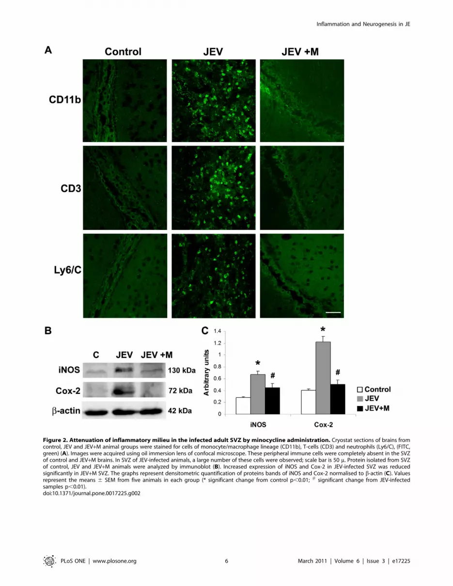
Figure 2. Attenuation of inflammatory milieu in the infected adult SVZ by minocycline administration. Cryostat sections of brains fromcontrol, JEV and JEV+M animal groups were stained for cells of monocyte/macrophage lineage (CD11b), T-cells (CD3) and neutrophils (Ly6/C), (FITC,green) (A). Images were acquired using oil immersion lens of confocal microscope. These peripheral immune cells were completely absent in the SVZof control and JEV+M brains. In SVZ of JEV-infected animals, a large number of these cells were observed; scale bar is 50 m. Protein isolated from SVZof control, JEV and JEV+M animals were analyzed by immunoblot (B). Increased expression of iNOS and Cox-2 in JEV-infected SVZ was reducedsignificantly in JEV+M SVZ. The graphs represent densitometric quantification of proteins bands of iNOS and Cox-2 normalised to b-actin (C). Valuesrepresent the means 6 SEM from five animals in each group (* significant change from control p,0.01; # significant change from JEV-infectedsamples p,0.01).doi:10.1371/journal.pone.0017225.g002
Inflammation and Neurogenesis in JE
PLoS ONE | www.plosone.org 6 March 2011 | Volume 6 | Issue 3 | e17225
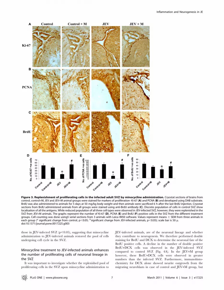
those in JEV-infected SVZ (p,0.05), suggesting that minocycline
administration to JEV-infected animals restored the pool of cells
undergoing cell cycle in the SVZ.
Minocycline treatment to JEV-infected animals enhancesthe number of proliferating cells of neuronal lineage inthe SVZ
It was important to investigate whether the replenished pool of
proliferating cells in the SVZ upon minocycline administration to
JEV-infected animals, are of the neuronal lineage and whether
they contribute to neurogenesis. We therefore performed double
staining for BrdU and DCX to determine the neuronal fate of the
BrdU positive cells. A decline in the number of double positive
BrdU+DCX cells was observed in the JEV-infected SVZ
compared to control SVZ (Fig. 4A). In the JEV+M group
however, these BrdU+DCX cells were observed in greater
numbers than the infected SVZ. Furthermore, immunohisto-
chemistry for DCX alone showed neurite outgrowth from the
migrating neuroblasts in case of control and JEV+M group, but
Figure 3. Replenishment of proliferating cells in the infected adult SVZ by minocycline administration. Cryostat sections of brains fromcontrol, control+M, JEV and JEV+M animal groups were stained for markers of proliferation- Ki-67 (A) and PCNA (B) and developed using DAB substrate.BrdU was also administered to animals for 5 days at 50 mg/kg body weight and then animals were sacrificed 6 h after the last BrdU injection. Cryostatsections from BrdU administered animals from all groups were stained using anti-BrdU antibody (C). Discrete population of cells in control SVZ showlocalisation of all the antigens. While reduced population of all three cell types were observed in JEV-infected SVZ, however, they were replenished in theSVZ from JEV+M animals. The graphs represent the number of Ki-67 (D), PCNA (E) and BrdU (F) positive cells in the SVZ from the different treatmentgroups. Cell counting was done using5 serial sections from 3 animals with Leica IM50 software. Values represent means 6 SEM from three animals ineach group (* significant change from control, p,0.05; #significant change from JEV-infected animals, p,0.05); scale bar is 50 m.doi:10.1371/journal.pone.0017225.g003
Inflammation and Neurogenesis in JE
PLoS ONE | www.plosone.org 7 March 2011 | Volume 6 | Issue 3 | e17225

such cells were completely absent from the JEV-infected group
(Fig. 4B). This suggests that the migrating neuroblasts in case of
control and JEV+M group with neurite protrusions maybe more
effective in promoting CNS repair than those cells without the
neurite outgrowths.
Quantification of both the proliferating cells and those of the
neuronal lineage was done by performing Western blotting for
PCNA and DCX respectively. Indeed, a significant decrease in
both PCNA and DCX protein expression was observed in JEV-
infected SVZ compared to control SVZ. Expression of both these
molecules were higher in SVZ from JEV+M group w.r.t JEV-
infected group (Fig. 4C, D) (p,0.05). We also checked for
expression of PSD-95 which is a post-synaptic density protein,
important for formation of functional synapses. A significant 2.5
fold decrease in PSD-95 was observed in JEV-infected SVZ w.r.t
control SVZ, probably suggesting the lack of functional synapse
formation in infected animals (Fig. 4C, D) (p,0.05). In JEV+M
animals, PSD-95 expression was significantly elevated by 2 fold
Figure 4. Population of migrating neuroblasts restored in adult infected SVZ by minocycline administration. Cryostat sections ofbrains from control, JEV and JEV+M animal groups were labelled for proliferating cells of neuronal lineage (BrdU+DCX double staining) (A) or formigrating neuroblasts (DCX positive cells) (B). Control SVZ show marked co-localization of BrdU (Alexa Fluor, red) and DCX (FITC, green). Thesedouble positive cells were distinctly lesser in the JEV-infected SVZ compared to control SVZ, which was however restored in JEV+M SVZ (A). DCXstaining (FITC, green) show migrating neuroblasts in control SVZ and in SVZ from JEV+M animals with neurite extending outwards (shown byarrowheads), was completely absent in JEV-infected SVZ (B); scale bar is 50 m. Protein isolated from SVZ of control, JEV and JEV+M animals wereanalyzed by immunoblot (C). Expression of DCX, PSD-95 and PCNA declined in JEV-infected SVZ, which in JEV+M SVZ was elevated almost to controllevels. The graph represents densitometric quantification of proteins bands of DCX, PSD-95 and PCNA, normalised to b-actin (D). Values represent themeans 6 SEM from five animals in each group (* significant change from control p,0.05; # significant change from JEV samples p,0.05).doi:10.1371/journal.pone.0017225.g004
Inflammation and Neurogenesis in JE
PLoS ONE | www.plosone.org 8 March 2011 | Volume 6 | Issue 3 | e17225

compared to JEV-infected SVZ, implying the functional role of
minocycline in CNS regeneration in the infected brain.
Minocycline attenuates microglial activation followingJEV infection in vitro
Mouse microglia BV2 were either control (C) or JEV-infected
(JEV) or infected with JEV and treated with minocycline, both pre
and post infection (JEV+M). After 6 h of post-treatment of
minocycline, fresh media was added to all the conditions and
another 6 h later, the media (CM) were collected for CBA and cell
protein were isolated for Western blotting. While the concentration
of TNF-a increased significantly in CM from JEV-infected BV2
compared to that from control BV2, however CM from JEV+M
cells showed a significant reduction to concentrations below control
(Fig. 5A) (p,0.05). Similar elevated concentrations of MCP-1 in
CM from JEV-infected BV2 was brought down to control levels in
CM from JEV+M treated BV2 (Fig. 5C) (p,0.05). IL-6
concentration was significantly higher in CM from JEV-infected
BV2 compared to CM from control cells. Although CM from
BV2(JEV+M) showed lower IL-6 concentrations than that from
BV2(JEV), however, the difference was not significant (Fig. 5B).
Apart from the cytokines, the expression levels of other
inflammatory mediators iNOS and Cox-2 was measured in BV2
cell lysates. Induction of both iNOS and Cox-2 expression was
distinct in JEV-infected BV2 compared to control BV2. This
increased expression of iNOS and Cox-2 was significantly
downregulated by 2 fold in JEV+M treated BV2, compared to
JEV-infected BV2 (Fig. 5D, E) (p,0.05).
Soluble inflammatory mediators from JEV-activatedmicroglia inhibit proliferation of NSPCs, which is reversedby minocycline treatment
In order to determine the extent to which the soluble factors
released from JEV activated microglia directly affect NSPC
development, the cells were grown in CM obtained from BV2
cells. After two passages, single cell suspensions of NSPCs were
grown for 3 days either alone (C), or in presence of 50% of CM
from BV2 cells in 3 conditions- BV2(C), BV2(JEV) and
BV2(JEV+M). The numbers of neurospheres formed after 3 days
in culture as well as their average diameter (both indicators of their
proliferative capacity) were calculated using Leica IM50 software
(Fig. 6). No significant differences in the number of spheres
generated were observed between the different conditions (Fig. 6A,
B). However, the average sphere diameter showed a significant
decrease when NSPCs were grown in CM from BV2(JEV)
compared to that from BV2(C) or control neurospheres (Fig. 6A,
C) (p,0.01). NSPCs grown in presence of CM from BV2(JEV+M)
however resulted in more number of spheres and with average
diameter greater than those grown in BV2(JEV) media (p,0.01).
In another set of experiments, NSPCs grown alone or in
presence of BV2-derived CM was collected after 3 days for cell
cycle analysis. Single cell suspensions of NSPCs were stained with
PI, and acquired in FACS Calibur. Following gating of specific
population, the percentages of cells lying in the different stages of
cell cycle (G0/G1, S and G2/M phase) were analysed using Cell
Quest Pro software. In case of control neurospheres, approxi-
mately 10% of cells were in the S-phase, which was comparable to
Figure 5. Expression of inflammatory molecules in JEV activated BV2 is attenuated by minocycline treatment. Mouse microglia BV2were either control (C), JEV-infected (JEV) or JEV-infected and treated with minocycline (JEV+M). After 6 h of minocycline treatment p.i., fresh DMEM-F12 media was added to all conditions. After additional 6 h, CM from BV2 cells from all conditions was collected for CBA and protein extracted forimmunoblot. The graphs depict the concentration in pg/ml of cyto/chemokines TNF-a (A), IL-6 (B) and MCP-1 (C) in Control, JEV-infected and JEV+Mtreated BV2 cells. Values represent means 6 SEM from 3 independent experiments performed in duplicate (* significant change from control, p,0.05;# significant change from JEV, p,0.05). Protein isolated from BV2 cells from C, JEV and JEV+M conditions were analyzed by immunoblot (D). Thegraphs represent the densitometric quantification of protein bands- iNOS and Cox-2 normalised to b-actin (E). Values represent mean 6 SEM from 3independent experiments performed in duplicate (* significant change from control, p,0.05; # significant change from JEV, p,0.05).doi:10.1371/journal.pone.0017225.g005
Inflammation and Neurogenesis in JE
PLoS ONE | www.plosone.org 9 March 2011 | Volume 6 | Issue 3 | e17225

the NSPCs grown in CM from BV2(C) (Fig. 7A). However, the
percentage of cells in S-phase decreased significantly to 7.7%
when grown in CM from BV2(JEV), and was restored back to
8.7% in presence of CM from BV2(JEV+M). Interestingly, as the
percentages of S-phase NSPCs declined in presence of BV2(JEV),
more cells were observed in the G0/G1 phase (Fig. 7A). This
possibly indicates a G1RS phase arrest in these NSPCs when
grown in CM from BV2(JEV) compared to that from BV2(C), and
the arrest was partially unblocked when CM from BV2(JEV+M)
was used. These are indications that the soluble inflammatory
mediators from JEV-activated microglia have anti-proliferative
effect on NSPCs, which is partially recovered by minocycline
treatment to BV2 cells due to reduced inflammatory molecules
production.
Considering that the CM from activated microglia modulates
G1RS phase transition, we determined the expression of cell cycle
checkpoint proteins by western blotting from cell lysates of NSPCs
grown in different CM. The expression of p21, p27, p53 and p107
was determined, all of which act during progression of cells from
G1RS stage. Both p21 and p27 are specific checkpoint proteins
for G1RS phase transition and are significantly up-regulated in
NSPCs grown in CM from BV2(JEV) compared to CM from
BV2(C) (Fig. 7B, C) (p,0.05). Similar increase in expression was
observed for p53 and p107 (both Retinoblastoma proteins) in case
of NSPCs grown in CM from BV2(JEV) (p,0.05). Interestingly,
all these checkpoint proteins were significantly down-regulated in
NSPCs when CM from BV2(JEV+M) was used compared to that
from BV2(JEV) (Fig. 7B, C) (p,0.05). Taken together, these
findings clearly indicate that soluble inflammatory mediators
released from JEV-activated BV2 cells blocks G1RS phase cell
cycle transition via upregulation of various checkpoint proteins- an
effect which is reversed using soluble mediators from JEV-infected
and minocycline treated BV2 cells.
Soluble inflammatory mediators from JEV-activatedmicroglia inhibit neuronal differentiation of NSPCs, whichis reversed by minocycline treatment
Neurospheres grown alone or in presence of CM from BV2 cells
for 3 days were plated on PDL-coated plates and allowed to
differentiate for 2 days. Micrographs of adhered neurospheres
show the migration of the cells from the periphery of the spheres
and differentiation into neurons (Fig. 8A). After induction of
differentiation, while neurons with elongated neurites extended
noticeably from the periphery of the spheres in control and in CM-
BV2(C) treated, lesser proportion of differentiating cells was
observed in CM from BV2(JEV) treated neurospheres. Neuro-
Figure 6. Impaired neurosphere formation by soluble mediators from JEV activated microglia is reversed by minocyclinetreatment. Single cell suspensions of NSPCs were cultured alone (C) or in presence of BV2-derived CM under 3 conditions- BV2(C), BV2(JEV),BV2(JEV+M) for 3 days. Phase-contrast micrographs of neurospheres were acquired under 106magnification (A). Scale bar is 100 m. The number ofneurospheres was counted from 10 different fields for each condition and the average diameter of the spheres was measured using Leica IM50software. The graphs represent the average number of spheres (B) and their average diameter in microns (C). The numbers of colony formingneurospheres do not show any significant difference between groups (B). The reduction in size of neurospheres grown in CM from BV2(JEV) wasprominent compared to CM-BV2(C) or CM-BV2(JEV+M) conditions (C). Values represent mean 6 SEM from 3 independent experiments performed induplicate (** significant change from control, p,0.05; * significant change from CM-BV2(C), p,0.01; # significant change from CM-BV2(JEV), p,0.01).doi:10.1371/journal.pone.0017225.g006
Inflammation and Neurogenesis in JE
PLoS ONE | www.plosone.org 10 March 2011 | Volume 6 | Issue 3 | e17225

spheres grown in CM from BV2(JEV+M) however showed
considerable proportion of differentiating cells emerging from
the sphere periphery (Fig. 8A).
In a simultaneous experiment, neurospheres from the different
conditions were dissociated into single cell suspensions and then
allowed to differentiate for 2 days on PDL-coated chamber slides.
Immunocytochemistry revealed the presence of b-III tubulin positive
cells indicative of neuronal differentiation and GFAP positive cells
indicative of astrocytic differentiation. The percentages of these
newly formed neurons (b-III tubulin) and astrocytes (GFAP) were
calculated for each condition. Differentiation of CM-BV2 (JEV)
treated NSPCs yielded significantly fewer numbers of neurons than
the NSPCs from CM of BV2(C) or control conditions (Fig. 8B, C)
(p,0.01). Moreover, a 2 fold increase in the percentage of b-III
tubulin cells was observed in NSPCs from CM of BV2(JEV+M) as
compared to that of BV2(JEV) (p,0.01). Additionally, we also
assessed for the transcript levels of b-III tubulin in these
differentiating NSPCs by qRT-PCR. A significant decrease in b-III
tubulin expression in differentiated NSPCs from CM of BV2(JEV)
was observed compared to CM from BV2(C) or from BV2(JEV+M)
(Fig. 8D) (p,0.01). Overall, the neurospheres/NSPCs grown in CM
from JEV-activated microglia exhibit impaired neuronal differenti-
ation even when the CM was removed during the differentiation
regime, thereby suggesting that the exposure to soluble mediators
from microglia for 3 days modulated the developmental pattern of
these NSPCs. To extend further, neurospheres/NSPCs grown in
CM from BV2(JEV+M) differentiate normally, possibly due to the
reduction of anti-neurogenic inflammatory mediators in the BV2-
derived CM by minocycline treatment.
Discussion
As the primary immune effector cells of the CNS, microglia
responds to injury or to the presence of pathogens by becoming
activated. Upon activation, microglia undergoes proliferation,
chemotaxis, and morphological alterations and generates numer-
ous mediators involved in the inflammatory and immunomodu-
latory response. The ensuing neuroinflammation have a myriad of
effects- from clearing the viral pathogen, to remodelling brain
parenchyma, as well as modulating neuronal health and
neurogenesis by the long-range diffusible soluble mediators and
the local inflammatory milieu.
Figure 7. Induction of cell cycle arrest by soluble mediators from JEV activated microglia is reversed by minocycline treatment. Cellcycle analysis of single cell suspensions of neurospheres grown alone or in presence of BV2-CM was performed using RNase/PI buffer and detectedby FACS. Gates were set to assess the percentages of G0/G1 (2n DNA), S (.2n DNA) and G2/M (4n DNA) cells using Cell Quest Pro Software, and therepresentative histograms have been shown (A). Data is representative of 3 independent experiments. Protein isolated from control and BV2-CMtreated neurospheres were immunoblotted for checkpoint proteins p21, p27, p53 and p107 (B). The expression of all these proteins increased inneurospheres grown in CM from BV2(JEV) compared to that from BV2(C) or from BV2(JEV+M) (C). Densitometric analysis of immunoblot normalisedto b-actin was plotted as a bar graph. Values represent mean 6 SEM from three independent experiments. (** significant change from control,p,0.05; * significant change from CM-BV2(C), p,0.05; # significant change from CM-BV2(JEV), p,0.05).doi:10.1371/journal.pone.0017225.g007
Inflammation and Neurogenesis in JE
PLoS ONE | www.plosone.org 11 March 2011 | Volume 6 | Issue 3 | e17225

Our previous studies have already established how JEV infection
in adult BALB/c mice leads to an inflammatory upheaval in the
adult CNS [30]. In this study, we observed that there is differential
distribution of the various cyto/chemokines in the different areas of
the brain; the most affected areas being the cortex and the
neurogenic niche of SVZ. Minocycline, a second-generation
tetracycline derived antibiotic has been proven as an effective
measure of attenuating JEV-induced microglial activation, as well as
alleviating the clinical symptoms of JE [30]. This present study
further corroborated that minocycline administration to adult
animals following JEV infection dramatically lowered the level of
cytokines in all the investigated brain areas and therefore promoted
animal survivability. The response to cerebral JEV infection is
characterized at the pathological level by significant recruitment
and extravasation of inflammatory cells [36,37], which is, partly
mediated by chemokines released from the activated microglia [38].
Though such infiltration of peripheral immune cells was prominent
in the infected SVZ, minocycline treated animals showed barely any
of these peripheral immune infiltrates. This partly results from the
ability of minocycline to reduce production of inflammatory
molecules and chemokines which signal to the peripheral immune
cells as well as their ability to maintain the integrity of BBB in JEV
infections [30,39]. Besides the cyto/chemokines, the expression of
other inflammatory molecules iNOS and Cox-2 was elevated in
JEV-infected SVZ but reduced significantly following minocycline
treatment. Taken together, these findings clearly established that
inflammatory milieu created in the SVZ during JEV infection is
completely alleviated by minocycline treatment.
The central role of inflammation in modulating neurogenesis is
still debatable, and often dependent on the disease model, and the
Figure 8. Decreased neuronal differentiation of NSPCs by soluble mediators from JEV activated microglia is reversed byminocycline treatment. Phase contrast micrographs of differentiating neurospheres from control and BV2 derived CM treated NSPCs 2 days afterinduction of differentiation on PDL-coated plates (A). Impaired migration and differentiation of cells from the periphery of the spheres is observed inCM-BV2(JEV) conditions. Control and BV2-Cm treated neurospheres were dissociated and single cell suspensions of NSPCs were differentiated for 2days. The cells were fixed and double immunocytochemistry for b-III tubulin (neuronal phenotype) and GFAP (astrocytic phenotype) was performedand mounted with Vectashield containing DAPI. Distinct b-III tubulin positive cells (FITC, green) and GFAP positive cells (Alexa 594, red) wereobserved, indicating NSPCs undergoing differentiation (B). The percentage of b-III tubulin positive cells were calculated from 5 different fields andplotted as a representative graph (C). RNA was isolated from these NSPCs after 2 days of differentiation and qRT-PCR was performed for b-III tubulinexpression. The graph represents relative mRNA expression of b-III tubulin normalised to the expression of constitutive 18S rRNA (D). Valuesrepresent mean 6 SEM from three independent experiments performed in duplicate. (** significant change from control, p,0.05; * significant changefrom CM-BV2(C), p,0.01; # significant change from CM-BV2(JEV), p,0.01). Scale bar corresponds to 50 microns.doi:10.1371/journal.pone.0017225.g008
Inflammation and Neurogenesis in JE
PLoS ONE | www.plosone.org 12 March 2011 | Volume 6 | Issue 3 | e17225

duration and intensity of the inflammatory response. One of the
best characterised neurogenic niches is SVZ [40], the endogenous
brain compartment housing the self-renewing NSPCs, which have
immense potential of tissue repair upon CNS injury [41,42].
Functional impairment of the SVZ germinal niche has been shown
following either cranial irradiation [43], or LPS-mediated acute
inflammation [44] or EAE-induced persistent inflammation [45].
Inflammation associated with ischemic insults and stroke compro-
mise the survivability of the newly formed striatal neurons that are
generated from the SVZ after stroke [46,47]. Our investigation
also revealed a significant reduction in the actively replicating cells
including those belonging to the neuronal lineage in the JEV-
infected SVZ niche. Administration of the anti-inflammatory
compound minocycline was able to restore the NSPC population
back to that in control SVZ. Minocycline readily crosses the BBB
and suppress microglial activation in a number of neurodegener-
ative disorders as well as virus infections [30,48]. Similarly, in
models of LPS inflammation and stroke, minocycline decreased
microglial activation, accounting for the potentiation of NSPC
pool and neurogenesis [14,28]. However, no direct effect of
minocycline in stimulating endogenous neurogenesis in control
animals has been reported, which is corroborated by our in vivo
findings. Minocycline administration to control animals did not
result in any significant increase in the number of proliferating
cells in the neurogenic niche, i.e. the SVZ. Hence, it is plausible
that minocycline has no direct effect on neurogenesis. Inflamma-
tory blockade usually is accompanied by a broad spectrum of
effects which promote neurogenesis, like reduction of cell death of
new born neurons [14], attenuation of HPA axis activation [49],
or the decrease in pro-inflammatory cytokines and serum
glucocorticoid levels [15,50].
Our in vitro studies provided further evidence that the
detrimental effects of JEV-induced microglial activation on the
development of NSPCs is via release of soluble inflammatory
mediators from the activated microglia, which is reversed upon
minocycline treatment. Upregulation of various inflammatory
markers iNOS and Cox-2 indicated the activation of mouse
microglial cells BV2 by JEV, which was significantly attenuated
with minocycline treatment to BV2 cells. The stress signaling
kinases were also elevated upon JEV infection in BV2, which was
considerably reduced by minocycline treatment (data not shown).
CM from JEV-activated BV2 cells was enriched with pro-
inflammatory mediators and cyto/chemokines, and mouse neuro-
spheres grown in this CM showed impaired proliferation and
neuronal differentiation. This was corroborated by previous
findings where CM from LPS-activated BV2 cells inhibited the
formation of new neurons (DCX+) and led to apoptosis [15].
Acutely activated microglia, or their CM, reduced NSPC survival,
prevented neuronal differentiation and strongly increased glial
differentiation, likely through the release of pro-inflammatory
cytokines, whereas chronically activated microglia promoted both
neuronal/glial differentiation and cell survival of NSPCs [51]. Pro-
inflammatory cytokine TNF-a contributes to apoptotic death of
hippocampal progenitor cells [52] and mouse SVZ derived
neurospheres [53]. Although low concentrations of TNF-astimulate NSPC proliferation [54], and are regarded as pro-
neurogenic [55], however, TNF-a has also been shown to inhibit
proliferation of striatal NSPCs [16]. Microglia-derived IL-6 is a
potent anti-neurogenic cytokine which promotes astrocytic
differentiation of NSPCs at the expense of neuronal differentiation
[15,56]. The physiological role of iNOS/NO as a negative
regulator of SVZ neurogenesis [57,58] as well as an inhibitor of
neurosphere proliferation by blocking EGF-receptor and PI3K/
Akt pathway has been established [59]. NO exposure to adult
NSPCs inhibits neurogenesis by decreasing expression of the
proneural gene Ngn-2 and b-III tubulin, and is instead beneficial
for astroglial differentiation [60]. Interestingly, CM from
BV2(JEV+M) rescued both the proliferative and differentiation
potential of these NSPCs, thereby indicating the restorative effect
of anti-inflammatory minocycline treatment on neurogenesis.
Other p38MAPK inhibitor with potency to inhibit microglial
activation, have been shown to be neuroprotective and promote
neurogenesis in hypoxic hippocampal slice cultures [61].
We have further elucidated that the inhibition of NSPC
proliferation by CM from BV2 results from a blockade of cell
cycle progression mainly at G1RS phase due to the upregulation
of various checkpoint proteins. The retinoblastoma proteins p53
and p107 are up-regulated which in turn induces other checkpoint
proteins, like p21 and p27. All these proteins together act on the
cyclin/CDK complexes, and inhibit their functions, thereby
inducing cell cycle arrest. NSPCs grown in CM from
BV2(JEV+M) however, exhibited low expression of these check-
point kinases, which allows them to cycle normally. Induction of
quiescence in NSPCs by chemokines has been reported via
upregulation of p21 and p27 [22].
Microglial activation and CNS inflammation have been
associated with cognitive impairment in a number of brain
disorders like Lewy Body dementia, AD, and AIDS dementia.
Cranial irradiation also leads to inflammation in the SGZ and
progressive cognitive deterioration. A link between onset of AIDS
dementia complex in HIV-individuals to compromised neurogen-
esis as a consequence of uncontrolled inflammatory processes has
been proposed [62,63]. Therefore, the use of anti-inflammatory
compounds and NSAIDs has shown promise in improving the
cognitive attributes in inflammatory neurodegenerative disorders
and in virus-induced inflammation. Since we observed no direct
effect of minocycline on neurogenesis, our findings highlights its
role in reduction of the inflammatory milieu in the SVZ germinal
niche and resultant rescue NSPCs from the toxic effects of the
inflammatory mediators. The restoration of SVZ neurogenesis by
minocycline administration in JEV-infected animals may have
important implications in promoting brain’s regenerative capacity
following neuronal loss in JE. The use of minocycline in a wide
variety of neurological disorders has been reported [64].
Minocycline has also been proposed as a potential therapeutic
measure which can possibly reduce HIV-induced cognitive
problems [65] and human trials for such are in progress (http://
clinicaltrials.gov/ct2/show/NCT00855062). From our study we
see that besides exerting neuroprotective effects, minocycline, by
virtue of its anti-inflammatory properties, stimulate neurogenesis
in JEV infection, thereby reasserting its immense therapeutic
potential in treatment of JE.
Supporting Information
Figure S1 Decreased viral protein expression andinfective virl particle production following minocyclinetreatment. Immunoblot showing significantly decreased expres-
sion of viral NS3 and NS5 proteins in SVZ of infected mice treated
with minocycline (A). Densitometric analysis of immunoblot
normalised to b-actin was plotted as a bar graph. Values represent
mean 6 SEM from three independent experiments (B). A
significant decrease in the number of infective viral particle
formation was also observed in the SVZ of minocycline treated
infected animals when compared to non-minocycline treated
animals. Values are mean 6 SEM from 2 independent
experiments (C). (* significantly increased in JEV-infected as
compared to control, p,0.01; # significantly decreased following
Inflammation and Neurogenesis in JE
PLoS ONE | www.plosone.org 13 March 2011 | Volume 6 | Issue 3 | e17225

minocycline treatment as compared to only JEV-infected,
p,0.01).
(TIF)
Acknowledgments
The authors would like to thank Mr. R. Khader Valli for his help in qRT-
PCR experiments.
Author Contributions
Conceived and designed the experiments: SD AB. Performed the
experiments: SD KD KLK AG DA. Analyzed the data: SD KD AB.
Wrote the paper: SD KD AB.
References
1. Buckner CM, Luers AJ, Calderon TM, Eugenin EA, Berman JW (2006)
Neuroimmunity and the blood-brain barrier: molecular regulation of leukocyte
transmigration and viral entry into the nervous system with a focus on
neuroAIDS. J Neuroimmune Pharmacol 1: 160–181.
2. Olson JK, Miller SD (2004) Microglia initiate central nervous system innate and
adaptive immune responses through multiple TLRs. J Immunol 173:
3916–3924.
3. Ghoshal A, Das S, Ghosh S, Mishra MK, Sharma V, et al. (2007)
Proinflammatory mediators released by activated microglia induces neuronal
death in Japanese encephalitis. Glia 55: 483–496.
4. Kaul M, Zheng J, Okamoto S, Gendelman HE, Lipton SA (2005) HIV-1
infection and AIDS: consequences for the central nervous system. Cell Death
Differ 12 Suppl 1: 878–892.
5. Kaul M, Lipton SA (2006) Mechanisms of neuronal injury and death in HIV-1
associated dementia. Curr HIV Res 4: 307–318.
6. Ghosh D, Basu A (2009) Japanese encephalitis-a pathological and clinical
perspective. PLoS Negl Trop Dis 3: e437.
7. Misra UK, Kalita J Overview: Japanese encephalitis. Prog Neurobiol 91:
108–120.
8. Richter RW, Shimojyo S (1961) Neurologic sequelae of Japanese B encephalitis.
Neurology 11: 553–559.
9. Monnet FP (2003) Behavioural disturbances following Japanese B encephalitis.
Eur Psychiatry 18: 269–273.
10. Swarup V, Das S, Ghosh S, Basu A (2007) Tumor necrosis factor receptor-1-
induced neuronal death by TRADD contributes to the pathogenesis of Japanese
encephalitis. J Neurochem 103: 771–783.
11. Das S, Basu A (2008) Inflammation: A New Candidate in Modulating Adult
Neurogenesis. J Neurosci Res 86: 1199–1208.
12. Ming GL, Song H (2005) Adult neurogenesis in the mammalian central nervous
system. Annu Rev Neurosci 28: 223–250.
13. Ma DK, Bonaguidi MA, Ming GL, Song H (2009) Adult neural stem cells in the
mammalian central nervous system. Cell Res 19: 672–682.
14. Ekdahl CT, Claasen JH, Bonde S, Kokaia Z, Lindvall O (2003) Inflammation is
detrimental for neurogenesis in adult brain. Proc Natl Acad Sci U S A 100:
13632–13637.
15. Monje ML, Toda H, Palmer TD (2003) Inflammatory blockade restores adult
hippocampal neurogenesis. Science 302: 1760–1765.
16. Ben-Hur T, Ben-Menachem O, Furer V, Einstein O, Mizrachi-Kol R, et al.
(2003) Effects of proinflammatory cytokines on the growth, fate, and motility of
multipotential neural precursor cells. Mol Cell Neurosci 24: 623–631.
17. Liu YP, Lin HI, Tzeng SF (2005) Tumor necrosis factor-alpha and interleukin-
18 modulate neuronal cell fate in embryonic neural progenitor culture. Brain
Res 1054: 152–158.
18. Barkho BZ, Song H, Aimone JB, Smrt RD, Kuwabara T, et al. (2006)
Identification of astrocyte-expressed factors that modulate neural stem/
progenitor cell differentiation. Stem Cells Dev 15: 407–421.
19. Kuzumaki N, Ikegami D, Imai S, Narita M, Tamura R, et al. (2010) Enhanced
IL-1beta production in response to the activation of hippocampal glial cells
impairs neurogenesis in aged mice. Synapse 64: 721–728.
20. Iosif RE, Ekdahl CT, Ahlenius H, Pronk CJ, Bonde S, et al. (2006) Tumor
necrosis factor receptor 1 is a negative regulator of progenitor proliferation in
adult hippocampal neurogenesis. J Neurosci 26: 9703–9712.
21. Ekdahl CT, Kokaia Z, Lindvall O (2009) Brain inflammation and adult
neurogenesis: the dual role of microglia. Neuroscience 158: 1021–1029.
22. Krathwohl MD, Kaiser JL (2004) Chemokines promote quiescence and survival
of human neural progenitor cells. Stem Cells 22: 109–118.
23. Tran PB, Miller RJ (2005) HIV-1, chemokines and neurogenesis. Neurotox Res
8: 149–158.
24. Peng H, Whitney N, Wu Y, Tian C, Dou H, et al. (2008) HIV-1-infected and/
or immune-activated macrophage-secreted TNF-alpha affects human fetal
cortical neural progenitor cell proliferation and differentiation. Glia 56: 903–
916.
25. Poluektova L, Meyer V, Walters L, Paez X, Gendelman HE (2005)
Macrophage-induced inflammation affects hippocampal plasticity and neuronal
development in a murine model of HIV-1 encephalitis. Glia 52: 344–353.
26. Monje ML, Mizumatsu S, Fike JR, Palmer TD (2002) Irradiation induces neural
precursor-cell dysfunction. Nat Med 8: 955–962.
27. Hoehn BD, Palmer TD, Steinberg GK (2005) Neurogenesis in rats after focal
cerebral ischemia is enhanced by indomethacin. Stroke 36: 2718–2724.
28. Liu Z, Fan Y, Won SJ, Neumann M, Hu D, et al. (2007) Chronic treatment with
minocycline preserves adult new neurons and reduces functional impairment
after focal cerebral ischemia. Stroke 38: 146–152.
29. Marchetti B, Abbracchio MP (2005) To be or not to be (inflamed)–is that the
question in anti-inflammatory drug therapy of neurodegenerative disorders?Trends Pharmacol Sci 26: 517–525.
30. Mishra MK, Basu A (2008) Minocycline neuroprotects, reduces microglial
activation, inhibits caspase 3 induction, and viral replication following Japaneseencephalitis. J Neurochem 105: 1582–1595.
31. Das S, Basu A (2008) Japanese encephalitis virus infects neural progenitor cellsand decreases their proliferation. J Neurochem 106: 1624–1636.
32. Vrati S, Agarwal V, Malik P, Wani SA, Saini M (1999) Molecularcharacterization of an Indian isolate of Japanese encephalitis virus that shows
an extended lag phase during growth. J Gen Virol 80(Pt 7): 1665–1671.
33. Das S, Ghosh D, Basu A (2009) Japanese encephalitis virus induce immuno-competency in neural stem/progenitor cells. PLoS One 4: e8134.
34. Mishra MK, Koli P, Bhowmick S, Basu A (2007) Neuroprotection conferred byastrocytes is insufficient to protect animals from succumbing to Japanese
encephalitis. Neurochem Int 50: 764–773.
35. Tewari R, Sharma V, Koul N, Sen E (2008) Involvement of miltefosine-
mediated ERK activation in glioma cell apoptosis through Fas regulation.
J Neurochem 107: 616–627.
36. Chaturvedi UC, Mathur A, Tandon P, Natu SM, Rajvanshi S, et al. (1979)
Variable effect on peripheral blood leucocytes during JE virus infection of man.Clin Exp Immunol 38: 492–498.
37. Mathur A, Khanna N, Chaturvedi UC (1992) Breakdown of blood-brain barrier
by virus-induced cytokine during Japanese encephalitis virus infection. Int J ExpPathol 73: 603–611.
38. Swarup V, Ghosh J, Das S, Basu A (2008) Tumor necrosis factor receptor-associated death domain mediated neuronal death contributes to the glial
activation and subsequent neuroinflammation in Japanese encephalitis.Neurochem Int 52: 1310–1321.
39. Mishra MK, Dutta K, Saheb SK, Basu A (2009) Understanding the molecular
mechanism of blood-brain barrier damage in an experimental model of Japaneseencephalitis: correlation with minocycline administration as a therapeutic agent.
Neurochem Int 55: 717–723.
40. Doetsch F (2003) A niche for adult neural stem cells. Curr Opin Genet Dev 13:
543–550.
41. Martino G, Pluchino S (2006) The therapeutic potential of neural stem cells. Nat
Rev Neurosci 7: 395–406.
42. Romanko MJ, Rola R, Fike JR, Szele FG, Dizon ML, et al. (2004) Roles of themammalian subventricular zone in cell replacement after brain injury. Prog
Neurobiol 74: 77–99.
43. Lazarini F, Mouthon MA, Gheusi G, de Chaumont F, Olivo-Marin JC, et al.
(2009) Cellular and behavioral effects of cranial irradiation of the subventricular
zone in adult mice. PLoS One 4: e7017.
44. Mori K, Kaneko YS, Nakashima A, Nagatsu T, Nagatsu I, et al. Peripherally
injected lipopolysaccharide induces apoptosis in the subventricular zone ofyoung adult mice. Neurosci Lett.
45. Pluchino S, Muzio L, Imitola J, Deleidi M, Alfaro-Cervello C, et al. (2008)Persistent inflammation alters the function of the endogenous brain stem cell
compartment. Brain 131: 2564–2578.
46. Arvidsson A, Collin T, Kirik D, Kokaia Z, Lindvall O (2002) Neuronalreplacement from endogenous precursors in the adult brain after stroke. Nat
Med 8: 963–970.
47. Heldmann U, Thored P, Claasen JH, Arvidsson A, Kokaia Z, et al. (2005) TNF-
alpha antibody infusion impairs survival of stroke-generated neuroblasts in adultrat brain. Exp Neurol 196: 204–208.
48. Tikka T, Fiebich BL, Goldsteins G, Keinanen R, Koistinaho J (2001)
Minocycline, a tetracycline derivative, is neuroprotective against excitotoxicityby inhibiting activation and proliferation of microglia. J Neurosci 21:
2580–2588.
49. Reyes TM, Coe CL (1998) The proinflammatory cytokine network: interactions
in the CNS and blood of rhesus monkeys. Am J Physiol 274: R139–144.
50. Tanapat P, Galea LA, Gould E (1998) Stress inhibits the proliferation of granule
cell precursors in the developing dentate gyrus. Int J Dev Neurosci 16: 235–239.
51. Cacci E, Ajmone-Cat MA, Anelli T, Biagioni S, Minghetti L (2008) In vitroneuronal and glial differentiation from embryonic or adult neural precursor cells
are differently affected by chronic or acute activation of microglia. Glia 56:412–425.
Inflammation and Neurogenesis in JE
PLoS ONE | www.plosone.org 14 March 2011 | Volume 6 | Issue 3 | e17225

52. Cacci E, Claasen JH, Kokaia Z (2005) Microglia-derived tumor necrosis factor-
alpha exaggerates death of newborn hippocampal progenitor cells in vitro.J Neurosci Res 80: 789–797.
53. Wong G, Goldshmit Y, Turnley AM (2004) Interferon-gamma but not TNF
alpha promotes neuronal differentiation and neurite outgrowth of murine adultneural stem cells. Exp Neurol 187: 171–177.
54. Widera D, Mikenberg I, Elvers M, Kaltschmidt C, Kaltschmidt B (2006) Tumornecrosis factor alpha triggers proliferation of adult neural stem cells via IKK/
NF-kappaB signaling. BMC Neurosci 7: 64.
55. Bernardino L, Agasse F, Silva B, Ferreira R, Grade S, et al. (2008) Tumornecrosis factor-alpha modulates survival, proliferation, and neuronal differen-
tiation in neonatal subventricular zone cell cultures. Stem Cells 26: 2361–2371.56. Nakanishi M, Niidome T, Matsuda S, Akaike A, Kihara T, et al. (2007)
Microglia-derived interleukin-6 and leukaemia inhibitory factor promoteastrocytic differentiation of neural stem/progenitor cells. Eur J Neurosci 25:
649–658.
57. Matarredona ER, Murillo-Carretero M, Moreno-Lopez B, Estrada C (2005)Role of nitric oxide in subventricular zone neurogenesis. Brain Res Brain Res
Rev 49: 355–366.58. Romero-Grimaldi C, Gheusi G, Lledo PM, Estrada C (2006) Chronic inhibition
of nitric oxide synthesis enhances both subventricular zone neurogenesis and
olfactory learning in adult mice. Eur J Neurosci 24: 2461–2470.
59. Torroglosa A, Murillo-Carretero M, Romero-Grimaldi C, Matarredona ER,
Campos-Caro A, et al. (2007) Nitric oxide decreases subventricular zone stem
cell proliferation by inhibition of epidermal growth factor receptor and
phosphoinositide-3-kinase/Akt pathway. Stem Cells 25: 88–97.
60. Covacu R, Danilov AI, Rasmussen BS, Hallen K, Moe MC, et al. (2006) Nitric
oxide exposure diverts neural stem cell fate from neurogenesis towards
astrogliogenesis. Stem Cells 24: 2792–2800.
61. Strassburger M, Braun H, Reymann KG (2008) Anti-inflammatory treatment
with the p38 mitogen-activated protein kinase inhibitor SB239063 is
neuroprotective, decreases the number of activated microglia and facilitates
neurogenesis in oxygen-glucose-deprived hippocampal slice cultures.
Eur J Pharmacol 592: 55–61.
62. Ziv Y, Schwartz M (2008) Orchestrating brain-cell renewal: the role of immune
cells in adult neurogenesis in health and disease. Trends Mol Med 14: 471–478.
63. Ziv Y, Schwartz M (2008) Immune-based regulation of adult neurogenesis:
implications for learning and memory. Brain Behav Immun 22: 167–176.
64. Yong VW, Wells J, Giuliani F, Casha S, Power C, et al. (2004) The promise of
minocycline in neurology. Lancet Neurol 3: 744–751.
65. Zink MC, Uhrlaub J, DeWitt J, Voelker T, Bullock B, et al. (2005)
Neuroprotective and anti-human immunodeficiency virus activity of minocy-
cline. Jama 293: 2003–2011.
Inflammation and Neurogenesis in JE
PLoS ONE | www.plosone.org 15 March 2011 | Volume 6 | Issue 3 | e17225