viral vectors | uf
TRANSCRIPT
Viral Vectors
Viral Vectors Page 1 of 17 05/2021
WHAT IS A VIRAL VECTOR?
Viral vectors work like a “nanosyringe” to deliver nucleic acids to a target. They are often more efficient than other transfection methods, are useful for whole organism studies, have a relatively low toxicity, and are promising candidates for human gene transfer.
All viral vectors require a host for replication. The production of a viral vector is typically separated from the ability of the viral vector to cause disease. While viral vectors maintain their ability to “infect” cells, most viral vectors are replication deficient under experimental conditions (although there are some replication competent viral vectors in use). Therefore, replication deficient viral vectors are not capable of establishing a productive disease and are generally not considered to be infectious biological agents.
It is important to note that if a viral vector can transduce a human cell line in the lab, it can likely deliver genetic material to YOUR cells upon accidental exposure. Viral vectors should always be handled with caution.
SAFETY CONSIDERATIONS FOR ALL VIRAL VECTORS
1. Does your method of viral vector production present a risk of generating replication competent virus? a. The generation system of a viral vector refers to the number of recombination events required to form a
replication competent virus. For example, if you’re using a lentiviral vector that is split up between 4 plasmids (i.e. Gag/Pol, VSV-G, Rev, Transgene), 3 recombination events must take place to create a replication competent virus; therefore, this is a 3rd generation lentiviral production system.
2. What specific hazard(s) does the transgene present? a. If your transgene is GFP, the hazard is relatively low; whereas, if your transgene is oncogenic/tumorigenic
(such as KRAS), the hazard is much greater. b. Using a viral vector to intentionally deliver genomic modification platforms (such as Cas9) may pose direct
target as well as unintentional off-target effects. In some cases, this may also increase the frequency of integrations into the host genome.
3. What is the tropism of the virus? a. Ecotropic viral vectors have a narrow host range and can only infect mouse or rat cells. Amphotropic viral
vectors have a wide host range and are capable of infecting numerous species or cell lines. Pantropic (VSV-G pseudotyped) virus can infect all mammalian cells.
4. Is your viral vector integrative? a. Certain viral vectors, such as AAV, are typically utilized for transient gene expression. However, other viral
vector platforms, such as lentiviral/retroviral vectors, are capable of integrating in the genome and may confer long-term effects.
Viral Vectors
Viral Vectors Page 2 of 17 05/2021
What is a viral vector? ................................................................................................................................................................. 1
Safety Considerations for all Viral Vectors .................................................................................................................................. 1
Adeno-Associated Virus ............................................................................................................................................................... 3
ADENOVIRUS ............................................................................................................................................................................... 4
Baculovirus................................................................................................................................................................................... 5
Epstein-Barr Virus ........................................................................................................................................................................ 6
Herpes Virus ................................................................................................................................................................................. 7
Poxvirus........................................................................................................................................................................................ 9
Rabies virus ................................................................................................................................................................................ 10
Retroviruses ............................................................................................................................................................................... 11
Lentivirus Vectors ...................................................................................................................................................................... 11
Oncoviruses (Gammaretroviruses) ............................................................................................................................................ 13
Replication Competency Testing ............................................................................................................................................... 16
Viral Vectors
Viral Vectors Page 3 of 17 05/2021
ADENO-ASSOCIATED VIRUS
GENERAL DESCRIPTION
Adeno-associated virus (AAV), as members of family Parvoviridae, are icosahedral single-stranded DNA viruses with a protein capsid. At only 22nm in diameter, they are among the smallest characterized viruses. AAV is most often found in cells that are simultaneously infected with adenovirus. During the natural course of infection, wild-type adenovirus or herpesvirus must be present for AAV to replicate. In the absence of helper virus, AAV will stably integrate into the host cell genome. Co-infection with helper virus triggers a lytic infection cycle.
There are at least 11 natural serotypes of AAV. These serotypes differ in their tropism, or the types of cells they infect, making AAV a very useful system for preferentially transducing specific cell types.
Recombinant AAV vectors are typically packaged through plasmid co-transfection of HEK293 cells (which inherently express adenovirus E1). Using distinct plasmids, this system employs the following key features:
1. Transgene flanked by inverted terminal repeats 2. AAV Rep/Cap proteins* 3. Helper plasmid encoding adenoviral E4, E2a and VA*
* The AAV Rep/Cap and adenoviral helper factors can be combined in a single plasmid vector.
Alternately, AAV vectors can be produced through adenovirus or herpesvirus co-infection. Large-scale packaging of AAV often utilize a recombinant herpes simplex virus platform.
POTENIAL HEALTH HAZARDS
• There are no known health hazards associated with AAV. • Contrary to wild-type AAV which readily integrate into the AAVS1 site on human chromosome 19, recombinant
AAV which lack the Rep protein are maintained extrachromosomally. As such, recombinant AAV have a very low likelihood of non-homologous integration (with the notable exception of Cas9 delivery applications).
• The low immunogenicity of AAV leads to long-term gene expression, the effects of which are not entirely understood.
LABORATORY HAZARDS
• Inhalation of aerosolized droplets, mucous membrane contact, parenteral injection, or ingestion.
• No specific treatment for infection with AAV.
BIOSAFETY CONTAINMENT
• Construction of AAV should be performed in accordance with BSL-2 practices.
• Once constructed, AAV may be manipulated at BSL-1.
o PPE required for manipulation at BSL-1: eye protection, lab coat, disposable gloves.
ANIMAL BIOSAFETY CONTAINMENT
Viral Vectors
Viral Vectors Page 4 of 17 05/2021
• Animal procedures may be conducted using ABSL-1 practices and containment.
• Animal housing must be maintained at ABSL-1.
• ABSL-2 containment is required if helper virus is present.
DISINFECTION AND INACTIVATION
• Susceptible to the following disinfectants: o 0.5% Sodium hypochlorite (freshly-prepared 10% bleach solution) [recommended] o 2% Glutaraldehyde o 5% Phenol
• Alcohol is NOT an effective disinfectant against AAV.
• Inactivated by autoclaving for 30 minutes at 121°C under 15 lbs per square inch (psi) of steam pressure.
ADENOVIRUS
GENERAL DESCRIPTION
Adenoviruses are non-enveloped, icosahedral, viruses with a linear, double-stranded DNA genome of approximately 36kb with a lytic infection cycle. There are 57 immunologically distinct types of adenovirus that can cause human infection. Recombinant adenoviruses used for biomedical research are primarily based on Adenovirus 5.
First generation adenoviral vectors result from the deletion of the E1 region. The 1st generation adenoviral vectors can be packaged using HEK293 or PER.C6 cell lines which stably express the viral E1A and E1B proteins.
Second generation adenoviral vectors have additional deletions of E2 and/or E4 function.
Deletion of the adenoviral E3 gene accommodates increased transgene packaging (>8 Kb).
Contrary to integrative viral vectors, adenoviral genomes reside episomally in the host nucleus. This mitigates the risk of insertional mutagenesis, but results in transient transgene expression.
POTENIAL HEALTH HAZARDS
Adenovirus is a pathogen of respiratory, gastrointestinal, and other mucous membranes. Symptoms of respiratory illness resulting from adenovirus infection can range from the common cold to pneumonia, croup, and bronchitis. Additional clinical symptoms include conjunctivitis (pink eye), cystitis, gastroenteritis (stomach flu), tonsillitis, rash-associated illness, and rare cases of severe disease (especially in immune compromised individuals). Adenoviral vectors DO NOT have to be replication competent to cause corneal and conjunctival damage.
LABORATORY HAZARDS
• Routes of exposure include inhalation of aerosolized droplets, mucous membrane contact, parenteral inoculation, or ingestion.
• Adenovirus is unusually stable in the environment. Adenovirus can still be infective after having been extracted with ether and/or chloroform.
• First generation systems may recover replication competent virus.
Viral Vectors
Viral Vectors Page 5 of 17 05/2021
BIOSAFETY CONTAINMENT
• BSL-2
o Active manipulation should be performed in a certified biosafety cabinet, BSC.
o Required personal protective equipment (PPE) includes:
Disposable gloves, laboratory coat, and eye/face protection (as needed)
Proper use of a BSC affords splash protection.
o When centrifuging adenovirus, rotors/buckets must be loaded/unloaded in the BSC and wiped down with appropriate disinfectant.
o Centrifuge tubes must be sealed (i.e. plates sealed with Parafilm) or capped.
ANIMAL BIOSAFETY CONTAINMENT
• Injections:
o Adenoviral vector must be administered under ABSL-1+ for injections (the ‘+’ requiring the use of safe sharps, mucous membrane protection, and wiping of the injection site), but may be enhanced, depending upon the transgene.
o Animals must be housed at ABSL-1 containment, but may be enhanced, depending upon the transgene.
• Intranasal inoculations:
o Performed at ABSL-2 containment and practices
o Following the first cage change, animals may be housed at ABSL-1 containment, provided this occurs at least 24 hours after inoculation. The cage from the first cage change will be autoclaved as described below.
DISINFECTION AND INACTIVATION
• Susceptible to the following disinfectants: o 0.5% Sodium hypochlorite (freshly-prepared 10% bleach solution) [recommended] o 2% Glutaraldehyde o 5% Phenol
• Alcohol is NOT an effective disinfectant against adenovirus.
• Inactivated by autoclaving for 30 minutes at 121°C under 15 lbs per square inch (psi) of steam pressure.
BACULOVIRUS
GENERAL DESCRIPTION
Baculoviruses are lytic DNA viruses that are primarily pathogenic to insects. The nucleocapsids of Baculoviruses are rod-shaped and enveloped, with circular genomes of double-stranded DNA, ranging in size from 80-180kbp.
Baculoviruses produce two distinct types of virions: occlusion-derived virions (ODV), which are embedded in large protein crystals called occlusion bodies, and budded virions (BV). ODV are responsible for horizontal transmission between insects, whereas BV help spread infection from cell to cell.
Viral Vectors
Viral Vectors Page 6 of 17 05/2021
There have been more than 500 baculovirus isolates identified based on hosts of origin. Apart from their utility as gene expression vectors, they are also useful as biological pesticides. The two most common isolates used for gene expression are Autographa californica multiple nuclear polyhedrosis virus (AcMNPV) and Bombyx mori (silkworm) nuclear polyhedrosis virus (BmNPV).
POTENTIAL HEALTH HAZARDS
Wild-type baculoviruses are typically not capable of replicating in vertebrate cells, and therefore do not pose much risk to laboratory personnel. Baculoviruses for gene expression which utilize polyhedrin or p10 promoters will only transfect insect cells. Baculoviruses that have been engineered with mammalian specific promoters do achieve expression of foreign genes in mammalian cell lines and primary cell cultures.
LABORATORY HAZARDS
• Budded virions (BV) are not infectious to insect hosts, minimizing potential spread to the environment.
• Baculovirus is very sensitive to human complement. Should exposure occur, rapid inactivation of the virus is expected.
• Pseudotyping with VSV-G may confer increased resistance to complement inactivation.
BIOSAFETY CONTAINMENT
• Baculoviruses with insect specific promoters (i.e. polyhedrin or p10) may be handled at BSL-1.
• Baculoviruses with mammalian specific promoters must be handled at BSL-1.
• PPE required for manipulation at BSL-1: eye protection, lab coat, disposable gloves.
ANIMAL BIOSAFETY CONTAINMENT
• Baculovirus with mammalian specific promoters may be administered using ABSL-1 practices and containment.
• Animals may be housed at ABSL-1 containment.
DISINFECTION AND INACTIVATION
• Susceptible to the following disinfectants: o 0.5% Sodium hypochlorite (freshly-prepared 10% bleach solution) [recommended] o 70% Ethanol
• Inactivated by autoclaving for 30 minutes at 121°C under 15 lbs per square inch (psi) of steam pressure.
EPSTEIN-BARR VIRUS
GENERAL DESCRIPTION
Epstein-Barr virus (EBV) is a ubiquitous B-lymphotrophic herpesvirus. EBV causes the common childhood disease mononucleosis. It is an icosahedral, lipid enveloped, double-stranded DNA virus sized 120-150 nm in diameter. EBV has been found in the tumor cells of a heterogeneous group of malignancies (i.e. Burkitt’s lymphoma, lymphomas associated with immunosuppression, other non-Hodgkin’s lymphomas, Hodgkin’s disease, nasopharyngeal carcinoma, gastric adenocarcinoma, lymphoepithelioma-like carcinomas, and immunodeficiency-related leiomyosarcoma). 80-90% of adults worldwide are infected with EBV.
Viral Vectors
Viral Vectors Page 7 of 17 05/2021
POTENTIAL HEALTH HAZARDS
• Infectious mononucleosis – acute viral syndrome with fever, sore throat, splenomegaly and lymphadenopathy; lasting one to several weeks; rarely fatal.
• Burkitt’s lymphoma – monoclonal tumors of B cells; typically involving children; jaw involvement also common; hyperendemic in highly malarial areas.
• Nasopharyngeal carcinoma – malignant tumor of epithelial cells of the nasopharynx; usually involving adults between 20 and 40 years of age.
LABORATORY HAZARDS
• Inhalation of aerosolized droplets, mucous membrane contact, parenteral injection, or ingestion.
• Cell lines are often immortalized by transformation with EBV. EBV is classified as a group 1 carcinogen by the International Agency for Research on Cancer
BIOSAFETY CONTAINMENT
• BSL-2
o Active manipulation should be performed in a certified biosafety cabinet, BSC.
o Required personal protective equipment (PPE) includes:
Disposable gloves, laboratory coat, and eye/face protection (as needed)
Proper use of a BSC affords splash protection.
o When centrifuging EBV, rotors/buckets must be loaded/unloaded within the BSC and wiped down with appropriate disinfectant prior to being removed from BSC.
o Centrifuge tubes must be sealed (i.e. plates sealed with Parafilm) or capped.
ANIMAL BIOSAFETY CONTAINMENT
• EBV vector must be administered using ABSL-2 practices and containment.
• Animals must be housed at ABSL-2.
DISINFECTION AND INACTIVATION
• EBV is susceptible to
o 0.5% Sodium hypochlorite (freshly-prepared 10% bleach solution) [recommended]
o 70% Ethanol
o Glutaraldehyde
o Formaldehyde
• Inactivated by autoclaving for 30 minutes at 121°C under 15 lbs per square inch (psi) of steam pressure.
HERPES VIRUS
GENERAL DESCRIPTION
Viral Vectors
Viral Vectors Page 8 of 17 05/2021
Herpes Simplex Virus (Types I and II) are icosahedral, lipid enveloped, double-stranded linear DNA viruses approximately 110-200nm in diameter. HSV types I and II can be differentiated immunologically. HSV-I is herpes gingivostomatitis; whereas HSV-II is herpes genitalis, or genital herpes. HSV-derived vectors are unique in that the vectors have a wide host range and cell tropism in dividing and non-dividing cells, and are able to infect almost every cell type in most vertebrates. HSV has a dual life cycle – a lytic growth cycle in epithelial cells and latent infection of neuronal cells. This latency in neuronal cells leads to persistent, long-term expression.
POTENIAL HEALTH HAZARDS
• Oral herpes – primary infection is typically mild and occurs in early childhood; reactivation of latent infection results in fever blisters or cold sores, usually on the face and lips, which crust and heal within a few days; possible CNS involvement (meningoencephalitis), 70% mortality rate if left untreated; causes approximately 2% of acute pharyngotonsilitis.
• Genital herpes – sexually transmitted, associated with aseptic meningitis; vaginal delivery may pose risk to newborn (encephalitis and death).
• Both HSV-I and HSV-II are capable of infecting the genital tract or oral mucosa.
• Latency and re-activation from latency are not well understood.
LABORATORY HAZARDS
• Inhalation of aerosolized droplets, mucous membrane contact, parenteral injection, or ingestion.
• Only treatment available is anti-viral drug therapy for symptoms.
BIOSAFETY CONTAINMENT
• BSL-2
o Active manipulation performed in a certified biosafety cabinet, BSC.
o Required PPE includes:
Disposable gloves, laboratory coat, and face/eye protection (as needed)
Proper use of a BSC affords splash protection.
o When centrifuging HSV, rotors/buckets must be loaded/unloaded within the BSC and wiped down with appropriate disinfectant prior to being removed from BSC.
o Centrifuge tubes must be sealed (i.e. plates sealed with Parafilm) or capped.
ANIMAL BIOSAFETY CONTAINMENT
• HSV must be administered using ABSL-2 practices and containment.
• Animals must be housed at ABSL-2.
DISINFECTION AND INACTIVATION
• HSV is susceptible to the following disinfectants: o 0.5% Sodium hypochlorite (freshly-prepared 10% bleach solution) [recommended] o 70% Ethanol o Glutaraldehyde o Formaldehyde
Viral Vectors
Viral Vectors Page 9 of 17 05/2021
o Iodine solutions containing ethanol
• Inactivated by autoclaving for 30 minutes at 121°C under 15 lbs per square inch (psi) of steam pressure.
POXVIRUS
GENERAL DESCRIPTION
The Poxviridae family is divided into two subfamilies: Chordopoxviridae, with a vertebrate host range, and Entomopoxviridae, with an insect host range. Chordopoxviridae is further broken down into eight genera: Avipoxvirus, Capripoxvirus, Leporipoxvirus, Molluscipoxvirus, Orthopoxvirus, Parapoxvirus, Suipoxvirus, and Yatapoxvirus.
Poxviruses are enveloped, with a double-stranded DNA genome with hairpin loops at each end and a lytic infection cycle. Poxviruses do not integrate into the host’s genome because they remain in the cytoplasm and utilize virus encoded polymerases to carry out replication and transcription. Members of the Orthopoxvirus genus have both narrow and broad host range. Variola, the agent of smallpox, only infects humans. The absence of other host species has made the eradication of smallpox possible. On the other hand, Vaccinia virus has a very broad host range. Vaccinia is used as a live vaccine for protection against smallpox. Vaccinia’s large genome (approximately 190kb) allows for the stable insertion of DNA as large as 25kb.
POTENIAL HEALTH HAZARDS
Unlike other viral vectors utilized, vaccinia is a replication competent vector. Vaccinia virus presents varying levels of health risk to laboratory personnel, depending on the strain utilized. Highly attenuated strains are typically unable to replicate or replicate poorly in human cells. Non-highly attenuated strains can replicate in human cells and pose a health risk. The classical symptom of poxvirus infection is a vesicular or pustular lesion on the skin at the inoculation site. Vaccinia can cause severe disease in people with active skin disorders (i.e. eczema, psoriasis), pregnant women, and immune compromised individuals.
LABORATORY HAZARDS
• Ingestion, parenteral inoculation, droplet or aerosol exposure to mucous membranes or exposure to broken skin.
• Vaccinia and other poxviruses are stable at ambient temperatures when dried and can remain infectious for long periods of time.
BIOSAFETY CONTAINMENT
• BSL-2 containment and practices are required, unless otherwise stated in the IBC approval letter.
• PPE required for BSL-2 work: eye protection, disposable gloves, and laboratory coat in addition to BSC.
ANIMAL BIOSAFETY CONTAINMENT
• Poxviruses must be administered using ABSL-2 practices and containment, unless otherwise stated in the IBC approval.
• Animals must be housed at ABSL-2, unless otherwise stated in the IBC approval.
DISINFECTION AND INACTIVATION
• Poxvirus is susceptible to the following disinfectants: o 0.5% Sodium hypochlorite (freshly-prepared 10% bleach solution) [recommended] o 70% Ethanol o Glutaraldehyde
Viral Vectors
Viral Vectors Page 10 of 17 05/2021
o Formaldehyde o Iodine solutions containing ethanol
• Inactivated by autoclaving for 30 minutes at 121°C under 15 lbs per square inch (psi) of steam pressure.
RABIES VIRUS
GENERAL DESCRIPTION
Rabies virus is a member of Rhabdoviridae family. These are bullet shaped enveloped, single-stranded, negative-sense RNA viruses. The viruses have an affinity for neurons and spread within and between neurons in a retrograde fashion. Because of this unique property, the recombinant vectors are often used to track neuronal circuits.
Rabies glycoprotein (G) is essential for its packaging and trans-synaptic spread. The viral genome encodes for 5 proteins. Initially, the viral coat is removed in the cytoplasm of infected cells. Individual viral proteins are transcribed with the help of viral RNA-dependent RNA polymerase. Replication produces a positive strand RNA, which serves as a template for negative strand RNA (genomic RNA) synthesis. The complete life cycle occurs in the cytoplasm.
Use of recombinant rabies virus with deleted G-glycoprotein (RV∆G) limits its trans- synaptic neuronal spread. RV∆G can be pseudotyped with VSV-G (Vesicular Stomatitis Virus Glycoprotein), EnvA and EnvB from avian sarcoma leucosis virus or HIV to incorporate non-native envelope proteins and change its tropism. The virus can be labelled with reporter genes (e.g. GFP, mCherry etc.) to allow tracking of the virus in infected cells.
POTENIAL HEALTH HAZARDS
The most likely sources for exposure of laboratory and animal care personnel are accidental parenteral inoculation, cuts, or needle sticks with contaminated laboratory equipment, bites by infected animals, and exposure of mucous membranes or broken skin to infectious tissue or fluids.
Potential inhalation exposure when working with high concentrations of infectious aerosols.
Rabies viral vectors are non-integrative, presenting little risk of insertional mutagenesis.
LABORATORY HAZARDS
• Direct contact with skin and mucous membranes, parenteral injection.
BIOSAFETY CONTAINMENT
• BSL-2 or BSL-1, depending upon the viral vector tropism and IBC approval.
• Eye protection, disposable gloves, laboratory coat required in addition to BSC.
• When centrifuging rabies viral vectors, rotors/buckets must be loaded/unloaded within the BSC and wiped down with appropriate disinfectant prior to being removed from BSC.
• Centrifuge tubes must be sealed (i.e. plates sealed with Parafilm) or capped.
ANIMAL BIOSAFETY CONTAINMENT
• Administration may be done using ABSL-2 or ABSL-1 containment, dependent on the viral vector tropism.
• Animals must be housed at ABSL-1 or ABSL-2.
DISINFECTION AND INACTIVATION
Viral Vectors
Viral Vectors Page 11 of 17 05/2021
• Rabies Virus is susceptible to the following disinfectants: o 0.5% Sodium hypochlorite (freshly-prepared 10% bleach solution) [recommended] o 70% Ethanol o Glutaraldehyde o Formaldehyde o Iodine solutions containing ethanol
• Inactivated by autoclaving for 30 minutes at 121°C under 15 lbs per square inch (psi) of steam pressure.
RETROVIRUSES
GENERAL OVERVIEW
Retroviruses are RNA viruses that replicate through an integrated DNA intermediate. Based on their genome structures, retroviruses are classified into simple and complex retroviruses. Simple and complex retroviruses encode Gag (group-specific antigen), Pro (protease), Pol (polymerase), and Env (envelope) genes. In addition to these genes, complex retroviruses also encode several accessory genes.
Retroviruses can also be classified into lentiviruses, oncoviruses, and spumaviruses. Most oncoviruses are simple retroviruses. Lentiviruses, spumaviruses, and some oncoviruses are complex retroviruses. Currently, all three types of viruses are being exploited as gene therapy tools. Common examples of each type include:
• Lentiviruses: Human Immunodeficiency virus 1 (HIV-1)
• Oncoviruses: Gammaretroviruses, including murine leukemia virus (MLV)-based retrovirus vectors
• Spumaviruses: Simian Foamy Virus (SFV)
LENTIVIRUS VECTORS
GENERAL DESCRIPTION
Lentiviruses are non-oncogenic retroviruses that produce multi-organ diseases characterized by long incubation periods and persistent infection. There are five (5) serotypes recognized, based upon the mammalian hosts with which they are associated: Bovine, Equine, Feline, Ovine/Caprine, and Primate.
Most lentiviral vectors (LVV) in use today are HIV-derived vectors. The cis- and trans- acting factors of the lentiviruses are often on separate plasmid vectors, with packaging being provided in trans. The vector constructs contain the viral cis elements, packaging sequences, the Rev response element (RRE), and a transgene. Lentiviral vectors can transfect dividing and non-dividing cells. Replacing the HIV envelope glycoprotein with VSV-G allows for a broad host-range for the vectors, allows the viral particles to be concentrated via centrifugation, and alters the mode of transmission.
Second-generation lentiviral packaging systems utilize plasmids encoding the Gag, Pol, Rev, and Tat genes. The transfer plasmid contains the viral LTRs and psi packaging signal. Unless an internal promoter is provided, gene expression is driven by the 5 'LTR, which requires the presence of Tat to activate expression.
Third-generation lentiviral packaging systems have a chimeric 5' LTR which is fused to a heterologous promoter on the transfer plasmid, thereby eliminating dependence upon Tat for transactivation. It is important to note that 3rd generation transfer plasmid can be packaged by either a 2nd generation or 3rd generation packaging system. If a 2nd generation packaging system is utilized, the resulting product is considered to be a 2nd generation lentiviral vector.
Viral Vectors
Viral Vectors Page 12 of 17 05/2021
POTENIAL HEALTH HAZARDS
Lentiviruses are transmitted via direct exposure to infected bodily fluids, sexual contact, and sharing unclean needles. Lentiviruses persist lifelong – being both a function of their ability to integrate into the host chromosome and ability to evade host immunity. Lentiviruses replicate, mutate, and undergo selection by host immune responses. The clinical manifestations of infection include non-specific symptoms such as lymphadenopathy, anorexia, chronic diarrhea, weight loss, fever, and fatigue.
The use of lentiviral vectors also presents the risk of insertional mutagenesis, potentially leading to cancer. The nature of the transgene may pose additional risk.
LABORATORY HAZARDS
• Direct contact with skin and mucous membranes, parenteral injection, or ingestion.
• Exposures associated with a hazardous transgene (i.e. an oncogene or toxin), should consider use of an antiretroviral agent (reverse transcriptase and integrase inhibitors, not protease inhibitors).
BIOSAFETY CONTAINMENT
• BSL-2+ containment and practices
o The research space should have inward (negative) directional airflow
o Active manipulation should be performed in a certified biological safety cabinet (BSC).
o Required personal protective equipment (PPE) includes:
Laboratory coat, disposable gloves, and mucous membrane protection.
Proper use of the BSC affords mucous membrane protection.
o When centrifuging lentiviral vectors, gasket-sealed rotors/cups/buckets must be loaded/unloaded within the BSC and wiped down with appropriate disinfectant prior to being removed from BSC.
o Centrifuge tubes/plates must be sealed (i.e. plates sealed with Parafilm) or screw-capped.
• BSL-1 containment is acceptable for handling non-human/primate cells that have been transduced with either a 3rd generation LVV or is accompanied with negative RCV testing data.
ANIMAL BIOSAFETY CONTAINMENT
• Second-generation packaging systems:
o Second-generation lentiviral vectors must be administered using ABSL-2+ (the ‘+’ requiring the use of safe sharps, mucous membrane protection, and wiping of the injection site) practices and containment.
o Animals must be housed at ABSL-2.
• Third-generation packaging system or if negative RCV data is provided to the Biosafety Office :
o Animal procedures and housing may be reduced to ABSL-1+ and ABSL-1, respectively
DISINFECTION AND INACTIVATION
• Lentiviral vectors are susceptible to the following disinfectants: o 0.5% Sodium hypochlorite (freshly-prepared 10% bleach solution) [recommended] o 70% Ethanol o Glutaraldehyde
Viral Vectors
Viral Vectors Page 13 of 17 05/2021
o Formaldehyde o Iodine solutions containing ethanol
• Inactivated by autoclaving for 30 minutes at 121°C under 15 lbs per square inch (psi) of steam pressure.
ONCOVIRUSES (GAMMARETROVIRUSES)
GENERAL DESCRIPTION
Gammaretroviruses are enveloped, icosahedral, single-stranded virus with a linear RNA genome, approximately 100nm in diameter. Akin to the lentiviruses, Gammaretroviruses integrate into the host genome. However, in contrast to lentiviral vectors, infectivity is limited to dividing cells. Both Gammaretroviral and lentiviral vectors utilize canonical gene products for packaging (i.e., Gag, Pol, and Env); however, the specific isoforms of these proteins, as well as the viral LTRs, are distinct. Therefore, the packaging systems for these viral vector systems are generally not interchangeable (with the exception of the VSV-G envelope plasmid, if applicable).
Murine leukemia virus (MLV)-based retrovirus vectors have been widely used to transfer genes into dividing eukaryotic cells. Most MLV-derived retroviral vectors, including MoMLV, are not active in undifferentiated mouse embryonic stem cells or in embryonic carcinoma cells due to several inhibitory mechanisms. Murine Stem Cell Virus (MSCV), a newer-generation vector developed from MoMLV, exhibits transcriptional activity in embryonic stem cells and in embryonic carcinoma cells.
The host range of Gammaretroviral vectors is dependent on the specificity of the viral envelope. The ecotropic Env gene produces particles that infect only rodent cells. Amphotropic Env gene allows infection of both murine and non-murine cells, including human. VSV-G envelope allows infection in a wide range of mammalian and non-mammalian cells.
POTENIAL HEALTH HAZARDS
Recent data suggests a pathogenic mechanism by which chronic productive retroviral infection allows insertional mutagenesis leading to cell transformation and tumor formation. The nature of the transgene or additional introduced genetic element(s) may pose additional risk. The provirus integrates randomly into the genome which can lead to inactivation of genes for protein expression. The 5’ and 3’ LTRs have promoter functions that can deregulate the expression of genes.
LABORATORY HAZARDS
• In mice, virus is transmitted via blood from infected mother to offspring; may also occur via germline infection.
• In vivo infection in humans appear to require direct parenteral injection with amphotropic or pseudotyped vectors.
• Exposures associated with a hazardous transgene (i.e. an oncogene or toxin) should consider the use of an antiretroviral agent (reverse transcriptase and integrase inhibitors, not protease inhibitors).
BIOSAFETY CONTAINMENT
• BSL-1 containment for ecotropic vectors.
o PPE required for BSL-1 work: Lab coat, disposable gloves, and face/eye protection (as needed).
• BSL-2 containment for amphotropic or pseudotyped vectors.
o Biological safety cabinet (BSC) is required for all active manipulation.
o Required personal protective equipment (PPE) includes:
Laboratory coat, disposable gloves, and mucous membrane protection.
Proper use of the BSC affords mucous membrane protection.
Viral Vectors
Viral Vectors Page 14 of 17 05/2021
o When centrifuging, gasket-sealed rotors/buckets must be loaded/unloaded within BSC and wiped down with appropriate disinfectant prior to being removed from BSC.
o Centrifuge tubes must be sealed (i.e. plates sealed with Parafilm) or capped.
ANIMAL BIOSAFETY CONTAINMENT
• Animals administered ecotropic vectors are typically approved at ABSL-2 containment and practices.
• Administration of amphotropic/pseudotyped vectors must done using ABSL-2 containment and practices.
DISINFECTION AND INACTIVATION
• Gammaretroviral vectors are susceptible to the following disinfectants: o 0.5% Sodium hypochlorite (freshly-prepared 10% bleach solution) [recommended] o 70% Ethanol o Glutaraldehyde o Formaldehyde o Iodine solutions containing ethanol
• Inactivated by autoclaving for 30 minutes at 121°C under 15 lbs per square inch (psi) of steam pressure.
SPUMAVIRUSES (FOAMY VIRUSES)
GENERAL DESCRIPTION
Spuma- or foamy viruses have co-evolved with their natural hosts and are endemic to most non-human primates, cats, cattle and horses. Although humans are not a natural host for foamy viruses, zoonotic infection can occur.
Of the foamy viruses, Simian Foamy Virus (SFV) is predominately utilized as a viral vector.
POTENIAL HEALTH HAZARDS
Recent data suggests a pathogenic mechanism by which chronic productive retroviral infection allows insertional mutagenesis leading to cell transformation and tumor formation. The nature of the transgene or additional introduced genetic element(s) may pose additional risk. The provirus integrates randomly into the genome which can lead to inactivation of genes for protein expression. The 5’ and 3’ LTRs have promoter functions that can deregulate the expression of genes.
LABORATORY HAZARDS
• In mice, virus is transmitted via blood from infected mother to offspring; may also occur via germline infection.
• Human infections appear to require direct parenteral injection with amphotropic or pseudotyped SFV.
• Exposures associated with a hazardous transgene (i.e. an oncogene or toxin) should consider the use of an antiretroviral agent (reverse transcriptase and integrase inhibitors, not protease inhibitors).
BIOSAFETY CONTAINMENT
• BSL-2+
o Active manipulation should be performed in a certified biological safety cabinet (BSC).
o Required personal protective equipment (PPE) includes:
Laboratory coat, disposable gloves, and mucous membrane protection.
Viral Vectors
Viral Vectors Page 15 of 17 05/2021
Proper use of the BSC affords mucous membrane protection.
o When centrifuging SFV vectors, gasket-sealed rotors/cups/buckets must be loaded/unloaded within the BSC and wiped down with appropriate disinfectant prior to being removed from BSC.
o Centrifuge tubes/plates must be sealed (i.e. plates sealed with Parafilm) or screw-capped.
ANIMAL BIOSAFETY CONTAINMENT
• In vivo administration is typically conducted at ABSL-2 containment and practices.
o Containment may be reduced to ABSL-1+ for inoculation and ABSL-1 for subsequent housing.
DISINFECTION AND INACTIVATION
• SFV vectors are susceptible to the following disinfectants: o 0.5% Sodium hypochlorite (freshly-prepared 10% bleach solution) [recommended] o 70% Ethanol o Glutaraldehyde o Formaldehyde o Iodine solutions containing ethanol
• Inactivated by autoclaving for 30 minutes at 121°C under 15 lbs per square inch (psi) of steam pressure.
Viral Vectors
Viral Vectors Page 16 of 17 05/2021
REPLICATION COMPETENCY TESTING
Most viral vectors used today are disabled such that replication competent viruses are not readily formed by any biological process that might occur in normal hosts. The Department of Biosafety encourages the use of such vectors in all relevant applications. In particularly sensitive applications, demonstrating that the viral stock used has no apparent contamination with replication competent vectors is essential. The issue is not whether replication competent virus (RCV) is present, but how much replication competent virus is present. Of course, assays for replication competence will never be perfect or absolute, so the Institutional Biosafety Committee (IBC) asks that one use a current procedure of demonstrated sensitivity and specificity.
In general, the IBC will require the use of such an assay whenever viruses or virus-infected cells are used in whole animals. Even more rigorous testing may be required in some instances, such as a vector bearing a pathogenic gene, in human gene transfer, or in any materials that could be released to the environment. Replication competency testing is not generally required if experiments are conducted entirely in tissue culture.
CONSIDERATIONS FOR AN APPROPRIATE RCV TEST
1. The test is an experiment
• Any experiment should have adequate controls. A positive control is necessary to demonstrate that you would have detected replication competent virus if it were present.
2. The test must be quantitative
• What is the level of detection for replication competent virus? Typically, this requires running a dilution curve. This should also be reported with the results of your test.
3. What is an appropriate cut-off for declaring it is safe to use this virus in animals?
SETTING AN APPROPRIATE CUT-OFF
1. FDA requirements for human use of recombinant adenovirus are less than 1 replication competent virus per 1013 viruses
• This is a very strict requirement because the quantity of virus used is high, containment is very difficult, and accidental release potentially disastrous
2. When working with small, easily sequestered animals (i.e. rats, mice), the cut-off can be considerably less
• One replication competent virus per 106 viruses is common, but is this appropriate?
• How many virus particles are being introduced into an animal?
• Best estimate for number of replication competent virus in that bolus?
• How many animals are likely to receive replication competent virus?
• What is an acceptable risk?
CONSIDERATIONS WHEN DESIGNING THE TEST
1. What does a recombinant virus need to regain replication competency?
• Adenovirus needs E1
• Lentivirus needs gag, pol, env
2. Where can recombinant virus pick up the sequences needed to regain replication competency?
• Adenovirus has easy access to E1 in HEK293 cells
Viral Vectors
Viral Vectors Page 17 of 17 05/2021
• Retroviruses may pick up “assets” from endogenous retroviruses
3. How does a replication competent virus present itself?
METHODOLOGICAL APPROACHES
• Plaque Assays (for lytic viruses, i.e. adenovirus)* o Must screen more viruses than the cut-off limit
If <1 in 106 is the cut-off, must screen >106 viruses • ELISA for production of viral protein essential for replication
o p24 assay for HIV (lentivirus) o Sensitivity poor. Attempt amplification in a competent host
• Quantitative PCR for an essential viral gene o Very sensitive o Problem with background
THINGS TO REMEMBER
• Confirmation of absence of RCV must be documented by researcher prior to use of animals o Documentation of methodology and results must be made available to the Biosafety Office upon request
• Procedure must be of demonstrated sensitivity and specificity • RCV tests must include appropriate controls.
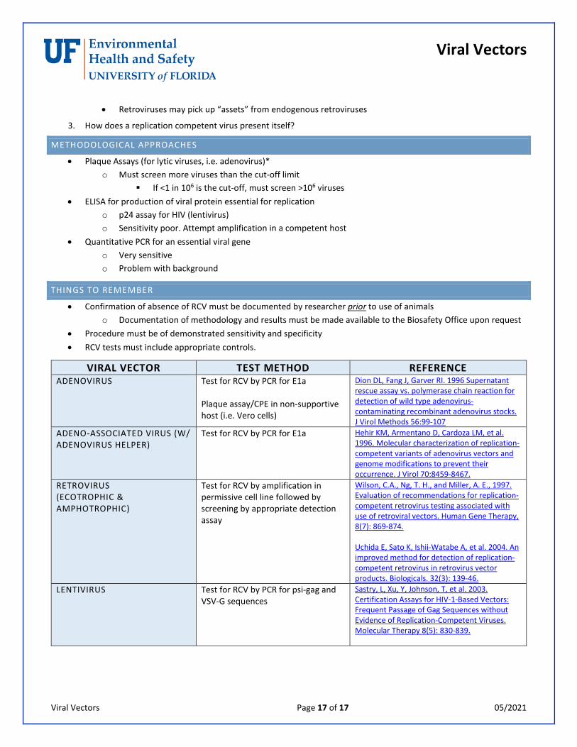
VIRAL VECTOR TEST METHOD REFERENCE ADENOVIRUS Test for RCV by PCR for E1a
Plaque assay/CPE in non-supportive host (i.e. Vero cells)
Dion DL, Fang J, Garver RI. 1996 Supernatant rescue assay vs. polymerase chain reaction for detection of wild type adenovirus-contaminating recombinant adenovirus stocks. J Virol Methods 56:99-107
ADENO-ASSOCIATED VIRUS (W/ ADENOVIRUS HELPER)
Test for RCV by PCR for E1a Hehir KM, Armentano D, Cardoza LM, et al. 1996. Molecular characterization of replication-competent variants of adenovirus vectors and genome modifications to prevent their occurrence. J Virol 70:8459-8467.
RETROVIRUS (ECOTROPHIC & AMPHOTROPHIC)
Test for RCV by amplification in permissive cell line followed by screening by appropriate detection assay
Wilson, C.A., Ng, T. H., and Miller, A. E., 1997. Evaluation of recommendations for replication-competent retrovirus testing associated with use of retroviral vectors. Human Gene Therapy, 8(7): 869-874. Uchida E, Sato K, Ishii-Watabe A, et al. 2004. An improved method for detection of replication-competent retrovirus in retrovirus vector products. Biologicals. 32(3): 139-46.
LENTIVIRUS Test for RCV by PCR for psi-gag and VSV-G sequences
Sastry, L, Xu, Y, Johnson, T, et al. 2003. Certification Assays for HIV-1-Based Vectors: Frequent Passage of Gag Sequences without Evidence of Replication-Competent Viruses. Molecular Therapy 8(5): 830-839.