induction of japanese encephalitis virus-specific cytotoxic t lymphocytes in humans by...
TRANSCRIPT

ELSEVIER
PII: SO264-410X(97)00265-X
Vaccine, Vol. 16, No. 8. pp. 842-849, 1998 0 1998 Elsewer Science Ltd. All rights reserved
Printed in Great Britain 0264-410X/98 $19+0.00
Induction of Japanese encephalitis virus-specific cytotoxic T lymphocytes in humans by poxvirus-based JE vaccine candidates
Eiji Konishi*2, Ichiro Kuyane-/-$, Peter W. Masons, Robert E. Shqpell/J, Niranjan Kanesa-Thasan’, John J. Smucny’, Charles H. Hoke, Jr.’ and Francis A. Ennis+
Poxvirus-based recombinant Japanese encephalitis (JE) vaccine candidates, NYKAC- JEV and ALVAC-JEY were examined for their ability to induce JE virus-specific cytotoxic T lymphocytes (CTLs) in a phase I clinical trial. These vaccine candidates encoded the JE virus premembrane (prM), envelope (E) and non-structural 1 (NSl) proteins. The volunteers received subcutaneous inoculations with each of these candi- dates on days 0 and 28, and blood was drawn 2 days before vaccination and on day 58. Anti-E and anti-NSl antibodies were elicited in most vaccinees inoculated with WAC-JEV and in some vaccinees inoculated with ALVAC-JECi Peripheral blood mononuclear cells (PBMCs) obtained fi-om approximately one half of vaccines showed positive proliferation in response to stimulation with live JE virus. Cytotoxic assays demonstrated the presence of JE virus-specific CTLs in in vitro-stimulated PBMCs obtained from two WAC-JEV and two ALVAC-JEV vaccinees. Cell depletion tests using PBMCs ji-om one WAC-JEV recipient indicated that the phenotype of CTLs was CD8’CD4-. 0 1998 Elsevier Science Ltd. All rights reserved
Keywords: Japanese encephalitis: recombinant vaccine; cytotoxic T lymphocytes
Japanese encephalitis (JE) virus is a medically important flavivirus distributed widely in East, through Southeast and South Asia’,“. The only JE vaccine currently approved in most nations for human use is a formalin-inactivated JE virion preparation purified from infected mouse brains (Biken vaccine). This inactivated JE vaccine is efficacious, but is expensive, poses safety risks in production, is occasionally aller-
*Department of Medical Zoology, Kobe University School of Medicine, Kobe 650, Japan. tDepartment of Medicine, University of Massachusetts Medical Center, Worcester, MA 01655, USA. *Present address: Department of Micro- biology, Kinki University School of Medicine, Osaka- Sayama 589, Japan. DPlum Island Animal Disease Center, Agricultural Research Service, United States Department of Aariculture. Greenoort. NY 11944. USA. llYale Arbovirus Research &tit, Department of Epidemiology and Public Health, Yale University School of Medicine, New Haven, CT 06510, USA. ItPresent address: Department of Pathology, University of Texas Medical Branch, Galveston, TX 77555, USA. ‘Department of Virus Diseases, Walter Reed Army Institute of Research, Washington, DC 20307, USA. ‘Author to whom all correspondence should be addressed. Tel.: (S?) 78 341 7451; Fax: (81) 78 796 4594; e-mail: [email protected]. (Received 1 September 1997; revised version received 29 September 1997; accepted 2 October 1997)
genie, and requires three doses. The development of second generation flavivirus vaccines using molecular technologies’ has been designed to address these concerns and to immunize in one dose, if possible.
A protective role of cytotoxic T lymphocytes (CTLs) against viral diseases has been reported with lympho- cytic choriomeningitis’ and influenza5. In JE virus infection, antibody has been considered to play an important role in protection as demonstrated by passive transfer of monoclonal antibodies against the envelope (E) protein”,‘. On the other hand, a role of cellular immunity in protection has been reported. The adoptive transfer of JE virus-immune T cells protected mice from lethal challenge”~“‘. Mice infected with JE virus developed CTLs that were able to lyse JE virus- infected cells”‘.“. Furthermore, a recent review has questioned whether humoral immunity is the only effective means of providing protection against flavi- virus infection”. These reports suggested that, in addition to antibodies, CTLs are important in protec- tion from JE.
Immune responses against infectious and non-infectious agents differ in some respects. Non-infectious antigens induce mainly specific CD4+T lymphocytes and antibody production, but usually not CD8+ CTLs. Infectious viruses tend to induce specific CD8+ T lymphocytes including CTLS’~. Therefore, it is
842 Vaccine 1998 Volume 16 Number 8

Human CTL induced by NYVACIALVAC-JEV: E. Konishi et al.
likely that inactivated vaccines, like the Biken vaccine, induce predominantly CD4’ T lymphocytes and humoral immunity in humans.
We have developed poxvirus-vectored recombinant JE viruses for use as human vaccines’J.‘5. These vaccine candidates contain the premcmbranc (prM), E and nonstructural 1 (NSI) genes of JE virus in tither a highly attenuated vaccinia virus strain (NYVAC) or a canarypox isolate (ALVAC)‘“. Immunization of outbred mice with these candidates induced neutral- izing (NEUT) antibodies and conferred protection against lethal JE virus challenge’J.‘S. Furthermore, our recent studies have demonstrated that these vaccine candidates are able to induce JE virus-specific CD8’ CTLs in mice”. Following safety and efficacy tests using rhesus monkeys ‘(Vaughn et rrl., in preparation), these candidates were subjected to a human phase I clinical trial (Kanesa-Tlhasan ef al., in submission). The present study was undertaken to determine whether these poxvirus-vectored JE vaccine candidates induce JE virus-specific CD8+ CTLs in humans.
MATERIALS AND METHODS
Viruses
NYVAC-based and ALVAC-based recombinants encoding the JE virus prM, E, and NSl genes of the Nakayama strain of JE virus (abbreviated as NYVAC- JEV and ALVAC-JEV, respectively) have been described’4,‘s. The expression of the JE virus sequences in both recombinants was under the control of the early/late vaccinia promoter (H6). The Nakayama strain of JE virusI was used for preparing antigens for radioimmunoprecipitation (RIP) tests and for stimu- lating peripheral blood mononuclear cells (PBMCs) in proliferation and cyto’toxic assays. Viruses used for infection of target cells in cytotoxic assays were the Copenhagen strain of vaccinia virus (vP410) and a vP410-based recombinant encoding the prM, E and NSl genes of the Nakayama strain of JE virus (vP55s)‘x.
Vaccination
A total of 19 male and 11 female adult volunteers were randomly grouped according to immunogens and preimmunity status against vaccinia. All volunteers were negative for neutralization antibody against JE virus. Results of one vaccinee (#ll) whose postimmu- nization samples were not obtained are not included in this paper.
Immunogens were (1) 4 x 10” plaque-forming units (p.f.u.) of NYVAC-JE.V, (2) 6 x 10’ p.f.u. of ALVAC- JEV, (3) 1 dose of Biken vaccine produced from the Nakayama strain of JE: virus, and (4) saline. The infec- tious titers of recombinant vaccines were based on the assay results of reconstituted inocula, although the titers of one dose prior to lyophilization were 5.8 x 10” p.f.u. for NYVAC-JEV and 3.1 x 10” p.f.u. for ALVAC-JEV. Volunteers received three subcutaneous inoculations on days 10, 7 and 28 with each of these immunogens, except for NYVAC-JEV and ALVAC- JEV recipients who were inoculated with saline in place of vaccines on day 7. Venous blood was drawn from all vaccinees on day -2 (2 days before vaccina-
tion) and on day 58, except one (#13) whose blood was collected on day 44.
Human plasma and PBMC samples
PBMCs were separated from blood by Ficoll- Hypaque density gradient centrifugation as described’“, and resuspended at a concentration of 1 x IO’ml~ in RPM11640 medium containing 10% fetal bovine serum and 10% dimethyl sulphoxide. These PBMCs were cryopreserved in liquid nitrogen until use for prolifera- tion and cytotoxic assays. Plasma samples obtained during PBMC preparation were kept frozen until use for RIP tests.
Radioimmunoprecipitation (RIP)
Plasma specimens were tested for their ability to immunoprecipitate ““[S]-labeled viral proteins harvested from the culture fluid of JE virus-infected cells as described”‘.
Proliferation assay of PBMCs
The viral antigen used in proliferation assays was live JE virus, which was prepared as previously described”. The culture fluid of infected C6136 cells containing JE virus at approximately 2 x 10’ p.f.u.per ml was used as JE viral antigen. The control antigen was culture fluid of mock-infected C6136 cells.
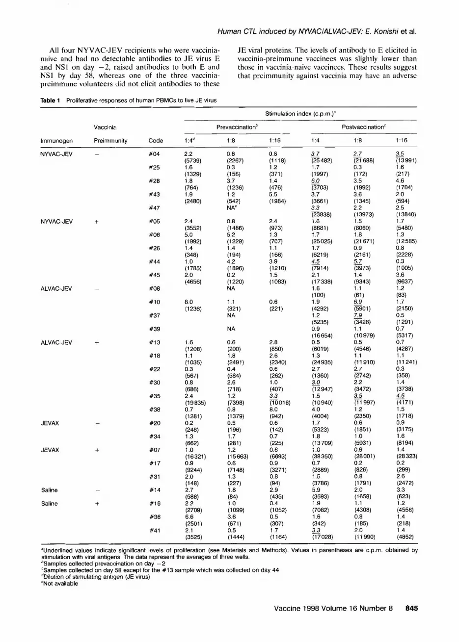
Proliferative responses of PBMCs were measured as previously described”. Briefly, 2 x IO’ PBMCs were cultured with each of the viral and control antigens at 1:4, 1:8 and 1:16 dilutions in 200~11 AIM/V medium (Gibco BRL, Gaithersburg, MD) containing 10% human AB serum in 96-well V-bottom microplates at 37°C. Six days after antigen addition, cells were pulsed with 1 LrCi of tritiated ([. H]) thymidine for 18 h before being harvested using a multiharvester (Titertek, Skatron Inc., Sterling, VA), and [“Hlthymidine incor- poration was determined by a liquid scintillation counter (1205 Betaplate; Pharmacia, Wallac Oy, Finland). Stimulation indices were calculated by the standard formula: counts per minute (c.p.m.) induced by JE antigen/c.p.m. induced by control antigen at the same dilution. The criteria to determine positive responses were; (i) a stimulation index equal to or greater than 2.7”, (ii) a significant difference between c.p.m. induced by the viral and control antigens as evaluated by the Student t-test, and (iii) a total c.p.m. greater than 1500 obtained with viral antigens. Prolifer- ative responses were defined as positive if all of these criteria were fulfilled in at least one dilution of stimu- lating antigens.
Cytotoxic assays
The target cells used for this assay system were autologous Epstein Barr virus-transformed lympho- blastoid cell lines (LCLs) infected with vP555 or vP410 at an m.o.i. of 10 p.f.u./cell or mock-infected 15-20 h before the assay. LCLs were prepared from PBMCs of each individual as described’-. Under these conditions, the proportion of target cells expressing viral antigens was usually 40-60s and occasionally higher as deter-
Vaccine 1998 Volume 16 Number 8 843

Human CTL induced by NWACIALVAC-JEV: E. Konishi et al.
mined by an indirect fluorescent antibody assay using a monoclonal antibody to the JE virus E protein (J3-llB9)‘“. LCLs were resistant to infection with JE virus: only 2-3% of the cells were JE antigen-positive even after infection at a high m.o.i.
Cytotoxic activities were measured as previously reported”. PBMCs (4-5 x 10”) were stimulated by incubation with live JE virus at a final dilution of I:8 in 2 ml of AIM/V containing 10% human AB serum per well of 24-well microplates at 37°C for 7-9 days. These cells were washed three times with RPMI-IO%FBS and distributed in microplates at different cell densities to provide various effector:target (E:T) ratios. The target cells were labeled with Na.“CrOJ, washed three times with RPMI-lO%FBS, counted for viable cells, and distributed at 1 x lo3 cells in 100 111 per well into microplates containing effector cells. The plates were incubated for 5 h at 37°C and “Cr release into the supernatant was measured using a gamma counter. Percent specific lysis was calculated by a formula: 100 x (experimental release - minimum release)/ (maximum release -minimum release), where the maximum release was obtained by lysing all the target cells with polyoxyethylene (12) tridecyl ether (Renex; Ruger Chemical Co.,Irvington, NJ) at a final dilution of 1:40 and the minimum release was obtained with target cells incubated alone in RPMI-lO%FBS.
Phenotypic analysis Cell depletion tests were performed using anti-CD4
(OKT4), anti-CD8 (OKT8; Ortho Diagnostic Systems, Inc., Raritan, NJ) and anti-CD16 (anti-natural killer (NK) cells) antibodies (Leull b; Becton Dickinson, Mountain View, CA) as previously described”. Briefly, approximately 1 x 10” PBMCs stimulated with live virus
at a 1:8 dilution for 7 days were incubated with each antibody at a 1:ll dilution at 4°C for 30 min and then treated with rabbit complement (Cedarlane Labora- tories, Ontario, Canada) at a I:5 dilution at 37°C for 1 h. These cells were used in cytotoxic assays at an E:T ratio of 75: 1.
RESULTS
Elicitation of antibody determined by RIP
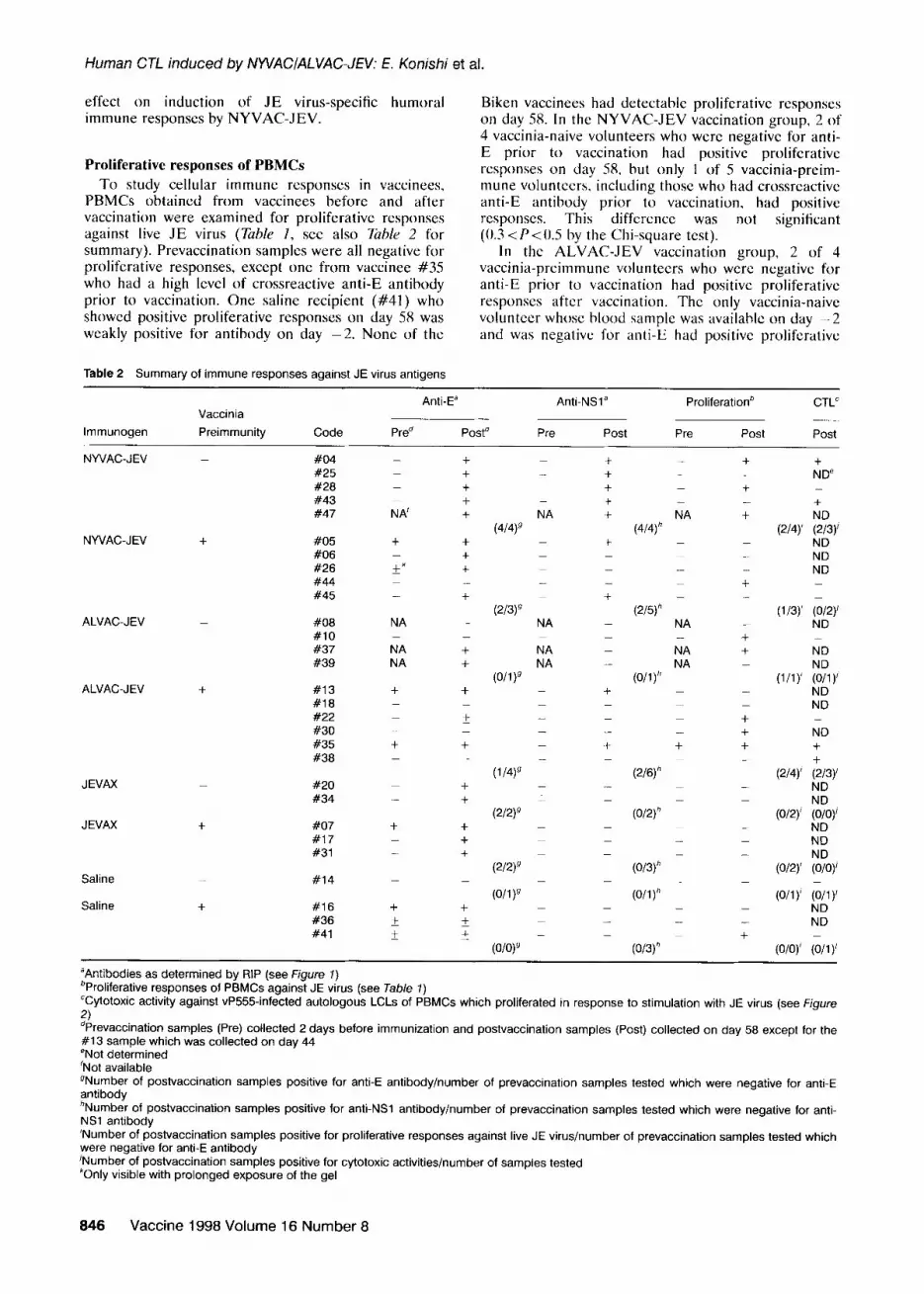
Plasma samples obtained from vaccinees before and after vaccination were examined by RIP for antibodies against E and NSl which were both encoded by candi- date vaccines (Figure I, see also Table 2 for summary). Although volunteers were originally screened for the absence of NEUT antibody and of travel history to any JE-endemic area before immunization, RIP detected anti-E antibody in volunteers #05, #26, #13, #35, #07, #16, #36 and #41 at various levels on day -2. probably because of crossreactive non-neutralizing antibody to JE virus produced by a prior yellow fever (YF) vaccination. Most of these reactivities were only detectable with prolonged exposure of the gels used to produce the autoradiographs shown in Figure 1.
The overall intensity of the bands on day 58 was strong in volunteers immunized with NYVAC-JEV and Biken vaccine, and less strong in ALVAC-JEV recipi- ents. Anti-E antibody was raised in all recipients of Biken vaccine, and anti-E and/or anti-NSl antibodies were raised in most recipients of NYVAC-JEV and some recipients of ALVAC-JEV. In saline recipients, intensity of the reaction was not different between days -2 and 58. These results demonstrate that recombi- nant JE vaccine candidates have an ability to induce antibodies to E and NSI in humans.
NSl’- NSl’-
E- ,.‘,
E- NS% NSl-
Figure 1 JE virus-specific reactivity of plasma samples obtained from vaccinees who received inoculations with NYVAC-JEV, ALVAC-JEV, Biken vaccine (JEVAX) or saline. Vaccinees were grouped according to vaccinia preimmunity, and each vaccinee is represented by a code
number. Plasma samples were collected 2 days before vaccination (represented as day 0) and on day 58 from all vaccinees except #13 whose plasma was collected on day 44. These specimens were tested for their ability to immunoprecipitate radiolabeled proteins harvested from JE virus-infected cells. The positions of JE virus proteins are shown at the left side of the autoradiogram: E, envelope protein; NSl , non-structural protein 1; NSl ‘, a higher molecular weight form of NSl produced by alternative processing of the sequences encoded by the NS2A region of the JE virus genome.
844 Vaccine 1998 Volume 16 Number 8
Human CTL induced by NWACIALVAC-JEV: E. Konishi et al.
All four NYVAC-JEV recipients who were vaccinia- naive and had no detectable antibodies to JE virus E and NSl on day -2, raised antihodies to both E and NSl by day 58, whereas one of the three vaccinia- preimmune volunteers #did not elicit antibodies to these
JE viral proteins. The levels of antibody to E elicited in vaccinia-preimmune vaccinees was slightly lower than those in vaccinia-naive vaccinees. These results suggest that preimmunity against vaccinia may have an adverse
Table 1 Proliferative responses of human PBMCs to live JE virus
Stimulation index (cpm.)”
lmmunogen
Vaccinia
Preimmunity Code
Prevaccinatior? PostvaccinationC
1 :4a 18 1:16 114 1:8 1:16
NWAC-JEV
NWAC-JEV
ALVAC-JEV
ALVAC-JEV
JEVAX
JEVAX
Saline
Saline
_ #04
#25
#28
#43
#47
+ #05
#06
#26
#44
#45
_ #08
#lO
#37
#39
+ #13
#18
#22
#30
#35
#38
_ #20
#34
+ #07
#17
#31
#14
+ #16
#36
#41
2.2 (5739) 1.6 (1329) 1.8
(764) 1.9 (2480)
0.8 (2267) 0.3
(156) 3.7 (1236) 1.2
(542) NA”
0.8 (1118) 1.2
(371) 1.4
(476) 5.5 (1984)
2.4 (3552) 5.0 (1992) 1.4
(348) 1 .o (1785) 2.0 (4656)
0.8 (1486) 5.2 (1229) 1.4
(194) 4.2 (1896) 0.2 (1220) NA
2.4
(973) 1.3
(707) 1.1
(166) 3.9 (1210) 1.5 (1083)
8.0 (1236)
1.1
(321) NA
0.6
(221)
NA
2.7 3.5 (21 688) (13991) 0.3 1.6
(172) (217) 3.5 4.6 (1992) (1704) 3.6 2.0 (1345) (594) 2.2 2.5 (13973) (13840) 1.5 1.7 (6060) (5480) 1.8 1.3 (21671) (12585) 0.9 0.8 (2161) (2228) z 0.3 (3973) (1005) 1.4 3.6 (9343) (9637) 1.1 1.2
(61) G I? (5901) (2150) 7.9 0.5 (3428) (1291) 1.1 0.7 (10979) (5317) 0.5 0.7 (4546) (4287) 1 .l 1.1 (11910) (11241) 2.7 0.3 (2742) (358) 2.2 1.4 (3472) (3738) s 4.6 (11997) (4171) 1.2 1.5 (2350) (1718) 0.6 0.9 (1851) (3175) 1 .o 1.6 (5931) (8194) 0.9 1.4 (28001) (28 323) 0.2 0.2
(826) (299) 0.8 2.6 (1791) (2472) 2.0 3.3 (1658) (623) 1.1 1.2 (4308) (4556) 0.8 1.4
(185) (218) 2.0 1.4 (11990) (4852)
“Underlined values indicate significant levels of proliferation (see Materials and Methods). Values in parentheses are c.p.m. obtained by stimulation with viral antigens. The data represent the averages of three wells. %amples collected prevaccination on day -2 “Samples collected on day 58 except for the #13 sample which was collected on day 44 dDilution of stimulating antigen (JE virus) eNot available
1.6 (1208) 1.1 (1035) 0.3
(567) 0.8
(686) 2.4 (19835) 0.7 (1281) 0.2
(248) 1.3
(662) 1 .o (16321) 0.9 (9244) 2.0
(‘48) 2.7
(588) 2.2 (2709) 6.6 (2501) 2.1 (3525)
0.6
(200) 1.8 (2491) 0.4
(584) 2.6
(718) 1.2 (7398) 0.8 (1379) 0.5
(196) 1.7
(281) 1.2 (15663) 0.6 (7148) 1.3
(227) 1.8
(84) 1 .o (1099) 3.6
(671) 0.5 (1444)
2.8
(850) 2.6 (2340) 0.6
(262) 1 .o
(407) e (10016) 8.0
(942) 0.6
(142) 0.7
(225) 0.6 (6693) 0.9 (3271) 0.8
(94) 2.9
(435) 0.4 (1052) 0.5
(307) 1.7 (1164)
3.7 (25 482) 1.7 (1997) 6.0 (3703) 3.7 (3661) 3.3 (23838) 1.6 (8681) 1.7 (25 025) 1.7 (6219) 4.5 (7914) 2.1 (17338) 1.6
(100) 1.9 (4292) 1.2 (5235) 0.9 (16654) 0.5 (6019) 1.3 (24 935) 2.7 (1360) g (12947) 1.5 (10940) 4.0 (4004) 1.7 (5323) 1.8 (13709) 1.0 (38 350) 0.7 (2889) 1.5 (3786) 5.9 (3593) 1.9 (7082) 1.6
(342) s (17028)
Vaccine 1998 Volume 16 Number 8 845
Human CTL induced by NYVACIALVAC-JEV: E. Konishi et al.
effect on induction of JE virus-specific humoral immune responses by NYVAC-JEV.
Biken vaccinees had detectable proliferative responses on day 58. In the NYVAC-JEV vaccination group, 2 of 4 vaccinia-naive volunteers who were negative for anti-
Proliferative responses of PBMCs
To study cellular immune responses in vaccinees, PBMCs obtained from vaccinees before and after vaccination were examined for proliferative responses against live JE virus (Table I, see also Tuhk 2 for
E prior to vaccination had positive proliferative responses on day 58, but only I of 5 vaccinia-preim- mune volunteers, including those who had crossreactivc anti-E antibody prior to vaccinatiun, had positive responses. This difference was not signilicant (0.3 <P<O.5 by the Chi-square test).
summary). Prevaccination samples were all negative for proliferative responses, except one from vaccinee #35
In the ALVAC-JEV vaccination group, 2 of 4
who had a high level of crossreactive anti-E antibody vaccinia-preimmune volunteers who were negative for anti-E prior to vaccination had positive proliferative
prior to vaccination. One saline recipient (#41) who responses after vaccination. The only vaccinia-naive showed positive proliferative responses On day 58 was weakly positive for antibody on day -2. None of the
volunteer whose blood sample was available on day -2 and was negative for anti-E had positive prolifcrativc
Table 2 Summary of immune responses against JE virus antigens
Anti-Ea Anti-NSla Proliferationb CTL” Vaccinia
lmmunogen Preimmunity Code Pred Postd Pre Post Pre Post Post
NYVAC-JEV + #05 #06 #26 #44 #45
ALVAC-JEV _ #08 #lO #37 #39
ALVAC-JEV + #13 #18 #22 #30 #35 #38
JEVAX #20 #34
JEVAX + #07 #17 #31
NYVAC-JEV _ #04 #25 #28 #43 #47
Saline
Saline
_ #14
t #16 #36 #41
_ + +
_ t t
NA’ +
+ + t
kk + _
_ +
NA _ _ _
NA + NA +
t t _ _
Z!Z _ _
t t _
t _ t
t t _ t _ +
_ _
+ + * * i f-
_
_ NA
(4/4)9 _ _
_
(2/3)’ NA
NA NA
(O/l )g _
_ _ _
(l/4)9 - _
(2/2)9
(2/2)9
(O/l )g
(O/O)9 -
t t t t +
+ _ _ _ t
_ _ _ _
t _ _ _ t -
- _
_ _ _
_
_ _ _
_ _ _ _ NA
(4/4)” _
_
(2/5)h - NA _
NA NA
(Oll)h
_
_
t
(2/6)”
(O/2)” -
_
(O/3)” -
(O/l)” - _ _
(O/3)”
t _ t _ +
_ _ _ + _
_ + t _
_ _ t + t _
_ _
_ _ -
_
_ - +
N+D’ _ N+D
(2/4)’ (2/3)’ ND ND ND _
(l/3)’ (O/2)’ ND _
ND
(l/l)’ $)I ND ND _
ND +
(2/4)’ k/3)’ ND
(O/2)’ ;,, ND ND
(O/2)’ %)I
(O/l )’ (O/l )’ ND ND
(O/O)’ (O/l )’
“Antibodies as determined by RIP (see Figure 1) bProliferative responses of PBMCs against JE virus (see Table 7) “Cytotoxic activity against vP555-infected autologous LCLs of PBMCs which proliferated in response to stimulation with JE virus (see Figure
2) dPrevaccination samples (Pre) collected 2 days before immunization and postvaccination samples (Post) collected on day 58 except for the #I 3 sample which was collected on day 44 eNot determined ‘Not available gNumber of postvaccination samples positive for anti-E antibody/number of prevaccination samples tested which were negative for anti-E antibody *Number of postvaccination samples positive for anti-NSl antibody/number of prevaccination samples tested which were negative for anti- NSl antibody ‘Number of postvaccination samples positive for proliferative responses against live JE virus/number of prevaccination samples tested which were negative for anti-E antibody ‘Number of postvaccination samples positive for cytotoxic activities/number of samples tested “Only visible with prolonged exposure of the gel
846 Vaccine 1998 Volume 16 Number 8

Human CTL induced by NWACIALVAC-JEV: E. Konishi et al.
responses on day 58. These results demonstrate that immunization of some of the subjects with recombinant JE vaccine candidate:, induced JE virus-specific T lymphocytes that proliferated after stimulation with live JE virus irz vitro.
Cytotoxic activity
The PBMCs, which showed stimulation indices equal to or greater than 2.7 on day 58 samples in prolifcra- tion assays, were examined for cytotoxic activity against autologous LCLs infected with vP555. Samples of donors #47 and #37 lacking day -2 PBMCs and the sample of donor #30 with only a small amount of cells available were excluded (see Tuhle 2). Four of the I I PBMC samples examined showed JE virus-specific cytotoxic activities. Two of the four PBMC samples were obtained from vaccinia-naive volunteers vacci- nated with NYVAC-JEV and the other two were obtained from vaccinia-preimmune volunteers vacci- nated with ALVAC-JEV. The CTL results obtained with the JE virus-stimulated PBMCs of these four vaccinecs at an E:T rario of 100: 1 are shown in Figure 2. Similar levels of cytotoxic activities were also observed in PBMC samples from the same vaccinees, after being stimulated twice with live JE virus in vitro (data not shown). The day -2 sample of vaccince #04 did not show cytotoxic activities after stimulation with live JE virus (data no1 shown). These results demon- strate that immunization with the recombinant vaccine candidates induced JE virus-specific memory cytotoxic cells in some of the vaccinated individuals. One of these four vaccinees shown in Figure 2 (#35) had anti- E antibody and positiv’? proliferative responses against live virus on day -2, but the authors did not examine
STIMULANT TARGET #04 it*2
JEV VP555
JEV VP410
JEV MOCK
NONE VP555
NONE VP41 0
NONE MOCK
#35 #38
JEV
JEV
JEV
NONE
NONE
NONE
VP555
VP410
MOCK
VP555
VP410
MOCK
Fr- IF 1 I
0 IO 20 30 0 10 20
% SPECIFIC LYSIS
Figure 2 Lysis of LCLs infected with a recombinant encoding the prM, E and NSl genes (vP555) or the parent vector virus (vP410), or uninfected (MOCK). PBMCs obtained from vaccinees #04, #43, #35 and #38 on day 58 were stimulated with a 1:8 dilution of live JE virus at 37°C for 7-9 days (JEV) or unstimulated (NONE). Cytotoxic activity was measured at an E:T ratio of 1OO:l using %r as a marker of cell lysis (see Materials and Methods for
details).
STIMULANT ANTIBODY
JEV NONE r
JEV ANTI
JEV ANTI
JEV ANT
MOCK NOh
CD4
CD8
-NK
IE
0 20 40 60 80
% SPECIFIC LYSIS
Figure 3 Phenotypic analysis of CTLs by cell depletion using indicated antibodies and complement. PBMCs obtained from
vaccinee #04 (vaccinia-naive, NYVAC-JEWvaccinated) on day 58 were stimulated with a 1:8 dilution of live JE virus for 7 days (JEV)
or unstimulated (MOCK). After cell depletion (see Materials and
Methods for details), cytotoxic activities were measured using %r-labeled autologous LCLs infected with vP555 at an E:T ratio of
75:l. iONe indicates treatment of cells only with complement.
whether cytotoxic activity was present in the day -2 PBMCs.
In order to characterize the cytotoxic cells, we depleted specific T lymphocyte populations from the JE virus-stimulated PBMCs using monoclonal antibodies and complement before the cytotoxic assay. The PBMC sample obtained from a vaccinia-naive vaccinee who received NYVAC-JEV vaccination (#04) was used in the experiment (Figure 3), since this sample showed the highest cytotoxic activity among the four samples shown in Figure 2. The cytotoxic activity against vP555-infected target cells was greatly reduced by treatment with anti-CD8 and complement, compared to the activity after treatment only with complement. On the other hand, reduction in cytotoxicity was not observed after treatment with anti- CD4 or anti-CD16 (anti-NK) in the presence of complement. These results demonstrate that cells responsible for lysis of JE virus-infected LCLs were CD8+CD4 T-lymphocytes.
DISCUSSION
The present report demonstrates that poxvirus-based recombinant JE vaccines, NYVAC-JEV and ALVAC- JEV, have the ability to induce JE virus-specific CD8’ memory CTLs in some of the vaccinated subjects. The results are summarized in T&e 2. In our cytotoxicity assays, vP555 was used for preparing target LCLs, since these cells were resistant to infection with JE virus. Our earlier studies indicated that recombinant JE viral proteins produced in eukaryotic cells infected with vP555 were equivalent to authentic proteins’x.“‘. Furthermore, vP555-infected P815 mastocytoma cells served as targets for murine CTLs, and the levels of specific lysis were similar between vP555-infected P815 cells and JE virus-infected primary kidney cells as target cells”.
Although similar levels of anti-E antibody were induced in Biken recipients and vaccinia-naive, NYVAC-JEV recipients in RIP tests, Biken recipients
Vaccine 1998 Volume 16 Number 8 847

Human CT/_ induced by NYVACIALVAC-JEV: E. Konishi et al.
had significantly higher NEUT antibody titers (a geometric mean titer of 1:211) than NYVAC-JEV recipients (a geometric mean titer of 1:61)( Kanesa- Thasan et al., in submission): the difference between results of RIP and NEUT tests is due to the ability of RIP to detect both neutralizing and non-neutralizing antibodies. Only one of the 10 ALVAC-JEV recipients developed detectable NEUT antibody. These results indicate that Biken vaccine elicited higher levels of NEUT antibodies than recombinant vaccine candidates at the present immunization doses. However, infectious recombinant vaccines were able to induce JE virus- specific memory CTLs. whereas the non-infectious Biken vaccine did not induce enough memory T lymphocytes to show positive proliferative responses of PBMCs to live JE virus in our tests.
Humoral immune responses induced by ALVAC- JEV were lower than those induced by NYVAC-JEV. The difference in humoral immune responses may be due to the difference in inoculation doses (4 x 10” p.f.u. for NYVAC-JEV and 6 x 10⁵ p.f.u. for ALVAC-JEV). However, lower NEUT titers were also observed in mice immunized with ALVAC-JEV than in those immunized with NYVAC-JEV, although the inocula- tion dose was identical”. Thus, these differences are probably due to different characteristics of the vectors, since the same cassette containing the promoter and JE virus genes was used in both recombinant viruses. It is of interest that both recombinants were able to induce CTLs to similar levels in mice” and in some vaccinated humans.
Humoral immune responses induced by immuniza- tion with NYVAC-JEV seem to be lower in the vaccinia-preimmune group than in the vaccinia-naive group as determined by RIP. Furthermore, results of NEUT tests using samples from NYVAC-JEV recipi- ents (Kanesa-Thasan et al., in submission) indicated a significant difference in positivity between the vaccinia- naive group (5 positive of 5 examined) and the vaccinia-preimmune group (0 positive of 5 examined). The effect of preimmunity against vaccinia on immuno- genicity of vaccinia-based recombinant viruses has also been reported by others’4_2h. Although the in Virgo events occurring in vaccinated hosts were not investi- gated in our study, this difference may be due to the vaccinia virus-specific memory CTLs probably present in vaccinia-preimmune vo1unteers’7’x. These CTLs may destroy NYVAC-infected cells before JE virus-specific antibody responses are induced to the levels that are attained in vaccinia-naive NYVAC-JEV recipients.
In clinical trials of other recombinant vaccine candi- dates, the induction of CTLs has been reported with vaccine candidates for human immunodeficiency virus type 1 (HIV-l) vectored by ALVAC’“-” and by vaccinia virus” and for human papilloma virus types 16 and 18 vectored by vaccinia virus-‘. Immunogenicity of ALVAC-JEV in humans observed in the present study seems somewhat lower than that of another experi- mental ALVAC vaccine against rabies”. The dose and inoculation route may be critical factors in the differ- ences trials.
In there CTLs
848
in immunogemcity determined in these vaccine
addition to recombinant virus-based vaccines, have been reports of successful induction of in animals by naked plasmid DNA” peptides
Vaccine 1998 Volume 16 Number 8
administered with adjuvants3”.‘7 or liposomes.‘“, and peptides expressed in particulate carrier?“. The analysis of memory T lymphocytes and antibodies will help us to evaluate new types of preventive and thera- peutic vaccine candidates for flaviviruscs.
ACKNOWLEDGEMENTS
This work was supported by grants from the National Institute of Health of the United States (NIH-ROI- A130624, AI10987-17) the US Army Medical Research and Development Command (DAMDI7-90-Z-0020), and the National Science Foundation of the United States (DMB 8515345). Written informed consent was obtained from each study volunteer. This protocol was approved by the Human Subjects Research Review Board of the Office of the Surgeon General, US Army. Research was conducted in accord with Army Regula- tion 70-25.
REFERENCES
1
2
3
4
5
6
7
8
9
10
11
12
13
14
Shope, R.E. Medical significance of togaviruses: an overview of diseases caused by togaviruses in man and in domestic and wild vertebrate animals. In: The Togaviruses (Ed. Schle- singer, R.W.) Academic Press, New York, 1980, pp. 47-82 Monath, T.P. Pathobiology of the flaviviruses. In: The Togavir- idae and flaviviridae (Eds Schlesinger, S. and Schlesinger, M.J.) Plenum Press, New York/London, 1986, pp. 375-440 Brandt, W.E. Current approaches to the development of dengue vaccines and related aspects of the molecular biology of flaviviruses. Journal of infectious Diseases 1988, 157, 1105-1111. Byrne, J.A. and Oldstone, M.B.A. Biology of cloned cytotoxic T lymphocytes specific for lymphocytic choriomeningitis virus: clearance of virus in vivo. Journal of Virology 1984, 51, 682-686. Kuwano, K., Scott, M., Young, J.F. and Ennis, F.A. HA2 subunit of influenza A Hl and H2 subtype viruses induces a protective cross-reactive cytotoxic T lymphocyte response. Journal of Immunology 1988,140,1264- 1268. Kimura-Kuroda, J. and Yasui, K. Protection of mice against Japanese encephalitis virus by passive administration with monoclonal antibodies. Journal of Immunology 1988, 141, 3606-3610. Mason, P.W., Dalrymple, J.M. and Gentry, M.K. et al. Molec- ular characterization of a neutralizing domain of the Japanese encephalitis virus structural glycoprotein. Journal of General Virology 1989, 70, 2037-2049. Mathur, A., Arora, K.L. and Chaturvedi, U.C. Host defense mechanisms against Japanese encephalitis virus infection in mice. Journal of Genera/ Virology 1983, 64, 805-811. Miura, K., Onodera, T., Nishida, A., Goto, N. and Fujisaki. Y. A single gene controls resistance to Japanese encephalitis virus in mice. Archives of Virology 1990, 1 i2, 261-270. Murali-Krishna. K.. Ravi. V. and Maniunath. R. Protection of adult but not newborn mice against lethal intracerebral challenge with Japanese encephalitis virus by adoptively transferred virus-specific cytotoxic T lymphocytes: require- ment for L3T4+ T cells. Journal of General Virology 1996, 77, 705-714. Konishi, E., Kurane, I., Mason, P.W., Shope, R.E. and Ennis, F.A. Poxvirus-based Japanese encephalitis vaccine candi- dates induce JE virus-specific CD8’ cytotoxic T lymphocytes in mice. Virology 1997, 227, 353-360. Venugopal, K. and Gould, E.A. Towards a new generation of flavivirus vaccines. Vaccine 1994, 12, 966-975. Germain, R.N. Antigen processing and presentation. In: Fundamental Immunology (Ed. Paul, W.E.), 3rd edn. Raven Press, New York, 1993, pp. 629-676 Konishi, E., Pincus, S. and Paoletti, E. et al. A highly attenu- ated host-range-restricted vaccinia virus strain, NYVAC, encoding the prM, E and NSl genes of Japanese encephalitis

Human CTL induced by NWACIALVAC-JEV: E. Konishi et al.
15
16
17
18
19
20
21
22
23
24
25
26
virus prevents JEV viremia in swine. Virology 1992. 190, 454-458. Konishi, E., Pincus, S., Paoletti, E., Shope, R.E. and Mason, P.W. Avipox virus-velctored Japanese encephalitis virus vaccines: use as vaccine candidates in combination with purified subunit immunogens. Vaccine 1994, 12, 633-638. Tartaglia, J., Perkus. M.E. and Taylor, J. et al. NYVAC: a highly attenuated strain of vaccinia virus. Viro/ogy 1992, 188, 2177232. Mason, P.W. Maturation of Japanese encephalitis virus glyco- proteins produced by infected mammalian and mosquito cells. Virology 1989, 18!9, 354-364. Mason, P.W., Pincus, t<. and Fournier, M.J. et al. Japanese encephalitis virus-vaccinia recombinants produce particulate forms of the structural membrane proteins and induce high levels of protection aga.inst lethal JEV infection. Virology 1991, 180,294-305. Kurane, I., Innis, B.L. and Nisalak, A. et a/. Human T cell responses to dengue virus antigens: proliferative responses and interferon gamma production. Journal of Clinical Invesfi- g&ion 1989, 83, 506-513. Konishi, E., Pincus, S. and Fonseca, B.A.L. et al. Comparison of protective immuniiy elicited by recombinant vaccinia viruses that synthesize E or NSl of Japanese encephalitis virus. Virology 1991, 185, 401-410. Konishi, E., Kurane, I., Mason, P.W., Innis, B.L. and Ennis, F.A. Japanese encephalitis virus-specific proliferative responses of humarl peripheral blood T lymphocytes. American Journal of Tropical Medicine and Hygiene 1995, 53, 278-283. Green, S., Kurane, I. and Edelman, Ft. et al. Dengue virus- specific human CD4’ T-lymphocyte responses in a recipient of an experimental live-attenuated dengue virus type 1 vaccine: bulk culture proliferation, clonal analysis, and precursor frequency determination. J. Viral. 1993, 67, 5962-5967. Livingston, P.G., Kurane, I., Lai, C.J., Bray, M. and Ennis, F.A. Recognition of envelope protein by dengue virus serotype- specific human CD4+ CD8 cytotoxic T-cell clones. Journal of Virology 1994, 88, 3283-3288. Rooney, J.F., Wohlenberg, C., Cremer, K.J., Moss, B. and Notkins, A.L. Immunization with a vaccinia virus recombinant expressing herpes simplex virus type 1 glycoprotein D: long- term protection and effect of revaccination. Journal of Virology 1988, 62,1530-l 534. Johnson, M.P., Meitin, C.A., Bender, B.S. and Small, P.A. Jr. Passive immune serum inhibits antibody response to recom- binant vaccinia virus. In: Vaccines 88 (Eds Ginsberg, H., Brown, F., Lerner, R.A. and Chanock, R.M.) Cold Spring Harbor Laboratory, New York, 1988, pp. 189-l 92 Cooney, E.L., Collier, A.C. and Greenberg, P.D. et a/. Safety of and immunological response to a recombinant vaccinia virus vaccine expressing HIV envelope glycoprotein. Lancet 1991, 337,567-572.
27
28
29
30
31
32
33
34
35
36
37
38
39
Demkowicz, W.E. Jr. and Ennis, F.A. Vaccinia virus-specific CD8’ cytotoxic T lymphocytes in humans. Journal of Virology 1993, 67,1538-1544. Littaua, R.A.. Takeda, A., Cruz, J. and Ennis, F.A. Vaccinia virus-specific human CD4’ cytotoxic T-lymphocyte clones. Journal of Virology 1992, 66, 2274-2280. Pialoux, G., Excler, J.L. and Riviere, Y. et a/. A prime-boost approach to HIV preventive vaccine using a recombinant canarypox virus expressing glycoprotein 160 (MN) followed by a recombinant glycoprotein 160 (MN/LAI). ADS Research and Human Retroviruses 1995, 11,373-381. Egan, M.A., Pavlat, W.A. and Tartaglia, J. et al. Induction of human immunodeficiency virus type 1 (HIV-1)-specific cytotoxic T lymphocyte responses in seronegative adults by a nonreplicating, host-range-restricted canarypox vector (ALVAC) carrying the HIV-l MN env gene. Journal of infectious Diseases 1995,171, 1623- 1627. Fleury, B., Janvier, G. and Pialoux, G. et al. Memory cytotoxic T lymphocyte responses in human immunodeficiency virus type 1 (HIV-I)-negative volunteers immunized with a recombi- nant canarypox expressing gp 160 of HIV-l and boosted with a recombinant gpl60. Journal of lnfecfious Diseases 1996, 174,734-738. Perales, M.A., Schwartz, D.H., Fabry, J.A. and Lieberman, J. A vaccinia-gpl60-based vaccine but not a gp160 protein vaccine elicits anti-gp160 cytotoxic T lymphocytes in some HIV-l seronegative vaccinees. Journal of Acquired immune Deficiency Syndrome and Human Retrovirology 1995, 10, 27-35. Borysiewicz, L.K., Fiander, A. and Nimako, M. et al. A recom- binant vaccinia virus encoding human papillomavirus types 16 and 18, E6 and E7 proteins as immunotherapy for cervical cancer. Lancet 1996, 347, 1523-1527. Cadoz, M., Strady, A. and Meignier. B. et a/. lmmunisation with canarypox virus expressing rabies glycoprotein. Lancet 1992,339, 1429-l 432. Donnelly, J.J., Ulmer, J.B. and Liu, M.A. Immunization with DNA. Journal of immunological Methods 1994, 176, 145- 152. Jones, P.D., Tha-Hla, R., Morein, B., Lovgren, K. and Ada, G.L. Cellular immune responses in the murine lung to local immunization with influenza A virus glycoproteins in micelles and immunostimulatory complexes (iscoms). Scandinavian Journal of Immunology 1988,27, 645-652. Takahashi, H., Takeshita, T., Morein, B., Putney, S., Germain, R.N. and Berzofsky, J.A. Induction of CD8’ cytotoxic T cells by immunization with purified HIV-l envelope protein in ISCOMS. Nature 1990, 344, 873-875. Reddy, R., Zhou, F., Nair, S., Huang, L. and Rouse, B.T. In vivo cytotoxic T lymphocyte induction with soluble proteins administered in liposomes. Journal of immunology 1992, 148, 1585-1589. Layton, G.T., Harris, S.J. and Gearing, A.J. et a/. Induction of HIV-specific cytotoxic T lymphocytes in vivo with hybrid HIV-l V3:Ty-virus-like particles, Journal of Immunology 1993, 151, 1097-1107.
Vaccine 1998 Volume 16 Number 8 849