isogenic induced pluripotent stem cell lines from an adult with mosaic down syndrome model...
TRANSCRIPT
STEM CELLS 2014;00:00‐00 www.StemCells.com ©AlphaMed Press 2014
EMBRYONIC STEM CELLS/INDUCED PLURIPOTENT STEM CELLS 1The Blizard Institute, Barts and The London School of Medicine, Queen Mary University of London, UK; 2The LonDownS Consortium, Wellcome Trust, UK; 3Department of Genet‐ic Medicine and Development, University of Geneva Medical School, Geneva, Switzer‐land; 4Service of Genetic Medicine, Universi‐ty Geneva Hospitals, Geneva, Switzerland; 5Lee Kong Chian School of Medicine, Na‐nyang Technological University, Singapore; 6Human Genetics Laboratory, Galliera Hos‐pital, Genoa, Italy; 7Liguria Department of Genetics, Genoa, Italy; 8Department of Neuroscience, Physiology and Pharmacolo‐gy, University College London, UK; 9Department of Forensic and Analytical Science, King’s College, London, UK; 10Precursory Research for Embryonic Science and Technology, Japan Science and Technology Agency, Saitama, Japan; 11DNAVEC corporation, Ibaraki, Tokyo, Ja‐pan; 12 Stem Cell Laboratory, National Cen‐tre for Bowel Research and Surgical Innova‐tion, Queen Mary University of London, UK Correspondence to: Professor Dean Nizetic M.D. Ph.D., Lee Kong Chian School of Medi‐cine, Nanyang Technological University Singapore, Proteos Building, Level 4, 61 Biopolis Drive, Biopolis, Singapore 138673, E‐mail: [email protected], Tel: +65 6778 4728, Fax: +65 6778 5824 And Dr Jurgen Groet, PhD, The Blizard Institute, Barts & The London School of Medicine, Blizard Building, 4 Newark Street, London E1 2AT, United Kingdom, E‐mail: [email protected], Tel: +442078822271, Fax: +442078822183; †sadly deceased dur‐ing revision of this manuscript; #Present address: Dept. of Opthalmology, Keio Uni‐versity School of Medicine, Tokyo, Japan; *co‐corresponding authors; Grants support‐ing this work: Kay Kendall Leukaemia Fund, Fondation Jerome, Lejeune, The Wellcome Trust “LonDownS Consortium” Strategic Funding, Award to DN, the “AnEUploidy Consortium” grant from Framework Pro‐gramme, 6 from the EU Commission to SEA and DN, SNF and ERC grants to SEA, Tele‐thon, Network of Genetic Biobanks (Tele‐thon project GTB12001) Received October 12, 2014; accepted for publication January 17, 2015; available online without subscription through the open access option. ©AlphaMed Press 1066‐5099/2015/$30.00/0 This article has been accepted for publica‐tion and undergone full peer review but has not been through the copyediting, typeset‐ting, pagination and proofreading process which may lead to differences between this version and the Version of Record. Please cite this article as doi: 10.1002/stem.1968
Isogenic Induced Pluripotent Stem Cell Lines from an Adult with Mosaic Down Syndrome Model Accelerated neuronal Ageing and Neu‐rodegeneration
Aoife Murray1,2, Audrey Letourneau3, Claudia Canzonetta1, Eli‐
savet Stathaki4, Stefania Gimelli4, Frederique Sloan‐Bena4, Robert Abrehart1, Pollyanna Goh1,2, Shuhui Lim5, Chiara Baldo6, Franca Dagna‐Bricarelli7†, Saad Hannan8, Martin Mortensen8, David Ballard9, Denise Synder‐combe Court9, Noemi Fusaki10#, Mamoru Hasegawa11, Trevor G. Smart8, Cleo Bishop1, Stylianos E. Antonara‐kis3, Jürgen Groet1,2,12*, and Dean Nizetic1,2,5*
Key words. Down syndrome • neurodegeneration • neurogenesis • neuronal differentiation
ABSTRACT
Trisomy 21 (T21), Down Syndrome (DS) is the most common genetic cause of dementia and intellectual disability. Modelling DS is beginning to yield pharmaceutical therapeutic interventions for amelioration of intellectual disability, which are currently being tested in clinical trials. DS is also a unique genetic system for investigation of pathological and protective me‐chanisms for accelerated ageing, neurodegeneration, dementia, cancer and other important common diseases. New drugs could be identified and dis‐ease mechanisms better understood by establishment of well controlled cell model systems. We have developed a first non‐integration‐reprogrammed isogenic human induced‐pluripotent‐stem‐cells (iPSC) mod‐el of DS by reprogramming the skin fibroblasts from an adult individual with constitutional mosaicism for DS, and separately cloned multiple iso‐genic T21 and euploid (D21) iPSC lines. Our model shows a very low num‐ber of reprogramming rearrangements as assessed by a high‐resolution whole genome CGH‐array hybridisation and it reproduces several cellular pathologies seen in primary human DS cells, as assessed by automated high‐content microscopic analysis. Early differentiation shows an imbal‐ance of the lineage‐specific stem/progenitor cell compartments: T21 caus‐es slower proliferation of neural and faster expansion of hematopoietic lineage. T21 iPSC‐derived neurons show increased production of amyloid peptide‐containing material, a decrease in mitochondrial membrane po‐tential, and an increased number and abnormal appearance of mitochon‐dria. Finally, T21‐derived neurons show significantly higher number of DNA double‐strand breaks than isogenic D21 controls. Our fully isogenic system therefore opens possibilities for modelling mechanisms of developmental, accelerated ageing, and neurodegenerative pathologies caused by T21. STEM CELLS 2014; 00:000–000

Isogenic iPSC model from mosaic Down Syndrome
www.StemCells.com ©AlphaMed Press 2014
2
INTRODUCTION
Trisomy 21 (T21), Down Syndrome (DS) is the most common genetic cause of intellectual disability and de‐mentia with rising global prevalence [1]. Several pheno‐types have been observed at molecular and cellular level, that reproduce in primary tissues from human individuals with T21. Some of these cellular phenotypes directly map onto clinical components of DS; these in‐clude intellectual disability, defects in cognitive devel‐opment and age‐related cognitive decline, Alzheimer’s disease (AD)‐like dementia, epilepsy, congenital heart defect, childhood leukemia and others [2‐12]. Modelling DS is beginning to yield pharmaceutical therapeutic interventions for amelioration of intellectual disability which are currently being tested in clinical trials. New drugs could be identified by high throughput screening of chemical libraries using cellular assays, and therefore well controlled cellular model systems are required. In order to eliminate effects of wide phenotypic differenc‐es among individuals with DS, the requirement for many experimental purposes has become the use of an isogenic iPSC model for DS, where the sole difference between iPSC lines is the presence of the third chromo‐some 21. Several recent iPSC models of DS have been developed. All of these (with one exception) used integrational re‐programming. All of these reprogrammed cells were derived from fetal, neonatal, or 1 year old infant DS, actually, three studies [13‐15] used the same iPSC lines as a starting point [16]. Some of these studies report defects in NPC proliferation [17], neurogenesis [13], gliogenesis and neurite outgrowth [18], others defects in synaptic morphology and function, mitochondrial dysfunction [19] and increased amyloid deposition [15]. Other models show increased propensity to generate hematopoietic precursors and increased multilineage myeloid hematopoiesis potential [14,20]. Most of these results have been generated on non‐isogenic compari‐sons, with isogenic lines either serendipitously generat‐ed in cell culture [14,19], or using an ingenious but complex and laborious approach to silence the third chromosome 21 [13,21]. In some cases, non‐isogenic lines were pooled with one isogenic line [14,19]. In one study, a unique case of heterokaryotypic twin fetal cells was exploited [17]. None of these papers reported a genome‐wide high‐resolution array‐Comparative Ge‐nomic Hybridization (aCGH) analysis of the resulting iPSCs for the rigorous measurement of the artificial copy‐number rearrangements known to frequently oc‐cur during the reprogramming process. Here we present the first iPSC DS model which is both non‐integrationally reprogrammed and fully isogenic, and the first derived from an adult individual with DS which is a constitutional mosaic. Furthermore, we verify the high level of genome integrity of the resulting iPSCs, by showing a very low number of reprogramming rear‐rangements as assessed by a high‐resolution whole ge‐
nome aCGH, and we make entirely isogenic compari‐sons of 3 trisomic and 3 disomic iPSC lines derived from this model. This approach minimizes the influence of any copy number fluctuations additional to T21, and eliminates genotypic difference noise, allowing the “clean” detection of T21‐causing effects. Our model reproduces several T21 cellular pathologies seen in pri‐mary human DS cells, but hitherto not reported in iPSC models, such as abnormalities in mitochondrial number and size, and an increase in DNA double‐strand breaks in neurons. MATERIALS AND METHODS
Detailed methods are shown in Supplementary Data online. RESULTS
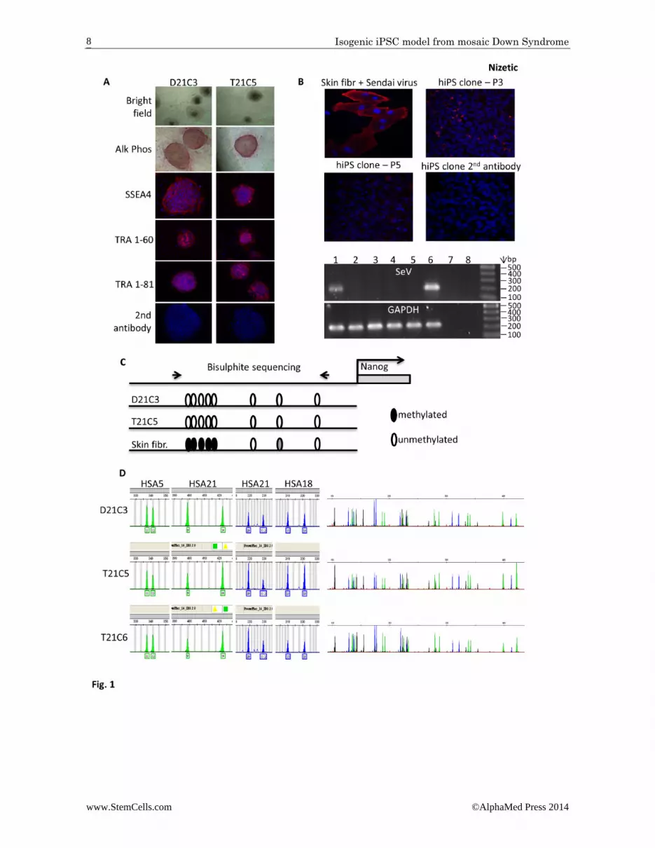
We generated the iPSCs by non‐integration repro‐gramming using temperature‐sensitive Sendai virus [22], from primary skin fibroblasts from a young adult diagnosed with constitutional mosaicism for DS (strate‐gy illustrated in Supplementary Data Fig. S1). We iso‐lated individual clones (labelled as C[number]), ex‐panded them, and confirmed that they tested positive for Alkaline‐phosphatase expression, and the presence of markers of pluripotency (Fig. 1A). The Sendai virus was efficiently removed after 7‐10 passages (Fig. 1B). De‐methylation of the endogenous NANOG promoter in the iPSCs, compared to the parental skin fibroblasts, was established via bisulfite sequencing (Fig. 1C). Indi‐vidual clones were analyzed by rigorous whole‐genome microsatellite DNA fingerprinting, which established the presence of clones with T21, and euploid genome (D21), which are otherwise isogenic (Fig. 1D). In a pre‐liminary RNA‐seq experiment, the isogenic iPSCs show an expected increase in transcript levels for the majority of HSA21 genes (not shown). The genome integrity of the resulting iPSCs is of a high level, as was assessed by high‐resolution, whole genome aCGH (Supplementary Data Fig. S2). The supernumerary HSA21 is intact and complete in both analyzed trisomic lines (T21C5 and T21C6), and T21 is stable for at least 17‐19 passages, which is as far as we tested for the presence of T21 (Supplementary Data Fig. S2A). After filtering out the copy number variations (CNVs) that occur commonly in healthy individuals (using comparison to the Database of Genomic Variation, DGV) we made an in silico com‐parison with the published survey of genome‐rearrangement artefacts in iPSC generated by conven‐tional integration‐reprogramming [23]. Selecting only events at the same aCGH resolution and the same pas‐sage number as in our study, we detected a significantly lower number of un‐common CNVs, affecting a signifi‐cantly lower number of genes in our lines, relative to those generated by classical integrational reprogram‐

Isogenic iPSC model from mosaic Down Syndrome
www.StemCells.com ©AlphaMed Press 2014
3
ming methods (Supplementary Data Fig. S2B and S2C). In compliance with international guidelines for iPSC nomenclature [24] we name these iPSCs: NIZEDSM1iD21‐C3, ‐C7 and ‐C9 for the disomic lines, respectively, and NIZEDSM1iT21‐C5, ‐C6, ‐C13 for the trisomic lines, respectively (henceforward abbreviated to D21C3, D21C7, D21C9, T21C5, T21C6 and T21C13). Microsatellite DNA fingerprinting was repeated at later passages and confirmed that trisomy 21 is retained through routine passaging (Supplementary Data Fig. S2D). The isogenic DS iPSC clones can differentiate into cell lineages of all 3 embryonic layers both in vitro and in vivo (Supplementary Data Fig. S3). After 45 days in culture, using neuronal differentiation via Neuro‐EB protocol (see Supplementary Methods), both disomic and trisomic neuronal differentiation cul‐tures were able to produce mature looking neurons expressing βIII‐tubulin, the inhibitory neurotransmitter GABA, as well as pre‐ and post‐synaptic markers of exci‐tatory synapses, PSD95 and VGlut (Supplementary Data Fig. S4A, S4B, respectively). In order to accelerate neu‐ronal differentiation and improve yields, we applied a directed neuronal differentiation protocol using dual SMAD inhibition (Noggin and SB431542), combined with stimulation of retinoid signaling and the addition of the Sonic Hedgehog agonist purmorphamine [25]. Both trisomic and disomic lines were able to produce electrophysiologically‐active neurons (Supplementary Data Fig. S4C) that supported spontaneous action po‐tential firing and functional whole‐cell current res‐ponses to saturating concentrations of externally‐applied GABA and glycine, observed in neurons from both D21C3 (at 28 days) and T21C5 lines (at 40 days). In order to gain an approximate estimate of the propor‐tion of neurons likely to fire spontaneous action poten‐tials, we used Ca2+ imaging and observed multiple Ca2+ transients, which would be indicative of regenerative spontaneous activity in accord with the firing of spon‐taneous action potentials (FigS5A,B). Quantification of the numbers of neurons exhibiting calcium transients over the 1 minute time‐course showed no significant difference between D21 and T21 neurons (Fig S5B). In addition, under voltage‐clamp conditions, we identified neuronal sensitivities to GABA and glycine which indi‐cates the cell surface expression of functional GABAA and glycine receptors. This was apparent in most cells tested, with no difference noted between T21 and D21 cells (not shown). To examine early stages of iPSC differentiation an em‐bryoid body (EB) protocol was adopted for both hema‐topoietic and neuronal differentiation, modifying the published method [26]. For hematopoietic EBs (HEBs) 3,000 live cells in single cell suspension were allowed to aggregate in a 96‐well. Imaging and analysis of HEBs after 5 days showed that T21 HEBs were significantly larger than the euploid controls (Supplementary Data Fig. S6A). This increase in HEB size was caused by an increase in the cell numbers (Fig. S6B), which at this stage are predicted to be early haematopoietic meso‐
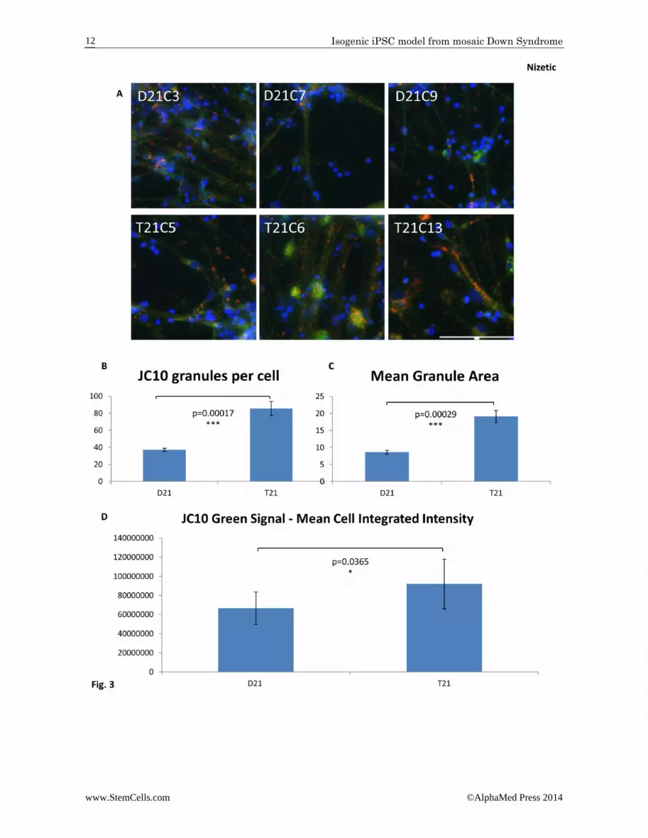
derm precursors. A similar result was obtained with early T21 mesodermal colonies derived from tran‐schromosomic mouse ES cells [7]. Although the T21 HEBs gave a higher proportion of CD34+ cells (haema‐topoietic stem/progenitor lineages) on both days 12 and 18 of further differentiation (not shown), there was no statistically significant difference. By contrast, neural progenitor cells (NPCs) derived from neuro EBs (NEBs) showed a reduction in proliferation rate. The cumula‐tive population doublings of trisomic cells was signifi‐cantly reduced during the expansion of NPCs (Supple‐mentary Data Fig. S6C). Increased cell death is unlikely to be the cause of the observed decrease population cell doubling, because the proportion of non‐viable cells in the same counts shows no increase in T21 NPCs (Fig S6D). iPSC‐derived neurons were analysed after 60 days of differentiation following the dual SMAD inhibition pro‐tocol (mentioned previously, but here minus purmor‐phamine) that generates multiple classes of projection neurons from different cortical layers [25]. We ob‐served the typical morphology of neurogenic cortical rosettes as published [27], and confirmed that after 60 days neuronal differentiation, >95% of cells were Tuj1+ (not shown). As this protocol generated 100% excitatory glutamatergic neurons [25,27], and as no differences in the composition of cortical layer sub‐populations were observed between DS and normal hiPSCs [15], we think it unlikely that difference in cell type profiles would be responsible for the differences between T21 and D21 observed in our results, though we cannot fully rule out this possibility. Immunostaining of fixed neurons with an antibody (6E10) raised against the epitopes in the Aβ–peptide, but reactive to all proteolytic fragments of the amyloid precursor protein (APP) that contain this epitope, revealed an increase in total amyloid staining in trisomic neurons compared to isogenic euploid con‐trols (Fig. 2A and B). This demonstrates that increased amyloid production can be successfully modelled in these cells. Trisomic neurons also appear to show an increase in size and number of 6E10‐reactive discreet punctiform aggregates (not quantified, Fig. 2B zoom). Our staining does not permit us to conclude if these aggregates are intracellular or extracellular. To assess mitochondrial membrane potential in neu‐rons, live cells were labelled with JC‐10 (Fig. 3A). In healthy cells JC‐10 selective accumulates in the mito‐chondria and forms aggregates with a characteristic fluorescent emission at 590 nm (orange/red). If mito‐chondrial membrane potential is decreased (due to damaged/unhealthy cells) JC‐10 monomers are formed, which are released into the cytoplasm, resulting in a shift to green emission at 525 nm. An increase in both the size and number of mitochondria were observed in the trisomic neurons (Fig. 3B and C), consistent with a previous study in primary T21 neurons which showed “generalized perturbations” in T21 mitochondrial struc‐ture and function, including a more fragmented mito‐chondrial network [9]. Trisomic neurons also showed a
Isogenic iPSC model from mosaic Down Syndrome
www.StemCells.com ©AlphaMed Press 2014
4
decreased mitochondrial membrane potential, as evi‐denced by the increase in green cytoplasmic staining with JC‐10 (Fig. 3D). Having shown that T21 neurons have a deficit in mito‐chondrial function similar to primary human T21 neu‐rons in vitro, we anticipate that reactive oxygen species (ROS) production is increased in T21 neurons. Increased ROS leads to increased DNA damage, as measured by the proportion of γH2AX foci, an in vitro marker of age‐ing, shown increased in primary fibroblasts from old, compared to young humans [28], and in primary neu‐rons from old, compared to young mice [29]. We as‐sessed the number of γH2AX foci (Fig. 4A). Fully auto‐mated quantification of approximately 18,000 cells per cell line showed a significantly increased number of γH2AX puncta per cell in T21 neurons (Fig. 4B). DISCUSSION
In conclusion, we report a first non‐integration‐reprogrammed isogenic and high genomic fidelity iPSC model from an adult with mosaic DS. The model repro‐duces several differentiation, ageing and neurodegene‐ration‐related cellular phenotypes associated with DS pathology, and attributable solely to T21 as a cause. Particularly intriguing are two observations (Fig. 3 and Fig. 4) that open a whole set of new interesting mecha‐nistic questions. Increased number of mitochondria in neurons could be related to increased mitochondrial fragmentation observed in primary DS cortical neurons in culture [9]. On the other hand, it is possible that T21 mitochondria in DS neurons, which are high consumers of energy exclusively derived from oxidative metabol‐ism, are hypofunctional (as has been observed in prima‐ry DS cortical neurons in culture [9]), and the demand for more energy increases mitochondrial biogenesis. These hypotheses remain to be tested. Increased num‐ber of DNA double‐strand breaks in T21 neurons (Fig. 4) could be further exploited as a cellular marker of acce‐lerated ageing observed in DS [10,30]. Alternatively, more genomic instability in the nuclei of post‐mitotic T21 neurons (such as transposition events [31]) could also explain this observation. Currently, several interdisciplinary consortia have been organized to study DS genetics and cellular models inte‐grated with the assessment of the adult population with DS for neurocognitive function, age‐related decline, presence or absence of dementia and other co‐morbidities (for example, see http://www.ucl.ac.uk/london‐down‐syndrome‐consortium, http://www.psychiatry.cam.ac.uk/ciddrg/research/dementia‐in‐downs‐syndrome‐dids/). It is therefore impor‐tant to verify that cellular phenotypes under‐pinning DS pathology can also be reproduced in iPSCs derived from an adult individual with DS, as this had not been re‐ported so far. Up to 75% of constitutionally T21 concepti sponta‐neously die in utero [10,32 ]suggesting that in random
allelic variation, the presence of a third copy of HSA21 has a 75% probability to cause severe phenotypes that are normally missed. In constitutional mosaicism, T21 with an otherwise deleterious genotype could be res‐cued by a significant (>50%) presence of normal (D21) cells, which results in varying intensity of clinical DS defects, often not correlating with the percentage of trisomic cells in tissues. This presents with a theoretical (but so far never explored) rationale for T21/D21 cells derived from a constitutionally mosaic DS individual to show more contrasting differences in cellular pheno‐types, than comparisons between cells of liveborn 100% trisomic individuals. Such phenotypes are more likely to be robust, reproducible, and therefore more amenable to developing into high throughput screening assays. ACKNOWLEDGMENTS
This work was supported by the project grants from the Kay Kendall Leukaemia Fund (KKL632), and Fondation Jerome Lejeune (2011B/960), by The Wellcome Trust “LonDownS Consortium” Strategic Funding Award (098330/Z/12/Z), and the Lee Kong Chian School of Medicine, Nanyang Technological University‐Singapore start‐up funding grant M4230024 to DN, by the “AnEUploidy Consorium” grant from Framework Pro‐gramme 6 from the EU Commission to DN and SEA, as well as SNF 144082 and ERC 249968 grants to SEA. We also thank Barts and The London Charity and the grant 520/1489: “Development of a National Centre for Bo‐wel Research and Surgical Innovation (NCBRSI)” for pur‐chase of high content imaging equipment. Galliera Ge‐netic Bank as member of the Telethon Network of Ge‐netic Biobanks (Telethon project GTB12001) provided us with specimens. We thank Li Pin for help in setting up the iPSC lab at LKCMedicine in Singapore. We thank Gary Warnes for help with the FACS analysis, and Domi‐nique Bonnet, Silvia Marino and the BSU Whitechapel for help in using NOD/SCID mice. We dedicate this pa‐per to Prof. Franca Dagna‐Bricarelli, our colleague and co‐author who sadly passed away during revision of this manuscript. Her dedicated work in human genetics made all this possible and will remain inspiration to all of us.

Isogenic iPSC model from mosaic Down Syndrome
www.StemCells.com ©AlphaMed Press 2014
5
AUTHOR CONTRIBUTIONS A.M.: Conception and design, Collection and assembly of data, Data analysis and interpretation, Manuscript writing, Final approval of manuscript; A.L.: Conception and design, Collection and assembly of data, Data anal‐ysis and interpretation, Manuscript writing; C.C.: Con‐ception and design, Collection and assembly of data, Data analysis and interpretation; E.S.: Collection of da‐ta; S.G.: Collection of data; F.S.‐B.: Collection of data, Data analysis and interpretation; R.A.: Collection of da‐ta; P.G.: Collection of data, Data analysis and interpreta‐tion; S.L.: Data analysis and interpretation; C.B.: Provi‐sion of study material or patients; F.D.‐B.: Provision of study material or patients; S.H.: Collection of data, Data analysis and interpretation; M.M.: Collection of data, Data analysis and interpretation; D.B.: Collection of da‐ta, Data analysis and interpretation; D.S.‐C.: Collection
of data, Data analysis and interpretation; N.F.: Provision of study material; M.H.: Provision of study material; T.S.: Assembly of data, Data analysis and interpretation; C.B.: Collection and assembly of data, Data analysis and interpretation, Manuscript writing, Final approval of manuscript; S.A.: Financial support, Data analysis and interpretation, Manuscript writing, Final approval of manuscript; J.G.: Conception and design, Collection and assembly of data, Data analysis and interpretation, Ma‐nuscript writing, Final approval of manuscript; D.N.: Conception and design, Financial support, Collection and assembly of data, Data analysis and interpretation, Manuscript writing, Final approval of manuscript DISCLOSURE OF POTENTIAL CONFLICTS OF INTEREST
The authors declare no conflict of interest
REFERENCES
1 Epstein CJ. 2001 William Allan Award
Address. From Down syndrome to the "human" in "human genetics". American journal of human genetics. Feb 2002;70(2):300‐313.
2 Antonarakis SE, Lyle R, Dermitzakis ET, Reymond A, Deutsch S. Chromosome 21 and down syndrome: from genomics to pathophysiology. Nat Rev Genet. Oct 2004;5(10):725‐738.
3 Bahn S, Mimmack M, Ryan M, et al. Neuronal target genes of the neuron‐restrictive silencer factor in neurospheres derived from fetuses with Down's syndrome: a gene expression study. Lancet. Jan 26 2002;359(9303):310‐315.
4 Busciglio J, Yankner BA. Apoptosis and increased generation of reactive oxygen species in Down's syndrome neurons in vitro. Nature. Dec 21‐28 1995;378(6559):776‐779.
5 Canzonetta C, Mulligan C, Deutsch S, et al. DYRK1A‐dosage imbalance perturbs NRSF/REST levels, deregulating pluripotency and embryonic stem cell fate in Down syndrome. American journal of human genetics. Sep 2008;83(3):388‐400.
6 Cataldo AM, Mathews PM, Boiteau AB, et al. Down syndrome fibroblast model of Alzheimer‐related endosome pathology: accelerated endocytosis promotes late endocytic defects. Am J Pathol. Aug 2008;173(2):370‐384.
7 De Vita S, Canzonetta C, Mulligan C, et al. Trisomic dose of several chromosome 21 genes perturbs haematopoietic stem and progenitor cell differentiation in Down's syndrome. Oncogene. Nov 18 2010;29(46):6102‐6114.
8 Delom F, Burt E, Hoischen A, et al. Transchromosomic cell model of Down syndrome shows aberrant migration, adhesion and proteome response to extracellular matrix. Proteome Sci. 2009;7:31.
9 Helguera P, Seiglie J, Rodriguez J, Hanna M, Helguera G, Busciglio J. Adaptive downregulation of mitochondrial function in down syndrome. Cell Metab. Jan 8 2013;17(1):132‐140.
10 Nizetic D, Groet J. Tumorigenesis in Down's syndrome: big lessons from a small chromosome. Nature reviews. Cancer. Oct 2012;12(10):721‐732.
11 Roper RJ, Baxter LL, Saran NG, Klinedinst DK, Beachy PA, Reeves RH. Defective cerebellar response to mitogenic Hedgehog signaling in Down's syndrome mice. Proc Natl Acad Sci U S A. Jan 31 2006;103(5):1452‐1456.
12 Wang Y, Mulligan C, Denyer G, et al. Quantitative proteomics characterization of a mouse embryonic stem cell model of Down syndrome. Molecular & cellular proteomics : MCP. Apr 2009;8(4):585‐595.
13 Jiang J, Jing Y, Cost GJ, et al. Translating dosage compensation to trisomy 21. Nature. Aug 15 2013;500(7462):296‐300.
14 Maclean GA, Menne TF, Guo G, et al. Altered hematopoiesis in trisomy 21 as revealed through in vitro differentiation of isogenic human pluripotent cells. Proc Natl Acad Sci U S A. Oct 23 2012;109(43):17567‐17572.
15 Shi Y, Kirwan P, Smith J, MacLean G, Orkin SH, Livesey FJ. A human stem cell model of early Alzheimer's disease pathology in Down syndrome. Sci Transl Med. Mar 7 2012;4(124):124ra129.
16 Park IH, Arora N, Huo H, et al. Disease‐specific induced pluripotent stem cells. Cell. Sep 5 2008;134(5):877‐886.
17 Hibaoui Y, Grad I, Letourneau A, et al. Modelling and rescuing neurodevelopmental defect of Down syndrome using induced pluripotent stem cells from monozygotic twins discordant for trisomy 21. EMBO molecular medicine. Feb 2014;6(2):259‐277.
18 Briggs JA, Sun J, Shepherd J, et al. Integration‐free induced pluripotent
stem cells model genetic and neural developmental features of down syndrome etiology. Stem Cells. Mar 2013;31(3):467‐478.
19 Weick JP, Held DL, Bonadurer GF, 3rd, et al. Deficits in human trisomy 21 iPSCs and neurons. Proc Natl Acad Sci U S A. Jun 11 2013;110(24):9962‐9967.
20 Chou ST, Byrska‐Bishop M, Tober JM, et al. Trisomy 21‐associated defects in human primitive hematopoiesis revealed through induced pluripotent stem cells. Proc Natl Acad Sci U S A. Oct 23 2012;109(43):17573‐17578.
21 Li LB, Chang KH, Wang PR, Hirata RK, Papayannopoulou T, Russell DW. Trisomy correction in down syndrome induced pluripotent stem cells. Cell Stem Cell. Nov 2 2012;11(5):615‐619.
22 Ban H, Nishishita N, Fusaki N, et al. Efficient generation of transgene‐free human induced pluripotent stem cells (iPSCs) by temperature‐sensitive Sendai virus vectors. Proc Natl Acad Sci U S A. Aug 23 2011;108(34):14234‐14239.
23 Hussein SM, Batada NN, Vuoristo S, et al. Copy number variation and selection during reprogramming to pluripotency. Nature. Mar 3 2011;471(7336):58‐62.
24 Luong MX, Auerbach J, Crook JM, et al. A call for standardized naming and reporting of human ESC and iPSC lines. Cell Stem Cell. Apr 8 2011;8(4):357‐359.
25 Shi Y, Kirwan P, Livesey FJ. Directed differentiation of human pluripotent stem cells to cerebral cortex neurons and neural networks. Nat Protoc. Oct 2012;7(10):1836‐1846.
26 Ng ES, Davis RP, Hatzistavrou T, Stanley EG, Elefanty AG. Directed differentiation of human embryonic stem cells as spin embryoid bodies and a description of the hematopoietic blast colony forming assay. Current protocols in stem cell biology. Jan 2008;Chapter 1:Unit 1D 3.
27 Shi Y, Kirwan P, Smith J, Robinson HP, Livesey FJ. Human cerebral cortex development from pluripotent stem cells
Isogenic iPSC model from mosaic Down Syndrome
www.StemCells.com ©AlphaMed Press 2014
6
to functional excitatory synapses. Nat Neurosci. Mar 2012;15(3):477‐486, S471.
28 Miller JD, Ganat YM, Kishinevsky S, et al. Human iPSC‐based modeling of late‐onset disease via progerin‐induced aging. Cell Stem Cell. Dec 5 2013;13(6):691‐705.
29 Jurk D, Wang C, Miwa S, et al. Postmitotic neurons develop a p21‐dependent senescence‐like phenotype driven by a
DNA damage response. Aging cell. Dec 2012;11(6):996‐1004.
30 Adorno M, Sikandar S, Mitra SS, et al. Usp16 contributes to somatic stem‐cell defects in Down's syndrome. Nature. Sep 19 2013;501(7467):380‐384.
31 Erwin JA, Marchetto MC, Gage FH. Mobile DNA elements in the generation of diversity and complexity in the brain.
Nature reviews. Neuroscience. Aug 2014;15(8):497‐506.
32 Spencer K. What is the true fetal loss rate in pregnancies affected by trisomy 21 and how does this influence whether first trimester detection rates are superior to those in the second trimester? Prenat Diagn. Sep 2001;21(9):788‐789.
See www.StemCells.com for supporting information available online. STEM
CELLS ; 00:000–000

Isogenic iPSC model from mosaic Down Syndrome
www.StemCells.com ©AlphaMed Press 2014
7
Figure 1. Isogenic iPSC model of Down Syndrome generated by reprogramming primary human skin fibroblasts from an adult individual with mosaic Down Syndrome, using a temperature‐sensitive Sendai virus. A: Images of undifferentiated iPSC colonies from two clones (D21C3, T21C5) after 3 passages; brightfield microphoto‐graphs and Alkaline Phosphatase expression. Further images after immunohistochemistry for pluripotency markers (SSEA4, TRA 1‐60, TRA 1‐81). B: Spontaneous elimination of the temperature sensitive, non‐integrating Sendai virus (Ts‐SeV) from iPSC cells through routine passaging. Primary human skin fibroblasts were infected with the Ts‐SeV for positive control, and were stained with an antibody against the SeV protein HN‐IL4.1 alongside the iPSC colonies at the indicated passage numbers (P[n]). The agarose gel shows amplification products after RT‐PCR, using SeV (and GAPDH) specific primers 1) D21C3 P5, 2) T21C5 P7, 3) T21C5 P10, 4) D21C3 P10, 5) untransfected fibroblasts, 6) SeV infected fibroblasts P0, 7) SeV infected fibroblasts P0 – reverse transcriptase, 8) H2O C: Demethylation of endogenous NANOG promoter following reprogramming: bisulfite sequencing analysis of eight CpG dinucleotides in the promoter region of NANOG using genomic DNA isolated from iPSC D21C3 and T21C5, compared to genomic DNA isolated from the primary mosaic DS skin fibroblasts that were used for reprogramming. D: (Left hand panels): Graphs showing semi‐quantitative microsatellite PCR analysis for two chromosome 21 (HSA21) markers and for markers from two euploid chromosomes (HSA5, 18) using genomic DNA isolated from iPSC clones. Clones T21C5 and T21C6 are trisomic and Clone D21C3 disomic for HSA21. (Right hand panels): Whole genome microsatellite fingerprint of genomic DNA isolated from iPSC clones, demonstrating that they are isogenic.
Isogenic iPSC model from mosaic Down Syndrome
www.StemCells.com ©AlphaMed Press 2014
8

Isogenic iPSC model from mosaic Down Syndrome
www.StemCells.com ©AlphaMed Press 2014
9
Figure 2. Trisomy 21 causes an increase in β‐amyloid containing material in and around neurons generated from iPSCs. A: Neurons were generated from iPSCs over a 60 day differentiation protocol. Cells were then fixed and stained with an anti‐amyloid peptide antibody (6E10), which is reactive to amino acids 1‐16 in β‐amyloid, but detects all APP poly‐peptide forms that contain the epitope. Nuclei were labelled with Hoechst. Scale bar: 100 μm. B: Quantification of the integrated intensity for the 6E10 stain shows an increase of APP expression in T21 neurons compared to the isogenic D21 neurons. Image capture and quantification was performed using automated multi‐parametric analysis on the ImageXpress Micro XL (Molecular Devices) wide‐field high content imaging system, and data analyzed using Meta‐Morph software. Three wells per cell line and a minimum of 6,000 cells per well were analyzed. Student’s T‐test, error bars SEM. Visually, T21 neurons appear to also show more 6E10‐reactive aggregates (not quantitated). Zoomed‐in images for T21C6 and D21C7 are shown at the same size and magnification.

Isogenic iPSC model from mosaic Down Syndrome
www.StemCells.com ©AlphaMed Press 2014
10

Isogenic iPSC model from mosaic Down Syndrome
www.StemCells.com ©AlphaMed Press 2014
11
Figure 3. Trisomy 21 results in an increase in size and number of mitochondria in neurons generated from iPSCs. A: Neurons were generated from iPSCs over a 60 day differentiation protocol. Live cells were then loaded with JC‐10 to assess mitochondrial membrane potential. Healthy mitochondria are labelled in red, while green cytoplasmic stain‐ing indicates that JC‐10 is diffusing out of the mitochondria due to decreased mitochondrial membrane potential. Representative images for each cell line are shown. Image capture and quantification was performed using auto‐mated multi‐parametric analysis on the ImageXpress Micro XL (Molecular Devices) wide‐field high content imaging system, and data analyzed using MetaMorph software. A total of 4 wells and a minimum of 1,500 cells per cell line were imaged and analyzed. Scale bar: 100 μm (identical scale for all images). B: Quantification of the number of mito‐chondria per cell, and (C) the mean mitochondrial area show that both are increased in T21 neurons compared to the isogenic D21 neurons. D: Quantification of the integrated intensity for the green signal generated by JC‐10 shows de‐creased mitochondrial membrane potential in T21 neurons compared to the isogenic D21 neurons. Student’s T‐test, error bars SEM.
Isogenic iPSC model from mosaic Down Syndrome
www.StemCells.com ©AlphaMed Press 2014
12

Isogenic iPSC model from mosaic Down Syndrome
www.StemCells.com ©AlphaMed Press 2014
13
Figure 4. Trisomy 21 causes an increase in DNA damage in neurons generated from iPSCs. A: Neurons were generated from iPSCs over a 60 day differentiation protocol. Cells were then fixed and stained with a γH2AX antibody to detect DNA double‐strand breaks. Nuclei were labelled with Hoechst. Scale bar: 100 μm. Enlarged insets for D21C3 and T21C5 double stained with Hoechst and γH2AX antibody are shown as examples below main images. B: The number of γH2AX puncta per cell is significantly increased in T21 neurons compared to the isogenic D21 neurons. Image capture and quantification was performed using automated multi‐parametric analysis on the ImageXpress Micro XL (Molecular Devices) wide‐field high content imaging system, and data analyzed using Meta‐Morph software. Three wells per cell line and a minimum of 6,000 cells per well were analyzed. Student’s T‐test, er‐ror bars SEM.