immunization with an immunodominant self-peptide derived from glucose-6-phosphate isomerase induces...
TRANSCRIPT

Available online http://arthritis-research.com/content/11/4/R117
Open AccessVol 11 No 4Research articleImmunization with an immunodominant self-peptide derived from glucose-6-phosphate isomerase induces arthritis in DBA/1 miceLisa Bruns1, Oliver Frey1, Lars Morawietz2, Christiane Landgraf3, Rudolf Volkmer3 and Thomas Kamradt1
1Universitätsklinikum Jena, Institut für Immunologie, Leutragraben 3, Jena 07740, Germany2Charité Universitätsmedizin Berlin, Institut für Pathologie, Charitéplatz 1, Berlin 10117, Germany3Charité Universitätsmedizin Berlin, Institut für Medizinische Immunologie, Charitéplatz 1, Berlin 10117, Germany
Corresponding author: Thomas Kamradt, [email protected]
Received: 17 Nov 2008 Revisions requested: 15 Dec 2008 Revisions received: 23 Jun 2009 Accepted: 29 Jul 2009 Published: 29 Jul 2009
Arthritis Research & Therapy 2009, 11:R117 (doi:10.1186/ar2777)This article is online at: http://arthritis-research.com/content/11/4/R117© 2009 Bruns et al.; licensee BioMed Central Ltd. This is an open access article distributed under the terms of the Creative Commons Attribution License (http://creativecommons.org/licenses/by/2.0), which permits unrestricted use, distribution, and reproduction in any medium, provided the original work is properly cited.
Abstract
Introduction T-helper (Th) lymphocytes are critically requiredfor the pathogenesis of glucose-6-phosphate isomerase (G6PI)-induced arthritis, but neither the G6PI epitopes recognized byarthritogenic T cells nor their pathogenic effector functions havebeen fully elucidated to date. We aimed at identifyingarthritogenic G6PI peptides.
Methods We used a library of overlapping peptides spanningthe entire G6PI sequence to identify the epitopes recognized byG6PI-specific Th cells. Immunodominant peptides were thenused to immunize mice. Arthritis development was evaluatedclinically and histologically. The humoral and cellular immuneresponses upon peptide immunization were analyzed by ELISAand multiparameter flow cytometry, respectively.
Results We identified six immunodominant T-cell epitopes inDBA/1 mice, of which three are arthritogenic. One of thesepeptides (G6PI469–483) is identical in man and mice.Immunization with this peptide induces arthritis, which is lesssevere and of shorter duration than arthritis induced byimmunization with full-length G6PI. Upon immunization witheither G6PI or peptide, the antigen-specific Th cells produce IL-17, RANKL, IFNγ and TNFα.
Conclusions We identified immunodominant and arthritogenicepitopes of G6PI. Not all immunodominant peptides arearthritogenic. This is the first description of arthritis induced byimmunization with a self-peptide in mice.
IntroductionAutoreactive CD4+ T-helper (Th) lymphocytes play a centralrole in the pathogenesis of autoimmune diseases [1]. Key tothe development of immune responses is the binding of T-cellreceptors on CD4+ Th cells to their cognate peptide/MHCcomplex on the surface of antigen-presenting cells (APC).Among the well-established genetic risk factors for rheuma-toid arthritis, HLA-DRB1, PTPN22 and STAT4 are relevant forT-cell function [2-4]. T cells are present in the inflamed syno-vial compartment [5,6]. These findings strongly suggest theconcept that rheumatoid arthritis is Th-cell dependent, andthat the associated HLA-DR molecules present peptides toautoreactive Th cells, which initiate the inflammatory processthat ultimately leads to rheumatoid arthritis. This assumption is
supported by the clinical benefits of treating rheumatoid arthri-tis patients with abatacept, a CTLA4–immunoglobulin fusionprotein that blocks Th-cell costimulation, thus selectively inhib-iting their activation [7,8]. Nevertheless, the specificity of thepathogenic Th cells in rheumatoid arthritis has been difficult todefine.
In experimental animals, arthritis can be induced by systemicimmunization with noncartilagenous antigens [9,10] or withcartilage-antigens including heterologous collagen type II[11], collagen type XI [12], cartilage oligomeric matrix protein[13] and proteoglycan [14] in complete Freund's adjuvant(CFA). The immune response of T cells to complex antigens iscommonly focused on a small number of major epitopes.
Page 1 of 11(page number not for citation purposes)
APC: antigen-presenting cells; CFA: complete Freund's adjuvant; ELISA: enzyme-linked immunosorbent assay; FCS: fetal calf serum; G6PI: glucose-6-phosphate-isomerase; H & E: hematoxylin and eosin; IFN: interferon; IL: interleukin; mAb: monoclonal antibody; RANKL: receptor activator of NFκβ ligand; Th: T-helper.

Arthritis Research & Therapy Vol 11 No 4 Bruns et al.
Although immunodominant collagen type II epitopes havebeen defined for different collagen-induced arthritis-suscepti-ble strains of mice [11,15], and for proteoglycan [16], experi-mental arthritis cannot be induced by immunization with theseimmunodominant peptides [15,16]. In fact, even denaturedcollagen type II or its cyanobromide fragments are less effi-cient for arthritis induction than full-length, native collagen typeII [17]. This lack of an identified arthritogenic epitope has beenan obstacle to studying the role of Th cells in mouse models ofarthritis. Collagen-induced arthritis is easily transferable withserum from arthritic animals or mixtures of monoclonal anti-bodies specific for collagen type II, reflecting a strong depend-ence on antibodies [18-20].
We recently described a model in which systemic immuneresponses to glucose-6-phosphate isomerase (G6PI) inducea peripheral symmetric polyarthritis in susceptible strains ofmice [21,22]. In this model, arthritis development depends onT cells, B cells and innate immunity [21-25]. CD4+ Th cells arecrucial not only for the induction of the disease but also duringthe effector phase. Depletion of CD4+ T cells in arthritic ani-mals induces arthritis remission [21]. To understand better therole of Th cells in this model, we sought to determine theimmunodominant epitopes in G6PI-induced arthritis. In thepresent article we describe the identification of six immunodo-minant G6PI epitopes and the induction of arthritis in DBA/1mice by immunization with three of these peptides.
Materials and methodsAnimals and arthritis inductionDBA/1 mice were bred and maintained under specific-patho-gen free conditions in our animal facility. All animal experi-ments were approved by the Government Commission forAnimal Protection (Registered Number 02-005/06).
Arthritis was induced in 6-week-old to 10-week-old DBA/1mice by subcutaneous immunization at the base of the tail witheither 400 μg recombinant human G6PI or 50 μg peptide incomplete Freund's adjuvant (Sigma-Aldrich, Taufkirchen, Ger-many).
Clinical scores were determined daily for each paw independ-ently, as previously described [21]. A score of 0 indicates noclinical signs of arthritis, 1 indicates slight swelling and red-ness, 2 indicates a strong swelling and redness, and 3 indi-cates massive swelling and redness. Arthritis incidence isalmost 100% in this model, and the natural history is highlysynchronized with arthritis onset on d9.
Antibodies and reagentsThe following mAbs were grown and purified from hybridomasupernatants in our laboratory: anti-CD16/CD32 (2.4G2) andanti-CD28 (37.51). Anti-IL-17A (eBio17B7)-Alexa 488, anti-TNFα (MP6-XT22)-Pacific Blue, anti-IFNγ (XMG1.2)-phyco-erythrin-Cy7, anti-CD4 (RM4-5)-allophycocyanin-Alexa750
(APC-A750), anti-IL-2 (JES6-5H4)-fluorescein isothiocyanate,anti-IL-6 (MP5-20F3)-fluorescein isothiocyanate, anti-IL-10(JES5-16E3)-APC, and anti-RANKL (IK22/5)-phycoerythrinwere purchased from ebiosciences (San Diego, CA, USA).Anti-CD154 (MR1)-APC was purchased from Miltenyi Biotec(Bergisch Gladbach, Germany). Recombinant human G6PIwas expressed in Escherichia coli BL21 as described previ-ously [21].
PeptidesCellulose-bound peptides were prepared according to thestandard SPOT synthesis protocol by a MultiPep SPOT-robot(INTAVIS Bioanalytical instruments AG, Köln, Germany) on aβ-alanine-modified cellulose membrane as described else-where [26].
Each spot was eluted in 200 μl distilled H2O containing 5%dimethylsulfoxide, resulting in an approximate concentration of350 to 650 μg/ml peptide solution. These peptide solutionswere taken to create peptide pools resulting in a concentrationof any single peptide of ~42 μg/ml. The final concentration forin vitro restimulation for every single peptide was ~1 μg/ml.Peptides for immunization were synthesized according tostandard Fmoc machine protocols with the multiple peptidesynthesizer SYRO II (MultiSynTec, Witten, Germany). The fol-lowing peptides derived from human G6PI were synthesized:G6PI65–79 (MRMLVDLAKSRGVEA), G6PI85–99(FNGEKINYTEGRAVL), G6PI325–339 (IWYINCFGCETHAML),G6PI469–483 (EGNRPTNSIVFTKLT), G6PI497–511 (KIFVQGII-WDINSFD) and G6PI517–531 (LGKQLAKKIEPELDG). Purity ofthe peptides was determined by high-performance liquid chro-matography and the composition was monitored by matrix-assisted laser desorption/ionization time-of-flight mass spec-troscopy.
HistopathologyMicrosections from mouse legs were prepared and stainedwith H & E as described previously [21]. Samples were viewedwith a DMRBE microscope (Leitz, Wetzlar, Germany) by apathologist who was blinded to the experimental setup. Theseverity was graded semiquantitatively in five steps from 0(normal finding) to 4 (strong inflammation) as described previ-ously [21,27].
Proliferation assaysAll cell cultures and assays were performed in RPMI 1640supplemented with 10% FCS, 100 U/ml penicillin, 100 μg/mlstreptomycin, and 50 μM 2-mercaptoethanol as described[21].
Cells were plated in a 96-well round-bottom plate (GreinerBio-One, Solingen, Germany) at a density of 1 × 10⁶ cells/mlculture medium. Cells were stimulated with either 10 μg/mlG6PI, 5 μl peptide pool or culture medium alone in triplicatefor 72 hours. For the last 18 hours 1 μCi/well [3H]thymidine
Page 2 of 11(page number not for citation purposes)
Available online http://arthritis-research.com/content/11/4/R117
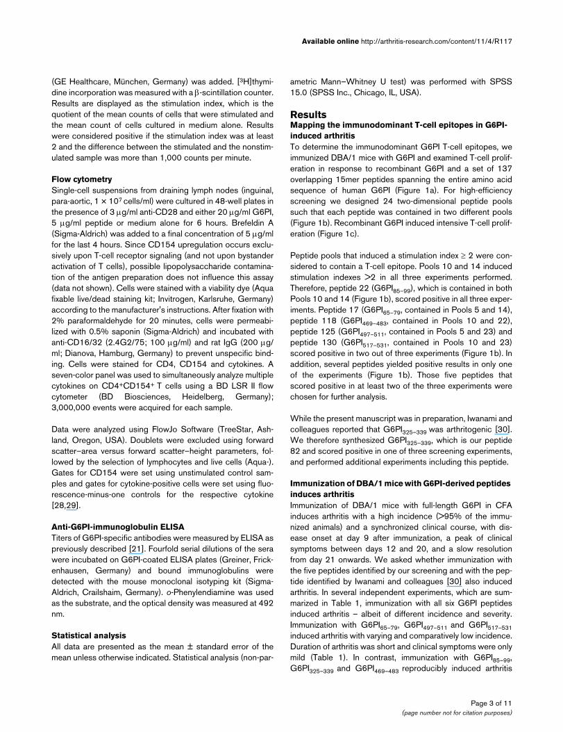
(GE Healthcare, München, Germany) was added. [3H]thymi-dine incorporation was measured with a β-scintillation counter.Results are displayed as the stimulation index, which is thequotient of the mean counts of cells that were stimulated andthe mean count of cells cultured in medium alone. Resultswere considered positive if the stimulation index was at least2 and the difference between the stimulated and the nonstim-ulated sample was more than 1,000 counts per minute.
Flow cytometrySingle-cell suspensions from draining lymph nodes (inguinal,para-aortic, 1 × 10⁷ cells/ml) were cultured in 48-well plates inthe presence of 3 μg/ml anti-CD28 and either 20 μg/ml G6PI,5 μg/ml peptide or medium alone for 6 hours. Brefeldin A(Sigma-Aldrich) was added to a final concentration of 5 μg/mlfor the last 4 hours. Since CD154 upregulation occurs exclu-sively upon T-cell receptor signaling (and not upon bystanderactivation of T cells), possible lipopolysaccharide contamina-tion of the antigen preparation does not influence this assay(data not shown). Cells were stained with a viability dye (Aquafixable live/dead staining kit; Invitrogen, Karlsruhe, Germany)according to the manufacturer's instructions. After fixation with2% paraformaldehyde for 20 minutes, cells were permeabi-lized with 0.5% saponin (Sigma-Aldrich) and incubated withanti-CD16/32 (2.4G2/75; 100 μg/ml) and rat IgG (200 μg/ml; Dianova, Hamburg, Germany) to prevent unspecific bind-ing. Cells were stained for CD4, CD154 and cytokines. Aseven-color panel was used to simultaneously analyze multiplecytokines on CD4+CD154+ T cells using a BD LSR II flowcytometer (BD Biosciences, Heidelberg, Germany);3,000,000 events were acquired for each sample.
Data were analyzed using FlowJo Software (TreeStar, Ash-land, Oregon, USA). Doublets were excluded using forwardscatter–area versus forward scatter–height parameters, fol-lowed by the selection of lymphocytes and live cells (Aqua-).Gates for CD154 were set using unstimulated control sam-ples and gates for cytokine-positive cells were set using fluo-rescence-minus-one controls for the respective cytokine[28,29].
Anti-G6PI-immunoglobulin ELISATiters of G6PI-specific antibodies were measured by ELISA aspreviously described [21]. Fourfold serial dilutions of the serawere incubated on G6PI-coated ELISA plates (Greiner, Frick-enhausen, Germany) and bound immunoglobulins weredetected with the mouse monoclonal isotyping kit (Sigma-Aldrich, Crailshaim, Germany). o-Phenylendiamine was usedas the substrate, and the optical density was measured at 492nm.
Statistical analysisAll data are presented as the mean ± standard error of themean unless otherwise indicated. Statistical analysis (non-par-
ametric Mann–Whitney U test) was performed with SPSS15.0 (SPSS Inc., Chicago, IL, USA).
ResultsMapping the immunodominant T-cell epitopes in G6PI-induced arthritisTo determine the immunodominant G6PI T-cell epitopes, weimmunized DBA/1 mice with G6PI and examined T-cell prolif-eration in response to recombinant G6PI and a set of 137overlapping 15mer peptides spanning the entire amino acidsequence of human G6PI (Figure 1a). For high-efficiencyscreening we designed 24 two-dimensional peptide poolssuch that each peptide was contained in two different pools(Figure 1b). Recombinant G6PI induced intensive T-cell prolif-eration (Figure 1c).
Peptide pools that induced a stimulation index ≥ 2 were con-sidered to contain a T-cell epitope. Pools 10 and 14 inducedstimulation indexes >2 in all three experiments performed.Therefore, peptide 22 (G6PI85–99), which is contained in bothPools 10 and 14 (Figure 1b), scored positive in all three exper-iments. Peptide 17 (G6PI65–79, contained in Pools 5 and 14),peptide 118 (G6PI469–483, contained in Pools 10 and 22),peptide 125 (G6PI497–511, contained in Pools 5 and 23) andpeptide 130 (G6PI517–531, contained in Pools 10 and 23)scored positive in two out of three experiments (Figure 1b). Inaddition, several peptides yielded positive results in only oneof the experiments (Figure 1b). Those five peptides thatscored positive in at least two of the three experiments werechosen for further analysis.
While the present manuscript was in preparation, Iwanami andcolleagues reported that G6PI325–339 was arthritogenic [30].We therefore synthesized G6PI325–339, which is our peptide82 and scored positive in one of three screening experiments,and performed additional experiments including this peptide.
Immunization of DBA/1 mice with G6PI-derived peptides induces arthritisImmunization of DBA/1 mice with full-length G6PI in CFAinduces arthritis with a high incidence (>95% of the immu-nized animals) and a synchronized clinical course, with dis-ease onset at day 9 after immunization, a peak of clinicalsymptoms between days 12 and 20, and a slow resolutionfrom day 21 onwards. We asked whether immunization withthe five peptides identified by our screening and with the pep-tide identified by Iwanami and colleagues [30] also inducedarthritis. In several independent experiments, which are sum-marized in Table 1, immunization with all six G6PI peptidesinduced arthritis – albeit of different incidence and severity.Immunization with G6PI65–79, G6PI497–511 and G6PI517–531induced arthritis with varying and comparatively low incidence.Duration of arthritis was short and clinical symptoms were onlymild (Table 1). In contrast, immunization with G6PI85–99,G6PI325–339 and G6PI469–483 reproducibly induced arthritis
Page 3 of 11(page number not for citation purposes)

Arthritis Research & Therapy Vol 11 No 4 Bruns et al.
with an incidence between 79 and 100%. Onset of arthritiswas delayed, the clinical scores were significantly lower (P <0.05 for G6PI-immunized vs. all peptide immunized groups),and the disease was of shorter duration in the peptide-immu-
nized mice compared with the mice immunized with full-lengthG6PI (Figure 2a).
Histopathological analysis revealed typical signs of arthritis inboth peptide-immunized and protein-immunized mice (Figure
Figure 1
T-cell epitope mappingT-cell epitope mapping. (a) The peptide library covering the entire human glucose-6-phosphate-isomerase (G6PI) sequence contained 137 pep-tides of 15 amino acids length, overlapping by 11 amino acids (aa). (b) Epitope mapping with two-dimensional peptide pools. Pools are represented by horizontal lines or vertical columns (white numbers on black background). For example, Pool 14 contains peptides 13 through 24 (numbers on white background). Each peptide is represented in two pools. For example, peptide 22 is represented in Pools 10 and 14. For epitope mapping, DBA/1 mice were immunized with 100 μg G6PI in complete Freund's adjuvant (n = 3 per experiment). Single-cell suspensions were prepared from draining lymph nodes and the spleen 12 days after immunization, and proliferation assays were performed as described in Materials and methods. Three independent experiments were performed. The one peptide (peptide 22) that scored positive in all three experiments is highlighted in bold italic; peptides identified in two experiments are highlighted by bold underlined numerals (peptides 17, 118, 125 and 30); peptides identified in only one experiment are highlighted in bold and the other peptides are given in grey. (c) Results from one exemplary experiment are shown. Results are displayed as the stimulation index. A stimulation index >2 was considered positive.
Page 4 of 11(page number not for citation purposes)
Available online http://arthritis-research.com/content/11/4/R117
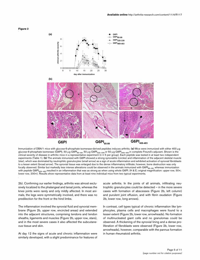
2b). Confirming our earlier findings, arthritis was almost exclu-sively localized to the phalangeal and tarsal joints, whereas theknee joints were rarely and only mildly affected. In most ani-mals, the legs were symmetrically involved, and there was nopredilection for the front or the hind limbs.
The inflammation involved the synovial fluid and synovial mem-brane (Figure 2b, upper row, encircled areas) and extendedinto the adjacent structures, comprising tendons and tendonsheaths, ligaments and muscles (Figure 2b, upper row, stars),and in the most severe cases it also affected the subcutane-ous tissue and skin.
At day 12 the signs of acute and chronic inflammation weresimilarly developed, with a slight predominance for features of
acute arthritis. In the joints of all animals, infiltrating neu-trophilic granulocytes could be detected – in the more severecases with formation of abscesses (Figure 2b, left column)and purulent joint effusion, and with fibrin exudation (Figure2b, lower row, long arrows).
In contrast, cell types typical of chronic inflammation like lym-phocytes, plasma cells and macrophages were found to alesser extent (Figure 2b, lower row, arrowheads). No formationof multinucleated giant cells and no granulomas could beobserved. A thickening of the synovial lining and a dense pro-liferation of fibroblasts were observed (Figure 2b, lower row,arrowheads), however, comparable with the pannus formationin human rheumatoid arthritis.
Figure 2
Immunization of DBA/1 mice with glucose-6-phosphate-isomerase-derived peptides induces arthritisImmunization of DBA/1 mice with glucose-6-phosphate-isomerase-derived peptides induces arthritis. (a) Mice were immunized with either 400 μg glucose-6-phosphate-isomerase (G6PI), 50 μg G6PI85–99, 50 μg G6PI325–339 or 50 μg G6PI469–483 in complete Freund's adjuvant. Shown is the clinical severity of disease in arthritic mice in a representative experiment (n = 5 per group). Each peptide was tested in at least two independent experiments (Table 1). (b) The animals immunized with G6PI showed a strong synovialitis (circles) and inflammation of the adjacent skeletal muscle (star), which was dominated by neutrophilic granulocytes (small arrow) as a sign of acute inflammation and exhibited activation of synovial fibroblasts to a lesser extent (broad arrow). The synovial tissue was enlarged due to the dense inflammatory infiltrate; however, bone destruction was only focally observed. Similar but markedly less intense alterations could be observed in the animals immunized with G6PI85–99, whereas immunization with peptide G6PI469–483 resulted in an inflammation that was as strong as when using whole G6PI. (H & E; original magnification: upper row, 50×; lower row, 200×). Results show representative data from at least nine individual mice from two typical experiments.
Page 5 of 11(page number not for citation purposes)

Arthritis Research & Therapy Vol 11 No 4 Bruns et al.
Taken together, our data show that three of the peptides wererobustly arthritogenic, whereas immunization with the otherthree peptides resulted only in a slight arthritis induction. Wetherefore restricted the following analyses of the T-cell and B-cell responses to the arthritogenic peptides G6PI85–99,G6PI325–339 and G6PI469–483.
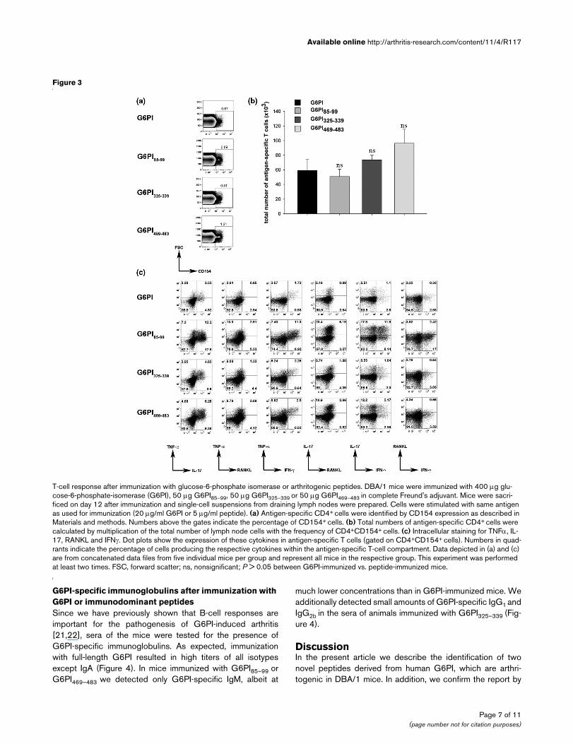
Ex vivo cytokine production after immunization with G6PI or arthritogenic peptidesTo identify G6PI-specific or peptide-specific T cells we usedintracellular staining of CD154 after a brief restimulation of exvivo isolated draining lymph node cells with full-length G6PI orthe respective peptide. CD154 expression is strictly depend-ent on T-cell receptor engagement, and therefore expressionof CD154 identifies antigen-specific cells in ex vivo stimula-tion assays [28,29].
We first analyzed the total number of CD154+CD4+ cells fromthe draining lymph nodes at day 12 after immunization, andfound no statistically significant difference between G6PI-
immunized or peptide-immunized mice (Figure 3a, b). Next weexamined the cytokine production of the CD154+ antigen-spe-cific T cells. As shown in Figure 3c, the most abundantcytokine in all groups was IL-17, followed by TNFα andRANKL or IFNγ. The frequency of antigen-specific IL-2-pro-ducing, IL-4-producing or IL-6-producing T cells wasextremely low, and IL-10-producing Th cells were neverdetected in G6PI-immunized mice (data not shown).
Compared with mice immunized with full-length G6PI, pep-tide-immunized mice showed a more prominent cytokineresponse. It is important to note here that the T-cell responsedevelops much faster in the animals immunized with full-lengthG6PI, with a maximum response at day 9, compared with thepeptide-immunized mice, in which the peak response is at day12 after immunization ([21] and data not shown).
Table 1
Summary of immunization experiments
Peptide Arthritic mice per experiment Cumulative incidence (%)
G6PI65–79 3/5 60
5/5
1/5
G6PI85–99 5/5 79
8/10
5/5
4/8
4/5
5/5
3/5
G6PI325–339 5/5 100
4/4
G6PI469–483 5/5 95
7/8
5/5
5/5
G6PI497–511 3/5 53
5/5
0/5
G6PI517–531 1/5 40
5/5
0/5
Page 6 of 11(page number not for citation purposes)
Available online http://arthritis-research.com/content/11/4/R117
G6PI-specific immunoglobulins after immunization with G6PI or immunodominant peptidesSince we have previously shown that B-cell responses areimportant for the pathogenesis of G6PI-induced arthritis[21,22], sera of the mice were tested for the presence ofG6PI-specific immunoglobulins. As expected, immunizationwith full-length G6PI resulted in high titers of all isotypesexcept IgA (Figure 4). In mice immunized with G6PI85–99 orG6PI469–483 we detected only G6PI-specific IgM, albeit at
much lower concentrations than in G6PI-immunized mice. Weadditionally detected small amounts of G6PI-specific IgG1 andIgG2b in the sera of animals immunized with G6PI325–339 (Fig-ure 4).
DiscussionIn the present article we describe the identification of twonovel peptides derived from human G6PI, which are arthri-togenic in DBA/1 mice. In addition, we confirm the report by
Figure 3
T-cell response after immunization with glucose-6-phosphate isomerase or arthritogenic peptidesT-cell response after immunization with glucose-6-phosphate isomerase or arthritogenic peptides. DBA/1 mice were immunized with 400 μg glu-cose-6-phosphate-isomerase (G6PI), 50 μg G6PI85–99, 50 μg G6PI325–339 or 50 μg G6PI469–483 in complete Freund's adjuvant. Mice were sacri-ficed on day 12 after immunization and single-cell suspensions from draining lymph nodes were prepared. Cells were stimulated with same antigen as used for immunization (20 μg/ml G6PI or 5 μg/ml peptide). (a) Antigen-specific CD4+ cells were identified by CD154 expression as described in Materials and methods. Numbers above the gates indicate the percentage of CD154+ cells. (b) Total numbers of antigen-specific CD4+ cells were calculated by multiplication of the total number of lymph node cells with the frequency of CD4+CD154+ cells. (c) Intracellular staining for TNFα, IL-17, RANKL and IFNγ. Dot plots show the expression of these cytokines in antigen-specific T cells (gated on CD4+CD154+ cells). Numbers in quad-rants indicate the percentage of cells producing the respective cytokines within the antigen-specific T-cell compartment. Data depicted in (a) and (c) are from concatenated data files from five individual mice per group and represent all mice in the respective group. This experiment was performed at least two times. FSC, forward scatter; ns, nonsignificant; P > 0.05 between G6PI-immunized vs. peptide-immunized mice.
Page 7 of 11(page number not for citation purposes)

Arthritis Research & Therapy Vol 11 No 4 Bruns et al.
Iwanami and colleagues identifying G6PI325–339 as an arthri-togenic peptide [30]. The peptide sequence of G6PI469–483 isidentical in man and mouse. G6PI-induced arthritis is thereforecurrently the only mouse model in which arthritis can beinduced by immunization with a peptide derived from a self-antigen.
Our screen identified at least six G6PI peptides that are immu-nodominant for I-Aq-restricted T-cell responses. The T-cellresponse towards the ubiquitously expressed autoantigenG6PI is therefore not focused on one dominant epitope.Instead, at least three peptide–epitopes derived from G6PIare immunodominant and arthritogenic in DBA/1 mice. Thereis precedence for several immunodominant antigens withinone protein for T-cell responses restricted towards one partic-ular MHC molecule [31]. For hen egg lysozyme, which con-sists of only 129 amino acids, there are at least six dominant
peptide epitopes for I-Ak-restricted T-cell responses alone[32].
The different number of peptides identified by Iwanami andcolleagues [30] and in the present report reflects the funda-mentally different approaches to identifying immunodominantG6PI epitopes. Iwanami and colleagues examined knownsequences of I-Aq-restricted T-cell epitopes and deduced apossible I-Aq binding motif from these sequences. Peptidesderived from G6PI that fit this possible I-Aq binding motif werethen synthesized and tested. These peptides covered 399/558 (71.5%) amino acid residues of the human GPI protein.Consequently, as acknowledged by Iwanami and colleagues[30], this approach carries the risk of missing relevant peptideepitopes. Moreover, several groups including ours have shownpreviously that several peptides that do not fit the consensussequence for peptides binding to a given MHC molecule canactivate T cells restricted to that MHC molecule very efficiently
Figure 4
Analysis of glucose-6-phosphate-specific immunoglobulin productionAnalysis of glucose-6-phosphate-specific immunoglobulin production. DBA/1 mice were immunized with either 400 μg glucose-6-phosphate (G6PI), 50 μg G6PI85–99, 50 μg G6PI325–339or 50 μg G6PI469–483 in complete Freund's adjuvant. Mice were sacrificed on day 12 after immunization and titers of G6PI-specific immunoglobulins of the indicated isotypes were measured by ELISA. Data are representative of at least two different experiments (n = 4 per group; *P < 0.05 for G6PI-immunized vs. peptide-immunized mice; §P < 0.05 for G6PI325–339 vs. G6PI85–99 or G6PI469–483). OD, optical density.
Page 8 of 11(page number not for citation purposes)
Available online http://arthritis-research.com/content/11/4/R117
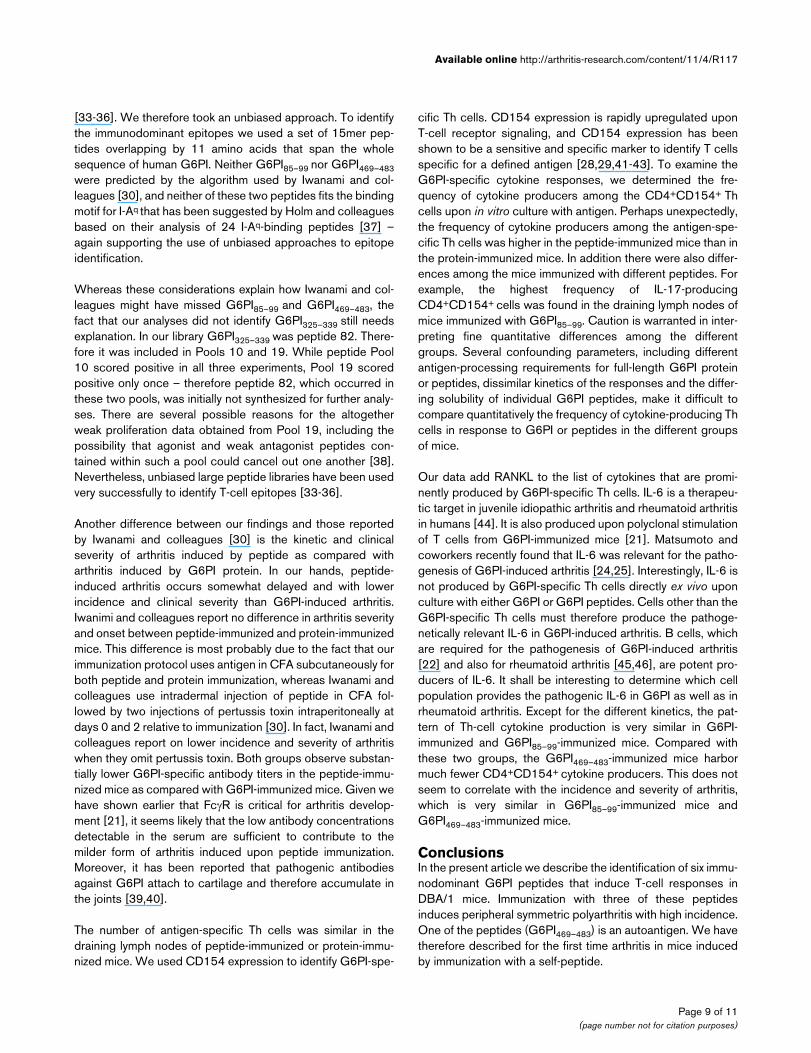
[33-36]. We therefore took an unbiased approach. To identifythe immunodominant epitopes we used a set of 15mer pep-tides overlapping by 11 amino acids that span the wholesequence of human G6PI. Neither G6PI85–99 nor G6PI469–483were predicted by the algorithm used by Iwanami and col-leagues [30], and neither of these two peptides fits the bindingmotif for I-Aq that has been suggested by Holm and colleaguesbased on their analysis of 24 I-Aq-binding peptides [37] –again supporting the use of unbiased approaches to epitopeidentification.
Whereas these considerations explain how Iwanami and col-leagues might have missed G6PI85–99 and G6PI469–483, thefact that our analyses did not identify G6PI325–339 still needsexplanation. In our library G6PI325–339 was peptide 82. There-fore it was included in Pools 10 and 19. While peptide Pool10 scored positive in all three experiments, Pool 19 scoredpositive only once – therefore peptide 82, which occurred inthese two pools, was initially not synthesized for further analy-ses. There are several possible reasons for the altogetherweak proliferation data obtained from Pool 19, including thepossibility that agonist and weak antagonist peptides con-tained within such a pool could cancel out one another [38].Nevertheless, unbiased large peptide libraries have been usedvery successfully to identify T-cell epitopes [33-36].
Another difference between our findings and those reportedby Iwanami and colleagues [30] is the kinetic and clinicalseverity of arthritis induced by peptide as compared witharthritis induced by G6PI protein. In our hands, peptide-induced arthritis occurs somewhat delayed and with lowerincidence and clinical severity than G6PI-induced arthritis.Iwanimi and colleagues report no difference in arthritis severityand onset between peptide-immunized and protein-immunizedmice. This difference is most probably due to the fact that ourimmunization protocol uses antigen in CFA subcutaneously forboth peptide and protein immunization, whereas Iwanami andcolleagues use intradermal injection of peptide in CFA fol-lowed by two injections of pertussis toxin intraperitoneally atdays 0 and 2 relative to immunization [30]. In fact, Iwanami andcolleagues report on lower incidence and severity of arthritiswhen they omit pertussis toxin. Both groups observe substan-tially lower G6PI-specific antibody titers in the peptide-immu-nized mice as compared with G6PI-immunized mice. Given wehave shown earlier that FcγR is critical for arthritis develop-ment [21], it seems likely that the low antibody concentrationsdetectable in the serum are sufficient to contribute to themilder form of arthritis induced upon peptide immunization.Moreover, it has been reported that pathogenic antibodiesagainst G6PI attach to cartilage and therefore accumulate inthe joints [39,40].
The number of antigen-specific Th cells was similar in thedraining lymph nodes of peptide-immunized or protein-immu-nized mice. We used CD154 expression to identify G6PI-spe-
cific Th cells. CD154 expression is rapidly upregulated uponT-cell receptor signaling, and CD154 expression has beenshown to be a sensitive and specific marker to identify T cellsspecific for a defined antigen [28,29,41-43]. To examine theG6PI-specific cytokine responses, we determined the fre-quency of cytokine producers among the CD4+CD154+ Thcells upon in vitro culture with antigen. Perhaps unexpectedly,the frequency of cytokine producers among the antigen-spe-cific Th cells was higher in the peptide-immunized mice than inthe protein-immunized mice. In addition there were also differ-ences among the mice immunized with different peptides. Forexample, the highest frequency of IL-17-producingCD4+CD154+ cells was found in the draining lymph nodes ofmice immunized with G6PI85–99. Caution is warranted in inter-preting fine quantitative differences among the differentgroups. Several confounding parameters, including differentantigen-processing requirements for full-length G6PI proteinor peptides, dissimilar kinetics of the responses and the differ-ing solubility of individual G6PI peptides, make it difficult tocompare quantitatively the frequency of cytokine-producing Thcells in response to G6PI or peptides in the different groupsof mice.
Our data add RANKL to the list of cytokines that are promi-nently produced by G6PI-specific Th cells. IL-6 is a therapeu-tic target in juvenile idiopathic arthritis and rheumatoid arthritisin humans [44]. It is also produced upon polyclonal stimulationof T cells from G6PI-immunized mice [21]. Matsumoto andcoworkers recently found that IL-6 was relevant for the patho-genesis of G6PI-induced arthritis [24,25]. Interestingly, IL-6 isnot produced by G6PI-specific Th cells directly ex vivo uponculture with either G6PI or G6PI peptides. Cells other than theG6PI-specific Th cells must therefore produce the pathoge-netically relevant IL-6 in G6PI-induced arthritis. B cells, whichare required for the pathogenesis of G6PI-induced arthritis[22] and also for rheumatoid arthritis [45,46], are potent pro-ducers of IL-6. It shall be interesting to determine which cellpopulation provides the pathogenic IL-6 in G6PI as well as inrheumatoid arthritis. Except for the different kinetics, the pat-tern of Th-cell cytokine production is very similar in G6PI-immunized and G6PI85–99-immunized mice. Compared withthese two groups, the G6PI469–483-immunized mice harbormuch fewer CD4+CD154+ cytokine producers. This does notseem to correlate with the incidence and severity of arthritis,which is very similar in G6PI85–99-immunized mice andG6PI469–483-immunized mice.
ConclusionsIn the present article we describe the identification of six immu-nodominant G6PI peptides that induce T-cell responses inDBA/1 mice. Immunization with three of these peptidesinduces peripheral symmetric polyarthritis with high incidence.One of the peptides (G6PI469–483) is an autoantigen. We havetherefore described for the first time arthritis in mice inducedby immunization with a self-peptide.
Page 9 of 11(page number not for citation purposes)

Arthritis Research & Therapy Vol 11 No 4 Bruns et al.
Competing interestsThe authors declare that they have no competing interests.
Authors' contributionsLB and OF participated in the in vivo studies, the T-cell assaysand the ELISA studies, and drafted parts of the manuscript.LM performed the histopathological analyses. CL and RV pre-pared the peptide libraries, the peptides and participated indesigning the peptide pools. TK conceived of the study andparticipated in its design and coordination, and wrote the man-uscript. All authors read and approved the final manuscript.
AcknowledgementsThe authors thank Kai Kaufmann, Caroline Bocklisch, Gabriele Fernahl and Christine Baier for excellent technical assistance, and thank Bärbel Matz and Regina Musack for expert mouse care. The present work was supported by the Interdisciplinary Centre for Clinical Research (IZKF) Jena – Pathogenesis and Modulation of G6PI-induced Arthritis (to TK), and Pathogenic and Protective Role of T helper Cells in Arthritis (to OF) – and by the ENDO-Stiftung-Gemeinnütziger Verein ENDO-Klinik e.V. (to LM). The present work forms part of the PhD thesis of LB.
References1. Kamradt T, Mitchison NA: Tolerance and autoimmunity. N Engl
J Med 2001, 344:655-664.2. The Wellcome Trust Case Control Consortium: Genome-wide
association study of 14,000 cases of seven common diseasesand 3,000 shared controls. Nature 2007, 447:661-678.
3. Plenge RM, Seielstad M, Padyukov L, Lee AT, Remmers EF, DingB, Liew A, Khalili H, Chandrasekaran A, Davies LR, Li W, Tan AK,Bonnard C, Ong RT, Thalamuthu A, Pettersson S, Liu C, Tian C,Chen WV, Carulli JP, Beckman EM, Altshuler D, Alfredsson L,Criswell LA, Amos CI, Seldin MF, Kastner DL, Klareskog L,Gregersen PK: TRAF1-C5 as a risk locus for rheumatoid arthri-tis – a genomewide study. N Engl J Med 2007, 357:1199-1209.
4. Klareskog L, Catrina AI, Paget S: Rheumatoid arthritis. Lancet2009, 373:659-672.
5. Lundy SK, Sarkar S, Tesmer LA, Fox DA: Cells of the synoviumin rheumatoid arthritis. T lymphocytes. Arthritis Res Ther 2007,9:202.
6. Firestein GS: Evolving concepts of rheumatoid arthritis. Nature2003, 423:356-361.
7. Genovese MC, Schiff M, Luggen M, Becker JC, Aranda R, Teng J,Li T, Schmidely N, Le Bars M, Dougados M: Efficacy and safetyof the selective co-stimulation modulator abatacept following2 years of treatment in patients with rheumatoid arthritis andan inadequate response to anti-tumour necrosis factor ther-apy. Ann Rheum Dis 2008, 67:547-554.
8. Kremer JM, Genant HK, Moreland LW, Russell AS, Emery P, Abud-Mendoza C, Szechinski J, Li T, Teng J, Becker JC, Westhovens R:Results of a two-year followup study of patients with rheuma-toid arthritis who received a combination of abatacept andmethotrexate. Arthritis Rheum 2008, 58:953-963.
9. Jirholt J, Lindqvist AB, Holmdahl R: The genetics of rheumatoidarthritis and the need for animal models to find and under-stand the underlying genes. Arthritis Res 2001, 3:87-97.
10. Berg WB van den, Joosten LA, van Lent PL: Murine antigen-induced arthritis. Methods Mol Med 2007, 136:243-253.
11. Holmdahl R, Bockermann R, Backlund J, Yamada H: The molecu-lar pathogenesis of collagen-induced arthritis in mice – amodel for rheumatoid arthritis. Ageing Res Rev 2002,1:135-147.
12. Cremer MA, Ye XJ, Terato K, Owens SW, Seyer JM, Kang AH:Type XI collagen-induced arthritis in the Lewis rat. Characteri-zation of cellular and humoral immune responses to nativetypes XI, V, and II collagen and constituent alpha-chains. JImmunol 1994, 153:824-832.
13. Carlsen S, Hansson AS, Olsson H, Heinegard D, Holmdahl R:Cartilage oligomeric matrix protein (COMP)-induced arthritisin rats. Clin Exp Immunol 1998, 114:477-484.
14. Glant TT, Finnegan A, Mikecz K: Proteoglycan-induced arthritis:immune regulation, cellular mechanisms, and genetics. CritRev Immunol 2003, 23:199-250.
15. Bayrak S, Holmdahl R, Travers P, Lauster R, Hesse M, Dolling R,Mitchison NA: T cell response of I-Aq mice to self type II colla-gen: meshing of the binding motif of the I-Aq molecule withrepetitive sequences results in autoreactivity to multipleepitopes. Int Immunol 1997, 9:1687-1699.
16. Buzas EI, Vegvari A, Murad YM, Finnegan A, Mikecz K, Glant TT:T-cell recognition of differentially tolerated epitopes of carti-lage proteoglycan aggrecan in arthritis. Cell Immunol 2005,235:98-108.
17. Brand DD, Myers LK, Terato K, Whittington KB, Stuart JM, KangAH, Rosloniec EF: Characterization of the T cell determinantsin the induction of autoimmune arthritis by bovine alpha 1(II)-CB11 in H-2q mice. J Immunol 1994, 152:3088-3097.
18. Holmdahl R, Jansson L, Larsson A, Jonsson R: Arthritis in DBA/1mice induced with passively transferred type II collagenimmune serum. Immunohistopathology and serum levels ofanti-type II collagen auto-antibodies. Scand J Immunol 1990,31:147-157.
19. Stuart JM, Tomoda K, Yoo TJ, Townes AS, Kang AH: Serumtransfer of collagen-induced arthritis. II. Identification andlocalization of autoantibody to type II collagen in donor andrecipient rats. Arthritis Rheum 1983, 26:1237-1244.
20. Terato K, Hasty KA, Reife RA, Cremer MA, Kang AH, Stuart JM:Induction of arthritis with monoclonal antibodies to collagen.J Immunol 1992, 148:2103-2108.
21. Schubert D, Maier B, Morawietz L, Krenn V, Kamradt T: Immuni-zation with glucose-6-phosphate isomerase induces T cell-dependent peripheral polyarthritis in genetically unalteredmice. J Immunol 2004, 172:4503-4509.
22. Bockermann R, Schubert D, Kamradt T, Holmdahl R: Induction ofa B-cell-dependent chronic arthritis with glucose-6-phosphateisomerase. Arthritis Res Ther 2005, 7:R1316-R1324.
23. Kamradt T, Schubert D: The role and clinical implications ofG6PI in experimental models of rheumatoid arthritis. ArthritisRes Ther 2005, 7:20-28.
24. Iwanami K, Matsumoto I, Tanaka-Watanabe Y, Inoue A, Mihara M,Ohsugi Y, Mamura M, Goto D, Ito S, Tsutsumi A, Kishimoto T,Sumida T: Crucial role of the interleukin-6/interleukin-17cytokine axis in the induction of arthritis by glucose-6-phos-phate isomerase. Arthritis Rheum 2008, 58:754-763.
25. Matsumoto I, Zhang H, Yasukochi T, Iwanami K, Tanaka Y, InoueA, Goto D, Ito S, Tsutsumi A, Sumida T: Therapeutic effects ofantibodies to tumor necrosis factor-alpha, interleukin-6 andcytotoxic T-lymphocyte antigen 4 immunoglobulin in mice withglucose-6-phosphate isomerase induced arthritis. ArthritisRes Ther 2008, 10:R66.
26. Lunemann JD, Frey O, Eidner T, Baier M, Roberts S, Sashihara J,Volkmer R, Cohen JI, Hein G, Kamradt T, Munz C: Increased fre-quency of EBV-specific effector memory CD8+ T cells corre-lates with higher viral load in rheumatoid arthritis. J Immunol2008, 181:991-1000.
27. Krenn V, Morawietz L, Haupl T, Neidel J, Petersen I, Konig A: Grad-ing of chronic synovitis – a histopathological grading systemfor molecular and diagnostic pathology. Pathol Res Pract2002, 198:317-325.
28. Frentsch M, Arbach O, Kirchhoff D, Moewes B, Worm M, Rothe M,Scheffold A, Thiel A: Direct access to CD4+ T cells specific fordefined antigens according to CD154 expression. Nat Med2005, 11:1118-1124.
29. Chattopadhyay PK, Yu J, Roederer M: A live-cell assay to detectantigen-specific CD4+ T cells with diverse cytokine profiles.Nat Med 2005, 11:1113-1117.
30. Iwanami K, Matsumoto I, Tanaka Y, Inoue A, Goto D, Ito S, Tsut-sumi A, Sumida T: Arthritogenic T cell epitope in glucose-6-phosphate isomerase-induced arthritis. Arthritis Res Ther2008, 10:R130.
31. Roy S, Scherer MT, Briner TJ, Smith JA, Gefter ML: Murine MHCpolymorphism and T cell specificities. Science 1989,244:572-575.
32. Gammon G, Klotz J, Ando D, Sercarz EE: The T cell repertoire toa multideterminant antigen. Clonal heterogeneity of the T cell
Page 10 of 11(page number not for citation purposes)
Available online http://arthritis-research.com/content/11/4/R117
response, variation between syngeneic individuals, and in vitroselection of T cell specificities. J Immunol 1990,144:1571-1577.
33. Grogan JL, Kramer A, Nogai A, Dong L, Ohde M, Schneider-Mer-gener J, Kamradt T: Cross-reactivity of myelin basic protein-specific T cells with multiple microbial peptides: experimentalautoimmune encephalomyelitis induction in TCR transgenicmice. J Immunol 1999, 163:3764-3770.
34. Hemmer B, Gran B, Zhao Y, Marques A, Pascal J, Tzou A, KondoT, Cortese I, Bielekova B, Straus SE, McFarland HF, Houghten R,Simon R, Pinilla C, Martin R: Identification of candidate T-cellepitopes and molecular mimics in chronic Lyme disease. NatMed 1999, 5:1375-1382.
35. Maier B, Molinger M, Cope AP, Fugger L, Schneider-Mergener J,Sonderstrup G, Kamradt T, Kramer A: Multiple cross-reactiveself-ligands for Borrelia burgdorferi-specific HLA-DR4-restricted T cells. Eur J Immunol 2000, 30:448-457.
36. Nino-Vasquez JJ, Allicotti G, Borras E, Wilson DB, Valmori D,Simon R, Martin R, Pinilla C: A powerful combination: the use ofpositional scanning libraries and biometrical analysis to iden-tify cross-reactive T cell epitopes. Mol Immunol 2004,40:1063-1074.
37. Holm L, Frech K, Dzhambazov B, Holmdahl R, Kihlberg J, LinussonA: Quantitative structure–activity relationship of peptidesbinding to the class II major histocompatibility complex mole-cule Aq associated with autoimmune arthritis. J Med Chem2007, 50:2049-2059.
38. Kamradt T, Volkmer-Engert R: Cross-reactivity of T lymphocytesin infection and autoimmunity. Mol Divers 2004, 8:271-280.
39. Wipke BT, Wang Z, Kim J, McCarthy TJ, Allen PM: Dynamic visu-alization of a joint-specific autoimmune response throughpositron emission tomography. Nat Immunol 2002, 3:366-372.
40. Studelska DR, Mandik-Nayak L, Zhou X, Pan J, Weiser P, McDow-ell LM, Lu H, Liapis H, Allen PM, Shih FF, Zhang L: High affinityglycosaminoglycan and autoantigen interaction explains jointspecificity in a mouse model of rheumatoid arthritis. J BiolChem 2009, 284:2354-2362.
41. Huaman MC, Martin LB, Malkin E, Narum DL, Miller LH, MahantyS, Long CA: Ex vivo cytokine and memory T cell responses tothe 42-kDa fragment of Plasmodium falciparum merozoitesurface protein-1 in vaccinated volunteers. J Immunol 2008,180:1451-1461.
42. Mittrucker HW, Steinhoff U, Kohler A, Krause M, Lazar D, Mex P,Miekley D, Kaufmann SH: Poor correlation between BCG vacci-nation-induced T cell responses and protection against tuber-culosis. Proc Natl Acad Sci USA 2007, 104:12434-12439.
43. Stubbe M, Vanderheyde N, Pircher H, Goldman M, Marchant A:Characterization of a subset of antigen-specific human centralmemory CD4+ T lymphocytes producing effector cytokines.Eur J Immunol 2008, 38:273-282.
44. Nishimoto N, Kishimoto T: Interleukin 6: from bench to bedside.Nat Clin Pract Rheumatol 2006, 2:619-626.
45. Browning JL: B cells move to centre stage: novel opportunitiesfor autoimmune disease treatment. Nat Rev Drug Discov 2006,5:564-576.
46. Martin F, Chan AC: B cell immunobiology in disease: evolvingconcepts from the clinic. Annu Rev Immunol 2006, 24:467-496.
Page 11 of 11(page number not for citation purposes)