an unusual triosephosphate isomerase from the early divergent eukaryotegiardia lamblia
TRANSCRIPT
An Unusual Triosephosphate Isomerase from the EarlyDivergent Eukaryote Giardia lambliaGabriel Lopez-Velazquez,1* Dora Molina-Ortiz,1 Nallely Cabrera,2 Gloria Hernandez-Alcantara,1
Jorge Peon-Peralta,3 Lilian Yepez-Mulia,4 Ruy Perez-Montfort,2 and Horacio Reyes-Vivas1*1Laboratorio de Bioquımica-Genetica, Instituto Nacional de Pediatrıa, Mexico, D.F. Mexico2Instituto de Fisiologıa Celular, Universidad Nacional Autonoma de Mexico, Mexico, D.F. Mexico3Instituto de Quımica, Universidad Nacional Autonoma de Mexico, Mexico, D.F. Mexico4UIMEIP-Pediatrıa, Centro Medico Nacional-Siglo XXI, IMSS
ABSTRACT Recombinant triosephosphate iso-merase from the parasite Giardia lamblia (GlTIM) wascharacterized and immunolocalized. The enzyme is dis-tributeduniformlythroughoutthecytoplasm.Sizeexclu-sion chromatography of the purified enzyme showedtwo peaks with molecular weights of 108 and 55 kDa.Under reducing conditions, only the 55-kDa protein wasdetected. In denaturing gel electrophoresis without di-thiothreitol, theenzymeshowedtwobandswithmolecu-lar weights of 28 and 50 kDa; with dithiotretitol, only the28-kDa protein was observed. These data indicate thatGlTIMmayexistasatetrameroradimerandthat, intheformer, the two dimers are covalently linked by disul-fide bonds. The kinetics of the dimer were similar tothose of other TIMs. The tetramer exhibited half of thekcat of the dimer without changes in the Km. Studies onthe thermal stability and the apparent association con-stants between monomers showed that the tetramerwas slightly more stable than the dimer. This findingsuggests the oligomerization is not related to enzymethermostability as in Thermotoga maritima. Instead, itcould be that oligomerization is related to the regula-tion of catalytic activity in different states of the lifecycle of this mesophilic parasite. Proteins 2004;55:824–834. © 2004 Wiley-Liss, Inc.© 2004 Wiley-Liss, Inc.
Key words: glycolysis; giardiasis; protein purifica-tion; oligomerization state; disulfides
INTRODUCTION
The protozoan parasite Giardia lamblia merits exten-sive studies for several reasons. From the evolutionarypoint of view, G. lamblia has been considered a ratherremarkable organism because its morphological, meta-bolic, and molecular features correspond to those of one ofthe earliest branching lineages of eukaryotes.1–5 Indeed,the G�C content, gene complementation, and rRNA sub-units of G. lamblia are markedly similar to those ofprokaryotic organisms. In addition, the parasite is thecausative agent of human giardiasis, a worldwide diseasethat affects millions of people, in particular children.6
Although metronidazole is effective in giardiasis,7 it exertsstrong side effects in the host,8 and there is clear evidencethat strains of G. lamblia resistant to metronidazole arenow existent.9 This finding underlines the need for devel-
oping drugs that are effective against giardiasis. In thisregard, it is noted that an additional feature of G. lambliais that it lacks mitochondria and oxidative phosphoryla-tion.10 Thus, in G. lamblia, the main source of ATP is theglycolytic pathway.11 This finding suggests that the en-zymes of glycolysis could be potential targets for drugdesign. The nucleotide sequence of the triosephosphateisomerase gene from G. lamblia has been previouslyreported.12 However, there are no data on the kinetics andstructure of triosephosphate isomerase from G. lamblia.Therefore, because of its importance in the evolutionaryprocesses and its potential as a target for drug design, wecharacterized triosephosphate isomerase from G. lamblia(GlTIM) and also determined its distribution within theparasite.
TIM is a ubiquitous glycolytic enzyme that catalyzes thereversible isomerization between (R)-glyceraldehyde3-phosphate (GAP) and dihydroxyacetone phosphate(DHAP). The kinetics and energetics of the catalyticreaction are well established,13–16 and the three-dimen-sional (3D) structure of the enzyme from 13 differentspecies has been determined. The active site residues of allknown TIMs are located in the central region of the�/�-barrel and belong to the same main-chain. Most TIMsso far described are homodimers in which each of the twomonomers exhibits an �/�-barrel structure. However, someTIMs from hyperthermophilic organisms have a differentquaternary structure, which apparently is related to theirthermal stability. For example, TIMs from Pyrococcuswoesei17,18 and Methanothermus fervidus19 are tetramericenzymes. In Thermotoga neapolitana and T. maritima, theenzyme is a tetramer fused with a phosphoglyceratekinase.20,21 TIM from T. maritima was separated fromphosphoglycerate kinase and crystallized.21 The modifiedTIM was a tetramer in which two dimers were in closecontact through hydrophobic and polar residues. However,
Grant sponsor: Consejo Nacional de Ciencia y Tecnologıa, Mexico;Grant number: J37071-B.
*Correspondence to: Gabriel Lopez-Velazquez and Horacio Reyes-Vivas. Laboratorio de Bioquımica-Genetica, Instituto Nacional dePediatrıa, Apartado Postal 04530, Mexico, D.F. Mexico. E-mail:[email protected].
Received 7 August 2003; Accepted 11 December 2003
Published online 1 April 2004 in Wiley InterScience(www.interscience.wiley.com). DOI: 10.1002/prot.20097
PROTEINS: Structure, Function, and Bioinformatics 55:824–834 (2004)
© 2004 WILEY-LISS, INC.
it is relevant to this work that the dimers of T. maritimawere joined through two disulfide bridges, between Cys142 of each monomer. Our studies on GlTIM show that it isan enzyme with characteristics different from those ofTIMs from other mesophilic organisms.
MATERIALS AND METHODSParasites
Trophozoites of G. lamblia WB strain were cultured inTYI-S-33 as previously described.22 Cultures were grownat 37°C for 72 h to semiconfluence and washed withphosphate buffered saline (PBS), pH 7.3.
Amplification of Triosephosphate Isomerase GeneFrom Giardia lamblia
Genomic DNA from G. lamblia trophozoites (WB strain)was purified by the phenol-chloroform method with slightmodifications.23 The polymerase chain reaction (PCR)conditions were those described by Gibco-BRL (Taq poly-merase supplier) using the following oligonucleotides:sense 5�-AATAACATATGCCTGCTCGTC-3� and anti-sense 5�-CCAGGATCCTATGTACGGG-3�, which containNdeI and BamHI restriction sites at the 5� end, respec-tively. The reaction mixture contained 400 ng of gDNA, 1.5mM MgCl2, 0.8 mM of dNTP, and 2.5 units of TaqPolymerase (Gibco-BRL). Amplification was performed byusing 30 cycles of 1 min at 94°C, 1 min at 57°C, and 1 minat 72°C.
Cloning of the GlTIM Gene and Sequence Analysis
The amplified DNA fragment was purified by electro-phoresis using an extraction kit (Concert; Gibco-BRL) andcloned into the pCR2.1 vector, as recommended (Novagen).E. coli TOP10 competent cells were transformed for clon-ing. The cloned fragment was subcloned into the pET3avector after digestion with NdeI and BamHI enzymes andused to transform E. coli TOP10 competent cells. Enzymerestriction assays and electrophoresis were performed toconfirm the correct fragment insertion into the vector.
The plasmid from positive clones was purified by usingan extraction plasmid kit (Concert; Gibco-BRL) and se-quenced by the enzymatic method with an automaticsequencer. A clone named 13* showed the correct sequenceand orientation of the gene of G. lamblia TIM. This wasused for the production of wild-type recombinant GlTIM.
Overexpression of GlTIM in E. coli
The plasmid from clone 13* was extracted and used totransform competent E. coli BL21(DE3)pLys cells. BL21cells contain the gene codifying for the T7 RNA pol neededfor expression of the recombinant enzyme. The trans-formed cells were grown in Luria-Bertani medium with100 �g/mL ampicillin at 37°C to an A600nm of 0.2–0.5. Toinduce overexpression, 0.4 mM isopropyl �-D-thiogalacto-pyranoside was added, and growth was continued over-night at 30°C.
Purification of Recombinant GlTIM
Transformed bacteria from 1 L culture were suspendedin 80 mL of a buffer containing 25 mM Tris, 1 mM EDTA,
0.2 mM phenylmethylsulfonyl fluoride, pH 7.9. The cellswere disrupted by sonication at 4°C, six cycles of 45 s with75 s of resting intervals. The sonicate was centrifuged at45,000 rpm for 1 h, and the supernatant was taken to 45%saturation ammonium sulfate. The suspension was al-lowed to stand for 3 h at 4°C and thereafter was centri-fuged at 10,000 rpm for 30 min. The pellet was discarded,and the concentration of ammonium sulfate in the superna-tant was increased to 75% saturation. The suspension wasincubated at 4°C overnight. It was then centrifuged at10,000 rpm for 30 min, and the supernatant was dis-carded. The pellet was suspended in 5 mL of a buffercontaining 25 mM Tris, 1 mM EDTA, 1 mM NaN3, pH 8,and filtered (0.45-�m pore diameter). The dissolved pro-tein was applied to a G-75 Sephadex (2.5 � 100 cm) columnequilibrated with the same buffer. The proteins wereeluted at a flow rate of 1 mL/min. Fractions of 2.5 mL werecollected. GlTIM eluted after 140 mL had passed throughthe column. The fractions with TIM activity were pooledand concentrated in Amicon filters (YM 10) to a volume of10 mL. The protein was dialyzed against 0.6 L of a buffercontaining 10 mM Tris, 1 mM EDTA, pH 8.7. The enzymewas then applied to a Q-Sepharose FF (1.5 � 12 cm)column equilibrated and washed with the same buffer.GlTIM was eluted with a gradient of 30–100 mM NaCl.Fractions with activity were pooled and concentrated.
To increase the purity of the enzyme, GlTIM was appliedto a SW300 (7.5 � 300 mm) column equilibrated with abuffer containing 50 mM triethanolamine hydrochloride,10 mM EDTA, 150 mM NaCl, 30% glycerol, pH 7.5.Fractions with activity were pooled, concentrated, andstored at 4°C as a 50% glycerol solution in the same buffer.For the studies, the enzyme was dialyzed against a buffercontaining 100 mM triethanolamine hydrochloride, 10mM EDTA, pH 7.4. Densitometric analysis in sodiumdodecyl sulfate-polyacrylamide gel electrophoresis (SDS-PAGE) under reducing conditions showed that GlTIM was�95% pure. This protocol was followed several times, andthe enzyme yield ranged between 18 and 23 mg/L ofculture. Table I summarizes the purification of GlTIM.
T. brucei and T. cruzi recombinant TIMs were purifiedas previously described.24,25 Protein concentration of pureTIM was determined spectrometrically at 280 nm. Themolar extinction coefficients at 280 nm of TcTIM andTbTIM were 36440 and 34950, respectively.25 The molar
TABLE I. Purification of Recombinant TriosephosphateIsomerase From G. lamblia†
Step
Totalprotein
(mg)
Total activity(�mol GAP
min�1)aSpecific activity
(�mol min�1 mg�1)
Homogenate(sonicated) 336 559,440 1794
G-75 column 60 457,523 3305Q-Sepharose
column 20 76,000 3800†The enzyme was purified as described in Materials and Methods,starting from 1-L culture.aGAP, glyceraldehyde 3-phosphate.
TRIOSEPHOSPHATE ISOMERASE OF GIARDIA LAMBLIA 825
extinction coefficient of GlTIM was 26600, calculatedaccording to Pace et al.26
To ascertain the best conditions for storage of GlTIM,the purified enzyme was incubated in buffers with differ-ent pH, maintaining a constant ionic strength. After this,aliquots were taken and diluted in a buffer containing 100mM triethanolamine, 10 mM EDTA at pH 7.4. The activi-ties of all samples were measured at this pH. The activityas a function of pH showed a bell-shaped curve, after it wasincubated for 2 h, with a maximum activity between pH6.5–9.0. The activity dropped significantly outside of thispH range. At pH 7.4, the activity of the enzyme wasunaffected for at least 2 weeks.
Activity Assays
Enzyme activity in the direction of GAP to DHAP wasmeasured at 25°C by following the decay in absorbance at340 nm of 1 mL reaction mixture at pH 7.4 that contained100 mM triethanolamine, 10 mM EDTA, 0.2 mM NADH,
0.9 U �-glycerol-3-phosphate dehydrogenase, 1 mM GAP(except when catalytic constants were determined), and 10ng TIM per mL. The kinetic constants were calculatedfrom initial velocities (vi) obtained at different substrateconcentrations.
For the determination of Km, kcat, and Ki values for2-phosphoglycolate (PG), the concentration of GAP rangedbetween 0.5 and 4 mM. The determination of catalyticconstants in the direction of DHAP to GAP were deter-mined at 25°C in 100 mM triethanolamine, 10 mM EDTA,1 mM NAD, 4 mM arsenate, 120 �M dithiothreitol, 1 Uglyceraldehyde 3-phosphate dehydrogenase per mL, 100ng GlTIM per mL, and 0.3–10 mM DHAP.
Polyclonal Anti-GlTIM Antibodies
Chickens (16-week-old Delkab-Warren) were immu-nized subcutaneously with 240 �g of highly purified Gl-TIM dissolved in complete Freund’s adjuvant. The immu-nization dose was repeated two times at intervals of 1
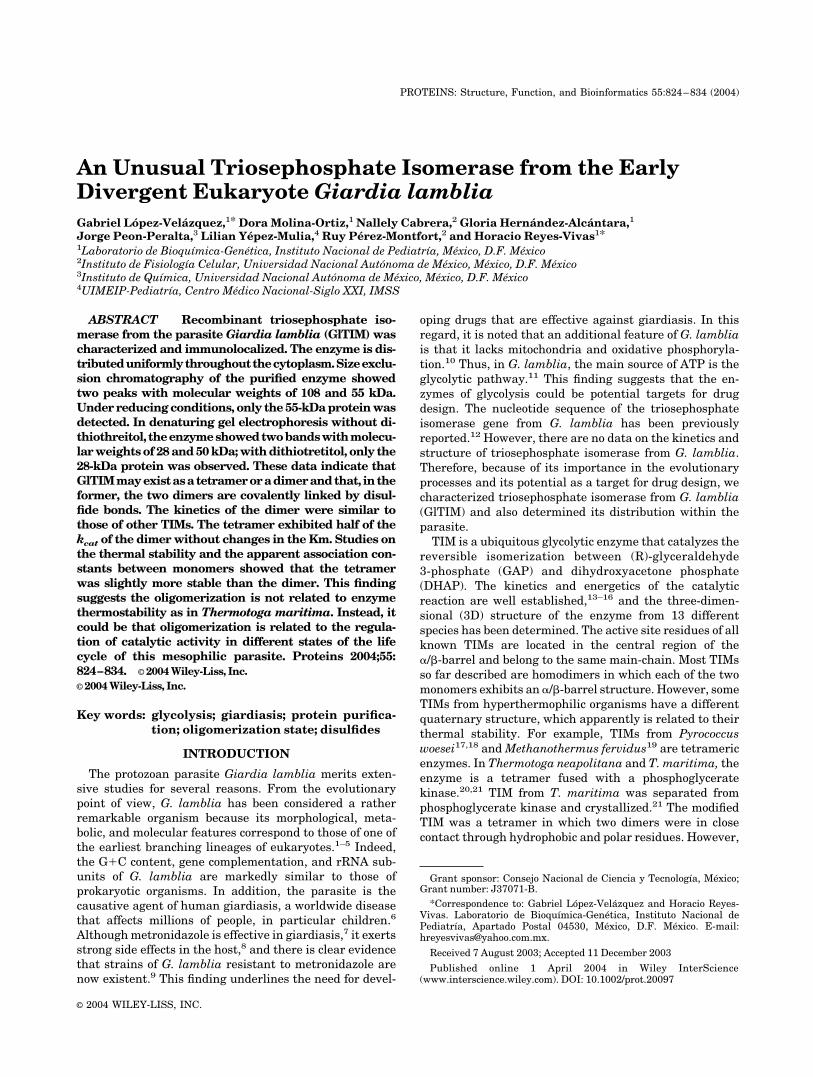
Fig. 1. Sequence alignment of GlTIM with other TIMs. The sequence of EhTIM, PfTIM, TbTIM, TcTIM,TmTIM, and HuTIM are shown. The active site residues are marked (*).
826 G. LOPEZ-VELAZQUEZ ET AL.
week. After the final boost, eggs were collected daily andprocessed individually. Chicken polyclonal antibodies (IgY)were purified from the egg yolk by using the methoddescribed by Polson et al.27 The IgY proteins were dialyzedextensively against PBS buffer and applied to a sizeexclusion column equilibrated with the same buffer. Thefractions enriched with IgY were pooled and concentrated.Western blot analysis was used to evaluate antibodyspecificity.
Light and Electron Microscopy
For light microscopy, cells were grown over glass cover-slips into six-well cell culture clusters (Costar). For elec-tron microscopy, pellets of G. lamblia trophozoites with106 cells were obtained by centrifugation and embeddedin acrylic resin Lowicryl K4M. Samples were fixed by usingfreshly prepared 4% paraformaldehyde in PBS.
Immunocytochemistry
Coverslips with a monolayer of trophozoites were incu-bated in 0.5% Triton X-100 for 5 min at 4°C to permeatecells. Samples were rinsed with PBS and incubated onTBS buffer (20 mM Tris, pH 7.6, 150 mM NaCl, 20 mMNaN3, 1% Tween 20, 5% BSA, 5% normal goat serum) for1 h. Samples were incubated with anti-GlTIM IgY diluted1:4000 in PBS at 4°C overnight, rinsed with PBS, andincubated with rabbit anti-chicken IgG at 1:500 in PBS for2 h at room temperature. Anti-rabbit IgG coupled tofluorescein isothiocyanate diluted 1:500 in PBS was used.Nuclei were stained by using DAPI (4�,6-diamidino-2-phenylindole) for specific DNA localization. Mountedsamples were photographed with a fluorescence micro-scope (Axiovert 200; Carl Zeiss).
Ultrathin sections (70 nm wide) were placed on nickelgrids. Grids were floated on TBS buffer for 1 h and
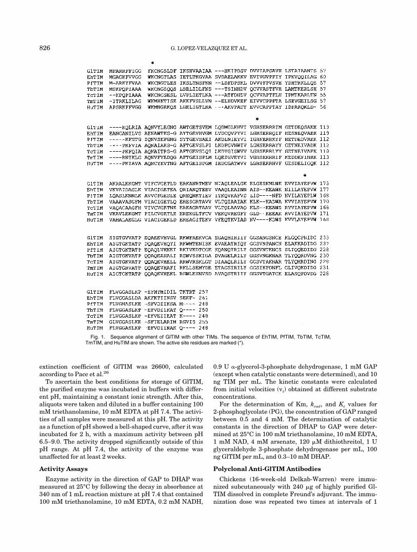
Fig. 2. Size exclusion chromatography of GlTIM. The chromatographic analysis under nonreducing (A) andreducing (B) conditions were performed by using a SW300 column equilibrated with buffer containing 50 mMtriethanolamine, 10 mM EDTA, 150 mM NaCl, 30% glycerol, pH 7.5. The proteins (25 �g) were eluted at a rateflow of 1 mL/min. For calibration, the following molecular weight markers were used: thyroglobulin, bovine-globulin, chicken ovalbumin, equine myoglobin, and vitamin B12.
TRIOSEPHOSPHATE ISOMERASE OF GIARDIA LAMBLIA 827
incubated at 4°C overnight with a 1:4000 solution ofanti-GlTIM antibody. Grids were rinsed with PBS andincubated for 1.5 h with a secondary antibody against IgY(Jackson Immunoresearch) at 1:200 in PBS. Anti-rabbitIgG coupled to 10-nm colloidal gold particles (ICN) diluted1:100 was added and incubated for 2 h. Samples werestained with 0.5% uranyl acetate and lead citrate for 1 mineach. Photographs were taken in a transmission electronmicroscope (EM109; Carl Zeiss).
RESULTSSequence Analysis
In consonance with the data of Mowatt et al.,12 we foundthat the gene of GlTIM is formed by 774 pb with apredicted sequence of 257 amino acid residues and amolecular mass of 27,903 Da. Figure 1 shows the sequencealignment of TIMs from G. lamblia (GlTIM), Entamoebahistolytica (EhTIM), Plasmodium falciparum (PfTIM), T.brucei (TbTIM), T. cruzi (TcTIM), T. maritima (TmTIM),and human (HuTIM). The identity of GlTIM with theenzymes from these organisms is 42, 41, 44, 46, 42, and45%, respectively. From the amino acid sequence of Gl-TIM, a pI of 7.05 was calculated. This is around 1 unithigher than the pI of TIM from E. coli (5.89). This allowedthe separation of the two enzymes by ion exchange chroma-tography (see Materials and Methods). GlTIM has fivecysteines per monomer; human TIM has the same numberof cysteines, but in different positions. The other enzymes
have a lower number of cysteines. It is noted that exceptfor TmTIM and HuTIM, the rest of enzymes have acysteine in their interface (Cys 14 in GlTIM). This region ofthe interface formed by the side-chain of Cys14 and itssurrounding loop 3 of the other subunit has been describedas a potential target for drug design.28,29
Hydrodynamic Parameters and SDS-PAGE ofGlTIM
To explore the oligomerization state of GlTIM, theenzyme was incubated overnight with or without thethiol-reducing agent 2-mercaptoethanol and then appliedto an analytical SEC column (Fig. 2). The chromatographicprofile of the enzyme incubated without 2-mercaptoetha-nol showed a minor peak with the Stokes radius of aprotein with a molecular mass of 108 kDa and a majorpeak of 55 kDa. The enzyme that had been exposed to2-mercaptoethanol showed only the 55-kDa protein. Thepeak that corresponded to the 55 kDa was larger for theenzyme treated with 2-mercaptoethanol. The same datawere obtained when GlTIM was incubated with dithiothre-itol (data not shown).
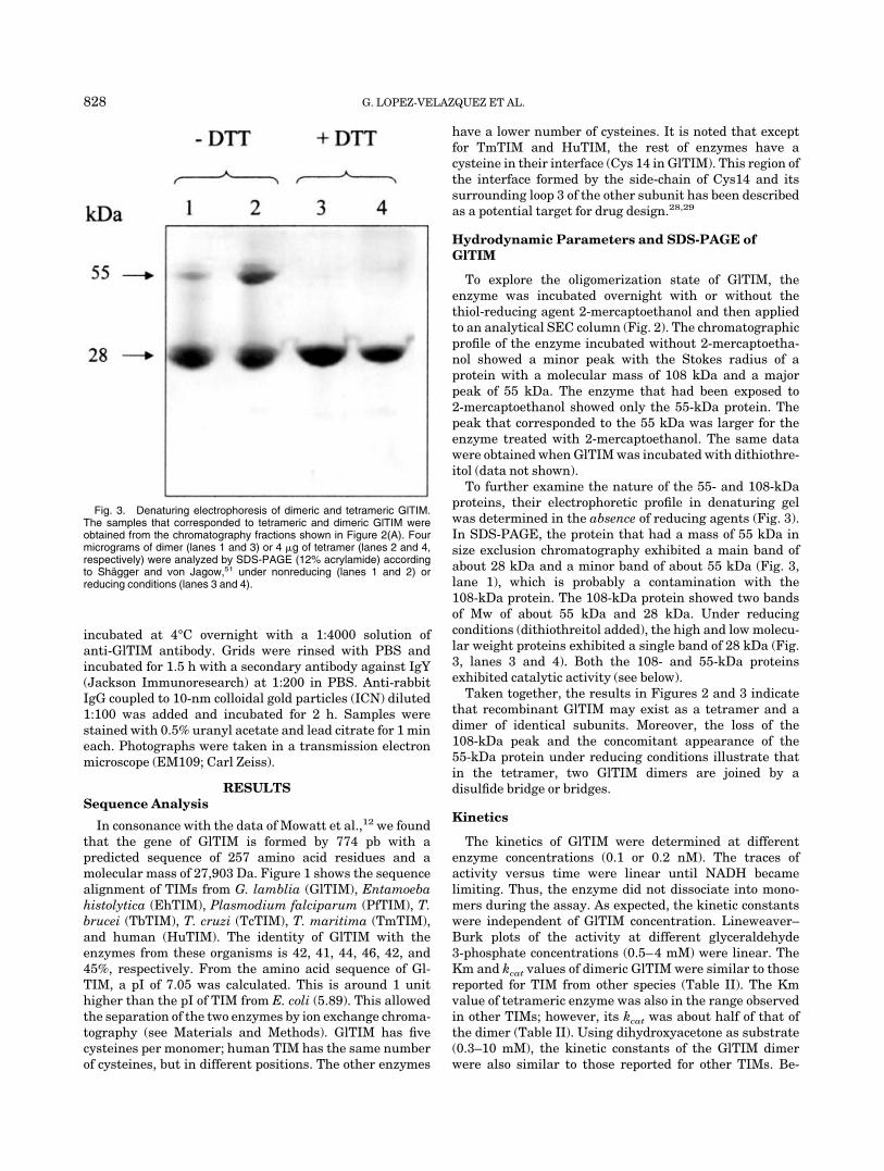
To further examine the nature of the 55- and 108-kDaproteins, their electrophoretic profile in denaturing gelwas determined in the absence of reducing agents (Fig. 3).In SDS-PAGE, the protein that had a mass of 55 kDa insize exclusion chromatography exhibited a main band ofabout 28 kDa and a minor band of about 55 kDa (Fig. 3,lane 1), which is probably a contamination with the108-kDa protein. The 108-kDa protein showed two bandsof Mw of about 55 kDa and 28 kDa. Under reducingconditions (dithiothreitol added), the high and low molecu-lar weight proteins exhibited a single band of 28 kDa (Fig.3, lanes 3 and 4). Both the 108- and 55-kDa proteinsexhibited catalytic activity (see below).
Taken together, the results in Figures 2 and 3 indicatethat recombinant GlTIM may exist as a tetramer and adimer of identical subunits. Moreover, the loss of the108-kDa peak and the concomitant appearance of the55-kDa protein under reducing conditions illustrate thatin the tetramer, two GlTIM dimers are joined by adisulfide bridge or bridges.
Kinetics
The kinetics of GlTIM were determined at differentenzyme concentrations (0.1 or 0.2 nM). The traces ofactivity versus time were linear until NADH becamelimiting. Thus, the enzyme did not dissociate into mono-mers during the assay. As expected, the kinetic constantswere independent of GlTIM concentration. Lineweaver–Burk plots of the activity at different glyceraldehyde3-phosphate concentrations (0.5–4 mM) were linear. TheKm and kcat values of dimeric GlTIM were similar to thosereported for TIM from other species (Table II). The Kmvalue of tetrameric enzyme was also in the range observedin other TIMs; however, its kcat was about half of that ofthe dimer (Table II). Using dihydroxyacetone as substrate(0.3–10 mM), the kinetic constants of the GlTIM dimerwere also similar to those reported for other TIMs. Be-
Fig. 3. Denaturing electrophoresis of dimeric and tetrameric GlTIM.The samples that corresponded to tetrameric and dimeric GlTIM wereobtained from the chromatography fractions shown in Figure 2(A). Fourmicrograms of dimer (lanes 1 and 3) or 4 �g of tetramer (lanes 2 and 4,respectively) were analyzed by SDS-PAGE (12% acrylamide) accordingto Shagger and von Jagow,51 under nonreducing (lanes 1 and 2) orreducing conditions (lanes 3 and 4).
828 G. LOPEZ-VELAZQUEZ ET AL.
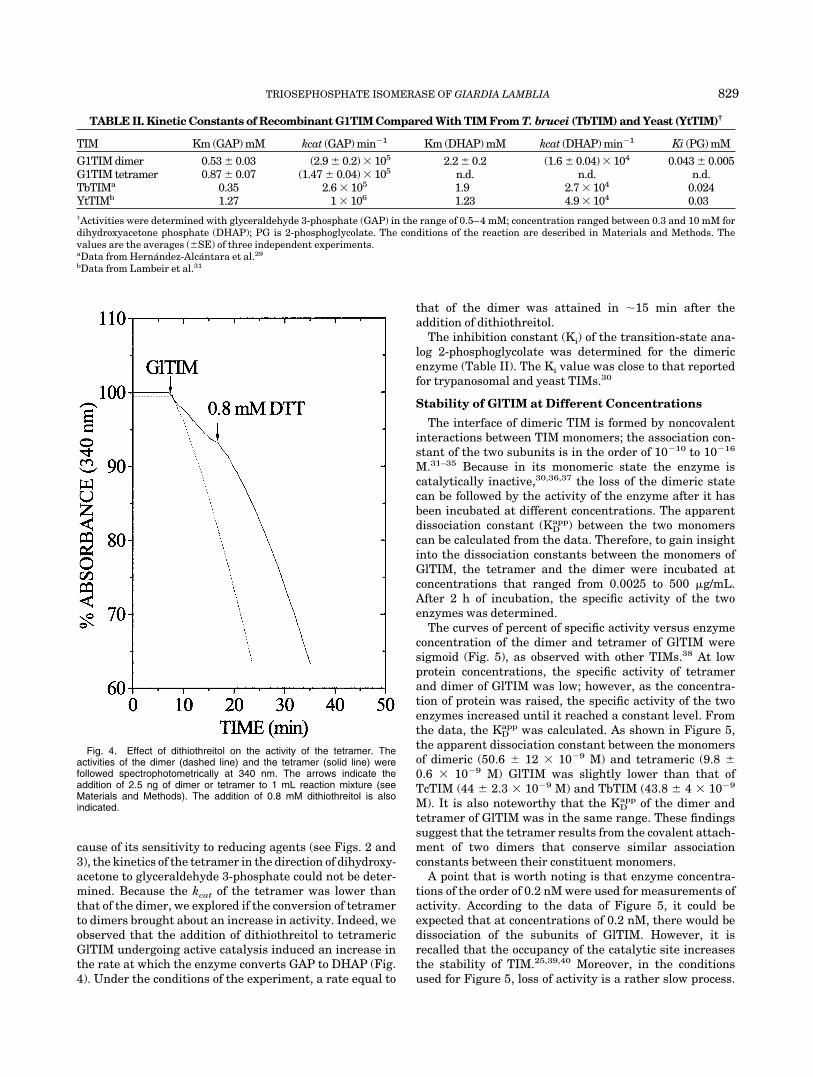
cause of its sensitivity to reducing agents (see Figs. 2 and3), the kinetics of the tetramer in the direction of dihydroxy-acetone to glyceraldehyde 3-phosphate could not be deter-mined. Because the kcat of the tetramer was lower thanthat of the dimer, we explored if the conversion of tetramerto dimers brought about an increase in activity. Indeed, weobserved that the addition of dithiothreitol to tetramericGlTIM undergoing active catalysis induced an increase inthe rate at which the enzyme converts GAP to DHAP (Fig.4). Under the conditions of the experiment, a rate equal to
that of the dimer was attained in 15 min after theaddition of dithiothreitol.
The inhibition constant (Ki) of the transition-state ana-log 2-phosphoglycolate was determined for the dimericenzyme (Table II). The Ki value was close to that reportedfor trypanosomal and yeast TIMs.30
Stability of GlTIM at Different Concentrations
The interface of dimeric TIM is formed by noncovalentinteractions between TIM monomers; the association con-stant of the two subunits is in the order of 10�10 to 10�16
M.31–35 Because in its monomeric state the enzyme iscatalytically inactive,30,36,37 the loss of the dimeric statecan be followed by the activity of the enzyme after it hasbeen incubated at different concentrations. The apparentdissociation constant (KD
app) between the two monomerscan be calculated from the data. Therefore, to gain insightinto the dissociation constants between the monomers ofGlTIM, the tetramer and the dimer were incubated atconcentrations that ranged from 0.0025 to 500 �g/mL.After 2 h of incubation, the specific activity of the twoenzymes was determined.
The curves of percent of specific activity versus enzymeconcentration of the dimer and tetramer of GlTIM weresigmoid (Fig. 5), as observed with other TIMs.38 At lowprotein concentrations, the specific activity of tetramerand dimer of GlTIM was low; however, as the concentra-tion of protein was raised, the specific activity of the twoenzymes increased until it reached a constant level. Fromthe data, the KD
app was calculated. As shown in Figure 5,the apparent dissociation constant between the monomersof dimeric (50.6 � 12 � 10�9 M) and tetrameric (9.8 �0.6 � 10�9 M) GlTIM was slightly lower than that ofTcTIM (44 � 2.3 � 10�9 M) and TbTIM (43.8 � 4 � 10�9
M). It is also noteworthy that the KDapp of the dimer and
tetramer of GlTIM was in the same range. These findingssuggest that the tetramer results from the covalent attach-ment of two dimers that conserve similar associationconstants between their constituent monomers.
A point that is worth noting is that enzyme concentra-tions of the order of 0.2 nM were used for measurements ofactivity. According to the data of Figure 5, it could beexpected that at concentrations of 0.2 nM, there would bedissociation of the subunits of GlTIM. However, it isrecalled that the occupancy of the catalytic site increasesthe stability of TIM.25,39,40 Moreover, in the conditionsused for Figure 5, loss of activity is a rather slow process.
Fig. 4. Effect of dithiothreitol on the activity of the tetramer. Theactivities of the dimer (dashed line) and the tetramer (solid line) werefollowed spectrophotometrically at 340 nm. The arrows indicate theaddition of 2.5 ng of dimer or tetramer to 1 mL reaction mixture (seeMaterials and Methods). The addition of 0.8 mM dithiothreitol is alsoindicated.
TABLE II. Kinetic Constants of Recombinant G1TIM Compared With TIM From T. brucei (TbTIM) and Yeast (YtTIM)†
TIM Km (GAP) mM kcat (GAP) min�1 Km (DHAP) mM kcat (DHAP) min�1 Ki (PG) mM
G1TIM dimer 0.53 � 0.03 (2.9 � 0.2) � 105 2.2 � 0.2 (1.6 � 0.04) � 104 0.043 � 0.005G1TIM tetramer 0.87 � 0.07 (1.47 � 0.04) � 105 n.d. n.d. n.d.TbTIMa 0.35 2.6 � 105 1.9 2.7 � 104 0.024YtTIMb 1.27 1 � 106 1.23 4.9 � 104 0.03†Activities were determined with glyceraldehyde 3-phosphate (GAP) in the range of 0.5–4 mM; concentration ranged between 0.3 and 10 mM fordihydroxyacetone phosphate (DHAP); PG is 2-phosphoglycolate. The conditions of the reaction are described in Materials and Methods. Thevalues are the averages (�SE) of three independent experiments.aData from Hernandez-Alcantara et al.29
bData from Lambeir et al.31
TRIOSEPHOSPHATE ISOMERASE OF GIARDIA LAMBLIA 829

Thus, in the times in which activity was measured, nodissociation to monomers took place. Indeed, we alwaysobserved that the activity traces were linear with time.
Stability to Temperature
The thermostabilities of the dimer and the tetramer ofGlTIM were determined from measurements of the decayof activity at different temperatures. The data were com-
pared with those of TcTIM and TbTIM. In all cases,inactivation followed a simple exponential decay. Thus,the data were expressed as kobs (Table III). At 45 and 55°C,the GlTIM dimer exhibited the highest decay rate. At60°C, however, the rate of inactivation was similar in thefour enzymes. It is noted that the loss of activity in thetetramer was not markedly different from that of the otherenzymes tested, albeit at 45 and 55°C, it was slightly morestable than the dimer.
Fig. 5. Stability of the dimer and tetramer of GlTIM at different concentrations. The dimer and tetramer wereincubated at concentrations that range from 0.0025 to 500 �g/mL at 40°C in a buffer containing 100 mMtriethanolamine, 10 mM EDTA, pH 7.4. After 2-h samples were incubated for 1 min at 25°C, subsequently theresidual activity was determined with 10 ng of protein/mL reaction mixture. The percent of specific activityversus enzyme concentration is shown; 100% of activity was 1700 and 714 �mol min�1 mg�1 for the dimer andtetramer, respectively. The apparent KD (�SE) of three independent experiments were calculated and fittedwith nonlinear regression plots.
830 G. LOPEZ-VELAZQUEZ ET AL.

In Situ Localization of GlTIM
Fluorescence immunolocalization of cellular GlTIMshowed that the enzyme was quite abundant in all thecytoplasm of G. lamblia trophozoites [Fig. 6(A), anti-GlTIM panel]. Its distribution was uniform, albeit thenuclei were not labeled [Fig. 6(A), DAPI panel]. Electronmicroscopy corroborated the homogeneous cellular distri-bution of GlTIM [Fig. 6(B)]; that is, the enzyme is notcontained in membranous vesicles (as in the glycosomes oftrypanosomes).41
DISCUSSION
The kinetics, the association constant between mono-mers, and the thermostability of GlTIM are similar tothose of other TIMs. Nonetheless, in size exclusion chroma-tography, two catalytically active proteins with molecularmasses of 108 and 55 kDa were clearly evident. The sameresults were obtained when the catalytically active pro-teins were analyzed in nonreducing native gel electrophore-sis. It is also noteworthy that only the 28-kDa protein wasobserved in SDS-PAGE under reducing conditions, whereasin the absence of reducing agents, two proteins withmolecular masses of 55 and 28 kDa were detected. Takentogether, these findings indicate that TIM from G. lambliacan exist as a tetramer or a dimer of identical subunits.Moreover, because reducing agents induce the transforma-tion of tetramers to dimers, it may be concluded that twoGlTIM dimers are covalently linked by disulfide bonds inthe tetramer. In this regard, it is noted that in thetetramer, the linkage of two dimers must be through an-S-S- bridge between only one of the monomers of eachdimer; otherwise, the tetrameric form in SDS-PAGE undernonreducing conditions would have exhibited only the55-kDa protein; instead, the gel showed bands of 55 and 28kDa.
GlTIM has cysteines at positions 14, 127, 202, 222, and228. PfTIM has cysteines at positions 13, 126, 196, and217. The cysteines of PfTIM and GlTIM have differentpositions in the primary sequence. This is because GlTIMhas an insertion of seven residues. However, the positionsof the cysteines between both organisms are equivalent(Fig. 1). Therefore, the crystal structure of the PfTIM42
was used to model the accessible solvent area (ASA) of thefive cysteines of GlTIM. The predicted ASA of the five
lateral chains of the cysteines from GlTIM43 showed thatCys 202, located in helix 6, could be the residue mostexposed to solvent (47.1 Å2). Alternatively, GlTIM pos-sesses a Cys 228 residue, but the residue that PfTIMcontains at this equivalent position is Gln 223. Conse-quently, Cys 228 of GlTIM would be located on loop 8 withalso a highly predicted ASA (26.7 Å2). Therefore, it isprobable that the cysteines involved in the disulfide of thetetramer were either Cys 202 or Cys 228 from each dimer.
As noted, TIM from T. maritima is a tetramer fused withphosphoglycerate kinase.44 The crystal structure of thetetrameric TIM after it was separated from glyceratekinase by molecular engineering has been reported.21 Inthis tetramer, the two dimers are linked through contactsbetween hydrophobic and polar residues; however, thepredominant linkages in the tetramer are the two disulfidebonds that are established between cysteines 142 of eachmonomer. In regard to the structural features of TIM fromT. maritima, it has been proposed that through tetramer-ization, the enzyme acquires a higher thermostability.44
Here, we found that the dimer and tetramer of GlTIM donot exhibit important differences in thermostability nor intheir association constant between monomers. Therefore,it is possible that in the mesophile G. lamblia, the occur-rence of tetrameric and dimeric forms serves a differentpurpose.
In the latter respect, the comparison of the kinetics ofthe GlTIM dimer and tetramer may be illustrative. Thedata show that although the two enzymes have the sameKm for glyceraldehyde 3-phosphate, the kcat of the tet-ramer is about half of that of the dimer. This suggests thatthe tetramer is an enzyme with only two catalyticallycompetent sites. In this connection, it is relevant to pointout that TIM dimers, in which one of its two catalytic siteshas been poisoned with a covalently linked inhibitor,express 50% of its maximal activity without importantchanges in Km,45,46 indicating that the catalytic sites oftwo monomers work independently. However, Biemannand Koshland47 reported that a protein with two potentialbinding sites exhibited Michaelis–Menten behavior with aHill coefficient of 1 and that, nonetheless, the proteinexpressed what they called half of the site reactivity. Thatis, the occupancy of one binding site suppressed thefunction of the other site. With the present data, it is notpossible to distinguish between the two alternatives. How-ever, it is noted that the kinetics of the dimer and tetramerexhibited classical Michaelis–Menten behavior with Hillcoefficients of 1.1 and 1.02, respectively.
In sum, our data on TIM from G. lamblia show that ithas characteristics that set it apart from all the otherTIMs so far reported. Indeed, to our knowledge, GlTIM isthe first example of a TIM from a mesophile that mayacquire a tetrameric structure. Likewise, GlTIM is also theonly known eukaryotic TIM that can acquire a tetramericstructure. Along this line, it may not be a coincidence thatG. lamblia is one of the earliest branches of eukaryotes. Inaddition, from the point of view of catalytic mechanisms,GlTIM also seems rather unique, because the kinetics of
TABLE III. First-Order Rate Constants for Inactivation asa Function of Temperature for G1TIM, TcTIM, and TbTIM†
TIM
kobs (h�1)
45°C 55°C 60°C
G1TIM dimer 1.9 � 0.1 65 � 3 173 � 9G1TIM tetramer 0 43 � 5.4 169 � 7.5TcTIM (4 � 0.37) � 10�2 36 � 4.3 198 � 17TbTIM (9 � 0.72) � 10�2 23.04 � 1.44 120 � 2.4†The enzymes were incubated at 100 �g/mL in 100 mM triethanol-amine, 10 mM EDTA buffer (pH 7.4) at the indicated temperatures. Atdifferent times of incubation, aliquots were withdrawn and activitiesmeasured at 25°C. The kobs (�SE) were calculated from nonlinearregression plots.
TRIOSEPHOSPHATE ISOMERASE OF GIARDIA LAMBLIA 831

the enzyme showed that in the tetramer only two of its fourcatalytic sites are catalytically competent.
The aforementioned considerations raise the question ofwhether the characteristics of GlTIM, particularly in itsability to tetramerize with a concomitant decrease incatalytic efficiency, are of physiological significance. Im-plicit in this question is the possibility that tetrameriza-tion of GlTIM is an “artifact” of laboratory manipulationsduring its purification or storage. Indeed, when we carryout the purification procedure with solutions supple-
mented with DTT, we only observed GlTIM dimers. This iswhat would be expected if the tetrameric structure weremaintained by disulfide bounds. Nonetheless, regardlessof whether tetramerization is of physiological significance,the fact remains that dimers of GlTIM are prone totetramerization. In fact, we have observed that in a fewhours at room temperatures, 5% of the dimers aretransformed into tetramers. Conversely, under reducingconditions, tetramers are converted into dimers with acorresponding gain in catalytic activity. Thus, the possibil-
Fig. 6. (A) Immunofluorescence of cellular GlTIM and (B) its distribution at the electron-microscopical level.Colloidal gold particles (arrow heads) show the localization of GlTIM. ES, extracellular space; VD, ventral disc;Cy, cytoplasm; N, nucleus. Bar 0.5 �m.
832 G. LOPEZ-VELAZQUEZ ET AL.
ity that tetramerization is relevant to the life of theparasite should be considered. The life cycle of G. lambliainvolves transformation of cysts into trophozoites; there-fore, it could be that the transition of GlTIM tetramers todimers, or vice versa, is guided by the intracellular condi-tions in a given physiological state. The tetramer could bea “storage conformation,” which participates in a transi-tion from a state with a low metabolism toward a highermetabolic state. Thus, it seems clear that the reversiblereaction between GlTIM dimers and tetramers deservesfurther studies.
Before closing, we call attention to two points that arosefrom the data of this work. The first is that, although thebulk of eukaryotic genome appears to share commonancestry with archaeobacteria,48,49 the phylogenetic anal-ysis of the TIM gene from several species (including G.lamblia) supports the notion that eukaryotic TIM has analpha-proteobacterial origin.50 Therefore, it would be inter-esting to determine if the different oligomerization statesof GlTIM are a consequence of parasitic adaptations orwhether they are an archaebacterial relic. Finally, thedata on the intracellular localization of GlTIM show that itis an enzyme that is evenly distributed throughout thecytoplasm and separated from the extracellular milieuonly by the cytoplasmic membrane. Therefore, the penetra-tion of drugs that target on GlTIM would not be hinderedby internal permeability barriers.
ACKNOWLEDGMENTS
D.M.-O. is the recipient of a fellowship from CONACyT.The authors are indebted to Dr. M. Tuena de Gomez-Puyou and Dr. A. Gomez-Puyou for their advice on theimprovement of the manuscript. We also thank Dr. E.Chavez Cosio for invaluable help on IgY development, Dr.Antonio Lazcano-Araujo for his comments of the manu-script, and Janet Flores for providing us with HPLCresources. The technical assistance of Carmen Ortiz, SaraNavarrete, and Amparo Tapia is acknowledged.
REFERENCES
1. Edlin TD, Charkraborty PR. Unusual ribosomal RNA of theintestinal parasite Giardia lamblia. Nucleic Acids Res 1987;15:7889–7901.
2. Sogin ML, Gunderson JH, Elwood HJ, Alonso RA, Peattie DA.Phylogenetic meaning of the kingdom concept: an unusual ribo-somal RNA from Giardia lamblia. Science 1989;6:75–77.
3. Narcisi EM, Glover VC, Fechheimer M. Fibrillarin, a conservedpre-ribosomal RNA processing protein of Giardia. J Euk Microbiol1998;45:105–111.
4. Hilario E, Gogarten JP. The prokaryote-to-eukaryote transitionreflected in the evolution of the V/F/A-ATPase catalytic andproteolipid subunits. J Mol Evol 1998;46:703–715.
5. Adam RD. The Giardia lamblia genome. Int J Parasitol 2000;30:475–484.
6. Boreham PFL. Giardiasis and its control. Pharm J 1991;234:271–274.
7. Towson SM, Boreham PFL, Upcroft P, Upcroft JA. Resistance tothe nitroheterocyclic drugs. Acta Trop 1994;56:173–194.
8. Upcroft J , Upcroft P. My favorite cell: Giardia. BioEssays1998;20:256–263.
9. Upcroft J, Upcroft P. Drug resistance and Giardia. ParasitolToday 1993;9:187–190.
10. Muller M. Energy metabolism of ancestral eukaryotes: a hypoth-esis based on the biochemistry of the amitochondriate parasiticprotists. Biosystems 1992;28:33–40
11. Schofield PJ, Edwards MR, Kranz P. Glucose metabolism inGiardia intestinalis. Mol Biochem Parasitol 1991;45:39–48.
12. Mowatt MR, Weinbach EC, Howard TC, Nash TE. Complementa-tion of an Escherichia coli glycolysis mutant by Giardia lambliatriosephosphate isomerase. Exp Parasitol 1994;78:85–92.
13. Albery J, Knowles JR. Evolution of enzyme function and thedevelopment of catalytic efficiency. Biochemistry 1976;64:5631–5640.
14. Albert T, Banner DW, Bloomer AC, Petsko GA, Phillips C, RiversP S, Wilson IA. On the thee-dimensional structure and catalyticmechanism of triosephosphate isomerase. Phil Trans Roy Soc B1981;293:159–171.
15. Nickbarg EB, Knowles JR. Triosephosphate isomerase; energeticsof the reaction catalyzed by the yeast enzyme expressed inEscherichia coli. Biochemistry 1988;27:5939–5947.
16. Knowles JR. Enzyme catalysis; not different, just better. Nature1991;350:121–124.
17. Bell GS, Russell RJ, Kohlhoff HM, Hensel R, Danson MJ, HoughDW, Taylor GL. Preliminary crystallographic studies of triosephos-phate isomerase (TIM) from the hyperthermophilic ArchaeonPyrococcus woesei. Acta Crystalogr Sect D Biol Crystallogr 1998;54:1419–1421.
18. Walden H, Bell GS, Russell RJM, Siebers B, Hensel R, Taylor GL.Tiny TIM: a small, tetrameric, hyperthermostable triosephos-phate isomerase. J Mol Biol 2001;306:745–757.
19. Kohlhoff M, Dahm A, Hensel R. Tetrameric triosephosphateisomerase from hyperthermophilic achaea. FEBS Lett 1996;383:245–250.
20. Yu JS, Noll KM. The hyperthermophilic bacterium Thermotoganeapolitana possesses two isozymes of the 3-phosphoglyceratekinase/triosephosphate isomerase fusion protein. FEMS MicrobiolLett 1995;131:307–312.
21. Maes D, Zeelen JP, Thanki N, Beaucamp N, Alvarez M, Thi MHD,Backmann J, Marital JA, Wyns L, Jaenicke R, Wierenga R. Thecrystal structure of triosephosphate isomerase (TIM) from Thermo-toga maritima: a comparative thermostability structural analysisof ten different TIM structures. Proteins 1999;37:441–453.
22. Mowatt MR, Aggarwal A, Nash TE. Carboxy-terminal sequenceconservation among variant-specific surface proteins of Giardialamblia. Mol Biochem Parasitol 1991;4:215–228.
23. Lopez-Velazquez G, Segura-Valdez MA, Alcantara-Ortigoza MA,Jimenez-Garcıa LF. Localization of intranuclear RNA by electronmicroscopy in situ hybridization using a genomic DNA probe. ArchMed Res 1998;29:185–190.
24. Borchert TV, Pratt K, Zeelen JP, Callens M, Noble ME, OpperdoesFR, Michels PA, Wierenga RK. Overexpression of trypanosomaltriosephosphate isomerase in Escherichia coli and characteriza-tion of a dimer-interface mutant. Eur J Biochem 1993;253:684–691.
25. Ostoa-Saloma P, Garza-Ramos G, Ramırez J, Becker I, BerzunzaM, Landa A, Gomez-Puyou A, Gomez-Puyou MT, Perez-MontfortR. Cloning, expression, purification and characterization of triose-phosphate isomerase from Trypanosoma cruzi. Eur J Biochem1997;244:700–705.
26. Pace NC, Vajdos F, Fee L, Grimsley G, Gray V. How to measureand predict the molar absorption coefficient of a protein. ProteinSci 1995;4:2411–2423.
27. Polson A, von Wechmar B, Van Regenmortel MHV. Isolation ofviral IgY antibodies from yolks of immunized hens. ImmunolComm 1980;9:475–493.
28. Garza-Ramos G, Perez-Montfort R, Rojo Domınguez A, Tuena deGomez-Puyou, M, Gomez-Puyou A. Species-specific inhibition ofhomologous enzymes by modification of non-conserved aminoacids residues. The cysteine residues of triosephosphate isomer-ase. Eur J Biochem 1996;241:114–120.
29. Hernandez-Alcantara G, Garza-Ramos G, Mendoza-Hernandez G,Gomez-Puyou A, Perez-Montfort R. Catalysis and stability oftriosephosphate isomerase from Trypanosoma brucei with differ-ent residues at position 14 of the dimer interface. Characteriza-tion of a catalytically competent monomeric enzyme. Biochemistry2002;41:4230–4238.
30. Garza-Ramos G, Tuena de Gomez-Puyou M, Gomez-Puyou A,Gracy RW. Dimerization and reactivation of triosephosphateisomerase in reverse micelles. Eur J Biochem 1992;208:389–395.
31. Lambeir AM, Opperdoes RF, Wierenga RK. Kinetic properties oftriose-phosphate isomerase from Trypanosoma brucei. Eur J Bio-chem 1987;168:69–74.
TRIOSEPHOSPHATE ISOMERASE OF GIARDIA LAMBLIA 833
32. Mainfroid V, Terpstra P, Beauregard M, Frere JM, Mande SC, HolWG, Martial JA, Goraj K. Three hTIM mutants that provide newinsights on why TIM is a dimer. J Mol Biol 1996;257:441–456.
33. Landa A, Rojo-Domınguez A, Jimenez L, Fernandez-Velasco A.Sequencing, expression and properties of triosephosphate isomer-ase from Entamoeba histolytica. Eur J Biochem 1997;247:348–355.
34. Borchert TV, Pratt K, Zeelen JP, Callens M, Noble ME, OpperdoesFR, Michels PA, Wierenga RK. Overexpression of trypanosomaltriosephosphate isomerase in Escherichia coli and characteriza-tion of a dimer-interface mutant. Eur J Biochem 1993;211:703–710.
35. Borchert TV, Abagyan R, Jaenicke R, Wierenga RK. Design,creation, and characterization of a stable, monomeric triosephos-phate isomerase. Proc Natl Acad Sci USA 1994;91:1515–1518.
36. Waley SG. Refolding of triosephosphate isomerase. Biochem J1973;135:165–172.
37. Zabori S, Rudolph F, Jaenicke R. Folding and association oftriosephosphate isomerase from rabbit muscle. Z Naturforsch1980;35:999–1004.
38. Reyes-Vivas H, Martınez-Martınez E, Mendoza-Hernandez G,Lopez-Velazquez G, Perez-Montfort R, Tuena de Gomez-Puyou M,Gomez-Puyou A. Susceptibility to proteolysis of triosephosphateisomerase from two pathogenic parasites: characterization of anenzyme with an intact and a nicked monomer. Proteins 2002;48:580–590.
39. Long CW, Levitzki A, Koshland DE. The subunit structure andsubunit interactions of cytidine triphosphate synthetase. J BiolChem 1970;10:80–87.
40. Jimenez L, Fernandez-Velasco DA, Willms K, Landa A. A compara-tive study of biochemical and immunological properties of triose-phosphate isomerase from Taenia solium and Sus scrofa. JParasitol 2003;89:209–214.
41. Opperdoes FR, Borst P. Localization of nine glycolytic enzymes ina microbody-like organelle in Trypanosoma brucei: the glycosome.FEBS Lett 1977;80:360–364.
42. Velanker SS, Ray SS, Gokhale RS, Suma S, Balaram H, BalaramP, Murthy MRN. Triosephosphate isomerase from Plasmodiumfalciparum: the crystal structure provides insights into antima-larial drug design. Structure 1997;5:751–761.
43. Gerstein M. A resolution-sensitive procedure for comparing pro-tein surfaces and its application to the comparison of antigen-combining sites. Acta Crystallogr 1992;A48:271–276.
44. Beaucamp N, Hofmann A, Kellerer B, Jaenicke R. Dissection ofthe gene of the bifunctional PGK-TIM fusion protein from thehyperthermophilic bacterium Thermotoga maritima: design andcharacterization of the separate triosephosphate isomerase. Pro-tein Sci 1997;6:2159–2165.
45. Schnackerz DD, Gracy RW. Probing the catalytic sites of triose-phosphate isomerase by 31P-NMR with reversibly and irreversiblybinding substrate analogues. Eur J Biochem 1991;199:231–238.
46. Sun A-Q, Yuksel U, Gracy RW. Interactions between the catalyticcenters and subunit interface of triosephosphate isomerase probedby refolding, active site modification, and subunit exchange. J BiolChem 1992;267:20168–20174.
47. Biemann HP, Koshland DE. Aspartate receptor of Escherichia coliand Salmonella typhimurium bind ligand with negative andhalf-of-the sites cooperativity. Biochemistry 1994;33:629–634.
48. Iwabe N, Kuma KI, Hasegawa M, Osawa S, Miyata T. Evolution-ary relationship of archaebacteria, eubacteria, and eukaryotesinferred from phylogenetic trees of duplicated genes. Proc NatlAcad Sci USA 1989;86:9355–9359.
49. Brown JR, Doolittle WF. Root of the universal tree of life based onancient aminoacyl-tRNA synthetase gene duplications. Proc NatlAcad Sci USA 1995;92:2441–2445.
50. Keeling PJ, Doolittle WF. Evidence that eukaryotic triosephos-phate isomerase is of alpha-proteobacterial origin. Proc Natl AcadSci USA 1997;94:1270–1275.
51. Shagger H, von Jagow G. Tricine-sodium dodecyl sulphate poly-acrylamide gel electrophoresis for the separation of proteins in therange from 1 to 100 kDa. Anal Biochem 1987;166:368–379.
834 G. LOPEZ-VELAZQUEZ ET AL.