hypoxia-ischemia, but not hypoxia alone, induces the expression of heme oxygenase-1 (hsp32) in...
TRANSCRIPT
Journal of Cerebral Blood Flow and Metabolism 17:647-658 © 1997 The International Society of Cerebral Blood Flow and Metabolism Published by Lippincott-Raven Publishers, Philadelphia
Hypoxia-Ischemia, But Not Hypoxia Alone, Induces the
Expression of Heme Oxygenase-l (HSP32)
in Newborn Rat Brain
*tMarcelle Bergeron, tDonna M, Ferriero, :j:Hendrik J. Vreman, :j:David K. Stevenson, and *tFrank R. Sharp
Department of Neurology, *Veterans Affairs Medical Center and fUniversity of California, San Francisco, California; and
:j:Department of Pediatrics, Stanford University School of Medicine, Stanford, California, USA
Summary: Heme oxygenase (HO) is the rate-limiting enzyme in the degradation of heme to produce bile pigments and carbon monoxide, The HO-I isozyme is induced by a variety of agents such as heat, heme, and hydrogen peroxide, Evidence suggests that the bile pigments serve as antioxidants in cells with compromised defense mechanisms, Because hypoxia-ischemia (HI) increases the level of oxygen free radicals, the induction of HO-I expression in the brain during ischemia could modulate the response to�oxidative stress, To study the possible involvement of HO- l in neonatal hypoxia-induced ischemic tolerance, we examined the brains of newborn rat pups exposed to 8% O2 (for 2,5 to 3 hours), and the brain of chronically hypoxic rat pups with congenital cardiac defects (Wi star Kyoto; WKYI NCr), Heme oxygenase-l immunostaining did not change after either acute or chronic hypoxia, suggesting that HO- l is not a good candidate for explaining hypoxia preconditioning in new-
Hypoxia induces the expression of a set of stress pro
teins called oxygen-regulated proteins, which have been
implicated in the development of drug and radiation re
sistance in tumor cells (Heacock and Sutherland, 1990).
Among these proteins, heme oxygenase-l (HO-I; also
called HSP32) has received increasing attention (Maines,
1992). Heme oxygenase catalyzes the degradation of
Received September 1 2, 1 996; final revision received January 6, 1997; accepted January 24, 1 997.
This work was supported by National Institutes of Health grants nos. NS28 1 67 and NS 1 4543 (F.R.S . ) , P20NS32553 (D.M.F.) , and HD-14426; The Hess Research Fund, and The Mary L. Johnson Research Fund (D.K.F). M.B . was a recipient of a postdoctoral fellowship from The Medical Research Council of Canada.
Address correspondence and reprint requests to: Dr. Marcelle Bergeron, Department of Neurology (V 1 27), UCSF and V A Medical Center, 4 1 50 Clement Street, San Francisco, CA 94 1 2 1
Abbreviations used: BSA, bovine serum albumin; CO, carbon monoxide; HI, hypoxia-ischemia; HO, heme oxygenase; HSF, heat shock factor; P7, postnatal day 7; PB , sodium phosphate buffer; WKY/NCr, Wistar Kyoto rats with congenital heart diseases, normotensive, and chronically hypoxic.
647
born rat brain. To study the role of HO- l in neonatal HI, l -week-old rats were subjected to right carotid coagulation and exposure to 8% 02/92% N2 for 2.5 hours. Whereas HO enzymatic activity was unchanged in ipsilateral cortex and subcortical regions compared with the contralateral hemisphere or control brains, immunocytochemistry and Western blot analysis showed increased HO-l staining in ipsilateral cortex, hippocampus, and striatum at 12 to 24 hours up to 7 days after HI. Double fluorescence immunostaining showed that HO-l was expressed mostly in ED-l positive macrophages. Because activated brain macrophages have been associated with the release of several cytotoxic molecules, the presence of HO-l positive brain macrophages may determine the tissue vulnerability after HI injury. Key Words: Heme oxygenaseHSP32-Hypoxia-Ischemia-Macrophage-Neonatal brain-Tolerance.
heme molecules derived from hemoproteins such as cy
tochrome P-450, nitric oxide synthase, tryptophan pyr
rolase, and several peroxidases and catalases, to biliver
din, ferrous iron, and carbon monoxide (CO). Biliverdin
is then converted to bilirubin by biliverdin reductase
(Maines, 1992). Two different gene products that both
contribute to HO enzymatic activity (microsomal iso
zymes HO-l and HO-2) have been characterized (Maines et aI., 1986; Shibahara et aI., 1993). Whereas
HO-2 activity is refractory to most types of stress or
injury, HO-l is induced by various stimuli. In healthy
unstressed adult rat brain, most of HO activity has been
attributed to the HO-2 isozyme whereas the HO-l iso
zyme seems to be present only at very low levels (Sun et aI.,
1990; Ewing and Maines, 1991, 1992; Maines, 1992).
The 5'-flanking promoter region of HO-l gene con
tains several important regulatory elements including at
least one copy of the following: a heat shock element (Shibahara et aI., 1987; Okinaga and Shibahara, 1993), a site for nuclear factor-kappa B (NFkB) (Lavrosky et
648 M. BERGERON ET AL.
aI., 1994), an AP-1-Iike binding site (MUlier et aI., 1987;
Alam and Zhining, 1992), and a metal regulatory element (MUller et aI., 1987) that may modulate the re
sponses to heat shock or denatured proteins, hypoxia or
oxidative stress, Fos/Jun immediate early genes, and
heme, respectively. Indeed, after global and focal ischemia (Paschen et aI., 1994; Takeda et aI., 1994; Nimura
et aI., 1996), hyperthermia (Ewing and Maines, 199 1)
and subarachnoid administration of lysed blood or he
moglobin (Matz et aI., 1996; Turner et aI., 1997) in adult
rats, as well as heat or hydrogen peroxide exposure in
primary cultures of neurons and astrocyteS (Dwyer et aI.,
1995), the induction of HO-l messenger RNA and pro
tein is observed mainly in nonneuronal cells. Increased
HO-l messenger RNA and protein have also been reported in rat brain after glutathione depletion (Ewing and
Maines, 1993). There is evidence to suggest that the
bilirubin produced by HO activity may serve as an antioxidant in cells with compromised defense mechanisms
(Stocker et aI., 1987). This may be important for neurons
which have low levels of the common antioxidants glu
tathione and ascorbate (Raps et aI., 1989). Because hyp
oxia and ischemia have been associated with increased
free oxygen species (Siesjo et aI., 1989), the production
of antioxidants by HO-l could help protect the brain from oxidativ,e injury.
Hypoxia pretreatment has been shown to confer neu
roprotection against hypoxic-ischemic (HI) injury in
newborn rats (Gidday et aI., 1994). Though the molecu
lar mechanism underlying this hypoxic preconditioning
is still poorly understood, HO-l could play a role be
cause it is an oxygen-regulated protein. Indeed, protec
tion against subsequent lethal insults has been shown
after prior induction of HO-l either by UV A exposure in
skin fibroblasts (Vile et aI., 1994), hemoglobin pretreat
ment in rat (Otterbein et aI., 1995), or overexpression in
rabbit coronary microvessel endothelial cells (Abraham
et aI., 1995). To elucidate the role of HO-l in neonatal
hypoxia and ischemia, the present study describes the
effect of acute and chronic hypoxia and the effect of
ischemia on HO-l expression in newborn rat brain. In
view of the suggested protective effects of hypoxia pre
conditioning against ischemic brain damage in the neo
natal rat (Gidday et aI., 1994), we also investigated the
role of oxygen-regulated HO-l protein as a potential can
didate responsible for neonatal hypoxia-induced isch
emic tolerance.
MATERIALS AND METHODS
Animal preparation All procedures were perfonned in accordance with the Na
tional Institutes of Health Guide for Care and Use of Laboratory Animals, and all protocols were approved by the University of California at San Francisco Committee on Animal Research. Male and female Sprague-Dawley rats (Bantam Kingman, Fremont, CA, U.S.A.) were used. Seven litters each
J Cereb Blood Flow Metab. Vol. 17. No.6. 1997
containing 10 Sprague-Dawley pups at postnatal day 7 (P7), 4 litters each containing 7 to 10 normotensive chronically hypoxic Wi star Kyoto pups at P7 (WKY/NCr) bearing various combinations of cardiac anomalies including hypertrophic cardiomyopathy, defects of the aortic arch system, and Tetralogy of Fallot (Kuribayashi et a!., 1990) and, 3 litters of 10 P7 Wi star pups as the control strain were used as previously described (Rice et a!., 1981; Ferriero et aI., 1990). Rat pups at P7 were used because their brain maturity is grossly comparable to that of a late-term gestation human fetus or newborn infant (Dobbing and Sands, 1979). After anesthesia with a gas mixture containing 1 % halothane in 70% N20 and 30% O2, rat pups underwent the right common carotid artery coagulation through a ventral midline neck incision. The wound was sutured and pups returned to their dam for 2 hours. Pups were then placed in an 8% 02/92% N2 humidified atmosphere in a chamber partially submerged in a water bath maintained at a constant temperature of 37°C (HI group; coagulation, hypoxia). Shamoperated animals underwent the same operative procedure except that the carotid artery was not ligated (hypoxia-treated group; no coagulation, hypoxia). Control untreated group (no coagulation, no hypoxia) animals and a group of coagulationonly animals (with no hypoxia; n = 5) were also studied. Because the latter two groups showed no difference in cell integrity and HO-l expression, the control animals shown in the present study refer to the untreated control group (no coagulation, no hypoxia). In general, animals from each litter were divided into control (1 to 2 per litter), hypoxia-treated (2 to 3 per litter), and HI groups (6 to 7 per litter). The systemic oxygen saturation in live P7 animals (untreated WKY/NCr and normal Wi star) was determined with an oxygen transducer probe (Oxisensor II N-25, Nellcor Inc., Hayward, CA, U.S.A.) wrapped around the pups abdomen and connected to a Nellcor Pulse Oximeter.
Western blot analysis At 24 hours after hypoxia or HI, rats were deeply anesthe
tized with an intraperitoneal injection (0.3 g/kg) of Nembutal (pentobarbital sodium; Abbott Lab, North Chicago, IL, U.S.A.) and killed by decapitation. Brains were quickly removed and dissected on ice. The cerebral cortex, hippocampus, and striatum from each hemisphere were placed in Laemmli solubilizing buffer (2.5% sodium dodecyl sulfate, 10% glycerol, 62.5 mmol/L Tris-HCI, pH 6.8, 5% 2-mercaptoethanol) and boiled for 10 minutes. The whole tissue extract was then frozen at -70°C. Western immunoblot analysis was performed as described previously (Bergeron et aI., 1996) with modifications. Protein concentration was determined using the bicinchoninic acid method (Pierce, Rockford, II, U.S.A.). Equal amounts (55 fLg) of protein per sample were separated on 12% sodium dodecyl sulfate polyacrylamide gels with 4.5% stacking gel. After electrotransfer onto a nitrocellulose membrane (0.2 fLm; Schleicher and SchueH, Keene, NH, U.S.A.), immobilized proteins were stained with Ponceau solution to verify equal protein loading. After a brief rinse in deionized water, the membranes were incubated overnight at 4°C in 0.1 moUL sodium phosphate buffer (PB) pH 7.4, containing 5% nonfat dry milk, 1% bovine serum albumin (BSA) and 0.1 % Tween-20, rinsed briefly in 0.1 mollL PB containing 1 % BSA and 0.1 % Tween-20, then incubated for 2 hours with a I :3500 dilution of rabbit polyclonal anti-rat HO-I antibody (StressGen, Victoria, BC, Canada). This polyclonal antibody, raised against rat liver purified HO-l protein, was originally described by Maines et aI., (1986). After three washes, membranes were incubated with a 1 :2500 dilution of anti-rabbit Ig-horseradish peroxidase antibody (Amersham, Arlington Heights, IL, U.S.A.) for 1.5 to 2

HO-I EXPRESSION IN ISCHEMIC NEWBORN RAT BRAIN 649
hours. Finally, the membranes were washed three times and the bound antibody was visualized with the ECL chemiluminescence system according to the manufacturer's protocol (Amersham). A computer-based imaging system (MCID, Imaging Research, St-Catherines, Ontario, Canada) was used to measure the areas of HO-I protein on Western immunoblot autoradiograms. The relative density of HO-l protein (32 kDa) bands was analyzed after subtraction of the film background.
Immunocytochemistry At the appropriate times after each treatment, rats were anes
thetized with -Nembutal and perfused through the left ventricle with cold 4% paraformaldehyde made up in 0.1 moUL PB, pH 7.4. Brains were removed from the skulls, postfixed in 4% paraformaldehyde for 1 to 4 hours and stored in a 30% sucrose overnight at 4°C. Fifty micrometer-thick coronal sections were cut on a vibratome and washed twice with 0.05 moUL PB. After 1 hour incubation in a peroxidase-inhibiting solution (0.65% sodium azide and 0.2% hydrogen peroxide in 0.05 moUL PB, pH 7.4), sections were incubated for 2 hours in a blocking solution (5% nonfat dry milk, 2% goat serum, 1 % BSA, 0.1 % Triton X-IOO, and 0.1 % rat serum, made up in 0.1 moUL PB, pH 7.4). The sections were then incubated for 12 to 48 hours at 4°C with the same anti-rat HO- l antibody used for Western blotting (StressGen, Victoria, BC, Canada) diluted I :4000 in 2% goat serum, 1 % BSA, 0.1 % Triton X-IOO, made up in 0.1 mollL PB, pH 7.4. Alternate sections from each brain were incubated without primary antibody (as negative controls). After three 10 minute-washes in 0.05 mollL PB, sections were incubated at room temperature for 2 hours with a I :200 dilution of biotinylatep goat anti-rabbit IgG antibody (Vector Laboratories, Burlingame, CA, U.S.A.). Sections were then incubated in an avidin-horseradish peroxidase solution (Elite Vectastain, Vector Laboratories) for 2 hours, followed by threc washes with PB. Staining was visualized with 0.015% diaminobenzidine (Sigma, St-Louis, MO, U.S.A.) and 0.001 % hydrogen peroxide. Sections were then washed, mounted on gelatin-coated slides and coverslipped.
Double immunofluorescence labeling To identify which cell type stained for HO-I, some sections
were coincubated with rabbit polyclonal anti-rat HO-l antibody (1 :4000) and either the mouse monoclonal anti-rat antibody ED-l (uncharacterized cytoplasmic antigen expressed by all cells of the rat monocyte/macrophage lineage; Serotec, Oxford, U.K.; 1:2000), OX-42 (complement type 3 receptor found on both resting and activated monocytes, macrophages, neutrophils, and microglia; Serotec, Oxford, U.K.; I :4000). or glial fibrillary acidic protein (GFAP) present in astrocytes (ICN, Costa Mesa, CA, U.S.A; 1 :4000). All dilutions were performed in 2% goat serum, 1 % BSA. 0.1 % Triton X-IOO, made up in 0.1 mollL PB, pH 7.4. After 24 to 48 hours incubation at 4°C, sections were washed three times for 10 minutes in PB and incubated for 2 hours in the dark with a Texas-Red-conjugated, goat anti-rabbit IgG antibody (I : 150; Vector Laboratories) together with a biotinylated goat anti-mouse IgG antibody (1: 150; Vector Laboratories). After three 10 minute washes, labeled sections were incubated for 2 hours with avidinconjugated fluorescein isothiocynate (FITC; Vector Laboratories; 1: 150 made up in a solution containing 0.1 moUL sodium bicarbonate and 0.15 mollL sodium chloride, pH 8.2). Sections were mounted onto slides and immediately coverslipped with Fluoromount-G (Southern Biotech. Assoc. Inc., Birmingham, AL, U.S.A.). Sections were photographed on a Leitz varioorthomat microscope using a Ploemopak 2.1 fluorescence illuminator. The same area of representative sections was photo-
graphed with interchangeable filters for Texas-Red and FITC fluorescence.
Histopathological evaluation Histopathological scoring of each newborn rat brain was
performed blindly on alternate coronal sections stained with cresyl violet (Nissl). Because increased OX-42 and GFAP staining occurs in areas of neuronal loss and brain damage after HI (Sheldon et a!., 1996), alternate sections stained with OX-42 and GF AP antibodies were also examined. The scoring scale was as follows: 0, no injury or no detectable neuronal loss; l, minimal neuronal loss with occasional gliosis; 2, columnar damage in cortex involving predominantly layers II through IV, moderate cell loss with areas of infarction and concomitant gliosis; and 3, severe cell loss and gliosis associated with extensive tissue infarction.
Determination of heme oxygenase enzymatic
activity Twenty-four hours after HI, rat pups were anesthetized with
Nembutal and decapitated. Brains were removed and dissected on ice. The cerebral cortex and a subcortical region comprised of hippocampal, striatal, thalamic, and hypothalamic tissue were isolated from each hemisphere. To account for the CO that may be bound to erythrocytes in the cerebral blood vessels, we perfused some animals with cold saline (0.9% sodium chloride) before processing the tissue and found no difference in HO activity compared with nonperfused animals. The tissue was weighted and homogenized by sonication (4 pulses of 1 second; Branson Sonifier Cell Disrupter 185 with microtip) in 4 volumes of ice-cold 0.1 moUL potassium phosphate buffer, pH 7.4. The protein concentration was determined using the bicinchoninic acid method (Pierce, Rockford, IL, U.S.A.). Heme oxygenase activity was determined using a gas chromatographic method as described previously (Vreman and Stevenson, 1988) with some modifications. Briefly, 20 f.1L of brain homogenate was reacted with 20 f.1L of 4.5 mmollL NADPH (reduced form of nicotinamide adenine dinucleotide phosphate) and 20 f.1L of 150 f.1mollL heme in a septum-sealed, amber-colored vial at 37°C in the dark. For blank values, the NADPH solution was replaced by an equal volume of potassium phosphate buffer. For negative controls, I f.1L of 600 f.1mollL chromium mesoporphyrin (potent inhibitor of HO activity) was added to the complete reaction mixture (Vreman et a!., 1993). This procedure inhibited the HO-induced CO production down to the blank value, suggesting that the entire CO production in the reaction vials was derived from HO activity. As a positive control, rat pup brain HO activity was induced by a subarachnoid (intracisternal) injection of purified hemoglobin, which has been shown to markedly increase HO-I expression in adult rat brain (Turner et a!., 1997). Twenty-four hours after injection, HO activity values in these brain homogenates ranged from 1.02 to 1.33 nmol CO produced/h/mg protein which is about two- to threefold higher than the activity measured in normal newborn brain (Table I). After 5 minutes of preincubation at 37°C in the dark, vials were purged with COfree air and allowed to incubate for an additional 15 minutes. The reaction was stopped by addition of 2 f.1L of sulfosalicylic acid solution (60% weight-to-volume ratio) and by cooling in wet ice. The amount of CO generated by the enzyme in the vial headspace was analyzed by gas chromatography (Vreman and Stevenson, 1988). Carbon monoxide concentration in tissue extracts was calculated from the peak area of the sample compared with that of CO external standards. The chromatographic assay was linear in the range of CO values obtained with brain tissue homogenates.
J Cereb Blood Flow Metab, Vol. 17, No.6, 1997
650 M. BERGERON ET AL.
TABLE 1. HO activity in homogenates of contralateral and
ipsilateral cerebral cortex and subcortical regions of P7 newborn rats 24 hours after hypoxia-ischemia
Control (n = 2) Contralateral (n = 7) Ipsilateral (n = 7)
Cerebral cortex
0.43 ± 0. 1 0 0.43 ± 0.06 0.47 ± 0.08
Subcortical regions
0.56 ± 0.08 0.48 ± 0.07 0.5 1 ± 0.06
Values represent the mean ± SD of triplicate determinations from "n" animals in each group and are expressed in nanomole of carbon monoxide produ'ced/h/mg protein. Intergroup comparisons (between controls and the contralateral and ipsilateral hemispheres) determined by analysis of variance showed no significant difference.
Statistical analysis Histopathological scores are reported as median score val
ues. Other data represent the mean ± SD expressed as percent of normal (oxygen saturation data) and nanomole of CO produced/h/mg protein (HO activity data). Intergroup comparisons for HO activity data were performed by one-way analysis of variance. All other data were analyzed by unpaired two-tailed Mann-Whitney non parametric test.
RESULTS
Heme oxygenase-! expression in untreated controls
Because there was no difference in the intensity and the profile of HO-l expression between the brain of untreated controis and the cerebral hemisphere contralateral
to the carotid occlusion (Fig. I, Lanes 1-3 and 4-6,
respectively), only the results obtained from the contra
lateral hemisphere were included in Figs. 2 to 4. In un
treated P7 rats, constitutive HO-I immunoreactivity was
detectable throughout the brain (Figs. 2C,E 3A,B, and
4A) with more intense staining in areas of myelinogen
esis of the white matter such as the cingulum, corpus callosum, internal and external capsule, and around the
periventricular ependyma (not shown). Double t1uorescence staining of normal P7 rat white matter showed that
almost all HO-l positive cells were ED-I positive mac
rophages (Fig. 5E,F) and occasionally OX-42 positive
50.0 -
34.9 -
28.7 -
20.9
1
. ,
2 3 4 5
,-
microglia (not shown). In addition to the white matter
staining, constitutive HO-l expression was also found in
the endothelium throughout the brain, in glia-like cells
and some neurons in the cortex (Fig. 4A) and in portions
of the hippocampus such as the hilus of the dentate gyrus, the stratum oriens, the pyramidal cell layer and the
stratum lucidum of the CA I to CA3 subfields of Am
mons horn (Figs. 2E and 3B).
Heme oxygenase-! expression after acute and
chronic hypoxia
Acute exposure to 8% O2 (for 3 hours) I day before
ischemia has been shown to confer tolerance to the new
born Sprague Dawley rat pup brain against ischemic in
jury (Gidday et aI., 1994). Using the same experimental
protocol, we studied the possible role of HO-I in the
phenomenon of hypoxia-induced ischemic tolerance by
examining brains from rat pups 24 hours after exposure
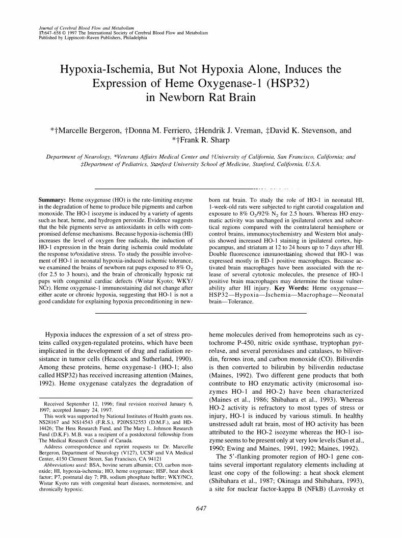
to 8% O2 for 3 hours. Western immunoblot analysis of
crude brain extracts (Fig. I) showed that the rabbit anti
rat HO-I antibody used in the present study recognized a
characteristic -32 kDa band that is consistent with the rat
HO-I protein (HSP32) described previously (Maines et
aI., 1986; Ewing and Maines, 1991). In all regions in
vestigated (striatum, hippocampus, and cortex), there
was no significant difference between untreated controls
(Fig. I, lanes I to 3), the contralateral side that received
the hypoxia without the coagulation (in animals sub
jected to HI) (Fig. I, lanes 4 to 6), or sham-operated
animals with hypoxia only (not shown). In agreement
with these observations, immunocytochemistry experi
ments showed no detectable change in HO-I expression
in newborn brain 24 hours after hypoxia (Figs. 2C,E and
4A), compared with untreated control animals (not
shown). Heme oxygenase-I expression in pup brain was
similar to controls whether animals had been exposed to 8% O2 for 2.5 hours up to 3.5 hours or whether brain
tissue was analyzed 0, I, 5, 24, 48, or 72 hours or 4 days
after hypoxia (not shown).
6 7 8 9
- - - ....c HO-1
, .
FIG. 1. Western immunoblot analysis of rat brain heme oxygenase-1 (HO-1 ) protein levels, 24 hours after a hypoxic-ischemic insult induced at postnatal day 7 (P7). Whole tissue extracts of striatum (Lanes 1 , 4, and 7), hippocampus (Lanes 2, 5, and 8), and cerebral cortex (Lanes 3, 6, and 9) were prepared as described in Materials and Methods and equal protein samples (55 I-1g) were analyzed by sodium dodecyl sulfate polyacrylamide gel electrophoresis and immunoblotting using rabbit anti-rat HO-1 antibody. Lanes 1 to 3: untreated control rat. Lanes 4 to 6: contralateral hemisphere exposed to hypoxia (8%02 for 2.5 hours) without the common carotid artery coagulation. Lanes 7 to 9: ipSilateral hemisphere with both the common carotid artery coagulation and exposure to hypoxia (8%02 for 2.5 hours). Molecular weight markers in kilodalton are indicated on the left. This experiment was repeated four times with similar results. HO-1, heme oxygenase-1 .
J Cereb Blood Flow Metab. Vol. 17, No.6. 1997

HO-I EXPRESSION IN ISCHEMIC NEWBORN RAT BRAIN 651
A B
FIG. 2. Effect of hypoxia and hypoxia-ischemia on brain HO-1 expression in newborn Sprague Dawley rat. Twenty-four hours after the hypoxic-ischemic insult induced at P7, increased HO-1 immunostaining was observed throughout the cortex (0) and in the hilus of the dentate gyrus of the hippocampus (arrows in F) ipsilateral to the common carotid artery ligation. Note the columnar pattern of HO-1 expression in this moderately injured (damage score = 2) ipsilateral cortex (arrows in 0). Contralateral cortex (e) and hippocampus (E) subjected to hypoxia alone showed no increased HO-1 staining compared with normal untreated newborn rat brain. Alternate brain sections incubated without HO-1 primary antibody showed no endogenous staining in corresponding areas of cortex (A) and hippocampus (8). Magnification: 10x, scale bar = 200 [.1m.
Induction of HO-l has been reported after chronic in vitro hypoxia in tumor cells (Heacock and Sutherland,
1990). Rat pups (P7) generally survive a maximum of 3.5 to 4 hours in 8% O2 before dying from cardiac arrest
related to severe hypotension and hypoglycemia (Rice et
aI., 1981; Vannucci and Yager, 1992). In the present
study, because brain HO-I expression was not induced
by 3 hours of in vivo hypoxia at 8% O2 , we examined the effect of chronic hypoxia on brain HO-l expression by
studying rats with congenital cardiac defects (WKY /
NCr; normotensive, chronically hypoxic Wistar rats).
The rats of this inbred strain spontaneously develop vari
ous heart anomalies including Tetralogy of Fallot, ven
tricular septal defect, pulmonary valve stenosis and/or
hypertrophic cardiomyopathy in association with hypo
plasia of the ductus arteriosus, and occasional anomalies
in the aortic arch systems (Kuribayashi et aI., 1990).
Because the WKY!NCr rats are from a Wi star rat lineage, we used normal Wistar rats as untreated controls.
The percentage of oxygen saturation in live untreated
J Cereb Blood Flow Metab, Vol. 17, No.6, 1997
652 M. BERGERON ET AL.
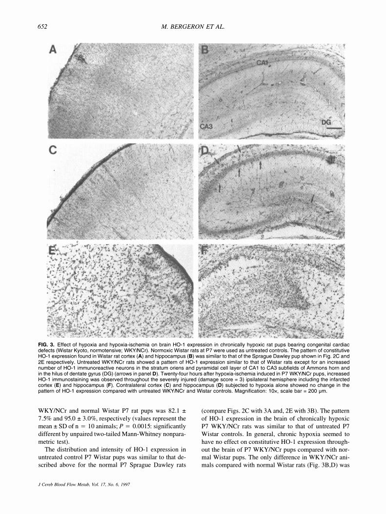
FIG. 3. Effect of hypoxia and hypoxia-ischemia on brain HO-1 expression in chronically hypoxic rat pups bearing congenital cardiac defects (Wistar Kyoto, normotensive; WKY/NCr). Normoxic Wistar rats at P7 were used as untreated controls. The pattern of constitutive HO-1 expression found in Wistar rat cortex (A) and hippocampus (8) was similar to that of the Sprague Dawley pup shown in Fig. 2C and 2E respectively. Untreated WKY/NCr rats showed a pattern of HO-1 expression similar to that of Wistar rats except for an increased number of HO-1 immunoreactive neurons in the stratum oriens and pyramidal cell layer of CA1 to CA3 subfields of Ammons horn and in the hilus of dentate gyrus (DG) (arrows in panel 0). Twenty-four hours after hypoxia-ischemia induced in P7 WKY/NCr pups, increased HO-1 immunostaining was observed throughout the severely injured (damage score = 3) ipsilateral hemisphere including the infarcted cortex (E) and hippocampus (F). Contralateral cortex (C) and hippocampus (0) subjected to hypoxia alone showed no change in the pattern of HO-1 expression compared with untreated WKY/NCr and Wistar controls. Magnification: 10x, scale bar = 200 iJm.
WKY /NCr and normal Wi star P7 rat pups was 82.1 ±
7.5% and 95.0 ± 3.0%, respectively (values represent the
mean ± SD of n = 10 animals; P = 0.0015: significantly
different by unpaired two-tailed Mann-Whitney nonpara
metric test).
The distribution and intensity of HO-l expression in
untreated control P7 Wistar pups was similar to that de
scribed above for the normal P7 Sprague Dawley rats
J Cereb Blood Flow Metab. Vol. 17, No.6. 1997
(compare Figs. 2C with 3A and, 2E with 3B). The pattern
of HO-l expression in the brain of chronically hypoxic
P7 WKY/NCr rats was similar to that of untreated P7
Wi star controls. In general, chronic hypoxia seemed to
have no effect on constitutive HO-l expression through
out the brain of P7 WKY INCr pups compared with normal Wistar pups. The only difference in WKY/NCr ani
mals compared with normal Wistar rats (Fig. 3B,D) was
HO-I EXPRESSION IN ISCHEMIC NEWBORN RAT BRAIN 653
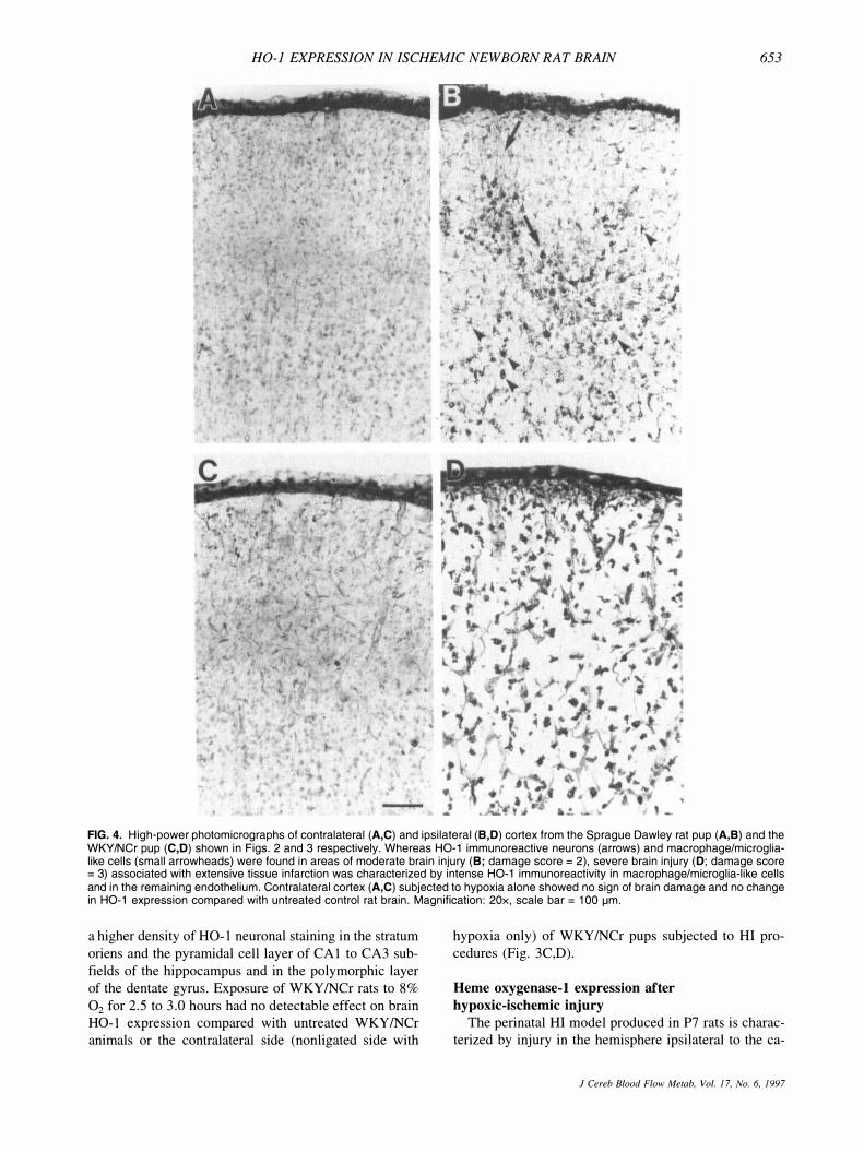
FIG. 4. High-power photomicrographs of contralateral (A,C) and ipsilateral (8,0) cortex from the Sprague Dawley rat pup (A,8) and the WKY/NCr pup (C,O) shown in Figs. 2 and 3 respectively. Whereas HO-1 immunoreactive neurons (arrows) and macrophage/microglialike cells (small arrowheads) were found in areas of moderate brain injury (8; damage score = 2), severe brain injury (0; damage score = 3) associated with extensive tissue infarction was characterized by intense HO-1 immunoreactivity in macrophage/microglia·like cells and in the remaining endothelium. Contralateral cortex (A,C) subjected to hypoxia alone showed no sign of brain damage and no change in HO-1 expression compared with untreated control rat brain. Magnification: 20x, scale bar = 100 IJm.
a higher density of HO-l neuronal staining in the stratum
oriens and the pyramidal cell layer of CAl to CA3 sub
fields of the hippocampus and in the polymorphic layer
of the dentate gyrus. Exposure of WKY/NCr rats to 8%
O2 for 2.5 to 3.0 hours had no detectable effect on brain HO-l expression compared with untreated WKY/NCr
animals or the contralateral side (nonligated side with
hypoxia only) of WKY/NCr pups subjected to HI pro
cedures (Fig. 3C,D).
Heme oxygenase-l expression after
hypoxic-ischemic injury
The perinatal HI model produced in P7 rats is charac
terized by injury in the hemisphere ipsilateral to the ca-
J Cereb Blood Flow Metab, Vol. 17, No. 6, 1997

654 M. BERGERON ET AL.
rotid occlusion (Rice et aI., 1981). Damage ranges from
the loss of a few isolated cells up to gross infarction in
cortex, hippocampus, striatum, thalamus, and white mat
ter ipsilateral to the ligation. Moderate (damage score =
2) to severe injury (damage score = 3) is more common
than little (damage score = I) or no damage (damage score = 0) (Table 2). Exposure to hypoxia alone (con
tralateral side) does not produce any evidence of cellular
damage.
Western immunoblot analysis of crude brain extracts
obtained from Sprague-Dawley rats pups 24 hours after
HI showed that HO-l protein expression was increased 5 to lO-fold in ipsilateral striatum, hippocampus, and cor
tex (Fig. 1, lanes 7 to 9), compared with the contralateral
side (Fig. 1, lanes 4 to 6) or untreated controls (Fig. 1, lanes 1 to 3). In agreement with these observations, increased HO-l immunoreactivity was detected as early as
6 to 12 hours after HI in the ipsilateral cortex near the
corpus callosum (not shown). Markedly increased HO-l
immunostaining was observed in cells from cortex, hip
pocampus, striatum, and thalamus ipsilateral to the ca
rotid occlusion by 24 hours after HI (Fig. 2D,F). Heme
oxygenase-l immunostaining was still quite prominent at
4 days after HI (Fig. 5C,D), and still detectable in the remaining tissue at 7 days (not shown). Interestingly, the increased Hq-l expression found in ipsilateral cortex
often displayed a columnar pattern (Fig. 2D, arrows).
Sections incubated without HO-l antibody showed no
endogenous staining (Fig. 2A,B). WKY/NCr pups sub
jected to HI showed a pattern of brain injury and HO-l
expression very similar to that described above for the
Sprague Dawley pups. As shown in Table 2, there was
no significant difference in the median histological dam
age score between the two groups (P = 0.9812, Mann
Whitney nonparametric test). Increased HO-l expression was observed throughout ipsilateral ischemic hemisphere
(Fig. 3E,F) compared with the contralateral hypoxic side
TABLE 2. Distribution of histopathologic damage scores in Sprague Dawley and WKYINCr newborn rats 24 hours
afier hypoxia-ischemia
Animals
Damage SPD WKY/NCr score* (n = 19) (n = I I )
0 I I 1 3 2 2 II 5 3 4 3
Median scoret 2 2
SPD, Sprague Dawley; WKY/NCr, Wi star Kyoto rats with congenital heart diseases, normotensive. and chronically hypoxic.
* Histopathological scores: 0, no gross or histological damage; I, no gross damage. minimal neuronal loss; 2, columnar cortical infarction, moderate neuronal loss; 3, extensive infarction and gliosis. severe neuronal loss.
t p = 0.98 1 2 : the median damage scores for the two groups are not significantly different by Mann-Whitney nonparametric test.
J Cereb Blood Flow Metab, Vol. 17, No.6, 1997
(Fig. 3C,D). For all animals investigated in this study,
the intensity of HO-l staining was generally proportional
to the degree of brain injury. Whereas moderate injury (Fig. 2D,F) resulted in increased HO-l expression in
some neurons and glia-like cells (Fig. 4B), severe injury (Fig. 3E,F) resulting in large areas of brain tissue infarc
tion revealed an increase in HO-l-positive macrophage
like cells and sustained HO-l staining in the remaining
endothelium (Fig. 4D). Induction of HO-l protein ex
pression did not increase total HO enzymatic activity in
ipsilateral cortex and subcortical regions 24 hours after
HI (Table 1).
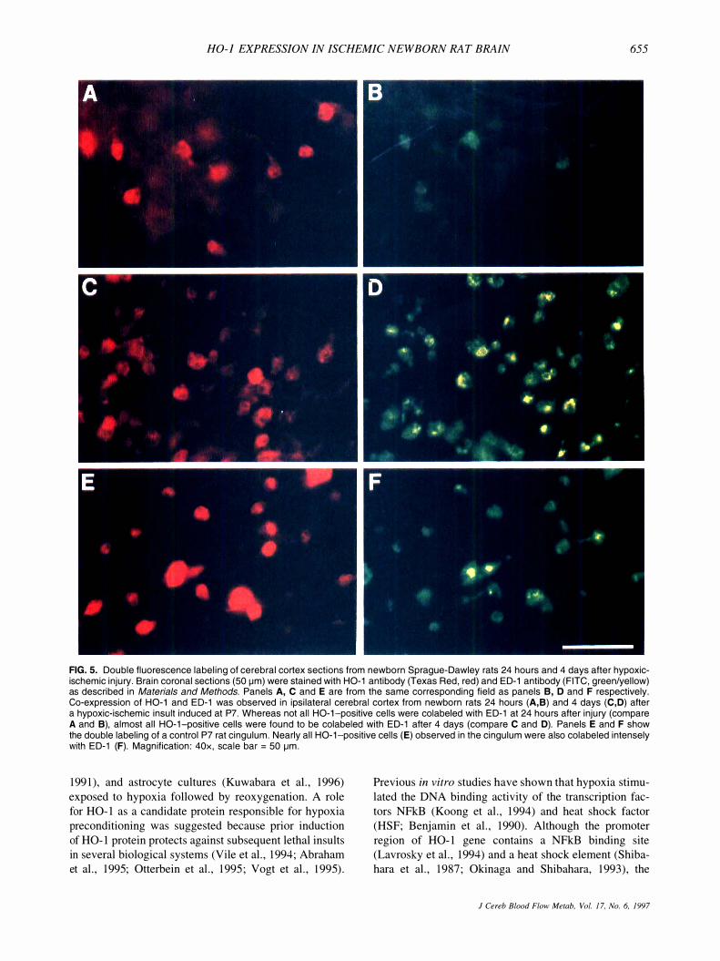
Double immunofluorescence staining after
hypoxia-ischemia
To define the type of cells expressing HO-l protein,
double fluorescence labeling using antibodies against
HO-I and ED-I, OX-42 or GFAP was performed 24
hours and 4 days after HI in newborn Sprague-Dawley
rats. Heme oxygenase-I expression in ipsilateral cortex
(Fig. 5A to D) was found mainly in brain macrophages
expressing the ED-l antigen. Double immunolabeling
with HO-l and OX-42 or GFAP antibodies showed occasional co staining of HO-l/OX-42 or HO-l/GFAP (not
shown). Twenty-four hours after moderate HI injury,
most but not all of the HO-l-positive cells were found to
be ED-l positive (Fig. 5A,B). Almost all HO-l positive
cells were found to be colabeled with the ED-I antigen 4
days after HI (Fig. 5C,D).
DISCUSSION
Hypoxia preconditioning and heme oxygenase-1
expression
Hypoxia pretreatment (8% 02/3 hours) confers neuro
protection in newborn rats against ischemic injury 24
hours after the initial preconditioning (Gidday et aI.,
1994). Such hypoxia treatment has relatively no effect on
neuronal integrity (Rice et aI., 1981) and on several
physiological parameters such as regional cerebral blood
flow and water content (Vannucci et aI., 1988; Mujsce et
aI., 1990), NADH fluorescence (Welsh et aI., 1982),
brain protein synthesis (Dwyer et aI., 1987), and cerebral
calcium uptake (Stein and Vannucci, 1988). In addition,
the expression of several genes including the immediate
early genes fos and jun (Munell et aI., 1994), HSP72
(Ferriero et aI., 1990; Munell et aI., 1994) and GFAP
(Burtrum and Silverstein, 1994) are unaffected by such
in vivo hypoxia pretreatment in newborn rat brain.
Recent studies have suggested that the induction of
oxygen-regulated proteins could be involved in the
mechanism of hypoxia-induced ischemic tolerance. In
creased expression of certain proteins including HO-l
has been reported in tumor cell lines (Heacock and Sutherland, 1990), endothelial cells (Zimmerman et aI.,
HO-i EXPRESSiON iN ISCHEMIC NEWBORN RAT BRAIN 655
FIG. 5. Double fluorescence labeling of cerebral cortex sections from newborn Sprague-Dawley rats 24 hours and 4 days after hypoxicischemic injury. Brain coronal sections (50 fJm) were stained with HO-1 antibody (Texas Red, red) and ED-1 antibody (FITC, green/yellow) as described in Materials and Methods. Panels A, C and E are from the same corresponding field as panels B, 0 and F respectively. Co-expression of HO-1 and ED-1 was observed in ipsilateral cerebral cortex from newborn rats 24 hours (A,B) and 4 days (C,D) after a hypoxic-ischemic insult induced at P7. Whereas not all HO-1-positive cells were colabeled with ED-1 at 24 hours after injury (compare A and B), almost all HO-1-positive cells were found to be colabeled with ED-1 after 4 days (compare C and D). Panels E and F show the double labeling of a control P7 rat cingulum. Nearly all HO-1-positive cells (E) observed in the cingulum were also colabeled intensely with ED-1 (F). Magnification: 40x, scale bar = 50 fJm.
1991), and astrocyte cultures (Kuwabara et aI., 1996)
exposed to hypoxia followed by reoxygenation. A role
for HO-l as a candidate protein responsible for hypoxia
preconditioning was suggested because prior induction
of HO-l protein protects against subsequent lethal insults in several biological systems (Vile et aI., 1994; Abraham et aI., 1995; Otterbein et a!., 1995; Vogt et aI., \995).
Previous in vitro studies have shown that hypoxia stimu
lated the DNA binding activity of the transcription fac
tors NFkB (Koong et aI., 1994) and heat shock factor
(HSF; Benjamin et aI., 1990). Although the promoter
region of HO-l gene contains a NFkB binding site (Lavrosky et aI., 1994) and a heat shock element (Shiba
hara et aI., 1987; Okinaga and Shibahara, 1993), the
J Cereb Blood Flow Metah, Va!. 17. No.6. 1997

656 M. BERGERON ET AL.
present study failed to show an induction of HO-l pro
tein expression after the 2.5 to 3.5 hour-hypoxia treat
ment necessary for preconditioning and neuroprotection.
The HO-l induction observed in cultured cells may be
caused by low oxygen tensions for long periods of time that cannot be achieved in vivo. For this reason, we investigated the effect of chronic in vivo hypoxia on brain
HO-l expression in rat pups with congenital heart de
fects (WKY/NCr). These animals also failed to show
increased HO-l protein expression. In contrast to the
study of Gidday et aI., (1994) which used acute hypoxia
(8% O2 for 3 hours) to induce ischemic 'tolerance, the
present study showed that chronic neonatal hypoxia associated with congenital heart defects (WKY/NCr rats) did not protect the brain against subsequent HI injury
(Table 2). Taken together, these observations suggest that the induction of HO-l expression is not the mecha
nism responsible for hypoxia preconditioning in new
born rat brain.
Effect of hypoxia-ischemia on heme oxygenase-!
expression
Perinatal HI brain damage is associated with increased HO-l protein expression. The degree of HO-l expression in the hemisphere ipsilateral to the common carotid ar
tery occlusioq was dependent on the severity of the insult. After moderate injury, HO-l expression was in
creased mostly in brain macrophages found throughout
the focal areas of tissue damage and in scattered neurons,
some astrocytes, and endothelial cells ipsilateral to the
carotid occlusion. Severe insults resulting in extensive
tissue infarction were accompanied by HO-l staining in
ED-I-positive macrophages and the surviving endothe
lium almost exclusively. Heme oxygenase-I-positive astrocytes were infrequent despite massive and progressive
reactive astrogliosis in areas of HI brain damage in the
newborn (Burtrum and Silverstein, 1994; Sheldon et aI.,
1996). In contrast, HO-IIED-l positive macrophages in
creased in number and became the major cellular com
ponent expressing HO-l after HI. The distribution of
these HO-l-positive cells was found to be similar to that
recently reported for ED-I-positive cells in the same animal model of neonatal HI (Ivacko et aI., 1996). The
origin of these HO-l-positive macrophages is not
known. Some of the HO-IIED-l macrophages observed
in the present study may have entered the brain from blood vessels possibly in response to the breakdown of
blood-brain barrier (Vannucci et aI., 1993) and signals
from damaged neurons and glia. Alternatively, some of the HO-IIED-l macrophages might have been derived
from brain-resident macrophages and possibly microglia
(Thomas, 1992). Glutathione depletion (Ewing and Maines, 1993) as
well as hyperthermia (Ewing and Maines, 1991) in rats
significantly induce HO-l messenger RNA and protein
J Cereb Blood Flow Metab. Vol. 17. No.6. 1997
without concomitant increased brain HO activity. Simi
larly, the present study showed that despite a 5- to 10-
fold increase in HO-l expression after HI, HO activity
remained unchanged compared with controls. Heme oxy
genase-2 protein, which is found mainly in neurons (Ew
ing and Maines, 1992; Maines, 1992), is the most abundant HO isozyme in the brain and is responsible for the
bulk of brain HO activity (Sun et aI., 1990; Ewing and
Maines, 1991). With the ongoing neuronal loss associ
ated with HI injury, it is possible that the increased HO-l
expression observed mainly in proliferating macro
phages may compensate for the loss of neuronal HO-2
protein and thus, maintain the overall HO activity at a
normal leveL Although it is also possible that HO-l protein becomes inactive after HI, the lack of detectable changes in brain HO activity after neonatal HI does not rule out the possibility for a local effect of HO-l activity
resulting in the release of free iron, bile pigments,and CO.
Carbon monoxide (like nitric oxide) has been pro
posed as a putative neurotransmitter acting as a physi
ologic regulator of guanylyl cyclase and cGMP
dependent protein kinase (Maines, 1993; Verma et aI.,
1993). Recent studies have shown that the activation of metabotropic receptors could modulate neuronal HO ac
tivity and in tum, CO could be involved in the signal transduction pathway coupling these receptors to the ac
tivity of the NaK-ATPase pump (Glaum and Miller,
1993; Nathanson et aI., 1995). In view of this suggested
relationship between glutamate receptor activation and
increased HO activity in the brain, it is possible that the
increased HO-l expression observed in neurons after moderate HI injury may be caused by, at least in part, the
overactivation of glutamate receptors (Rothman and Ol
ney, 1986). Ischemia-induced oxidative stress and cellular protein denaturation may also induce neuronal HO-l
through activation of specific transcription factors such
as fos and jun, NFkB, and HSF. Indeed, a region
selective induction of the immediate early genes fos and
jun has been reported ipsilateral to the HI injury in new
born rat brain (Munell et aI., 1994). Moreover, increased
of DNA-binding activity at the AP-l binding site and the
heat shock element has been reported in cerebral cortex
after transient focal ischemia in adult rats (Salminen et
aI., 1995) and in gerbil hippocampus after global isch
emia (Nowak and Abe, 1994). However, because neu
rons only express HSF-2, which is less able than HSF-l
to direct a strong heat shock response (Marcuccilli et aI.,
1996), a major increase of HO-l expression in neurons
through HSF activation seems less likely. Interestingly,
increased NFkB binding activity was noted in ischemic
cortex only 5 days after focal ischemia (Salminen et aI.,
1995), suggesting that this late NFkB surge may be related
to postischemic infiltration of inflammatory macrophages.
Increased production of the antioxidant bilirubin
(Stocker et aI., 1987) after induction of HO-l expression
HO-J EXPRESSiON IN ISCHEMIC NEWBORN RAT BRAIN 657
could protect neurons from subsequent delayed injury.
However, this study showed that although there was a
clear attempt by neurons to synthesize more HO-l pro
tein after moderate injury, there was also a clear failure
for them to survive more severe HI insults. These observations are consistent with previous reports suggesting
that when ischemia is sufficiently severe to produce in
farction, transcription and/or translation of the heat shock
genes is blocked in neurons and glia destined to die,
whereas the surviving endothelial cells, which have been
shown to induce both HSP72 and HO-l after focal isch
emia in adult rat (Nimura et aI., 1996) or neonatal HI
(Ferriero et aI., 1990; present study), continue to synthe
size stress proteins (Gonzalez et aI., 1989; Kinouchi et aI., 1993). In addition, studies have shown that neurons
in culture have a very limited capacity to induce HO-I
expression even after hydrogen peroxide exposure. In
fact, this may contribute to their selective vulnerability to
oxidative stress (Dwyer et aI., 1995). The protective
mechanism suggested for HO-l activity may also in
volve the potential lethal effects of ferrous iron, which if
un sequestered, may exacerbate HI brain injury by in
creasing the formation of hydroxyl radicals through the
Fenton reaction (Braughler et aI., 1986). Almost two
thirds of the iron in the brain is stored as ferritin (H and L isoforms). l-I-ferritin (heavy-chain) is found mainly in
neurons and has a low storage capacity consistent with
the high iron utilization. L-ferritin (light-chain) which is
localized mainly in brain macrophages is involved in
long-term iron storage, consistent with the major role of
this cell type as a scavenger (Connor et aI., 1994). As a
result of HI, the increased pro-oxidant levels produced
by HO-l activity combined with the low iron-storage
capacity of neurons (H-ferritin) could contribute to HI neuronal injury. In contrast, because of the greater iron
sequestering capacity of L-ferritin, brain macrophages
may be able to survive greater increases in iron release
and thus, may better benefit from the antioxidant activity
of HO-I induced expression. Because inhibition of
phagocytic and secretory functions in mononuclear
phagocytes after ischemia has been shown to reduce
ischemic injury in the spinal cord (Giulian and Robert
son, 1990), it is suggested that HO-I-protected newborn
macrophages, besides scavenging iron and removing cel
lular debris from developing and/or injured brain tissue,
may also contribute to neuronal damage after HI and
reoxygenation by releasing neurotoxic molecules (Giu
han et aI., 1993).
Though the bilirubin produced by HO-l may act as an
antioxidant in many tissues, it may also be toxic to the
brain. Hyperbilirubinemia is commonly observed during
the first week of life in humans and rats (Maines, 1992).
Whereas physiological hyperbilirubinemia alone does not cause bilirubin encephalophathy (kernicterus), cer
tain conditions such as asphyxia and acidosis can predis-
pose the brain to bilirubin toxicity by decreasing biliru
bin binding to serum albumin and increasing tissue bind
ing of bilirubin (Maines, 1992). Besides its suggested
role as an antioxidant at moderate concentrations (Stocker et aI., 1987), bilirubin has also been shown to be toxic to cultured astrocytes, neurons, and neural cell lines
(Amit and Brenner, 1993). Thus, the elevated levels of
bilirubin produced by local increases in HO-l expression
after HI injury could contribute to neuronal damage after
ischemia and to the neuronal injury in kernicterus after
perinatal asphyxia.
Acknowledgment: The authors thank Ronald J. Wang for expert technical assistance with the heme oxygenase activity assay.
REFERENCES
Abraham NG. Lavrovsky Y. Schwartzman ML, Stoltz RA, Levere RD, Gerritsen ME, Shibahara S, Kappas A ( 1 995) Transfection of the human heme oxygenase gene into rabbit coronary microvessel endothelial cells: protective effect against heme and hemoglobin toxicity. Proc Natl Acad Sci USA 92:6798-6802
Alam J, Zhining D ( 1 992) Distal AP- I binding sites mediate basal1evel enhancement and TP A induction of the mouse heme oxygenase- l gene. J Bioi Chern 267 : 2 1 894-2 1 900
Amit Y, Brenner T ( 1 993) Age-dependent sensitivity of cultured rat glial cells to bilirubin toxicity. Exp Neural 1 2 1 :248-255
Benjamin 11, Kroger B, Williams RS ( 1 990) Activation of the heat shock transcription factor by hypoxia in mammalian cells. Prac Natl Acad Sci USA 87:6263-6267
Bergeron M, Mivechi NF, Giaccia AJ, Giffard RG ( 1 996) Mechanism of heat shock protein 72 induction in primary cultured astrocytes after oxygen-glucose deprivation. Neural Res 18:64-72
Braughler JM, Duncan LA, Chase RL ( 1 986) The involvement of iron in lipid peroxidation. Importance of ferric to ferrous ratios in initiation. J Bioi Chern 26 1 : 1 0282- 1 0289
Burtrum D, Silverstein FS ( 1 994) Hypoxic-ischemic brain injury stimulates glial fibrillary acidic protein mRNA and protein expression in neonatal rats. Exp Neural 1 26: 1 1 2-1 1 8
Connor JR, Boeshore KL, Benkovic SA, Menzies SL ( 1 994) lsoforms of ferritin have a specific cellular distribution in the brain. J Neurasci Res 37:461-465
Dobbing J, Sands J ( 1 979) Comparative aspects of the brain growth spurt. Early Hum Dev 3 :79-83
Dwyer BE, Nishimura RN, Powell CL, Mailheau SL (1987) Focal protein synthesis inhibition in a model of neonatal hypoxicischemic brain injury. Exp Neural 95:277-289
Dwyer BE, Nishimura RN, Lu S-Y ( 1 995) Differential expression of heme oxygenase- l in cultured cortical neurons and astrocytes determined by the aid of a new heme oxygenase antibody. Response to oxidative stress. Mol Brain Res 30:37-47
Ewing JF, Maines MD ( 1 99 1 ) Rapid induction of heme oxygenase I mRNA and protein by hyperthermia in rat brain: heme oxygenase 2 is not a heat shock protein. Proc Natl Acad Sci USA 88:5364-5368
Ewing JF, Maines MD (1992) In situ hybridization and immunohistochemical localization of heme oxygenase-2 mRNA and protein in normal rat brain : differential distribution of isozyme I and 2. Mol Cell Neurasci 3 : 559-570
Ewing JF, Maines MD ( 1 993) Glutathione depletion induces heme oxygenase- I (HSP32) mRNA and protein in rat brain. J Neurachern 60: 1 5 1 2- 1 5 1 9
Ferriero DM, Soberano HQ, Simon RP, Sharp FR ( 1 990) Hypoxiaischemia induces heat shock protein-like (HSP72) immunoreactivity in neonatal rat brain. Dev Brain Res 53 : 1 45- 1 50
Gidday JM, Fitzgibbons JC, Shah AR, Park TS ( 1 994) Neuroprotection from ischemic brain injury by hypoxic preconditioning in the neonatal rat. Neurasci Lett 168 :22 1 -224
Glaum SR, Miller RJ ( 1 993) Zinc protoporphyrin-IX blocks the effects
J Cereb Blood Flow Metab, Vol. 17, No.6, 1997

658 M. BERGERON ET AL.
of metabotropic glutamate receptor activation in the rat nucleus tractus solitaris . Mol Pharmacal 43: 965-969
Giulian 0, Robertson C ( 1 990) Inhibition of mononuclear phagocytes reduces ischemic injury in the spinal chord. Ann Neurol 27 :33-42
Giulian 0, Vaca K, Corpuz M ( 1 993) Brain glia release factors with opposing actions upon neuronal survival . J Neurosci 1 3 : 29-37
Gonzalez MF, Shiraishi K, Hisanaga K, Sagar SM, Mandabach M, Sharp FR ( 1 989) Heat shock proteins as markers of neuronal injury. Mol Brain Res 6: 93- 1 00
Heacock CS, Sutherland RM ( 1 990) Enhanced synthesis of stress proteins caused by hypoxia and relation to altered cell growth and metabolism. Br J Cancer 62: 2 1 7-225
Ivacko JA, Sun R, Silverstein FS ( 1 996) Hypoxic-ischemic brain injury induces an �cute microglial reaction in perinatal rats. Pediatr Res 39:39-47
Kinouchi H, Sharp FR, Koistinaho J, Hicks K, K'amii H, Chan PH ( 1 993) Induction of heat shock hsp70 mRNA and HSP70 kDa protein in neurons in the 'penumbra' following focal cerebral ischemia in the rat. Brain Res 6 1 9 : 334-3 3 8
Koong AC, Chen EY, Giaccia AJ ( 1 994) Hypoxia causes the activation of nuclear factor kB through the phosphorylation of IkBa on tyrosine residues. Cancer Res 54: 1 425- 1 430
Kuribayashi T, Shimoo K, Nakamura T, Taniwaki H, Hamaoka K, Nakagawa M, Ibata Y, Komeda T, Nagaoka A ( 1 990) Tetralogy of Fallot, pulmonary valve stenosis, ventricular septal defect, and hypertrophic cardiomyopathy in WKY/NCrj rats. Pediatr Res 27 : 483-487
Kuwabara K, Matsumoto M, Ikeda J, Hori 0, Ogawa S, Maeda Y, Kitagawa K, Imuta N, Kinoshita T, Stern 0, Yanagi H, Kamada T ( 1 996) Purification and characterization of a novel stress protein, the 1 50-kDa oxygen-regulated protein (ORP 1 50), from cultured rat astrocytes and its expression in ischemic mouse brain. J Bioi Chem 27 1 : 5025-5032
Lavrovsky Y, Schwartzman ML, Levere RD, Kappas A, Abraham NG ( 1 994) Identification of binding sites for transcription factors NFkB and AP-2 in the promoter region of the human heme oxygenase 1 gene. Proc Natl Acad Sci US A 91 :5987-599 1
Maines MD, Trakshel GM, Kutty RK ( 1 986) Characterization of two constitutive forms of rat liver microsomal heme oxygenase. J Bioi
Chem 26 1 :4 1 1 -4 1 9 Maines M D ( 1992) Heme oxygenase: clinical applications and func
tions, Boca Raton, Florida, CRC Press, pp 1 -276 Maines MD ( 1 993) Carbon monoxide: an emerging regulator of cGMP
in the brain. Mol Cell Neurosci 4 :3 89-397 Marcuccilli CJ, Mathur SK, Morimoto RI, Miller RJ ( 1 996) Regulatory
differences in the stress response of hippocampal neurons and glial cells after heat shock. J Neurosci 1 6 :478-485
Matz P, Turner C, Weinstein PR, Massa SM, Panter SS, Sharp FR ( 1 996) Heme-oxygenase- I induction in glia throughout rat brain following experimental subarachnoid hemmorrhage. Brain Res 7 1 3 : 2 1 1 -222
Muller RM, Taguchi H, Shibahara S ( 1 987) Nucleotide sequence and organization of the rat heme oxygenase gene. J Bioi Chem 262: 6795-6802
Mujsce DJ, Christensen MA, Vannucci RC ( 1 990) Cerebral blood flow and edema in perinatal hypoxic-ischemic brain damage. Pediatr Res 27 :450-453
Munell F, Burke RE, Bandele A, Gubits RM ( 1 994) Localization of c-fos, c-jun, and hsp70 mRNA expression in brain after neonatal hypoxia-ischemia. Dev Brain Res 77: 1 1 1 - 1 2 1
Nathanson JA, Scavone C, Scanlon C, McKee M ( 1 995) The cellular Na+ pump as a site of action for carbon monoxide and glutamate: a mechanism for long-term modulation of cellular activity. Neuron
1 4 : 7 8 1 -794
Nimura T, Weinstein PR, Massa SM, Panter S, Sharp FR ( 1 996) Heme oxygenase- I (HO- I ) protein induction i n rat brain following focal ischemia. Mol Brain Res 37 :20 1 -208
Nowak TS, Jr. , Abe H ( 1 994) Post-ischemic stress response in brain . In: The Biology of Heat Shock Proteins and Molecular Chaperones (Morimoto RI, Tissieres A, Georgopoulos C. eds) , New York, Cold Spring Harbor Laboratory Press, pp 553-575
Okinaga S , Shibahara S ( 1 993) Identification of a nuclear protein that constitutively recognizes the sequence containing a heat-shock el-
J Cereb Blood Flow Me/ab, Vol. 17, No. 6, 1997
ement. Its binding properties and possible function modulating heat-shock induction of the rat heme oxygenase gene. Eur J Biochem 2 1 2: 1 67- 1 75
Otterbein L, Sylvester SL, Choi AM ( 1 995) Hemoglobin provides protection against lethal endotoxemia in rats. The role of heme oxygenase- I . Am J Resp Cell Mol Bioi 1 3 : 595-60 1
Paschen W, Uto A, Djuricic B, Schmitt J ( 1 994) Hemeoxygenase expression after reversible ischemia of rat brain. Neurosci Lett 1 80:5-8
Raps SP, Lai JC, Hertz L, Cooper AJ ( 1 989) Glutathione is present in high concentrations in cultured astrocytes but not in cultured neurons . Brain Res 493 :398-40 1
Rice JE, Vannucci RC, Brierley JB ( 1 9 8 1 ) The influence of immaturity on hypoxic-ischemic brain damage in the rat. Ann NeuroI 9: 1 3 1 - 1 4 1
Rothman SM, Olney JW ( 1 986) Glutamate and the pathophysiology of hypoxic-ischemic brain damage. Ann Neurol 1 9 : 1 05-1 1 1
Salminen A, Liu PK, Hsu CY ( 1 995) Alteration of transcription factor binding activities in the ischemic rat brain. Biochem Biophys Res Comm 2 1 2:939-944
Sheldon RA, Chuai J, Ferriero DM ( 1 996) A rat model for hypoxicischemic brain damage in very premature infants. Bioi Neonate 69: 327-34 1
Shibahara S, Muller RM, Taguchi H ( 1 987) Transcriptional control of rat heme oxygenase by heat shock. J Bioi Chem 262: 1 2889- 1 2892
Shibahara S , Y oshizawa M, Suzuki H, Takeda K, Meguro K, Endo K ( 1 993) Functional analysis of cDNA for two types of human heme oxygenase and evidence for their separate regulation. J Biochem 1 1 3 : 2 1 4-2 1 8
Siesj6 BK, Agardh CD, Bengtsson F ( 1 989) Free radicals and brain damage. Cerebrovasc. Brain Metab Rev 1 : 1 65-2 1 1
Stein DT, Vannucci RC ( 1 988) Calcium accumulation during the evolution of hypoxic-ischemic brain damage in the immature rat. J Cerebr Blood Flow Metab 8 :834-842
Stocker R, Yamamoto Y, McDonagh AF, Glazer AN, Ames BN ( 1 987) Bilirubin is an antioxidant of possible physiological importance. Science 235: 1 043- 1 046
Sun Y, Rotenberg MO, Maines MD ( 1 990) Developmental expression of heme oxygenase isozymes in rat brain. Two HO-2 mRNAs are detected. J Bioi Chem 265 : 8 2 1 2-82 1 7
Takeda A , Onodera H , Sugimoto A , Itoyama Y , Kogure K , Shibahara S ( 1 994) Increased expression of heme oxygenase mRNA in rat brain following transient forebrain ischemia. Brain Res 666: 1 20- 1 24
Thomas WE ( 1 992) Brain macrophages: evaluation of microglia and their functions. Brain Res Rev 1 7 : 6 1 -74
Turner CP, Bergeron M, Matz P, Noble L, Panter SS, Sharp FR ( 1 997) HemeOxygenase- l (HO- l , HSP32) i s induced in microglia throughout brain by subarachnoid injection of hemoglobin. J Cereb Blood Flow Metab (submitted)
Vannucci RC, Lyons DT, Vasta F ( 1 988) Regional cerebral blood flow during hypoxia-ischemia in immature rats. Stroke 1 9 : 245-250
Vannucci RC, Yager Y ( 1 992) Glucose, lactic acid, and perinatal hypoxic-ischemic brain damage. Pediatr NeuroI 8 : 3 - 1 2
Vannucci RC, Christensen MA, Yager JY ( 1 993) Nature, time-course, and extent of cerebral edema in perinatal hypoxic-ischemic brain edema. Pediatr Neurol 9 :29-34
Verma A, Hirsch OJ, Glatt CE, Ronnett GV, Snyder SH ( 1 993) Carbon monoxide: putative neural messenger. Science 259:38 1-384
Vile GF, Basu-Modak S , Waltner C, Tyrrell RM ( 1 994) Heme oxygenase 1 mediates an adaptive response to oxidative stress in human skin fibroblasts . Proc Natl Acad Sci US A 9 1 : 2607-26 1 0
Vogt BA, Alam J , Croatt AJ, Vercellotti GM, Nath K A ( 1 995) Acquired resistance to acute oxidative stress. Possible role of heme oxygenase and ferritin. Lab Invest 72:474-483
Vreman HJ, Ekstrand BC, Stevenson DK ( 1 993) Selection of metalloporphyrin heme oxygenase inhibitors based on potency and photoreactivity. Pediatr Res 33 : 1 95-200
Vreman HJ, Stevenson DK ( 1 988) Heme oxygenase activity as measured by carbon monoxide production. Anal Biochem 1 68 : 3 1-38
Welsh FA, Vannucci RC, Brierley JB ( 1 982) Columnar alterations of NADH tluorescence during hypoxia-ischemia in immature rat brain. J Cerebr Blood Flow Metab 2 : 22 1 -228
Zimmerman LH, Levine RA, Farber HW ( 1 99 1 ) Hypoxia induces a specific set of stress proteins in cultured endothelial cells . J CUn Invest 87 :908-9 1 4