divergent cardiopulmonary actions of heme oxygenase enzymatic products in chronic hypoxia
TRANSCRIPT
Divergent Cardiopulmonary Actions of Heme OxygenaseEnzymatic Products in Chronic HypoxiaSally H. Vitali1, S. Alex Mitsialis2, Olin D. Liang2, Xiaoli Liu3, Angeles Fernandez-Gonzalez2, Helen
Christou4, Xinqi Wu4, Francis X. McGowan5, Stella Kourembanas2*
1 Division of Critical Care Medicine, Children’s Hospital Boston, Boston, Massachusetts, United States of America, 2 Division of Newborn Medicine, Children’s Hospital
Boston, Boston, Massachusetts, United States of America, 3 Pulmonary Division, Brigham and Women’s Hospital, Boston, Massachusetts, United States of America,
4 Division of Newborn Medicine, Brigham and Women’s Hospital, Boston, Massachusetts, United States of America, 5 Division of Cardiac Anesthesia, Children’s Hospital
Boston, Boston, Massachusetts, United States of America
Abstract
Background: Hypoxia and pressure-overload induce heme oxygenase-1 (HO-1) in cardiomyocytes and vascular smoothmuscle cells (VSMCs). HO-12/2 mice exposed to chronic hypoxia develop pulmonary arterial hypertension (PAH) withexaggerated right ventricular (RV) injury consisting of dilation, fibrosis, and mural thrombi. Our objective was to indentifythe HO-1 product(s) mediating RV protection from hypoxic injury in HO-12/2 mice.
Methodology/Principal Findings: HO-12/2 mice were exposed to seven weeks of hypoxia and treated with inhaled CO orbiliverdin injections. CO reduced right ventricular systolic pressure (RVSP) and prevented hypoxic pulmonary arteriolarremodeling in both HO-12/2 and control mice. Biliverdin had no significant effect on arteriolar remodeling or RVSP in eithergenotype. Despite this, biliverdin prevented RV failure in the hypoxic HO-12/2 mice (0/14 manifested RV wall fibrosis orthrombus), while CO-treated HO-12/2 mice developed RV insults similar to untreated controls. In vitro, CO inhibited hypoxicVSMC proliferation and migration but did not prevent cardiomyocyte death from anoxia-reoxygenation (A-R). In contrast,bilirubin limited A-R-induced cardiomyocyte death but did not inhibit VSMC proliferation and migration.
Conclusions/Significance: CO and bilirubin have distinct protective actions in the heart and pulmonary vasculature duringchronic hypoxia. Moreover, reducing pulmonary vascular resistance may not prevent RV injury in hypoxia-induced PAH;supporting RV adaptation to hypoxia and preventing RV failure must be a therapeutic goal.
Citation: Vitali SH, Mitsialis SA, Liang OD, Liu X, Fernandez-Gonzalez A, et al. (2009) Divergent Cardiopulmonary Actions of Heme Oxygenase Enzymatic Productsin Chronic Hypoxia. PLoS ONE 4(6): e5978. doi:10.1371/journal.pone.0005978
Editor: Rory Edward Morty, University of Giessen Lung Center, Germany
Received February 19, 2009; Accepted April 29, 2009; Published June 19, 2009
Copyright: � 2009 Vitali et al. This is an open-access article distributed under the terms of the Creative Commons Attribution License, which permitsunrestricted use, distribution, and reproduction in any medium, provided the original author and source are credited.
Funding: Research was suppported by the NICHD-sponsored Pediatric Critical Care Scientist Development Program, and NHLBI K08 HL077344 to Dr. Vitali andNIH RO1 HL055454 and HL085446 to Dr. Kourembanas. The funders had no role in study design, data collection and analysis, decision to publish, or preparation ofthe manuscript.
Competing Interests: The authors have declared that no competing interests exist.
* E-mail: [email protected]
Introduction
Chronic hypoxia causes remodeling of the pulmonary vascula-
ture with increased proliferation and migration of vascular smooth
muscle cells (VSMC), increased pulmonary vascular resistance,
increased pulmonary artery pressure, and right ventricular
hypertrophy. This clinical condition of ‘‘pulmonary hypertension’’
ultimately leads to death as the right ventricular hypertrophy
progresses to dilation and failure. Heme oxygenase-1 (HO-1), an
inducible enzyme which degrades the oxidant heme to produce
equimolar products of carbon monoxide (CO), biliverdin, and
ferrous iron, has been reported to protect animals from
proliferation of VSMC [1,2], apoptosis of cardiomyocytes [3],
hypertrophy of cardiomyocytes [4,5], and cardiac ischemia –
reperfusion injury [6–8] in a variety of in vitro and in vivo studies.
HO-1 is known to be potently induced in VSMC under
conditions of hypoxia [9,10] and increased shear stress [11], and in
cardiomyocytes under conditions of hypoxia and pressure
overload [12]. We have previously reported that HO-12/2 mice
exposed to seven weeks of chronic hypoxia develop pulmonary
vascular remodeling which is similar to wild-type mice, but their
right ventricle (RV) develops a more severe injury pattern
characterized by areas of wall fibrosis, apoptosis, lipid peroxida-
tion, and mural thrombi [13]. Although the lack of HO-1 is not
associated with worsened pulmonary vascular remodeling in
response to hypoxia, constitutive overexpression of HO-1 by type
II pneumocytes reduces hypoxic pulmonary vascular remodeling
[14]. In the heart, cardiac-specific overexpression of HO-1
protects the myocardium from ischemia-reperfusion injury [6].
Taken together, these data suggest that HO-1 and its enzymatic
products provide protection of both the myocardium and the lung
vasculature under conditions of hypoxia.
The protective effects of HO-1 are likely the result of the action
of its enzymatic products, CO and biliverdin, which is converted
by biliverdin reductase to bilirubin. CO activates guanylyl cyclase
to produce cGMP, a vasorelaxant second messenger molecule with
anti-thrombotic properties [15,16]. CO also has anti-inflammatory
actions and is anti-proliferative in VSMC [17–20]. Inhaled CO
has been shown to be protective in animal models of inflammatory
conditions such as sepsis, hyperoxic lung injury [21], ventilator-
PLoS ONE | www.plosone.org 1 June 2009 | Volume 4 | Issue 6 | e5978
induced lung injury [22], and transplant rejection [23]. More
recently, Zuckerbraun, et al. have reported that intermittently
inhaled CO reverses pulmonary hypertension in different animal
models, including hypoxia-induced pulmonary hypertension in
mice and rats [24]. In the heart, CO delivered by a CO releasing
molecule has been shown to reduce infarct size in mouse and rat
models of cardiac ischemia-reperfusion [25,26].
Increasing evidence suggests that bilirubin and biliverdin may
have protective effects that rival those of the better-studied HO-1
product, CO. Biliverdin and bilirubin are potent antioxidants that
have anti-inflammatory properties and also reduce vascular
intimal growth and wound migration in models of vascular injury
[27–30]. Injected biliverdin hydrochloride (BV), which is rapidly
converted to bilirubin in vivo [31], has been shown to be protective
in models of cardiac transplant rejection [31], vascular intimal
injury [32], and sepsis-induced acute lung injury [33]. In the heart,
bilirubin ameliorated postischemic myocardial dysfunction and
infarct size in response to ischemia-reperfusion in an isolated-
perfused rat heart model [34].
Since HO-1 deficiency results in the characteristic pattern of
right heart fibrotic injury with overlying mural thrombus and
failure in response to chronic hypoxia, we sought to identify the
enzymatic product of HO-1 that is critical for cardioprotection
under hypoxia. The main findings of our study are that biliverdin
treatment protects the HO-12/2 mouse from RV injury and an
exaggerated increase in RV weight after seven weeks of chronic
hypoxia without diminishing pulmonary hypertension. In contrast,
HO-12/2 mice treated with inhaled CO are protected from
pulmonary vascular remodeling, however, they still develop RV
failure and thrombus with significant mortality, despite normal
right ventricular pressures. The divergent effects of two enzymatic
products of HO-1 in the same disease model highlight the
complexity of HO-1’s protective actions in the cardiovascular
system. Moreover, the finding that CO protected from pulmonary
hypertension but failed to protect from RV injury indicates that
hypoxia has a direct effect on the right ventricle that is not
mediated by pulmonary vascular constriction or remodeling.
Methods
Animal Model and Hypoxia ExposureHO-12/2 mice have been previously described [13]. Controls
were HO-1+/2 littermates as these mice do not develop the RV
pathology seen in HO-12/2 mice after 7 weeks of hypoxia (data
not shown). All animal experiments were approved by the
Children’s Hospital Animal Care and Use Committee.
Mice between 8–12 weeks of age were exposed to normobaric
hypoxia at 8–10% O2 with or without 20–60 ppm CO in a
plexiglass chamber where gas delivery is controlled by an
OxyCycler (BioSpherix, Redfield, NY). Ventilation is adjusted so
that CO2 does not exceed 5,000 ppm (0.5%) and ammonia is
removed with charcoal filtration. Animals were pretreated with
either inhaled CO (20–60 ppm) or biliverdin IX hydrochloride
(Frontier Scientific, Logan, UT) (50 mmol/kg ip) one hour prior to
the experiment and them maintained in continuous CO or daily
BV (or PBS vehicle) injection. CO-oximetry was performed weekly
using sentinel animals. Cages were changed and food and water
replenished weekly for all animals.
RVSP MeasurementsMice were anesthetized with pentobarbital (60 mg/kg) and
remained spontaneously breathing. A transverse incision was
made in the abdominal wall, a 23-gauge needle with tubing
attached to a pressure transducer was inserted through the
diaphragm into the RV, and pressure was recorded with
PowerLab monitoring hardware and software (ADInstruments,
Colorado Springs, CO). Animals with heart rates less than 300
BPM were excluded. Mean RVSP over the first ten stable
heartbeats was recorded.
Histological Analysis and RV Weight MeasurementsMice were anesthetized as described above and perfused
through the RV with PBS. After inflation of the lungs under
constant pressure (15–20 cm H2O) with 4% paraformaldehyde
(PFA), lungs and hearts were removed and postfixed in PFA
overnight. Tissues were paraffin-embedded and 5 mm thick
sections obtained for histological analysis. For RV weight
measurements, hearts were removed before fixation and both
ventricles and LV+septum weighed. Heart weight was normalized
for animal weight differences.
For pulmonary histology, H&E stained sections were analyzed
and 50–100 mm arterioles were captured with a microscope digital
camera system (DXM1200F, Nikon, Japan), and areas obtained
using computer-based analysis (NIH Image 1.55). Percentage wall
thickness (%) = areaext – areaint/areaext 6100 where areaext and
areaint are the area bounded by external and internal elastic
lamina, respectively.
For cardiac histology, sections were stained with H&E and
Masson’s trichrome to determine RV fibrosis or analyzed with
terminal deoxynucleotide transferase-mediated dUTP nick end-
labeling (TUNEL) to detect DNA breaks in apoptotic cells in situ.
Hearts were considered positive for RV fibrosis if they contained
an area staining positive for Masson’s trichrome which extended
from the inner aspect to the outer aspect of the RV free wall.
Cardiomyocyte (CM) Isolation and Anoxia-Reoxygenation(A-R) Exposure
Neonatal ventricular CMs were isolated from 1 day-old Wistar
rats and cultured using a commercially available system (Cellu-
tron, Highland Park NJ). Contaminating cardiac fibroblasts were
removed by pre-plating cells for 2 hours in uncoated culture flasks.
CMs were plated at a density of 25,000 cells/well in 96-well plates.
After 48 hours of culture, media were changed in a hypoxic (,1%
O2) workstation (Ruskinn Technologies, Ltd., Bridgeend, UK) to
glucose-free substrate deprivation media pre-equilibrated in
hypoxia for 18 hours. CMs were treated with bilirubin HCl
(Sigma-Aldrich St. Louis, MO) in PBS or the CO-releasing
molecule tricarbonyldichlororuthenium (II) dimer (CORM-1)
(Sigma-Aldrich, St. Louis MO) in DMSO or with vehicle control.
CO gas at 250 ppm (21% O2, 5% CO2, balance N2) was also used
to treat CMs on a separate plate for one hour prior to A-R using
Billups chambers (Billups-Rothenberg, Inc., Del Mar, CA).
Conditions were replicated in 4–6 wells per plate. Plates were
placed in anoxic bags (BD Biosciences, Sparks MD) or left
normoxic (controls) and cultured at 37u C for 6 hours. Media were
changed to normoxic glucose-containing media with the same
concentrations of BR, CORM I, or vehicle and incubated at 37u C
for an additional 42 hours.
Cardiomyocyte Cell Viability/Death AssayCalcein and ethidium staining was performed using a
commercially available viability/cytotoxicity assay (Molecular
Probes, Eugene OR). Plates were read on a Packard Fusion
fluorescence platereader (Perkin Elmer, Wellesley MA). Calcein to
ethidium fluorescence ratio was calculated for each well. Lactate
Dehydrogenase (LDH) was also measured using a commercially
available kit (Sigma, St.Louis).
HO Products Protect in Hypoxia
PLoS ONE | www.plosone.org 2 June 2009 | Volume 4 | Issue 6 | e5978

Cardiomyocyte Apoptosis AssayCMs were exposed to A-R as described above except that
reoxygenation was 20 hours. CMs were collected and stained for
phosphatidylserine (PS) using FITC-labeled Annexin V. Disrup-
tion of CM membranes was detected using 7-alpha actinomycin D
(7AAD) (Invitrogen, Carlsbad, CA). Labeling was assessed with a
Dako Cytomation MoFlo flow cytometer (Dako, Glostrup, Den-
mark). Quadrants were defined using unstained and single-stained
cell populations. CMs staining positive for PS but negative for 7-
AAD were counted and presented as a proportion of all CMs in
the sample.
PASMC ProliferationRat PASMC (passage,15) were plated on 96-well plates and
serum-deprived for 72 hours. Platelet-derived growth factor
(PDGF) stimulated (25 ng/mL)- or unstimulated controls were
exposed to 1% O2 in a hypoxia workstation in the presence or
absence of 1 or 5 mM BR or PBS vehicle. Other cells were exposed
to 1% O2 and 250 ppm CO inside an airtight Billups chamber.Hypoxic media from the workstation and the Billups chambers
were analyzed for pO2 levels using a blood gas analyzer and were
found to range between 14 and 25 mmHg in both chambers. After
21 hours, BrdU was added for the final 3 hours of incubation.
BrdU incorporation was assessed using a commercially available
ELISA (Roche Diagnostics, Mannheim, Germany).
PASMC MigrationPASMCs were incubated as above in normoxia or 1% hypoxia
with or without 250 ppm CO or 5 mM BR for 18 hours before
counting and plating in triplicate on the inserts of an 8 mm pore
Costar Transwell Plate (Corning, Inc., Corning, NY). PDGF
(50 ng/mL) was added to the lower chamber. After 6 hours, an
acid phosphatase assay for cell number was performed by
incubating the transmigrated cells in substrate solution (10 mM
P-nitrophenol phosphate (Sigma), 10 mM sodium acetate, 0.1%
Triton X-100, pH 5.8) for 1.5 h at 37uC. After addition of 0.05 ml
1 N NaOH to quench the reaction, OD410 was measured using a
microplate reader.
StatisticsGraphs and statistics were performed using the GraphPad Prism
4 software and (GraphPad, San Diego, CA). Significance of
differences was assessed non-parametrically using Mann-Whitney
U test.
Results
Inhaled CO but not biliverdin inhibits pulmonaryhypertension
To assess the impact of inhaled CO and biliverdin injections on
the development of pulmonary hypertension, we first measured
RVSP in spontaneously breathing mice after 7 weeks of chronic
hypoxia. Hypoxia caused a significant increase in RVSP for both
HO-12/2 and HO-1+/2 control animals, as expected. We were
able to use HO-1+/2 mice as littermate controls since the HO-1+/2
mice manifest the same degree of pulmonary hypertension as
wild-type and none develop RV fibrosis or thrombus under
hypoxia ([13], and results not shown). Continuously inhaled CO
inhibited elevation of RVSP in chronic hypoxia for both HO-12/2
and HO-1+/2 animals. For both genotypes, CO treatment
resulted in significantly lower RVSP values compared with
untreated hypoxic animals and with no significant difference
from the values obtained in normoxic control animals. Biliverdin
injections did not prevent elevated RVSP for either HO-12/2 or
HO-1+/2 control animals, as biliverdin-treated animals of both
genotypes had RVSP which was significantly higher than
normoxic controls but not significantly different from untreated
hypoxic mice (Figure 1).
Exposure to chronic hypoxia caused significant pulmonary
arteriolar wall remodeling in both the HO-12/2 and HO-1+/2
animals. Treatment with continuous inhaled CO for 7 weeks
prevented arteriolar wall thickening in both HO-12/2 and HO-
1+/2 mice, while biliverdin injections had no effect on pulmonary
arteriolar remodeling for either genotype (Figure 2). In follow-up
experiments HO-12/2 and HO-1+/2 animals were treated with
intermittent inhaled CO at 250 ppm for one hour per day
throughout a seven-week hypoxic exposure. Intermittent CO was
equally efficacious as continuous CO in preventing arteriolar
remodeling and elevated RVSP during chronic hypoxia (data not
shown).
Biliverdin but not inhaled CO prevents RV fibrotic injuryin HO-12/2 mice exposed to chronic hypoxia
To assess the impact of inhaled CO and injected biliverdin on
the development of RV injury in HO-12/2 mice, we weighed
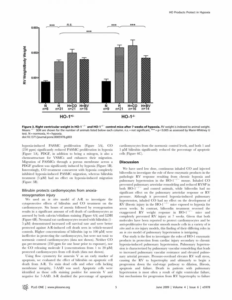
hearts and evaluated them grossly and histologically. Hypoxic
exposure caused HO-12/2 mice to have a more profound
elevation in RV weight (normalized to animal weight) as
compared with HO-1+/2 control animals (Figure 3). Daily
Figure 1. Right ventricular systolic pressure (RVSP) after 7weeks of hypoxia in HO-12/2 and HO-1+/2 control mice. Meanvalues +/2 SEM are indicated with each dot representing RVSPmeasurement for one animal. n.s. = not significant, * = p,0.05,*** = p,0.005 as assessed by Mann-Whitney U test.doi:10.1371/journal.pone.0005978.g001
HO Products Protect in Hypoxia
PLoS ONE | www.plosone.org 3 June 2009 | Volume 4 | Issue 6 | e5978

injections of biliverdin (50 mmol/kg) prevented the exaggerated
RV weight gain in the HO-12/2 mice, while having no effect on
the RV weight of the HO-1+/2 control animals. Treatment with
continuous inhaled CO throughout the hypoxic exposure did not
prevent the elevation in RV weight in the HO-12/2 animals and
had no significant effect on the RV weight of the HO-1+/2 control
animals (Figure 3).
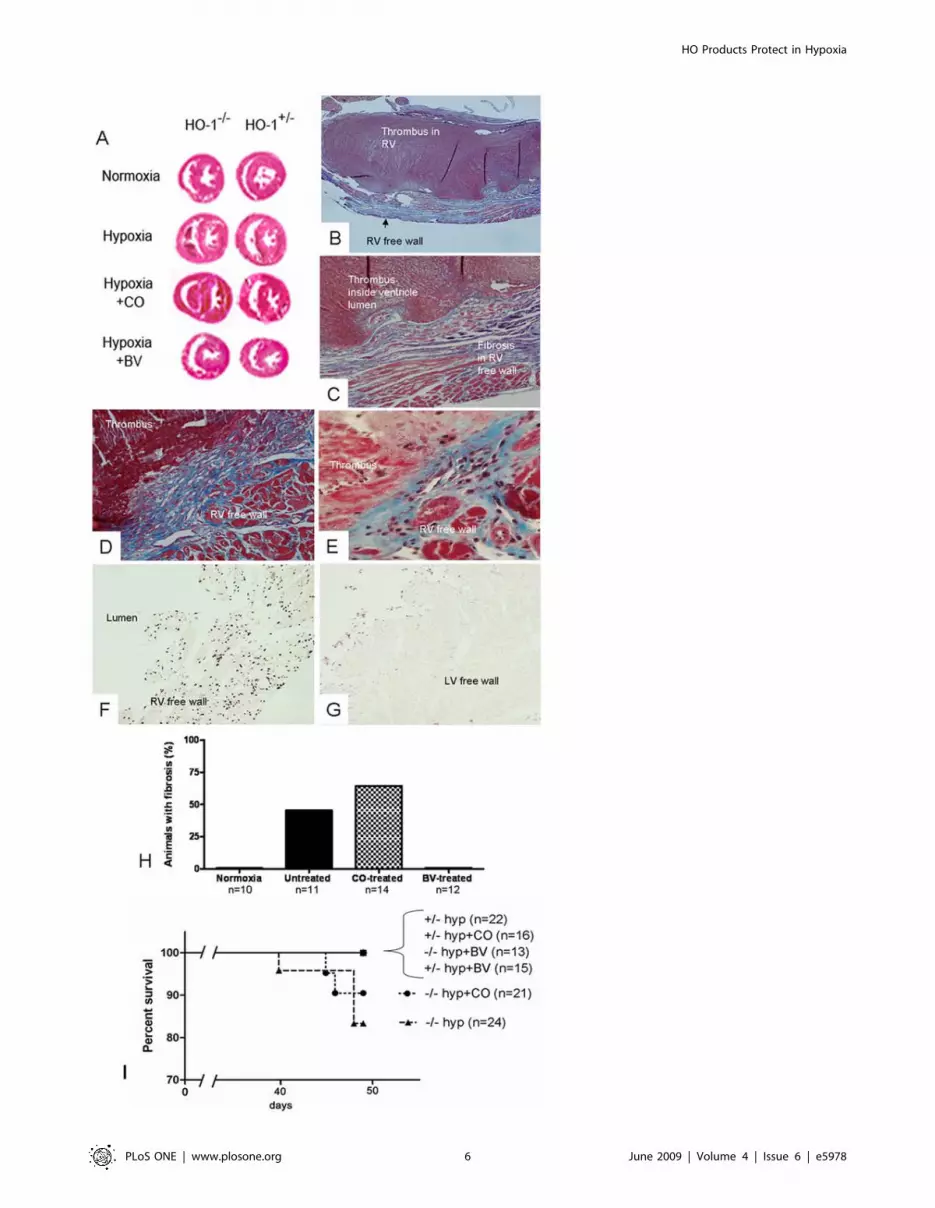
As we have previously reported [13], exposure of HO-12/2
mice to chronic hypoxia for seven weeks caused areas of full-
thickness RV wall fibrosis, with the majority of the animals having
an overlying mural thrombus (Figure 4A, B, and C). RV injury
was evident in many HO-12/2 mice on gross examination, with
white thrombus protruding from the RV once atria were removed
and often visible through a translucent, dilated RV. 45.4% of
untreated HO-12/2 animals developed an area of full-thickness
right ventricular wall fibrosis as detected with Masson’s Trichrome
stain after seven weeks (Figure 4H). Biliverdin injections entirely
prevented this response; no HO-12/2 animals treated with
biliverdin injections had full-thickness RV wall fibrosis after seven
weeks (Figure 4A and H). Unlike biliverdin, continuous CO
administration did not prevent RV wall fibrosis in the HO-12/2
animals (Figure 4D, E), with 64.3% of HO-12/2 animals treated
with CO developing injury (Figure 4H). These areas of fibrotic
injury in the untreated and CO-treated animals had many
TUNEL-positive apoptotic cells throughout (Figure 4F), while left
ventricular cardiomyocytes on the same slide are negative for
TUNEL staining (Figure 4G). No HO-1+/2 control animals
developed RV wall fibrosis or TUNEL-positive areas. In follow up
experiments we have found that HO-12/2 mice treated with
intermittent inhaled CO (250 ppm CO for one hour a day)
developed RV wall fibrosis and thrombus at a rate similar to HO-
12/2 animals treated with continuous CO and hypoxic HO-12/2
controls (data not shown).
In addition to its impact on RV weight, development of RV
injury affected death prior to 7 weeks. Approximately 12–18% of
HO-12/2 animals died between the 6th and 7th weeks of hypoxia
in the untreated or CO-treated group (Figure 4I). Interestingly,
there were no deaths prior to seven weeks in the biliverdin-treated
HO-12/2 groups, or in the HO-1+/2 controls. Necropsy on the
untreated and CO-treated HO-12/2 animals dying in the 6th
week showed white thrombus protruding from the RV when the
atria were removed.
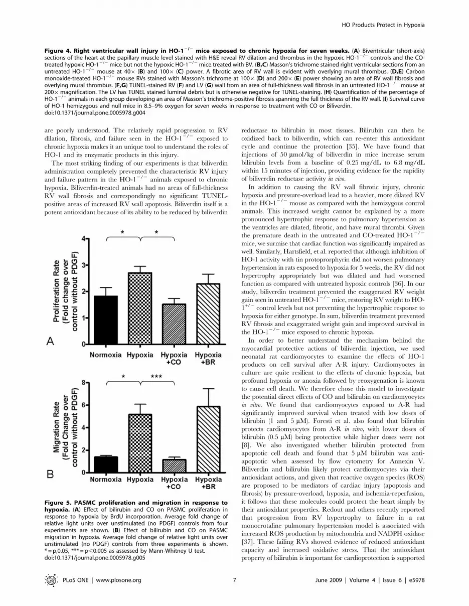
CO but not bilirubin inhibits hypoxic PASMC proliferationand migration
VSMC exposed to hypoxia have increased proliferation in
response to a mitogenic stimulus over normoxic controls, and as
we and others have previously reported, treatment of these cells
with CO inhibits their proliferation rate [17,18,20]. To begin to
investigate the differential responses of the pulmonary vasculature
to CO and biliverdin, PASMCs were cultured in vitro and
evaluated for proliferation and migration under hypoxia. Cultured
rat PASMCs were treated with either bilirubin (5 mM) or CO
(250 ppm) and cell proliferation was assessed in response to PDGF
stimulation. The specific doses of bilirubin and CO were selected
based on our findings in the cardiomyocyte anoxia-reoxygenation
experiments (see below) and our previously published work [17].
We used bilirubin instead of biliverdin in case biliverdin reductase
was absent or inactive in the cultured cells. Bilirubin treatment did
not affect PASMC growth under normoxia and modestly reduced
Figure 2. Pulmonary arteriolar remodeling in HO-12/2 animals exposed to chronic hypoxia for seven weeks and treated with CO orBiliverdin, as indicated. (A) Quantification of percent wall thickness. Bars represent means +/2 SEM. Each dot represents one animal and tenvessels were averaged for each animal. * = p,0.05 as assessed by Mann-Whitney U test. (B) Representative pulmonary arterioles of HO-12/2 micestained with H&E for each of the different conditions and treatments above.doi:10.1371/journal.pone.0005978.g002
HO Products Protect in Hypoxia
PLoS ONE | www.plosone.org 4 June 2009 | Volume 4 | Issue 6 | e5978
hypoxia-induced PASMC proliferation (Figure 5A). CO
(250 ppm) significantly reduced PASMC proliferation in hypoxia
(Figure 5A). PDGF, in addition to being a mitogen, is also a
chemoattractant for VSMCs and enhances their migration.
Migration of PASMCs through a porous membrane across a
PDGF gradient was significantly induced by hypoxia (Figure 5B).
Interestingly, CO treatment concurrent with hypoxia completely
inhibited hypoxia-induced PASMC migration, whereas bilirubin
treatment (5 mM) had no effect on hypoxia-induced migration
(Figure 5B).
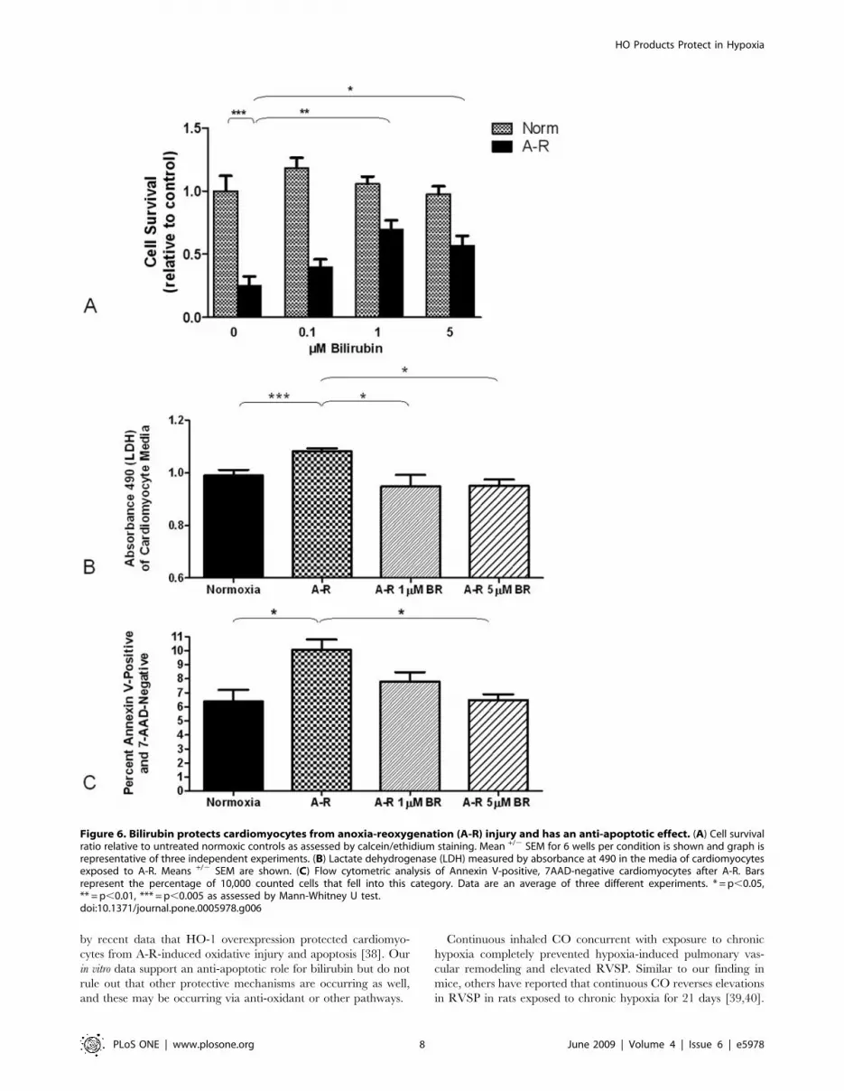
Bilirubin protects cardiomyocytes from anoxia-reoxygenation injury
We used an in vitro model of A-R to investigate the
cytoprotective effects of bilirubin and CO treatment on the
cardiomyocyte. Six hours of anoxia followed by reoxygenation
results in a significant amount of cell death of cardiomyocytes as
assessed by both calcein/ethidium staining (Figure 6A) and LDH
(Figure 6B). Neonatal rat cardiomyocytes treated with bilirubin (1–
5 mM) demonstrated increased cell survival rate and were better
protected against A-R-induced cell death seen in vehicle-treated
controls. Higher concentrations of bilirubin (up to 100 mM) were
ineffective in protecting the cardiomyocytes, but were not toxic to
normoxic control cardiomyocytes (data not shown). Neither CO
gas pre-treatment (250 ppm for one hour prior to exposure), nor
the CO releasing molecule I (concentrations from 1 to 20 mM)
protected cardiomyocytes from cell death (data not shown).
Using flow cytometry for annexin V as an early marker of
apoptosis, we evaluated the effect of bilirubin on apoptotic cell
death from A-R. To stain dead cells and other cells lacking
membrane integrity, 7-AAD was used. Apoptotic cells were
identified as those cells staining positive for annexin V and
negative for 7-AAD. A-R doubled the percentage of apoptotic
cardiomyocytes from the normoxic control levels, and both 1 and
5 mM bilirubin significantly reduced the percentage of apoptotic
cells (Figure 6C).
Discussion
We have used low dose, continuous inhaled CO and injected
biliverdin to investigate the role of these enzymatic products in the
pathologic RV response resulting from chronic hypoxia and
pulmonary hypertension in the HO-12/2 mouse. Inhaled CO
prevented pulmonary arteriolar remodeling and reduced RVSP in
both HO-12/2 and control animals, while biliverdin had no
significant effect on the pulmonary arteriolar response or RV
pressure. Although it prevented hypoxia-induced pulmonary
hypertension, inhaled CO had no effect on the development of
RV fibrotic injury in the HO-12/2 mice exposed to hypoxia for
seven weeks. In contrast, biliverdin treatment reversed the
exaggerated RV weight response in HO-12/2 mice and
completely prevented RV injury at 7 weeks. Given that both
molecules have been reported to protect cardiomyocytes and be
anti-proliferative for vascular smooth muscle cells in a variety of in
vitro and in vivo injury models, this finding of their differing roles in
an in vivo model of pulmonary hypertension is intriguing.
Our study is the first to investigate the roles of HO-1 enzymatic
products in protection from cardiac injury secondary to chronic
hypoxia-induced pulmonary hypertension. Pulmonary hyperten-
sion is characterized by pulmonary vascular remodeling that leads
to increased pulmonary vascular resistance and elevated pulmo-
nary arterial pressure. Pressure-overload elevates RV wall stress,
causing the RV to hypertrophy and ultimately to begin a
progression down the end-stage pathway to dilation, fibrosis,
apoptosis and failure. Death in patients with pulmonary
hypertension is most often a result of right ventricular failure,
but mechanisms for progression from RV hypertrophy to failure
Figure 3. Right ventricular weight in HO-12/2 and HO-1+/2 control mice after 7 weeks of hypoxia. RV weight is indexed to animal weight.Means +/2 SEM are shown for the number of animals listed below each column. n.s. = not significant, *** = p,0.005 as assessed by Mann-Whitney Utest. N = normoxia, H = hypoxia.doi:10.1371/journal.pone.0005978.g003
HO Products Protect in Hypoxia
PLoS ONE | www.plosone.org 5 June 2009 | Volume 4 | Issue 6 | e5978
HO Products Protect in Hypoxia
PLoS ONE | www.plosone.org 6 June 2009 | Volume 4 | Issue 6 | e5978
are poorly understood. The relatively rapid progression to RV
dilation, fibrosis, and failure seen in the HO-12/2 exposed to
chronic hypoxia makes it an unique tool to understand the roles of
HO-1 and its enzymatic products in this injury.
The most striking finding of our experiments is that biliverdin
administration completely prevented the characteristic RV injury
and failure pattern in the HO-12/2 animals exposed to chronic
hypoxia. Biliverdin-treated animals had no areas of full-thickness
RV wall fibrosis and correspondingly no significant TUNEL-
positive areas of increased RV wall apoptosis. Biliverdin itself is a
potent antioxidant because of its ability to be reduced by biliverdin
reductase to bilirubin in most tissues. Bilirubin can then be
oxidized back to biliverdin, which can re-enter this antioxidant
cycle and continue the protection [35]. We have found that
injections of 50 mmol/kg of biliverdin in mice increase serum
bilirubin levels from a baseline of 0.25 mg/dL to 6.8 mg/dL
within 15 minutes of injection, providing evidence for the rapidity
of biliverdin reductase activity in vivo.
In addition to causing the RV wall fibrotic injury, chronic
hypoxia and pressure-overload lead to a heavier, more dilated RV
in the HO-12/2 mouse as compared with the hemizygous control
animals. This increased weight cannot be explained by a more
pronounced hypertrophic response to pulmonary hypertension as
the ventricles are dilated, fibrotic, and have mural thrombi. Given
the premature death in the untreated and CO-treated HO-12/2
mice, we surmise that cardiac function was significantly impaired as
well. Similarly, Hartsfield, et al. reported that although inhibition of
HO-1 activity with tin protoprorphyrin did not worsen pulmonary
hypertension in rats exposed to hypoxia for 5 weeks, the RV did not
hypertrophy appropriately but was dilated and had worsened
function as compared with untreated hypoxic controls [36]. In our
study, biliverdin treatment prevented the exaggerated RV weight
gain seen in untreated HO-12/2 mice, restoring RV weight to HO-
1+/2 control levels but not preventing the hypertrophic response to
hypoxia for either genotype. In sum, biliverdin treatment prevented
RV fibrosis and exaggerated weight gain and improved survival in
the HO-12/2 mice exposed to chronic hypoxia.
In order to better understand the mechanism behind the
myocardial protective actions of biliverdin injection, we used
neonatal rat cardiomyocytes to examine the effects of HO-1
products on cell survival after A-R injury. Cardiomyoctes in
culture are quite resilient to the effects of chronic hypoxia, but
profound hypoxia or anoxia followed by reoxygenation is known
to cause cell death. We therefore chose this model to investigate
the potential direct effects of CO and bilirubin on cardiomyocytes
in vitro. We found that cardiomyocytes exposed to A-R had
significantly improved survival when treated with low doses of
bilirubin (1 and 5 mM). Foresti et al. also found that bilirubin
protects cardiomyocytes from A-R in vitro, with lower doses of
bilirubin (0.5 mM) being protective while higher doses were not
[8]. We also investigated whether bilirubin protected from
apoptotic cell death and found that 5 mM bilirubin was anti-
apoptotic when assessed by flow cytometry for Annexin V.
Biliverdin and bilirubin likely protect cardiomyocytes via their
antioxidant actions, and given that reactive oxygen species (ROS)
are proposed to be mediators of cardiac injury (apoptosis and
fibrosis) by pressure-overload, hypoxia, and ischemia-reperfusion,
it follows that these molecules could protect the heart simply by
their antioxidant properties. Redout and others recently reported
that progression from RV hypertrophy to failure in a rat
monocrotaline pulmonary hypertension model is associated with
increased ROS production by mitochondria and NADPH oxidase
[37]. These failing RVs showed evidence of reduced antioxidant
capacity and increased oxidative stress. That the antioxidant
property of bilirubin is important for cardioprotection is supported
Figure 4. Right ventricular wall injury in HO-12/2 mice exposed to chronic hypoxia for seven weeks. (A) Biventricular (short-axis)sections of the heart at the papillary muscle level stained with H&E reveal RV dilation and thrombus in the hypoxic HO-12/2 controls and the CO-treated hypoxic HO-12/2 mice but not the hypoxic HO-12/2 mice treated with BV. (B,C) Masson’s trichrome stained right ventricular sections from anuntreated HO-12/2 mouse at 406 (B) and 1006 (C) power. A fibrotic area of RV wall is evident with overlying mural thrombus. (D,E) Carbonmonoxide-treated HO-12/2 mouse RVs stained with Masson’s trichrome at 1006 (D) and 2006 (E) power showing an area of RV wall fibrosis andoverlying mural thrombus. (F,G) TUNEL-stained RV (F) and LV (G) wall from an area of full-thickness wall fibrosis in an untreated HO-12/2 mouse at2006magnification. The LV has TUNEL stained luminal debris but is otherwise negative for TUNEL-staining. (H) Quantification of the percentage ofHO-12/2 animals in each group developing an area of Masson’s trichrome-positive fibrosis spanning the full thickness of the RV wall. (I) Survival curveof HO-1 hemizygous and null mice in 8.5–9% oxygen for seven weeks in response to treatment with CO or Biliverdin.doi:10.1371/journal.pone.0005978.g004
Figure 5. PASMC proliferation and migration in response tohypoxia. (A) Effect of bilirubin and CO on PASMC proliferation inresponse to hypoxia by BrdU incorporation. Average fold change ofrelative light units over unstimulated (no PDGF) controls from fourexperiments are shown. (B) Effect of bilirubin and CO on PASMCmigration in hypoxia. Average fold change of relative light units overunstimulated (no PDGF) controls from three experiments is shown.* = p,0.05, *** = p,0.005 as assessed by Mann-Whitney U test.doi:10.1371/journal.pone.0005978.g005
HO Products Protect in Hypoxia
PLoS ONE | www.plosone.org 7 June 2009 | Volume 4 | Issue 6 | e5978
by recent data that HO-1 overexpression protected cardiomyo-
cytes from A-R-induced oxidative injury and apoptosis [38]. Our
in vitro data support an anti-apoptotic role for bilirubin but do not
rule out that other protective mechanisms are occurring as well,
and these may be occurring via anti-oxidant or other pathways.
Continuous inhaled CO concurrent with exposure to chronic
hypoxia completely prevented hypoxia-induced pulmonary vas-
cular remodeling and elevated RVSP. Similar to our finding in
mice, others have reported that continuous CO reverses elevations
in RVSP in rats exposed to chronic hypoxia for 21 days [39,40].
Figure 6. Bilirubin protects cardiomyocytes from anoxia-reoxygenation (A-R) injury and has an anti-apoptotic effect. (A) Cell survivalratio relative to untreated normoxic controls as assessed by calcein/ethidium staining. Mean +/2 SEM for 6 wells per condition is shown and graph isrepresentative of three independent experiments. (B) Lactate dehydrogenase (LDH) measured by absorbance at 490 in the media of cardiomyocytesexposed to A-R. Means +/2 SEM are shown. (C) Flow cytometric analysis of Annexin V-positive, 7AAD-negative cardiomyocytes after A-R. Barsrepresent the percentage of 10,000 counted cells that fell into this category. Data are an average of three different experiments. * = p,0.05,** = p,0.01, *** = p,0.005 as assessed by Mann-Whitney U test.doi:10.1371/journal.pone.0005978.g006
HO Products Protect in Hypoxia
PLoS ONE | www.plosone.org 8 June 2009 | Volume 4 | Issue 6 | e5978

We demonstrate here that continuous CO inhalation prevents
pulmonary vascular remodeling and elevation of RVSP in mice by
chronic hypoxia, and likely does so, at least in part, by prevention
of hypoxia-induced pulmonary arterial smooth muscle cell
proliferation and migration. Bilirubin, in contrast, only modestly
reduced hypoxia-induced PASMC proliferation and had no effect
on migration in vitro. While we have reported the inhibitory effect
of CO on VSMC proliferation in the past, to our knowledge this is
the first report of the profound anti-migratory action of CO in
hypoxic vascular smooth muscle cells. We have previously
reported that the antiproliferative effect of CO in hypoxic VSMC
is through cGMP-mediated down-regulation of the cell cycle
transcription factor E2F-1 [17], while others have implicated
cGMP-dependent phosphorylation of p38 MAP kinase, dephos-
phorylation of ERK MAP kinase, and regulation of p21 to control
cell proliferation [1,41]. Although the mechanism for hypoxia-
induced PASMC migration has not been studied, PASMC
migration toward PDGF has been reported to be dependent on
ERK MAP kinase phosphorylation [42] and therefore it is
plausible that CO inhibits migration via dephosphorylation of
ERK 1/2.
A key finding of our study is that despite the protection from
pulmonary arteriolar remodeling and elevated RVSP seen in HO-
12/2 and HO-1+/2 control mice treated with inhaled CO,
increased RV weight was not prevented in either genotype and
RV fibrosis and apoptosis was not prevented in the HO-12/2
mouse. Other authors who have used CO to treat hypoxia-
induced pulmonary hypertension in rats have had conflicting
results pertaining to RV hypertrophy. Otterbein, et.al. treated rats
with 250 ppm CO once daily during the last three weeks of a six
week hypoxic exposure and found improvement in Fulton’s Index
[24]. Gautier, et.al. treated rats with 50 ppm CO continuously
during the last week of a three week hypoxic exposure and found
that although RVSP was significantly improved, Fulton’s Index
was not affected and RV function was significantly worsened in the
CO-treated group. In addition, RV infarction developed in some
of these animals [39]. For the HO-12/2 mouse that cannot
respond to hypoxia by increasing CO production, it appears that
neither reducing pulmonary vascular resistance (which occurs as a
consequence of CO therapy) nor replacement of CO at the
cardiomyocyte level is sufficient to prevent RV injury. Interest-
ingly, for the HO-1+/2 control animals treated with CO, elevation
of RVSP was prevented but the RV hypertrophied to the same
extent as untreated hypoxic HO-1+/2 controls. One possible
explanation for this effect is CO toxicity, but we monitored COHb
levels weekly throughout the hypoxic exposure and found levels
less than 9% regardless of CO dose within the range of 20–
60 ppm. Others have reported similar COHb levels in rats
exposed to chronic hypoxia and continuous inhaled CO at
50 ppm for 3 weeks [39]. These levels are lower than those of
chronic smokers and are not thought to be toxic. Another possible
explanation is that in hypoxia-induced pulmonary hypertension,
CO may be preventing the pulmonary vascular smooth muscle cell
proliferative and pro-migratory effects of hypoxia but not
protecting from the direct effects of hypoxia on the right ventricle.
In support of this, we found that in vitro, neither pre-treatment with
CO gas nor CORM-1 treatment improved cardiomyocyte cell
survival in a model of anoxia-reoxygenation. A previous study by
Clark, et.al. showed that CO was protective when delivered using
CORM-3 in H9C2 cells exposed to A-R [43]. The use of a
different CO-releasing molecule in a transformed cell line (H9C2)
in the Clark study may account for the observed difference
between their results and ours.
The fact that CO reduced RVSP and pulmonary arteriolar
remodeling but did not prevent RV fibrotic injury in the HO-12/2
mice points to a possible direct effect of hypoxia on the RV. When
chronic hypoxia is the cause of pulmonary hypertension, the RV is
subjected not only to elevated pulmonary vascular resistance from
vascular remodeling, but also to systemic hypoxia. Both pressure-
overload [12,44] and chronic hypoxia [13] are known to
independently increase HO-1 expression in the heart, pointing to
a possible independent role of these two stressful stimuli. As patients
with PAH secondary to hypoxia (e.g. secondary to chronic
obstructive pulmonary disease) make up a large percentage of
patients with PAH, it is important that we investigate this
independent effect of systemic hypoxia on myocardial performance
and progression to failure.
In summary, biliverdin exerted a direct cardioprotective effect
in HO-12/2 mice exposed to chronic hypoxia, but did not
ameliorate pulmonary vascular pathology. In the same model,
inhalation of low dose continuous CO ameliorated hypoxia-
induced pulmonary hypertension in both HO-1+/2 and HO-12/2
animals, but did not protect from RV dilation, fibrosis, and mural
thrombus. Given that HO-12/2 animals appear to have an
accelerated progression to apoptosis, fibrosis, and heart failure, we
propose that biliverdin administration slows this progression to
resemble the wild-type phenotype while CO either has no effect or
accelerates the progression. At the same time, CO dramatically
inhibits hypoxia-induced proliferation and migration of vascular
smooth muscle cells and prevents hypoxic pulmonary vascular
remodeling and RV pressure overload, but has no protective
effects on the RV. The distinctly different roles of these two
protective enzymatic products of HO-1 in our model of hypoxia-
induced pulmonary hypertension are intriguing and may
demonstrate how HO-1 can have a variety of protective effects
that are dependent on the type of stress and the type of cell
involved in injury. As therapeutic strategies that involve
upregulation of HO-1 or delivery of its enzymatic products are
developed, there will need to be thorough investigation of the
possible effects of these strategies in each organ system. In general,
our results support the emerging concept that treatments for
pulmonary hypertension targeted solely at reducing pulmonary
arterial pressure may not necessarily be cardioprotective and may
not ultimately improve outcome for these patients. Potential
treatments must be evaluated for their protective actions in both
organ systems.
Acknowledgments
The authors wish to thank Dimitrios Poutias for his assistance with
cardiomyocyte isolation techniques, Hilary Mehler and Xianlan Liu for
technical assistance, and Stephanie Giannetto and Sarah Gately for their
expert assistance in preparation of the manuscript.
Author Contributions
Performed the experiments: SV. Wrote the paper: SV. PI of laboratory,
provided overall scientific direction and funded the research: SK. Edited
the manuscript: SK SAM HC. Performed all animal experiments,
cardiomyocyte work, proliferation studies: SV. Provided intellectual input,
maintained HO-1 null line, engineered hypoxia chamber setup and
software control, and taught several animal techniques to Dr. Vitali: SAM.
Performed right ventricular pressure measurements for several experi-
ments: ODL XL. Taught Dr. Vitali histological techniques and lung
inflation: AFG. Analyzed pulmonary arteriolar remodeling: HC. Per-
formed smooth muscle cell migration experiments: XW. Cardiac
anesthesiologist who directed cardiomyocyte experiments: FXM.
HO Products Protect in Hypoxia
PLoS ONE | www.plosone.org 9 June 2009 | Volume 4 | Issue 6 | e5978

References
1. Duckers HJ, Boehm M, True AL, Yet SF, San H, et al. (2001) Heme oxygenase-
1 protects against vascular constriction and proliferation. Nat Med 7: 693–698.2. Zhang M, Zhang BH, Chen L, An W (2002) Overexpression of heme
oxygenase-1 protects smooth muscle cells against oxidative injury and inhibitscell proliferation. Cell Res 12: 123–132.
3. Foo RS, Siow RC, Brown MJ, Bennett MR (2006) Heme oxygenase-1 gene
transfer inhibits angiotensin II-mediated rat cardiac myocyte apoptosis but nothypertrophy. J Cell Physiol 209: 1–7.
4. Hu CM, Chen YH, Chiang MT, Chau LY (2004) Heme oxygenase-1 inhibitsangiotensin II-induced cardiac hypertrophy in vitro and in vivo. Circulation 110:
309–316.
5. Tongers J, Fiedler B, Konig D, Kempf T, Klein G, et al. (2004) Hemeoxygenase-1 inhibition of MAP kinases, calcineurin/NFAT signaling, and
hypertrophy in cardiac myocytes. Cardiovasc Res 63: 545–552.6. Yet SF, Tian R, Layne MD, Wang ZY, Maemura K, et al. (2001) Cardiac-
specific expression of heme oxygenase-1 protects against ischemia andreperfusion injury in transgenic mice. Circ Res 89: 168–173.
7. Liu X, Wei J, Peng DH, Layne MD, Yet SF (2005) Absence of heme oxygenase-
1 exacerbates myocardial ischemia/reperfusion injury in diabetic mice. Diabetes54: 778–784.
8. Foresti R, Goatly H, Green CJ, Motterlini R (2001) Role of heme oxygenase-1 inhypoxia-reoxygenation: requirement of substrate heme to promote cardiopro-
tection. Am J Physiol Heart Circ Physiol 281: H1976–1984.
9. Morita T, Perrella MA, Lee M-E, Kourembanas S (1995) Smooth muscle cell-derived carbon monoxide is a regulator of vascular cGMP. Proc Natl Acad Sci
USA 92: 1475–1479.10. Kacimi R, Chentoufi J, Honbo N, Long CS, Karliner JS (2000) Hypoxia
differentially regulates stress proteins in cultured cardiomyocytes: Role of thep38 stress-activated signaling cascade, and relation to cytoprotection. Cardiovasc
Res 46: 139–150.
11. Wagner CT, Durante W, Christodoulides N, Hellums JD, Schafer AI (1997)Hemodynamic forces induce the expression of heme oxygenase in cultured
vascular smooth muscle cells. J Clin Invest 100: 589–596.12. Katayose D, Isoyama S, Fujita H, Shibahara S (1993) Separate regulation of
heme oxygenase and heat shock protein 70 mRNA expression in the rat heart by
hemodynamic stress. Biochem Biophys Res Commun 191: 587–594.13. Yet S-F, Perrella MA, Layne MD, Hsieh C-M, Maemura K, et al. (1999)
Hypoxia induces severe right ventricular dilatation and infarction in hemeoxygenase-1 null mice. J Clin Invest 103: R23–R29.
14. Minamino T, Christou H, Hsieh CM, Liu Y, Dhawan V, et al. (2001) Targetedexpression of heme oxygenase-1 prevents the pulmonary inflammatory and
vascular responses to hypoxia. Proc Natl Acad Sci U S A 98: 8798–8803.
15. Motterlini R, Gonzales A, Foresti R, Clark JE, Green CJ, et al. (1998) Hemeoxygenase-1-derived carbon monoxide contributes to the suppression of acute
hypertensive responses in vivo. Circ Res 83: 568–577.16. Brune B, Ullrich V (1987) Inhibition of platelet aggregation by carbon monoxide
is mediated by activation of guanylate cyclase. Mol Pharmacol 32: 497–504.
17. Morita T, Mitsialis SA, Koike H, Liu Y, Kourembanas S (1997) Carbonmonoxide controls the proliferation of hypoxic vascular smooth muscle cells.
J Biol Chem 272: 32804–32809.18. Stanford SJ, Walters MJ, Hislop AA, Haworth SG, Evans TW, et al. (2003)
Heme oxygenase is expressed in human pulmonary artery smooth muscle wherecarbon monoxide has an anti-proliferative role. Eur J Pharmacol 473: 135–141.
19. Peyton KJ, Reyna SV, Chapman GB, Ensenat D, Liu XM, et al. (2002) Heme
oxygenase-1-derived carbon monoxide is an autocrine inhibitor of vascularsmooth muscle cell growth. Blood 99: 4443–4448.
20. Zhen G, Xue Z, Zhang Z, Xu Y (2003) Carbon monoxide inhibits proliferationof pulmonary smooth muscle cells under hypoxia. Chin Med J (Engl) 116:
1804–1809.
21. Otterbein LE, Mantell LL, Choi AMK (1999) Carbon monoxide providesprotection against hyperoxic lung injury. Am J Physiol 276: L688–L694.
22. Dolinay T, Szilasi M, Liu M, Choi AM (2004) Inhaled carbon monoxide confersantiinflammatory effects against ventilator-induced lung injury. Am J Respir Crit
Care Med 170: 613–620.
23. Sato K, Balla J, Otterbein L, Smith RN, Brouard S, et al. (2001) Carbonmonoxide generated by heme oxygenase-1 suppresses the rejection of mouse-to-
rat cardiac transplants. J Immunol 166: 4185–4194.
24. Zuckerbraun BS, Chin BY, Wegiel B, Billiar TR, Czsimadia E, et al. (2006)
Carbon monoxide reverses established pulmonary hypertension. J Exp Med 203:
2109–2119.
25. Guo Y, Stein AB, Wu WJ, Tan W, Zhu X, et al. (2004) Administration of a CO-
releasing molecule at the time of reperfusion reduces infarct size in vivo.
Am J Physiol Heart Circ Physiol 286: H1649–1653.
26. Stein AB, Guo Y, Tan W, Wu WJ, Zhu X, et al. (2005) Administration of a CO-
releasing molecule induces late preconditioning against myocardial infarction.
J Mol Cell Cardiol 38: 127–134.
27. Stocker R, Yamamoto Y, McDonagh AF, Glazer AN, Ames BN (1987) Bilirubin
is an antioxidant of possible physiological importance. Science 235: 1043–1046.
28. Wang HD, Yamaya M, Okinaga S, Jia YX, Kamanaka M, et al. (2002) Bilirubin
ameliorates bleomycin-induced pulmonary fibrosis in rats. Am J Respir Crit
Care Med 165: 406–411.
29. Overhaus M, Moore BA, Barbato JE, Behrendt FF, Doering JG, et al. (2006)
Biliverdin protects against polymicrobial sepsis by modulating inflammatory
mediators. Am J Physiol Gastrointest Liver Physiol 290: G695–703.
30. Nakao A, Murase N, Ho C, Toyokawa H, Billiar TR, et al. (2005) Biliverdin
administration prevents the formation of intimal hyperplasia induced by vascular
injury. Circulation 112: 587–591.
31. Yamashita K, McDaid J, Ollinger R, Tsui TY, Berberat PO, et al. (2004)
Biliverdin, a natural product of heme catabolism, induces tolerance to cardiac
allografts. Faseb J 18: 765–767.
32. Ollinger R, Bilban M, Erat A, Froio A, McDaid J, et al. (2005) Bilirubin: a
natural inhibitor of vascular smooth muscle cell proliferation. Circulation 112:
1030–1039.
33. Sarady-Andrews JK, Liu F, Gallo D, Nakao A, Overhaus M, et al. (2005)
Biliverdin administration protects against endotoxin-induced acute lung injury in
rats. Am J Physiol Lung Cell Mol Physiol 289: L1131–1137.
34. Clark JE, Foresti R, Sarathchandra P, Kaur H, Green CJ, et al. (2000) Heme
oxygenase-1-derived bilirubin ameliorates postchemic myocardial dysfunction.
Am J Physiol Heart Circ Physiol 278: H643–H651.
35. Baranano DE, Rao M, Ferris CD, Snyder SH (2002) Biliverdin reductase: a
major physiologic cytoprotectant. Proc Natl Acad Sci U S A 99: 16093–16098.
36. Hartsfield CL, McMurtry IF, Ivy DD, Morris KG, Vidmar S, et al. (2004)
Cardioprotective and vasomotor effects of HO activity during acute and chronic
hypoxia. Am J Physiol Heart Circ Physiol 287: H2009–2015.
37. Redout EM, Wagner MJ, Zuidwijk MJ, Boer C, Musters RJ, et al. (2007) Right-
ventricular failure is associated with increased mitochondrial complex II activity
and production of reactive oxygen species. Cardiovasc Res 75: 770–781.
38. Pachori AS, Smith A, McDonald P, Zhang L, Dzau VJ, et al. (2007) Heme-
oxygenase-1-induced protection against hypoxia/reoxygenation is dependent on
biliverdin reductase and its interaction with PI3K/Akt pathway. J Mol Cell
Cardiol 43: 580–592.
39. Gautier M, Antier D, Bonnet P, Le Net JL, Hanton G, et al. (2007) Continuous
inhalation of carbon monoxide induces right ventricle ischemia and dysfunction
in rats with hypoxic pulmonary hypertension. Am J Physiol Heart Circ Physiol
293: H1046–1052.
40. Dubuis E, Potier M, Wang R, Vandier C (2005) Continuous inhalation of
carbon monoxide attenuates hypoxic pulmonary hypertension development
presumably through activation of BKCa channels. Cardiovasc Res 65: 751–761.
41. Kim HP, Wang X, Nakao A, Kim SI, Murase N, et al. (2005) Caveolin-1
expression by means of p38beta mitogen-activated protein kinase mediates the
antiproliferative effect of carbon monoxide. Proc Natl Acad Sci U S A 102:
11319–11324.
42. Yamboliev IA, Gerthoffer WT (2001) Modulatory role of ERK MAPK-
caldesmon pathway in PDGF-stimulated migration of cultured pulmonary artery
SMCs. Am J Physiol Cell Physiol 280: C1680–1688.
43. Clark JE, Naughton P, Shurey S, Green CJ, Johnson TR, et al. (2003)
Cardioprotective actions by a water-soluble carbon monoxide-releasing
molecule. Circ Res 93: e2–8.
44. Delcayre C, Samuel JL, Marotte F, Best-Belpomme M, Mercadier JJ, et al.
(1988) Synthesis of stress proteins in rat cardiac myocytes 2–4 days after
imposition of hemodynamic overload. J Clin Invest 82: 460–468.
HO Products Protect in Hypoxia
PLoS ONE | www.plosone.org 10 June 2009 | Volume 4 | Issue 6 | e5978