heme protein-induced chronic renal inflammation: suppressive effect of induced heme oxygenase-11
TRANSCRIPT

Kidney International, Vol. 59 (2001), pp. 106–117
CELL BIOLOGY – IMMUNOLOGY – PATHOLOGY
Heme protein-induced chronic renal inflammation:Suppressive effect of induced heme oxygenase-11
KARL A. NATH, GREGORY M. VERCELLOTTI, JOSEPH P. GRANDE, HIROKO MIYOSHI,CARLOS V. PAYA, J. CARLOS MANIVEL, JILL J. HAGGARD, ANTHONY J. CROATT,WILLIAM D. PAYNE, and JAWED ALAM
Nephrology Research Unit, Departments of Pathology and Immunology, Mayo Clinic/Foundation, Rochester; Departments ofMedicine, Pathology, and Surgery, University of Minnesota, Minneapolis, Minnesota; and the Department of Biochemistry,Ochsner Clinic, New Orleans, Louisiana, USA
in the kidney in humans and rats repetitively exposed to hemeHeme protein-induced chronic renal inflammation: Suppressiveproteins. Such up-regulation represents an anti-inflammatoryeffect of induced heme oxygenase-1.response since the genetic deficiency of HO-1 markedly in-Background. Heme oxygenase (HO) is the rate-limiting en-creases activation of NF-kB, MCP-1 expression, and tubuloin-zyme in the degradation of heme; its inducible isozyme, HO-1,
protects against acute heme protein-induced nephrotoxicity terstitial cellular inflammation.and other forms of acute tissue injury. This study examinesthe induction of HO-1 in the kidney chronically inflamed byheme proteins and the functional significance of such an induc-
Heme oxygenase (HO) is the rate-limiting enzyme intion of HO-1.Methods. Studies were undertaken in a patient with chronic the degradation of heme, facilitating the conversion of
tubulointerstitial disease in the setting of paroxysmal nocturnal heme to biliverdin, in the course of which iron is releasedhemoglobinuria (PNH), in a rat model of chronic tubulointer- and carbon monoxide is emitted [1–3]. Its isozyme, HO-1,stitial nephropathy caused by repetitive exposure to heme pro-
is induced by diverse stressors [1–3], and in some instances,teins, and in genetically engineered mice deficient in HO-1(HO-1 2/2) in which hemoglobin was repetitively adminis- such induction of HO-1 confers cytoprotection againsttered. acute toxicity [1–3]. For example, induction of HO-1
Results. The kidney in PNH evinces robust induction of HO-1 protects against acute cytotoxicity induced by heme pro-in renal tubules in the setting of chronic inflammation. The hemeteins [4–6], hypoxia [7], hyperoxia [8], sepsis [9], acuteprotein-enriched urine from this patient, but not urine from a
healthy control subject, induced expression of HO-1 in renal nephrotoxicity from chemotherapeutic agents [10], andtubular epithelial cells (LLC-PK1 cells). A similar induction ischemia-reperfusion injury in the transplanted organ [11].of HO-1 and related findings are recapitulated in a rat model of In addition to the recognition that HO-1 can guardchronic inflammation induced by repetitive exposure to heme
against acute cytotoxicity, there is growing evidence thatproteins. Additionally, in the rat, the administration of hemeHO-1 down-regulates the inflammatory response [2, 3,proteins induces monocyte chemoattractant protein (MCP-1).
The functional significance of HO-1 so induced was uncovered 12]. For example, acute cellular inflammation followingin the HO-1 knockout mouse: Repeated administration of he- pleurisy in a rat model is significantly ameliorated bymoglobin to HO-1 1/1 and HO-1 2/2 mice led to intense
the up-regulation of HO-1 [13], while in a model ofinterstitial cellular inflammation in HO-1 2/2 mice accompa-cardiac xenotransplant rejection, the deficiency of HO-1nied by striking up-regulation of MCP-1 and activation of one
of its stimulators, nuclear factor-kB (NF-kB). These findings exacerbates xenotransplantation-related cell injury andwere not observed in similarly treated HO-1 1/1 mice or in inflammatory responses [14]. Chronic vascular inflam-vehicle-treated HO-1 2/2 and HO-1 1/1 mice.
mation associated with vascular rejection in antibody-Conclusion. We conclude that up-regulation of HO-1 occursinduced transplant arteriosclerosis is also ameliorated byup-regulation of HO-1 [15]. The mechanisms by which
1 See Editorial by Kanwar, p. 378. HO-1 influences the inflammatory response is one ofintense interest [2, 3, 12]. Understanding the basis forKey words: tubulointerstitial inflammation, nuclear factor-kB, mono-
cyte chemoattractant protein-1, paroxysmal nocturnal hemoglobinuria. such anti-inflammatory effects would be widely applica-ble to a number of disease states, including assorted
Received for publication March 24, 2000nephritides, chronic heme protein-induced tissue injury,and in revised form July 6, 2000
Accepted for publication July 12, 2000 chronic inflammation in other organs, rejection of solidorgans, transplant arteriopathy, arteriosclerosis, local 2001 by the International Society of Nephrology
106
Nath et al: HO-1 and chronic renal inflammation 107
and systemic microbial infections, and the systemic in- logic alterations and deposition of iron, respectively. Ad-ditional sections of the kidney biopsy were studied byflammatory response syndrome.
Our laboratory has demonstrated the functional sig- immunoperoxidase, as previously described [28, 29], forthe expression of HO-1 and ferritin (vide infra).nificance of induced HO-1 in protecting the kidney
against the acute cytotoxic effects of heme proteins, as The capacity of hemoglobin-enriched urine from thispatient to induce HO-1 in renal proximal tubular epithe-occurs in settings in which the kidney is exposed to myo-
globin and hemoglobin [4, 6]. Such studies focus on acute lial cells was examined in LLC-PK1 cells. LLC-PK1 cellswere grown to confluence, as previously described [30].heme protein-induced nephrotoxicity that occurs in dis-
eases such as rhabdomyolysis and intravascular hemoly- Samples of urine from the patient and a healthy controlsubject were diluted 1/5 with Hank’s balanced salt solu-sis [16]. Heme proteins may also contribute to a chronic
inflammatory response in the kidney. For example, tion (HBSS), titrated to pH 7.4, and then filtered througha 0.2 micron filter. Confluent monolayers were then ex-chronic inflammation appears in the kidney continually
exposed to heme proteins as occurs in hematuric nephrit- posed to HBSS alone, urine from the healthy subjectdiluted with HBSS (1/5), or urine from the patient dilutedides [17], sickle cell disease [18, 19], and recurrent hemo-
lytic episodes [16]. Whether HO-1 is up-regulated in such with HBSS (1/5). Following incubation for four hours,the respective media were removed, the cell monolayerschronic, heme protein-induced, renal inflammation and
influences the nature and severity of the inflammatory were washed, and all cell monolayers were incubated foran additional two hours in Dulbecco’s modified Eagle’sresponse is an issue that, to date, has not been addressed.
Using a multilayered approach that incorporates find- medium (DMEM). In additional studies, cells were incu-bated in the presence of urine from the patient dilutedings in human disease and in relevant models in rats and
mice, this study examined the induction of HO-1 in the with HBSS 1/5, 1/10, and 1/25; cells were also incubatedin the presence or absence of pyruvate (5 mmol/L), akidney chronically inflamed by heme proteins. We then
explored the functional significance of such an induction scavenger for hydrogen peroxide [31]. In these and sub-sequent studies, RNA was extracted using the Trizolof HO-1. This line of investigation was initially stimu-
lated by a patient who developed progressive renal insuf- method (GIBCO BRL, Gaithersburg, MD, USA), as pre-viously described [30]. Ten micrograms of total RNA fromficiency in the setting of paroxysmal nocturnal hemoglo-
binuria (PNH); the relevant findings in this patient thus each sample were separated on an agarose gel and trans-ferred to a nylon membrane. Membranes were hybrid-provide the provenance for subsequent explorations in
relevant disease models. PNH is characterized by recur- ized overnight with a 32P-labeled human HO-1 cDNAprobe. Autoradiograms were standardized, as previouslyrent episodes of hemolysis, which intermittently exposes
the kidney to heme proteins [20–22]. We demonstrate described [30], by factoring the optical density of themessage for HO-1 with the optical density of the 18Sthat in this condition, the kidney evinces robust induction
of HO-1 in the presence of smoldering renal inflamma- rRNA, the latter obtained on a negative of the ethidiumbromide-stained nylon membrane.tion. Similar induction of HO-1 and related findings in
the kidney in PNH are recapitulated in a rat model ofStudies in ratschronic inflammation induced by repetitive exposure to
heme proteins. This latter model in the rat suggested a The glycerol model was employed in the study of hemeprotein-induced renal injury in the rat. Hypertonic glyc-mechanism based on the expression of the chemokine
monocyte chemoattractant protein-1 (MCP-1)—a mono- erol (50%, 7.5 mL/kg body wt) was injected intramuscu-larly in rats dehydrated overnight and was repeated atcyte chemoattractant protein that contributes to chronic
renal inflammation in diverse states [23–27]—by which weekly intervals, as previously described [32]. After thefourth injection, the kidneys were fixed in formalin, andHO-1 may modulate the inflammatory response. To ex-
plore this mechanism further, as well as the significance immunoperoxidase studies for the detection of HO-1 andferritin were performed [28, 29]. Staining for HO-1 wasof induced HO-1, we examined renal alterations in genet-
ically engineered mice deficient in HO-1 that were repet- carried out using a polyclonal rat antibody (SPA-895;Stressgen, Victoria, British Columbia, Canada) as theitively exposed to heme proteins.primary antibody, a horseradish peroxidase-conjugatedsecondary antibody (SAB-300; Stressgen) and diamino-
METHODSbenzidine as substrate for localization; kidney sections
Clinical case report were also stained for ferritin using an anti-rat ferritinantibody, as previously described [4, 29]. Kidney sectionsA renal biopsy was obtained from an adult patient
with a history of PNH who developed chronic renal insuf- were stained for the assessment of the deposition of ironusing Prussian blue.ficiency. The kidney biopsy specimen was immersion fixed
in formalin, and sections were stained with hematoxylin In additional studies, the effect of rat hemoglobin ongene expression in the kidney was examined. Rats wereand eosin and with Prussian blue for assessment of histo-

Nath et al: HO-1 and chronic renal inflammation108
dehydrated overnight and then given rat hemoglobin (60 tion of 100 mL of Nonidet-P40, the sample was centri-fuged at 13,000 3 g for five minutes at 48C; the resultantmg/100 g body wt) or saline vehicle intravenously. The
kidneys were harvested, and RNA from kidneys was pellet was resuspended in 60 mL of a buffer consistingof 50 mmol/L HEPES, 10% glycerol, 300 mmol/L NaCl,extracted using the Trizol method; total RNA from each
sample was separated on an agarose gel and transferred 50 mmol/L KCl, and protease inhibitors. Following cen-trifugation at 13,000 3 g for 10 minutes, the supernatantto a nylon membrane. Membranes were hybridized over-
night with a 32P-labeled mouse HO-1 or MCP-1 cDNA was harvested and used in the EMSA. The oligonucleo-tide employed corresponded to the two NF-kB bindingprobe. Autoradiograms were standardized as described
previously in this article. sequences present within the enhancer of the HIV LTR(59-ACAAGGGACTTTCCGCTGGGGACTTTCCA
Studies in mice GGGA-39), labeled with [32P] gATP, as described byPaya et al [35, 36]. Ten micrograms of protein extractHemoglobin or vehicle was administered on a weekly
basis to HO-1 1/1 and HO-1 2/2 mice. Homozygous were incubated with 4 mL of binding buffer (5 mmol/LMgCl2, 50 mmol/L Tris-HCl, pH 7.5, 250 mmol/L NaCl,HO-1 null mutants were generated by targeted disrup-
tion of the HO-1 gene as described by Poss and Tone- 20% glycerol, 2.5 mmol/L EDTA, 2.5 mmol/L DTT, 0.25mg/mL poly dI.dC) and 2 mL of 32P-labeled NF-kB probe.gawa [33]. Colonies of mice were maintained by breeding
HO-1 2/2 males with HO-1 1/2 females. Offspring Termination was effected after 30 minutes with 2 mL ofgel-loading buffer (250 mmol/L Tris-HCl, pH 7.5, 0.2%were genotyped at the time of weaning by using polymer-
ase chain reaction to amplify the wild-type and mutant bromophenol blue, 40% glycerol). The binding reactionwas analyzed by electrophoresis in a nondenaturing 7%alleles of genomic DNA obtained from tail samples.
HO-1 1/1 mice (wild-type) were used as controls. polyacrylamide gel and visualized by autoradiography.Groups of HO-1 1/1 and HO-1 2/2 mice comprised
Statisticssimilar numbers of male and female mice. Prior to theinitiation of chronic intermittent administration of hemo- Results are expressed as means 6 SEM and are consid-
ered statistically significant for P , 0.05. For comparisonglobin, the body weights of the HO-1 2/2 mice (mean,22.7 6 1.6 g; range, 18.3 to 29.2 g) were matched to HO-1 between unpaired groups, the Student’s t-test or the
Mann–Whitney test was employed as appropriate.1/1 mice (mean, 22.9 6 1.4; range, 17.7 to 27.2 g); themean age of the HO-1 2/2 mice was 13 6 1 weeks(range, 11 to 16 weeks), while the mean age of the HO-1
RESULTS1/1 mice was 10 6 1 weeks (range, 8 to 14 weeks).
Clinical case description: Renal expression of HO-1At weekly intervals, mice were administered mousefollowing recurrent exposure to heme proteins inhemoglobin (90 mg/100 g body weight; Sigma, St. Louis,paroxysmal nocturnal hemoglobinuriaMO, USA) via a tail vein injection; prior to the adminis-
tration of hemoglobin, HO-1 1/1 and HO-1 2/2 mice The patient presented with aplastic anemia at age 22years and, subsequently, exhibited a positive Ham’s test.were deprived of water for 16 hours but were allowed
free access to rodent chow. Plasma creatinine concentra- He was treated intermittently with prednisone and aspi-rin, and for most of his clinical course, he experiencedtions were determined on tail vein blood samples using a
Beckman Creatinine Analyzer II (Beckman Instruments, hemoglobinuria. Seventeen years after his initial presen-tation, the patient developed an upper respiratory tractInc., Fullerton, CA, USA) [4]. Seven days after the last
injection of hemoglobin, the kidneys were harvested for infection and was treated with amoxicillin/clavulanicacid; one week later, he presented with arthritis, facialhistologic examination with hematoxylin and eosin and
Northern analyses for the expression of MCP-1 mRNA. edema, and urticaria; the serum creatinine was elevatedat 2.1 mg/dL. Serum sickness was diagnosed, and highIn these kidneys, the electrophoretic mobility shift
assay (EMSA) was performed to assess activation of doses of corticosteroids were commenced. Although thearthritis and edema rapidly improved, the renal insuffi-nuclear factor-kB (NF-kB). A modified version of the
method described by Rangan et al was used to prepare ciency did not, and renal function declined in subsequentyears. A kidney biopsy showed chronic tubulointerstitialnuclear protein extracts from the kidney and to perform
the EMSA [34]. Half of a mouse kidney was homoge- disease and tubular deposition of iron (Fig. 1A), alongwith marked tubular staining for the major iron-bindingnized in 400 mL of an ice-cold buffer, the latter consti-
tuted by 10 mmol/L HEPES (pH 7.9), 10 mmol/L KCl, protein, ferritin (Fig. 1B). The glomeruli appeared nor-mal. The heme-degrading enzyme, HO-1, was markedly2 mmol/L MgCl2, 0.1 mmol/L ethylenediaminetetraacetic
acid (EDTA), and assorted protease inhibitors [0.5 mmol/L induced in renal tubules (Fig. 1C). The normal humankidney does not express HO-1 by immunoperoxidasedithiothreitol (DTT), 1 mmol/L phenylmethylsulfonyl
fluoride (PMSF), 0.05 mg/mL leupeptin, 0.1 mg/mL aprot- staining in renal tubules, nor does it evince such expres-sion of ferritin, or deposits of iron (data not shown).inin, and 0.01 mg/mL pepstatin A]. Following the addi-

Fig. 1. (A) Prussian blue staining of the kid-ney biopsy demonstrating chronic tubuloin-terstitial disease with extensive tubular depos-its of iron (original magnification 3105). (B)Immunoperoxidase staining of the kidneybiopsy demonstrating expression of ferritin inrenal tubules (original magnification 3105).(C) Immunoperoxidase staining of the kidneybiopsy demonstrating expression of HO-1 inrenal tubules (original magnification 3105).(D) Northern analysis for HO-1 expression inrenal tubular epithelial cells (LLC-PK1) ex-posed to control medium, urine from a healthysubject diluted in control medium (1/5), andurine from the patient diluted in control me-dium (1/5). Each lane represents RNA ex-tracted from an individual plate.
Fig. 6. Histologic sections of the kidney inmice subjected to weekly intravenous admin-istration of mouse hemoglobin (90 mg/100 gbody weight) and harvested seven days afterthe eighth injection and stained with hematox-ylin and eosin (original magnification 3200).(A) Kidney histology in HO-1 1/1 mice. (B)Kidney histology in HO-1 2/2 mice.

Nath et al: HO-1 and chronic renal inflammation110
Fig. 2. Heme oxygenase-1 (HO-1) mRNA expression in renal tubular epithelial cells (LLC-PK1) exposed to urine from the patient with paroxysmalnocturnal hemoglobinuria (PNH) diluted 1/5, 1/10, 1/25, and urine from the patient diluted 1/5 and in the presence of pyruvate (Pyr; 5 mmol/L).Each lane represents RNA extracted from a single plate. The individual standardized densitometric readings are provided below the Northernanalyses.
To determine whether hemoglobin-enriched urine may tial inflammation in the setting of up-regulation of inter-stitial collagens [32]. To determine whether up-regulationprovide a mechanism accounting for the induction of
HO-1, we exposed renal proximal tubular epithelial cells, of HO-1 occurs in this model—and thereby to under-score its similarity to findings observed in PNH—weLLC-PK1 cells, to urine from this patient and similarlyexamined the expression of HO-1 in the kidneys of ratsdiluted urine from a healthy subject. LLC-PK1 cells insubjected to weekly intramuscular injections of glycerol.culture readily expressed HO-1 mRNA when exposedIn such kidneys, we demonstrate that the renal tubules into urine from this patient but not to urine from a healthyrats, as in the patient exposed to heme proteins, exhibitedsubject or to control medium (Fig. 1D).marked up-regulation of HO-1 by immunoperoxidaseSuch induction of HO-1 in LLC-PK1 cells exposed to(Fig. 3A). These kidneys also displayed increased stain-urine from the patient was dose dependent, since theing for ferritin (Fig. 3B) and marked deposition of ironexpression of HO-1 mRNA was less with increased dilu-in renal tubules and in the interstitial macrophages (Fig.tion of the urine (Fig. 2). The induction of HO-1 mRNA3C). Thus, this recently described rat model of chronicin LLC-PK1 cells by urine from the patient with PNHtubulointerstitial disease recapitulates the characteristicswas not affected by the incubation of urine with pyruvatedescribed in the patient with PNH, namely, up-regulation(5 mmol/L), the latter representing an effective scaven-of HO-1 in tubules, increased ferritin expression, andger of hydrogen peroxide in urine. Hydrogen peroxideincreased deposition of iron in the tubulointerstitium.may be present in urine in concentrations up to 150
In this rat model of chronic tubulointerstitial inflam-mmol/L, and such concentrations of hydrogen peroxidemation, MCP-1, a potent chemoattractant for leukocytes,can induce HO-1 [31]. The inability of pyruvate to atten-is incriminated in the tubulointerstitial inflammation thatuate the expression of HO-1 indicates that hydrogenensues [32]. In the present studies, we considered theperoxide per se is unlikely to serve as the stimulus forpossibility that expression of MCP-1 may be regulatedthe induction of HO-1.by HO-1, a hypothesis based on the following grounds:Chronic renal insufficiency in this patient progressedSince MCP-1 is an oxidant-inducible gene [37], whereasto end-stage disease, which was treated initially by hemo-induction of HO-1 represents an antioxidant responsedialysis and subsequently by transplantation. The post-[1–3], then the induction of an antioxidant gene such astransplant course was complicated by acute rejection andHO-1 may render cellular redox less disposed to the up-cytomegalovirus infections. Biopsy of the renal trans-regulation of an oxidant-inducible gene such as MCP-1.plant showed pronounced staining of renal tubules for
To address this possibility, we first determined whetherHO-1 and ferritin (data not shown); tubular staining forheme proteins per se induce MCP-1. Since induction ofHO-1 is not seen in healthy, adequately functioning renalMCP-1 in the kidney in the glycerol-induced model ofallografts in patients without PNH (data not shown).heme protein-instigated renal injury in the rat may sim-
Studies in rats: Expression of heme oxygenase-1 in the ply represent a nonspecific manifestation of a chronicallyrat kidney following recurrent exposure to inflamed kidney—and one driven by mechanisms otherheme proteins than heme proteins—we first examined the direct effect
of heme proteins on kidney expression of MCP-1.We have recently described the occurrence of tubulo-interstitial disease in rats induced by repetitive exposure As demonstrated in Figure 4, the administration of a
heme protein, hemoglobin, to rats led to marked up-to heme proteins. Rats subjected weekly to glycerol-induced myolysis and hemolysis exhibited tubulointersti- regulation of MCP-1. Such an induction of MCP-1 was

Nath et al: HO-1 and chronic renal inflammation 111
Fig. 3. (A) Expression of HO-1 by immuno-peroxidase staining in control (left panel) andglycerol-treated rats (right panel; originalmagnification 3400). (B) Expression of ferri-tin by immunoperoxidase staining in control(left panel) and glycerol-treated rats (rightpanel; original magnification 3400). (C) Ironstaining by prussian blue in control (left panel)and glycerol-treated rats (right panel; originalmagnification 3400).

Nath et al: HO-1 and chronic renal inflammation112
Fig. 4. Expression of HO-1 and MCP-1 mRNA in the kidney in ratssubjected to rat hemoglobin (60 mg/100 g body wt) or saline vehicleadministered intravenously six hours previously. Each lane representsRNA extracted from a single kidney from an individual rat. Fig. 5. Plasma creatinine concentrations in HO-1 1/1 (h) and HO-1
2/2 (j) mice one day after weekly intravenous administration of mousehemoglobin (90 mg/100 g body wt).
accompanied by the expected induction of HO-1 mRNA.This induction of MCP-1 is notable for the following
absence of HO-1 in HO-1 2/2 mice led to a greaterreasons: First, it identifies a novel stimulus for this che-elevation in plasma creatinine after the first dose of he-mokine, namely, heme proteins such as hemoglobin; sec-moglobin in the HO-1 2/2 mice, and this greater eleva-ond, such early expression in the kidney of MCP-1 occurstion in plasma creatinine again persisted after the secondin the absence of tubulointerstitial inflammation. Thus,dose (Fig. 5). However, from the third dose onward,heme proteins, such as hemoglobin, can directly up-regu-there was a failure of plasma creatinine to rise in eitherlate the chemokine MCP-1 in the kidney.group after the injection of hemoglobin. Thus, resistance
Studies in mice: Renal responses following repetitive to acute decline in kidney function eventually occurs inexposure to heme proteins in HO-1 1/1 and HO-1 2/2 mice and appears independent of HO-1 inHO-1 2/2 mice this setting (Fig. 5). However, at the end of the study
when renal histology was examined, the HO-1–deficientTo determine whether expression of HO-1 influencesmice demonstrated markedly worse histologic injury,tubulointerstitial disease and MCP-1 expression inducedwhereas such injury was relatively mild in HO-1 1/1by heme proteins, we utilized genetically engineeredmice following repetitive exposure to heme proteinsmice that are deficient in HO-1 and in which heme pro-(Fig. 6, vide infra). At the end of the study, the meanteins were administered at weekly intervals. We use thisplasma creatinine in HO-1 2/2 mice subjected to weeklyapproach rather than a pharmacologic approach basedinjections of hemoglobin was not significantly differenton chronic administration of competitive inhibitors offrom the mean plasma creatinine in similarly treatedHO in the glycerol model of heme protein-mediatedHO-1 1/1 mice (0.20 6 0.04 vs. 0.22 6 0.04 mg/dL, P 5injury for several reasons. First, there are concerns re-NS). There were no significant differences in plasmagarding the capacity of these inhibitors to influence othercreatinine in HO-1 1/1 and HO-1 2/2 mice prior tosystems besides HO [38–40]. Second, as a model of repet-the administration of saline vehicle, and in neither groupitive exposure to heme protein in mice, we used hemoglo-was there any significant acute alteration in plasma creat-bin rather than repeated intramuscular administrationinine in response to saline, or at the end of the studyof glycerol because hemoglobin can be dosed precisely,when kidneys were harvested (data not shown).and its use avoids the vagaries associated with variable
Figure 6 shows the marked interstitial cellular in-amounts of myolysis and hemolysis that occur in theflammation in conjunction with interstitial fibrosis, tubu-glycerol model. These latter considerations are especiallylar atrophy and dilation, in the kidneys of HO-1 2/2 micegermane given the much smaller anterior thigh musclesubjected to repetitive administration of heme proteinsmass in the mouse.(Fig. 6B). Such features were scant in similarly treatedMouse hemoglobin was thus administered weekly toHO-1 1/1 mice (Fig. 6A). In neither group was thereHO-1 1/1 mice and HO-1 2/2 mice. Renal function,evidence of glomerular injury. The histology of the kid-as reflected by plasma creatinine, was sequentially fol-neys in HO-1 1/1 and HO-1 2/2 mice, harvested afterlowed, and after eight weekly injections, histologic exam-eight infusions of saline vehicle administered at weeklyination of the kidney was performed. During the courseintervals, appeared normal (data not shown). Thus, theof these studies, three out of seven HO-1 2/2 mice died
with no deaths occurring in the HO-1 1/1 mice. The absence of HO-1 predisposes toward increased mortality
Nath et al: HO-1 and chronic renal inflammation 113
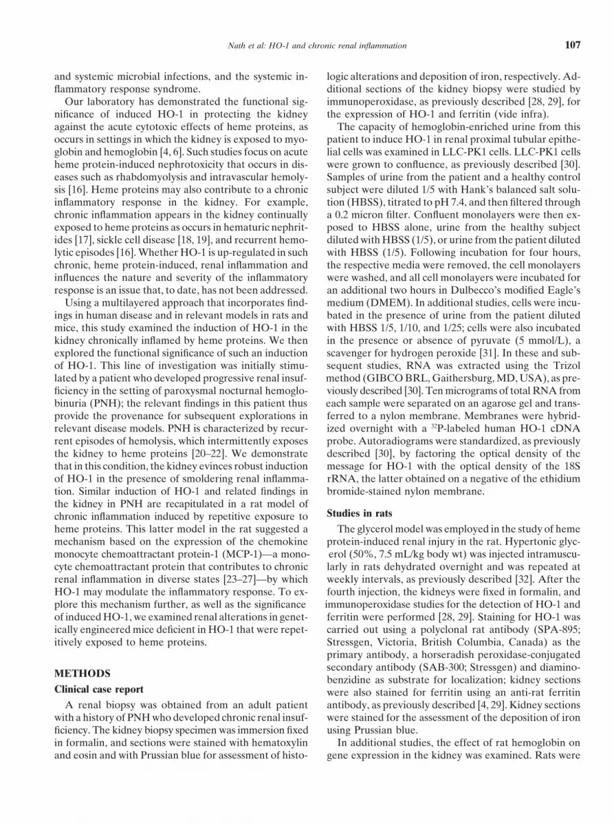
Fig. 7. Expression of MCP-1 mRNA in the kidney in HO-1 1/1 miceand HO-1 2/2 mice subjected to weekly intravenous administrationof mouse hemoglobin (90 mg/100 g body weight) or saline vehicle.Northern analyses for MCP-1 expression were performed using RNAextracted from kidneys harvested seven days after the eighth injectionof hemoglobin. Each lane represents RNA extracted from a singlekidney from an individual mouse.
and marked tubulointerstitial inflammation with repeti-tive doses of hemoglobin.
To determine the mechanism accounting for suchmarked leukocytic infiltration, we considered the possi-bility that the chemokine MCP-1, a chemoattractant formonocytes and T cells, would be up-regulated. Northernanalyses conducted in these kidneys demonstrated markedup-regulation of MCP-1 mRNA in HO-1 2/2 mice sub-jected to repetitive injections of hemoglobin, whereasthere was scant, if any, expression of MCP-1 in HO-1
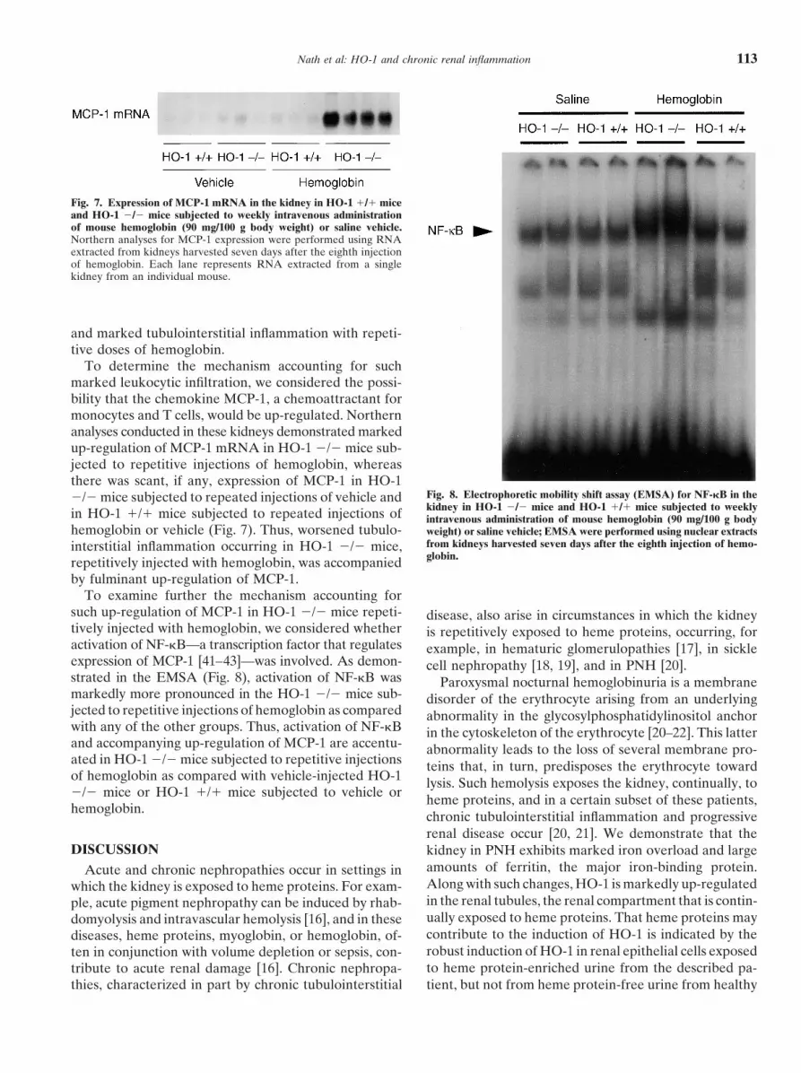
Fig. 8. Electrophoretic mobility shift assay (EMSA) for NF-kB in the2/2 mice subjected to repeated injections of vehicle andkidney in HO-1 2/2 mice and HO-1 1/1 mice subjected to weeklyin HO-1 1/1 mice subjected to repeated injections of intravenous administration of mouse hemoglobin (90 mg/100 g body
hemoglobin or vehicle (Fig. 7). Thus, worsened tubulo- weight) or saline vehicle; EMSA were performed using nuclear extractsfrom kidneys harvested seven days after the eighth injection of hemo-interstitial inflammation occurring in HO-1 2/2 mice,globin.repetitively injected with hemoglobin, was accompanied
by fulminant up-regulation of MCP-1.To examine further the mechanism accounting for
such up-regulation of MCP-1 in HO-1 2/2 mice repeti- disease, also arise in circumstances in which the kidneytively injected with hemoglobin, we considered whether is repetitively exposed to heme proteins, occurring, foractivation of NF-kB—a transcription factor that regulates example, in hematuric glomerulopathies [17], in sickleexpression of MCP-1 [41–43]—was involved. As demon- cell nephropathy [18, 19], and in PNH [20].strated in the EMSA (Fig. 8), activation of NF-kB was Paroxysmal nocturnal hemoglobinuria is a membranemarkedly more pronounced in the HO-1 2/2 mice sub- disorder of the erythrocyte arising from an underlyingjected to repetitive injections of hemoglobin as compared abnormality in the glycosylphosphatidylinositol anchorwith any of the other groups. Thus, activation of NF-kB in the cytoskeleton of the erythrocyte [20–22]. This latterand accompanying up-regulation of MCP-1 are accentu- abnormality leads to the loss of several membrane pro-ated in HO-1 2/2 mice subjected to repetitive injections teins that, in turn, predisposes the erythrocyte towardof hemoglobin as compared with vehicle-injected HO-1 lysis. Such hemolysis exposes the kidney, continually, to2/2 mice or HO-1 1/1 mice subjected to vehicle or heme proteins, and in a certain subset of these patients,hemoglobin.
chronic tubulointerstitial inflammation and progressiverenal disease occur [20, 21]. We demonstrate that the
DISCUSSION kidney in PNH exhibits marked iron overload and largeamounts of ferritin, the major iron-binding protein.Acute and chronic nephropathies occur in settings inAlong with such changes, HO-1 is markedly up-regulatedwhich the kidney is exposed to heme proteins. For exam-in the renal tubules, the renal compartment that is contin-ple, acute pigment nephropathy can be induced by rhab-ually exposed to heme proteins. That heme proteins maydomyolysis and intravascular hemolysis [16], and in thesecontribute to the induction of HO-1 is indicated by thediseases, heme proteins, myoglobin, or hemoglobin, of-robust induction of HO-1 in renal epithelial cells exposedten in conjunction with volume depletion or sepsis, con-to heme protein-enriched urine from the described pa-tribute to acute renal damage [16]. Chronic nephropa-
thies, characterized in part by chronic tubulointerstitial tient, but not from heme protein-free urine from healthy
Nath et al: HO-1 and chronic renal inflammation114
controls. It would be of interest to determine the extent HO-1 influences the triphasic response to heme proteins.The greater rise in plasma creatinine in HO-1 2/2 mice,to which HO-1 and ferritin are induced in the kidney in
patients with renal diseases other than PNH. Also of as compared to the HO-1 1/1 mice, after the first andsecond doses of hemoglobin, attests to the importanceinterest is the extent to which HO-1 and ferritin can be
induced in LLC-PK1 cells by urine from patients with of HO-1 in protecting against acute sensitivity to hemeproteins. However, by the time of the third injection,renal diseases other than PNH and by urine enriched
with proteins other than hemoglobin. HO-1 2/2 mice did not evince a rise in plasma creatininefollowing the administration of hemoglobin. Thus, it ap-We have recently described a rat model of chronic
tubulointerstitial inflammation that is induced by recur- pears that unlike its involvement in attenuating the acute,early, sensitivity to heme proteins, HO-1 may not berent exposure to heme proteins [32]. This model exhibits
histologic features that resemble the kidney in PNH, required in the acquisition of renal resistance that ap-pears by the third injection of hemoglobin, at least asexhibiting as it does, interstitial inflammation in conjunc-
tion with chronic up-regulation of interstitial and base- reflected by the rise in plasma creatinine. However, thethird phase, which is characterized by chronic inflamma-ment membrane collagens [32]. We now provide further
evidence in support of the similarity of renal features of tion, is clearly one that is influenced by HO-1. The ab-sence of HO-1 leads to striking exacerbation of intersti-this rat model and the clinical disease: In the rat kidney,
large deposits of iron are present in conjunction with tial inflammation and tubulointerstitial disease. Thesefindings demonstrate that within the kidney, a chronicprominent up-regulation of ferritin, and notably, HO-1
is markedly up-regulated in renal tubules. inflammatory response is influenced by HO-1. Specifi-cally, the absence of HO-1 transforms what is otherwiseTo analyze the functional effect of induced HO-1, we
examined the influence of HO-1 on each aspect of the a mild lesion into an aggressive interstitial cellular infil-tration. From these findings, we suggest that the induc-renal response to repetitive exposure to heme proteins.
As recently characterized, the renal response to repeti- tion of HO-1 in chronic interstitial disease may serve asa governor that limits the chronic inflammatory cellulartive exposure to heme proteins in the rat revealed a
triphasic pattern consisting of initial sensitivity to the response that would otherwise occur.To explore a potential mechanism whereby the defi-acute toxicity of heme proteins, acquired resistance to
these effects of heme proteins when repetitively adminis- ciency of HO-1 promotes interstitial cellular infiltrationin the kidney, we focused on MCP-1. This chemokine,tered, and eventually, a progressive inflammatory response
[32]. Acute sensitivity represents the acute functional a potent chemoattractant for monocytes and T cells, isincriminated in the initiation and perpetuation of chronicand structural deterioration that occurs in response to
the first insult. Acquired renal resistance to injury is tubulointerstitial inflammation in assorted nephritides[23–27]. An additional consideration motivating the exam-reflected by the blunted fall in glomerular filtration rate
that is accompanied by reduced amounts of necrosis and ination of MCP-1 arose from our recent description ofa model of repetitive administration of heme proteins inapoptosis in renal tubular epithelial cells with each suc-
cessive insult, and chronic inflammation is characterized the rat wherein MCP-1, an oxidant-inducible chemokine[37], was up-regulated [32]. Based on such findings, weby progressive tubulointerstitial inflammation in con-
junction with up-regulation of MCP-1. suggested that MCP-1 may contribute to the attendantchronic tubulointerstitial inflammation in this model [32].In the present study, we examined the functional sig-
nificance of expression of HO-1 in the kidney repetitively In the present studies, we provide the novel findingthat heme proteins, such as hemoglobin, directly induceexposed to heme proteins. To this end, we utilized geneti-
cally engineered mice that are deficient in HO-1 and MCP-1. When examined seven days after the last doseof hemoglobin, fulminant induction of MCP-1 in thein which heme proteins were administered at weekly
intervals. We used this knockout model rather than a kidney occurred in HO-1 2/2 mice subjected to re-peated exposure to hemoglobin, but not in HO-1 1/1pharmacologic approach based on repeated administra-
tion of competitive inhibitors of HO over an extended mice subjected to hemoglobin or vehicle, or in HO-12/2 mice subjected to saline vehicle. Thus, the markedperiod, since there are concerns regarding the nonspe-
cific actions of these pharmacologic inhibitors of HO-1. exacerbation of interstitial inflammation in the absenceof HO-1 is attended by intense up-regulation of a potentAs a model of repetitive exposure to heme protein in
mice, we used mouse hemoglobin rather than repeated chemoattractant for monocytes and T lymphocytes,namely, MCP-1.administration of glycerol: Hemoglobin can be dosed
precisely, and its use avoids the vagaries associated with The lack of expression of MCP-1 in the kidney inHO-1 1/1 mice in these studies (Fig. 7) merits comment.variable amounts of muscle and red cell lysis that occur
in the glycerol model. These studies in mice were undertaken seven days afterthe administration of hemoglobin, whereas the study ofThe repetitive administration of hemoglobin to HO-1
2/2 mice allowed the examination of the extent to which MCP-1 expression in rats was undertaken six hours after

Nath et al: HO-1 and chronic renal inflammation 115
the administration of hemoglobin (Fig. 4). The intense of renal inflammation, HO-1 may subserve a functionalrole. Studies along these lines examining the effect ofup-regulation of MCP-1 observed six hours after a given
dose of hemoglobin in rodents with inducible HO-1 thus induced HO-1 on the inflammatory response in suchstates would be of interest.subsides by seven days. The lack of expression of MCP-1
in the kidney in HO-1 1/1 mice when studied seven Our studies demonstrating that hemoglobin, directlyadministered, induces MCP-1, offer a mechanism account-days after hemoglobin likely represents the abatement in
expression of previously up-regulated MCP-1. As such, it ing for progressive tubulointerstitial injury in assortedhematuric glomerulonephritides [17]. In such conditions,underscores the persistence in, and intensity of, expres-
sion of MCP-1 in the HO-1–deficient state following a renal tubules are exposed to a burden of hemoglobin asred blood cells are engulfed and degraded within thesimilar exposure to hemoglobin.
Among the factors that regulate MCP-1 is NF-kB. tubular epithelial cells [55]. Moreover, the direct micro-injection of red blood cells into rat proximal tubulesActivation of NF-kB occurs in renal inflammation and
is achieved, in some instances, by alterations in cellular eventually incurs an inflammatory response [56]. Wespeculate that such inflammatory responses in the tubu-redox [25, 34, 41, 42]. We thus examined activation of
NF-kB in HO-1 1/1 and HO-1 2/2 mice subjected to lointerstitium may reflect the induction of MCP-1 byhemoglobin. It would be of interest to determine thehemoglobin or vehicle. As demonstrated, the activation
of NF-kB mirrored the pattern of expression found for renal expression of MCP-1 in chronic hematuric condi-tions and hemoglobinuric states (PNH, prosthetic heartMCP-1: Activation of NF-kB was much more prominent
in HO-1 2/2 mice subjected to hemoglobin as compared valves, etc.). Perhaps the extent to which MCP-1 is ex-pressed in these states—and thus the propensity to chronicwith HO-1 1/1 mice exposed to hemoglobin or vehicle
or HO-1 2/2 mice subjected to vehicle. We speculate tubulointerstitial disease—is inversely influenced by theextent to which HO-1 is induced. This latter relationshipthat the deficiency of HO-1 promotes activation of
NF-kB, which, in turn, induces such cytokines as MCP-1. may underlie the lack of progressive renal injury in statesof low grade hemoglobinuria, as occurs in patients withThat the deficiency of HO-1 promotes chronic cellularprosthetic heart valves.inflammation through up-regulation of NF-kB and MCP-1
In summary, we demonstrate the induction of HO-1adds to the mechanistic underpinnings whereby HO-1in the kidney in PNH. We suggest that such inductiondown-regulates inflammation. Products of HO such asrepresents an adaptive response that enables the degra-carbon monoxide may suppress proinflammatory cyto-dation of heme, an oxidant abundant in tissues in PNH.kines such as platelet-derived growth factor and endo-This induction of HO-1 and other germane features isthelin-1 [44]. Other products such as bilirubin can attenu-faithfully reproduced in the setting of chronic tubuloin-ate adhesion of leukocytes to the venular endotheliumterstitial disease in the rat kidney following repetitive[45] or inhibit the activation of NADPH oxidase [46].exposure to heme proteins. The importance of such anHO-1 reduces the up-regulation of intercellular adhesioninduction of HO-1 in down-regulating the inflammatorymolecule-1 that otherwise occurs in endothelial cells inresponse is uncovered by the fulminant interstitial in-response to heme [47]. The striking up-regulation offlammation, striking up-regulation of the chemokineMCP-1 in the absence of HO-1 provides another mecha-MCP-1, and activation of the transcription factor NF-kBnism by which HO-1 may act as an anti-inflammatoryoccurring in HO-1–deficient mice repetitively exposedagent. By suppressing MCP-1, a major chemoattractantto heme proteins. We conclude by speculating that up-for monocytes and T cells, HO-1 may thus deprive theseregulation of HO-1 in the injured kidney may assist incells of a potent stimulus that recruits them into an in-down-regulating cellular infiltration by suppressing NF-jured area.kB–driven activation of MCP-1.Besides its induction in toxic models of renal injury
[1–3], induction of HO-1 is increasingly described inACKNOWLEDGMENTSmodels of renal inflammation, including acute transplant
rejection [48], acute glomerular inflammation associated These studies were supported by the following grants: DK47060(K.A.N.), HL55552 (G.M.V., K.A.N.), and DK43135 (J.A.). We grate-with nephrotoxic serum nephritis [49–51], chronic ad-fully acknowledge the secretarial expertise of Mrs. Sharon Heppel-ministration of angiotensin II [52], and acute obstruction mann in the preparation of this work.
to the urinary tract [53]. Expression of HO-1 has beenReprint requests to Dr. Karl Nath, Nephrology Research Unit, Mayorecently described in renal tubules in case reports of
Clinic, 200 First Street, SW, 542 Guggenheim Building, Rochester, Min-patients with acute tubular necrosis, IgA nephropathy, nesota 55905, USA.and remission from minimal change disease [54]. The E-mail: [email protected]
capacity of HO-1 to down-regulate the inflammatoryresponse—as indicated by the intense up-regulation of REFERENCESinflammation observed in its absence—raises the inter- 1. Nath KA, Agarwal A, Vogt B: Functional consequences of induc-
tion of heme oxygenase, in Contemporary Issues in Nephrology:esting possibility that in these, and possibly other states

Nath et al: HO-1 and chronic renal inflammation116
Acute Renal Failure: Emerging Concepts and Therapeutic Strate- but not glomerular injury, in nephrotoxic serum nephritis. J ClinInvest 103:73–80, 1999gies, edited by Goligorsky MS, Stein J, New York, Churchill
Livingstone, 1995, pp 97–118 27. Wada T, Furuichi K, Segawa-Takeda C, et al: MIP-1a and MCP-1contribute to crescents and interstitial lesions in human crescentic2. Nath KA: Heme oxygenase-1: A redoubtable response that limits
reperfusion injury in the transplanted adipose liver. J Clin Invest glomerulonephritis. Kidney Int 56:995–1003, 199928. Dvergsten J, Manivel JC, Correa-Rotter R, et al: Expression104:1485–1486, 1999
3. Agarwal A, Nick HS: Renal response to tissue injury: Lessons of clusterin in human renal diseases. Kidney Int 45:828–835, 199429. Nath KA, Dvergsten J, Correa-Rotter R, et al: Induction offrom heme oxygenase-1 gene ablation and expression. J Am Soc
Nephrol 11:965–973, 2000 clusterin in acute and chronic oxidative renal disease in the ratand its dissociation from cell injury. Lab Invest 71:209–218, 19944. Nath KA, Balla G, Vercellotti GM, et al: Induction of heme
oxygenase is a rapid, protective response in rhabdomyolysis in the 30. Nath KA, Croatt AJ, Likely S, et al: Renal oxidant injury andoxidant response induced by mercury. Kidney Int 50:1032–1043,rat. J Clin Invest 90:267–270, 1992
5. Abraham NG, Lavrovsky Y, Schwartzman ML, et al: Transfec- 199631. Nath KA, Ngo EO, Hebbel RP, et al: a-Ketoacids scavenge H2O2tion of the human heme oxygenase gene into rabbit coronary
microvessel endothelial cells: Protective effect against heme and in vitro and in vivo and reduce menadione-induced DNA injuryand cytotoxicity. Am J Physiol 268( 1 Pt 1):C227–C236, 1995hemoglobin toxicity. Proc Natl Acad Sci USA 92:6798–6802, 1995
6. Nath KA, Haggard JJ, Croatt AJ, et al: The indispensability of 32. Nath KA, Croatt AJ, Haggard JJ, et al: Renal response to repeti-tive exposure to heme proteins: Chronic injury induced by an acuteheme oxygenase-1 (HO-1) in protecting against acute heme pro-
tein-induced toxicity in vivo. Am J Pathol 156:1527–1535, 2000 insult. Kidney Int 57:2423–2433, 200033. Poss KD, Tonegawa S: Heme oxygenase 1 is required for mamma-7. Yet S-F, Perrella MA, Layne MD, et al: Hypoxia induces severe
right ventricular dilatation and infarction in heme oxygenase-1 null lian iron reutilization. Proc Natl Acad Sci USA 94:10919–10924,1997mice. J Clin Invest 103:R23–R29, 1999
8. Otterbein LE, Kolls JK, Mantell LL, et al: Exogenous adminis- 34. Rangan GK, Wang Y, Tay Y-C, et al: Inhibition of nuclearfactor-kB activation reduces cortical tubulointerstitial injury intration of heme oxygenase-1 by gene transfer provide protection
against hyperoxia-induced lung injury. J Clin Invest 103:1047–1054, proteinuric rats. Kidney Int 56:118–134, 199935. Paya CV, Ten RM, Bessia C, et al: NF-kB-dependent induction1999
9. Poss KD, Tonegawa S: Reduced stress defense in heme oxy- of the NF-kB p50 subunit gene promoter underlies self-perpetua-tion of human immunodeficiency virus transcription in monocyticgenase-1 deficient cells. Proc Natl Acad Sci USA 94:10925–10930,
1997 cells. Proc Natl Acad Sci USA 89:7826–7830, 199236. McElhinny JA, MacMorran WS, Bren GD, et al: Regulation of10. Shiraishi F, Curtis LM, Truong L, et al: Heme oxygenase-1 gene
ablation or expression modulates cisplatin-induced renal tubular IkBa and p105 in monocytes and macrophages persistently infectedwith human immunodeficiency virus. J Virol 69:1500–1509, 1995apoptosis. Am J Physiol Renal Physiol 278:F726–F736, 2000
11. Amersi F, Buelow R, Kato H, et al: Upregulation of heme oxy- 37. Navab M, Imes SS, Hama SY, et al: Monocyte transmigrationinduced by modification of low density lipoprotein in cocultures ofgenase-1 protects genetically fat Zucker rat livers from ischemia/
reperfusion injury. J Clin Invest 104:1631–1639, 1999 human aortic wall cells is due to induction of monocyte chemotacticprotein 1 synthesis and is abolished by high density lipoprotein.12. Platt JL, Nath KA: Heme oxygenase: Protective gene or Trojan
horse. Nat Med 4:1364–1365, 1998 J Clin Invest 88:2039–2046, 199138. Meffert MK, Haley JE, Schuman EM, et al: Inhibition of hippo-13. Willis D, Moore AR, Frederick R, et al: Heme oxygenase: A
novel target for the modulation of the inflammatory response. Nat campal heme oxygenase, nitric oxide synthase, and long-term po-tentiation by metalloporphyrins. Neuron 13:1225–1233, 1994Med 2:87–90, 1996
14. Soares MP, Lin Y, Anrather J, et al: Expression of heme oxy- 39. Grundemar L, Ny L: Pitfalls using metalloporphyrins in carbonmonoxide research. Trends Pharmacol Sci 18:193–195, 1997genase-1 can determine cardiac xenograft survival. Nat Med
4:1073–1077, 1998 40. Serfass L, Burstyn JN: Effect of heme oxygenase inhibitors onsoluble guanylyl cyclase activity. Arch Biochem Biophys 359:8–16,15. Hancock WW, Buelow R, Sayegh MH, et al: Antibody-induced
transplant arteriosclerosis is prevented by graft expression of anti- 199841. Morrissey J, Klahr S: Transcription factor NF-kB regulation ofoxidant and anti-apoptotic genes. Nat Med 4:1392–1396, 1998
16. Zager RA: Rhabdomyolysis and myohemoglobinuric acute renal renal fibrosis during ureteral obstruction. Semin Nephrol 18:603–611, 1998failure. Kidney Int 49:314–326, 1996
17. Nath KA: Tubulointerstitial changes as a major determinant in 42. Ishikawa Y, Sugiyama H, Stylianou E, et al: Bioflavonoid querce-tin inhibits interleukin-1-induced transcriptional expression ofthe progression of renal damage. Am J Kidney Dis 20:1–17, 1992
18. Saborio P, Scheinman JI: Sickle cell nephropathy. J Am Soc monocyte chemoattractant protein-1 in glomerular cells via sup-pression of nuclear factor-kB. J Am Soc Nephrol 10:2290–2296,Nephrol 10:187–192, 1999
19. Pham T-TT, Pham P-CT, Wilinson AH, et al: Renal abnormalities 199943. Stylianou E, Nie M, Ueda A, et al: c-Rel and p65 trans-activatein sickle cell disease. Kidney Int 57:1–8, 2000
20. Clark DA, Butler SA, Braren V, et al: The kidney in paroxysmal the monocyte chemoattractant protein-1 gene in interleukin-1 stim-ulated mesangial cells. Kidney Int 56:873–882, 1999nocturnal hemoglobinuria. Blood 57:83–89, 1981
21. Hillmen P, Lewis SM, Bessler M, et al: Natural history of paroxys- 44. Morita T, Kourembanas S: Endothelial cell expression of vasocon-strictors and growth factors is regulated by smooth muscle cell-mal nocturnal hemoglobinuria. N Engl J Med 333:1253–1258, 1995
22. Rosse WF: Paroxysmal nocturnal hemoglobinuria as a molecular derived carbon monoxide. J Clin Invest 96:2676–2682, 199545. Hayashi S, Takamiya R, Yamaguchi T, et al: Induction of hemedisease. Medicine (Baltimore) 76:63–93, 1997
23. Wenzel UO, Abboud HE: Chemokines and renal disease. Am J oxygenase-1 suppresses venular leukocyte adhesion elicited by oxi-dative stress. Circ Res 85:663–671, 1999Kidney Dis 26:982–994, 1995
24. Wenzel U, Schneider A, Valente AJ, et al: Monocyte chemoat- 46. Kwak JY, Takeshige K, Cheung BS, et al: Bilirubin inhibits theactivation of superoxide-producing NADPH oxidase in a neutro-tractant protein-1 mediates monocyte-macrophage influx in anti-
thymocyte antibody-induced glomerulonephritis. Kidney Int 51: phil cell-free system. Biochem Biophys Acta 1076:369–373, 199147. Frank AD, Wagener TG, Da Silva J-L, et al: Differential effects770–776, 1997
25. Ruiz-Ortega M, Bustos C, Hernandez-Presa Lorenzo O, et al: of heme oxygenase isoforms on heme mediation of endothelialintracellular adhesion molecule 1 expression. J Pharmacol ExpAngiotensin II participates in mononuclear cell recruitment in
experimental immune complex nephritis through nuclear factor-kB Ther 291:416–423, 199948. Agarwal A, Kim Y, Matas AJ, et al: Gas-generating systems inactivation and monocyte chemoattractant protein-1 synthesis.
J Immunol 161:430–439, 1998 acute renal allograft rejection in the rat: Co-induction of hemeoxygenase and nitric oxide synthase. Transplantation 61:93–98,26. Tesch GH, Schwarting A, Kinoshita K, et al: Monocyte chemoat-
tractant protein-1 promotes macrophage-mediated tubular injury, 1996

Nath et al: HO-1 and chronic renal inflammation 117
49. Vogt BA, Shanley TP, Croatt AJ, et al: Glomerular inflammation oxidant stress in vivo and heme oxygenase-1 in vivo and in vitro.Kidney Int 58:144–152, 2000induces resistance to tubular injury in the rat: A novel form of
53. Kawada N, Moriyama T, Ando A, et al: Increased oxidative stressacquired heme oxygenase-dependent resistance to renal injury.in mouse kidneys with unilateral ureteral obstruction. Kidney IntJ Clin Invest 98:2139–2145, 199656:1004–1013, 199950. Mosley K, Wembridge DE, Cattell V, et al: Heme oxygenase is
54. Ohta K, Yachie A, Fujimoto K, et al: Tubular injury as a cardinalinduced in nephrotoxic nephritis and hemin, a stimulator of hemepathologic feature in human heme oxygenase-1 deficiency. Am Joxygenase synthesis, ameliorates disease. Kidney Int 53:672–678,Kidney Dis 35:863–870, 20001998 55. Hill PA, Davies DJ, Kincaid-Smith P, et al: Ultrastructural
51. Datta PK, Koukouritaki SB, Hopp KA, et al: Heme oxygenase- changes in renal tubules associated with glomerular bleeding. Kid-1 induction attenuates inducible nitric oxide synthase expression ney Int 36:992–997, 1989and proteinuria in glomerulonephritis. J Am Soc Nephrol 10:2540– 56. Madsen KM, Applegate CW, Tisher CC: Phagocytosis of erythro-2550, 1999 cytes by the proximal tubule of the rat kidney. Cell Tissue Res
226:363–374, 198252. Haugen EN, Croatt AJ, Nath KA: Angiotensin II induces renal