upregulation of heme oxygenase-1 protects genetically fat zucker rat livers from...
TRANSCRIPT
IntroductionOrthotopic liver transplantation (OLT) is an effectivetherapeutic modality for end-stage liver disease.Advances in surgical technique, patient management,and immunosuppression have improved patient sur-vival after OLT, resulting in expansion of the list ofindications for the procedure. However, a serious prob-lem limiting OLT is the widening disparity between theincreasing number of potential recipients who vie for aconstant donor supply. This demand has renewedinterest in the use of marginal livers to expand thenumber of available donors. The prevalence of fattyinfiltration in the liver ranges from 6 to 26% based onautopsy studies and donor liver biopsies (1, 2). Indeed,steatotic donor livers are frequently discarded becauseof the fear of primary nonfunction, as compared withnormal livers, further accentuating the critical short-age of human donor livers (3, 4). Because 10–30% ofpotential liver recipients on transplant waiting lists die
without being transplanted, considerable effort hasbeen made to identify methods that would allow suc-cessful use of steatotic grafts.
Ischemia/reperfusion (I/R) injury, an antigen-inde-pendent event, often leads to primary nonfunction orearly dysfunction of steatotic liver grafts. Moreover,ischemia of more than 12 hours strongly correlates notonly with primary nonfunction after OLT, but also withbiopsy-proven centrilobular necrosis and a higher inci-dence of both acute and chronic rejection (5). Themechanism of liver injury following I/R involves a com-plex interaction of events, which include neutrophilactivation, increased expression of adhesion molecules,Kupffer cell activation, cytokine release, sinusoidalendothelial cell death, and hepatocyte injury (6, 7).Hepatocytes are more susceptible to ischemic injurywith damage to proteins, lipids, and DNA, altered reg-ulation of signal transduction, and release of growthfactors (8). The exact mechanism that leads to the ulti-
The Journal of Clinical Investigation | December 1999 | Volume 104 | Number 11 1631
Upregulation of heme oxygenase-1 protects genetically fat Zucker rat livers from ischemia/reperfusion injury
Farin Amersi,1 Roland Buelow,2 Hirohisa Kato,1 Bibo Ke,1 Ana J. Coito,1 Xiu-Da Shen,1
Delai Zhao,1 Joseph Zaky,1 Judy Melinek,3 Charles R. Lassman,3 Jay K. Kolls,4
J. Alam,5 Thomas Ritter,6 Hans-Dieter Volk,6 Douglas G. Farmer,1
Rafik M. Ghobrial,1 Ronald W. Busuttil,1 and Jerzy W. Kupiec-Weglinski1
1The Dumont-UCLA Transplant Center, Division of Liver and Pancreas Transplantation, Department of Surgery, and2Sangstat Medical Corporation, Fremont, California 94555, USA 3Department of Pathology and Laboratory Medicine, UCLA School of Medicine, Los Angeles, California 90095, USA4Section of Pulmonary/Critical Care, Louisiana State University School of Medicine, New Orleans, Louisiana 70112, USA5Department of Molecular Genetics, Alton Ochsner Medical Foundation, and Department of Biochemistry/Molecular Biology,Louisiana State University Medical Center, New Orleans, Louisiana 70121, USA
6Institute of Medical Immunology, Humboldt University, D-10098 Berlin, Germany
Address correspondence to: Jerzy W. Kupiec-Weglinski, The Dumont-UCLA Transplant Center, Room 77-120 CHS, 10833 Le Conte Avenue, Los Angeles, California 90095, USA. Phone: (310) 825-4196; Fax: (310) 267-2358; E-mail: [email protected].
Received for publication July 19, 1999, and accepted in revised form October 18, 1999.
We examined the effects of upregulation of heme oxygenase-1 (HO-1) in steatotic rat liver models ofex vivo cold ischemia/reperfusion (I/R) injury. In the model of ischemia/isolated perfusion, treat-ment of genetically obese Zucker rats with the HO-1 inducer cobalt protoporphyrin (CoPP) or withadenoviral HO-1 (Ad-HO-1) significantly improved portal venous blood flow, increased bile pro-duction, and decreased hepatocyte injury. Unlike in untreated rats or those pretreated with the HO-1 inhibitor zinc protoporphyrin (ZnPP), upregulation of HO-1 by Western blots correlated with ame-lioration of histologic features of I/R injury. Adjunctive infusion of ZnPP abrogated the beneficialeffects of Ad-HO-1 gene transfer, documenting the direct involvement of HO-1 in protection againstI/R injury. Following cold ischemia/isotransplantation, HO-1 overexpression extended animal sur-vival from 40% in untreated controls to about 80% after CoPP or Ad-HO-1 therapy. This effect corre-lated with preserved hepatic architecture, improved liver function, and depressed infiltration by Tcells and macrophages. Hence, CoPP- or gene therapy–induced HO-1 prevented I/R injury in steatot-ic rat livers. These findings provide the rationale for refined new treatments that should increase thesupply of usable donor livers and ultimately improve the overall success of liver transplantation.
J. Clin. Invest. 104:1631–1639 (1999).
mate decline in liver function and eventual organ fail-ure remains elusive, but data suggest that the oxidativestress of I/R plays a major role (9). The consequences ofoxidative stress include lipid peroxidation, as well asdamage to proteins and nucleic acids. Because of thenature of liver disease, transplant patients may be fur-ther disadvantaged by starting from a baseline deletionof normal antioxidative mechanisms. Normal cellularantioxidant systems involve a delicate balance of sever-al enzymes and proteins that are increasingly recognizedas critical regulators of cell function and viability (10).
Heme oxygenases (HO) are the rate-limiting enzymesthat catalyze the conversion of heme into biliverdin,carbon monoxide (CO), and free iron (11). They consistof 3 isoforms, the inducible HO-1, also known as heatshock protein 32 (hsp32), the constitutive HO-2, andthe not fully defined HO-3. HO-1 activity is upregulat-ed following various stimuli and is considered one ofthe most sensitive indicators of cellular stress. Inter-estingly, HO-1 levels may also increase in animals afteradministration of HO inhibitors, perhaps because ofthe accumulation of intracellular heme upon inhibi-tion of HO (12). In analogy with heat-shock regulation,upregulation of HO-1 may be an adaptive mechanismprotecting cells from stress, in particular hyperoxia,ischemia, or inflammation.
In transplantation models increased expression ofHO-1 was associated with long-term acceptance of car-diac xenografts, whereas donor hearts from HO-1–defi-cient donors were vigorously rejected (13). In our ownstudies, pretreatment of mice with cobalt protopor-phyrin (CoPP), a known HO-1 inducer, prolonged car-diac allograft survival (14), whereas exogenous HO-1overexpression prevented the development of cardiactransplant arteriosclerosis and interstitial fibrosis char-acteristic of chronic rejection (15). Moreover, a recentstudy demonstrated that induction of HO-1 was neu-roprotective and attenuated cellular injury caused byischemic stroke (16). Hence, pharmacological stimula-tion of HO-1 activity may represent a novel therapeu-tic approach in the amelioration of ischemic injuryduring the acute period of stroke.
This study was designed to determine the role of HO-1 expression in well-defined steatotic rat liver modelsof ex vivo cold ischemia followed by perfusion or syn-geneic OLT. To our knowledge, we have demonstratedfor the first time that overexpression of HO-1 after pre-treatment with CoPP or gene therapy with a recombi-nant adenovirus encoding rat HO-1 cDNA (Ad-HO-1)can prevent severe I/R insult suffered by steatotic livers.The beneficial effects upon hepatocyte injury and liverfunction were abrogated after adjunctive infusion ofzinc protoporphyrin (ZnPP), the HO-1 antagonist,which implicates a direct involvement of HO-1 in theprotection against I/R injury. Our present findingsraise the possibility of refined new treatment regimens,which, in turn, may increase the OLT donor poolthrough modulation of marginal livers and ultimatelyimprove the overall success of liver transplantation.
MethodsAnimals. Genetically obese (fa/fa) male Zucker rats(220–275 g) and lean (fa/–) Zucker rats (250–300 g)were used (Harlan Sprague Dawley Inc., Indianapolis,Indiana, USA). Animals were fed standard rodentchow and water ad libitum and cared for according toguidelines approved by the American Association ofLaboratory Animal Care.
Synthetic metalloporphyrins. Metalloporphyrins (CoPPand ZnPP) were purchased from Porphyrin ProductsInc. (Logan, Utah, USA). They were dissolved in 0.2 MNaOH, subsequently adjusted to a pH of 7.4, and dilut-ed in 0.85% NaCl. The stock concentration of metallo-porphyrins was 1 mg/mL.
Ad-HO-1 construct. A 1.0-kbp XhoI-HindIII fragmentfrom the rat HO-1 cDNA clone pRHO-1 (17), con-taining the entire coding region was cloned into plas-mid pAC-CMVpLpA (18, 19). Ad-HO-1 was generatedby homologous recombination in 911 cells (20) aftercotransfection with the pAC-HO-1 plasmid and plas-mid pJM17 (21). The recombinant Ad-HO-1 cloneswere screened by Southern blot analysis. Ad contain-ing the Escherichia coli β-galactosidase gene (Ad-βGal)has been described (22).
Ex vivo cold ischemia model. Genetically obese Zucker ratsunderwent ether anesthesia and systemic heparinization.After skeletonization of the liver, the portal vein, bileduct, and inferior vena cava were cannulated, and theliver was flushed with 10 mL of University of Wisconsin(UW) solution. Control livers from untreated obeseZucker rats were stored for 6 hours at 4°C in UW solu-tion (n = 6). There were 4 treatment groups. Group 1animals received CoPP, the HO-1 inducer (5 mg/kgintraperitoneally) 24 hours before liver harvest (n = 6).Group 2 rats were infused with Ad-HO-1 or Ad-β Gal(2.4 × 10⁹ plaque-forming units [pfu] intraperitoneally)
1632 The Journal of Clinical Investigation | December 1999 | Volume 104 | Number 11
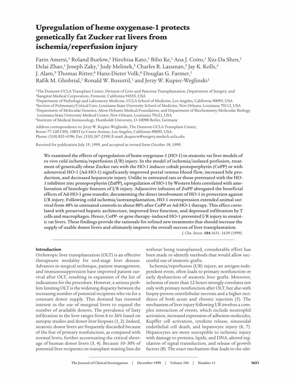
Figure 1HO-1–inducing agents decrease resistance to portal vein blood flow.Livers harvested from obese Zucker rats were perfused for 2 hours onthe isolated perfusion rat liver apparatus after 6 hours of cold ischemia.Pretreatment with CoPP or Ad-HO-1 gene transfer (day –1) significantlyimproved portal venous blood flow compared with untreated, ZnPP, orAd-βGal pretreated controls throughout the reperfusion period. Thesedata represent the mean ± SE of 4–10 independent perfusions for eachgroup. *P = 0.0001 versus untreated/ZnPP-treated controls.
24 to 48 hours before the procurement (n = 4–10). Group3 donors were treated with Ad-HO-1 (2.4 × 10⁹ pfu intra-venously) at day –2, followed 1 day later by infusion ofZnPP (20 mg/kg intraperitoneally), the HO-1 inhibitor(n = 4). Group 4 rats received ZnPP alone (20 mg/kgintraperitoneally) at 24 hours before harvest (n = 4). Alllivers were procured at day 0, stored for 6 hours at 4°Cin UW solution, and then perfused on an isolated perfu-sion rat liver apparatus, as described (6). The Zucker liv-ers were perfused ex vivo for 2 hours while temperature,pH, and inflow pressure were kept constant. Portal veinblood flow and pressure were recorded every 15 minutes,whereas bile output was monitored every 30 minutes.Portal vein blood flow was adjusted to maintain portalpressures of 13 to 18 cmH20. Blood was collected at 30-minute intervals and serum glutamic-oxaloacetic trans-aminase (sGOT) levels were measured using an autoan-alyzer from ANTECH Diagnostics (Irvine, California,USA). Following 2 hours of perfusion, a portion of theliver was snap-frozen for mRNA extraction and Westernblot analysis of HO-1 expression; the remaining tissuesamples were fixed in formalin for hematoxylin andeosin (H&E) staining.
Syngeneic OLT model. Syngeneic liver transplants wereperformed using fatty livers that were harvested fromobese Zucker rats and stored for 4 hours at 4°C in UWsolution before being transplanted into lean Zuckerrecipients. OLTs were performed with revascularizationwithout hepatic artery reconstruction (23). There were 2treatment groups. In the first group, obese Zucker rats (n= 10) received CoPP (5 mg/kg intraperitoneally) 24 hoursbefore the procurement. Group 2 donors (n = 11) were
treated with Ad-HO-1 (2.4 × 10⁹ pfu intravenously) 24hours before harvest. OLT recipients were followed forsurvival and sGOT levels. Separate groups of rats (n =2/group) were sacrificed at 1, 7, 14, and 100 days afterOLT, and liver samples were collected for H&E/immuno-histology staining and Western blot analysis.
Histology and immunohistochemistry. Liver specimenswere fixed in a 10% buffered formalin solution and em-bedded in paraffin. Sections were made at 4 µm andstained with H&E. The histologic severity of I/R injuryin the ex vivo perfusion model was graded using Inter-national Banff Criteria (24). Using these criteria, lobulardisarray and ballooning changes are graded from 1 to 4,where no change is given a score of 1 and severe disarrayor ballooning changes is given a score of 4. The previ-ously published Suzuki’s criteria (25) were modified toevaluate the histologic severity of I/R injury in the OLTmodel. In this classification sinusoidal congestion, hepa-tocyte necrosis, and ballooning degeneration are gradedfrom 0 to 4. No necrosis, congestion or centrilobular bal-looning is given a score of 0, whereas severe congestionand ballooning degeneration as well as greater than 60%lobular necrosis is given a value of 4.
OLTs were examined serially by immunohistochem-istry for mononuclear cell (MNC) infiltration (26, 27).Briefly, liver tissue was embedded in Tissue Tek OCTcompound (Miles Inc., Elkhart, Indiana, USA), snapfrozen in liquid nitrogen, and stored at –70° C. Cryostatsections (5 µm) were fixed in acetone, and then endoge-nous peroxidase activity was inhibited with 0.3% H2O2
in PBS. Normal heat-inactivated donkey serum (10%)was used for blocking. Appropriate primary mouse Ab
The Journal of Clinical Investigation | December 1999 | Volume 104 | Number 11 1633
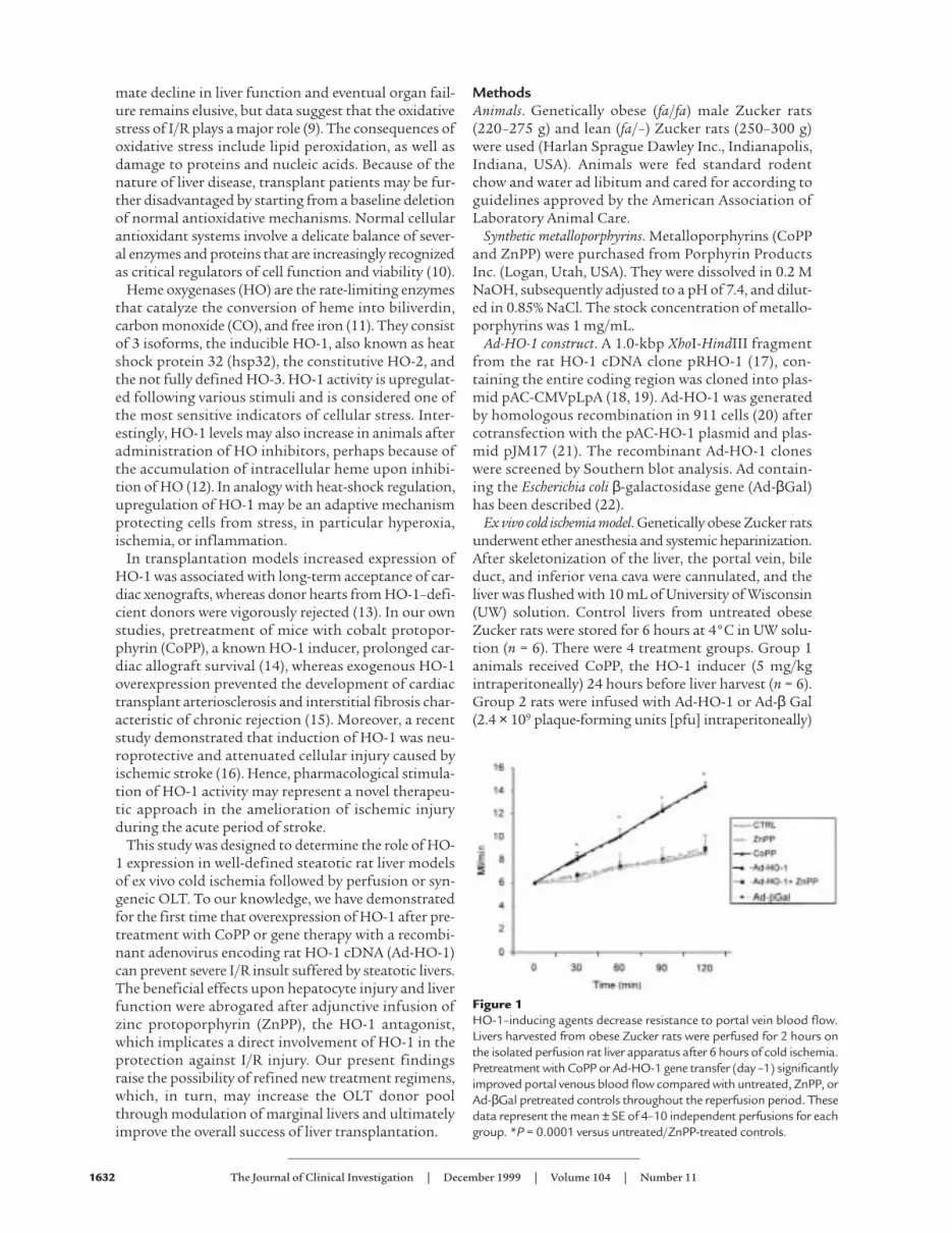
Figure 2Bile production in fatty livers perfused for 2 hours on the isolatedperfusion rat liver apparatus after 6 hours of cold ischemia. Animalswere pretreated with metalloporphyrins, or with Ad-HO-1 genetransfer, or left untreated. Bile production at 30-minute intervalsthroughout the reperfusion period was significantly higher in theCoPP/Ad-HO-1 groups (*P < 0.05) as compared with untreated,ZnPP-, or Ad-βGal–pretreated controls. These data represent themean ± SE of 4–10 independent perfusions for each group. *P < 0.05versus untreated/ZnPP-treated controls.
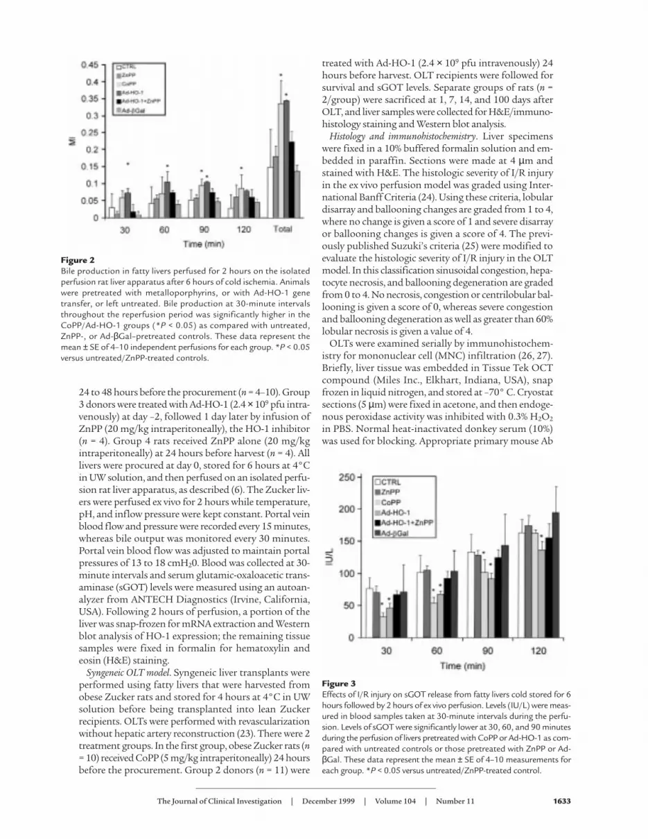
Figure 3Effects of I/R injury on sGOT release from fatty livers cold stored for 6hours followed by 2 hours of ex vivo perfusion. Levels (IU/L) were meas-ured in blood samples taken at 30-minute intervals during the perfu-sion. Levels of sGOT were significantly lower at 30, 60, and 90 minutesduring the perfusion of livers pretreated with CoPP or Ad-HO-1 as com-pared with untreated controls or those pretreated with ZnPP or Ad-βGal. These data represent the mean ± SE of 4–10 measurements foreach group. *P < 0.05 versus untreated/ZnPP-treated control.

against rat T cells (R73) and monocytes/macrophages(ED1) (Harlan Bioproducts for Science, Indianapolis,Indiana, USA) were added at optimal dilutions. Boundprimary Ab was detected using biotinylated donkeyanti-mouse IgG and streptavidin peroxidase–conjugat-ed complexes (DAKO Corp., Carpinteria, California,USA). The control sections were performed by replacingthe primary Ab with either dilution buffer or normalmouse serum. The peroxidase reaction was developedwith 3,3-diaminobenzidine tetrahydrochloride (SigmaChemical Co., St. Louis, Missouri, USA). The sectionswere evaluated blindly by counting the labeled cells intriplicates in 10 high-power fields.
Western blots. Protein was extracted from liver tissuesamples with PBSTDS buffer (50 mM Tris, 150 mMNaCl, 0.1% SDS, 1% sodium deoxycholate, and 1% tri-ton X-100, pH 7.2). Proteins (30 µg/sample) in SDS-loading buffer (50 mM Tris, pH 7.6, 10% glycerol, 1%SDS) were subjected to 12% SDS-PAGE and trans-ferred to nitrocellulose membrane (Bio-Rad Labora-tories Inc., Hercules, California, USA). The gel wasthen stained with Coomassie blue to document equalprotein loading. The membrane was blocked with 3%dry milk and 0.1% Tween-20 (Amersham, ArlingtonHeights, Illinois, USA) in PBS and incubated with rab-bit anti-rat HO-1 polyclonal Ab (Sangstat Corp., SanFrancisco, California, USA). The filters were washedand then incubated with horseradish peroxidase don-key anti-rabbit Ab (Amersham Life Sciences, Arling-ton Heights, Illinois, USA). Relative quantities of HO-1 protein were determined using a densitometer(Kodak Digital Science 1D Analysis Software, Ro-chester, New York, USA).
Statistics. Results are expressed as mean ± SEM. Sta-tistical comparisons between the groups in the ex-vivoperfusion model were performed using repeated meas-ure ANOVA. We used the Tukey-Fisher least-significantdifference (LSD) criterion for judging statistical signif-icance where P values of less than 0.05 were consideredstatistically significant.
ResultsThe effects of HO-1–inducing agents in the ex vivo steatotic ratliver cold ischemia model followed by reperfusion. To test ourhypothesis that overexpression of HO-1 decreases I/R-mediated hepatocyte injury, we monitored portal veinblood flow, bile production, and sGOT levels in liversfrom obese Zucker rats that were either untreated or pre-treated with HO-1–inducing agents and then perfusedfor 2 hours on the isolated perfusion rat liver apparatus.
Pretreatment of Zucker rats with synthetic metallo-porphyrin CoPP or Ad-HO-1 gene transfer exertedequally protective effects against liver I/R injury (Fig-ure 1). Both modalities significantly improved portalblood flow throughout the 2-hour reperfusion period,as compared with untreated controls (P = 0.0001). Inaddition, as shown in Figure 2, both CoPP and Ad-HO-1 significantly increased bile production (P <0.05), as compared with controls. The I/R-inducedhepatocyte injury measured by sGOT release was alsomarkedly reduced in the CoPP/Ad-HO-1 treatmentgroups as compared with controls (Figure 3). Forinstance, at 60 minutes of reperfusion, sGOT concen-trations were 53.3 ± 8.23 IU/L and 68.8 ± 10.1 IU/L inthe CoPP and Ad-HO-1 groups, respectively, versus102 ± 8.23 IU/L in untreated controls (P < 0.02). In
1634 The Journal of Clinical Investigation | December 1999 | Volume 104 | Number 11
Figure 4Photomicrographs of representative rat fatty liversafter 6 hours of cold ischemia and 2 hours of reper-fusion on the isolated perfusion rat liver apparatus.(a) Control untreated group with severe lobular dis-tortion, zone 3 ballooning, and hepatocyte necrosis(Banff’s score = 3.0 ± 0.63); (b) ZnPP-pretreatedgroup with marked sinusoidal and vascular conges-tion (arrow) and profound zone 3 ballooning change(score = 2.86 ± 0.12); (c) CoPP-pretreated and (d)Ad-HO-1–pretreated groups with minimal vacuolardegeneration and almost complete preservation oflobular architecture (scores = 1.21 ± 0.39 and 1.68± 0.51, respectively); (e) Ad-HO-1 plus ZnPP–treat-ed group, similar to untreated controls, with pro-found zone 3 ballooning change accompanied byconfluent hepatocyte necrosis (arrow; score = 2.74± 0.26); (f) Ad-βGal–pretreated group, similar tountreated controls, showing profound zone 3 bal-looning change accompanied by severe vascular con-gestion and confluent hepatocyte necrosis (arrow;score = 3.0 ± 1.41). ×100, H&E stain; n = 2–3/group.

contrast, Ad-β Gal gene transfer did not affect theextent of I/R insult suffered otherwise by steatotic ratlivers (Figures 1–3; n = 4).
ZnPP abrogates the beneficial effects of HO-1 upon hepaticI/R injury. To determine if the amelioration of hepato-cyte injury in this I/R model was indeed mediated by anincrease in HO-1 activity, prospective liver donors werepretreated with ZnPP, a potent HO-1 inhibitor. Unlikein the CoPP group, livers procured from obese Zuckerrats pretreated with ZnPP alone exhibited diminishedportal blood flow (Figure 1) and bile production (Fig-ure 2), effects that were accompanied by augmentedhepatocyte injury (Figure 3) comparable with otherwiseuntreated fatty controls. Interestingly, infusion of ZnPPabolished Ad-HO-1–mediated protective effects uponI/R injury in steatotic rat livers. Therefore, portal bloodflow (Figure 1) and bile production (Figure 2) were sig-nificantly (P < 0.05) decreased, and hepatocyte functionbecame impaired (Figure 3) after adjunctive ZnPP treat-ment, as compared with Ad-HO-1 monotherapy.
Liver histology in the ex vivo cold ischemia model followed byreperfusion. The I/R-induced hepatocyte injury in the exvivo model was graded using Banff’s criteria (24). In theuntreated fatty Zucker group, there was severe disrup-tion of lobular architecture with marked zone 3 bal-looning change, focally associated with hepatocytenecrosis (Figure 4a; Banff ’s score = 3.0 ± 0.63). TheZnPP-treated livers showed somewhat less lobular bal-looning changes, but more sinusoidal and vascular con-gestion (Figure 4b; score = 2.86 ± 0.12). In marked con-trast, CoPP-treated livers showed complete preservationof the lobular architecture with no signs of hepatocytenecrosis (Figure 4c; score = 1.21 ± 0.39). Similarly, liverstransduced with Ad-HO-1 revealed focal areas of mildvacuolar degeneration with minimal hepatocyte necro-sis (Figure 4d; score = 1.68 ± 0.51). However, livers pro-cured from animals treated with Ad-HO-1 plus ZnPPwere characterized by severe disruption of the lobulararchitecture, similar to the control untreated group,with profound zone 3 ballooning change accompaniedby confluent areas of hepatocyte necrosis (Figure 4e;score = 2.74 ± 0.26). Livers treated with Ad-β Gal
revealed less necrosis compared with the untreatedgroup, but had severe architectural disruption and vas-cular congestion (Figure 4f; score = 3.0 ± 1.41).
Western analysis of HO-1 expression in the ex vivo I/Rinjury model. We used Western analysis to evaluate HO-1 expression in liver samples following cold ischemiaat the completion of 2-hour perfusion period. The rel-ative expression levels of HO-1 protein in absorbanceunits (AU) were analyzed by densitometer. As shown inFigure 5, preservation of hepatic function after CoPPpretreatment or Ad-HO-1 gene transfer was accompa-nied by enhanced HO-1 expression (2.46 and 2.12 AU,respectively). In contrast, HO-1 was diminished afteradjunctive ZnPP infusion (1.18 AU) and virtuallyundetectable in untreated (0.11 AU) and ZnPP-pre-treated (0.12 AU) controls.
HO-1 overexpression prolongs OLT survival and improveshepatic function. We then sought to examine whetherexogenous manipulation of HO-1 expression could alsoconfer protection against I/R injury in the in vivo set-ting. Hence, we performed OLTs using steatotic Zuckerlivers that were cold stored for 4 hours before transplantinto syngeneic lean Zucker rats. The treatment groupsreceived a single dose of CoPP or Ad-HO-1 gene trans-fer 24 hours before liver procurement. As shown in Fig-ure 6, recipients of liver isografts that were stored beforetransplantation in UW solution alone had a 40% sur-vival rate at 14 days (4 out of 10). In contrast, recipientsof liver isografts pretreated with CoPP showed 80% sur-vival rate (8 out of 10). Livers pretreated with Ad-HO-1had 81.8% survival rate at 2 weeks (9 out of 11). Indeed,8 out of 10 lean Zucker rats engrafted with livers fromCoPP-treated obese Zucker donors are still alive at wellover 100 days after transplant. Prolonged survival afterCoPP or Ad-HO-1 pretreatment correlated withimproved OLT function as evidenced by sGOT levels.Hence, at day 1, 7, and 14 posttransplant sGOT levels
The Journal of Clinical Investigation | December 1999 | Volume 104 | Number 11 1635
Figure 5Western blot analysis of HO-1 protein expression in fatty Zucker liv-ers stored for 6 hours at 4°C, followed by 2 hours of perfusion on theisolated perfusion rat liver apparatus. HO-1 expression was detectedby using a polyclonal rabbit anti-rat HO-1 Ab. Lane 1, CoPP treat-ment; lane 2, Ad-HO-1 gene transfer; lane 3, Ad-HO-1 gene transferplus ZnPP treatment; lane 4, ZnPP treatment; lane 5, untreated con-trol. HO-1 migrates as a 32-kDa protein. The relative HO-1 expres-sion levels analyzed by densitometer were (in absorbance units): 2.46,2.12, 1.18, 0.12, and 0.11 for lanes 1, 2, 3, 4, and 5, respectively.Results shown are representative of 2–3 independent experiments.
Figure 6Prolongation of liver isograft survival. Lean Zucker rats served as recip-ients of liver transplants from obese Zucker donors. Donor rats wereeither pretreated with CoPP or Ad-HO-1 or remained untreatedbefore liver procurement followed by 4 hours of cold ischemia. Con-trol animal survival at 14 days was 40% versus 80% and 81.8% in theCoPP and the Ad-HO-1 group, respectively (n = 10–11 rats/group).

(IU/L) in control untreated OLTs of 2695, 1570, and460, respectively, were significantly higher as comparedwith corresponding CoPP-pretreated (1838, 477, and198, respectively; P < 0.05) or Ad-HO-1–pretreated(1628, 244, and 137, respectively; P < 0.05) OLTs.
Liver histology and MNC infiltration in the OLT model.Hepatocyte damage in the OLT model was assessed bya modified Suzuki’s classification (25). At day 1 aftertransplant, control untreated liver isografts showedsevere disruption of lobular architecture by ballooningchange, significant edema around portal areas, andmoderate to severe bile duct proliferation (Figure 7a;Suzuki score = 3.33 ± 0.58). In addition, moderate neu-trophil infiltration and hepatocyte necrosis withextreme pallor that signifies glycogen depletion in thedamaged hepatocytes, were prominent in this OLTgroup. In contrast, CoPP pretreated liver isografts atday 1 showed less neutrophil infiltration and signifi-cantly less pallor in addition to complete preservationof lobular architecture with no evidence of congestionor necrosis (Figure 7b; score = 1.33 ± 0.70). The Ad-HO-1–pretreated isografts showed much less neutrophilinfiltration as compared with untreated controls; therewas no sinusoidal congestion or hepatocyte necrosisand complete preservation of lobular architecture (Fig-ure 7c; score 1.50 ± 0.5). Most histologic features char-acteristic for ischemic pathology resolved by 14–100days in those 40% of untreated OLT recipients that sur-vived 2 weeks (not shown). However, unlike in theCoPP/Ad-HO-1–pretreated groups, untreated controlsstill showed significant bile duct proliferation.
Figure 8 depicts representative staining for T cells (a,b, e, f) and macrophages (c, d, g, h) in liver isografts at day1 (a–d) and day 100 (e–h) after transplantation. Liver iso-grafts from untreated obese Zucker donors showed mas-sive MNC infiltration as early as at 24 hours (T cells: 9 ±3; monocytes/macrophages: 136 ± 31). In contrast, Zuck-er rats pretreated with CoPP revealed significantly
decreased numbers of intragraft MNC by day 1 (T cells:2 ± 1; monocytes/macrophages 71 ± 12; P < 0.03 and P <0.05, respectively). We found some heterogeneity in long-term liver grafts harvested at day 100. Thus, about 50%of untreated grafts showed dense infiltration by T cellsand monocytes/macrophages, followed by severe hepa-tocellular injury; the remainder were characterized bymoderate MNC infiltration and largely preserved hepa-tocyte architecture. In contrast, all grafts in the CoPPgroup showed good preservation of hepatocyte archi-tecture and only mild MNC infiltrate.
Western analysis of HO-1 expression in the OLT model.Finally, we employed Western blots to correlate histo-logic findings with local HO-1 expression in liver iso-grafts. The relative expression levels in absorbanceunits were analyzed by densitometer. As shown in Fig-ure 9, improved hepatic function after CoPP treatmentwas accompanied by enhanced HO-1 expression at day1, 7, 14, and 100 after transplant (lines 1–4, respective-ly; 1.21–2.14 AU). In contrast, the corresponding liverisografts from untreated Zucker rats showed little HO-1 expression (lines 5–8; 0.09–0.85 AU).
DiscussionWe report here the results of our studies on the protec-tive effects of HO-1 against I/R injury in steatotic ratlivers. The principal findings of this work are as follows:(a) CoPP or gene therapy–induced HO-1 overexpres-sion prevented I/R insult in ex vivo models of hepaticcold ischemia followed by reperfusion or syngeneicOLT; (b) HO-1 overexpression, as documented by West-ern blot analysis, improved liver function, preservedhepatocyte integrity, and decreased inflammatoryMNC infiltration, with resultant prolongation of sur-vival after transplantation; and (c) treatment withZnPP, the HO-1 inhibitor, abolished these beneficialeffects, documenting the direct involvement of HO-1in protection against I/R injury. By demonstrating, for
1636 The Journal of Clinical Investigation | December 1999 | Volume 104 | Number 11
Figure 7Photomicrographs of representative liver isografts transplanted from obese Zucker donors into lean Zucker recipients and harvested at day1. (a) Untreated group with significant edema and pallor around periportal hepatocytes and severe disruption of lobular architecture withzone 3 necrosis (arrow; Suzuki score = 3.33 ± 0.58); (b) CoPP- and (c) Ad-HO-1–treated groups with minimal pallor, no edema, and com-plete preservation of lobular architecture (score = 1.33 ± 0.70 and 1.5 ± 0.5, respectively). ×100, H&E stain; n = 2–3/group.

the first time to our knowledge, that exogenous HO-1induction prevents severe I/R insult in fatty livers, ourresults provide the rationale for novel therapeuticapproaches to maximize organ use and functionthrough the safer use of marginal steatotic livers.
To test our hypothesis that stress-induced upregu-lation of HO-1 reduces I/R insult in steatotic rat liv-ers, we have chosen 2 distinct HO-1–inducingapproaches. First, donor rats were pretreated withCoPP (5 mg/kg intraperitoneally), a regimen that
increases HO-1 protein levels in rat livers by 250% ina rat sandwich ELISA (R. Buelow, unpublished data).In our present study, a single dose of CoPP induced aprolonged increase in HO-1 expression that lastedmore than 100 days after OLT. Second, because infu-sion of CoPP in high doses may modulate other hemeenzymes such as nitric oxide synthase (NOS) andguanylate cyclase (28, 29), we have also used Ad-basedgene delivery to provide “proof of principle” and toselectively upregulate HO-1 expression in prospectiveliver donors. Indeed, as recently shown in a hyperox-ia-induced lung injury rat model (30), our Westernblot analysis confirmed increased HO-1 proteinexpression in the ex vivo I/R model using Ad-HO-1–transduced rat steatotic livers.
Ischemia/reperfusion is markedly increased in steatot-ic livers compared with normal livers, and reactive oxy-gen species have been shown to contribute, at least inpart, to this event (31). Moreover, the antioxidantdefenses of hepatocytes in steatotic livers are decreasedin comparison with normal livers (32). Exogenousupregulation of HO-1 in our study prevented or signif-icantly decreased hepatic injury in 2 clinically relevantand well-defined ex vivo rat fatty liver models of coldischemia followed by reperfusion or syngeneic OLT. Thebeneficial effects in the ex-vivo I/R-injury model werereflected by the ability of exogenously upregulated HO-1 to improve portal vein blood flow, increase bile pro-duction, and depress sGOT levels, all well-acceptedparameters of hepatic function (6). Portal blood flow ismostly affected by resistance in the graft caused by lob-ular ballooning, hepatocyte swelling, and sinusoidalcongestion. In this ex vivo perfusion model, the im-proved portal venous blood flow represents less hepa-tocyte injury and lobular disarray in the liver rather thanthe endothelium-dependant vasodilatory effects of car-bon monoxide. In the in vivo liver isotransplant model,enhanced HO-1 expression improved animal survivalfrom 40% in untreated controls to about 80% afterCoPP treatment or local Ad-HO-1 gene delivery, anultimate test for the liver function. Collectively, theseresults are consistent with the ability of HO-1 to protectcells from oxidative injury (33–35).
Upregulation of HO-1 inhibits inflammatory re-sponses (36–38) consistent with our present immuno-histochemical findings of markedly decreased MNCinfiltration in CoPP-pretreated liver isografts, as com-pared with untreated controls. Whereas it is still unclearhow HO-1 upregulation influenced graft infiltration,several possibilities can be envisioned: (a) decreased pro-duction of cytokines and chemokines by infiltrating cellsmay have prevented local cell sequestration at the graftsite; (b) reduced cytokine levels could diminish endothe-lial cell activation; and (c) HO-1 may have influenceddirectly endothelial cell activation. Indeed, in our ongo-ing real-time RT-PCR studies, markedly diminishedexpression of mRNA coding for Th1-type IFN-γ/IL-2 aswell as Th2-type IL-10 was readily detectable in HO-1-overexpressed and functioning liver isografts despite
The Journal of Clinical Investigation | December 1999 | Volume 104 | Number 11 1637
Figure 8Immunohistochemical staining for infiltrating T cells and macrophagesin steatotic rat livers at day 1 and 100 following transplantation intolean Zucker recipients. Sections of liver isografts from untreated orCoPP-pretreated obese Zucker donors were stained with primarymouse mAb against rat T cells (R73) and monocytes/macrophages(ED1). (a) Control day 1 (T cells); (b) CoPP day 1 (T cells); (c) controlday 1 (macrophages); (d) CoPP day 1 (macrophages); (e) control day100 (T cells); (f) CoPP day 100 (T cells); (g) control day 100(macrophages); (h) CoPP day 100 (macrophages). Original ×272.

previous I/R injury (F. Amersi, unpublished data). More-over, consistent with our preliminary findings in the I/Rsteatotic OLT model, we have recently shown that CoPPtreatment suppressed both proliferative responses andelaboration of TNF-α, IFN-γ, and IL-10 in a murine car-diac allograft model (14). CoPP-mediated in vivo effects,such as HO-1 upregulation, inhibition of T-cell prolifer-ation, cytokine elaboration, T- and NK-cell cytotoxicity,and prolongation of allograft survival (14) were allobserved after treatment with an immunomodulatoryMHC Class I–derived peptide (D2702.75-84) (33). Mostrecently, we have also shown that a rationally designedHO-1–inducing immunomodulatory peptide (RDP1258) inhibited rat renal transplant vasculopathy (36).Moreover, in agreement with the current report, ourongoing studies show that CoPP-triggered HO-1 over-expression in small-bowel donors may decrease preser-vation/reperfusion injury and improve survival of boththe animal and of the transplanted bowel (39). Clearly,our present findings support the idea that HO-1 upreg-ulation does not associate just with exogenous immuno-suppression, but it may well represent an essential com-ponent of stress-mediated immunomodulation. Toprove this hypothesis, we are currently generating HO-1transgenic rats to use as liver donors and/or recipients inour hepatic I/R injury models.
The results of our experiments in which ZnPP-medi-ated inhibition of HO-1 negated beneficial effects seenin vivo after CoPP treatment or Ad-HO-1 gene transferendorse the hypothesis that the mechanism behind pro-tection against hepatic I/R injury involves HO-1 induc-tion rather than modulation of other biochemical path-ways that may protect hepatocytes from oxidativeinjury. In other studies rat epithelial cells transfectedwith HO-1 cDNA exhibited marked increases of HO-1protein and resistance against hyperoxia, whereas inhi-bition of HO-1 with tin protoporphyrin (SnPP) ablatedprotection against hyperoxia (40). Similarly, in rodentmodels of renal failure, elevated HO-1 expressionreduced tissue injury (41, 42). Again, protection couldbe reversed by SnPP-induced inhibition of HO-1. In ani-
mal models of inflammatory disease, enhanced HO-1activity downregulated inflammation, whereas inhibi-tion of HO exacerbated the inflammatory response (37,38). In septic shock models, increased HO-1 activity pro-tected animals from LPS-induced death (43). Consistentwith these observations, HO-1–deficient mice sufferfrom progressive chronic inflammatory disease and areextremely sensitive to stressful injury (44). Similar ob-servations were made with the first known human caseof HO-1 deficiency (45).
The mechanisms behind HO-1–mediated cytopro-tection remain unclear. However, all of the end prod-ucts of heme degradation, including biliverdin, biliru-bin, and CO, are known to modulate immune effectorfunctions (46–48). Biliverdin has also been shown toinhibit hu-man complement in vitro (46). Bilirubininhibits human lymphocyte responses, including PHA-induced proliferation, IL-2 production, and antibody-dependent and -independent cell-mediated cytotoxici-ty (44, 45). Moreover, because of the heme proteinnature of NOS, induction of HO-1 is likely to modu-late nitric oxide (NO) production, an important effec-tor molecule involved in inflammation and immuneregulation (49). Indeed, HO-1 upregulation correlateswith increased production of NO, which in turn mayinhibit lymphocyte proliferation following CoPP ther-apy, as in our present studies. On the other hand, NOis also known to induce HO-1 expression. This effectmay be of significance because CO directly inhibitsNOS activity by binding to the heme moiety of theNOS enzyme and thus downregulating NO produc-tion. Like NO, CO contributes to endothelium-dependent vasodilatation and inhibits platelet aggre-gation by elevating intracellular cGMP levels (50). Thedeleterious effects of hyperoxia are thought to be medi-ated by reactive oxygen species (ROS). Both biliverdinand bilirubin are efficient peroxyl radical scavengersthat inhibit lipid peroxidation (51). Bilirubin scavengesperoxyl radicals as efficiently as α-tocopherol, which isregarded as the most potent antioxidant of lipid per-oxidation. On the other hand, oxygen radicals may trig-ger cascade of antiapoptotic events, including thosethat involve activation of bcl-2 protooncogene. Indeed,as shown by us (15) and others (13, 16), increasedexpression of bcl-2 may represent one of the mecha-nisms by which increased HO-1 expression may pro-mote protection against tissue injury. All these factorspoint to a complex picture of putative regulatory inter-actions between the HO system and the host cytokinenetwork set in motion through the biological activityof heme degradation products.
In conclusion, CoPP- or gene therapy–induced HO-1 overexpression protects against severe I/R injury insteatotic rat liver models of ex vivo cold ischemia fol-lowed by reperfusion or OLT. To our knowledge, thisis the first report that documents the potential utilityof HO-1 in increasing the donor transplant poolthrough modulation of marginal steatotic livers orconditions of prolonged ischemia. Our findings raise
1638 The Journal of Clinical Investigation | December 1999 | Volume 104 | Number 11
Figure 9Western blot analysis of HO-1 protein expression in fatty livers with orwithout CoPP pretreatment. Livers were stored for 4 hours at 4°C,transplanted into lean Zucker recipients, and then harvested at 1, 7,14, and 100 days. The expression of HO-1 was detected by a poly-clonal rabbit anti-rat HO-1 Ab. Lane 1, CoPP day 1; lane 2, CoPP day7; lane 3, CoPP day 14; lane 4, CoPP day 100; lane 5, untreated day 1;lane 6, untreated day 7; lane 7, untreated day 14; lane 8, untreated day100. HO-1 migrates as a 32-kDa protein. The relative HO-1 expressionlevels analyzed by densitometer were (in absorbance units): 1.21, 1.27,1.63, and 2.14 for CoPP-treated livers in lanes 1, 2, 3, and 4, respec-tively. The HO-1 expression for untreated controls in lanes 5, 6, 7, and8 were (in absorbance units): 0.09, 0.17, 0.85, and 0.75, respectively.Data shown are representative of 3 independent experiments.

the possibility of refined new treatment regimens inOLT that may ultimately improve the overall successof liver transplantation.
AcknowledgmentsThe authors thank Lou Ignarro for helpful discussionand critical reviewing of the manuscript. This work wassupported by National Institutes of Health grants RO1AI-23847, RO1 AI-42223, Dumont Research Founda-tion, and Sangstat Medical Corp.
1. Markin, R.S., et al. 1993. Frozen section evaluation of donor livers beforetransplantation. Transplantation. 56:1403–1409.
2. Hornboll, P., and Olsen, T.S. 1982. Fatty changes in the liver: the relationto age, overweight, and diabetes mellitus. Acta Path. Microbiol. Immunol.Scand. [A.] 90:199–205.
3. D’Allessandro, A.M., Kalayoglu, M., and Sollinger, H.W. 1991. The predic-tive value of donor liver biopsies for the development of primary nonfunc-tion after orthotopic liver transplantation. Transplantation. 51:157–162.
4. Strasberg, S.M., Howard, J.K., Melmenti, E.P., and Hersh, M. 1994. Select-ing the donor liver: risk factors for poor function after orthotopic livertransplantation. Hepatology. 20:829–838.
5. Fellstrom, B., Akuyrek, L.M., and Zezina, L. 1998. Post ischemic reperfu-sion injury and allograft arteriosclerosis. Transplant. Proc. 38:4278–4280.
6. Dulkanchainun, T.S., et al. 1998. Reduction of hepatic ischemia/reper-fusion injury by a soluble p-selectin glycoprotein ligand-1. Ann. Surg.227:832–840.
7. Thurman, R.G., et al. 1988. Hepatic reperfusion injury following ortho-topic liver transplantation in the rat. Transplantation. 46:502–506.
8. Serizawa, A., Nakamure, S., Suzuki, S., Baba, S., and Nakano, M. 1996.Involvement of platelet activating factor in cytokine production andneutrophil activation after hepatic ischemia reperfusion. Hepatology.23:1656–1663.
9. Goode, H.F., et al. 1994. Reperfusion injury, antioxidants and hemody-namics during OLT. Hepatology. 19:354–360.
10. Shau, H., and Kim, A. 1994. Identification of natural killer enhancingfactor as a major antioxidant in human red blood cells. Biochem. Biophys.Res. Commun. 199:83–87.
11. Maines, M.D. 1992. Heme oxygenase: clinical applications and functions. CRCPress. Boca Raton, FL. 276 pp.
12. Rodgers, P.A., Seidman, P.S., Wei, P.L., Denney, P.A., and Stevenson, D.C.1996. Duration of action and tissue distribution of zinc protoporphyrinin neonatal rats. Pediatr. Res. 39:1041–1049.
13. Soares, M.P., et al. 1998. Expression of HO-1 can determine cardiacxenograft survival. Nat. Med. 4:1073–1077.
14. Woo, J., et al. 1998. Stress protein-induced immunosuppression: inhibi-tion of cellular immune effector functions following overexpression ofhaem oxygenase (HSP 32). Transpl. Immunol. 6:84–93.
15. Hancock, W.W., Buelow, R., Sayegh, M.H., and Turka, L.A. 1998. Anti-body induced transplant arteriosclerosis is prevented by graft expressionof anti-oxidant and anti-apoptotic genes. Nat. Med. 12:1392–1396.
16. Panahian, N., Yoshiura, M., and Maines, M.D. 1999. Overexpression ofheme oxygenase-1 is neuroprotective in a model of permanent middlecerebral artery occlusion in transgenic mice. J. Neurochem. 72:1187–1203.
17. Shibahara, S., Muller, R., Taguchi, H., and Yoshida, T. 1985. Cloning andexpression of cDNA for rat heme oxygenase. Proc. Natl. Acad. Sci. USA.93:10393–10398.
18. Graham, F.L., and Prevec, L. 1992. Manipulation of adenoviral vectorsin molecular biology. Gene transfer and expression protocols. Biotech-nology. 20:363–390.
19. Gomez-Foix, A.M., et al. 1992. Adenovirus mediated gene transfer of themuscle glycogen phosphorylase gene into hepatocyte confers altered reg-ulation of glycogen metabolism. J. Biol. Chem. 267:25129–25134.
20. Fallaux, F.J., et al. 1996. Characterization of 911: a new helper cell linefor the titration and propagation of early region I-deleted adenoviral vec-tors. Hum. Gene Ther. 7:215–222.
21. McGrory, W.J., Bautista, D.S., and Graham, F.L. 1988. A simple tech-nique for the rescue of early region 1 mutations into infectious humanadenovirus type 5. Virology. 163:614–617.
22. Kolls, J., Peppel, K., Silva, M., and Beutler, B. 1994. Prolonged and effec-tive blockade of tumor necrosis factor activity through adenovirus-medi-ated gene transfer. Proc. Natl. Acad. Sci. USA. 91:215–219.
23. Kamada, N., and Calne, R.Y. 1979. Orthotopic liver transplantation inthe rat: technique using cuff for portal vein anastomosis and biliarydrainage. Transplantation. 28:47–48.
24. Demetris, A.J., et al. International Banff Schema Consensus Conference.The Third Banff Conference on Allograft Pathology. June 21–25, 1995.
Banff, Alberta, Canada.25. Suzuki, S., Toledo-Pereyra, L.H., Rodriguez, F.J., and Cejalvo, D. 1993.
Neutrophil infiltration as an important factor in liver ischemia andreperfusion injury. Transplantation. 55:1265–1272.
26. Coito, A.J., Binder, J., de Sousa, M., and Kupiec-Weglinski, J.W. 1994. Theexpression of extracellular matrix proteins during accelerated rejectionof cardiac allografts in sensitized rats. Transplantation. 57:599–605.
27. Coito, A.J., et al. 1995. Anti-TNF-α antibody treatment downregulatesthe expression of fibronectin and decreases cellular infiltration of car-diac allografts in rats. J. Immunol. 154:2949–2958.
28. Ignarro, L.J., Ballot, B., and Woods, K.S. 1984. Regulation of solubleguanylate cyclase activity by porphyrins and metalloporphyrins. J. Biol.Chem. 259:6201–6207.
29. Wolff, D., Naddelman, R.A., Lubeskie, A., and Sulcs, D.A. 1996. Inhibi-tion of nitric oxide synthase isoforms by porphyrins. Arch. Biochem. Bio-phys. 333:27–34.
30. Otterbein, L.E., et al. 1999. Exogenous administration of heme oxyge-nase-1 by gene transfer provides protection against hyperoxia inducedlung injury. J. Clin. Invest. 103:1047–1054.
31. Taneja, C., Prescott, L., and Koneru, B. 1998. Critical preservation injuryin rat fatty livers is to hepatocytes, not sinusoidal lining cells. Transplan-tation. 65:167–172.
32. Gonzales-Flecha, B., Cutrin, J.C., and Bovens, A. 1993. Time course andmechanism of oxidative stress and tissue damage in rat liver subjectedto ischemia reperfusion. J. Clin. Invest. 91:456–460.
33. Iyer, J., et al. 1998. Characterization and biological significance ofimmunosuppressive peptide D2702.75-84 (E-V) binding protein. J. Biol.Chem. 273:2692–2697.
34. Nath, K.A., et al. 1994. The functional significance of the induction ofheme oxygenase by oxidative stress. J. Lab. Clin. Med. 123:461–463.
35. Nath, K.A., et al. 1992. Induction of heme oxygenase is a rapid, protec-tive response in rhabdomyolysis in the rat. J. Clin. Invest. 90:267–270.
36. Parry, N., Buelow, R., Jiang, J., Garcia, B., and Zhong, R. 1999. A rationallydesigned immunomodulatory peptide upregulates expression of hemo-xygenase-1 and attenuates chronic rejection in a rat renal allograft model.Transplantation. 67:S252.
37. Laniado-Schwartzman, M., et al. 1996. Heme oxygenase induction withattenuation of experimentally induced corneal inflammation. Biochem.Pharmacol. 53:1069–1073.
38. Willis, D., Moore, A.R., Frederick, R., and Willoughby, D.A. 1996. Hemeoxygenase: a novel target for the modulation of the inflammatoryresponse. Nat. Med. 2:87–90.
39. Squiers, E.C., Bruch, D., Buelow, R., and Tice, D.G. 1999. Pretreatmentof small bowel isograft donors with cobalt-protoporphyrin decreasespreservation injury. Transplant. Proc. 31:585–589.
40. Lee, P.J., Alam, J., Wiegand, G.W., and Choi, A.M.K. 1996. Overexpressionof hemoxygenase-1 in human pulmonary epithelial cells results in cellgrowth arrest and increased resistance to hyperoxia. Proc. Natl. Acad. Sci.USA. 93:10393–10399.
41. Vogt, B.A., et al. 1996. Glomerular inflammation induces resistance totubular injury in the rat. J. Clin. Invest. 98:2139–2145.
42. Agarwal, A., Balla, J., Alam, J., Croatt, J., and Nath, K.A. 1995. Inductionof heme oxygenase (HO) in renal injury: a protective role in acute cis-platin nephrotoxicity in the rat. Kidney Int. 48:1298–1307.
43. Otterbein, L., Sylvester, S.L., and Choi, A.M.K. 1995. Hemoglobin pro-vides protection against lethal endotoxemia in rats: the role of heme exy-genase-1. Am. J. Respir. Cell Mol. Biol. 13:595–602.
44. Poss, K.D., and Tonegawa, S. 1997. Reduced stress defense in heme oxy-genase 1 deficient cells. Proc. Natl. Acad. Sci. USA. 9:10925–10930.
45. Yachie, A., et al. 1999. Oxidative stress causes enhanced endothelial cellinjury in human heme oxygenase-1 deficiency. J. Clin. Invest. 103:129–135.
46. Nagakami, T., Toyomura, K., Kinoshita, T., and Morisawa, S. 1993. Abeneficial role of bile pigments as an endogenous tissue protector: Anti-complement effects of biliverdin and conjugated bilirubin. Biochem. Bio-phys. Acta. 1158:189–193.
47. Haga, Y., Temepero, A.M., Kay, D., and Zetterman, R.K. 1996. Intracel-lular accumulation of unconjugated bilirubin inhibits phytohemagglu-tinin-induced proliferation and interleukin-2 production of human lym-phocytes. Dig. Dis. Sci. 41:1468–1474.
48. Haga, Y., Tempero, M.A., and Zetterman, R.K. 1996. Unconjugatedbilirubin inhibits in vitro major histocompatibility complex-unrestrict-ed cytotoxicity in human lymphocytes. Biochem. Biophys. Acta.1316:29–34.
49. Maines, M.D. 1997. The heme oxygenase system: a regulator of secondmessenger gases. Annu. Rev. Pharmacol. Toxicol. 37:517–554.
50. Brune, B., and Ulrich, V. 1987. Inhibition of platelet aggregation by car-bon monoxide is mediated by activation of guanylate cyclase. Mol. Phar-macol. 32:497–504.
51. Neuzil, J., and Stocker, R. 1994. Free and albumin-bound bilirubin areefficient antioxidants for α-tocopherol, inhibiting plasma and low-den-sity lipoprotein peroxidation. J. Biol. Chem. 269:16712–16719.
The Journal of Clinical Investigation | December 1999 | Volume 104 | Number 11 1639