human l1 retrotransposition: cis preference versus trans complementation
TRANSCRIPT
10.1128/MCB.21.4.1429-1439.2001.
2001, 21(4):1429. DOI:Mol. Cell. Biol. V. MoranEric M. Ostertag, Haig H. Kazazian, Jef D. Boeke and John Wei Wei, Nicolas Gilbert, Siew Loon Ooi, Joseph F. Lawler,
ComplementationtransPreference versus cisHuman L1 Retrotransposition:
http://mcb.asm.org/content/21/4/1429Updated information and services can be found at:
These include:
REFERENCEShttp://mcb.asm.org/content/21/4/1429#ref-list-1at:
This article cites 44 articles, 21 of which can be accessed free
CONTENT ALERTS more»articles cite this article),
Receive: RSS Feeds, eTOCs, free email alerts (when new
http://journals.asm.org/site/misc/reprints.xhtmlInformation about commercial reprint orders: http://journals.asm.org/site/subscriptions/To subscribe to to another ASM Journal go to:
on October 8, 2013 by guest
http://mcb.asm
.org/D
ownloaded from
on O
ctober 8, 2013 by guesthttp://m
cb.asm.org/
Dow
nloaded from
on October 8, 2013 by guest
http://mcb.asm
.org/D
ownloaded from
on O
ctober 8, 2013 by guesthttp://m
cb.asm.org/
Dow
nloaded from
on October 8, 2013 by guest
http://mcb.asm
.org/D
ownloaded from
on O
ctober 8, 2013 by guesthttp://m
cb.asm.org/
Dow
nloaded from
on October 8, 2013 by guest
http://mcb.asm
.org/D
ownloaded from
on O
ctober 8, 2013 by guesthttp://m
cb.asm.org/
Dow
nloaded from
on October 8, 2013 by guest
http://mcb.asm
.org/D
ownloaded from
on O
ctober 8, 2013 by guesthttp://m
cb.asm.org/
Dow
nloaded from
on October 8, 2013 by guest
http://mcb.asm
.org/D
ownloaded from
on O
ctober 8, 2013 by guesthttp://m
cb.asm.org/
Dow
nloaded from
MOLECULAR AND CELLULAR BIOLOGY,0270-7306/01/$04.0010 DOI: 10.1128/MCB.21.4.1429–1439.2001
Feb. 2001, p. 1429–1439 Vol. 21, No. 4
Copyright © 2001, American Society for Microbiology. All Rights Reserved.
Human L1 Retrotransposition: cis Preference versustrans Complementation
WEI WEI,1 NICOLAS GILBERT,1 SIEW LOON OOI,2 JOSEPH F. LAWLER,2 ERIC M. OSTERTAG,3
HAIG H. KAZAZIAN,3 JEF D. BOEKE,2 AND JOHN V. MORAN1*
Departments of Human Genetics and Internal Medicine, The University of Michigan Medical School, Ann Arbor,Michigan 481091; Department of Molecular Biology and Genetics, Johns Hopkins School of Medicine, Baltimore,
Maryland 212052; and Department of Genetics, The University of Pennsylvania Medical School,Philadelphia, Pennsylvania 191043
Received 21 August 2000/Returned for modification 18 October 2000/Accepted 6 November 2000
Long interspersed nuclear elements (LINEs or L1s) comprise approximately 17% of human DNA; however,only about 60 of the ;400,000 L1s are mobile. Using a retrotransposition assay in cultured human cells, wedemonstrate that L1-encoded proteins predominantly mobilize the RNA that encodes them. At much lowerlevels, L1-encoded proteins can act in trans to promote retrotransposition of mutant L1s and other cellularmRNAs, creating processed pseudogenes. Mutant L1 RNAs are mobilized at 0.2 to 0.9% of the retrotranspo-sition frequency of wild-type L1s, whereas cellular RNAs are mobilized at much lower frequencies (ca. 0.01 to0.05% of wild-type levels). Thus, we conclude that L1-encoded proteins demonstrate a profound cis preferencefor their encoding RNA. This mechanism could enable L1 to remain retrotransposition competent in thepresence of the overwhelming number of nonfunctional L1s present in human DNA.
Retrotransposons are DNA sequences that can move (i.e.,retrotranspose) to different genomic locations via an RNAintermediate. They are present in the genomes of virtually alleukaryotes and can be subdivided into two general structuralclasses. Long terminal repeat (LTR) retrotransposons resem-ble simple retroviruses but lack a functional envelope (Env)gene (2). Non-LTR retrotransposons lack LTRs and generallyterminate in a polyadenylic acid [poly(A)] tail (20, 23).
L1s are the most abundant non-LTR retrotransposons in thehuman genome and comprise approximately 17% of nuclearDNA (42). The overwhelming majority of L1s are retrotrans-position defective (RD-L1s) and cannot retrotranspose be-cause they are 59 truncated, internally rearranged, or mutated(23); however, an estimated 30 to 60 human L1s remain ret-rotransposition competent (RC-L1s) (40). RC-L1s are 6.0 kbin length and contain a 59 untranslated region (UTR) harbor-ing an internal promoter (43), two nonoverlapping open read-ing frames (open reading frame 1 [ORF1] and ORF2) (7, 41),and a 39 UTR ending in an unorthodox poly(A) tail (20, 46). Inaddition, these elements are flanked by variable-length targetsite duplications, which are hallmarks of the retrotranspositionprocess (20).
Non-LTR retrotransposons encode endonuclease activities,which can generate either site-specific (4, 11, 47) or relativelynon-site-specific nicks in chromosomal DNA (5, 10). The lib-erated 39 hydroxyl residue then acts as a primer for reversetranscription of the retrotransposon RNA by the retrotrans-poson-encoded reverse transcriptase (RT) by a mechanismtermed target site-primed reverse transcription (TPRT) (28,
29). Thus, the processes of integration and reverse transcrip-tion are coupled for non-LTR retrotransposons.
Biochemical studies revealed that ORF1 encodes a 40-kDaRNA binding protein that colocalizes with L1 RNA in cyto-plasmic ribonucleoprotein particles (RNPs) (17, 18). ORF2encodes a multifunctional protein containing endonucleaseand RT activities (10, 34) and also has a carboxyl-terminalcysteine-rich domain (C) of unknown function (9). Using anassay to monitor L1 retrotransposition in cultured humanHeLa cells, we demonstrated that a wide variety of site-di-rected point mutations in conserved domains of the ORF1-and ORF2-encoded proteins essentially abolish L1 retrotrans-position (10, 37).
L1 retrotransposition can be mutagenic and has resulted invarious genetic disorders (23, 24). The characterization of mu-tagenic L1 insertions in humans and mice yielded the unex-pected finding that each insertion is derived from a progenitorL1 containing intact ORFs (7, 19, 25, 38). Thus, despite thevast majority of RD-L1s in the genome, it appears that onlyRNAs derived from RC-L1s efficiently retrotranspose (i.e.,the L1 proteins demonstrate an apparent cis preference) (7,8, 37). Paradoxically, it also is proposed that the proteinsencoded by RC-L1s function in trans to promote both pro-cessed pseudogene formation and the retrotransposition ofcertain short interspersed nuclear elements (SINEs) (1, 6, 8,21, 23, 30, 44).
Here, we use a two-plasmid complementation assay to dem-onstrate that the RC-L1 proteins preferentially mobilize thetranscript from which they are encoded. This cis-preferencemechanism likely allows RC-L1s to persist despite the pres-ence of overwhelming numbers of nonfunctional elements. Wefurther show that the RC-L1 proteins can function at a lowlevel in trans to retrotranspose both mutant L1 RNAs andcellular mRNAs, resulting in the formation of processed pseu-dogenes.
* Corresponding author. Mailing address: Departments of HumanGenetics and Internal Medicine, The University of Michigan MedicalSchool, Ann Arbor, MI 48109. Phone: (734) 615-0456. Fax: (734)763-3784. E-mail: [email protected].
1429
MATERIALS AND METHODS
The oligonucleotides used in this study were as follows: 437SNEO, 59-CAGCCCCTGATGCTCTTCGTCC; 6664NEO, 59CCCTTCCCGCTTCAGTGACA;1808ASNEO, 59-CATTGAACAAGATGGATTGCACGC; RT TESTB, 59-CGATTTCGAACCCTGACGTC; ORF1END, 59-TACCAGCCGCTGCAAAATCATGCC; PAI1B59, 59-GCCCTCACCTGCCTAGTCC; PAI1BMID, 59-GGGAGAGAAGTTTGAAGCAC; PAI1B39, 59-CAGAGTGAATGTCCCCCATC;ABL59, 59-TTTATGGGGCAGCAGCCTGGAAAAGTACTTGGG; ABL39, 59-TCACTGGGTCCAGCGAGAAGGTTTTCCTTGGAGTT; IPCRPAI1B1, 59-GATGGGGGACATTCACTCTG; IPCRPAI1B2, 59-CTGTCACCAGCCTCCTCCG; L1PCRA, 59-GGTTCGAAATCGATAAGCTTGG; L1IPCRB, 59-GGACAAACCACAACTAGAATGC; JB3169, 59-TAATACGACTCACTATAGGGGTTGACGCAAATGGGCGGTAGGCGTGTACGG; JB3165, 59-AATTAACCCTCACTAAAGGGCAGGTTGACGCAAATGGGCGGTAGGCGTGTACGG; JB3168, 59-TAATACGACTCACTATAGGGCAGCGGGCAGTTCGGTTTCAGGCAGGTCTTGC; and JB3167, 59-AATAACCCTCACTAAAGGGCAGCCAGCGTCTTGTCATTGGCGAATTCGAACACGC.
Recombinant DNA plasmids. The following recombinant plasmids contain theindicated restriction fragments of L1 DNA cloned into pCEP4 (Invitrogen)unless otherwise indicated.
pJM108/L1.3 contains a 7.2-kb NotI-BamHI fragment containing L1.3 ORF1,L1.3 ORF2, and the mneoI indicator cassette. A nonsense mutation (S119X) ispresent in ORF1. The mutation introduces a BclI restriction site.
pJM111/L1.3 contains a 7.2-kb NotI-BamHI fragment containing L1.3 ORF1,L1.3 ORF2, and the mneoI indicator cassette. Two missense mutations (R261Aand R262A) are present in ORF1. The mutation introduces a SacII restrictionsite.
pJM116/L1.3 contains a 7.2-kb NotI-BamHI fragment containing L1.3 ORF1,L1.3 ORF2, and the mneoI indicator cassette. A missense mutation (H230A) ispresent in the endonuclease domain of ORF2. The mutation introduces an NheIrestriction site.
pJM105/L1.3 contains a 7.2-kb NotI-BamHI fragment containing L1.3 ORF1,L1.3 ORF2, and the mneoI indicator cassette. A missense mutation (D702A) ispresent in the RT domain of ORF2. The mutation introduces a PvuII site intothe plasmid.
pJM124/L1.3 contains a 7.2-kb NotI-BamHI fragment containing L1.3 ORF1,L1.3 ORF2, and the mneoI indicator cassette. The construct contains two mis-sense mutations (R261A and R262A) in ORF1 and a missense mutation(D702A) in the RT domain of ORF2.
pJM101/L1.3 Dneo and pJM101/L1RP Dneo (and mutant derivatives) contain6.0-kb NotI-BamHI fragments containing the complete sequence of L1.3 orL1RP, respectively. These clones lack the mneoI indicator cassette.
L1.3 ORF1mneoI contains a 3.8-kb NotI-BamHI fragment containing the L1.359 UTR, L1.3 ORF1, and the mneoI cassette.
pPAI1amneoI contains a 2.8-kb NotI-BamHI fragment containing a 1.0-kbfragment of PAI1 cDNA and the mneoI indicator cassette. The 1.0-kb PAI1cDNA fragment is in the antisense orientation.
pPAI1bmneoI contains a 3.8-kb NotI-BamHI fragment containing a 2.0-kbfragment of PAI1 cDNA and the mneoI indicator cassette. The increased lengthof the PAI1 cDNA is due to a length increase in the 39 UTR because of the useof an alternative polyadenylation site.
pPAI1cmneoI contains a 2.8-kb NotI-BamHI fragment containing a 1.0-kbfragment of PAI1 cDNA and the mneoI indicator cassette.
pbGAL-aNLS and pbGAL-VNLS contain the a or V fragments, respectively,of the b-galactosidase gene 35 in the pRK5 mammalian expression vector. Eachfragment is expressed from the cytomegalovirus (CMV) immediate-early pro-moter and uses the simian virus 40 (SV40) late polyadenylation signal; therefore,they are in expression contexts similar to that of the L1s used in this study.
DNA preparation and DNA sequencing. Plasmid DNAs were purified onQiagen Maxi or Midi prep columns according to the procedures specified by themanufacturer. DNAs for transfection experiments were checked for superhelicityby electrophoresis on 0.7% agarose-ethidium bromide gels. Only highly super-coiled preparations of DNA (.90%) were used for transfection. Genomic DNAfrom tissue culture cells was isolated using the Blood and Cell Midi Prep Kit(Qiagen). DNA sequencing was performed on an Applied Biosystems DNAsequencer (ABI 377) at the University of Michigan Core facilities.
Growth of HeLa cells. HeLa cells were grown at 37°C in an atmospherecontaining 7% carbon dioxide and 100% humidity in Dulbecco modified Eaglemedium (DMEM)–high-glucose medium lacking pyruvate (Gibco-BRL).DMEM was supplemented with 10% fetal bovine calf serum, 0.4 mM glutamine,and 20 U of penicillin-streptomycin per ml (DMEM-complete). Cell passage andcloning of cells by limiting dilution was performed using standard techniques.
Transfection conditions. We used a modified version of a transient-transfec-tion protocol (45). Approximately 2 3 105 cells/ml were plated in each well of asix-well dish, and the cells were grown to about 50 to 80% confluency. Thefollowing day, duplicate dishes were cotransfected with equal amounts of areporter plasmid (pGreen Lantern) and an L1 allele tagged with the mneoIindicator cassette. We routinely use 3 ml of Fugene-6 transfection reagent(Roche Molecular Biochemicals) and 0.5 to 1.0 mg of Qiagen prepared DNA pertransfection reaction for HeLa cells plated in six-well dishes. For 175-cm2 plates,we typically plate 6 3 106 HeLa cells/dish and use 90 ml of Fugene and 30 mg ofDNA per transfection reaction (45). At 72 h posttransfection, the HeLa cells inone set of tissue culture dishes were trypsinized and subjected to flow cytometry.The percentage of green fluorescent cells was used to determine the transfectionefficiency of each sample (39, 45). The remaining samples were visualized toensure that they were transfected and then were subjected to G418r selection(400 mg/ml) to score for retrotransposition. After 12 days, the media wereaspirated, the cells were washed in 13 phosphate-buffered saline (PBS), and thewashed cells were fixed to plates by treating with FIX solution (2% formaldehyde[of a 37% stock solution in water], 0.2% glutaraldehyde, 13 PBS) at 4°C for 30min. The fixed cells were then stained with 0.4% Giemsa at room temperatureovernight. The retrotransposition efficiency was then scored as the number ofG418r foci/number of cells transfected with green fluorescent protein (GFP).
Fluorescent microscopy and fluorescence-activated cell scanning (FACS). Flu-orescence microscopy was performed using a Leica DM-IL inverted microscopewith an ultra-high-pressure lamp (HBO/50W), a vertical fluorescence illumina-tor, and a fluorescein isothiocyanate filter set (530-nm peak excitation; Chroma).The cells were prepared for cell sorting by washing them once with 2 ml of PBSand then were removed from six-well dishes with trypsin (0.05% solution; Gibco-BRL). The suspended cells were transferred to polystyrene tubes and kept on iceuntil FACS analysis. Cells were analyzed with a Coulter Epics Elite tabletopanalysis instrument (Beckman-Coulter) containing a blue argon laser (488 nm)and fluorescein filter sets (525-band-pass filter). Between 10,000 and 20,000 cellswere analyzed per sample. Live-dead gating was performed based on the for-ward-scatter versus the side-scatter profile. Living cells were analyzed for fluo-rescence intensity, and the proportion of GFP-positive cells was determined.Mock-transfected HeLa cells were used as negative controls in these experi-ments. Data analysis was performed using the Coulter Elite software package.
PCR analysis. PCR reactions were carried out in 50-ml volumes. Each reactioncontained 10 U of Taq polymerase, 0.2 mM concentrations of deoxynucleosidetriphosphates (dNTPs), and 200 ng of each primer in the buffer supplied by thevendor (Perkin-Elmer). In general, reactions were conducted at an annealingtemperature 5°C below the Tm of the primer. One-fifth of the reaction volumewas separated on 1% agarose gels containing ethidium bromide.
Inverse PCR. The procedure described below was adapted from that of Li etal. (27). HeLa cell DNA (5 mg) derived from G418r clonal lines was digested tocompletion with either XbaI or SspI (New England Biolabs) in a total reactionvolume of 50 ml; heating at 65°C for 30 min stopped the reactions. RestrictedDNA was circularized by dilution and ligation using T4 DNA ligase (3,200 U;New England Biolabs) in a volume of 600 ml at 16°C for at least 16 h. The ligatedDNA was precipitated with ethanol and dissolved in 40 ml of distilled water.Then, 2 ml of DNA was used in the primary PCR reaction in a 50-ml reactionvolume containing a 20 nM concentration of each dNTP, 10 pmol of primersIPCRPAI1B1 and L1IPCRA, 13 buffer 2, and 2.5 U of enzyme mix in theExpand Long Template PCR system (Roche Molecular Biochemicals). We useda Hybaid Thermocycler programmed as follows: 95°C for 2 min, followed by 30cycles of 94°C for 10 s, 63°C for 30 s, and 68°C for 15 min, and a final extensionstep at 68°C for 30 min. The amount of primary PCR was semiquantified on a0.7% agarose-ethidium bromide gel, and 1 ml was used in a secondary PCRreaction using the same conditions, except that we used primers IPCRPAI1B2and L1IPCRB. The secondary PCR product was separated on a 0.7% agarose-ethidium bromide gel, the product was band isolated using GeneClean (Bio 101),and the gel purified fragment was cloned into pGEM-T easy (Promega) using themanufacturer’s protocols.
RNase protection analysis. A total of 106 HeLa cells were transfected with 2.5mg of plasmid using Lipofectamine-Plus reagent as described by the manufac-turer (Gibco-BRL). Approximately 52 h after transfection, transfected cells werelysed directly in 1 ml of TRIzol reagent (Gibco-BRL), and the total RNA wasisolated as described by the manufacturer. The total RNA was subjected to RQ1DNase (Promega) digestion at 37°C for 20 min. The resultant RNA was ex-tracted with phenol-chloroform and collected by ethanol precipitation. PCRproducts containing T7 promoter sequences were used as template for in vitrotranscription, which was carried out using T7 RNA polymerase in the presenceof [a-32P]CTP using the Maxiscript in vitro transcription kit (Ambion). Theprimers JB3169 and JB3165 were used to generate the L1 probes, while JB3168
1430 WEI ET AL. MOL. CELL. BIOL.
and JB3167 were used to generate the hyg probe (see above). JB3169 and JB3168contain T7 promoter sequences, while JB3165 and JB3167 contain T3 promotersequences. The RNA ladder was similarly transcribed using RNA CenturyMarker Plus Template Set (Ambion) as a template. Incubating the resultantsamples with 2 U of DNase at 37°C for 15 min (Ambion) degraded the DNAtemplates. RNase protection assays were performed using the RPA III nucleaseprotection kit as described by the manufacturer (Ambion). Briefly, 20 mg of totalRNA were hybridized to gel-purified labeled RNA probes at 42°C overnight. Thehybridization products were digested using a mixture of RNase A (0.375 U) andRNase T1 (15 U) for 12 h. The remaining products were precipitated andresolved on 5% denaturing polyacrylamide gels.
RESULTS
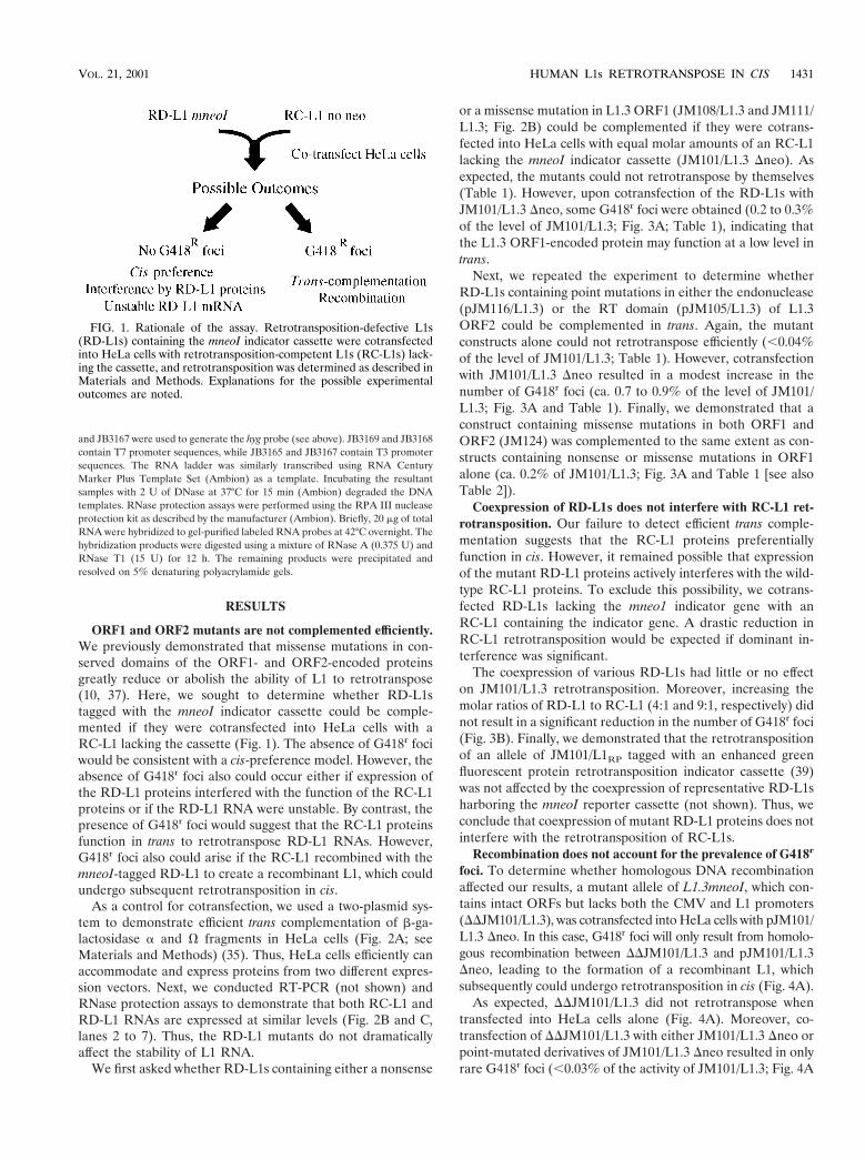
ORF1 and ORF2 mutants are not complemented efficiently.We previously demonstrated that missense mutations in con-served domains of the ORF1- and ORF2-encoded proteinsgreatly reduce or abolish the ability of L1 to retrotranspose(10, 37). Here, we sought to determine whether RD-L1stagged with the mneoI indicator cassette could be comple-mented if they were cotransfected into HeLa cells with aRC-L1 lacking the cassette (Fig. 1). The absence of G418r fociwould be consistent with a cis-preference model. However, theabsence of G418r foci also could occur either if expression ofthe RD-L1 proteins interfered with the function of the RC-L1proteins or if the RD-L1 RNA were unstable. By contrast, thepresence of G418r foci would suggest that the RC-L1 proteinsfunction in trans to retrotranspose RD-L1 RNAs. However,G418r foci also could arise if the RC-L1 recombined with themneoI-tagged RD-L1 to create a recombinant L1, which couldundergo subsequent retrotransposition in cis.
As a control for cotransfection, we used a two-plasmid sys-tem to demonstrate efficient trans complementation of b-ga-lactosidase a and V fragments in HeLa cells (Fig. 2A; seeMaterials and Methods) (35). Thus, HeLa cells efficiently canaccommodate and express proteins from two different expres-sion vectors. Next, we conducted RT-PCR (not shown) andRNase protection assays to demonstrate that both RC-L1 andRD-L1 RNAs are expressed at similar levels (Fig. 2B and C,lanes 2 to 7). Thus, the RD-L1 mutants do not dramaticallyaffect the stability of L1 RNA.
We first asked whether RD-L1s containing either a nonsense
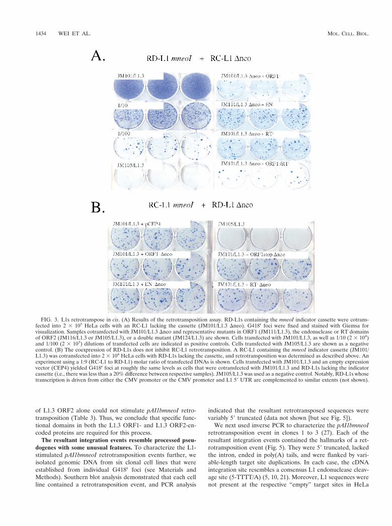
or a missense mutation in L1.3 ORF1 (JM108/L1.3 and JM111/L1.3; Fig. 2B) could be complemented if they were cotrans-fected into HeLa cells with equal molar amounts of an RC-L1lacking the mneoI indicator cassette (JM101/L1.3 Dneo). Asexpected, the mutants could not retrotranspose by themselves(Table 1). However, upon cotransfection of the RD-L1s withJM101/L1.3 Dneo, some G418r foci were obtained (0.2 to 0.3%of the level of JM101/L1.3; Fig. 3A; Table 1), indicating thatthe L1.3 ORF1-encoded protein may function at a low level intrans.
Next, we repeated the experiment to determine whetherRD-L1s containing point mutations in either the endonuclease(pJM116/L1.3) or the RT domain (pJM105/L1.3) of L1.3ORF2 could be complemented in trans. Again, the mutantconstructs alone could not retrotranspose efficiently (,0.04%of the level of JM101/L1.3; Table 1). However, cotransfectionwith JM101/L1.3 Dneo resulted in a modest increase in thenumber of G418r foci (ca. 0.7 to 0.9% of the level of JM101/L1.3; Fig. 3A and Table 1). Finally, we demonstrated that aconstruct containing missense mutations in both ORF1 andORF2 (JM124) was complemented to the same extent as con-structs containing nonsense or missense mutations in ORF1alone (ca. 0.2% of JM101/L1.3; Fig. 3A and Table 1 [see alsoTable 2]).
Coexpression of RD-L1s does not interfere with RC-L1 ret-rotransposition. Our failure to detect efficient trans comple-mentation suggests that the RC-L1 proteins preferentiallyfunction in cis. However, it remained possible that expressionof the mutant RD-L1 proteins actively interferes with the wild-type RC-L1 proteins. To exclude this possibility, we cotrans-fected RD-L1s lacking the mneo1 indicator gene with anRC-L1 containing the indicator gene. A drastic reduction inRC-L1 retrotransposition would be expected if dominant in-terference was significant.
The coexpression of various RD-L1s had little or no effecton JM101/L1.3 retrotransposition. Moreover, increasing themolar ratios of RD-L1 to RC-L1 (4:1 and 9:1, respectively) didnot result in a significant reduction in the number of G418r foci(Fig. 3B). Finally, we demonstrated that the retrotranspositionof an allele of JM101/L1RP tagged with an enhanced greenfluorescent protein retrotransposition indicator cassette (39)was not affected by the coexpression of representative RD-L1sharboring the mneoI reporter cassette (not shown). Thus, weconclude that coexpression of mutant RD-L1 proteins does notinterfere with the retrotransposition of RC-L1s.
Recombination does not account for the prevalence of G418r
foci. To determine whether homologous DNA recombinationaffected our results, a mutant allele of L1.3mneoI, which con-tains intact ORFs but lacks both the CMV and L1 promoters(DDJM101/L1.3), was cotransfected into HeLa cells with pJM101/L1.3 Dneo. In this case, G418r foci will only result from homolo-gous recombination between DDJM101/L1.3 and pJM101/L1.3Dneo, leading to the formation of a recombinant L1, whichsubsequently could undergo retrotransposition in cis (Fig. 4A).
As expected, DDJM101/L1.3 did not retrotranspose whentransfected into HeLa cells alone (Fig. 4A). Moreover, co-transfection of DDJM101/L1.3 with either JM101/L1.3 Dneo orpoint-mutated derivatives of JM101/L1.3 Dneo resulted in onlyrare G418r foci (,0.03% of the activity of JM101/L1.3; Fig. 4A
FIG. 1. Rationale of the assay. Retrotransposition-defective L1s(RD-L1s) containing the mneoI indicator cassette were cotransfectedinto HeLa cells with retrotransposition-competent L1s (RC-L1s) lack-ing the cassette, and retrotransposition was determined as described inMaterials and Methods. Explanations for the possible experimentaloutcomes are noted.
VOL. 21, 2001 HUMAN L1s RETROTRANSPOSE IN CIS 1431
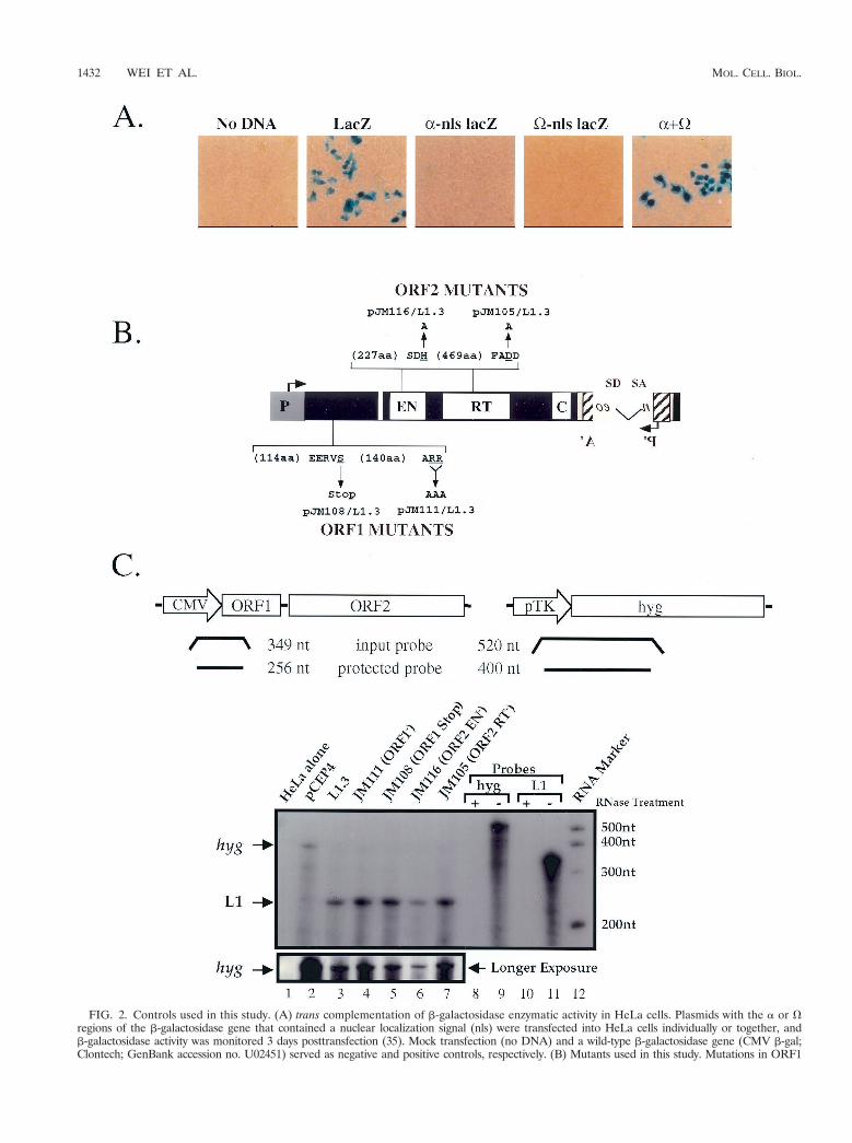
FIG. 2. Controls used in this study. (A) trans complementation of b-galactosidase enzymatic activity in HeLa cells. Plasmids with the a or Vregions of the b-galactosidase gene that contained a nuclear localization signal (nls) were transfected into HeLa cells individually or together, andb-galactosidase activity was monitored 3 days posttransfection (35). Mock transfection (no DNA) and a wild-type b-galactosidase gene (CMV b-gal;Clontech; GenBank accession no. U02451) served as negative and positive controls, respectively. (B) Mutants used in this study. Mutations in ORF1
1432 WEI ET AL. MOL. CELL. BIOL.
and data not shown). Thus, homologous recombination cannotaccount for the G418r foci observed in Fig. 3A.
G418r foci must arise by trans complementation. To provethat the RC-L1 proteins could function in trans, we sought todetermine whether the retrotransposition of L1.3 ORF1mneoIcould be stimulated by the cotransfection of JM101/L1.3 Dneo(Fig. 4B). Here, G418r foci will arise only if the JM101/L1.3ORF2-encoded protein functions in trans to retrotranspose theL1.3 ORF1mneoI RNA. Since L1.3 ORF1mneoI completelylacks ORF2 sequences, it is difficult to envision how homology-dependent DNA recombination would recreate a recombinantL1 that could undergo subsequent retrotransposition in cis.
As expected, L1.3 ORF1mneoI was unable to retrotransposewhen transfected into HeLa cells alone (Table 1). However,upon cotransfection with JM101/L1.3 Dneo, G418r foci wereobtained at levels comparable to those of RD-L1s containingmutations in L1.3 ORF2 (Table 1). Next, we pooled the G418r
foci obtained in these experiments and established three poly-clonal cell lines. We isolated genomic DNA from each cell lineand conducted PCR to determine whether the resultant retro-transposition events had the predicted structures. In each sam-ple we detected the predicted product, indicating that ORF1was linked physically to the retrotransposed mneoI indicatorcassette (Fig. 4C). Thus, we conclude that the G418r foci ob-tained in these experiments arise because of trans complemen-tation.
L1-encoded proteins can promote the retrotransposition ofother cellular mRNAs. The finding that the RC-L1 proteinscould function in trans led us to ask whether other cellularmRNAs could also serve as substrates for the L1 retrotrans-
position machinery. Thus, we constructed a variety of plasmin-ogen activator inhibitor 1 expression cassettes tagged with themneoI indicator cassette [pPAI1(a-c)mneoI; see Materials andMethods]. We chose these cDNAs because they are expressedat relatively high levels in human cells (13). Indeed, the ex-pression of each cDNA was confirmed by RT-PCR (notshown). Notably, we only used DNA sequences correspondingto the gene region of PAI1; thus, polyadenylation will occur atthe SV40 pA site present in the pCEP4 expression vector (36,37).
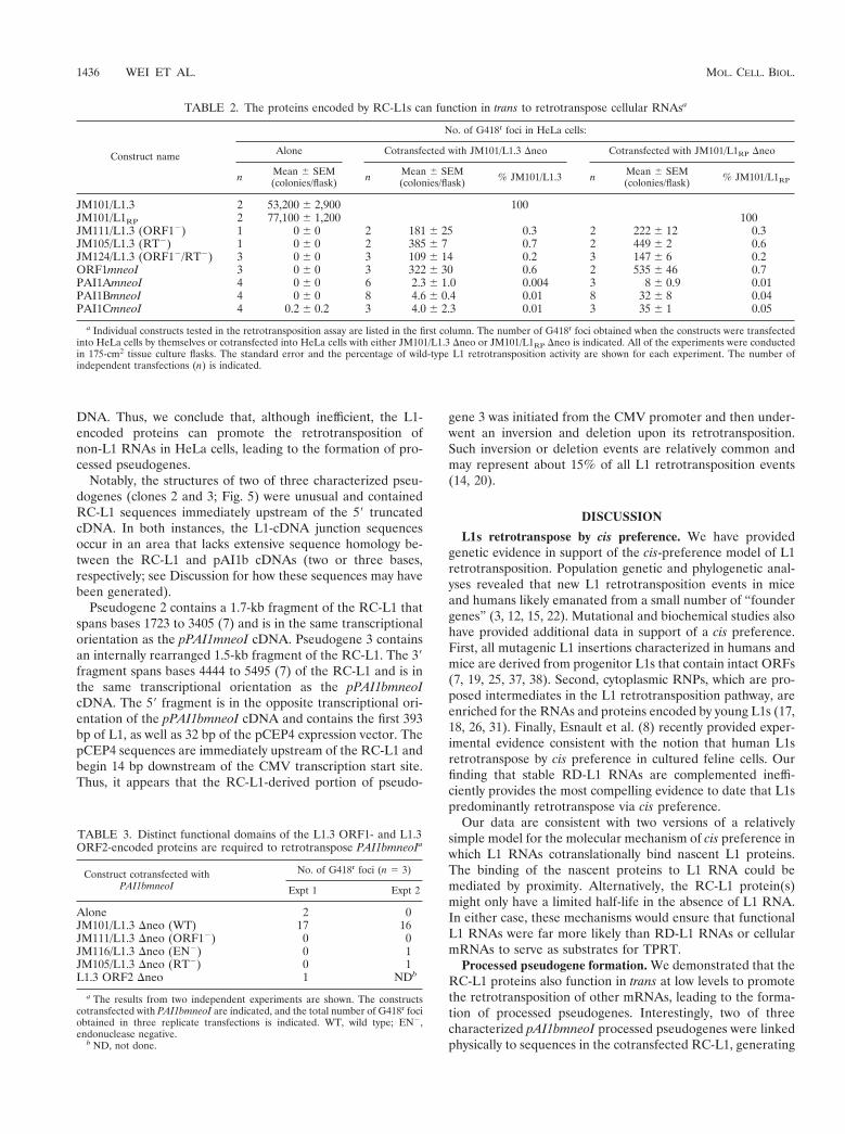
The resultant constructs were cotransfected into HeLa cellswith either JM101/L1.3 Dneo or JM101/L1RP Dneo, a secondRC-L1 that retrotransposes at a slightly higher frequency thanJM101/L1.3 (25). As before, JM101/L1.3 and JM101/L1RP ret-rotransposed extremely efficiently, and L1.3 ORF1mneoI andRD-L1s were complemented at about 0.3 to 0.7% of the levelof their respective controls (Table 2). By contrast, the PAI1(a-c)mneoI constructs were complemented at reproducibly far lowerlevels (ca. 0.004 to 0.05% of the respective wild-type controls;Table 2). Notably, a construct containing a von Willebrand factorexpression cassette tagged with mneoI retrotransposed at a simi-lar low frequency (not shown).
Functional domains in both the L1.3 ORF1- and ORF2-encoded proteins are required for the retrotransposition ofcellular RNAs. We determined that retrotransposition ofpAI1bmneoI was dependent on both the L1.3 ORF1- and L1.3ORF2-encoded proteins. Missense mutations in either L1.3ORF1 or in the endonuclease or RT domains of L1.3 ORF2were unable to stimulate the retrotransposition of pAI1bmneoIabove background levels (Table 3). Moreover, cotransfection
TABLE 1. Retrotransposition frequencies of the constructsa
Group Constructname
No. of G418r foci in HeLa cells:
Alone Cotransfected with JM101/L1.3 Dneo
n Mean 6 SEM(colonies/well) n Mean 6 SEM
(colonies/well) % JM101/L1.3
Wild type JM101/L1.3 12 6,100 6 910 100
ORF1 mutantsMissense JM111/L1.3 12 0 6 0 21 16 6 1 0.3Nonsense JM108/L1.3 12 0 6 0 18 13 6 1 0.2
ORF2 mutantsEN2 JM116/L1.3 12 2.7 6 0.6 21 55 6 7 0.9RT2 JM105/L1.3 12 0.1 6 0.1 21 41 6 4 0.7ORF12
ORF22JM124/L1.3 6 0 6 0 6 12 6 1 0.2
ORF1 alone ORF1mneoI 18 0 6 0 18 25 6 4 0.4
a Individual constructs tested in the retrotransposition assay are listed. The number of G418r foci obtained when the constructs were transfected into HeLa cells bythemselves or cotransfected into HeLa cells with JM101/L1.3 Dneo is indicated. All of the experiments were conducted in six-well tissue culture dishes. The standarderror and percentage of JM101/L1.3 retrotransposition activity are shown for each experiment. The number of independent transfections (n) is also given. EN2,endonuclease negative.
or the endonuclease or RT domains of L1.3mneoI are indicated. The wild-type amino acids that were mutated are underlined. The arrows indicatethe mutant amino acid sequence changes (e.g., ARR was changed to AAA). (C) RNA expression of representative L1 constructs. Structures ofthe hyg (hygromycin resistance gene) and L1 probes. Sizes of the full-length input and protected bands are indicated at the top of the figure. RNaseprotection assays were carried out of total RNAs prepared from HeLa cells transfected with the indicated plasmids. Probes that have undergonethe RNase protection assay with (lanes 8 and 10) or without (lanes 9 and 11) the addition of RNase are shown. A longer exposure of thepCEP4-derived hyg transcripts, which serves as an internal control, is shown in the bottom panel. Consistent with earlier studies, we were unableto detect the expression of endogenous L1 transcripts in HeLa cells (43).
VOL. 21, 2001 HUMAN L1s RETROTRANSPOSE IN CIS 1433
of L1.3 ORF2 alone could not stimulate pAI1bmneoI retro-transposition (Table 3). Thus, we conclude that specific func-tional domains in both the L1.3 ORF1- and L1.3 ORF2-en-coded proteins are required for this process.
The resultant integration events resemble processed pseu-dogenes with some unusual features. To characterize the L1-stimulated pAI1bmneoI retrotransposition events further, weisolated genomic DNA from six clonal cell lines that wereestablished from individual G418r foci (see Materials andMethods). Southern blot analysis demonstrated that each cellline contained a retrotransposition event, and PCR analysis
indicated that the resultant retrotransposed sequences werevariably 59 truncated (data not shown [but see Fig. 5]).
We next used inverse PCR to characterize the pAI1bmneoIretrotransposition event in clones 1 to 3 (27). Each of theresultant integration events contained the hallmarks of a ret-rotransposition event (Fig. 5). They were 59 truncated, lackedthe intron, ended in poly(A) tails, and were flanked by vari-able-length target site duplications. In each case, the cDNAintegration site resembles a consensus L1 endonuclease cleav-age site (5-TTTT/A) (5, 10, 21). Moreover, L1 sequences werenot present at the respective “empty” target sites in HeLa
FIG. 3. L1s retrotranspose in cis. (A) Results of the retrotransposition assay. RD-L1s containing the mneoI indicator cassette were cotrans-fected into 2 3 105 HeLa cells with an RC-L1 lacking the cassette (JM101/L1.3 Dneo). G418r foci were fixed and stained with Giemsa forvisualization. Samples cotransfected with JM101/L1.3 Dneo and representative mutants in ORF1 (JM111/L1.3), the endonuclease or RT domainsof ORF2 (JM116/L1.3 or JM105/L1.3), or a double mutant (JM124/L1.3) are shown. Cells transfected with JM101/L1.3, as well as 1/10 (2 3 104)and 1/100 (2 3 103) dilutions of transfected cells are indicated as positive controls. Cells transfected with JM105/L1.3 are shown as a negativecontrol. (B) The coexpression of RD-L1s does not inhibit RC-L1 retrotransposition. A RC-L1 containing the mneoI indicator cassette (JM101/L1.3) was cotransfected into 2 3 104 HeLa cells with RD-L1s lacking the cassette, and retrotransposition was determined as described above. Anexperiment using a 1:9 (RC-L1 to RD-L1) molar ratio of transfected DNAs is shown. Cells transfected with JM101/L1.3 and an empty expressionvector (CEP4) yielded G418r foci at roughly the same levels as cells that were cotransfected with JM101/L1.3 and RD-L1s lacking the indicatorcassette (i.e., there was less than a 20% difference between respective samples). JM105/L1.3 was used as a negative control. Notably, RD-L1s whosetranscription is driven from either the CMV promoter or the CMV promoter and L1 59 UTR are complemented to similar extents (not shown).
1434 WEI ET AL. MOL. CELL. BIOL.
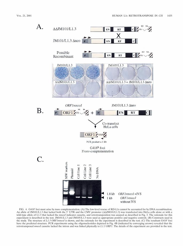
FIG. 4. G418r foci must arise by trans complementation. (A) The low-level rescue of RD-L1s cannot be accounted for by DNA recombination.An allele of JM101/L1.3 that lacked both the 59 UTR and the CMV promoter (DDJM101/L1.3) was transfected into HeLa cells alone or with awild-type allele of L1.3 that lacked the mneoI indicator cassette, and retrotransposition was assayed as described in Fig. 3. The rationale for thisexperiment is described in the text. JM101/L1.3 and JM105/L1.3 were used as appropriate positive and negative controls. (B) Constructs used inthe study. The structure of L1.3 ORF1mneoI is shown, and the rationale for the experiment is described in the text. (C) The resultant G418r focihave the predicted structure. PCR experiments using the oligonucleotides depicted in Fig. 4B (indicated by converging arrows) revealed that theretrotransposed mneoI cassette lacked the intron and was linked physically to L1.3 ORF1. The details of the experiment are provided in the text.
VOL. 21, 2001 HUMAN L1s RETROTRANSPOSE IN CIS 1435
DNA. Thus, we conclude that, although inefficient, the L1-encoded proteins can promote the retrotransposition ofnon-L1 RNAs in HeLa cells, leading to the formation of pro-cessed pseudogenes.
Notably, the structures of two of three characterized pseu-dogenes (clones 2 and 3; Fig. 5) were unusual and containedRC-L1 sequences immediately upstream of the 59 truncatedcDNA. In both instances, the L1-cDNA junction sequencesoccur in an area that lacks extensive sequence homology be-tween the RC-L1 and pAI1b cDNAs (two or three bases,respectively; see Discussion for how these sequences may havebeen generated).
Pseudogene 2 contains a 1.7-kb fragment of the RC-L1 thatspans bases 1723 to 3405 (7) and is in the same transcriptionalorientation as the pPAI1mneoI cDNA. Pseudogene 3 containsan internally rearranged 1.5-kb fragment of the RC-L1. The 39fragment spans bases 4444 to 5495 (7) of the RC-L1 and is inthe same transcriptional orientation as the pPAI1bmneoIcDNA. The 59 fragment is in the opposite transcriptional ori-entation of the pPAI1bmneoI cDNA and contains the first 393bp of L1, as well as 32 bp of the pCEP4 expression vector. ThepCEP4 sequences are immediately upstream of the RC-L1 andbegin 14 bp downstream of the CMV transcription start site.Thus, it appears that the RC-L1-derived portion of pseudo-
gene 3 was initiated from the CMV promoter and then under-went an inversion and deletion upon its retrotransposition.Such inversion or deletion events are relatively common andmay represent about 15% of all L1 retrotransposition events(14, 20).
DISCUSSION
L1s retrotranspose by cis preference. We have providedgenetic evidence in support of the cis-preference model of L1retrotransposition. Population genetic and phylogenetic anal-yses revealed that new L1 retrotransposition events in miceand humans likely emanated from a small number of “foundergenes” (3, 12, 15, 22). Mutational and biochemical studies alsohave provided additional data in support of a cis preference.First, all mutagenic L1 insertions characterized in humans andmice are derived from progenitor L1s that contain intact ORFs(7, 19, 25, 37, 38). Second, cytoplasmic RNPs, which are pro-posed intermediates in the L1 retrotransposition pathway, areenriched for the RNAs and proteins encoded by young L1s (17,18, 26, 31). Finally, Esnault et al. (8) recently provided exper-imental evidence consistent with the notion that human L1sretrotranspose by cis preference in cultured feline cells. Ourfinding that stable RD-L1 RNAs are complemented ineffi-ciently provides the most compelling evidence to date that L1spredominantly retrotranspose via cis preference.
Our data are consistent with two versions of a relativelysimple model for the molecular mechanism of cis preference inwhich L1 RNAs cotranslationally bind nascent L1 proteins.The binding of the nascent proteins to L1 RNA could bemediated by proximity. Alternatively, the RC-L1 protein(s)might only have a limited half-life in the absence of L1 RNA.In either case, these mechanisms would ensure that functionalL1 RNAs were far more likely than RD-L1 RNAs or cellularmRNAs to serve as substrates for TPRT.
Processed pseudogene formation. We demonstrated that theRC-L1 proteins also function in trans at low levels to promotethe retrotransposition of other mRNAs, leading to the forma-tion of processed pseudogenes. Interestingly, two of threecharacterized pAI1bmneoI processed pseudogenes were linkedphysically to sequences in the cotransfected RC-L1, generating
TABLE 2. The proteins encoded by RC-L1s can function in trans to retrotranspose cellular RNAsa
Construct name
No. of G418r foci in HeLa cells:
Alone Cotransfected with JM101/L1.3 Dneo Cotransfected with JM101/L1RP Dneo
n Mean 6 SEM(colonies/flask) n Mean 6 SEM
(colonies/flask) % JM101/L1.3 n Mean 6 SEM(colonies/flask) % JM101/L1RP
JM101/L1.3 2 53,200 6 2,900 100JM101/L1RP 2 77,100 6 1,200 100JM111/L1.3 (ORF12) 1 0 6 0 2 181 6 25 0.3 2 222 6 12 0.3JM105/L1.3 (RT2) 1 0 6 0 2 385 6 7 0.7 2 449 6 2 0.6JM124/L1.3 (ORF12/RT2) 3 0 6 0 3 109 6 14 0.2 3 147 6 6 0.2ORF1mneoI 3 0 6 0 3 322 6 30 0.6 2 535 6 46 0.7PAI1AmneoI 4 0 6 0 6 2.3 6 1.0 0.004 3 8 6 0.9 0.01PAI1BmneoI 4 0 6 0 8 4.6 6 0.4 0.01 8 32 6 8 0.04PAI1CmneoI 4 0.2 6 0.2 3 4.0 6 2.3 0.01 3 35 6 1 0.05
a Individual constructs tested in the retrotransposition assay are listed in the first column. The number of G418r foci obtained when the constructs were transfectedinto HeLa cells by themselves or cotransfected into HeLa cells with either JM101/L1.3 Dneo or JM101/L1RP Dneo is indicated. All of the experiments were conductedin 175-cm2 tissue culture flasks. The standard error and the percentage of wild-type L1 retrotransposition activity are shown for each experiment. The number ofindependent transfections (n) is indicated.
TABLE 3. Distinct functional domains of the L1.3 ORF1- and L1.3ORF2-encoded proteins are required to retrotranspose PAI1bmneoIa
Construct cotransfected withPAI1bmneoI
No. of G418r foci (n 5 3)
Expt 1 Expt 2
Alone 2 0JM101/L1.3 Dneo (WT) 17 16JM111/L1.3 Dneo (ORF12) 0 0JM116/L1.3 Dneo (EN2) 0 1JM105/L1.3 Dneo (RT2) 0 1L1.3 ORF2 Dneo 1 NDb
a The results from two independent experiments are shown. The constructscotransfected with PAI1bmneoI are indicated, and the total number of G418r fociobtained in three replicate transfections is indicated. WT, wild type; EN2,endonuclease negative.
b ND, not done.
1436 WEI ET AL. MOL. CELL. BIOL.
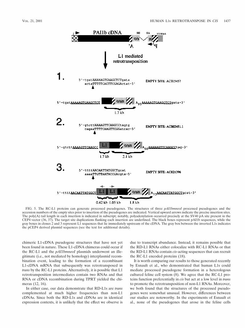
chimeric L1-cDNA pseudogene structures that have not yetbeen found in nature. These L1-cDNA chimeras could occur ifthe RC-L1 and the pAI1bmneoI plasmids underwent an ille-gitimate (i.e., not mediated by homology) interplasmid recom-bination event, leading to the formation of a recombinantL1-cDNA mRNA that subsequently was retrotransposed intrans by the RC-L1 proteins. Alternatively, it is possible that L1retrotransposition intermediates contain two RNAs and thatRNA or cDNA recombination during TPRT yielded the chi-meras (12, 16).
In either case, our data demonstrate that RD-L1s are transcomplemented at much higher frequencies than non-L1cDNAs. Since both the RD-L1s and cDNAs are in identicalexpression contexts, it is unlikely that the effect we observe is
due to transcript abundance. Instead, it remains possible thatthe RD-L1 RNAs either colocalize with RC-L1 RNAs or thatthe RD-L1 RNAs contain cis-acting sequences that can recruitthe RC-L1 encoded proteins (18).
It is worth comparing our results to those generated recentlyby Esnault et al., who demonstrated that human L1s couldmediate processed pseudogene formation in a heterologouscultured feline cell system (8). We agree that the RC-L1 pro-teins function preferentially in cis but act at a low level in transto promote the retrotransposition of non-L1 RNAs. Moreover,we both found that the structures of the processed pseudo-genes were somewhat unusual. However, differences betweenour studies are noteworthy. In the experiments of Esnault etal., none of the pseudogenes that arose in the feline cells
FIG. 5. The RC-L1 proteins can generate processed pseudogenes. The structures of three pAI1bmneoI processed pseudogenes and theaccession numbers of the empty sites prior to insertion of the pseudogenes are indicated. Vertical upward arrows indicate the precise insertion sites.The poly(A) tail length in each insertion is indicated in subscript; notably, polyadenylation occurred precisely at the SV40 pA site present in theCEP4 vector (36, 37). The target site duplications flanking each insertion are underlined. The black boxes represent pAI1b sequences, while thegray boxes in clones 2 and 3 represent L1 sequences that lie immediately upstream of the cDNA. The gray box between the inverted L1s indicatesthe pCEP4 derived plasmid sequences (see the text for additional details).
VOL. 21, 2001 HUMAN L1s RETROTRANSPOSE IN CIS 1437
integrated at consensus L1 endonuclease cleavage sites. In-deed, two of three “pseudogenes” actually lacked poly(A) se-quences. All three of our pseudogenes had all the character-istics of retrotransposition events generated via TPRT.
In addition, our data demonstrate that pseudogene forma-tion in human cells is extremely rare (ca. 0.01 to 0.05% of therate of L1 retrotransposition) and only is detected when ourmost “active” L1s are used as sources of the L1-encoded pro-teins. By contrast, in feline-cultured cells, the frequency ofprocessed pseudogene formation is only 10-fold lower than thefrequency of L1 retrotransposition. While it remains possiblethat these discrepancies reflect subtle differences that existbetween our assays, it is interesting to note that both we andDhellin et al. were unable to detect pseudogene formationwhen L1.2 was overexpressed in human cells (6; J. V. Moran etal., unpublished data). Thus, it appears that the feline cells aremore permissive for L1-mediated processed pseudogene for-mation than human HeLa cells.
Finally, it is notable that the studies conducted by Esnault etal. were performed in the presence of phleomycin, a knownclastogen. Thus, it remains possible that interplasmid recom-bination occurred more frequently in their study. Moreover,the unusual structures of the pseudogenes suggest that theymay have integrated into chromosomal DNA by an L1 endo-nuclease-independent mechanism.
Evolutionary implications and practical considerations ofthe cis-preference model. The cis-preference model would ex-plain how a small number of autonomous L1s remain retro-transposition competent among an overwhelming abundanceof nonfunctional elements. Indeed, such a mechanism wouldselect for the retrotransposition of RC-L1 RNAs and would beconsistent with the apparent patterns of concerted evolutionthat L1s display in different species (15, 32). It also would limitthe extent to which the proteins encoded by RC-L1s couldfunction to retrotranspose other cellular RNAs (e.g., RD-L1sand other cellular RNAs). However, it is noteworthy that par-ticular RNAs (e.g., Alu RNAs) likely have evolved ways tousurp the cis-preference retrotransposition machinery of hu-man RC-L1s (possible mechanisms are discussed further else-where [1, 23, 33]).
Finally, the finding that L1s retrotranspose by cis preferencemay have practical value. For example, if engineered L1s wereused as transposon mutagens, there is a high likelihood thatany resultant mutations would be due to the retrotranspositionof the engineered L1 RNA and would not be caused by thetrans mobilization of endogenous retrotransposons. Indeed,the inability of the RC-L1 proteins to efficiently mobilize cel-lular RNAs may prove useful when considering L1 as a poten-tial gene delivery vehicle.
ACKNOWLEDGMENTS
We thank Anne Marie DesLauriers at the University of MichiganFlow Core for help with flow cytometry, Robert Lyons at the Univer-sity of Michigan DNA Sequencing Core for help with oligonucleotidesynthesis and DNA sequencing, and Ali Lotia for help with generatingcomputer graphics. We thank David Ginsburg for providing pAI1cDNAs. We thank Alice Telesnitsky, John Goodier, Eline LuningPrak, Tom Glaser, Dennis Hartigan-O’Connor, and current membersof the Moran Lab for critical reading of the manuscript and for helpfuldiscussions during the course of this work.
This work was supported in part by a Damon Runyon Scholar
Award (J.V.M.) and National Institutes of Health grants GM60518(J.V.M.) and CA16519 (J.D.B.).
REFERENCES
1. Boeke, J. D. 1997. LINEs and Alus—the polyA connection. Nat. Genet.16:6–7.
2. Boeke, J. D., and J. P. Stoye. 1997. Retrotransposons, endogenous retrovi-ruses, and the evolution of retroelements, p. 343–435. In J. M. Coffin, S. H.Hughes, and H. E. Varmus (ed.), Retroviruses. Cold Spring Harbor Labo-ratory Press, Cold Spring Harbor, N.Y.
3. Boissinot, S., P. Chevret, and A. V. Furano. 2000. L1 (LINE-1) retrotrans-poson evolution and amplification in recent human history. Mol. Biol. Evol.17:915–928.
4. Christensen, S., G. Pont-Kingdon, and D. Carroll. 2000. Target specificity ofthe endonuclease from the Xenopus laevis non-lung terminal repeat retro-transposon, Tx1L. Mol. Cell. Biol. 20:1219–1226.
5. Cost, G. J., and J. D. Boeke. 1998. Targeting of human retrotransposonintegration is directed by the specificity of the L1 endonuclease for regionsof unusual DNA structure. Biochemistry 37:18081–18093.
6. Dhellin, O., J. Maestre, and T. Heidmann. 1997. Functional differencesbetween the human LINE retrotransposon and retroviral reverse transcrip-tases for in vivo mRNA reverse transcription. EMBO J. 16:6590–6602.
7. Dombroski, B. A., S. L. Mathias, E. Nanthakumar, A. F. Scott, and H. H.Kazazian, Jr. 1991. Isolation of an active human transposable element.Science 254:1805–1808.
8. Esnault, C., J. Maestre, and T. Heidmann. 2000. Human LINE retrotrans-posons generate processed pseudogenes. Nat. Genet. 24:363–367.
9. Fanning, T., and M. Singer. 1987. The LINE-1 DNA sequences in fourmammalian orders predict proteins that conserve homologies to retrovirusproteins. Nucleic Acids Res. 15:2251–2260.
10. Feng, Q., J. V. Moran, H. H. Kazazian, Jr., and J. D. Boeke. 1996. HumanL1 retrotransposon encodes a conserved endonuclease required for retro-transposition. Cell 87:905–916.
11. Feng, Q., G. Schumann, and J. D. Boeke. 1998. Retrotransposon R1Bmendonuclease cleaves the target sequence. Proc. Natl. Acad. Sci. USA 95:2083–2088.
12. Furano, A. V. 2000. The biological properties and evolutionary dynamics ofmammalian LINE-1 retrotransposons. Prog. Nucleic Acid Res. Mol. Biol.64:255–294.
13. Ginsburg, D., R. Zeheb, A. Y. Yang, U. M. Rafferty, P. A. Andreasen, L.Nielsen, K. Dano, R. V. Lebo, and T. D. Gelehrter. 1986. cDNA cloning ofhuman plasminogen activator-inhibitor from endothelial cells. J. Clin. Inves-tig. 78:1673–1680.
14. Goodier, J. L., E. M. Ostertag, and H. H. Kazazian, Jr. 2000. Transductionof 39-flanking sequences is common in L1 retrotransposition. Hum. Mol.Genet. 9:653–657.
15. Hardies, S. C., S. L. Martin, C. F. Voliva, C. A. D. Hutchison, and M. H.Edgell. 1986. An analysis of replacement and synonymous changes in therodent L1 repeat family. Mol. Biol. Evol. 3:109–125.
16. Hayward, B. E., M. Zavanelli, and A. V. Furano. 1997. Recombinationcreates novel L1 (LINE-1) elements in Rattus norvegicus. Genetics 146:641–654.
17. Hohjoh, H., and M. F. Singer. 1996. Cytoplasmic ribonucleoprotein com-plexes containing human LINE-1 protein and RNA. EMBO J. 15:630–639.
18. Hohjoh, H., and M. F. Singer. 1997. Sequence-specific single-strand RNAbinding protein encoded by the human LINE-1 retrotransposon. EMBO J.16:6034–6043.
19. Holmes, S. E., B. A. Dombroski, C. M. Krebs, C. D. Boehm, and H. H.Kazazian, Jr. 1994. A new retrotransposable human L1 element from theLRE2 locus on chromosome 1q produces a chimaeric insertion. Nat. Genet.7:143–148.
20. Hutchison, C. A., S. C. Hardies, D. D. Loeb, W. R. Shehee, and M. H. Edgell.1989. LINES and related retroposons: long interspersed sequences in theeucaryotic genome, p. 593–617. In D. E. Berg and M. M. Howe (ed.), MobileDNA. ASM Press, Washington, D.C.
21. Jurka, J. 1997. Sequence patterns indicate an enzymatic involvement inintegration of mammalian retroposons. Proc. Natl. Acad. Sci. USA 94:1872–1877.
22. Kaplan, N., T. Darden, and C. H. Langley. 1985. Evolution and extinction oftransposable elements in Mendelian populations. Genetics 109:459–480.
23. Kazazian, H. H., Jr., and J. V. Moran. 1998. The impact of L1 retrotrans-posons on the human genome. Nat. Genet. 19:19–24.
24. Kazazian, H. H., Jr., C. Wong, H. Youssoufian, A. F. Scott, D. G. Phillips,and S. E. Antonarakis. 1988. Haemophilia A resulting from de novo inser-tion of L1 sequences represents a novel mechanism for mutation in man.Nature 332:164–166.
25. Kimberland, M. L., V. Divoky, J. Prchal, U. Schwahn, W. Berger, and H. H.Kazazian, Jr. 1999. Full-length human L1 insertions retain the capacity forhigh-frequency retrotransposition in cultured cells. Hum. Mol. Genet.8:1557–1560.
26. Kolosha, V. O., and S. L. Martin. 1995. Polymorphic sequences encoding the
1438 WEI ET AL. MOL. CELL. BIOL.
first open reading frame protein from LINE-1 ribonucleoprotein particles.J. Biol. Chem. 270:2868–2873.
27. Li, J., H. Shen, K. L. Himmel, A. J. Dupuy, D. A. Largaespada, T. Nakamura,J. D. Shaughnessy, Jr., N. A. Jenkins, and N. G. Copeland. 1999. Leukaemiadisease genes: large-scale cloning and pathway predictions. Nat. Genet. 23:348–353.
28. Luan, D. D., and T. H. Eickbush. 1995. RNA template requirements fortarget DNA-primed reverse transcription by the R2 retrotransposable ele-ment. Mol. Cell. Biol. 15:3882–3891.
29. Luan, D. D., M. H. Korman, J. L. Jakubczak, and T. H. Eickbush. 1993.Reverse transcription of R2Bm RNA is primed by a nick at the chromosomaltarget site: a mechanism for non-LTR retrotransposition. Cell 72:595–605.
30. Maestre, J., T. Tchenio, O. Dhellin, and T. Heidmann. 1995. mRNA ret-roposition in human cells: processed pseudogene formation. EMBO J. 14:6333–6338.
31. Martin, S. L. 1991. Ribonucleoprotein particles with LINE-1 RNA in mouseembryonal carcinoma cells. Mol. Cell. Biol. 11:4804–4807.
32. Martin, S. L., C. F. Voliva, S. C. Hardies, M. H. Edgell, and C. A. D.Hutchison. 1985. Tempo and mode of concerted evolution in the L1 repeatfamily of mice. Mol. Biol. Evol. 2:127–140.
33. Mathews, D. H., A. R. Banerjee, D. D. Luan, T. H. Eickbush, and D. H.Turner. 1997. Secondary structure model of the RNA recognized by thereverse transcriptase from the R2 retrotransposable element. RNA 3:1–16.
34. Mathias, S. L., A. F. Scott, H. H. Kazazian, Jr., J. D. Boeke, and A. Gabriel.1991. Reverse transcriptase encoded by a human transposable element.Science 254:1808–1810.
35. Mohler, W. A., and H. M. Blau. 1996. Gene expression and cell fusionanalyzed by lacZ complementation in mammalian cells. Proc. Natl. Acad.Sci. USA 93:12423–12427.
36. Moran, J. V., R. J. DeBerardinis, and H. H. Kazazian. 1999. Exon shufflingby L1 retrotransposition. Science 283:1530–1534.
37. Moran, J. V., S. E. Holmes, T. P. Naas, R. J. DeBerardinis, J. D. Boeke, and
H. H. Kazazian, Jr. 1996. High frequency retrotransposition in culturedmammalian cells. Cell 87:917–927.
38. Naas, T. P., R. J. DeBerardinis, J. V. Moran, E. M. Ostertag, S. F.Kingsmore, M. F. Seldin, Y. Hayashizaki, S. L. Martin, and H. H. Kazazian.1998. An actively retrotransposing, novel subfamily of mouse L1 elements.EMBO J. 17:590–597.
39. Ostertag, E. M., E. T. Prak, R. J. DeBerardinis, J. V. Moran, and H. H.Kazazian, Jr. 2000. Determination of L1 retrotransposition kinetics in cul-tured cells. Nucleic Acids Res. 28:1418–1423.
40. Sassaman, D. M., B. A. Dombroski, J. V. Moran, M. L. Kimberland, T. P.Naas, R. J. DeBerardinis, A. Gabriel, G. D. Swergold, and H. H. Kazazian,Jr. 1997. Many human L1 elements are capable of retrotransposition. Nat.Genet. 16:37–43.
41. Scott, A. F., B. J. Schmeckpeper, M. Abdelrazik, C. T. Comey, B. O’Hara,J. P. Rossiter, T. Cooley, P. Heath, K. D. Smith, and L. Margolet. 1987.Origin of the human L1 elements: proposed progenitor genes deduced froma consensus DNA sequence. Genomics 1:113–125.
42. Smit, A. F. 1999. Interspersed repeats and other mementos of transposableelements in mammalian genomes. Curr. Opin. Genet. Dev. 9:657–663.
43. Swergold, G. D. 1990. Identification, characterization, and cell specificity ofa human LINE-1 promoter. Mol. Cell. Biol. 10:6718–6729.
44. Tchenio, T., E. Segal-Bendirdjian, and T. Heidmann. 1993. Generation ofprocessed pseudogenes in murine cells. EMBO J. 12:1487–1497.
45. Wei, W., T. A. Morrish, R. S. Alisch, and J. V. Moran. 2000. A transient assayreveals that cultured human cells can accommodate multiple LINE-1 retro-transposition events. Anal. Biochem. 284:435–438.
46. Weiner, A. M., P. L. Deininger, and A. Efstratiadis. 1986. Nonviral retro-posons: genes, pseudogenes, and transposable elements generated by thereverse flow of genetic information. Annu. Rev. Biochem. 55:631–661.
47. Xiong, Y. E., and T. H. Eickbush. 1988. Functional expression of a sequence-specific endonuclease encoded by the retrotransposon R2Bm. Cell 55:235–246.
VOL. 21, 2001 HUMAN L1s RETROTRANSPOSE IN CIS 1439