mechanisms for intragenic complementation at the human argininosuccinate lyase locus
TRANSCRIPT
Mechanisms for Intragenic Complementation at the Human Argininosuccinate LyaseLocus†
Bomina Yu,‡,§ Gawen D. Thompson,‡,§ Patrick Yip,‡ P. Lynne Howell,*,‡,§ and Alan R. Davidson*,§,|
Structural Biology and Biochemistry, Research Institute, Hospital for Sick Children, 555 UniVersity AVenue,Toronto, M5G 1X8, Ontario, Canada, and Departments of Biochemistry and of Molecular and Medical Genetics,
Faculty of Medicine, UniVersity of Toronto, Medical Sciences Building, Toronto, M5S 1A8, Ontario, Canada
ReceiVed July 20, 2001; ReVised Manuscript ReceiVed October 18, 2001
ABSTRACT: Argininosuccinate lyase (ASL) is a homotetrameric enzyme that catalyzes the reversible cleavageof argininosuccinate to arginine and fumarate. Deficiencies in the enzyme result in the autosomal, recessivedisorderargininosuccinic aciduria. Considerable clinical and genetic heterogeneity is associated withthis disorder, which is thought to be a consequence of the extensive intragenic complementation identifiedin patient strains. Our ability to predict genotype-phenotype relationships is hampered by the currentlack of understanding of the mechanisms by which complementation can occur. The 3-dimensional structureof wild-type ASL has enabled us to propose that the complementation between two ASL active site mutantsubunits, Q286R and D87G, occurs through a regeneration of functional active sites in the heteromutantprotein. We have reconstructed this complementation event, both in vivo and in vitro, using recombinantproteins and have confirmed this hypothesis. The complementation events between Q286R and twononactive site mutants, M360T and A398D, have also been characterized. The M360T and A398Dsubstitutions have adverse effects on the thermodynamic stability of the protein. Complementation betweeneither the M360T or the A398D mutant and the stable Q286R mutant occurs through the formation of amore stable heteromeric protein with partial recovery of catalytic activity. The detection and characterizationof a novel complementation event between the A398D and D87G mutants has shown how complementationin patients withargininosuccinic aciduriamay correlate with the clinical phenotype.
Intragenic complementation is a phenomenon wherebyparticular combinations of mutant alleles at a given locusproduce a less severe phenotype than the same alleles do inthe homozygous state, or in the presence of noncomple-menting alleles. Intragenic complementation has been dem-onstrated to occur in a number of human metabolic disorders,includingargininosuccinic aciduria(1), propionic acidemia(2, 3), andmethylmalonic aciduria(4), and is believed tocontribute to the extensive clinical heterogeneity observedamong patients suffering from these diseases. In all casesexamined, intragenic complementation has been found toinvolve genes encoding multimeric enzymes. Mutant subunitsof these enzymes, each possessing distinct amino acidsubstitutions that render the homomutant proteins inactive,interact to form heteromutant multimers possessing partialactivity. Since intragenic complementation may occur in allgenetic diseases involving multimeric proteins, the accurateprediction of genotype-phenotype relationships in thesediseases requires a complete understanding of the mecha-
nisms by which this phenomenon can occur. In this study,the metabolic enzyme argininosuccinate lyase (ASL)1 is usedas a model system to investigate intragenic complementation.
Argininosuccinate lyase (ASL, EC 4.3.2.1), a homotetra-meric protein of 50 kDa subunits (5-7), catalyzes the revers-ible breakdown of argininosuccinic acid into arginine andfumarate. This reaction is a required step in the urea cycle,the major pathway for the detoxification of ammonia. Muta-tions in the ASL gene causeargininosuccinic aciduria, anautosomal recessive disorder with considerable clinical andgenetic heterogeneity (1, 8-10). The clinical heterogeneityof the disease manifests itself in the variation of the age ofonset and the severity of the symptoms (8). Three distinctclinical phenotypes have been identified: neonatal, subacute,and late-onset. The biochemical basis of this clinical variationis unclear, as there is only partial correlation between theclinical phenotype and the residual enzyme activity detectedin cultured fibroblasts (1) and other tissues (8).
† This work was supported by Grant GP0155209 from the NaturalSciences and Engineering Research Council of Canada to P.L.H. andA.R.D. and by University of Toronto/Hospital for Sick ChildrenGraduate Research Scholarships to B.Y.
* Corresponding authors. A.R.D.: phone, 416-978-0332; fax, 416-978-6885; e-mail, [email protected]. P.L.H.: phone, 416-813-5378; fax, 416-813-5022; e-mail, [email protected].
‡ Structural Biology and Biochemistry, Hospital for Sick Children.§ Department of Biochemistry, University of Toronto.| Department of Molecular and Medical Genetics, University of
Toronto.
1 Abbreviations: ASL, argininosuccinate lyase; kDa, kilodalton(s);bp, base pair(s); wt, wild-type; Q286R-his, C-terminal 6-histidine taggedargininosuccinate lyase with a glutamine to arginine mutation at residue286; D87G-his, C-terminal 6-histidine tagged argininosuccinate lyasewith an aspartate to glycine mutation at residue 87; A398D-his,C-terminal 6-histidine tagged argininosuccinate lyase with an alanineto aspartate mutation at residue 398; M360T-his, C-terminal 6-histidinetagged argininosuccinate lyase with a methionine to threonine mutationat residue 360; IPTG, isopropyl-â-D-thiogalactopyranoside; PMSF,phenylmethylsulfonyl fluoride; MW, molecular weight; CRM, cross-reactive material; NADP-GDH, nicotinamide adenine dinucleotidephosphate-linked glutamic dehydrogenase.
15581Biochemistry2001,40, 15581-15590
10.1021/bi011526e CCC: $20.00 © 2001 American Chemical SocietyPublished on Web 11/29/2001
The genetic heterogeneity at the ASL locus was character-ized by complementation analysis of fibroblasts cultured from28 unrelated patients withargininosuccinic aciduria(1, 9).These studies showed that the fusion of certain pairs of celllines resulted in significant increases in ASL activity, whichcould be attributed to intragenic complementation. Intrigu-ingly, subsequent analysis demonstrated that patient strainscontaining an ASL allele encoding a glutamine to argininesubstitution at codon 286 (Q286R) participated in almost allof the observed complementation events (11). Intrageniccomplementation could not be detected in most othercombinations of patient strains. In addition, fusions of theQ286R-containing strains and those containing a mutantallele encoding an aspartate to glycine substitution at codon87 (D87G) resulted in 3-fold higher levels of activity thanthat seen in any other complementation event. The existenceof intragenic complementation between the Q286R andD87G mutants was directly demonstrated in COS celltransfection experiments (11). Plasmids expressing normalASL, the Q286R mutant, and the D87G mutant weretransfected into COS cells in various combinations, and therate of conversion of14C-labeled fumarate into14C-labeledargininosuccinate was measured. The D87G and Q286Rmutants showed little (∼4.5%) or no (<0.05%) activity,respectively, when transfected on their own. However, COScells transfected with both the D87G and Q286R mutantssimultaneously were found to exhibit approximately 30% ofwild-type ASL activity.
The determination of the high-resolution 3-dimensionalstructures of human ASL (12, 13) and the homologous eyelens protein, duckδ-crystallin (14-17), has suggested amechanism for the Q286R:D87G complementation event (12,18). Each of the four active sites of the ASL protein is locatedin a cleft formed by three different monomers (12, 14-17).Both D87 and Q286 lie in the active site region, but in anyone active site, each residue is contributed by a differentmonomer. A homotetramer of D87G or Q286R would beinactive since all four active sites would have an amino acidsubstitution. Heterotetramers, containing subunits with eitherthe D87G substitution or the Q286R substitution, could have0, 1, or 2 active sites with all wild-type residues dependingon the number and orientation of the different mutantsubunits in the tetrameric protein. If the subunits had equalprobability of associating, a mixture of five tetramers wouldform with D87G to Q286R ratios of 4:0, 3:1, 2:2, 1:3, and0:4 in a 1:4:6:4:1 distribution. By calculating the number ofactive sites containing all wild-type residues in each tetramerand assuming that these nativelike active sites are fullyfunctional and that both individual mutants are completelyinactive, the mixture of heterotetramers should have 25%of the catalytic activity of the wild-type protein. This valueis consistent with the 30% of wild-type activity observedfor the Q286R/D87G heterotetramers in the COS cellexperiments described above (11).
Thirteen additional mutations in the ASL gene associatedwith argininosuccinic aciduriahave been identified (11, 19-22). In contrast to the Q286R and D87G substitutions, whichare located in the ASL active site, the majority of these otherdisease-causing substitutions are dispersed throughout theASL structure. Nevertheless, the Q286R mutation was foundto participate in complementation events with almost all ofthese ASL mutants, regardless of their location in the tertiary
structure (1). In all cases, however, the recovered wild-typeASL activity was only about one-third of that for the Q286R:D87G complementation event (1).
The work presented in this paper is aimed at answeringthree questions raised by the previous studies on intrageniccomplementation at the ASL locus: (1) Why do most ofthe ASL alleles fail to display intragenic complementationin the fibroblast assay? (2) Why does the Q286R mutantcomplement almost all other ASL mutants? (3) Why doesQ286R:D87G complementation result in much higher activitythan any other observed ASL complementation event? Toaddress these questions, we have characterized the effectsof the Q286R and D87G substitutions, as well as an alanineto aspartate substitution at codon 398 (A398D) (11) and amethionine to threonine substitution at codon 360 (M360T)(21), on the recombinant ASL protein. The latter twomutants were identified in patient strains that complementwith Q286R. The A398 and M360 residues are removed fromthe active site of ASL. By coexpressing combinations ofthese mutants, we have been able to purify mutant hetero-tetramers and assess their complementation behavior in vitro.We have also assessed the thermal stability of the homo-and heteromutant proteins. The results of these experimentshave allowed us to identify two general mechanisms bywhich intragenic complementation occurs at the ASL locus.First, complementation can occur by the regeneration offunctional nativelike active sites in heterotetramers of twodistinct, stable, active site mutants. Furthermore, these stable,active site mutants can also oligomerize with unstable mutantsubunits to form hybrid tetramers with enough stability forcatalysis to occur.
MATERIALS AND METHODS
Expression Vectors.A pET-3c vector (T7 promoter, ampr,pBR322 origin) containing human ASL (pET-wtASL) wasobtained from Dr. M. Hershfield (23). The pET-3c D87G(pET-D87G) and Q286R (pET-Q286R) expression vectorswere constructed using restriction digestion and the pESP-SVTEXP expression vectors pESP-D87G and pESP-Q286Rconstructed previously (11). The pESP-D87G plasmid wasdigested withMluI andSpeI, and nucleotides 74-743 weresubcloned into the pET-wtASL plasmid, replacing the normalsequence in this region. Similarly, the pESP-Q286R plasmidwas digested withSpeI andSfiI, and nucleotides 743-1211were subcloned into the pET-wtASL plasmid. To simplifypurification, plasmids that expressed a C-terminal histidine-tagged version of the protein were also constructed. The pET-wtASL-his plasmid was constructed by amplifying the 5′ endof the gene using PCR with the addition of a six-histidinetag. The PCR product and the pET-wtASL were digestedwith SfiI and BamHI, and the digested PCR product wasligated into the analogous region of the pET-wtASL plasmidto construct pET-wtASL-his. The pET-D87G-his and pET-Q286R-his plasmids were constructed as described abovefor the pET-D87G and pET-Q286R plasmids except thatpET-wtASL-his was used instead of pET-wtASL.
The pET-3c A398D-his (pET-A398D-his) and M360T-his (pET-M360T-his) expression vectors were constructedby site-directed mutagenesis using the Unique Site Elimina-tion kit from Pharmacia. The oligonucleotides (Gibco BRL)CC TCC GGA AAA GCT GTG TTC ATGGAC GAG ACC
15582 Biochemistry, Vol. 40, No. 51, 2001 Yu et al.
and CAA GAG AACACG GGA CAG GCC TTA AGC CCwere used to generate the A398D and M360T mutations,respectively. The above mutagenic primers were used tointroduce both the desired codon (boldface) and a uniquerestriction site (underlined),Kpn2I or BspTI, respectively,in the region of the mutation. Screening for the presence ofthe new restriction site by restriction enzyme mappingidentified positive mutants. All plasmids were sequenced toensure that each mutation had been correctly introduced andthat no additional mutations existed.
For the in vivo complementation studies, coexpressionvectors containing the full-length cDNA and T7 promotorsite for either the D87G-his, A398D-his, or M360T-hismutant with the full-length cDNA and T7 promotor site forthe Q286R mutant were created. The pET-D87G-his plasmidwas digested withBglII and BamHI, and the resulting1537 bp fragment was subcloned into theBglII site of thepET-Q286R plasmid to generate the pET-D87G-his/Q286Rplasmid. The pET-A398D-his/Q286R and pET-M360T-his/Q286R plasmids were generated as above.
Protein Expression and Purification.Plasmids containingC-terminal histidine-tagged proteins were overexpressed intheE. coli strain BB101, genotypeara ∆(lac proAB)∆slyD(kanr) nalA argEam rif thiF′[lacI q proAB+] (λDE3), usingthe T7 polymerase system. Cells were harvested 3-4 h post-induction (1 mM IPTG, 37°C) for wtASL-his, Q286R-his,D87G-his, and D87G-his/Q286R, and 20 h post-induction(1 mM IPTG, 25 °C) for A398D-his, M360T-his, andheterotetramers containing either the A398D-his or theM360T-his subunit. Cells were stored at-20 °C untilrequired. Expression of the full-length A398D-his andM360T-his mutant proteins was confirmed by western blotanalysis (24) using the QIAexpressPenta•His antibody(Qiagen).
The frozen cells for the homomutant proteins were lysedby sonication in 20 mL of Buffer A (20 mM Tris-HCl, pH8, 0.5 M NaCl) with 0.5 mM PMSF. Cells were sonicatedfor a total of 10 min using a pulse sequence for 30 sinterspersed with 90 s of cooling on ice. The sonicated cellswere centrifuged (JA-20 rotor in a Beckman J2-21 centrifuge)at 15 000 rpm for 30 min at 4°C. The pellet was resuspendedin 20 mL of Buffer A and sonicated and centrifuged asdescribed above. The supernatant from both spins was pooledand applied to a Ni-affinity column (His-BindR resin,Novagen) preequilibrated with Buffer A. The column waswashed with 30 mL of 5 mM imidazole in Buffer A and 12mL each of 30 and 60 mM imidazole in Buffer A. Theprotein was eluted with 15 mL of 100 mM imidazole inBuffer A and the eluted fraction dialyzed overnight at 4°Cagainst 4 L of dialysis buffer (20 mM potassium phosphate,pH 6.5, 1 mM EDTA, 1 mM DTT, 300 mM NaCl).
The frozen cell pellets from the coexpressed heteromutantproteins were lysed and applied to the Ni-affinity columnas described above for the homomutant proteins. The columnwas washed with 30 mL of Buffer A, 30 mL of Buffer Awith 5 mM imidazole, and 12 mL each of Buffer A with thefollowing imidazole concentrations: 10, 20, 30, 60, 100, and200 mM. The fractions eluted with 30 and 60 mM imidazolewere pooled and dialyzed overnight at 4°C against 4 L ofdialysis buffer. All proteins were approximately 95% pure.
Circular Dichroism Spectroscopy.The CD spectra for allproteins were collected in a cell with a path length of 0.1
cm on an AVIV Circular Dichroism spectrophotometer(model 62A DS). The protein solution (0.15-0.20 mg/mLin 20 mM potassium phosphate, pH 7.5, 1 mM EDTA, 1mM DTT) was scanned from 200 to 260 nm in 1 nmincrements. To examine the thermal stability of each protein,the loss of ellipticity at 222 nm was monitored as the proteinsample was heat-denatured. The temperature was increasedfrom 25 to 101 °C in 2 °C increments with 1 minequilibration before each reading.
ASL ActiVity Assays.The ability of plasmid-borne ASLmutants to complement anE. coli strain with its endogenousASL gene deleted served as a simple in vivo assay for ASLactivity. E. coli strain W3678 (∆galT, LAM-, IN(rrnD-rrNE)1, ∆argH, F-) was obtained from V. L. Chan (25) andlysogenized using Novagen’sλDE3 Lysogenization Kit. Theresulting∆ASL cells transformed with various ASL-express-ing plasmids were assayed on plates containing M9 minimalmedia (24) with and without arginine supplementation. Thestreaked plates were incubated at 37°C for 48 h. The levelof growth of the transformed cells on M9 minimal mediawas compared to that for cells transformed with the plasmidcarrying the wild-type ASL protein.
The in vitro ASL enzyme activity of the wild-type andmutant proteins was determined spectrophotometrically; 10×stock solutions of sodium argininosuccinate (Sigma-Aldrich)in reaction buffer (20 mM potassium phosphate, pH 7.5,1 mM EDTA) were prepared such that the final concentrationof substrate in the reaction ranged from 0.02 to 2.0 mM.The reaction was initiated by adding 20-30µL of the proteinsolution (10-20 µg of enzyme) to the reaction mixture for atotal reaction volume of 500µL. The reaction was monitoredby measuring the increase in fumarate (λmax ) 240 nm;ε ) 2.44 mM-1 cm-1) concentration accessed by UVabsorption at 240 nm on a Milton Roy 3000 spectro-photometer. All ASL assays were performed in triplicate atroom temperature. Initial velocities were used to determinethe kinetic parameters.
In Vitro Complementation Studies.Equal quantities of theD87G-his and Q286R-his ASL mutants (0.2 mg/mL) weremixed together and incubated at either 0°C or roomtemperature (25°C). Aliquots (50µL) were taken from themixture at various intervals of time over a 6-day period anddiluted 10 times and allowed to equilibrate at room temper-ature for 30 min prior to performing the activity assay. Thediluted sample (50µL) was assayed for activity in thepresence of 5 mM substrate. The enzyme activity of allsamples was assayed in triplicate at 25°C. The activities ofwild-type ASL, individual D87G-his and Q286R-his homo-mutants, and the D87G-his/Q286R coexpressed protein,incubated at both 0°C and 25°C, were monitored as controls.The total protein concentration of the incubated mixtures was0.4 mg/mL. The results obtained were expressed as thepercentage of wild-type activity recovered over time.
RESULTS
Production of Q286R/D87G Heterotetramers by in ViVoCoexpression.To determine whether the ASL proteinexpressed inE. coli could be used as a model system forstudying the phenomenon of intragenic complementationobserved at the ASL locus, we first established that mutantheterotetramers would form when coexpressed inE. coli.
Mechanisms for Intragenic Complementation in ASL Biochemistry, Vol. 40, No. 51, 200115583
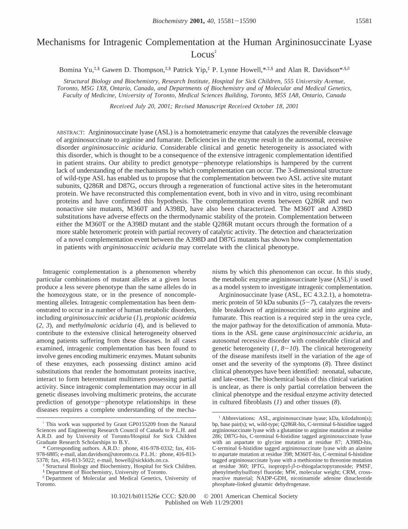
To facilitate purification of wild-type and mutant ASLproteins, the proteins were expressed with C-terminal 6-Histags. 6-His-tagged wild-type ASL and Q286R and D87GASL mutants (wtASL-his, Q286R-his, and D87G-his, respec-tively) were expressed inE. coli and yielded approximately10-20 mg of 95% pure protein per liter of cultured cells(Figure 1a). Plasmids were constructed to coexpress variouspairs of mutants within the sameE. coli cell. On eachplasmid, only one of the mutant ASL genes encoded aC-terminal 6-His tag. The objective of this approach was toproduce heterotetramers in vivo, which could subsequentlybe purified by Ni-affinity chromatography. There are fivedifferent heterotetramers that can be formed from thecombination of two different monomers (mutants 1 and 2).These tetramers would contain mutant 1 and mutant 2polypeptides in the following ratios: 0:4, 1:3, 2:2, 3:1, 4:0.Since only one of the two mutants has a 6-histidine tag, thetetramers produced are expected to have varying affinitiesfor a Ni column based on the number of histidine-taggedsubunits present.
Putative heterotetramers, produced in cells where theQ286R and D87G-his mutants were coexpressed, werepurified by Ni-affinity chromatography. ASL-containingfractions were found to elute at imidazole concentrationsbetween 30 and 60 mM (Figure 1b). These fractions werepresumed to contain heterotetramers of tagged and untagged
ASL monomers because homotetramers of 6-His-taggedASL elute at 100 mM imidazole (Figure 1a). A homotetramerwith none of its subunits tagged does not bind to the column(data not shown). Isoelectric focusing gels were used toconfirm the presence of heterotetrameric enzymes. Since theadditional histidines will increase the isoelectric point of atagged monomer, the isoelectric point of heterotetramers willincrease with the number of histidine-tagged subunits present.Homotetramers containing only Q286R or D87G-his homo-tetramers should migrate as single bands at distinctly differentpositions on an isoelectric focusing gel (Figure 1c). To date,we do not know why the Q286R tetramer appears to run asa doublet, although this observation is not unique to thisstudy. In previous immunoblot experiments, two bands of∼51 and∼49 kDa were seen for patient strains that carriedthe Q286R substitution (9). In contrast, ASL purified fromthe Q286R/D87G-his coexpression strain migrates in multiplebands with mobilities that are intermediate between the puremutant homotetramers. These data demonstrate that hetero-tetramers with varying ratios of tagged to untagged mono-mers are present in this enzyme preparation.
Coexpression Studies with the M360T and A398D Mutants.Unlike the Q286R and D87G mutants, both the M360T-hisand A398D-his mutants were found to accumulate ininsoluble aggregates when expressed inE. coli at 37 °C.Western blot analysis confirmed that the full-length proteinswere expressed at high levels similar to wild-type (data notshown), but remained in the pellet after cell lysis. Puresoluble M360T-his mutant protein (2 mg per liter of culturedcells) was obtained when the mutant protein was expressedat room temperature (Figure 1d). Varying the temperatureat which the A398D-his protein was expressed, from 15 to37 °C, failed to produce any soluble protein, preventingfurther investigation of this homomutant protein in vitro.
The solubility of the M360T-his and A398D-his mutantsdramatically increases when they are coexpressed with theQ286R mutant. Most remarkably, soluble subunits of theA398D-his mutant protein were observed when coexpressedwith Q286R, even though the A398D-his protein accumulatesexclusively in insoluble aggregates when expressed on itsown (data now shown). These results indicate that the M360Tand A398D substitutions cause defects in protein foldingwhich result in precipitation inside the cell. Coexpressionwith the Q286R mutant leads to a rescue of these insolublemutants by the formation of more stable heterotetramers.Consistent with this scenario, coexpression of the twoputative unstable mutants, M360T and A398D, did notincrease the solubility of the proteins (data not shown).
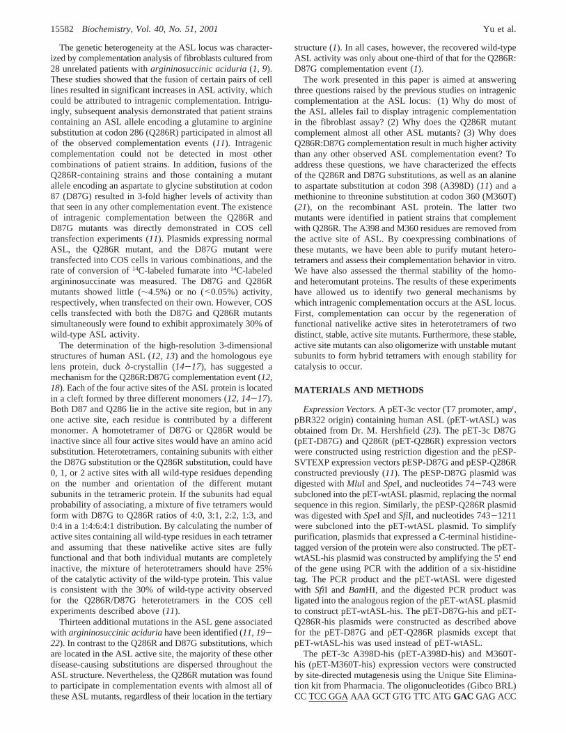
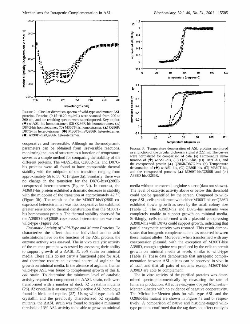
Stability of Wild-Type and Mutant Proteins.To determinethe effects of these substitutions on the structure and stabilityof the protein, the wild-type and mutant ASL proteins wereanalyzed by circular dichroism (CD) spectroscopy. All theASL proteins in the present study have a typicalR-helicalspectrum with the characteristic minima at 208 and 222 nm(Figure 2). The variations in the total amount of ellipticitymay reflect subtle alterations in the structure of somemutants. In this regard, it is notable that the homotetramerof the unstable M360T-his mutant displays the least ellip-ticity.
The unfolding of each protein was monitored by thechange in ellipticity at 222 nm as the temperature of thesample was increased. All transitions were found to be
FIGURE 1: Expression and purification of ASL proteins. (a)Purification of wtASL-his by Ni-affinity chromatography. Lane 1,flow-through; lanes 2-6, washes with 5, 60, 100, 100, and200 mM imidazole, respectively. The protein was eluted in the100 and 200 mM imidazole fractions (lanes 4-6). (b) Purificationof the coexpressed Q286R/D87G-his heterotetramers by Ni-affinity chromatography. The protein was eluted with increasingimidazole concentrations. The protein eluted with 30 and 60 mMimidazole was pooled and run on an isoelectric focusing gel. (c)Isoelectric focusing gel illustrating the heterogeneous mixture ofthe coexpressed protein. Lane 1, D87G-his homotetramer; lane 2,Q286R homotetramer; lane 3, Q286R/D87G-his-coexpressedheterotetramers. (d) Purification of M360T-his mutant ASL byNi-affinity chromatography. Lane 1, flow-through; lanes 2-7,washes with 5, 30, 60, 100, 100, and 200 mM imidazole,respectively. The protein eluted in the 100 mM imidazole fractions(lanes 5 and 6).
15584 Biochemistry, Vol. 40, No. 51, 2001 Yu et al.
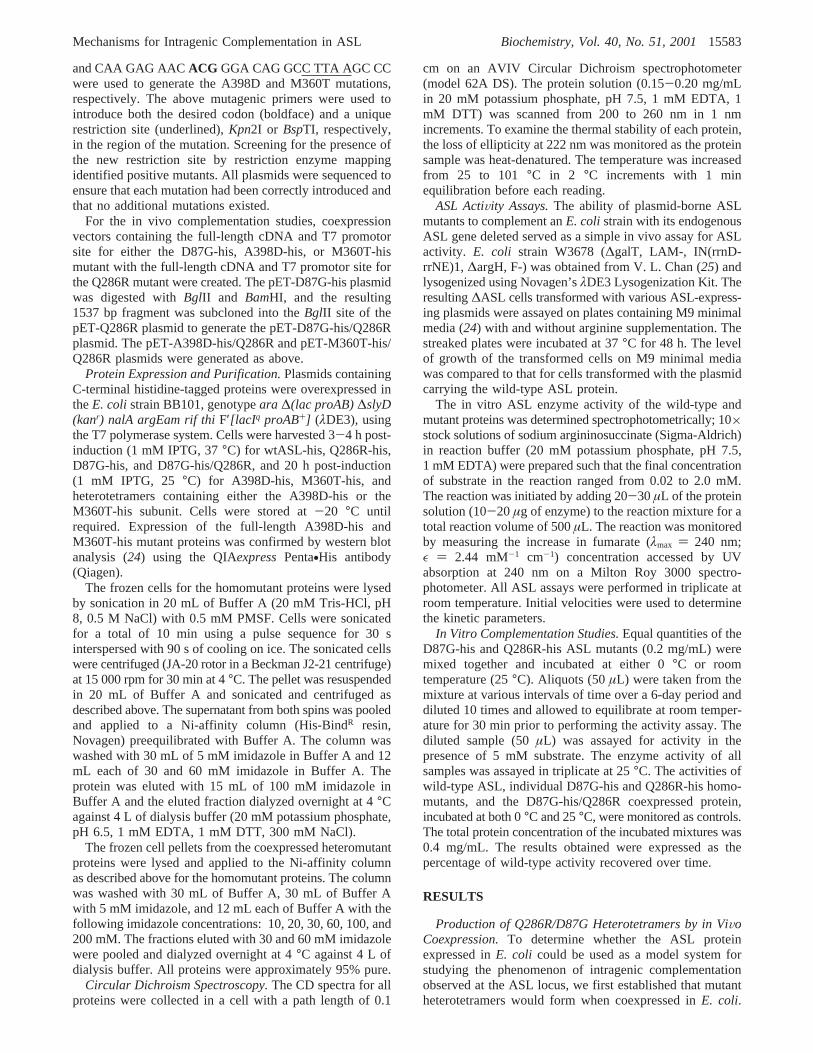
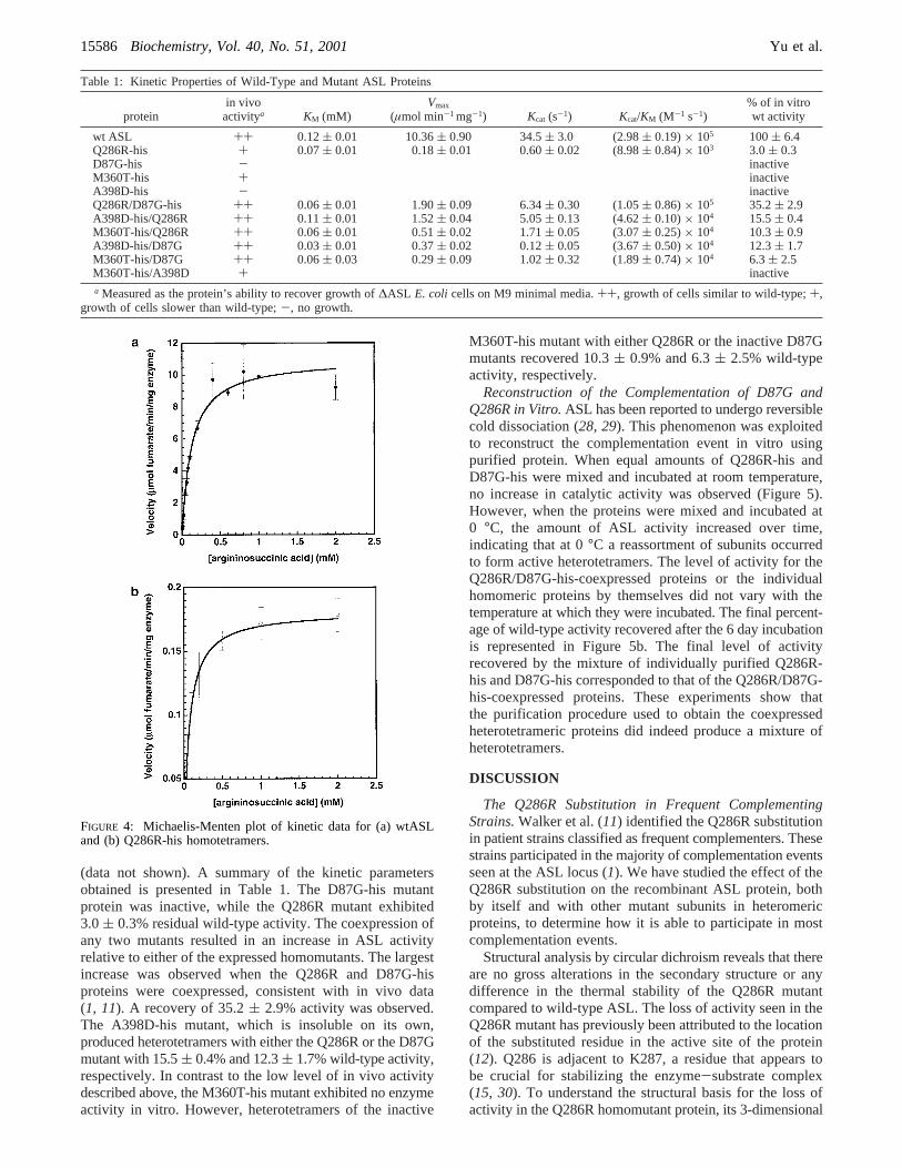
cooperative and irreversible. Although no thermodynamicparameters can be obtained from irreversible reactions,monitoring the loss of structure as a function of temperatureserves as a simple method for comparing the stability of thedifferent proteins. The wtASL-his, Q286R-his, and D87G-his proteins were all found to have comparable thermalstability with the midpoint of the transition ranging fromapproximately 56 to 58°C (Figure 3a). Similarly, there wasno change in the transition for the D87G-his/Q286R-coexpressed heterotetramers (Figure 3a). In contrast, theM360T-his protein exhibited a dramatic decrease in stabilitywith the midpoint of the transition at approximately 42°C(Figure 3b). The transition for the M360T-his/Q286R-co-expressed heterotetramers was less cooperative but exhibitedgreater resistance to thermal denaturation than the M360T-his homomutant protein. The thermal stability observed forthe A398D-his/Q286R-coexpressed heterotetramers was nearwild-type (Figure 3b).
Enzymatic ActiVity of Wild-Type and Mutant Proteins.Tocharacterize the effect that the individual amino acidsubstitutions have on the function of the ASL protein, theenzyme activity was assayed. The in vivo catalytic activityof the mutant proteins was tested by assessing their abilityto support growth of a∆ASL E. coli strain on minimalmedia. These cells do not carry a functional gene for ASLand therefore require an external source of arginine forgrowth on minimal media. In the absence of arginine, humanwild-type ASL was found to complement growth of thisE.coli strain. To determine the minimum level of catalyticactivity required to complement the∆ASL strain, cells weretransformed with a number of duckδ2 crystallin mutants(26). δ2 crystallin is an enzymatically active ASL homologuefound in birds and reptiles (27). Using wild-type duckδ2crystallin and the previously characterizedδ2 crystallinmutants, the∆ASL strain was found to require a minimumthreshold of 3% ASL activity to be able to grow on minimal
media without an external arginine source (data not shown).The level of catalytic activity above or below this thresholdcould not be quantified by the screen. Compared to wild-type ASL, cells transformed with either M360T-his or Q286Rexhibited slower growth as seen by the small colony size(Table 1). The A398D-his and D87G-his mutants werecompletely unable to support growth on minimal media.Strikingly, cells transformed with a plasmid coexpressingA398D-his with D87G could support growth, indicating thatpartial enzymatic activity was restored. This result demon-strates that intragenic complementation has occurred betweenthese mutant alleles. Moreover, when transformed with anycoexpression plasmid, with the exception of M360T-his/A398D, enough arginine was produced by the cells to permitgrowth on minimal media at levels similar to wild-type(Table 1). These data demonstrate that intragenic comple-mentation between ASL alleles can be observed in vivo inE. coli, and that all pairs of mutants except M360T-his/A398D are able to complement.
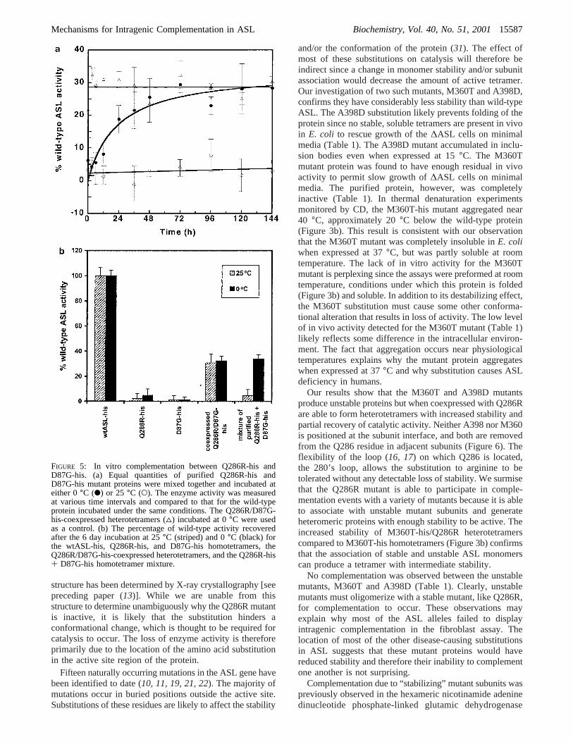
The in vitro activity of the purified proteins was deter-mined spectrophotometrically by measuring the rate offumarate production. All active enzymes obeyed Michaelis-Menten kinetics with no evidence of negative cooperativity.The Michaelis-Menten plots for wild-type ASL and theQ286R-his mutant are shown in Figure 4a and b, respec-tively. A comparison of native and histidine-tagged wild-type proteins confirmed that the tag does not affect catalysis
FIGURE 2: Circular dichroism spectra of wild-type and mutant ASLproteins. Proteins (0.15-0.20 mg/mL) were scanned from 200 to260 nm, and the resulting spectra were superimposed. Key to plot:([) wtASL-his homotetramer; (0) Q286R-his homotetramer; (4)D87G-his homotetramer; (O) M360T-his homotetramer; (2) Q286R/D87G-his heterotetramer; (b) M360T-his/Q286R heterotetramer;(9) A398D-his/Q286R heterotetramer.
FIGURE 3: Temperature denaturation of ASL proteins monitoredas a function of the circular dichroism signal at 222 nm. The curveswere normalized for comparison of data. (a) Temperature dena-turation of ([) wtASL-his, (O) Q286R-his, (0) D87G-his, andthe coexpressed protein (2) Q286R/D87G-his. (b) Temperaturedenaturation of ([) wtASL-his, (O) Q286R-his, (0) M360T-his,and the coexpressed proteins (2) M360T-his/Q286R and (×)A398D-his/Q286R.
Mechanisms for Intragenic Complementation in ASL Biochemistry, Vol. 40, No. 51, 200115585
(data not shown). A summary of the kinetic parametersobtained is presented in Table 1. The D87G-his mutantprotein was inactive, while the Q286R mutant exhibited3.0( 0.3% residual wild-type activity. The coexpression ofany two mutants resulted in an increase in ASL activityrelative to either of the expressed homomutants. The largestincrease was observed when the Q286R and D87G-hisproteins were coexpressed, consistent with in vivo data(1, 11). A recovery of 35.2( 2.9% activity was observed.The A398D-his mutant, which is insoluble on its own,produced heterotetramers with either the Q286R or the D87Gmutant with 15.5( 0.4% and 12.3( 1.7% wild-type activity,respectively. In contrast to the low level of in vivo activitydescribed above, the M360T-his mutant exhibited no enzymeactivity in vitro. However, heterotetramers of the inactive
M360T-his mutant with either Q286R or the inactive D87Gmutants recovered 10.3( 0.9% and 6.3( 2.5% wild-typeactivity, respectively.
Reconstruction of the Complementation of D87G andQ286R in Vitro.ASL has been reported to undergo reversiblecold dissociation (28, 29). This phenomenon was exploitedto reconstruct the complementation event in vitro usingpurified protein. When equal amounts of Q286R-his andD87G-his were mixed and incubated at room temperature,no increase in catalytic activity was observed (Figure 5).However, when the proteins were mixed and incubated at0 °C, the amount of ASL activity increased over time,indicating that at 0°C a reassortment of subunits occurredto form active heterotetramers. The level of activity for theQ286R/D87G-his-coexpressed proteins or the individualhomomeric proteins by themselves did not vary with thetemperature at which they were incubated. The final percent-age of wild-type activity recovered after the 6 day incubationis represented in Figure 5b. The final level of activityrecovered by the mixture of individually purified Q286R-his and D87G-his corresponded to that of the Q286R/D87G-his-coexpressed proteins. These experiments show thatthe purification procedure used to obtain the coexpressedheterotetrameric proteins did indeed produce a mixture ofheterotetramers.
DISCUSSION
The Q286R Substitution in Frequent ComplementingStrains.Walker et al. (11) identified the Q286R substitutionin patient strains classified as frequent complementers. Thesestrains participated in the majority of complementation eventsseen at the ASL locus (1). We have studied the effect of theQ286R substitution on the recombinant ASL protein, bothby itself and with other mutant subunits in heteromericproteins, to determine how it is able to participate in mostcomplementation events.
Structural analysis by circular dichroism reveals that thereare no gross alterations in the secondary structure or anydifference in the thermal stability of the Q286R mutantcompared to wild-type ASL. The loss of activity seen in theQ286R mutant has previously been attributed to the locationof the substituted residue in the active site of the protein(12). Q286 is adjacent to K287, a residue that appears tobe crucial for stabilizing the enzyme-substrate complex(15, 30). To understand the structural basis for the loss ofactivity in the Q286R homomutant protein, its 3-dimensional
Table 1: Kinetic Properties of Wild-Type and Mutant ASL Proteins
proteinin vivoactivitya KM (mM)
Vmax
(µmol min-1 mg-1) Kcat (s-1) Kcat/KM (M-1 s-1)% of in vitrowt activity
wt ASL ++ 0.12( 0.01 10.36( 0.90 34.5( 3.0 (2.98( 0.19)× 105 100( 6.4Q286R-his + 0.07( 0.01 0.18( 0.01 0.60( 0.02 (8.98( 0.84)× 103 3.0( 0.3D87G-his - inactiveM360T-his + inactiveA398D-his - inactiveQ286R/D87G-his ++ 0.06( 0.01 1.90( 0.09 6.34( 0.30 (1.05( 0.86)× 105 35.2( 2.9A398D-his/Q286R ++ 0.11( 0.01 1.52( 0.04 5.05( 0.13 (4.62( 0.10)× 104 15.5( 0.4M360T-his/Q286R ++ 0.06( 0.01 0.51( 0.02 1.71( 0.05 (3.07( 0.25)× 104 10.3( 0.9A398D-his/D87G ++ 0.03( 0.01 0.37( 0.02 0.12( 0.05 (3.67( 0.50)× 104 12.3( 1.7M360T-his/D87G ++ 0.06( 0.03 0.29( 0.09 1.02( 0.32 (1.89( 0.74)× 104 6.3( 2.5M360T-his/A398D + inactivea Measured as the protein’s ability to recover growth of∆ASL E. coli cells on M9 minimal media.++, growth of cells similar to wild-type;+,
growth of cells slower than wild-type;-, no growth.
FIGURE 4: Michaelis-Menten plot of kinetic data for (a) wtASLand (b) Q286R-his homotetramers.
15586 Biochemistry, Vol. 40, No. 51, 2001 Yu et al.
structure has been determined by X-ray crystallography [seepreceding paper (13)]. While we are unable from thisstructure to determine unambiguously why the Q286R mutantis inactive, it is likely that the substitution hinders aconformational change, which is thought to be required forcatalysis to occur. The loss of enzyme activity is thereforeprimarily due to the location of the amino acid substitutionin the active site region of the protein.
Fifteen naturally occurring mutations in the ASL gene havebeen identified to date (10, 11, 19, 21, 22). The majority ofmutations occur in buried positions outside the active site.Substitutions of these residues are likely to affect the stability
and/or the conformation of the protein (31). The effect ofmost of these substitutions on catalysis will therefore beindirect since a change in monomer stability and/or subunitassociation would decrease the amount of active tetramer.Our investigation of two such mutants, M360T and A398D,confirms they have considerably less stability than wild-typeASL. The A398D substitution likely prevents folding of theprotein since no stable, soluble tetramers are present in vivoin E. coli to rescue growth of the∆ASL cells on minimalmedia (Table 1). The A398D mutant accumulated in inclu-sion bodies even when expressed at 15°C. The M360Tmutant protein was found to have enough residual in vivoactivity to permit slow growth of∆ASL cells on minimalmedia. The purified protein, however, was completelyinactive (Table 1). In thermal denaturation experimentsmonitored by CD, the M360T-his mutant aggregated near40 °C, approximately 20°C below the wild-type protein(Figure 3b). This result is consistent with our observationthat the M360T mutant was completely insoluble inE. coliwhen expressed at 37°C, but was partly soluble at roomtemperature. The lack of in vitro activity for the M360Tmutant is perplexing since the assays were preformed at roomtemperature, conditions under which this protein is folded(Figure 3b) and soluble. In addition to its destabilizing effect,the M360T substitution must cause some other conforma-tional alteration that results in loss of activity. The low levelof in vivo activity detected for the M360T mutant (Table 1)likely reflects some difference in the intracellular environ-ment. The fact that aggregation occurs near physiologicaltemperatures explains why the mutant protein aggregateswhen expressed at 37°C and why substitution causes ASLdeficiency in humans.
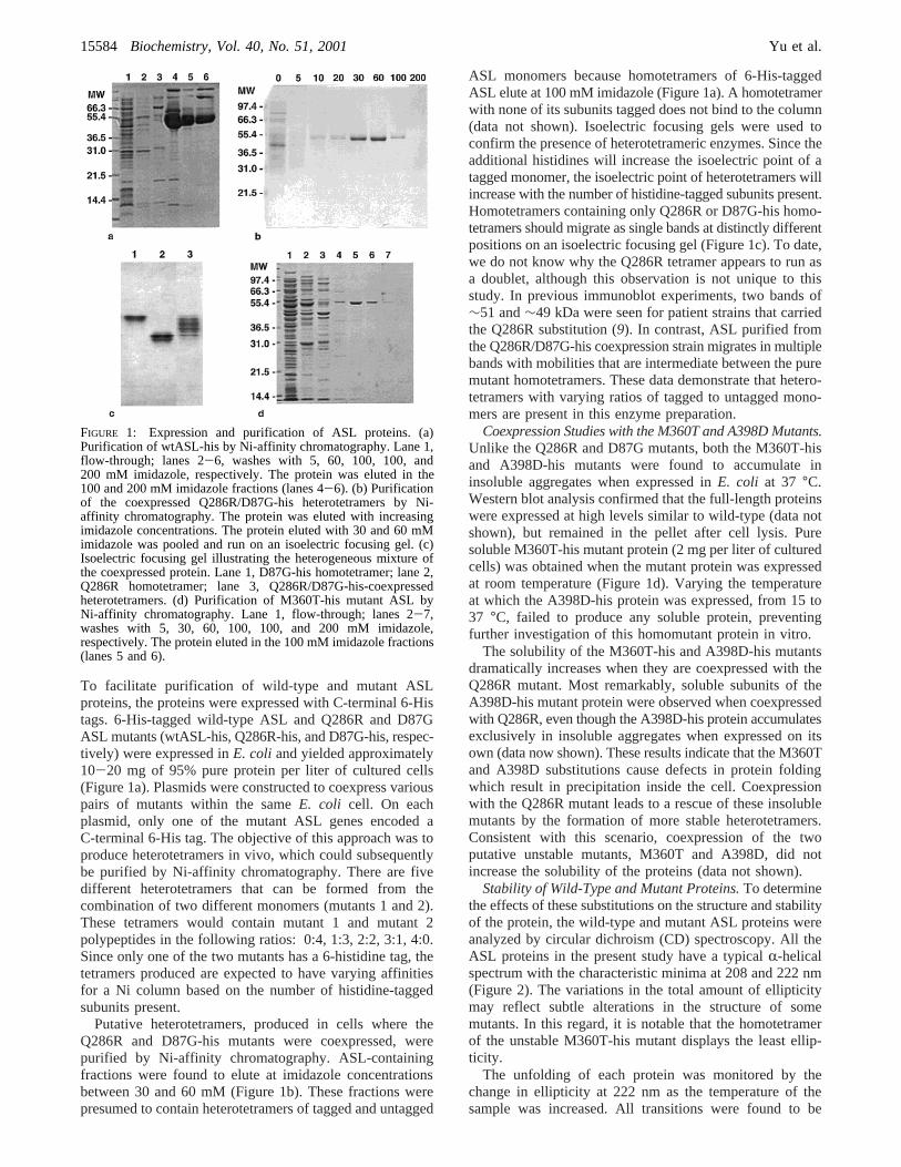
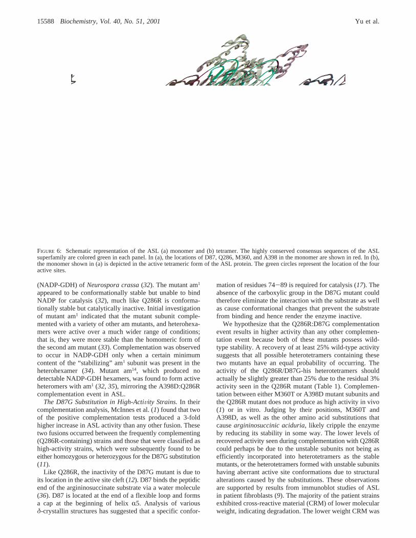
Our results show that the M360T and A398D mutantsproduce unstable proteins but when coexpressed with Q286Rare able to form heterotetramers with increased stability andpartial recovery of catalytic activity. Neither A398 nor M360is positioned at the subunit interface, and both are removedfrom the Q286 residue in adjacent subunits (Figure 6). Theflexibility of the loop (16, 17) on which Q286 is located,the 280’s loop, allows the substitution to arginine to betolerated without any detectable loss of stability. We surmisethat the Q286R mutant is able to participate in comple-mentation events with a variety of mutants because it is ableto associate with unstable mutant subunits and generateheteromeric proteins with enough stability to be active. Theincreased stability of M360T-his/Q286R heterotetramerscompared to M360T-his homotetramers (Figure 3b) confirmsthat the association of stable and unstable ASL monomerscan produce a tetramer with intermediate stability.
No complementation was observed between the unstablemutants, M360T and A398D (Table 1). Clearly, unstablemutants must oligomerize with a stable mutant, like Q286R,for complementation to occur. These observations mayexplain why most of the ASL alleles failed to displayintragenic complementation in the fibroblast assay. Thelocation of most of the other disease-causing substitutionsin ASL suggests that these mutant proteins would havereduced stability and therefore their inability to complementone another is not surprising.
Complementation due to “stabilizing” mutant subunits waspreviously observed in the hexameric nicotinamide adeninedinucleotide phosphate-linked glutamic dehydrogenase
FIGURE 5: In vitro complementation between Q286R-his andD87G-his. (a) Equal quantities of purified Q286R-his andD87G-his mutant proteins were mixed together and incubated ateither 0°C (b) or 25 °C (O). The enzyme activity was measuredat various time intervals and compared to that for the wild-typeprotein incubated under the same conditions. The Q286R/D87G-his-coexpressed heterotetramers (4) incubated at 0°C were usedas a control. (b) The percentage of wild-type activity recoveredafter the 6 day incubation at 25°C (striped) and 0°C (black) forthe wtASL-his, Q286R-his, and D87G-his homotetramers, theQ286R/D87G-his-coexpressed heterotetramers, and the Q286R-his+ D87G-his homotetramer mixture.
Mechanisms for Intragenic Complementation in ASL Biochemistry, Vol. 40, No. 51, 200115587
(NADP-GDH) of Neurospora crassa(32). The mutant am1
appeared to be conformationally stable but unable to bindNADP for catalysis (32), much like Q286R is conforma-tionally stable but catalytically inactive. Initial investigationof mutant am1 indicated that the mutant subunit comple-mented with a variety of other am mutants, and heterohexa-mers were active over a much wider range of conditions;that is, they were more stable than the homomeric form ofthe second am mutant (33). Complementation was observedto occur in NADP-GDH only when a certain minimumcontent of the “stabilizing” am1 subunit was present in theheterohexamer (34). Mutant am14, which produced nodetectable NADP-GDH hexamers, was found to form activeheteromers with am1 (32, 35), mirroring the A398D:Q286Rcomplementation event in ASL.
The D87G Substitution in High-ActiVity Strains.In theircomplementation analysis, McInnes et al. (1) found that twoof the positive complementation tests produced a 3-foldhigher increase in ASL activity than any other fusion. Thesetwo fusions occurred between the frequently complementing(Q286R-containing) strains and those that were classified ashigh-activity strains, which were subsequently found to beeither homozygous or heterozygous for the D87G substitution(11).
Like Q286R, the inactivity of the D87G mutant is due toits location in the active site cleft (12). D87 binds the peptidicend of the argininosuccinate substrate via a water molecule(36). D87 is located at the end of a flexible loop and formsa cap at the beginning of helixR5. Analysis of variousδ-crystallin structures has suggested that a specific confor-
mation of residues 74-89 is required for catalysis (17). Theabsence of the carboxylic group in the D87G mutant couldtherefore eliminate the interaction with the substrate as wellas cause conformational changes that prevent the substratefrom binding and hence render the enzyme inactive.
We hypothesize that the Q286R:D87G complementationevent results in higher activity than any other complemen-tation event because both of these mutants possess wild-type stability. A recovery of at least 25% wild-type activitysuggests that all possible heterotetramers containing thesetwo mutants have an equal probability of occurring. Theactivity of the Q286R/D87G-his heterotetramers shouldactually be slightly greater than 25% due to the residual 3%activity seen in the Q286R mutant (Table 1). Complemen-tation between either M360T or A398D mutant subunits andthe Q286R mutant does not produce as high activity in vivo(1) or in vitro. Judging by their positions, M360T andA398D, as well as the other amino acid substitutions thatcauseargininosuccinic aciduria, likely cripple the enzymeby reducing its stability in some way. The lower levels ofrecovered activity seen during complementation with Q286Rcould perhaps be due to the unstable subunits not being asefficiently incorporated into heterotetramers as the stablemutants, or the heterotetramers formed with unstable subunitshaving aberrant active site conformations due to structuralalterations caused by the substitutions. These observationsare supported by results from immunoblot studies of ASLin patient fibroblasts (9). The majority of the patient strainsexhibited cross-reactive material (CRM) of lower molecularweight, indicating degradation. The lower weight CRM was
FIGURE 6: Schematic representation of the ASL (a) monomer and (b) tetramer. The highly conserved consensus sequences of the ASLsuperfamily are colored green in each panel. In (a), the locations of D87, Q286, M360, and A398 in the monomer are shown in red. In (b),the monomer shown in (a) is depicted in the active tetrameric form of the ASL protein. The green circles represent the location of the fouractive sites.
15588 Biochemistry, Vol. 40, No. 51, 2001 Yu et al.
not present in patient strains containing the Q286R substitu-tion. It was also observed that the amount of normalmolecular mass CRM (49-51 kDa) present in a patient straincorrelated strongly with the frequency in which that patientstrain participated in complementation. This correlationagrees with our hypothesis that the more stable mutants arebetter able to complement.
The experiments performed in this work demonstrate thatthe D87G mutant complements unstable mutants, such asM360T and A398D, almost as well as Q286R (Table 1).This result was expected because we hypothesize that stableactive site mutants, such as D87G, should be able tocomplement most nonactive site unstable mutants. TheD87G-containing fibroblast strains, however, were not foundto be frequently complementing strains. At present, it isdifficult to explain the very different behaviors of the D87Gand Q286R mutants in the in vivo complementation analysis(1). Conformational changes that alter substrate bindingin D87G homotetramers may also affect the ability of theD87G mutants to form active tetramers with unstablemutants. The relatively efficient D87G-mediated comple-mentation that we observed in this work may be the resultof much higher concentrations of protein being present invitro and in E. coli as compared to the levels present infibroblasts. The lower concentrations of ASL within fibro-blasts could exacerbate any defects in multimerizationpossessed by the D87G mutant.
The A398D mutation was identified by Walker et al. asthe substitution in the second allele in a patient strainheterozygous for the D87G substitution (11). Walker et al.suggested that the A398D allele had significant residual ASLactivity due to the mild clinical phenotype of the heterozygote(late-onset) compared to a D87G homozygote (sub-acute)(11). Our results suggest that this is unlikely to be true sincethe A398D homomutant protein is completely inactive,cannot be expressed in a soluble form, and does not supportgrowth of the ∆ASL E. coli strain. There has been noreported case of an individual homozygous for the A398Dsubstitution, probably due to the severe nature of the aminoacid substitution on the homomeric protein. We hypothesizethat in this particular case, the difference in the clinicalphenotype observed is due to complementation betweenD87G and A398D.
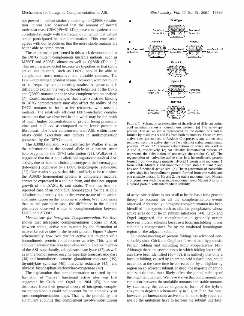
Mechanisms for Intragenic Complementation.We haveshown that intragenic complementation occurs in ASLbetween stable, active site mutants by the formation ofnativelike active sites in the hybrid protein. Figure 7 showsschematically how two distinct active site mutants of ahomodimeric protein could recover activity. This type ofcomplementation has also been observed in another memberof the ASL superfamily, adenylosuccinate lyase (37), as wellas in the homotrimeric enzyme aspartate transcarbamoylase(38) and homodimeric proteins glutathione reductase (39),thymidylate synthase (40), mercuric reductase (41), andribulose bisphosphate carboxylase/oxygenase (42).
The explanation that complementation occurred by theformation of “native” functional active sites was firstsuggested by Crick and Orgel in 1964 (43), but wasdismissed from their general theory of intragenic comple-mentation since it could not account for the complexity ofmost complementation maps. That is, the probability thatall mutant subunits that complement involve substitutions
of active site residues is too small to be the basis for a generaltheory to account for all the complementation eventsobserved. Additionally, intragenic complementation has beenidentified in enzymes, such as alkaline phosphatase, whoseactive sites do not lie in subunit interfaces (44). Crick andOrgel suggested that complementation generally occursbetween mutant subunits because a local misfolding in onesubunit is compensated for by the unaltered homologousregion of the adjacent subunit.
Our understanding of protein folding has advanced con-siderably since Crick and Orgel put forward their hypothesis.Protein folding and unfolding occur cooperatively (45).Although there are several cases in which folding intermedi-ates have been identified (46-48), it is unlikely that only alocal unfolding, caused by an amino acid substitution, couldoccur and at the same time be corrected for by a neighboringregion on an adjacent subunit. Instead, the majority of aminoacid substitutions most likely affect the global stability ofthe oligomeric protein. We have shown that complementationcan occur between thermolabile mutants and stable mutantsby stabilizing the active oligomeric form of the hybridenzyme as depicted schematically in Figure 7. In this case,however, an intersubunit active site is not strictly required,nor do the mutations have to lie near the subunit interface.
FIGURE 7: Schematic representation of the effects of different aminoacid substitutions on a homodimeric protein. (a) The wild-typeprotein. The active site is represented by the dashed box and isformed by residues (A and B) from both monomers. There are twoactive sites per molecule. Residue C represents any amino acidremoved from the active site. (b) Two distinct stable homomutantproteins. a* and b* represent substitutions of active site residuesA and B, respectively. (c) An unstable homomutant protein. c*represents the substitution of nonactive site residue C. (d) Theregeneration of nativelike active sites in a heterodimeric proteinformed from two stable mutants. Hybrid 1 consists of monomer 1from stable Mutant 1 and monomer 2 from stable Mutant 2 andhas one functional active site. (e) The regeneration of nativelikeactive sites in a heterodimeric protein formed from one stable andone unstable mutant. In Hybrid 2, the stable monomer from Mutant1 oligomerizes with the unstable monomer from Mutant 3 to forma hybrid protein with intermediate stability.
Mechanisms for Intragenic Complementation in ASL Biochemistry, Vol. 40, No. 51, 200115589
This mechanism for complementation therefore extends Crickand Orgel’s original theory by including global destabilizingmutants. We propose that both mechanisms reported herecan be used to broadly explain all of the complementationevents observed at the ASL locus.
ACKNOWLEDGMENT
We thank Dr. M. Hershfield for providing the expressionvector for wild-type ASL, Dr. R. R. McInnes for providingthe pESP-D87G and pESP-Q286R expression vectors, V. L.Chan for providing the∆ASL E. coli strain, and DavidBateman for technical assistance.
REFERENCES
1. McInnes, R. R., Shih, V., and Chilton, S. (1984)Proc. Natl.Acad. Sci. U.S.A. 81, 4480-4484.
2. Gravel, R. A., Lam, K. F., Scully, K. J., and Hsia, Y. (1977)Am. J. Hum. Genet. 29, 378-388.
3. Gravel, R. A., Akerman, B. R., Lamhonwah, A. M., Loyer,M., Leon-del-Rio, A., and Italiano, I. (1994)Am. J. Hum.Genet. 55, 51-58.
4. Qureshi, A. A., Crane, A. M., Matiaszuk, N. V., Rezvani, I.,Ledley, F. D., and Rosenblatt, D. S. (1994)J. Clin. InVest.93, 1812-1819.
5. Lusty, C. J., and Ratner, S. (1972)J. Biol. Chem. 247, 7010-7022.
6. O’Brien, W. E., and Barr, R. H. (1981)Biochemistry 20,2056-2060.
7. Palekar, A. G., and Mantagos, S. (1981)J. Biol. Chem. 256,9192-9194.
8. Brusilow, S. W., and Maestri, N. E. (1996)AdV. Pediatr. 43,127-170.
9. Simard, L., O’Brien, W. E., and McInnes, R. R. (1986)Am.J. Hum. Genet. 39, 38-51.
10. Barbosa, P., Cialkowski, M., and O’Brien, W. E. (1991)J.Biol. Chem. 266, 5286-5290.
11. Walker, D. C., Christodoulou, J., Craig, H. J., Simard, L. R.,Ploder, L., Howell, P. L., and McInnes, R. R. (1997)J. Biol.Chem. 272, 6777-6783.
12. Turner, M. A., Simpson, A., McInnes, R. R., and Howell, P.L. (1997)Proc. Natl. Acad. Sci. U.S.A. 94, 9063-9068.
13. Sampaleanu, L. M., Vallee, F., Thompson, G. D., and Howell,P. L. (2001)Biochemistry 41, 15570-15580.
14. Abu-Abed, M., Turner, M. A., Vallee, F., Simpson, A.,Slingsby, C., and Howell, P. L. (1997)Biochemistry 36,14012-14022.
15. Simpson, A., Bateman, O., Driessen, H., Lindley, P., Moss,D., Mylvaganam, S., Narebor, E., and Slingsby, C. (1994)Nat.Struct. Biol. 1, 724-734.
16. Vallee, F., Turner, M. A., Lindley, P. L., and Howell, P. L.(1999)Biochemistry 38, 2425-2434.
17. Sampaleanu, L. M., Vallee, F., Slingsby, C., and Howell, P.L. (2001)Biochemistry 40, 2732-2742.
18. Howell, P. L., Turner, M. A., Christodoulou, J., Walker, D.C., Craig, H. J., Simard, L. R., Ploder, L., and McInnes, R. R.(1998)J. Inherit. Metab. Dis. 21, 72-85.
19. Walker, D. C., McCloskey, D. A., Simard, L. R., and McInnes,R. R. (1990)Proc. Natl. Acad. Sci. U.S.A. 87, 9625-9629.
20. Barbosa, P., Wistow, G. J., Cialkowski, M., Piatigorsky, J.,and O’Brien, W. E. (1991)J. Biol. Chem. 266, 22319-22322.
21. Craig, H. J. (1993) inMolecular and Medical Genetics, p 77,University of Toronto, Toronto.
22. Linnebank, M., Homberger, A., Rapp, B., Winter, C., Mar-quardt, T., Harms, E., and Koch, H. G. (2000)J. Inherit.Metab. Dis. 23, 308-312.
23. Turner, M. A., Achyuthan, A. M., Hershfield, M. S., McInnes,R. R., and Howell, P. L. (1994)J. Mol. Biol. 239, 336-338.
24. Sambrook, J., Frtisch, E., and Maniatis, T. (1989)MolecularCloning: A Laboratory Manual, Cold Spring Harbor Labora-tory, Cold Spring Harbor, NY.
25. Hani, E. K., and Chan, V. L. (1994)J. Bacteriol. 176, 1865-1871.
26. Chakraborty, A. R., Davidson, A., and Howell, P. L. (1999)Biochemistry 38, 2435-2443.
27. Piatigorsky, J., O’Brien, W. E., Norman, B. L., Kalumuck,K., Wistow, G. J., Borras, T., Nickerson, J. M., and Wawrousek,E. F. (1988)Proc. Natl. Acad. Sci. U.S.A. 85, 3479-3483.
28. Havir, E. A., Tamir, H., Ratner, S., and Warner, R. C. (1965)J. Biol. Chem. 240, 3079-3088.
29. Schulze, I. T., Lusty, C. J., and Ratner, S. (1970)J. Biol. Chem.245, 4534-4543.
30. Saribas, A. S., Schindler, J. F., and Viola, R. E. (1994)J. Biol.Chem. 269, 6313-6319.
31. Yu, B., and Howell, P. L. (2000)Cell. Mol. Life Sci. 57, 1637-1651.
32. Fincham, J. R., Kinsey, J. A., Fuentes, A. M., Cummings, N.J., and Connerton, I. F. (2000)Genet. Res. 76, 1-10.
33. Fincham, J. R. S. (1962)J. Mol. Biol. 4, 257-274.34. Coddington, A., and Fincham, J. R. S. (1965)J. Mol. Biol.
12, 152-161.35. Sundaram, T. K., and Fincham, J. R. S. (1968)J. Mol. Biol.
95, 787-792.36. Sampaleanu, L. M., Yu, B., and Howell, P. L. (2002)J. Biol.
Chem.(in press).37. Lee, T. T., Worby, C., Bao, Z. Q., Dixon, J. E., and Colman,
R. F. (1999)Biochemistry 38, 22-32.38. Wente, S. R., and Schachman, H. K. (1987)Proc. Natl. Acad.
Sci. U.S.A. 84, 31-35.39. Scrutton, N. S., Berry, A., Deonarain, M. P., and Perham, R.
N. (1990)Proc. R. Soc. London, Ser. B: Biol. Sci. 242, 217-224.
40. Pookanjanatavip, M., Yuthavong, Y., Greene, P. J., and Santi,D. V. (1992)Biochemistry 31, 10303-10309.
41. Distefano, M. D., Moore, M. J., and Walsh, C. T. (1990)Biochemistry 29, 2703-2713.
42. Larimer, F. W., Lee, E. H., Mural, R. J., Soper, T. S., andHartman, F. C. (1987)J. Biol. Chem. 262, 15327-15329.
43. Crick, F. H. C., and Orgel, L. E. (1964)J. Mol. Biol. 8, 161-165.
44. Hehir, M. J., Murphy, J. E., and Kantrowitz, E. R. (2000)J.Mol. Biol. 304, 645-656.
45. Tanford, C. (1968)AdV. Protein Chem. 23, 121-282.46. Kim, P. S., and Baldwin, R. L. (1982)Annu. ReV. Biochem.
51, 459-489.47. Kim, P. S., and Baldwin, R. L. (1990)Annu. ReV. Biochem.
59, 631-660.48. Englander, S. W. (2000)Annu. ReV. Biophys. Biomol. Struct.
29, 213-238.
BI011526E
15590 Biochemistry, Vol. 40, No. 51, 2001 Yu et al.