systematic identification of cis-silenced genes by trans complementation
TRANSCRIPT
1
© 2008 The Author(s) This is an Open Access article distributed under the terms of the Creative Commons Attribution Non-Commercial License (http://creativecommons.org/licenses/by-nc/2.0/uk/) which permits unrestricted non-commercial use, distribution, and reproduction in any medium, provided the original work is properly cited.
Systematic identification of cis-silenced genes by trans complementation
Jae Hyun Lee1, Branimir Bugarija1, Enrique J. Millan1, Noah M. Walton1, Jedidiah
Gaetz1, Croydon J. Fernandes1, Wei-Hua Yu2, Nitzan Mekel-Bobrov1, Tammy W.
Vallender1, Gregory E. Snyder1, Andy Peng Xiang2, Bruce T. Lahn1, 2,* 1Department of Human Genetics, Howard Hughes Medical Institute, University of Chicago, Chicago, IL
60637, USA. 2Center for Stem Cell Biology and Tissue Engineering, Sun Yat-sen University, Guangzhou, 510080,
China.
*To whom correspondence should be addressed. Address: Department of Human Genetics, Howard
Hughes Medical Institute, University of Chicago, Chicago, IL 60637, USATel: 773-834-4393
Fax: 773-702-0271 Email: [email protected]
HMG Advance Access published December 2, 2008 by guest on A
ugust 28, 2014http://hm
g.oxfordjournals.org/D
ownloaded from
2
ABSTRACT
A gene’s transcriptional output is the combined product of two inputs: diffusible factors in the cellular
milieu acting in trans, and chromatin state acting in cis. Here, we describe a strategy for dissecting the
relative contribution of cis versus trans mechanisms to gene regulation. Referred to as trans
complementation, it entails fusing two disparate cell types and searching for genes differentially
expressed between the two genomes of fused cells. Any differential expression can be causally
attributed to cis mechanisms because the two genomes of fused cells share a single homogenized milieu
in trans. This assay uncovered a state of transcriptional competency that we termed “occluded” whereby
affected genes are silenced by cis-acting mechanisms in a manner that blocks them from responding to
the trans-acting milieu of the cell. Importantly, occluded genes in a given cell type tend to include
master triggers of alternative cell fates. Furthermore, the occluded state is maintained during cell
division and is extraordinarily stable under a wide range of physiological conditions. These results
support the model that the occlusion of lineage-inappropriate genes is a key mechanism of cell fate
restriction. The identification of occluded genes by our assay provides a hitherto unavailable functional
readout of chromatin state that is distinct from and complementary to gene expression status.
by guest on August 28, 2014
http://hmg.oxfordjournals.org/
Dow
nloaded from
3
INTRODUCTION
Multicellular life is defined by the presence, within a single organism, of a wide array of cell types
bearing the same genome but disparate physiological functions. This is typically achieved through the
progressive differentiation of multipotent stem cells into functionally specialized cells. As a general rule,
differentiated cell types can stably maintain their phenotypic identities despite fluctuations in
extracellular environment and intracellular regulatory networks (1). How cell type identity is maintained
at the molecular level is a central but poorly understood question in biology. One attractive idea is that
the phenotypic identity of differentiated cells is maintained via the silencing of lineage-inappropriate
genes – i.e., genes promoting alternative lineages which, if expressed aberrantly, would lead to the
manifestation of incorrect cellular phenotypes (2-5).
This idea is in line with the increasing recognition that the transcriptional output of a gene is the
combined product of two distinct inputs. The first is the trans-acting milieu of the cell, defined as all the
diffusible factors that collectively impinge on gene regulatory sequences to promote or repress
expression. The second is the cis-acting chromatin state of the gene itself, defined as the full
complement of chromatin marks at the locus such as DNA methylation, histone modifications, and the
binding of chromatin remodeling factors, which in combination determine how the locus responds to its
milieu. Numerous studies have found that particular chromatin marks such as DNA methylation and
histone hypoacetylation are enriched at silent loci of the genome (6-13). In most cases, however, the
exact contribution of these chromatin marks to the silent state cannot be teased apart from the
contribution of milieu. This is because it is difficult to know whether chromatin marks at silent loci are
the cause or consequence of silencing, or to what extent is the silent status of a gene and its associated
chromatin marks reversible when cellular milieu changes (9, 14). As such, whether gene silencing by
chromatin-based cis mechanisms plays a key role in restricting cell fate remains to be resolved.
Monoallelic silencing such as X inactivation and imprinting is a clear exception to the above
ambiguity. Here, it can be unequivocally ascertained that silencing is due to cis-acting chromatin
mechanisms in a manner independent of milieu. The hallmark of monoallelic silencing is the differential
expression of two copies of a gene – one silent and one active – in the same cell (15-17). The active
copy serves as a positive control, attesting to the presence of a milieu that is conducive to the expression
of the gene. In this context, the silent copy, which is bathed in the same milieu, must have been blocked
from the milieu’s action by the cis effect of its chromatin state. Thus, at least in the case of monoallelic
silencing, the transcriptional competency of a gene can be defined as existing in either of two states. One
by guest on August 28, 2014
http://hmg.oxfordjournals.org/
Dow
nloaded from
4
is the “competent” state whereby a gene is capable of responding to the milieu of the cell, such that it is
active if appropriate transcription activators are present, and silent if activators are absent or repressors
are present. The other can be called the “occluded” state whereby a gene is no longer capable of
responding to the cell’s milieu, and remains silent even in the presence of a transcriptionally conducive
milieu.
It is reasonable to hypothesize that during development, some genes might become biallelically
occluded by mechanisms similar to monoallelic silencing, and that this process could play an essential
role in maintaining the phenotypic identities of cells. A key test of this model is the identification of
biallelically occluded genes. However, the lack of a positive control – the equivalent of the active copies
for monoallelically silenced genes – poses a technical challenge in ascertaining the presence of
biallelically occluded genes. This is because it is impossible, without such a control, to definitively
differentiate whether a silent gene is in the occluded state or whether it is competent but not expressed
simply due to the lack of a conducive milieu. Furthermore, biochemical modifications of chromatin,
which regulate gene expression in cis, is immensely complex (for example, there are over 100 known
chromatin marks) (18), thus limiting the use of a “bottom up” approach to differentiate cis versus trans
regulation.
Here, we describe the trans complementation assay, which allows the systematic identification of
biallelically occluded genes. The approach is to fuse two disparate cell types, and search in fused cells
for genes silent in one genome but active in the other. Similar to monoallelic silencing, the active copies
of genes serve as a positive control, with which the occluded state of the silent copies can be ascertained.
RESULTS
Identification of occluded genes via cell fusion
To identify occluded genes within specific cell types, we employed a cell fusion strategy. For ease of
description, one of the two cell types being fused will be referred to as the responder and the other the
reprogrammer. The goal is to identify occluded genes in the responder, which are defined operationally
as genes silent in the responder genome of fused cells but active in the reprogrammer genome of the
same cells (Table 1). We chose human lung fibroblasts (hereon abbreviated hLF) as the responder and
mouse skeletal muscle myoblasts (mSMM) as the reprogrammer. By using cells from different species,
sequence divergence between orthologs can be exploited to distinguish whether a transcript in fused
cells is produced from the reprogrammer genome or the responder genome.
The two cell populations were labeled by different fluorescent dyes and fused by polyethylene
by guest on August 28, 2014
http://hmg.oxfordjournals.org/
Dow
nloaded from
5
glycol. Dual fluorescent cells, which represent a small fraction of the total, were isolated by fluorescence
activated cell sorting (FACS) (Figure S1A). Microscopy confirmed that FACS-isolated cells were
predominantly (>98%) fusions between hLF and mSMM, as they contained multiple nuclei of two
distinct morphologies (hLF nuclei are larger and have weaker DAPI staining relative to mSMM) (Figure
S1B). For a subset of experiments, cells of heterotypic fusion (i.e., fusion between hLF and mSMM)
were further enriched by antibiotics that eliminated unfused cells or cells of homotypic fusion. Of the
fused cells, more than 70% showed equal numbers of hLF versus mSMM nuclei, the great majority of
which possessed one hLF and one mSMM nucleus while the rest contained two hLF and two mSMM
nuclei. Less than 30% of cells showed unequal numbers of hLF and mSMM nuclei, the majority of
which had an overrepresentation of mSMM nuclei (Figure S1C). Fused cells were cultured for varying
periods of time to allow for the resetting of gene expression in the new cellular milieu. Regardless of
culture period and medium formulation, fused cells remained as multinucleated heterokaryons,
indicating that they had lost the ability to divide after fusion. We found that gene expression patterns
became stabilized within 3 days of fusion (see below). We therefore focused on day 4 post fusion for our
analysis of gene expression.
To interrogate gene expression in hLF and mSMM before and after fusion, we used human and
mouse Affymetrix microarrays. Although there is significant sequence divergence between human and
mouse genomes (average 16% in coding regions), a human transcript in fused cells may still hybridize to
orthologous probes on the mouse arrays and vice versa, given that the arrays are not designed for
species-specific hybridization. To examine how serious a problem cross-species hybridization might be,
we hybridized cRNA from each cell type to both the human and the mouse arrays. When cRNA from
the correct species was hybridized to the arrays, about 45% of all the genes were called present. By
contrast, cRNA from the wrong species only led to about 10% of the genes being called present. This
shows that the arrays have sufficient species specificity to interrogate expression of a considerable
fraction of genes in fused cells, but it also demonstrates the need for validation by more stringent
methods.
Four sets of array data were generated: hLF on human arrays, mSMM on mouse arrays, fused
cells on human arrays, and fused cells on mouse arrays. To ensure robustness of the analysis, we first
narrowed down to a list of genes shown by the array data to be active in mSMM but silent in hLF prior
to fusion. If, in the fused cells, these genes remain active in the mSMM genome and silent in the hLF
genome, they would be placed in a candidate list of occluded hLF genes.
This analysis generated a candidate list of 279 putatively occluded hLF genes, all of which were
by guest on August 28, 2014
http://hmg.oxfordjournals.org/
Dow
nloaded from
6
subject to RT-PCR validation. For each gene, we designed and confirmed mouse-specific and human-
specific RT-PCR primers to allow mouse and human gene expression in fused cells to be interrogated
independently. Consistent with previous Affymetrix microarray studies (19), our RT-PCR analysis
showed that absence calls in the array data are much less reliable than presence calls. As a result, a large
number of the candidate occluded hLF genes from the array data were shown by RT-PCR to be
expressed at appreciable levels in hLF both before and after fusion. Winnowing out these and other false
leads, 24 genes were confirmed by RT-PCR to exhibit expression patterns consistent with their occluded
status in hLF (Figure 1A and Table S1). Of these, 9 have known muscle-related functions (indicated in
Figure 1A). PCR on genomic DNA of fused cells using human-specific primers successfully amplified
the hLF copies of all these genes, indicating that their lack of expression in fused cells is not due to the
absence of hLF chromosomes (data not shown). Indeed, it is unlikely that chromosome loss should
occur in mitotically arrested heterokaryons.
Applying the criteria in Table 1, we also obtained a candidate list of 1040 putatively
transactivated hLF genes. Given that transactivation was not the focus of the study, we only selected a
subset of 202 genes for RT-PCR validation. Many genes failed validation because RT-PCR detected
appreciable levels of expression in hLF both before and after fusion. For a lot of these, RT-PCR did
show increased expression after fusion, but we did not consider them as transactivated genes per our
stringent definition of transactivation. This led to the identification of 10 transactivated hLF genes, of
which 7 have known muscle-related functions (Figure 1A and Table S1). For 3 of the genes (Ckm, Acta1
and Myl1), their transactivation is consistent with previous reports (20, 21). For transactivated genes that
showed significantly less amplification of the hLF transcripts than the mSMM transcripts in fused cells
(such as Myog, Acta1, and Rap1ga1), additional sets of primers confirmed that the differences in
amplification reflected actual gene expression differences between the human and mouse orthologs,
rather than differences in PCR efficiencies. In principle, the observed differences in gene expression
between human and mouse transcripts in fused cells could be due to at least three possibilities. First,
these genes could be partially occluded such that they turn on in response to the introduction of a
conducive milieu, but not to the full extent possible (see Discussion). Second, the hLF cell population
may be heterogeneous, with the genes in question being occluded in some but not all the cells. A third
possibility is that incompatibility between mouse transcription factors made from the mSMM genome
and human cis-regulatory sequences in the hLF genome results in only partial activation of these genes.
This possibility is addressed in greater detail later.
Our results demonstrate that, although occluded and transactivated genes are both silent in hLF
by guest on August 28, 2014
http://hmg.oxfordjournals.org/
Dow
nloaded from
7
prior to fusion, they clearly exist in two distinct states of transcriptional competency. Occluded genes do
not become active even in the presence of a transcriptionally conducive milieu. In contrast,
transactivated genes exist in a competent (though inactive) state that can turn on in response to the
introduction of trans-acting factors in the milieu.
Ad hoc RT-PCR analysis also uncovered 4 extinguished mSMM genes and 6 occluded mSMM
genes (Figure 1A). Extinction could result either from the introduction of transcriptional repressors or
from the dilution or disappearance of transcriptional activators upon fusion (22). For extinguished
mSMM genes, it is not possible to determine if their orthologs in hLF are occluded or not. The presence
of occluded mSMM genes indicates that a given cell fusion experiment can be used to identify occluded
genes in both fusion partners, even though we chose to only focus on hLF in this study.
The identification of occluded hLF genes was carried out in a systematic and unbiased fashion,
in the sense that all the candidate occluded genes based on the array data were subject to RT-PCR
validation. The final tally of 24 occluded hLF genes therefore likely represents a considerable fraction of
all occluded hLF genes in the context of the hLF-mSMM fusion experiment. By contrast, the
transactivated hLF genes, extinguished mSMM genes, and occluded mSMM genes were uncovered by
less systematic means.
The specification of the myogenic lineage is controlled by four transcription factors, Myod1,
Myf5, Myog, and Myf6 (23-27). Of these myogenic master triggers, Myod1 and Myf5 are occluded in
hLF, Myog is transactivated (and therefore competent) in hLF, and Myf6 is extinguished in mSMM (and
therefore may be either occluded or competent in hLF). Interestingly, Myod1 and Myf5 are known to be
upstream of Myog and Myf6 in driving myogenic programs, and they also engage in positive auto-
regulation and positive cross-regulation (28, 29). Given such a regulatory circuit, should Myod1 and
Myf5 not undergo occlusion in non-muscle cells, any low-level expression of these genes caused by
cellular noise is likely to get amplified through a positive feedback loop, which in turn could trigger the
erroneous manifestation of muscle phenotype in non-muscle cells. The fact that Myod1 and Myf5 are
occluded in hLF (and in other non-muscle cell types as shown below) is therefore consistent with the
model that the occlusion of key lineage-inappropriate genes serves to restrict cell identity against
aberrant transdifferentiation.
Temporal stability of occluded state in fused cells
To investigate how the resetting of gene expression in fused cells is influenced by culture time, we
incubated cells for 1, 2, 3, 4, 8 or 16 days after fusion. RT-PCR was used to examine the expression of
by guest on August 28, 2014
http://hmg.oxfordjournals.org/
Dow
nloaded from
8
genes listed in Figure 1A. This showed that the resetting of gene expression occurred mostly within the
first 3 days of fusion (data not shown), with expression patterns becoming stabilized after that.
Importantly, occluded genes remained silent regardless of post-fusion incubation time (Figure S2),
demonstrating the temporal stability of the occluded state in fused cells. This temporal stability is further
corroborated by experiments involving the fusion of other cell types (see below).
Observed gene occlusion not due to interspecies incompatibility
It is possible that what appears to be the occlusion of hLF genes may actually be the result of
interspecies incompatibility – i.e., failure of mouse transcription factors produced from the mSMM
genome to recognize the corresponding human cis-regulatory sequences in the hLF genome. To address
this issue, we fused two cell types that are both of mouse origin but from different strains. One of the
two cell types is mSMM that we have already used, which is of C3H strain background. The other is
mouse cloned embryonic fibroblasts (mCEF) of B6 strain background. We exploited sequence
polymorphisms between the B6 and C3H mouse strains to determine the origin of transcripts in fused
cells.
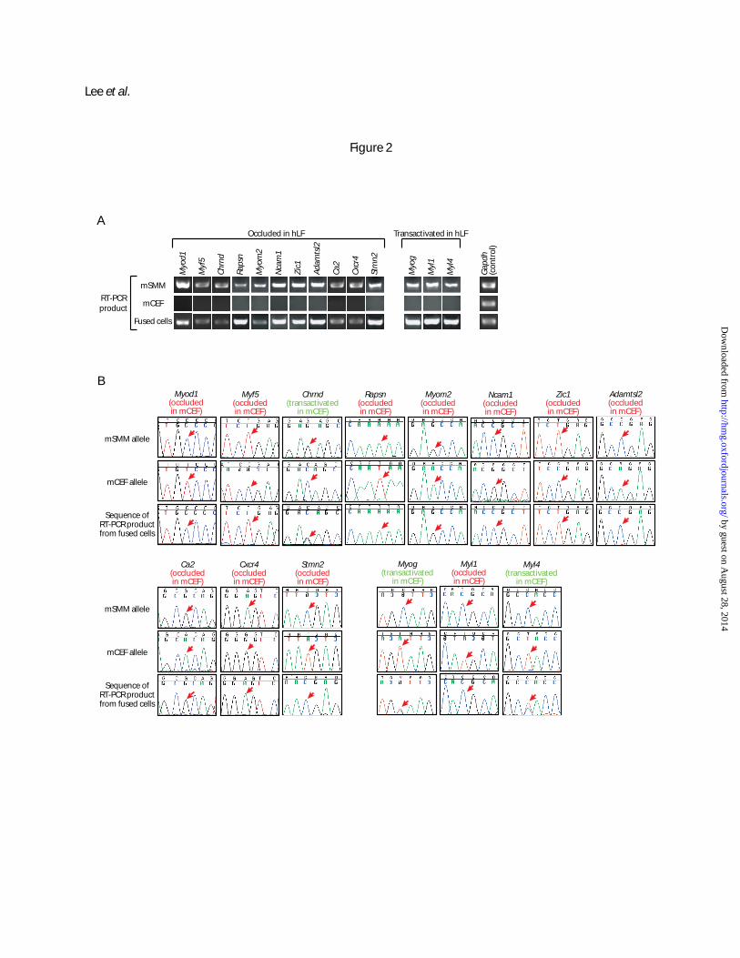
Among the 24 occluded and 10 transactivated hLF genes, 11 and 3, respectively, were found to
be informative in the mCEF-mSMM fusion, meaning that they bear exonic polymorphisms between the
two strains based on our resequencing data, and are expressed in mSMM but not mCEF based on RT-
PCR data. For each of these genes, RT-PCR primers were designed to flank an inter-strain polymorphic
site. The relative abundance of mCEF (B6 strain) versus mSMM (C3H strain) transcripts of the gene in
mCEF-mSMM fusion cells was then assessed by sequencing the RT-PCR product. This analysis showed
that, of the 11 informative genes occluded in hLF, all but one are also occluded in mCEF based on their
exclusive expression from the mSMM allele in fused cells, including the myogenic master triggers
Myod1 and Myf5 (Figure 2; data also summarized in Figure 1B). The single exception is Chrnd, which is
expressed at roughly equal levels from both mSMM and mCEF alleles, indicating transactivation. Of the
3 informative genes transactivated in hLF, 2 were also found to be transactivated in mCEF and one was
occluded in mCEF (Figure 2; also summarized in Figure 1B). Similar to the hDF-mSMM fusion
described above, genes found to be occluded in mCEF in the mCEF-mSMM fusion experiment
remained silent in fused cells independent of culture time (Figure S3). Thus, among the informative
genes, those occluded in hLF are almost all occluded in mCEF and those transactivated in hLF are
mostly transactivated in mCEF. These results offer strong evidence that interspecies incompatibility
played a negligible role in the identification of occluded genes in the hLF-mSMM fusion, though we
by guest on August 28, 2014
http://hmg.oxfordjournals.org/
Dow
nloaded from
9
cannot rule out the possibility that incompatibility might have affected a small number of genes. The
fact that Chrnd appears occluded in hLF but transactivated in mCEF suggests the possibility that the
observed occlusion of this gene in hLF might be an artifact of interspecies incompatibility in the hLF-
mSMM fusion. However, we think that this is unlikely based on data presented in the section below.
Conservation of occluded state across species
Comparison between hLF and mCEF suggests that the set of genes subject to occlusion in a given cell
type – fibroblasts in this case – is relatively conserved between divergent species. To further investigate
this conservation, we fused chimpanzee dermal fibroblasts (cDF) with human skeletal muscle myoblasts
(hSMM) in order to examine whether genes occluded in hLF are also occluded in cDF. The human-
chimpanzee genome divergence is about 1/30 of that between human and mouse, and is in fact less than
the polymorphism levels within many species. Interspecies incompatibility should therefore not be a
significant issue in this case.
Of the 24 occluded and 10 transactivated hLF genes, 12 and 8, respectively, were found to be
informative in the cDF-hSMM fusion. For these genes, RT-PCR was performed on cDF-hSMM fusion
cells using primers common to both species but flanking human-chimpanzee nucleotide substitutions.
Sequencing of the RT-PCR products revealed that of the 12 informative genes occluded in hLF, all are
occluded in cDF (Figure S4; summarized in Figure 1B). The occluded cDF genes include Chrnd, which
is transactivated in mCEF, suggesting that the occluded status of this gene in hLF is real. Of the 8
informative genes transactivated in hLF, 6 are transactivated in cDF while the other 2 are occluded in
cDF (Figure S4; summarized in Figure 1B).
Thus, the occluded or transactivated state of genes in hLF is closely recapitulated in both mCEF
and cDF, indicating that the set of genes subject to occlusion in a given cell type is strongly conserved
across species. Such conservation argues that the occlusion of lineage-inappropriate genes is a highly
regulated process with important biological functions.
Effect of DNA synthesis and nuclear merger on the occluded state
In the mCEF-mSMM fusion experiment, we observed that even though the majority of cells were
heterokaryons immediately after fusion and FACS purification, most cells became mononucleated after
a few days of culture. Furthermore, the average nuclear diameter of these mononucleated cells is about
40% larger than that of either mCEF or mSMM alone (Figure S5A). We suspected that this was due to
the formation of a single nucleus from the multiple nuclei in a given fused cell (i.e., nuclear merger).
by guest on August 28, 2014
http://hmg.oxfordjournals.org/
Dow
nloaded from
10
The most likely scenario that multiple nuclei of a heterokaryon could merge is the breakdown and
reassembly of the nuclear envelope as the cell undergoes mitosis. For this to occur, cells in the mCEF-
mSMM fusion must be capable of DNA synthesis and mitosis. This is consistent with the observation
that the mononucleated cells proliferated in number while in culture. By monitoring the incorporation of
the thymidine analog 5-bromo-2’-deoxyuridine (BrdU), we confirmed that the majority of fused mCEF-
mSMM cells underwent de novo DNA synthesis a few days after fusion (Figures S5B and S5C). To
further confirm that the single nucleus present in each of the mononucleated cells indeed contains both
mCEF and mSMM genomes, we labeled mCEF and mSMM DNA, prior to fusion, with the thymidine
analogs 5-chloro-2′-deoxyuridine (CldU) and 5-iodo-2′-deoxyuridine (IdU), respectively. Four days
after fusion and FACS purification, cells were co-immunostained for CldU and IdU. For the great
majority of mononucleated cells, the nuclei were found to be double positive for both CldU and IdU,
consistent with the merger of the mCEF and mSMM nuclei (Figure S5D).
One complicating factor in identifying occluded genes in fused cells that have undergone mitosis
is the possibility of chromosome loss. If some chromosomes are preferentially lost, they would be
underrepresented in fused cells and the genes they carry could appear occluded. We addressed this issue
by performing PCR on genomic DNA of the fused cells, amplifying across the same polymorphic sites
as those interrogated by RT-PCR. Sequencing of PCR products indicated the presence of both alleles at
comparable levels for all genes investigated, which are physically scattered across the genome (data not
shown). The allele-specific expression seen in Figure 2 is therefore not the result of chromosome loss.
This data also argues that the DNA synthesis observed in the fused mCEF-mSMM cells is likely
contributed by the replication of both the mCEF and mSMM genomes, because if only one of two
genomes has undergone replication, the alleles of the replicating genome should be consistently
overrepresented in the genomic PCR product over the alleles of the non-replicating genome, which is
not the case.
It is not clear why cells in the mCEF-mSMM fusion can undergo division whereas cells in the
other fusion experiments of this study remain largely as mitotically arrested heterokaryons. This
notwithstanding, the fact that occluded genes can be uncovered even after heterokaryons have
undergone division argues that the occluded state is robust to DNA replication, nuclear merger, and
changes in the cell cycle state.
Occlusion of muscle-related genes in diverse non-muscle cell types
If the occlusion of muscle-related genes, especially Myod1 and Myf5, indeed serves to safeguard hLF
by guest on August 28, 2014
http://hmg.oxfordjournals.org/
Dow
nloaded from
11
against the accidental activation of myogenic programs, then similar sets of muscle-related genes are
likely to be occluded in other cell types of non-myogenic lineages. To test this possibility, we fused
mSMM with non-muscle cell types of diverse lineages, and performed RT-PCR to examine if the 24
genes occluded in hLF are also occluded in these other cell types. The non-muscle cells used included
human mesenchymal stem cells (hMSC), human keratinocytes (hKe), and the human cervical cancer cell
line Hela. These cells provide a broad representation of both stem cells and differentiated cells, both
normal cells and transformed cells, and cells derived from different germ layers.
We found that of the 9 known muscle-related genes occluded in hLF, the majority are also
occluded in all these additional non-muscle cell types, including the myogenic master regulators Myod1
and Myf5 (Figure S6). Of the remaining 15 occluded hLF genes not known to be muscle-related, most
were either expressed prior to fusion or were transactivated upon fusion in at least one of the non-muscle
cell types interrogated. These results support the model that the occlusion of lineage-inappropriate genes,
especially key master triggers of alternative lineages, contributes to the restriction of cell fate.
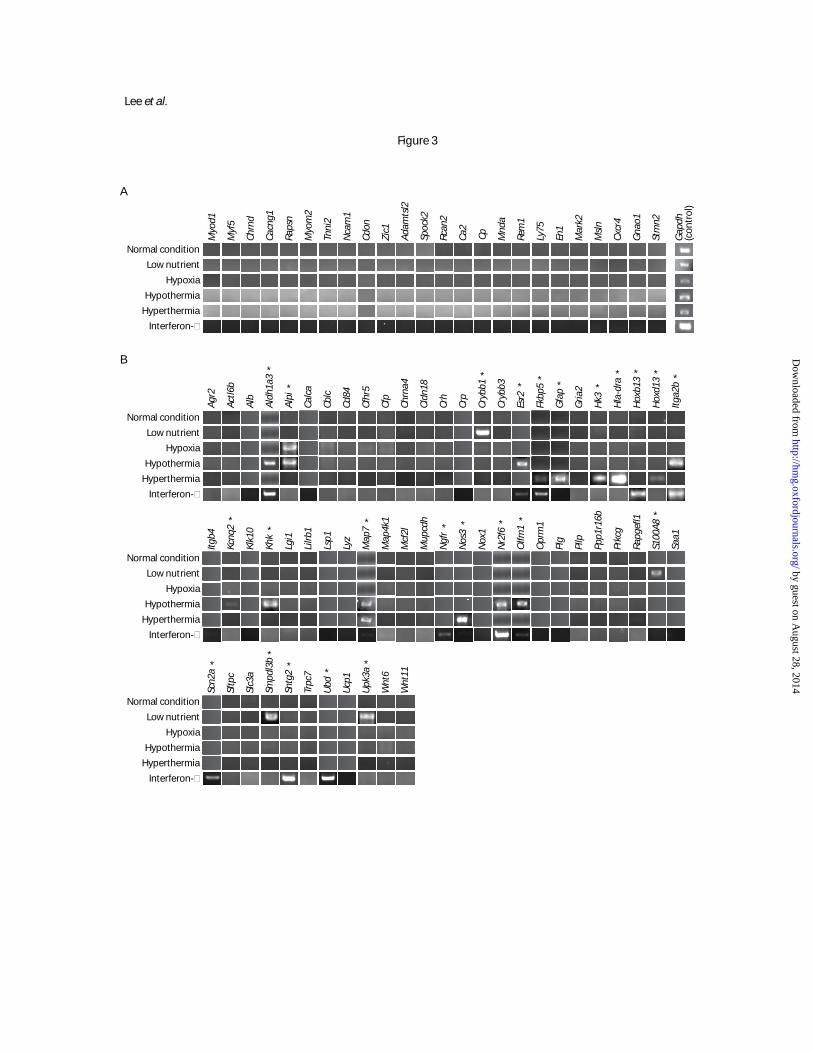
Stability of the occluded state under varying physiological conditions
If the occluded state is indeed critical in safeguarding cell identity as we have proposed, then it should
be stable under a variety of physiological conditions. To investigate this, we subjected hLF to a variety
of culture conditions mimicking various types of physiological stress, including low nutrient, hypoxia,
hypothermia, and hyperthermia. We also included interferon-γ treatment, which is known to have a
dramatic effect on the expression of many genes in a variety of cell types including fibroblasts (30). We
then examined the resulting expression patterns of the 24 occluded genes under these culture conditions.
All of them remained silent regardless of condition (Figure 3A). As a control, we identified a set of 61
genes silent in hLF under the normal culture condition based on microarray data and RT-PCR validation.
We then examined their expression patterns under the alternative culture conditions. A total of 24 of the
61 genes (39%) became active in at least one of the conditions (Figure 3B), which is statistically highly
distinct from the behavior of zero activation among the 24 occluded genes (p < 0.00007 by Fisher’s
exact test).
These results demonstrate the extraordinary stability of the occluded state under variable
physiological conditions, which stands in stark contrast to the transcriptional lability of other genes in
the genome. Researchers have often resorted to genome-wide gene expression patterns (i.e., the
transcriptome) as a means of defining cell type identity. However, as our data demonstrate, one cell type
has the potential to display considerably different gene expression patterns under different physiological
by guest on August 28, 2014
http://hmg.oxfordjournals.org/
Dow
nloaded from
12
conditions, making the transcriptome too labile to provide a consistent definition of cell type. Our results
suggest that genome-wide gene occlusion patterns (i.e., the “occludome”) might provide a much more
consistent definition of cell type than the physiologically labile transcriptome (see Discussion).
DISCUSSION
Cell fusion has been used in the past to investigate gene regulation, with most previous studies focusing
on the transactivation and extinction of tissue-specific genes in fused cells that indicate the presence of
trans-acting transcriptional activators or repressors (22, 31-33). In this report, we demonstrate an
important utility of cell fusion as implemented in the trans complementation assay. By fusing disparate
cell types and searching for genes differentially expressed between the two genomes of the fused cells,
the assay can dissect out the contribution of cis-acting mechanisms to gene silencing apart from the
contribution of trans-acting milieu. Using this assay, we identified a class of genes existing in what we
refer to as the occluded state, defined as a state of transcriptional competency whereby a gene remains
silent even in the presence of a transcriptionally conducive milieu. We further showed that the occluded
state is maintained during cell division and is highly stable under a wide range of physiological
conditions.
Monoallelic silencing such as X inactivation and imprinting clearly fits the definition of the
occluded state (15-17). Our work demonstrates that biallelic occlusion also occurs as a widespread
biological phenomenon, affecting many genes in diverse cell types. Indeed, monoallelic silencing can be
viewed as a special case of gene occlusion. It is plausible that biallelic occlusion is the ancestral state
that evolved into existence first, with monoallelic silencing evolving subsequently by adopting many of
the basic machineries of biallelic occlusion but adding a mechanism for targeting one allele (rather than
both alleles) during the silencing process. Biallelic occlusion may be key to defining and restricting the
phenotypic identities of cells by stably shutting down lineage-inappropriate genes that might otherwise
become active.
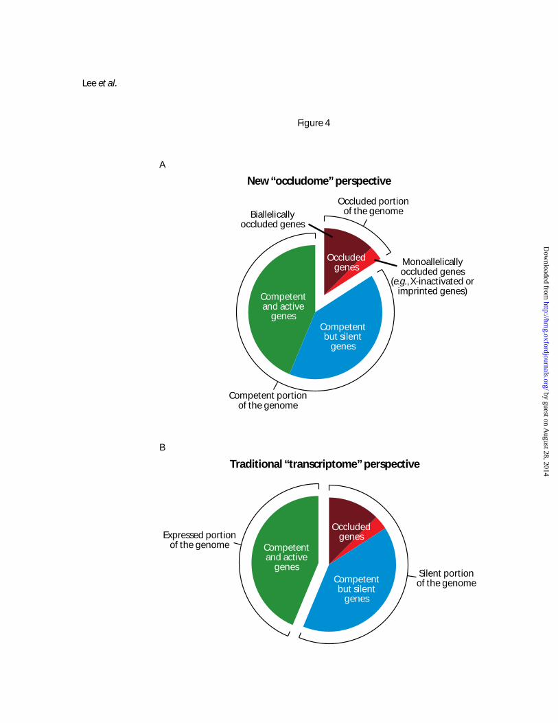
Extrapolating from our results, we argue that it may be meaningful to take an “occludome”
perspective of genome regulation – i.e., consider the genome of a cell type as comprising two portions,
one being the occluded genes and the other the competent genes (Figure 4A). Actively expressed genes
in a cell type are all competent, but silent genes can be either competent or occluded. This is a different
conceptual framework for understanding genome regulation from the traditional “transcriptome”
perspective whereby genes are considered to be expressed or silent (compare Figure 4A with 4B).
We propose that it should be possible to systematically map all the occluded genes in a cell type
by guest on August 28, 2014
http://hmg.oxfordjournals.org/
Dow
nloaded from
13
by fusing it with a wide variety of other cell types that collectively express the entire genome. Such an
occludome map might provide a definition of cell type that is physiologically more consistent – and
molecularly more fundamental – than the rather labile transcriptome. By comparing occludome maps
between cell types of different lineages, between stem cells and differentiated cells of the same lineage,
between young and old cells, between normal and pathological cells (such as cancer), and between cells
from different species, it might be possible to gain wide-ranging insights into fundamental mechanisms
of development, aging, disease processes, and evolution. Furthermore, for a given cell type, comparisons
could be made between the occludome map and genome-wide maps of chromatin marks such as DNA
methylation and histone modifications (34-36). Such comparisons could reveal the biochemical
underpinnings of the occluded state, and more importantly, provide a hitherto unavailable functional
readout of the complex chromatin code superimposed on the genetic code.
For operational simplicity, the current study has taken a binary, on/off view of gene occlusion.
However, it is plausible that occlusion can sometimes lead to partial silencing of some genes, in which
case a gene may show a quantitative expression difference between the two genomes of fused cells
rather than a qualitative on/off difference. In theory, the trans complementation assay should be able to
reveal both full and partial occlusion as long as a gene displays differential expression between the two
genomes of the fused cells (provided that confounding factors such as interspecies incompatibility are
ruled out).
The definition of the occluded state requires that a gene is silent (or nearly silent) even in the
presence of a transcriptionally conducive milieu. It is important to note, however, that this definition is
only in reference to a particular milieu. It may be the case that a gene occluded to one milieu might
become active in another milieu. This could happen if transcription factors in the first milieu are blocked
by repressive chromatin marks present in certain cis-regulatory sequences of a gene, but transcription
factors in the second milieu, distinct from the first, are able to drive expression by recognizing a
different set of cis-regulatory sequences of the gene not affected by repressive chromatin. Alternatively,
factors in the second milieu, unlike those in the first, can recognize their target sequences even in the
presence of repressive chromatin.
Another important possibility is that some milieus might have the ability to “deocclude” genes –
i.e., erasing the chromatin marks responsible for the occluded state. Such erasure could affect individual
genes or the whole genome, and could be an active process or a passive one. Reprogramming of somatic
cells by nuclear transfer into oocytes or by fusion with embryonic stem cells (ESC) or embryonic germ
cells (EGC) have demonstrated the ability of these cell types to erase most, if not all, of the chromatin
by guest on August 28, 2014
http://hmg.oxfordjournals.org/
Dow
nloaded from
14
marks in somatic cells established during development (37-43). Recent work indicates that such ability
may arise from just a few genes whose ectopic expression can reprogram fibroblasts into pluripotent,
ESC-like cells called induced pluripotent stem (iPS) cells (44-50). We hypothesize that shortly after the
blastocyst stage (where ESC is derived), cells lose their ability to deocclude the genome, perhaps by
occluding the very genes that are responsible for genome-wide deocclusion in the first place. We further
hypothesize that the progressive differentiation of cells in subsequent developmental stages is
accompanied by the irreversible or nearly irreversible occlusion of an increasing number of genes, with
distinct sets of genes becoming occluded in different lineages.
The occlusion of lineage-inappropriate genes could serve to safeguard the phenotypic stability of
the myriad cell types in multicellular organisms against noise in both extracellular environment and
intracellular regulatory networks. Furthermore, that different cell types are characterized by different
occludomes might also explain why the same signaling pathway often triggers the activation of different
sets of genes in different cell – a frequent phenomenon during the development of multicellular
organisms. The ability of the same transcription factors to play different roles in different cell types
allows increased cell type complexity in multicellular organisms without concomitant increases in
genome size/complexity. Thus, the evolution of some form of gene occlusion might have been a
prerequisite for the evolution of multicellularity.
The occluded state could be quite stable in order to maintain cell identity over the entire ontology
of the organism (the germline being an exception where the occluded state is either never fully
established for most genes or is erased during gametogenesis). For some genes, the occluded state might
be essentially irreversible in somatic cells under normal conditions (as is the case for X-inactivated and
imprinted genes). Nevertheless, some occluded genes might become deoccluded in certain somatic cell
types by deliberate mechanisms, which could contribute to the dedifferentiation/transdifferentiation of
cells during tissue regeneration, especially in species capable of regenerating entire body parts after
injury (51). On rare occasions, the competent/occluded status of genes could also change in a stochastic,
unregulated manner, which might contribute to aging and disease processes such as cancer. The use of
the trans complementation assay to systematically identify and characterize occluded genes should
therefore have wide-ranging applications in studies of health and disease.
by guest on August 28, 2014
http://hmg.oxfordjournals.org/
Dow
nloaded from
15
MATERIALS AND METHODS
Cell fusion
Mouse skeletal muscle myoblasts (mSMM) and human lung fibroblasts (hLF) have been described
previously, and are known by their common names as C2C12 and MRC-5, respectively (21, 52). C2C12
(CRL-1772), MRC-5 (CCL-171), Hela (CCL-2), mouse cloned embryonic fibroblasts (mCEF; TIB-81),
and human kerationcytes (hKe; CRL-2404) were obtained from ATCC; human mesenchymal stem cells
(hMSC) were derived as described (53) and are available from Cyagen Biosciences; chimpanzee dermal
fibroblasts (cDF; S006007) were obtained from Coriell Institute for Medical Research; and human
skeletal muscle myoblasts (hSMM; CC-2580T25) were obtained from Cambrex. Cell culture conditions
followed published or vendor-supplied protocols.
Neomycin-resistant mSMM cells were generated by transfection with the pEGFP-N1 plasmid
(Clontech) and selection in 800 μg/ml G418. EGFP fluorescence varied within this cell population, but
was negligible compared to dye fluorescence used for cell sorting. Puromycin-resistant hLF cells were
generated using pBabe-puro retroviral vector from Addgene (#1764) (54). The vector was transfected
into ProPakA.6 packaging cells (ATCC). 48 hours after transfection, the viral supernatant was filtered
and added to the cells in the presence of 8 ug/ml polybrene, and 24 hours later cells were selected using
2 μg/ml puromycin.
One day before fusion, cells were labeled with 30 μM CMTMR or 10 μM CMFDA Celltracker
dye (Invitrogen) for 30 min at 37°C in culture medium. Subsequently, the cells were incubated in basal
medium for one hour, and washed twice with PBS. After staining, mSMM cells were kept in low-serum
medium composed of DMEM supplemented with 2% horse serum. Cell fusion was performed with
polyethylene glycol (MW 1500) as described (55). Briefly, one of the two cell populations was plated on
10-cm tissue culture dishes and the other cell population was overlaid. After attachment, cells were
treated with warm PEG for 1 min, and then washed three times with warm basal medium. The fused
cells were incubated for two hours in low-serum medium until cell sorting. Fused cells were purified to
>98% purity by fluorescence activated cell sorting (FACS) with gating for dual fluorescence. After
FACS, purified fused cells were maintained in low-serum medium until RNA extraction. The unfused
mSMM used as control in expression studies are kept in low-serum medium for the same period as fused
cells. For experiments in which antibiotic-resistant cells were fused, 4-8 μg/ml puromycin and 400-800
μg/ml G418 were added to the medium one day after fusion.
by guest on August 28, 2014
http://hmg.oxfordjournals.org/
Dow
nloaded from
16
Microarray analysis
Total RNA was purified from hLF, mSMM, and fused cells using TRIZOL reagent (Invitrogen)
according to vendor’s protocol, and used for microarray probe synthesis following standard Affymetrix
protocols. Double-stranded cDNA samples generated using GeneChip One-Cycle cDNA Synthesis kit
with first strand synthesis using oligo(dT) primers (Affymetrix) were used to synthesize biotin-labeled
cRNA using GeneChip IVT Labeling kit (Affymetrix), and then the labeled cRNA samples were
fragmented using GeneChip Sample Cleanup Module (Affymetrix). Hybridization, labeling and
scanning were all performed by the Protein and Nucleic Acid (PAN) facility at Stanford University. The
labeled cRNA sample from each cell type was hybridized to both of mouse MG U74Av2 and human HG
U133A GeneChips (Affymetrix) with replicates to assess gene expression and cross-hybridization
between species. Probe-level analyses of the images from scanning of chips were performed using
Affymetrix GeneChip Operating Software (GCOS). Similar hybridization procedures were carried out
for the hLF-mOst fusion.
Threshold detection p-values were set to assign “present” (p < 0.05), “marginal” (0.05 ≤ p ≤
0.49), or “absent” (p > 0.49) decision calls for each gene assigned by MAS 5.0 criteria using GCOS.
Filtering gene lists based on absolute decision calls to get a candidate list of occluded genes was
performed by using GeneSpring (Silicon Genetics). Occluded genes were filtered based on the following
criteria: absent calls in all of the replicates with hLF hybridized to human chip, absent calls in all of the
replicates with fused cells hybridized to human chip, present or marginal calls in at least one of the
replicates with mSMM hybridized to mouse chip, and present or marginal calls in at least one of the
replicates with fused cells hybridized to mouse chip. Filtering of transactivated genes was performed by
1) comparing genes based on absolute decision calls using GeneSpring with criteria of absent calls in all
of the replicates with hLF hybridized to human chip and present or marginal calls in at least one of the
replicates with fused cells hybridized to human chip, 2) selecting genes showing differential expression
between the two cell types based on signal intensity after normalization by RMA using RMAexpress
(http://rmaexpress.bmbolstad.com), or 3) comparing the data from the two cell types by model-based
expression index analysis using dChip (http://biosun1.harvard.edu/complab/dchip).
RT-PCR and sequencing
RNA (up to 2 μg) was used to generate cDNA using M-MLV reverse transcriptase and random primers
(Invitrogen), or using SuperScript III First-Strand Synthesis System with random primers for RT-PCR
(Invitrogen) following vendor’s protocol. Semi-quantitative PCR was carried out with variable template
by guest on August 28, 2014
http://hmg.oxfordjournals.org/
Dow
nloaded from
17
concentrations and PCR cycles to obtain linear range amplification of each gene. For the human-
chimpanzee fusion experiment, primers were selected by identifying non-polymorphic primer sequences
flanking intron-spanning amplicons that contain at least one single-nucleotide substitution between the
two species based on genomic sequence alignment. For the mouse-mouse fusion experiment, amplicons
containing at least one polymorphism between the two mouse strains were identified by sequencing
randomly-chosen intron-spanning amplicons. Sequences of PCR primers and detailed conditions for RT-
PCR are available upon request. All DNA sequence analysis was performed with the ABI 3730 DNA
Analyzer using the ABI BigDye Terminator (Applied Biosystems).
Analysis of DNA synthesis and nuclear merger
For analysis of nuclear merger, unfused cells were labeled with 10 μM 5-iodo-2’-deoxyuridine (IdU) or
5-chloro-2’-deoxyuridine (CldU) in the media for 72 hours prior to fusion, and fused cells were stained
specifically with mouse monoclonal anti-IdU (Becton-Dickinson, #347580; 1:500 dilution) and rat
monoclonal anti-CldU antibodies (Accurate, #OBT0030; 1:250) based on published protocol (56). These
two antibodies do not cross-react when used for double-staining IdU and CldU, but both recognize 5-
bromo-2’-deoxyuridine (BrdU). Secondary antibodies were Oregon Green labeled goat anti-mouse
(Invitrogen; 1:1000) and Cy3 labeled mouse anti-rat (Jackson Immunoresearch; 1:300). For analysis of
DNA synthesis, BrdU was administered at 10 μM in the media immediately following cell fusion for 72
hours, and the cells stained with anti-BrdU antibody (Accurate, #OBT0030; 1:250) at a later time point.
Because the incorporation of halogenated nucleotides into DNA could affect gene expression, the fusion
experiment involving labeling with halogenated nucleotides is done separately from the fusion
experiment for ascertaining the expression status of genes.
Analysis of gene expression under physiological alterations
Cells were cultured under either the normal condition (10% fetal calf serum at 37°C) or one of the
conditions mimicking physiological alterations, including low nutrient (0.1% serum), hypoxia (380 µM
of the hypoxia mimetic deferoxamine), hypothermia (33°C), hyperthermia (41°C), and interferon-γ
treatment (100 ng/ml; Cell Sciences). Cells were maintained under each condition for 3 days, followed
by RT-PCR analysis of selected genes as described above.
by guest on August 28, 2014
http://hmg.oxfordjournals.org/
Dow
nloaded from
18
ACKNOWLEDGMENTS
We thank Ryan Duggan, David Leclerc, Michael Olson, Jaejung Kim, and Ingrid Kilgore for technical
assistance. This work was partly supported by National Institutes of Health grants F32HL922792 (to JG),
F32GM075503 (to GES), and HL07605 (to ECB).
REFERENCES
1. Waddington, C.H. (1966) Principles of Development and Differentiation. Macmillan, New York. 2. Caplan, A.I. and Ordahl, C.P. (1978) Irreversible gene repression model for control of
development. Science, 201, 120-130. 3. Fisher, A.G. and Merkenschlager, M. (2002) Gene silencing, cell fate and nuclear organisation.
Curr. Opin. Genet. Dev., 12, 193-197. 4. Macaluso, M. and Giordano, A. (2004) How does DNA methylation mark the fate of cells?
Tumori, 90, 367-372. 5. Sparmann, A. and van Lohuizen, M. (2006) Polycomb silencers control cell fate, development
and cancer. Nat. Rev. Cancer, 6, 846-856. 6. Jenuwein, T. and Allis, C.D. (2001) Translating the histone code. Science, 293, 1074-1080. 7. Jaenisch, R. and Bird, A. (2003) Epigenetic regulation of gene expression: how the genome
integrates intrinsic and environmental signals. Nat. Genet., 33 Suppl, 245-254. 8. Vermaak, D., Ahmad, K. and Henikoff, S. (2003) Maintenance of chromatin states: an open-and-
shut case. Curr. Opin. Cell Biol., 15, 266-274. 9. Goldberg, A.D., Allis, C.D. and Bernstein, E. (2007) Epigenetics: a landscape takes shape. Cell,
128, 635-638. 10. Kouzarides, T. (2007) Chromatin modifications and their function. Cell, 128, 693-705. 11. Li, B., Carey, M. and Workman, J.L. (2007) The role of chromatin during transcription. Cell,
128, 707-719. 12. Schuettengruber, B., Chourrout, D., Vervoort, M., Leblanc, B. and Cavalli, G. (2007) Genome
regulation by polycomb and trithorax proteins. Cell, 128, 735-745. 13. Surani, M.A., Hayashi, K. and Hajkova, P. (2007) Genetic and epigenetic regulators of
pluripotency. Cell, 128, 747-762. 14. Bird, A. (2007) Perceptions of epigenetics. Nature, 447, 396-398. 15. Bartolomei, M.S. and Tilghman, S.M. (1997) Genomic imprinting in mammals. Annu. Rev.
Genet., 31, 493-525. 16. Goldmit, M. and Bergman, Y. (2004) Monoallelic gene expression: a repertoire of recurrent
themes. Immunol. Rev., 200, 197-214. 17. Valley, C.M. and Willard, H.F. (2006) Genomic and epigenomic approaches to the study of X
chromosome inactivation. Curr. Opin. Genet. Dev., 16, 240-245. 18. Barrera, L.O. and Ren, B. (2006) The transcriptional regulatory code of eukaryotic cells--insights
from genome-wide analysis of chromatin organization and transcription factor binding. Curr. Opin. Cell Biol., 18, 291-298.
19. Maziarz, M., Chung, C., Drucker, D.J. and Emili, A. (2005) Integrating global proteomic and genomic expression profiles generated from islet alpha cells: opportunities and challenges to deriving reliable biological inferences. Mol. Cell. Proteomics, 4, 458-474.
by guest on August 28, 2014
http://hmg.oxfordjournals.org/
Dow
nloaded from
19
20. Chiu, C.P. and Blau, H.M. (1984) Reprogramming cell differentiation in the absence of DNA synthesis. Cell, 37, 879-887.
21. Blau, H.M., Pavlath, G.K., Hardeman, E.C., Chiu, C.P., Silberstein, L., Webster, S.G., Miller, S.C. and Webster, C. (1985) Plasticity of the differentiated state. Science, 230, 758-766.
22. Boshart, M., Nitsch, D. and Schutz, G. (1993) Extinction of gene expression in somatic cell hybrids--a reflection of important regulatory mechanisms? Trends Genet., 9, 240-245.
23. Davis, R.L., Weintraub, H. and Lassar, A.B. (1987) Expression of a single transfected cDNA converts fibroblasts to myoblasts. Cell, 51, 987-1000.
24. Braun, T., Buschhausen-Denker, G., Bober, E., Tannich, E. and Arnold, H.H. (1989) A novel human muscle factor related to but distinct from MyoD1 induces myogenic conversion in 10T1/2 fibroblasts. Embo J., 8, 701-709.
25. Wright, W.E., Sassoon, D.A. and Lin, V.K. (1989) Myogenin, a factor regulating myogenesis, has a domain homologous to MyoD. Cell, 56, 607-617.
26. Braun, T., Bober, E., Winter, B., Rosenthal, N. and Arnold, H.H. (1990) Myf-6, a new member of the human gene family of myogenic determination factors: evidence for a gene cluster on chromosome 12. Embo J., 9, 821-831.
27. Miner, J.H. and Wold, B. (1990) Herculin, a fourth member of the MyoD family of myogenic regulatory genes. Proc. Natl. Acad. Sci. U. S. A., 87, 1089-1093.
28. Thayer, M.J., Tapscott, S.J., Davis, R.L., Wright, W.E., Lassar, A.B. and Weintraub, H. (1989) Positive autoregulation of the myogenic determination gene MyoD1. Cell, 58, 241-248.
29. Tapscott, S.J. (2005) The circuitry of a master switch: Myod and the regulation of skeletal muscle gene transcription. Development, 132, 2685-2695.
30. Der, S.D., Zhou, A., Williams, B.R. and Silverman, R.H. (1998) Identification of genes differentially regulated by interferon alpha, beta, or gamma using oligonucleotide arrays. Proc. Natl. Acad. Sci. U. S. A., 95, 15623-15628.
31. Davidson, R.L. (1974) Gene expression in somatic cell hybrids. Annu. Rev. Genet., 8, 195-218. 32. Blau, H.M. (1989) How fixed is the differentiated state? Lessons from heterokaryons. Trends
Genet., 5, 268-272. 33. Gourdeau, H. and Fournier, R.E. (1990) Genetic analysis of mammalian cell differentiation.
Annu. Rev. Cell Biol., 6, 69-94. 34. van Steensel, B. and Henikoff, S. (2003) Epigenomic profiling using microarrays. Biotechniques,
35, 346-350, 352-344, 356-347. 35. Bernstein, B.E., Meissner, A. and Lander, E.S. (2007) The mammalian epigenome. Cell, 128,
669-681. 36. Jones, P.A. and Baylin, S.B. (2007) The epigenomics of cancer. Cell, 128, 683-692. 37. Gurdon, J.B. (1962) The developmental capacity of nuclei taken from intestinal epithelium cells
of feeding tadpoles. J. Embryol. Exp. Morphol., 10, 622-640. 38. Wilmut, I., Schnieke, A.E., McWhir, J., Kind, A.J. and Campbell, K.H. (1997) Viable offspring
derived from fetal and adult mammalian cells. Nature, 385, 810-813. 39. Hochedlinger, K. and Jaenisch, R. (2006) Nuclear reprogramming and pluripotency. Nature, 441,
1061-1067. 40. Tada, M., Tada, T., Lefebvre, L., Barton, S.C. and Surani, M.A. (1997) Embryonic germ cells
induce epigenetic reprogramming of somatic nucleus in hybrid cells. Embo J., 16, 6510-6520. 41. Tada, M., Takahama, Y., Abe, K., Nakatsuji, N. and Tada, T. (2001) Nuclear reprogramming of
somatic cells by in vitro hybridization with ES cells. Curr. Biol., 11, 1553-1558. 42. Do, J.T. and Scholer, H.R. (2004) Nuclei of embryonic stem cells reprogram somatic cells. Stem
Cells, 22, 941-949.
by guest on August 28, 2014
http://hmg.oxfordjournals.org/
Dow
nloaded from
20
43. Cowan, C.A., Atienza, J., Melton, D.A. and Eggan, K. (2005) Nuclear reprogramming of somatic cells after fusion with human embryonic stem cells. Science, 309, 1369-1373.
44. Takahashi, K. and Yamanaka, S. (2006) Induction of pluripotent stem cells from mouse embryonic and adult fibroblast cultures by defined factors. Cell, 126, 663-676.
45. Maherali, N., Sridharan, R., Xie, W., Utikal, J., Eminli, S., Arnold, K., Stadtfeld, M., Yachechko, R., Tchieu, J., Jaenisch, R. et al. (2007) Directly reprogrammed fibroblasts show global epigenetic remodeling and widespread tissue contribution. Cell Stem Cell, 1, 55-70.
46. Okita, K., Ichisaka, T. and Yamanaka, S. (2007) Generation of germline-competent induced pluripotent stem cells. Nature, 448, 313-317.
47. Wernig, M., Meissner, A., Foreman, R., Brambrink, T., Ku, M., Hochedlinger, K., Bernstein, B.E. and Jaenisch, R. (2007) In vitro reprogramming of fibroblasts into a pluripotent ES-cell-like state. Nature, 448, 318-324.
48. Takahashi, K., Tanabe, K., Ohnuki, M., Narita, M., Ichisaka, T., Tomoda, K. and Yamanaka, S. (2007) Induction of pluripotent stem cells from adult human fibroblasts by defined factors. Cell, 131, 861-872.
49. Yu, J., Vodyanik, M.A., Smuga-Otto, K., Antosiewicz-Bourget, J., Frane, J.L., Tian, S., Nie, J., Jonsdottir, G.A., Ruotti, V., Stewart, R. et al. (2007) Induced Pluripotent Stem Cell Lines Derived from Human Somatic Cells. Science.
50. Nakagawa, M., Koyanagi, M., Tanabe, K., Takahashi, K., Ichisaka, T., Aoi, T., Okita, K., Mochiduki, Y., Takizawa, N. and Yamanaka, S. (2007) Generation of induced pluripotent stem cells without Myc from mouse and human fibroblasts. Nat. Biotechnol., 26, 101-106.
51. Sanchez Alvarado, A. and Tsonis, P.A. (2006) Bridging the regeneration gap: genetic insights from diverse animal models. Nat. Rev. Genet., 7, 873-884.
52. Yaffe, D. and Saxel, O. (1977) Serial passaging and differentiation of myogenic cells isolated from dystrophic mouse muscle. Nature, 270, 725-727.
53. Zhang, A.X., Yu, W.H., Ma, B.F., Yu, X.B., Mao, F.F., Liu, W., Zhang, J.Q., Zhang, X.M., Li, S.N., Li, M.T. et al. (2007) Proteomic identification of differently expressed proteins responsible for osteoblast differentiation from human mesenchymal stem cells. Mol. Cell. Biochem., 304, 167-179.
54. Morgenstern, J.P. and Land, H. (1990) Advanced mammalian gene transfer: high titre retroviral vectors with multiple drug selection markers and a complementary helper-free packaging cell line. Nucleic Acids Res., 18, 3587-3596.
55. Davidson, R.L., O'Malley, K.A. and Wheeler, T.B. (1976) Polyethylene glycol-induced mammalian cell hybridization: effect of polyethylene glycol molecular weight and concentration. Somatic Cell Genet., 2, 271-280.
56. Vega, C.J. and Peterson, D.A. (2005) Stem cell proliferative history in tissue revealed by temporal halogenated thymidine analog discrimination. Nat. Methods, 2, 167-169.
57. Lee, T.I., Jenner, R.G., Boyer, L.A., Guenther, M.G., Levine, S.S., Kumar, R.M., Chevalier, B., Johnstone, S.E., Cole, M.F., Isono, K. et al. (2006) Control of developmental regulators by Polycomb in human embryonic stem cells. Cell, 125, 301-313.
by guest on August 28, 2014
http://hmg.oxfordjournals.org/
Dow
nloaded from
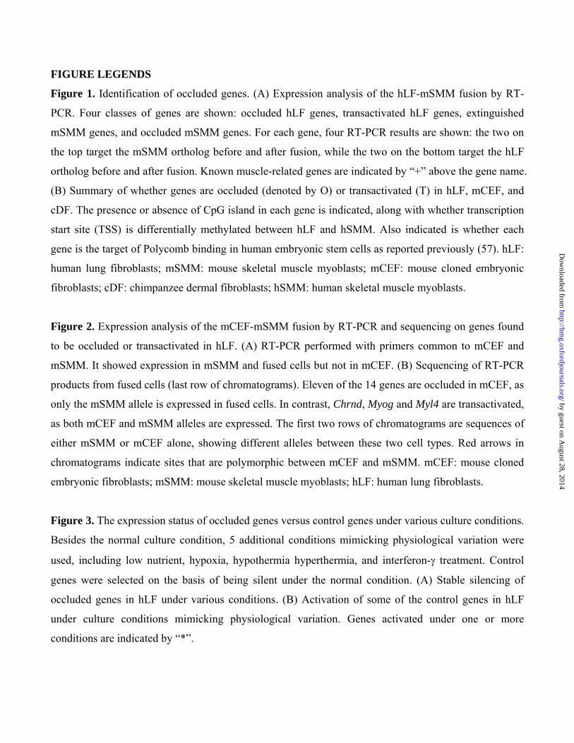
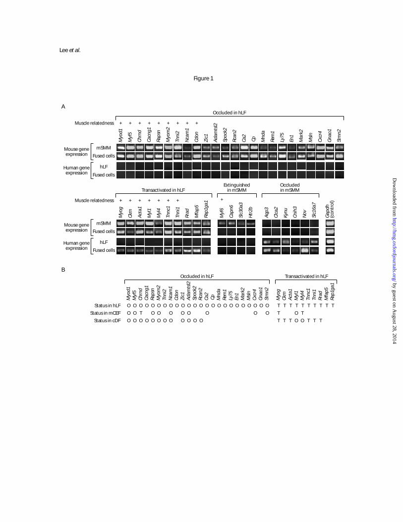
FIGURE LEGENDS
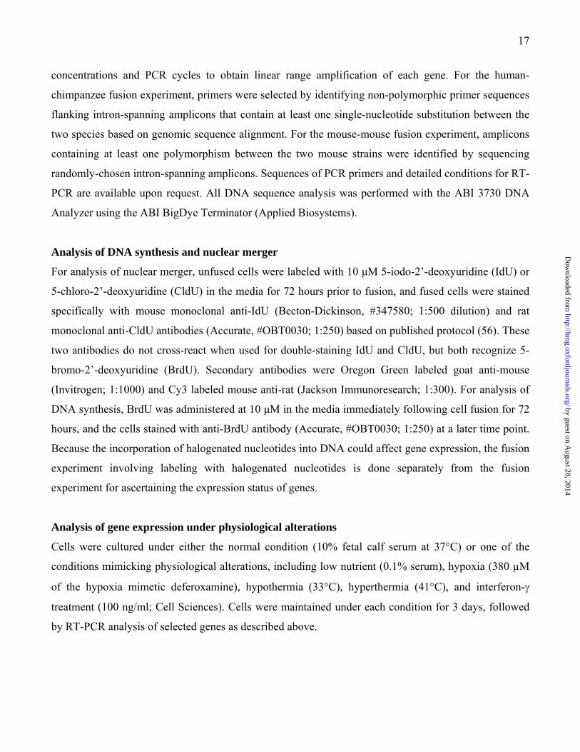
Figure 1. Identification of occluded genes. (A) Expression analysis of the hLF-mSMM fusion by RT-
PCR. Four classes of genes are shown: occluded hLF genes, transactivated hLF genes, extinguished
mSMM genes, and occluded mSMM genes. For each gene, four RT-PCR results are shown: the two on
the top target the mSMM ortholog before and after fusion, while the two on the bottom target the hLF
ortholog before and after fusion. Known muscle-related genes are indicated by “+” above the gene name.
(B) Summary of whether genes are occluded (denoted by O) or transactivated (T) in hLF, mCEF, and
cDF. The presence or absence of CpG island in each gene is indicated, along with whether transcription
start site (TSS) is differentially methylated between hLF and hSMM. Also indicated is whether each
gene is the target of Polycomb binding in human embryonic stem cells as reported previously (57). hLF:
human lung fibroblasts; mSMM: mouse skeletal muscle myoblasts; mCEF: mouse cloned embryonic
fibroblasts; cDF: chimpanzee dermal fibroblasts; hSMM: human skeletal muscle myoblasts.
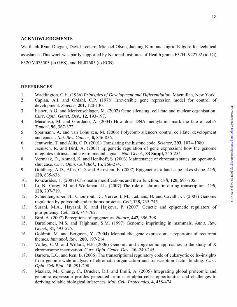
Figure 2. Expression analysis of the mCEF-mSMM fusion by RT-PCR and sequencing on genes found
to be occluded or transactivated in hLF. (A) RT-PCR performed with primers common to mCEF and
mSMM. It showed expression in mSMM and fused cells but not in mCEF. (B) Sequencing of RT-PCR
products from fused cells (last row of chromatograms). Eleven of the 14 genes are occluded in mCEF, as
only the mSMM allele is expressed in fused cells. In contrast, Chrnd, Myog and Myl4 are transactivated,
as both mCEF and mSMM alleles are expressed. The first two rows of chromatograms are sequences of
either mSMM or mCEF alone, showing different alleles between these two cell types. Red arrows in
chromatograms indicate sites that are polymorphic between mCEF and mSMM. mCEF: mouse cloned
embryonic fibroblasts; mSMM: mouse skeletal muscle myoblasts; hLF: human lung fibroblasts.
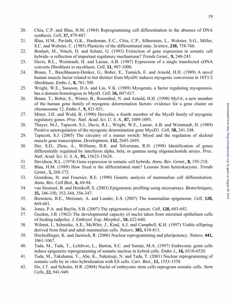
Figure 3. The expression status of occluded genes versus control genes under various culture conditions.
Besides the normal culture condition, 5 additional conditions mimicking physiological variation were
used, including low nutrient, hypoxia, hypothermia hyperthermia, and interferon-γ treatment. Control
genes were selected on the basis of being silent under the normal condition. (A) Stable silencing of
occluded genes in hLF under various conditions. (B) Activation of some of the control genes in hLF
under culture conditions mimicking physiological variation. Genes activated under one or more
conditions are indicated by “*”.
by guest on August 28, 2014
http://hmg.oxfordjournals.org/
Dow
nloaded from
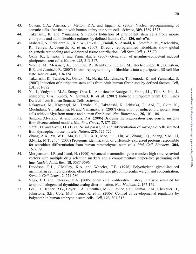
Figure 4. Different perspectives for understanding genome regulation. (A) New “occludome”
perspective that considers the genome as consisting of occluded genes and competent genes. (B)
Traditional “transcriptome” perspective that views the genome as comprising expressed genes and silent
genes. The occludome perspective may provide a molecularly more fundamental and physiologically
more stable definition of cell type than the transcriptome.
by guest on August 28, 2014
http://hmg.oxfordjournals.org/
Dow
nloaded from
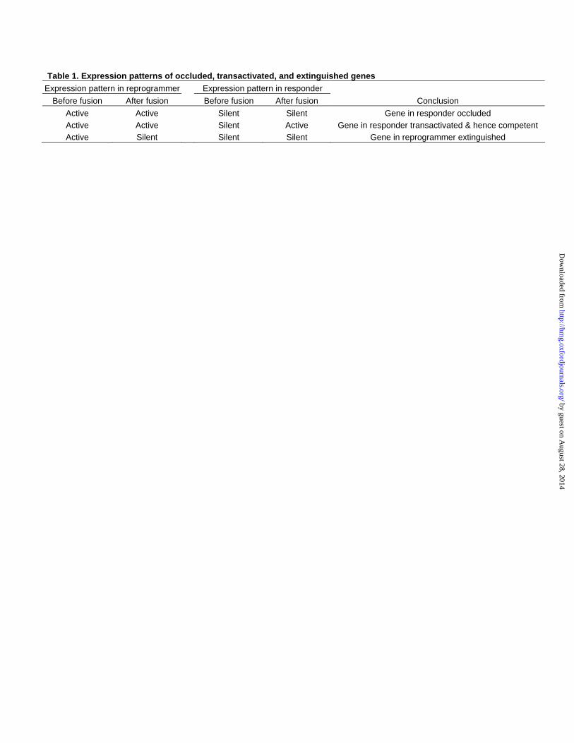
Table 1. Expression patterns of occluded, transactivated, and extinguished genes Expression pattern in reprogrammer Expression pattern in responder
Before fusion After fusion Before fusion After fusion Conclusion Active Active Silent Silent Gene in responder occluded Active Active Silent Active Gene in responder transactivated & hence competent Active Silent Silent Silent Gene in reprogrammer extinguished
by guest on August 28, 2014
http://hmg.oxfordjournals.org/
Dow
nloaded from
Gnao
1
Zic1
Adam
tsl2
Ca2
Rcan
2
Stm
n2
Cp Mnd
a
Rem
1
Spoc
k2
Ly75
En1
Mar
k2
Msln
Cxcr
4
Lee et al.
Figure 1
Occluded in hLF
Fused cells
Fused cells
Occluded in hLF
Mouse geneexpression
Human geneexpression
hLF
Fused cells
Mouse geneexpression
Human geneexpression
Capn
6
Myf
6
Slc3
0a3
Gapd
h(c
ontro
l)
Aqp3
Clca
2
Slc1
6a7
Kynu
Cnih
3
Nov
Extinguishedin mSMM
mSMM
Fused cells
hLF
mSMM
in mSMMOccluded
Transactivated in hLF
Transactivated in hLF
Rrad
Mfa
p5
Rap1
ga1
Htr2
b
Myo
g
+
Acta
1
+
Myl
1
+ +
Ckm
+
Myl
4
+
Tnni
1
+
Tnnc
1
+
Myf
5+
Myo
d1+Muscle relatedness
Muscle relatedness
Chrn
d
+
Ncam
1
+
Cacn
g1
+
Raps
n
+
Cdon
+
Myo
m2
+
Tnni
2
+
Status in cDFStatus in mCEF
Status in hLF
Myo
d1
OOO
Myf
5
OO
O
Chrn
d
OTO
Cacn
g1
O
O
Raps
n
OOO
Myo
m2
OOO
Tnni
2
O
O
Ncam
1
OOO
Cdon
O
Zic1
OOO
Rcan
2
O
O
Spoc
k2
O
O
Ca2
OO
Cp
O
Mnd
a
O
Rem
1
O
Ly75
O
En1
O
Mar
k2
O
Gnao
1
O
Stm
n2
OO
Cxcr
4
OO
Msln
O
Adam
tsl2
O
OO
Myo
g
TTT
Ckm
T
T
Acta
1
T
TTn
nc1
T
T
Tnni
1T
T
Myl
1
OOT
Myl
4
OTT
Rrad
T
T T
Mfa
p5Ra
p1ga
1
T
A
B
by guest on August 28, 2014
http://hmg.oxfordjournals.org/
Dow
nloaded from
mSMM allele
mCEF allele
mSMM allele
mCEF allele
Sequence ofRT-PCR productfrom fused cells
Lee et al.
Figure 2
(occludedin mCEF)
Adamtsl2
Sequence ofRT-PCR productfrom fused cells
(occludedin mCEF)
Myod1(occludedin mCEF)
Myf5(transactivated
in mCEF)
Chrnd(occludedin mCEF)
Ncam1 Zic1(occludedin mCEF)
(occludedin mCEF)
Myom2(occludedin mCEF)
Rapsn
A
B
mCEF
Fused cells
mSMMRT-PCRproduct
5fyM Ca
2 4rcxC
dnrhC
1doyM
1macN
1ciZ
2lstmadA
nspaR
2moy
M
2nmtS
Occluded in hLF Transactivated in hLF
Myl1
Myo
g
Myl4
Gapd
h(c
ontro
l)
(occludedin mCEF)
Ca2(occludedin mCEF)
Cxcr4(occludedin mCEF)
Stmn2(transactivated
in mCEF)
Myl4(occludedin mCEF)
Myl1(transactivated
in mCEF)
Myog
by guest on August 28, 2014
http://hmg.oxfordjournals.org/
Dow
nloaded from
Gnao
1
Zic1
Adam
tsl2
Ca2
Rcan
2
Stm
n2
Cp Mnd
a
Rem
1
Spoc
k2
Ly75
En1
Mar
k2
Msln
Cxcr
4
Myf
5
Myo
d1
Chrn
d
Ncam
1
Cacn
g1
Raps
n
Cdon
Myo
m2
Tnni
2
Gapd
h(c
ontro
l)
Low nutrientHypoxia
HypothermiaHyperthermia
Normal condition
Lee et al.
Figure 3
A
B
Interferon-
Normal conditionLow nutrient
HypoxiaHypothermia
HyperthermiaInterferon-
Alb
Aldh
1a3
Calca
Crp
Esr2
Fkbp
5
Gfap
*
*
**
* **
* * * * * **
**
* * ** * * *
*
Actl6
b
Alpi
Cryb
b3
Cd84
Hla-
dra
Cryb
b1
Itga2
b
Crh
Hk3
Agr2
Hoxd
13
Cldn
18
Cblc
Cfp
Chrn
a4
Hoxb
13
Gria
2
Normal conditionLow nutrient
HypoxiaHypothermia
HyperthermiaInterferon-
Itgb4
Klk1
0
Lsp1
Lyz
Map
7
Ngfr
Nos3
Nox1
Nr2f
6
Olfm
1
Opr
m1
Plg
S100
A8
Saa1
Mup
cdh
Map
4k1
Lgi1
Prkc
g
Mcf
2l
Rapg
efl1
Kcnq
2
Khk
Pllp
Ppp1
r16b
Lilrb
1
Normal conditionLow nutrient
HypoxiaHypothermia
HyperthermiaInterferon-
Scn2
a
Ubd
Ucp1
Upk3
a
Trpc
7
Smpd
l3b
Wnt
11
Slc3
a
Sntg
2
Sftp
c
Wnt
6
Cfhr
5
by guest on August 28, 2014
http://hmg.oxfordjournals.org/
Dow
nloaded from
Silent portionof the genome
Occluded portionof the genome
Monoallelicallyoccluded genes
Biallelicallyoccluded genes
Competent portionof the genome
Expressed portionof the genome
(e.g., X-inactivated orimprinted genes)
B
A
Lee et al.
Figure 4
New “occludome” perspective
Traditional “transcriptome” perspective
Occludedgenes
and active
Competentbut silent
genes
genes
Competent
Occludedgenes
and active
Competentbut silent
genes
genes
Competent
by guest on August 28, 2014
http://hmg.oxfordjournals.org/
Dow
nloaded from