head direction cells in the primate pre-subiculum
TRANSCRIPT

RESEARCH ARTICLES
Head Direction Cells in the Primate Pre-Subiculum
Robert G. Robertson, Edmund T. Rolls,*Pierre Georges-Francois, and Stefano Panzeri
Department of Experimental Psychology,University of Oxford, Oxford, England
ABSTRACT: The function of the primate hippocampus and relatedstructures was analysed by making recordings from the hippocampus,subiculum, presubiculum, and parahippocampal gyrus in monkeys ac-tively walking in the laboratory. Head direction cells were found in thepresubiculum. The firing rate of these cells was a function of the headdirection of the monkey, with a response that was typically 10–100 timeslarger to the best as compared to the opposite direction. The meanhalf-amplitude width of the tuning of the cells was 76°. The response ofhead direction cells in the presubiculum was not influenced by the placewhere the monkey was, there being the same tuning to head direction atdifferent places in a room, and even outside the room. The response ofthese cells was also independent of the ‘‘spatial view’’ observed by themonkey, and also the position of the eyes in the head. The averageinformation about head direction was 0.64 bits, about place was 0.10 bits,about spatial view was 0.27 bits, and about eye position was 0.04 bits. Thecells maintained their tuning for periods of at least several minutes whenthe view details were obscured or the room was darkened. This representa-tion of head direction could be useful together with the hippocampalspatial view cells and whole body motion cells found in primates in suchspatial and memory functions as path integration. Hippocampus 1999;9:206–219. r 1999 Wiley-Liss, Inc.
KEY WORDS: hippocampus; space; view; place; memory; monkey;subiculum
Damage to the temporal lobe, which includes the hippocampal forma-tion, or to one of its main connection pathways, the fornix, producesamnesia (see Scoville and Milner, 1957; Squire and Knowlton, 1994;Gaffan, 1994). One of the memory deficits in amnesic humans is a majorimpairment in remembering not just what objects have been seen recently,but also where they have been seen (Smith and Milner, 1981). Inexperimental studies in monkeys to define the crucial structures to whichdamage produces memory impairments, it has been shown that hippocam-pal or fornix damage produces deficits in learning about where objects havebeen seen, in object-place memory tasks (Parkinson et al., 1988; Angeli etal., 1993; Gaffan, 1994). To analyze how the hippocampus operates to helpimplement this type of memory, recordings have been made from singleneurons in the hippocampus while monkeys perform object-place memory
tasks in which they must remember where on a videomonitor a picture has been shown. It was found thatapproximately 10% of hippocampal neurons respondedwhen images were shown in some positions on the screen(Rolls et al., 1989). Moreover, the representation was inallocentric (world) rather than egocentric (related to thebody) coordinates, in that the spatial fields of theseneurons remained in the same position on the videomonitor even when the whole monitor was movedrelative to the monkey’s body axis (Feigenbaum andRolls, 1991).
However, in rats, the spatial representation providedby hippocampal neurons that has been described is ofthe place where the rat is (O’Keefe and Speakman,1987). A theory that the hippocampus is a computer forspatial navigation, computing bearings, and distances tothe next place, has been built on the basis of theproperties of rat place cells (Burgess et al., 1994).Because it is not clear whether the primate hippocampusshould be considered a spatial computer, with perhapsplace cells like those of rats (Ono et al., 1993), or isinstead a structure involved in storing memories includ-ing memories with a spatial component such as where anobject has been seen, we recorded from single hippocam-pal neurons while monkeys were moved to differentplaces in a cue-controlled spatial environment. Wefound ‘‘spatial view’’ cells that responded to the positionin space a monkey could see, rather than the place wherethe monkey was (Rolls and O’Mara, 1995). However,because it is only during active locomotion that the placefields of rat hippocampal neurons become evident(Foster et al., 1989), we set up a recording situation toallow active walking by the monkey, in a rich spatialenvironment. We again found ‘‘spatial view’’ cells thatresponded when the monkey looked at one part of theenvironment, but not when he looked at another (Rollset al., 1997a). The majority of these cells (in, e.g., CA1)can respond when the view details are obscured, so thatthey encode an abstract representation of space (Robert-son et al., 1998). The use of information theoretictechniques has shown that the information availablefrom an ensemble of these cells increases approximately
Grant sponsor: Medical Research Council; Grant number: PG8513579;Grant sponsor: Human Frontier Science Program.*Correspondence to: E.T. Rolls, Department of Experimental Psychology,University of Oxford, Oxford OX1 3UD England.E-mail: [email protected] for publication 5 January 1998
HIPPOCAMPUS 9:206–219 (1999)
r 1999 WILEY-LISS, INC.
linearly with the number of cells in the ensemble, so that anensemble of these cells provides rich information about spatialview (Rolls et al., 1998). It has also been shown that the encodingused by these cells is in allocentric coordinates, in that theirresponses do not depend on eye position, the place where themonkey is, or head direction (Georges-Francois et al., 1999).Evidence that these findings in macaques are very relevant tounderstanding human hippocampal function is that Epstein andKanwisher (1998) have reported in an fMRI study that views ofspace are very effective stimuli for activation of the humanhippocampus.
In addition to these spatial view cells, another class ofhippocampal cell in primates responds to whole body motion,such as linear translation or axial rotation (O’Mara et al., 1994).At least some of these cells appear to be influenced by vestibularinputs, in that they can respond when the view details areobscured.
While making the recordings of spatial view cells in the activelylocomoting monkey, we discovered a new population of cells,which we describe here. We call these cells head direction cellsbecause they have many similarities to head direction cells in rats.Rat head direction cells have a firing rate which is a simplefunction of head direction in the horizontal plane (see Taube et al.,1996; Muller et al., 1996). The firing does not depend on theplace where the rat is located. The cells in the rat are found in thedorsal presubiculum (also referred to as the postsubiculum), andalso in some other brain structures including the anterior thalamicnuclei (Taube et al., 1996). The rat head direction cells appear tobe able to be influenced by vestibular input, in that they maintaintheir tuning even when the rat is in darkness. The cells can be resetby visual landmarks. The discovery of these cells in primates is ofinterest, because it provides useful evidence with which to develophypotheses of primate hippocampal function in the context ofwhat is encoded in the primate hippocampus in terms of spatialview.
METHODS
To perform the experiments, we arranged for the monkey to seepositions in space with different head directions, with different eyepositions, and when the monkey was located at different positionsin the laboratory. The recordings were made both during activelocomotion, and when the monkey was still for a few secondsvisually exploring the environment by eye movements. Theneuronal activity for a cell was sorted according to each hypothesisto be tested (head direction, allocentric view, place, and eyeposition), and an ANOVA was performed to determine whetherthe cell had significantly different firing rates when sortedaccording to each of the hypotheses. In addition, the quantitativemeasure of the information that was available in the firing rate ofthe cell about the different hypotheses, was calculated. We wereable to show for example that these cells convey much moreinformation about head direction than about spatial view, the
place where the monkey is, or about eye position. We were alsoable to test the cells when the view details in the room werecompletely obscured with curtains.
Recordings
Single neurons were recorded with glass-insulated tungstenmicroelectrodes from two rhesus macaques with methods thathave been described previously (Rolls et al., 1989). (All procedureswere carried out in accordance with the ‘‘Guidelines for the Use ofAnimals in Neuroscience Research’’ of the Society for Neurosci-ence, and were licensed under the UK Animals (ScientificProcedures) Act 1986.) The monkey was free to walk on all fourswith his head in an upright position round an open 4 x 4 mlaboratory in a 2.7 x 2.7 m area in a modified chair on four wheels,using methods described more fully elsewhere (Rolls et al., 1997a,1998; Robertson et al., 1998; Georges-Francois et al., 1999). Thehead orientation was fixed with respect to the chair, so that thehead orientation and position at all times could be monitored bytracking the chair position and orientation (see below). The chairhad a removable bottom, so that testing could take place bothduring active locomotion by the monkey, and with the monkeystationary in the environment. Three of the wells were providedwith food to encourage the monkey to learn about the places offood in the spatial environment. Small pieces of food were alsosometimes scattered on the floor, to ensure that the monkeyexplored the environment fully. Eye position was measured withthe search coil technique, with the field coils attached to thewalker to which the head was also attached. The eye movementsmade by the monkey were approximately 357 left and right, and357 up and down, with respect to head direction. The headdirection and position in the room were measured using a videotracking device (Datawave, Tucson, AZ) with the camera in theceiling tracking two light emitting diodes placed in line 25 cmapart above the head on the top of the chair. We wrote software toprovide every 67 ms the position of the monkey’s head in theroom, the head direction, the eye position (i.e., the anglehorizontally and vertically of the eyes with respect to the head),and from these the position on the wall of the room at which themonkey was looking. Every time the cell fired, that time wasrecorded with an accuracy of 1 ms. The Datawave spike cuttingsoftware was used offline to ensure that the spikes of well-isolatedneurons were analyzed. The firing rate could then be analyzed as afunction of head direction, eye position, and place where themonkey was. Software was also written to measure the firing rateof the neuron whenever the monkey was looking at a position inspace. The algorithm took a fixed length record (usually 250 mslong) whenever the eyes were still (to within typically 17) duringthe record, and calculated the firing rate together with where themonkey was looking during that record. The next record wastaken immediately after the preceding one, if there was no eyemovement. The algorithm could lag its neuronal data collection ashort latency later than the eye position data. (If the neuronstarted to respond 100 ms after the monkey moved his eyes to aneffective location in space, this lag could be set to 100 ms. Inpractice, the lag was set for all neurons to a small value between 0
___________________________________________________________ PRIMATE HEAD DIRECTION CELLS 207

and 100 ms.) From the records containing a firing rate and theplace of the monkey, the head direction, and the eye position, itwas possible to plot diagrams and perform statistical and informa-tion theoretic analyses of the firing rate of the cell when differentlocations in the room were being viewed, and also in relation toeye position, place, and head direction. For allocentric position,the records were binned typically into 64 bins horizontally (16 foreach wall), and 16 vertically. The accuracy of the eye position datawas 6 37, of the measurement of the chair orientation 57, and ofthe chair position was 5 cm for 90% of the active locomotionarea.
Procedure
Cells were searched for while the monkey was actively locomot-ing. Cells that responded differently with respect to different headdirections were analyzed for this paper. Polar firing rate responseplots were made when the monkey was actively locomoting, andwhen he was stationary, in the following two types of experiment.
In the first type of experiment, the firing rate of hippocampalcells was measured when the monkey was walking round (or wasmoved to different places in) the environment. The firing rate wastypically measured throughout a 5–10-min period for eachcondition during which the monkey walked round the environ-ment, often picking up small pieces of food scattered on the floorto encourage exploration of all parts of the environment.An advantage of this type of experiment is that the responses ofcells were being studied during active locomotion by the mon-key.
In the second type of experiment, the firing rate of the neuronwas measured when the monkey’s chair was stationary in aparticular position in the environment, facing in a particulardirection (with the monkey’s feet touching a floor of his walker,not the lab floor, so that the monkey did not locomote). The eyeposition as well as the firing rate and the head direction and placewere recorded for several minutes. Then the procedure wasrepeated for a number of different head positions and directions.The advantage of this type of experiment was that the experi-menter could define by selecting the head position and directionthe spatial view that was seen by the monkey, and couldconcentrate the data collection on a number of different headposition and head direction combinations, to test hypotheses suchas that the firing of the neuron depended on the head directionindependently of the spatial view being seen by the monkey, or theplace where the monkey was located, or of eye position.
For statistical analysis of the responses of the neurons, 4– 50values of the firing rate for each condition (e.g., direction in whichthe head was facing, eye position, place, and spatial position atwhich the eyes were looking) were obtained. A one-way analysis ofvariance was then performed, to determine whether there weresignificant differences between the conditions. In some cases, asshown in Figures 1 and 2, tests of a hypothesis requiredcomparison of certain conditions. For example, to test whether acell responded differently to different head directions, a goodcomparison was to compare the rates statistically for two differenthead directions for each of which (because of different eye
positions or places of the monkey) the monkey could see thelocation in the environment that made the cell respond. The useof these different comparisons is made clear in the Results sectionwhen actual data are described. In addition to performingone-way ANOVAs to test for significant differences of neuronalfiring rate for the different conditions, the information the neuronconveyed about each parameter (head direction, eye position, etc.)was also calculated as follows. The usefulness of informationmeasures of neuronal responses, and an introduction to theapplication of information theory to neuronal activity, is providedby Rolls and Treves (1998, appendix 2).
Information Available in the Responsesof Single Neurons
The rigorous quantitative way to analyse the degree to whichthe neuronal responses enable stimuli or events to be discrimi-nated is to use an information theoretic measure. This will reflectboth the differences of the firing rates to the different stimuli, andhow significant those differences are taking into account thevariability of the neuronal responses (see Optican and Richmond,1987; Tovee et al., 1993; Rolls and Treves, 1998). The transmittedinformation carried by neuronal firing rates about the stimuli wascomputed with the use of techniques that have been describedfully previously (e.g., Rolls et al., 1997b; Rolls and Treves, 1998).In brief, the procedure was as follows. The response r of a neuronto a particular stimulus s from a set S of stimuli was computed bymeasuring the firing rate of the neuron in a time window, which inthis case was 3 s long. The measured firing rate responses takediscrete rather than continuous values, which consist of thenumber of spikes in the time window on a particular trial, andspan a discrete set of responses R across all stimuli and trials. Toprevent undersampling, and resulting incorrectly high values ofcalculated information, the number of firing rate bins that can beused should be smaller than (or equal to) the number of trialsavailable for each stimulus (Panzeri and Treves, 1996). We,therefore, quantized the measured firing rates into a smallernumber of bins d, chosen according to the following: d was thenumber of trials per stimulus (or the number of different rates thatactually occurred if this was lower). This procedure is veryeffective, when used with the appropriate correction for thelimited number of trials, in minimizing information loss due toover-regularization of responses, as shown by Panzeri and Treves(1996). After this response quantization, the experimental jointstimulus-response probability table P(s, r) was computed from thedata (where P(r) and P(s) are the experimental probability ofoccurrence of responses and of stimuli, respectively), and theinformation I(S, R) transmitted by the neurons averaged acrossthe stimuli was calculated by using the Shannon formula (Shan-non, 1948; Cover and Thomas, 1991):
I (S, R) 5 os,r
P(s, r) log2
P(s, r)
P(s)P(r) (1)
and then subtracting the correction for the limited number oftrials of Panzeri and Treves (1996). This procedure leads to the
208 ROBERTSON ET AL.

information available in the firing rates about the stimulus. Theinformation about individual stimuli is obtained using the sameprocedure (for full details see Rolls et al., 1997b).
We used this procedure to calculate the information available inthe firing rates about head direction, about allocentric spatial view(i.e., the position in the environment being looked at by themonkey), about the place where the monkey was in the environ-ment, and about eye position. We had sufficient trials anddifferent stimuli to enable the data to be discretized into typically4–15 different classes.
Information Available in the Responsesof a Population of Neurons
The first step in estimating the information carried by theresponses of several cells is the construction of a vector r for each
trial that describes the firing rates of each cell on that trial ( r is avector with one component for each of the C cells considered).The dataset then consists of a set R of such response vectors,which now contains the responses of all cells on all trials to allstimuli. (If the cells are not recorded simultaneously, as here, thenpseudosimultaneous response vectors r, are simply constructedfrom the data available. Of course in this case, it is not possible toaddress the issue of whether there is any information in the relativetime of firing of the cells.) Then from the probability table P(s,r),embodying a relationship between stimuli s and cell populationresponses r, one could compute directly the information in thepopulation responses by using Eq. 1 with the population responsevector r instead of the single cell response. The practical problemwith this approach is that the minimum number of trials neededto sample efficiently the response space, and prevent biases due to
FIGURE 1. The responses of a head direction cell. Polar responseplots of the firing rate (in spikes/s) when the monkey was stationaryat different positions (shown at the 0 on the firing rate scale) in (andone outside) the room are shown. The monkey was rotated to face in
each direction . The mean response of the cell from at least 4 differentfiring rate measurements in each head direction in pseudorandomsequence are shown. c1–c3: cups to which the monkey could walk toobtain food.
___________________________________________________________ PRIMATE HEAD DIRECTION CELLS 209

undersampling, grows exponentially with the populationsize. This rules out, in the case of limited datasets, any attemptto evaluate directly the information in the population formore than one or two cells (see Rolls and Treves, 1998). Aneffective solution is to use a decoding procedure to estimate forevery trial which stimulus was shown. (This is interestingalso because decoding might give insights into how the brainuses the information available in neuronal responses.) Thedecoding is performed by taking a single ‘‘test’’ trial of thedata, and predicting from the average response and its distribu-tion to each stimulus which was the stimulus on the test trial.When performing this, the population response probabilitydistributions to each stimulus are calculated without thedata in the test trial, as this provides the most effective cross-validation procedure (Rolls et al., 1997b; Rolls and Treves,1998). We call sP the stimulus predicted, on each trial,from the observation of the responses. The percentage ofcorrect predictions canbe obtained directly for different num-
bers of cells. The information about the true stimulus s thatwas shown can then be measured from the predictions sP , asfollows
I 5 os,s p
P(s) Q(s p 0 s) log2 Q(s p 0 s)/Q(s p) (2)
Q(sP 0s) is the fraction of times an actual stimulus s elicited atest response that led to a predicted stimulus sP. This procedureand the relevant correction for finite sampling that must beapplied to Eq. 2 are described in detail in Rolls et al. (1998).The prediction of the stimulus, (or ‘‘decoding’’), involves analgorithm that takes the current test response r and measureshow close it is to the response probability distributions foreach stimulus. We used the following five different stimulusprediction procedures (as comparing them sheds very interest-ing light on how information may be encoded by neuronalactivity):
FIGURE 2. The responses of a different head direction cell, recorded at two positions in and oneoutside the room.
210 ROBERTSON ET AL.

1. Probability Estimator (PE).The predicted stimulus is the one whichhas the maximal probability of being observed, conditional to responser. For the calculation of the maximally likely stimulus, a Gaussianmodel of probability distribution of responses (truncated at zero) todifferent stimuli was used. This method makes use of the completeshape of the response probabilities, not just of the preferred directions.2. Euclidean Distance (ED). The predicted stimulus s is the onewhose corresponding mean response population vector rs has theminimal Euclidean Distance d 5 ( rs2 r ) from the test vector r.3. Dot Product (DP). The predicted stimulus is the one whosecorresponding mean responses population vector has the maximalscalar (dot) product with the test vector r. The predicted stimulusis thus the one whose mean response is most aligned with thecurrent vector. The interest in the ED and DP algorithm ariseform the fact that they can potentially be performed by down-stream neurons using biophysically plausible operations. Theyboth might be performed by a cell that received the test vector as aset of input firings, and produced an output that depends on itssynaptic weight vector, which represents the average responsevector to a stimulus (see Rolls and Treves, 1998). The slightadditional factor introduced with the ED algorithm is that it isnot just the direction of the neuronal response vector that is used,but also the lengths of the vectors, which reflect the actual firingrates of the neurons. We refer to Rolls et al. (1997c, 1998) for acomplete description of the PE, ED, and DP algorithms.4. Vector Reconstruction (VR). This method (Georgopoulos etal., 1986), unlike the first three, can be applied only to neuronstuned to vectorial quantities, like the direction of the head, andcannot be used with stimuli that are not vectors in space (e.g.,scalar quantities, or separate stimuli such as different objects).Instead of decoding the stimuli on the basis of the properties ofresponse profiles in the abstract vector space of populationresponses, the VR algorithm supposes that each neuron codes forthe preferred direction (in this case of the head). The contributionof each neuron c to the estimation of the true head direction vectorSP is along the neurons’s preferred direction Vc, and proportional to theneuronal response (i.e., the difference between the actual neuronalfiring rate rc and the spontaneous activity bc). (In the case of headdirection cells, the spontaneous firing rate is taken to be the firing ratein the direction opposite to the preferred direction of the cell.)
SP 5 oc
(rc 2 bc)Vc (3)
5. Optimal Linear Estimator (OLE). This method is similar toVR, except that the contribution of each neuron to the estimationof the true head direction is not along the neuron’s preferreddirection, but along a possibly different direction vector, which iscalculated in order to correct for problems arising from non-symmetric tuning curves and inhomogeneous distributions ofpreferred directions (see Salinas and Abbott, 1994). This algo-rithm is the estimator linear in the neuronal firing rates whichminimizes the mean square reconstruction error. The Equation issimilar to that of Eq 3 except that the vector associated with eachcell is not Vc, but is computed according to the algorithmdescribed by Salinas and Abbott (1994).TA
BL
E1.
________
________
________
________
________
________
________
________
________
________
________
________
________
________
________
________
________
__H
ead
Dir
ecti
onC
ells
†
Cel
l
Hea
dd
irec
tion
Allo
cent
ric
view
Eye
posi
tion
Plac
e
Info
r-m
atio
n(b
its)
I max
(bit
s)
Ano
vaIn
for-
mat
ion
(bit
s)I m
ax
(bit
s)
Ano
vaIn
for-
mat
ion
(bit
s)I m
ax
(bit
s)
Ano
vaIn
for-
mat
ion
(bit
s)I m
ax
(bit
s)
Ano
va
F(d
f)P
F(d
f)P
F(d
f)P
F(d
f)P
av07
00.
582.
2651
.1(7
),
102
40.
080.
080.
776
(1)
0.39
1n.
a.0.
160.
491.
78(3
)0.
153
av11
50.
622.
5923
.5(7
),
102
40.
090.
102.
91(1
)0.
103
n.a.
0.03
0.05
1.08
(2)
0.34
2av
195
1.11
1.67
59.2
1(2
3)3
310
216
0.55
1.74
49.3
1(1
4)0.
0001
0.03
0.08
3.56
(7)
0.00
10.
160.
1926
.12
(2)
0.00
01az
080
0.41
1.14
19.2
6(2
3)3
310
213
0.32
0.54
40.7
8(7
)0.
0001
0.01
0.21
2.17
(12)
0.01
10.
050.
1210
.36
(4)
0.00
01av
192
0.38
0.99
13.9
8(7
),
102
40.
280.
290.
077
(1)
0.78
40.
030.
891.
88(1
4)0.
024*
0.12
0.15
8.01
(2)
0.00
1A
VG
0.64
1.71
0.27
0.55
0.02
0.39
0.10
0.20
†The
aver
age
info
rmat
ion,
and
the
max
imum
info
rmat
ion
abou
tany
one
cond
itio
n(I
max
)wit
hth
ed
ata
cast
acco
rdin
gto
head
dir
ecti
on,a
lloce
ntri
cvi
ew,e
yepo
siti
on,a
ndpl
ace.
n.a.
,not
avai
labl
e.
___________________________________________________________ PRIMATE HEAD DIRECTION CELLS 211

It is important to note that the decoded information, Eq. 2,cannot be bigger than the information encoded in the neuronalresponses r, which is Eq. 1 computed with the populationresponse vector r. This is because the decoding step cannot addnew information on its own to what is contained in the responses.Therefore, the information encoded in the neuronal responses isthe maximum amount of information inherent in the neuronalresponses that can be extracted for any stimulus prediction, andsets the natural scale for a comparison of efficiency of differentdecoding schemes. A comparison of the efficiency of vectorial vs.probabilistic methods for decoding spike trains has already beenperformed in a number of studies (Salinas and Abbott, 1994; Zhang etal., 1998, Brown et al., 1998). What is entirely new in our analysis isthe use of Shannon information for assessing the performance of thereconstruction, and the application to head direction cells.
Recording Sites
X-radiography was used to determine the position of themicroelectrode after each recording track relative to permanentreference electrodes and to the anterior sphenoidal process, a bony
landmark whose position is relatively invariant with respect todeep brain structures. Microlesions (60–100 mA, 100 s) madethrough the tip of the recording electrode during the final trackswere used to mark the location of typical neurons. Thesemicrolesions together with the associated X-radiographs allowedthe position of all cells to be reconstructed in the 50 mm brain sectionswith the methods described in Feigenbaum and Rolls (1991).
RESULTS
An example of a head direction cell recorded in a macaque isshown in Figure 1 (av070c2). The data for this diagram wereobtained with the monkey stationary in the positions shown at the0 on the firing rate scale. The mean response of the cell from atleast 4 different firing rate measurements in each head directionare shown. The polar firing rate response plot shows that the cellhas its maximum firing rate when the monkey was facing ‘‘West.’’
FIGURE 3. a: The response plots are shown for different places in the environment, and recordedduring active locomotion, of cell az080. The cell responded optimally to a head direction ofnortheast. b: The firing rate as a function of eye position. The darkness of each square represents thefiring rate of the cell in spikes/s.
212 ROBERTSON ET AL.
The polar response plots were remarkably similar for threedifferent positions in the room. A one-way ANOVA for thedifferent head directions showed highly significantly differentfiring for the different head directions (F(1, 7) 5 51.1, P ,0.0001) (see Table 1). The average information over the eight headdirections was 0.58 bits, and the maximal information about anyone head direction was 2.26 bits (see Table 1). The cell showed thesame head direction tuning outside the laboratory in the corridor(see Fig. 1), a place where the monkey had never been previouslywalking at floor level. (Being in this novel environment did excitethe monkey, and perhaps related to this the spontaneous firingrate of the neuron did increase a little to several spikes/s.) Whenthe data for the cell were cast to show how much information thecell firing provided about the place where the monkey was located,the information was low (0.16 bits), and the ANOVA acrossdifferent places was not significant (see Table 1). The neuronconveyed little information about spatial view (0.08 bits), in thatthe firing rate of the cell was very similar inside and outside theroom even though the spatial views were completely different.This is a complete contrast to spatial view cells. The cell waslocated in the presubiculum (see Fig. 7).
Another example of a head direction cell is shown in Figure 2(av115c3). The cell had its maximal firing to ‘‘East,’’ and the firingwas very similar in the three different places, including a location
outside the room in the corridor where the monkey was notnormally tested. The results of the ANOVAs and informationanalyses for the cell are shown in Table 1. It is clear that the neuronconveyed information about head direction, but very little aboutwhere the monkey was located, or about spatial view (see Table 1).The cell was located in the presubiculum (see Fig. 7).
Eye position did not have a marked effect on the firing rates ofthese head direction cells. An example is shown in Figure 3. Thehead-direction polar response plots are shown for different placesin the environment, and recorded during active locomotion, inFigure 3a (cell az080). The cell responded optimally to a headdirection of northeast. The firing rate as a function of eye positionis shown in Figure 3b. The cell clearly did not respond markedlydifferently for different eye positions, and this is borne out by thelow information about eye position (0.01 bits) contained in theresponses of the neuron (see Table 1). It was possible to repeat thistype of experiment on three head direction cells. In all cases, asshown in Table 1, the cells’ firing was little affected by eyeposition.
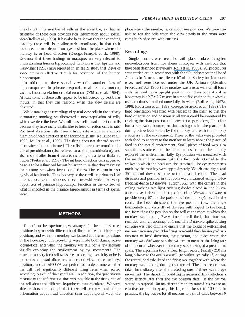
The results of an experiment in which the firing of a headdirection cell was recorded for many minutes while the room wascompletely obscured by ceiling-to-floor curtains is shown inFigure 4 , curve b (av115c3). The head direction tuning was verysimilar when the curtains were drawn closed (compare to Fig. 4,
FIGURE 4. a: The firing of a head direction cell recorded with thenormal view of the room. b: The firing rate when the room wascompletely obscured by ceiling-to-floor curtains. c: The responses of
the same cell recorded not only with the blackout curtain, but alsowith the lights off. The mean and standard error of the mean responseis shown.
___________________________________________________________ PRIMATE HEAD DIRECTION CELLS 213
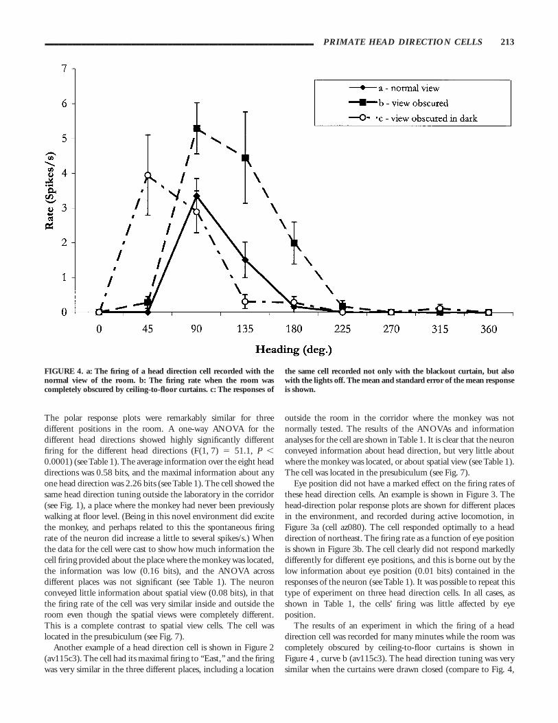
FIGURE 5. a: A cell with some of the characteristics of a head direction cell recorded in theparahippocampal gyrus. Conventions as in Figure 1, with the head direction tuning measured withthe monkey stationary. b: The head direction tuning for the same cell during active locomotionthroughout the laboratory.

curve a with the curtains open). When the room lights weresubsequently extinguished, a head direction tuning curve was stillpresent, though with no visual anchor, and the peak of the tuningdid drift a little during several minutes in darkness, as shown inFigure 4, curve c.
A cell with some of the characteristics of a head direction cellbut recorded in the parahippocampal gyrus is shown in Figure 5a(av192). The cell had similar head direction tuning in the threeplaces shown, but was different from other head direction cells inhaving a somewhat higher firing rate when in the Northeastcorner of the room. The head direction tuning for this cell duringactive locomotion throughout the laboratory is shown in Figure5b. The tuning is generally similar to that found when themonkey was stationary as in Figure 5a.
The results over all head direction cells fully tested are shown inTable 1, and in Table 2, which summarizes the half-amplitudetuning widths of the cells, and the peak firing rates. For cells av070and av115, data were not available (n.a.) for eye position. The firstfour cells in Table 1 were recorded in the presubiculum, and wereamong a set of 12 different cells analyzed in the presubiculum.The individual head direction cells shown in Table 1 had veryhighly significant head direction tuning, as shown. The probabil-ity of this set of significance values arising by chance calculatedusing the Fisher generalized significance test was P 9 0.001(Chi-square 5 167, df 5 24; critical value for P , 0.001 551.18). This test shows that the head direction tuning of thesecells is most unlikely to have arisen by chance.
The results of the multiple cell decoding and informationanalysis are shown in Figure 6. For homogeneity, only the fourcells from the same animal (av) were considered. Both thepercentage of correct decodings and the information in thepopulation are plotted for different supopulation sizes. TheProbability Estimator (PE) and Euclidean Distance (ED) algo-rithm perform very well. The information encoded in theneuronal responses, Eq 1, can be calculated directly for the singlecell case, and compared to the decoded information. Both the PEand ED decodings lead to information values that come close to
the directly calculated value (within 5%); thus PE and ED decode,at least for single cells, essentially all the information that can bedecoded by any algorithm. In contrast, the vector reconstructionmethods perform poorly, especially when information, not thepercent correct, is considered. The Dot Product (DP) decodingperforms for these data considerably less well than the PE and EDestimator, probably because some of the cells in this small samplewere tuned to similar head directions, and because the DPalgorithm does not make effective use of the absolute firing rates ofthe neurons.
The sites in the brain where the cells were located are shown inFigure 7. All the cells had low spontaneous firing rates (mean 50.8 spikes/s, interquartile range 0– 1.0). The peak firing rates werealso relatively low, mean 10.0 spikes/s, interquartile range 6– 13).These characteristics, together with the large amplitude and broadaction potentials, indicate that these neurons are likely to bepyramidal cells. Four cells were in the presubiculum, and 1 in theparahippocampal gyrus.
DISCUSSION
These findings show that there are head direction cells inprimates. They are found in the presubiculum, and some cellswith similar properties are found in the parahippocampal gyrus.We have not so far found head direction cells in the hippocampusitself (CA3, and CA1), nor in the dentate gyrus. Although becauseof the technical difficulties of recording from cells in thepresubiculum of the freely locomoting macaque we have not beenable to increase the number of cells recorded in the presubiculumbeyond 12, we note that the individual head direction cells shownin Table 1 had very highly significant head direction tuning, asshown. Further, the probability of this set of significance valuesarising by chance calculated using the Fisher generalized signifi-cance test was P 9 0.001. For these reasons, the actual finding ofhead direction cells reported for the first time in primates in thispaper can be taken as a firm result.
The head direction cells are very different from the spatial viewcells, which are found in the primate hippocampus and parahippo-campal gyrus. For example, for a given head direction, if themonkey is moved to different places in the environment where thespatial view is different, spatial view cells give different responses.In contrast, the response of head direction cells remains constantfor a given head direction, even when the spatial view is verydifferent, as the data shown in Figures 1–4 and Tables 1 and 2show. To provide a simple concept to emphasize the difference,one can think of head direction cells as responding like a compassattached to the top of the head, which will signal head directioneven when the compass is in different locations, including in atotally different, and even novel, spatial environment, as illus-trated in Figures 1 and 2. Each head direction cell is tuned todifferent directions, and indeed more information about headdirection is present from an ensemble of such cells than from asingle cell, which would not be the case if they were all tuned to
TABLE 2. _____________________________________________Head Direction Cells: Firing Rates
LocationCell
number
Peakrate
(spikes/s)
1⁄2amplitude
width(deg.)
Nullrate
(spikes/s)
Presubiculum (1) AV070c2 17.2 72 0.8Presubiculum (2) AV115c3 4.3 54 0.0Presubiculum (3) AV195 29.1 89 0.9Presubiculum (4) AZ080 2.3 90 0.0
Mean 13.2 76.3 0.4Parahippo-
campalgyrus (5) AV192c4 15.7 139 2.2
___________________________________________________________ PRIMATE HEAD DIRECTION CELLS 215

the same compass direction (apart from some noise reductionincreasing as the square root of the number of cells).
The head direction cells continued to signal head direction evenwhen the view details were obscured by curtains and the monkeywas in darkness (see Fig. 4). This type of experiment involved
rotating the monkey to different head directions during darkness. Theexperiment indicates that signals that are probably of vestibular originprovide inputs to head direction cells. However, in darkness the firingof head direction cells may drift a little, consistent with the hypothesisthat visual cues can reset the cells, and prevent them from drifting over
FIGURE 6. a: The percentage of correctdecodings for a set of four cells recorded fromanimal av as a function of the population size.Notations as in the main text. b: The same asin a, but the mutual information is plotted onthe ordinate axis.
216 ROBERTSON ET AL.

long periods. This is similar to the hypothesis for head direction cells inrats (see Taube et al., 1996; Muller et al., 1996).
A hypothesis that can be tested in primates is whether eyeposition affects the responses of head direction cells. They mightrespond to compass-related head direction, or to compass-relatedeye-gaze (i.e., the direction of the eye taking into account headdirection and eye position in the head). The evidence we have sofar indicates that their firing rate for a given head direction doesnot depend on eye position (see Fig. 3b and Table 1). Moreover,they carried little information about eye position (Table 1). Thus,the evidence so far available suggests that the cells signal headdirection rather than (allocentric or compass-related) eye gazeangle. However, because the tuning of the head direction cells isrelatively broad, the range of possible eye positions might notmove the firing to a part of the head direction tuning where theeffect of differences in eye position (in the head) would make asignificant difference to the (allocentric) eye gaze angle. It will be ofinterest in future research to explore this further when the headdirection is set to the steepest part of the head direction tuning of a cell.
Taken in the context of evidence on the neurophysiology andfunctions of the primate (including human) hippocampal system,head direction cells could perform a number of functions. Onewould be as part of a memory system. By remembering thecompass bearing (head direction) and distance travelled, it is
possible to find one’s way back to the origin, even with a numberof sectors of travel, and over a number of minutes. This is referredto as path integration, and can occur even without a view of theenvironment. Head direction cells provide part of the informationto be remembered for such spatial memory functions. Complemen-tary information also required for this is available in the wholebody motion cells that we have described in the primatehippocampus (O’Mara et al., 1994). These cells provide informa-tion about linear translation, for example, or axial whole bodyrotation. Part of the way in which head direction cell firing couldbe produced is by taking into account axial movements, which iswhat are signalled by some of these whole body motion cells(O’Mara et al., 1994), and it is an interesting hypothesis that thisfunction is performed by some of the structures related to thehippocampal system such as the presubiculum. Spatial memoryand navigation can also benefit from visual information aboutplaces being looked at, which can be used as landmarks, andspatial view cells added to the head direction cells and whole bodymotion cells would provide the basis for a good memory systemuseful in navigation. Another possibility is that primate headdirection cells are part of a system for computing duringnavigation which direction to head in next. For this, not onlywould a memory system of the type just described and elaboratedelsewhere be needed (Rolls, 1989, 1996; Treves and Rolls, 1994;
FIGURE 7. The hippocampal and parahippocampal sites at whichdifferent spatial view cells were recorded. Coronal sections at differentdistances in mm posterior (P) to the sphenoid reference are shown.The number inside each circle corresponds to the cell number shown
in Table 1. CA3, CA3 hippocampal pyramidal cell field; CA1, CA1hippocampal pyramidal cell field; DG, dentate gyrus; PHG, parahip-pocampal gyrus; PreSub, presubiculum.
___________________________________________________________ PRIMATE HEAD DIRECTION CELLS 217

Rolls and Treves, 1998), which can store spatial information of thetype found in the hippocampus, but also an ability to use thisinformation in spatial computation of the appropriate nextbearing would be needed. Such a system might be implementedusing a hippocampal memory system that associated togetherspatial views, whole body motion, and head direction informa-tion. The system would be different from that in the rat (Burgesset al., 1994; McNaughton et al., 1996), in that spatial view isrepresented in the primate hippocampus.
The results of the multiple cell decoding and informationanalysis show that decodings that use the response probabilitydistributions (like PE and ED), and operate in the abstract vectorspace of neuronal population responses, actually do decode mostof the information available in the firing rates. This means thatthese methods, which utilize the full shape of the neuronalresponse probabilities, not only the preferred head direction,would be good ones for the brain to use when other neurons‘‘interpret’’ (i.e., receive information about) the responses of apopulation of neurons. Further, the ED algorithm can be easilyimplemented by a set of receiving neurons, and therefore can beeasily implemented in the brain to achieve accurate head directionreconstruction. The decodings based on reconstruction from thephysical direction vectors of each neuron, perform much morepoorly (see Fig. 6). This is not due to the fact that the preferredhead direction of our limited sample of cells might not arise froma uniform distribution, nor it is due to possible non-symmetricityof tuning curves (see Fig. 4). These facts, which might produceproblems for the VR algorithm, are in fact corrected for by theOLE, which also performs poorly on this set of data. Theimplication is that methods in general that are based on reconstruc-tion from physical vectors estimated for different cells are muchless powerful that reading the output of neuronal populationsmore directly, using, for example, a Euclidean Distance measure.Such a measure is also very biologically plausible. Moreover, thefinding shows that direction-tuned cells, including head directioncells, convey much more information about head direction thancan be captured just by specifying the direction to which the neuronhas its optimal response, and weighting the contribution of thatphysical direction vector by the intensity of the neuronal response.
ACKNOWLEDGMENTS
This research was supported by the Medical Research Council,PG8513579, and by the Human Frontier Science Program.P.G.-F. and S.P. are European Community Research Fellows.
REFERENCES
Angeli SJ, Murray EA, Mishkin M. 1993 Hippocampectomized monkeyscan remember one place but not two. Neuropsychologia 31:1021–1030.
Brown EN, Frank LM, Tang D, Quirk C, Wilson MA. 1998. A statisticalparadigm for neural spike train decoding applied to position predic-tion from ensemble firing patterns of rat hippocampal place cells. JNeurosci 18:7411–7425.
Burgess N, Recce M, O’Keefe J. 1994. A model of hippocampal function.Neural Networks 7:1065–1081.
Cover TM, Thomas JA. 1991. Elements of information theory. JohnWiley: New York.
Epstein R, Kanwisher N. 1998. A cortical representation of the localvisual environment. Nature 392:598–601.
Feigenbaum JD, Rolls ET. 1991. Allocentric and egocentric spatialinformation processing in the hippocampal formation of the behavingprimate. Psychobiology 19:21–40.
Foster TC, Castro CA, McNaughton BL. 1989. Spatial selectivity of rathippocampal neurons: Dependence on preparedness for movement.Science 244:1580–1582.
Gaffan D. 1994. Scene-specific memory for objects: A model of episodicmemory impairment in monkeys with fornix transection. J CognNeurosci 6:305–320.
Georges-Francois P, Rolls ET, Robertson RG. 1999. Spatial view cells inthe primate hippocampus: Allocentric view not head direction or eyeposition or place. Cerebral Cortex, in press.
Georgopoulos AP, Schwartz AB, Kettner RE. 1986. Neuronal populationcoding of movement direction. Science 233:1416–1419.
McNaughton BL, Barnes CA, Gerrard JL, Gothard K, Jung MW,Knierim JJ, Kudrimoti H, Qin Y, Skaggs WE, Suster M, Weaver KL.1996. Deciphering the hippocampal polyglot: the hippocampus as apath integration system. J Exp Biol 199:173–185.
Muller RU, Ranck JB, Taube JS. 1996. Head direction cells: propertiesand functional significance. Curr Opin Neurobiol 6:196–206.
O’Keefe J, Speakman A. 1987. Single unit activity in the rat hippocam-pus during a spatial memory task. Exp Brain Res 68:1–27.
O’Mara SM, Rolls ET, Berthoz A, Kesner RP. 1994. Neurons respondingto whole-body motion in the primate hippocampus. J Neurosci14:6511–6523.
Ono T, Tamura R, Nishijo H, Nakamura K. 1993. Neural mechanisms ofrecognition and memory in the limbic system. In: Ono T, Squire LR,Raichle ME, Perrett DI, Fukuda M, editors. Brain mechanisms ofperception and memory: from neuron to behavior. New York: OxfordUniversity Press. p 330–355.
Optican LM, Richmond BJ. 1987. Temporal encoding of two-dimensional patterns by single units in primate inferior temporalcortex: III. Information theoretic analysis. J Neurophysiol 57:162–178.
Panzeri S, Treves A. 1996. Analytical estimates of limited sampling biasesin different information measures. Network 7:87–107.
Parkinson JK, Murray EA, Mishkin M. 1988. A selective mnemonic rolefor the hippocampus in monkeys: Memory for the location of objects.J Neurosci 8:4059–4167.
Robertson RG, Rolls ET, Georges-Francois P. 1998. Spatial view cells inthe primate hippocampus: Effects of removal of view details. JNeurophysiol 79:1145–1156.
Rolls ET 1989. Functions of neuronal networks in the hippocampus andneocortex in memory. In: Byrne JH, Berry WO, editors. Neuralmodels of plasticity: experimental and theoretical approaches. SanDiego: Academic Press. p 240–265.
Rolls ET 1996. A theory of hippocampal function in memory. Hippocam-pus 6:601–620.
Rolls ET, O’Mara SM. 1995. View-responsive neurons in the primatehippocampal complex. Hippocampus 5:409–424.
Rolls ET, Treves A. 1998. Neural networks and brain function. Oxford:Oxford University Press.
Rolls ET, Miyashita Y, Cahusac PMB, Kesner RP, Niki H, Feigenbaum J,Bach L. 1989. Hippocampal neurons in the monkey with activityrelated to the place in which a stimulus is shown. J Neurosci9:1835–1845.
218 ROBERTSON ET AL.

Rolls ET, Robertson RG, Georges-Francois P. 1997a. Spatial view cells inthe primate hippocampus. Eur J Neurosci 9:1789–1794.
Rolls ET, Treves A, Tovee M, Panzeri S. 1997b. Information in theneuronal representation of individual stimuli in the primate temporalvisual cortex. J Comput Neurosci 4:309–333.
Rolls ET, Treves A, and Tovee MJ. 1997c. The representational capacityof the distributed encoding of information provided by populations ofneurons in the primate temporal visual cortex. Exp Brain Res114:149–162.
Rolls ET, Treves A, Robertson RG, Georges-Francois P, Panzeri S. 1998.Information about spatial view in an ensemble of primate hippocam-pal cells. J Neurophysiol 79:1797–1813.
Salinas E, Abbott LF. 1994. Vector reconstruction from firing rates. JComput Neurosci 1:89–107.
Scoville WB, Milner B. 1957. Loss of recent memory after bilateralhippocampal lesions. J Neurol Neurosurg Psychiat 20:11–21.
Shannon CE. 1948. A mathematical theory of communication. AT&TBell Lab Tech J 27:379–423.
Smith ML, Milner B. 1981. The role of the right hippocampus in therecall of spatial location. Neuropsychologia 19:781–793.
Squire LR, Knowlton BJ. 1994. Memory, hippocampus, and brainsystems. In: Gazzaniga M, editor. The cognitive neurosciences.Cambridge, MA: MIT Press. p 825–837.
Taube JS, Goodridge JP, Golob EJ, Dudchenko PA, Stackman RW. 1996.Processing the head direction signal: a review and commentary. BrainRes Bull 40:477–486.
Tovee MJ, Rolls ET, Treves A, Bellis RP. 1993. Information encoding andthe responses of single neurons in the primate temporal visual cortex. JNeurophysiol 70:640–654.
Treves A, Rolls ET. 1994. A computational analysis of the role of thehippocampus in memory. Hippocampus 4:374–391.
Zhang K, Ginzburg I, McNaughton BL, Sejnowski TJ. 1998. Interpret-ing neuronal population activity by reconstruction: unified frameworkwith application to hippocampal place cells. J Neurophysiol 79:1017–1044.
___________________________________________________________ PRIMATE HEAD DIRECTION CELLS 219