vestibular and attractor network basis of the head direction cell signal in subcortical circuits
TRANSCRIPT
REVIEW ARTICLEpublished: 20 March 2012
doi: 10.3389/fncir.2012.00007
Vestibular and attractor network basis of the head directioncell signal in subcortical circuitsBenjamin J. Clark † and Jeffrey S. Taube*
Department of Psychological and Brain Sciences, Center for Cognitive Neuroscience, Dartmouth College, Hanover, NH, USA
Edited by:
Yasser Roudi, Norwegian Universityof Science and Technology, Norway
Reviewed by:
Francesca Sargolini, Aix MarseilleUniversité – CNRS, FranceDouglas Nitz, University ofCalifornia, San Diego, USA
*Correspondence:
Jeffrey S. Taube, Department ofPsychological and Brain Sciences,Dartmouth College, 6207 MooreHall, Hanover, NH 03755, USA.e-mail: [email protected]†Present address:
Canadian Centre for BehaviouralNeuroscience, University ofLethbridge, Lethbridge, AB, CanadaT1K 3M4.
Accurate navigation depends on a network of neural systems that encode themoment-to-moment changes in an animal’s directional orientation and location in space.Within this navigation system are head direction (HD) cells, which fire persistently whenan animal’s head is pointed in a particular direction (Sharp et al., 2001a; Taube, 2007). HDcells are widely thought to underlie an animal’s sense of spatial orientation, and researchover the last 25+ years has revealed that this robust spatial signal is widely distributedacross subcortical and cortical limbic areas. The purpose of the present review is tosummarize some of the recent studies arguing that the origin of the HD signal residessubcortically, specifically within the reciprocal connections of the dorsal tegmental andlateral mammillary nuclei. Furthermore, we review recent work identifying “bursting”cellular activity in the HD cell circuit after lesions of the vestibular system, and relate theseobservations to the long held view that attractor network mechanisms underlie HD signalgeneration. Finally, we summarize anatomical and physiological work suggesting that thisattractor network architecture may reside within the tegmento-mammillary circuit.
Keywords: entorhinal, hippocampus, head direction, spatial orientation, navigation
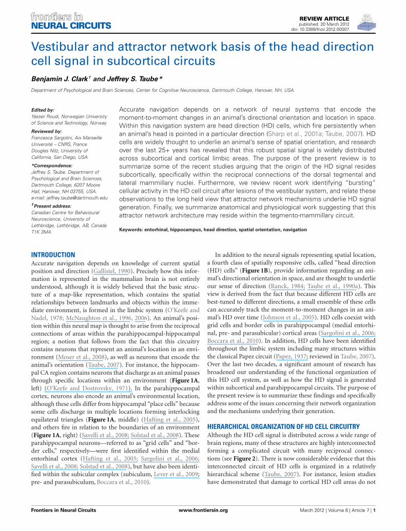
INTRODUCTIONAccurate navigation depends on knowledge of current spatialposition and direction (Gallistel, 1990). Precisely how this infor-mation is represented in the mammalian brain is not entirelyunderstood, although it is widely believed that the basic struc-ture of a map-like representation, which contains the spatialrelationships between landmarks and objects within the imme-diate environment, is formed in the limbic system (O’Keefe andNadel, 1978; McNaughton et al., 1996, 2006). An animal’s posi-tion within this neural map is thought to arise from the reciprocalconnections of areas within the parahippocampal-hippocampalregion; a notion that follows from the fact that this circuitrycontains neurons that represent an animal’s location in an envi-ronment (Moser et al., 2008), as well as neurons that encode theanimal’s orientation (Taube, 2007). For instance, the hippocam-pal CA region contains neurons that discharge as an animal passesthrough specific locations within an environment (Figure 1A,left) (O’Keefe and Dostrovsky, 1971). In the parahippocampalcortex, neurons also encode an animal’s environmental location,although these cells differ from hippocampal “place cells” becausesome cells discharge in multiple locations forming interlockingequilateral triangles (Figure 1A, middle) (Hafting et al., 2005),and others fire in relation to the boundaries of an environment(Figure 1A, right) (Savelli et al., 2008; Solstad et al., 2008). Theseparahippocampal neurons—referred to as “grid cells” and “bor-der cells,” respectively—were first identified within the medialentorhinal cortex (Hafting et al., 2005; Sargolini et al., 2006;Savelli et al., 2008; Solstad et al., 2008), but have also been identi-fied within the subicular complex (subiculum, Lever et al., 2009;pre- and parasubiculum, Boccara et al., 2010).
In addition to the neural signals representing spatial location,a fourth class of spatially responsive cells, called “head direction(HD) cells” (Figure 1B), provide information regarding an ani-mal’s directional orientation in space, and are thought to underlieour sense of direction (Ranck, 1984; Taube et al., 1990a). Thisview is derived from the fact that because different HD cells arebest-tuned to different directions, a small ensemble of these cellscan accurately track the moment-to-moment changes in an ani-mal’s HD over time (Johnson et al., 2005). HD cells coexist withgrid cells and border cells in parahippocampal (medial entorhi-nal, pre- and parasubicular) cortical areas (Sargolini et al., 2006;Boccara et al., 2010). In addition, HD cells have been identifiedthroughout the limbic system including many structures withinthe classical Papez circuit (Papez, 1937; reviewed in Taube, 2007).Over the last two decades, a significant amount of research hasbroadened our understanding of the functional organization ofthis HD cell system, as well as how the HD signal is generatedwithin subcortical and parahippocampal circuits. The purpose ofthe present review is to summarize these findings and specificallyaddress some of the issues concerning their network organizationand the mechanisms underlying their generation.
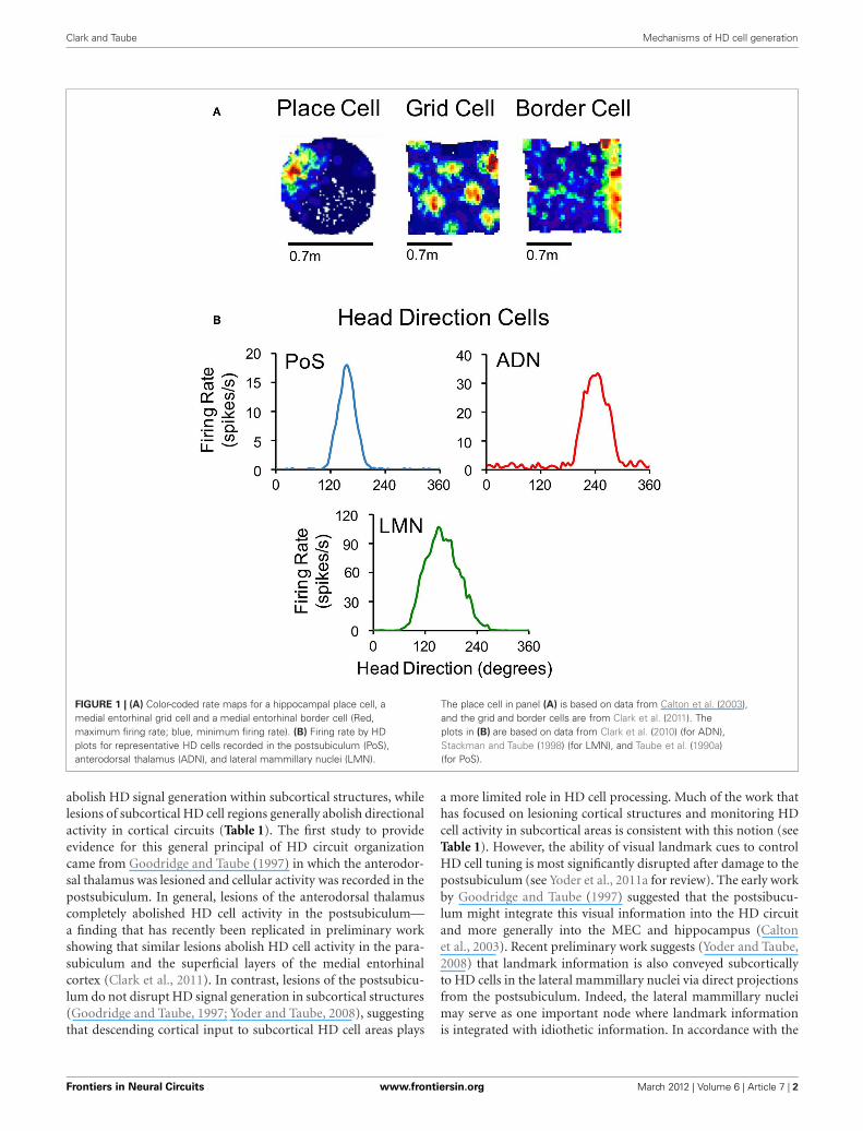
HIERARCHICAL ORGANIZATION OF HD CELL CIRCUITRYAlthough the HD cell signal is distributed across a wide range ofbrain regions, many of these structures are highly interconnectedforming a complicated circuit with many reciprocal connec-tions (see Figure 2). There is now considerable evidence that thisinterconnected circuit of HD cells is organized in a relativelyhierarchical scheme (Taube, 2007). For instance, lesion studieshave demonstrated that damage to cortical HD cell areas do not
Frontiers in Neural Circuits www.frontiersin.org March 2012 | Volume 6 | Article 7 | 1
NEURAL CIRCUITS
Clark and Taube Mechanisms of HD cell generation
FIGURE 1 | (A) Color-coded rate maps for a hippocampal place cell, amedial entorhinal grid cell and a medial entorhinal border cell (Red,maximum firing rate; blue, minimum firing rate). (B) Firing rate by HDplots for representative HD cells recorded in the postsubiculum (PoS),anterodorsal thalamus (ADN), and lateral mammillary nuclei (LMN).
The place cell in panel (A) is based on data from Calton et al. (2003),and the grid and border cells are from Clark et al. (2011). Theplots in (B) are based on data from Clark et al. (2010) (for ADN),Stackman and Taube (1998) (for LMN), and Taube et al. (1990a)(for PoS).
abolish HD signal generation within subcortical structures, whilelesions of subcortical HD cell regions generally abolish directionalactivity in cortical circuits (Table 1). The first study to provideevidence for this general principal of HD circuit organizationcame from Goodridge and Taube (1997) in which the anterodor-sal thalamus was lesioned and cellular activity was recorded in thepostsubiculum. In general, lesions of the anterodorsal thalamuscompletely abolished HD cell activity in the postsubiculum—a finding that has recently been replicated in preliminary workshowing that similar lesions abolish HD cell activity in the para-subiculum and the superficial layers of the medial entorhinalcortex (Clark et al., 2011). In contrast, lesions of the postsubicu-lum do not disrupt HD signal generation in subcortical structures(Goodridge and Taube, 1997; Yoder and Taube, 2008), suggestingthat descending cortical input to subcortical HD cell areas plays
a more limited role in HD cell processing. Much of the work thathas focused on lesioning cortical structures and monitoring HDcell activity in subcortical areas is consistent with this notion (seeTable 1). However, the ability of visual landmark cues to controlHD cell tuning is most significantly disrupted after damage to thepostsubiculum (see Yoder et al., 2011a for review). The early workby Goodridge and Taube (1997) suggested that the postsibucu-lum might integrate this visual information into the HD circuitand more generally into the MEC and hippocampus (Caltonet al., 2003). Recent preliminary work suggests (Yoder and Taube,2008) that landmark information is also conveyed subcorticallyto HD cells in the lateral mammillary nuclei via direct projectionsfrom the postsubiculum. Indeed, the lateral mammillary nucleimay serve as one important node where landmark informationis integrated with idiothetic information. In accordance with the
Frontiers in Neural Circuits www.frontiersin.org March 2012 | Volume 6 | Article 7 | 2
Clark and Taube Mechanisms of HD cell generation
FIGURE 2 | Circuit diagram showing the principle connections
between brain regions containing place cells (yellow), grid cells (red),
border cells (blue), HD cells (green), and angular velocity cells (orange)
(Sharp et al., 2001a; Taube and Bassett, 2003; Taube, 2007). Arrowsindicate excitatory projections, and lines that end with a bar representinhibitory projections. ADN, anterodorsal thalamus; AVN, anteroventralthalamus; DTN, dorsal tegmental nucleus; HPC, hippocampus; LDN,laterodorsal thalamus; LMN, lateral mammillary nuclei; MEC, medialentorhinal cortex; MVN, medial vestibular nuclei; NPH, nucleus prepositushypoglossi; PaS, parasubiculum; PoS, postsubiculum; RSP, retrosplenialcortex; SGN, supragenual nucleus.
working model presented in Figure 2, damage to the postsubicu-lum may abolish directional activity in the medial entorhinalcortex as well as landmark control over other spatial signals withinthis region, given that it provides one of the most prominentinputs to this region (van Haeften et al., 1997; Kerr et al., 2007)
and that postsubicular lesions disrupt landmark control in CA1place cells (Calton et al., 2003).
Supporting the general conclusion that HD signal genera-tion resides subcortically, several studies have shown that lesionscentered on structures upstream of the anterior thalamus alsoeliminate directional tuning downstream (Table 1). Specifically,bilateral damage of the dorsal tegmental or lateral mammil-lary nuclei abolishes HD cell activity in the anterodorsal tha-lamus (Blair et al., 1998, 1999; Bassett et al., 2007). Further,lateral mammillary lesions abolish HD tuning in the postsubicu-lum, parasubiculum, and medial entorhinal cortex (Sharp andKoester, 2008). Again, lesions of the anterodorsal thalamus com-pletely abolish directional tuning throughout the parahippocam-pal region, collectively indicating that given the known anatomyof these areas (see Taube, 2007 for review), the flow of infor-mation within the HD system likely follows a path from dorsaltegmental nucleus → lateral mammillary nuclei → anterodorsalthalamus → parahippocampal/retrosplenial cortex.
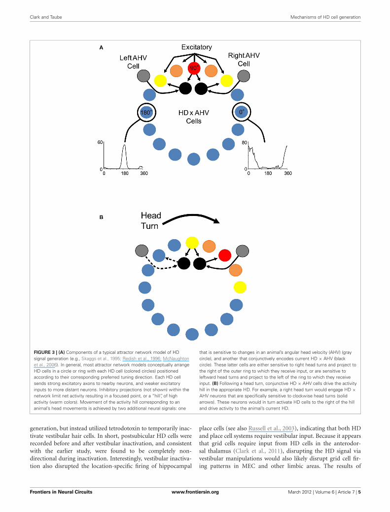
VESTIBULAR INPUT, “BURSTY” CELLS, AND ATTRACTORNETWORK HYPOTHESES OF HD SIGNAL GENERATIONHD cells are strongly influenced by external and internally gener-ated sources of information (Taube, 2007). For instance, externalcues such as visual landmarks can control HD cell tuning suchthat a change in the orientation of a landmark can induce a similarchange in the preferred directions of HD cells. In other words, HDcell preferred firing directions tend to shift in the same directionand angular distance as landmarks (Taube et al., 1990b; Taube,1995). Although landmark information exerts a strong influenceon HD cells, the directional firing preference of HD cells canbe maintained even when familiar external cues are eliminated,such as in darkness or in novel environments (Taube and Burton,1995; Goodridge et al., 1998; Stackman et al., 2003; Yoder et al.,2011b). This finding suggests that internally generated informa-tion, or idiothetic cues (i.e., vestibular, proprioception, and motorefference), can be utilized to keep track of changes in directionalheading over time, a process often referred to as angular path inte-gration. The precise sensory mechanisms underlying angular pathintegration are unclear, however, it was suggested very early onthat the vestibular system might play a particularly important rolein this process (Potegal, 1982; Taube et al., 1990b; McNaughtonet al., 1991). This view follows from the simple fact that angu-lar head velocity—a product of the mechanical properties of thesemicircular canals, which are sensitive to head acceleration—can be integrated over time to yield angular displacement, as hasbeen demonstrated in occulomotor pathways (Robinson, 1989).An estimate of current HD can, therefore, be accomplished bya vector summation of the angular displacement and the ani-mal’s previous HD. McNaughton et al. (1991) provided the firstcomputational model that captured this notion and subsequentmodels (e.g., Skaggs et al., 1995; Redish et al., 1996; Zhang, 1996)have expanded on this basic principal using continuous attractornetworks (Figure 3). In these models, HD cells are conceptu-ally arranged in a ring corresponding to their preferred firingdirections. Cells with preferred directions that have overlappingdirectional firing ranges share excitatory connections and cellswith greatly different preferred directions inhibit each other. This
Frontiers in Neural Circuits www.frontiersin.org March 2012 | Volume 6 | Article 7 | 3
Clark and Taube Mechanisms of HD cell generation
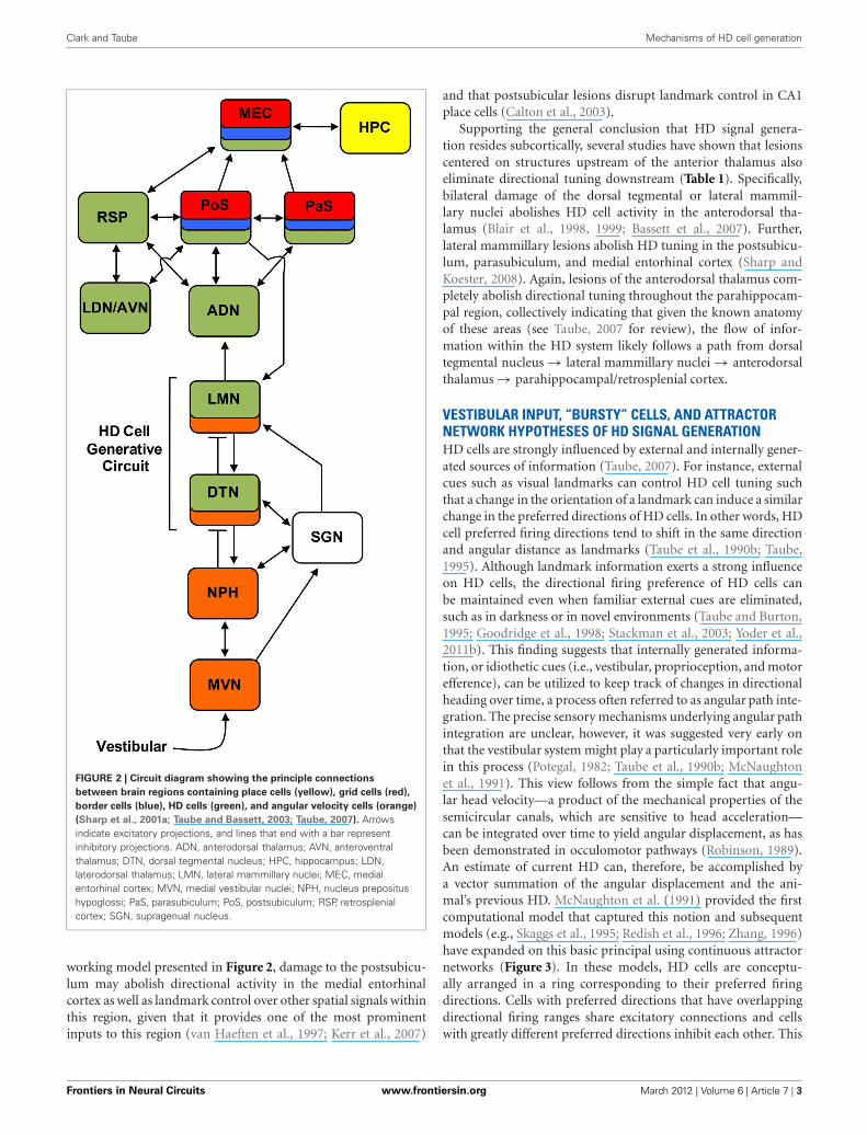
Table 1 | Summary of lesion results involving the HD cell circuit.
Lesion area Recording location HD signal Landmark Idiothetic Reference
Vestibular labyrinth Anterodorsal thalamus No – – Stackman and Taube, 1997
Postsubiculum No – – Stackman et al., 2002
Supragenual nuclei Anterodorsal thalamus No – – Clark et al., unpublished
observations
Dorsal tegmental nuclei Anterodorsal thalamus No – – Bassett et al., 2007
Lateral mammillary nuclei Anterodorsal thalamus No – – Blair et al., 1998; Bassett et al., 2007
Postsubiculum No – – Sharp and Koester, 2008
Parasubiculum No – – Sharp and Koester, 2008
Medial entorhinal cortex No – – Sharp and Koester, 2008
Anterodorsal thalamus Postsubiculum No – – Goodridge and Taube, 1997
Parasubiculum No – – Clark et al., 2011
Medial entorhinal cortex No – – Clark et al., 2011
Laterodorsal thalamus Postsubiculum Yes OK Not tested Golob et al., 1998
Postsubiculum Anterodorsal thalamus Yes Impaired Mildly impaired Goodridge and Taube, 1997
Lateral mammillary nuclei Yes Impaired Not tested Yoder and Taube, 2008
Retrosplenial cortex Anterodorsal thalamus Yes Mildly impaired OK Clark et al., 2010
Medial entorhinal cortex Anterodorsal thalamus Yes OK OK Clark and Taube, 2011
Hippocampus Anterodorsal thalamus Yes OK Impaired Golob and Taube, 1997, 1999
Postsubiculum Yes OK Impaired Golob and Taube, 1997, 1999
Parietal cortex Anterodorsal thalamus Yes OK Mildly impaired Calton et al., 2008
Medial entorhinal cortex Yes Not tested Not tested Whitlock et al., 2010
For each lesion the table shows whether: (1) the HD signal is present, (2) landmark cue control is affected (as judged by the ability of the HD cell to shift its preferred
direction following visual cue rotation), and (3) idiothetic cue processing (i.e., vestibular or motor) is affected (as judged by the ability of the HD cell to maintain a
stable preferred direction as the animal locomotes in darkness and when locomoting from a familiar to a novel environment). Note: if no HD cells were identified
following the lesion, then it was not possible to assess landmark or idiothetic cue processing.
neural architecture forms a sustained “hill” of excitation centeredon the animal’s current HD. This activity hill is observed as aburst of firing when the animal’s HD passes through the cell’s pre-ferred firing direction during a head turn. Angular head velocityinformation is commonly used to move the hill of activity aroundto different directions depending on changes to the animal’s HD(this process is typically modeled through an additional ring ofneurons that conjunctively encodes HD and angular head veloc-ity; see Figure 3). A specific consequence of removing angularhead velocity input is an activity hill that no longer moves alongthe ring in register with the animal’s HD. Instead, the hill movesin different directions at different rates depending on the remain-ing inputs that convey information about current heading (e.g.,visual and motor). In other words, neurons within the networkwould fire in bursts, as if the animal is passing its head througheach cell’s preferred firing direction. This latter feature of attrac-tor networks, which we have termed “bursting” activity, forms animportant prediction especially when considered with the lesionstudies described below.
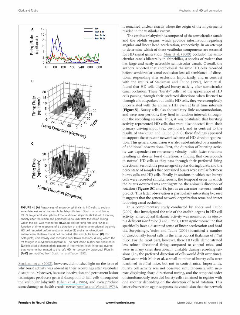
The hypothesis that vestibular information is critical forHD signal processing was first tested in a study conducted byStackman and Taube (1997) in which anterodorsal thalamus HDcells were recorded before and after sodium arsanilate lesionsof the vestibular system—a neurotoxic lesion technique thatdestroys hair cells throughout the vestibular labyrinth (Chenet al., 1986). In general, lesions of the peripheral vestibular sys-tem completely abolished directional activity in the anterodorsal
thalamus of rats (Figure 4A). Surprisingly, the absence of HD cellactivity persisted for up to 3 months (the post-lesion time periodin which cell recording was monitored), strongly indicating thatother sensory systems (e.g., vision or motor) were not capableof compensating for the loss of vestibular input, despite the factthat these cues can gain strong control over directional tuning(Goodridge et al., 1998; Stackman et al., 2003). An additional sur-prising result was the fact that Stackman and Taube did not detectburst firing in isolated units that had previously been modulatedby HD. In other words, HD cells did not appear to “turn into”bursty cells following the neurotoxic lesions. As noted above,the significance of this result is derived from predictions of ringattractor models, in which an attractor that is uncoupled fromits angular updating inputs should have the appearance of HDcell activity, but cell firing would be uncorrelated with the ani-mal’s HD. Because the activity hill would move around the ringattractor in random directions and at varying rates, the activityof a single cell recorded from such a network would resembleintermittent bursts of activity. Stackman and Taube identifiedsome anterodorsal thalamus neurons that exhibited intermittentbursts of activity (e.g., Figure 4C), but because the spatial andbehavioral correlates of these cells were not determined beforethe lesion, and because identified HD cells failed to display burstactivity after lesions were produced, the authors argued againstthis interpretation.
Subsequent work by Stackman et al. (2002) replicated thegeneral finding that vestibular input is necessary for HD cell
Frontiers in Neural Circuits www.frontiersin.org March 2012 | Volume 6 | Article 7 | 4
Clark and Taube Mechanisms of HD cell generation
FIGURE 3 | (A) Components of a typical attractor network model of HDsignal generation (e.g., Skaggs et al., 1995; Redish et al., 1996; McNaughtonet al., 2006). In general, most attractor network models conceptually arrangeHD cells in a circle or ring with each HD cell (colored circles) positionedaccording to their corresponding preferred tuning direction. Each HD cellsends strong excitatory axons to nearby neurons, and weaker excitatoryinputs to more distant neurons. Inhibitory projections (not shown) within thenetwork limit net activity resulting in a focused point, or a “hill”, of highactivity (warm colors). Movement of the activity hill corresponding to ananimal’s head movements is achieved by two additional neural signals: one
that is sensitive to changes in an animal’s angular head velocity (AHV) (graycircle), and another that conjunctively encodes current HD × AHV (blackcircle). These latter cells are either sensitive to right head turns and project tothe right of the outer ring to which they receive input, or are sensitive toleftward head turns and project to the left of the ring to which they receiveinput. (B) Following a head turn, conjunctive HD × AHV cells drive the activityhill in the appropriate HD. For example, a right head turn would engage HD ×AHV neurons that are specifically sensitive to clockwise head turns (solidarrows). These neurons would in turn activate HD cells to the right of the hilland drive activity to the animal’s current HD.
generation, but instead utilized tetrodotoxin to temporarily inac-tivate vestibular hair cells. In short, postsubicular HD cells wererecorded before and after vestibular inactivation, and consistentwith the earlier study, were found to be completely non-directional during inactivation. Interestingly, vestibular inactiva-tion also disrupted the location-specific firing of hippocampal
place cells (see also Russell et al., 2003), indicating that both HDand place cell systems require vestibular input. Because it appearsthat grid cells require input from HD cells in the anterodor-sal thalamus (Clark et al., 2011), disrupting the HD signal viavestibular manipulations would also likely disrupt grid cell fir-ing patterns in MEC and other limbic areas. The results of
Frontiers in Neural Circuits www.frontiersin.org March 2012 | Volume 6 | Article 7 | 5
Clark and Taube Mechanisms of HD cell generation
FIGURE 4 | (A) Responses of anterodorsal thalamic HD cells to sodiumarsanilate lesions of the vestibular labyrinth (from Stackman and Taube,1997). In general, disruption of the vestibular labyrinth abolished HD tuningshortly after the lesion and persisted up to 96 h after the lesion duringwhich the cell was monitored. (B,C) 3D plot of firing rate and HD as afunction of time in epochs of 5 s duration of a distinct anterodorsal thalamicHD cell recorded before vestibular lesion (B) and a non-directionalanterodorsal thalamic burst cell recorded after vestibular lesion (C). Forboth plots, unit activity was recorded over 8 min sessions, during which therat foraged in a cylindrical apparatus. The post-lesion bursty cell depicted in(C) exhibited a characteristic pattern of intermittent high firing rate eventsthat were neither related to the rat’s HD nor temporally organized. Plots in(A–C) are modified from Stackman and Taube (1997).
Stackman et al. (2002), however, did not shed light on the issue ofwhy burst activity was absent in their recordings after vestibulardisruption. Moreover, because inactivation and permanent lesiontechniques produce a general disruption to hair cells throughoutthe vestibular labyrinth (Chen et al., 1986), and even producesome damage to the 8th cranial nerve (Anniko and Wersäll, 1976),
it remained unclear exactly where the origin of the impairmentsresided in the vestibular system.
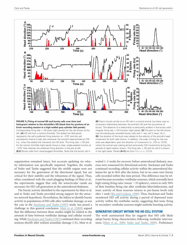
The vestibular labyrinth is composed of the semicircular canalsand the otolith organs, which provide information regardingangular and linear head acceleration, respectively. In an attemptto determine which of these vestibular components are essentialfor HD signal generation, Muir et al. (2009) occluded the semi-circular canals bilaterally in chinchillas, a species of rodent thathas large and easily accessible semicircular canals. Overall, theauthors reported that anterodorsal thalamic HD cells recordedbefore semicircular canal occlusion lost all semblance of direc-tional responding after occlusion. Importantly, and in contrastwith the results of Stackman and Taube (1997), Muir et al.found that HD cells displayed bursty activity after semicircularcanal occlusion. These “bursty” cells had the appearance of HDcells passing through their preferred directions when listened tothrough a loudspeaker, but unlike HD cells, they were completelyuncorrelated with the animal’s HD, even at brief time intervals(Figure 5). Bursty cells also showed very little accommodation,and were non-periodic; they fired in random intervals through-out the recording session. Thus, it was postulated that burstingactivity represented HD cells that were disconnected from theirprimary driving input (i.e., vestibular), and in contrast to theresults of Stackman and Taube (1997), these findings appearedto support the attractor network scheme of HD circuit organiza-tion. This general conclusion was also substantiated by a numberof additional observations. First, the duration of bursting activ-ity was dependent on movement velocity—with faster rotationsresulting in shorter burst durations, a finding that correspondsto normal HD cells as they pass through their preferred firingdirections. Second, the percentage of spikes during bursts and thepercentage of samples that contained bursts were similar betweenbursty cells and HD cells. Finally, in sessions in which two burstycells were recorded simultaneously, the temporal order in whichthe bursts occurred was contingent on the animal’s direction ofrotation (Figures 5C and 6), just as an attractor network wouldpredict. This latter observation is particularly interesting becauseit suggests that the general network organization remained intactfollowing canal occlusion.
In a complimentary study conducted by Yoder and Taube(2009) that investigated the role of the otolith organs in HD cellactivity, anterodorsal thalamic activity was monitored in otoco-nia deficient tilted mice (Lane, 1986), a transgenic mouse line thatspecifically have a disrupted sense of linear acceleration and headtilt. Surprisingly, Yoder and Taube (2009) identified a numberof directionally tuned cells in the anterodorsal thalamus of tiltedmice. For the most part, however, these HD cells demonstratedless robust directional firing compared to control mice, andwere in many cases directionally unstable during recording ses-sions (i.e., the preferred direction of cells would drift over time).Consistent with Muir et al. a small number of bursty cells wereidentified in tilted mice, but not in control mice. Importantly,bursty cell activity was not observed simultaneously with neu-rons displaying sharp directional tuning, and the temporal orderof simultaneously recorded bursty cells remained in register withone another depending on the direction of head rotation. Thislatter observation again supports the conclusion that the network
Frontiers in Neural Circuits www.frontiersin.org March 2012 | Volume 6 | Article 7 | 6
Clark and Taube Mechanisms of HD cell generation
FIGURE 5 | Firing of normal HD and bursty cells over time (red
histogram) relative to the chinchilla’s HD (black line) for portions of an
8 min recording session in a high-walled gray cylinder (left panels).
Corresponding firing rate × HD plots (right panels) for the cell shown at theleft. (A) HD cell from a control chinchilla. The dotted line (left panel)represents the cell’s preferred firing direction at ∼270◦ and this cellconsistently fired at a high rate whenever the animal’s head pointed at 270◦(i.e., when the dotted line intersects the HD plot). The firing rate × HD plotfor the control chinchilla (right panel) shows a clear, single-peaked function at∼270◦ that matches the preferred firing direction in the plot at left.(B,C) Bursty cells from canal-plugged chinchillas. Note that the bursty cell in
(B) fired in bursts similar to an HD cell in a control animal, but there was noconsistent relationship between the animal’s HD and the occurrence ofbursts. This absence of a relationship is particularly evident in the bursty cell’sirregular firing rate × HD function (right panel). (C) The plot on the left showstwo simultaneously recorded bursty cells (cell 1, red; cell 2, blue). As in(B), the duration of the burst was related to the velocity of the animal’s headmovement for both cells shown. In addition, the order of firing for the twocells was dependent upon the direction of movement (i.e., CW vs. CCW),where the animal was making almost exclusively CW movements during theperiods of rapid rotation shown. The firing rate × HD plot for cell 2 is shownin the right panel. Panels (A–C) are from Muir et al. (2009).
organization remained intact, but accurate updating via veloc-ity information was specifically impaired. Together, the resultsof Yoder and Taube suggested that the otolith organs were notnecessary for the generation of the directional signal, but arecritical for their stability and the robustness of the signal. Thus,when considered with the canal-plugging findings of Muir et al.,the experiments suggest that only the semicircular canals arenecessary for HD cell generation in the anterodorsal thalamus.
The bursty activity identified in the experiments by Muir et al.and in Yoder and Taube provided strong support for the attrac-tor network hypothesis. Nevertheless, the failure to identify burstyactivity in populations of HD cells after vestibular damage, as wasthe case in the Stackman and Taube (1997) study, has posed achallenge to this general conclusion. Muir et al. (2009) arguedthat the difference between these studies might be related to theamount of time between vestibular damage and cellular record-ing. While Stackman and Taube (1997) continued their recordingsessions shortly after sodium arsanilate damage (1 h), Muir et al.
waited 1–2 weeks for recovery before anterodorsal thalamic neu-rons were reassessed for directional activity. Stackman and Taubecontinued recording cellular activity within the anterodorsal tha-lamus for up to 96 h after the lesion, but in no cases were burstycells recorded within this time period. This difference may be rel-evant because secondary vestibular neurons, which normally havehigh resting firing rates (mean: ∼35 spikes/s), return to only 50%of their baseline-firing rate after vestibular labyrinthectomy, andtonic activity of these neurons returns to pre-lesion levels onlyafter 1 week (Ris and Godaux, 1998). Thus, Stackman and Taubemonitored HD cell activity during a period of depressed tonicactivity within the vestibular nuclei, suggesting that tonic firingby secondary vestibular neurons might underlie bursting activity.
GENERATIVE CIRCUIT WITHIN THE HD CELL SYSTEMThe work summarized thus far suggests that HD cells likelyadopt bursty firing characteristics following vestibular interven-tions (Muir et al., 2009; Yoder and Taube, 2009). Preliminary
Frontiers in Neural Circuits www.frontiersin.org March 2012 | Volume 6 | Article 7 | 7
Clark and Taube Mechanisms of HD cell generation
FIGURE 6 | The direction of the animal’s movement strongly predicted
the order of cell firing when pairs of HD or bursty cells were recorded
simultaneously (HD, solid black line). Two different episodes are shownfor the same pair of cells. During an episode when the animal made CCWturns (top), cell 1 (dark gray fill) fired a burst before cell 2 (light gray fill). Incontrast, during an episode with CW head turns (bottom), cell 1 fired aftercell 2. Plots in (A,B) are from Muir et al., 2009.
work from our laboratory has also corroborated these observa-tions following lesions of putative vestibular relay centers such asthe supragenual nucleus (Clark and Taube, unpublished observa-tions). In contrast to these studies, however, Bassett et al. (2007) didnot identify bursting activity in the anterodorsal thalamus of ani-mals with bilateral lesions of the dorsal tegmental nuclei or lateralmammillary nuclei (see also Blair et al., 1998, 1999). Expandingon the interpretation that bursty activity after vestibular damagereflects an attractor network uncoupled from external vestibularinput, these latter observations may constitute evidence that theHD signal and an attractor-based architecture resides in the recip-rocal connectivity of the dorsal tegmental and lateral mammillarynuclei (Allen and Hopkins, 1989; Hayakawa and Zyo, 1989; Taube,1998, 2007; Sharp et al., 2001a; Blair and Sharp, 2002).
The earliest support for the hypothesis above came fromstudies showing that both the lateral mammillary and dorsal
tegmental nuclei contain populations of HD cells (Blair et al.,1998; Stackman and Taube, 1998; Sharp et al., 2001b), althoughwith relatively smaller proportions reported in the dorsal tegmen-tal nuclei compared to other diencephalic and telencephalicstructures (reviewed in Taube and Bassett, 2003). Several anatom-ical studies also support the hypothesis, as both regions occupya pivotal position between vestibular and landmark processingsystems. Most notable among the vestibular system projectionsare the supragenual nuclei and nucleus prepositus hypoglossi(Figure 2; Brown et al., 2005; Biazoli et al., 2006); the latter ofthese two regions is thought to be the site of the neural integra-tor for the vestibulo-occular reflex (Robinson, 1989). Projectionsstemming from the nucleus prepositus hypoglossi are largelyrestricted to the dorsal tegmental nuclei (Brown et al., 2005;Biazoli et al., 2006), whereas output from the supragenual nucleireaches all levels of the tegmento-mammillary pathway. These lat-ter projections, however, are topographically organized such thatoutput to the lateral mammillary nucleus is directed mostly ipsi-lateral, and output to the dorsal tegmental nucleus is directedlargely to the contralateral side (Biazoli et al., 2006). The lat-eral mammillary nuclei also receive prominent projections fromthe postsubiculum (Shibata, 1989), suggesting that landmarkinformation is integrated at this subcortical level (Yoder et al.,2011a).
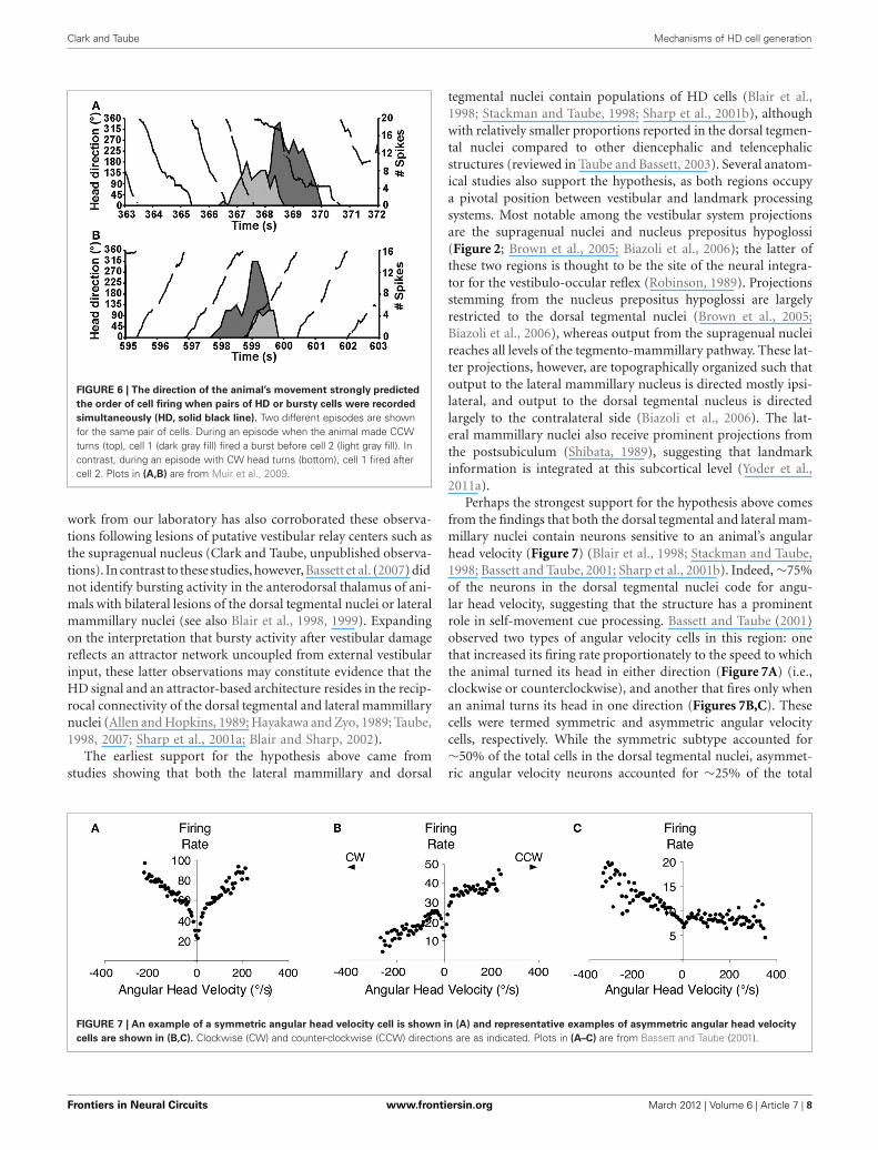
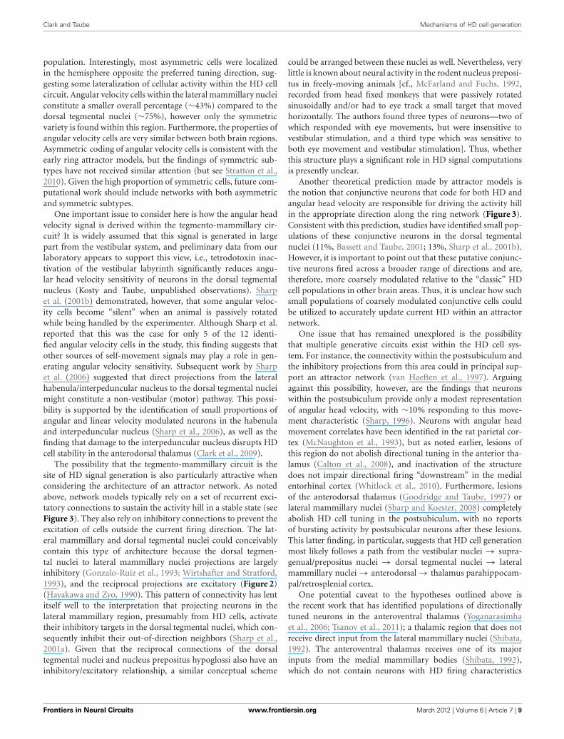
Perhaps the strongest support for the hypothesis above comesfrom the findings that both the dorsal tegmental and lateral mam-millary nuclei contain neurons sensitive to an animal’s angularhead velocity (Figure 7) (Blair et al., 1998; Stackman and Taube,1998; Bassett and Taube, 2001; Sharp et al., 2001b). Indeed, ∼75%of the neurons in the dorsal tegmental nuclei code for angu-lar head velocity, suggesting that the structure has a prominentrole in self-movement cue processing. Bassett and Taube (2001)observed two types of angular velocity cells in this region: onethat increased its firing rate proportionately to the speed to whichthe animal turned its head in either direction (Figure 7A) (i.e.,clockwise or counterclockwise), and another that fires only whenan animal turns its head in one direction (Figures 7B,C). Thesecells were termed symmetric and asymmetric angular velocitycells, respectively. While the symmetric subtype accounted for∼50% of the total cells in the dorsal tegmental nuclei, asymmet-ric angular velocity neurons accounted for ∼25% of the total
FIGURE 7 | An example of a symmetric angular head velocity cell is shown in (A) and representative examples of asymmetric angular head velocity
cells are shown in (B,C). Clockwise (CW) and counter-clockwise (CCW) directions are as indicated. Plots in (A–C) are from Bassett and Taube (2001).
Frontiers in Neural Circuits www.frontiersin.org March 2012 | Volume 6 | Article 7 | 8
Clark and Taube Mechanisms of HD cell generation
population. Interestingly, most asymmetric cells were localizedin the hemisphere opposite the preferred tuning direction, sug-gesting some lateralization of cellular activity within the HD cellcircuit. Angular velocity cells within the lateral mammillary nucleiconstitute a smaller overall percentage (∼43%) compared to thedorsal tegmental nuclei (∼75%), however only the symmetricvariety is found within this region. Furthermore, the properties ofangular velocity cells are very similar between both brain regions.Asymmetric coding of angular velocity cells is consistent with theearly ring attractor models, but the findings of symmetric sub-types have not received similar attention (but see Stratton et al.,2010). Given the high proportion of symmetric cells, future com-putational work should include networks with both asymmetricand symmetric subtypes.
One important issue to consider here is how the angular headvelocity signal is derived within the tegmento-mammillary cir-cuit? It is widely assumed that this signal is generated in largepart from the vestibular system, and preliminary data from ourlaboratory appears to support this view, i.e., tetrodotoxin inac-tivation of the vestibular labyrinth significantly reduces angu-lar head velocity sensitivity of neurons in the dorsal tegmentalnucleus (Kosty and Taube, unpublished observations). Sharpet al. (2001b) demonstrated, however, that some angular veloc-ity cells become “silent” when an animal is passively rotatedwhile being handled by the experimenter. Although Sharp et al.reported that this was the case for only 5 of the 12 identi-fied angular velocity cells in the study, this finding suggests thatother sources of self-movement signals may play a role in gen-erating angular velocity sensitivity. Subsequent work by Sharpet al. (2006) suggested that direct projections from the lateralhabenula/interpeduncular nucleus to the dorsal tegmental nucleimight constitute a non-vestibular (motor) pathway. This possi-bility is supported by the identification of small proportions ofangular and linear velocity modulated neurons in the habenulaand interpeduncular nucleus (Sharp et al., 2006), as well as thefinding that damage to the interpeduncular nucleus disrupts HDcell stability in the anterodorsal thalamus (Clark et al., 2009).
The possibility that the tegmento-mammillary circuit is thesite of HD signal generation is also particularly attractive whenconsidering the architecture of an attractor network. As notedabove, network models typically rely on a set of recurrent exci-tatory connections to sustain the activity hill in a stable state (seeFigure 3). They also rely on inhibitory connections to prevent theexcitation of cells outside the current firing direction. The lat-eral mammillary and dorsal tegmental nuclei could conceivablycontain this type of architecture because the dorsal tegmen-tal nuclei to lateral mammillary nuclei projections are largelyinhibitory (Gonzalo-Ruiz et al., 1993; Wirtshafter and Stratford,1993), and the reciprocal projections are excitatory (Figure 2)(Hayakawa and Zyo, 1990). This pattern of connectivity has lentitself well to the interpretation that projecting neurons in thelateral mammillary region, presumably from HD cells, activatetheir inhibitory targets in the dorsal tegmental nuclei, which con-sequently inhibit their out-of-direction neighbors (Sharp et al.,2001a). Given that the reciprocal connections of the dorsaltegmental nuclei and nucleus prepositus hypoglossi also have aninhibitory/excitatory relationship, a similar conceptual scheme
could be arranged between these nuclei as well. Nevertheless, verylittle is known about neural activity in the rodent nucleus preposi-tus in freely-moving animals [cf., McFarland and Fuchs, 1992,recorded from head fixed monkeys that were passively rotatedsinusoidally and/or had to eye track a small target that movedhorizontally. The authors found three types of neurons—two ofwhich responded with eye movements, but were insensitive tovestibular stimulation, and a third type which was sensitive toboth eye movement and vestibular stimulation]. Thus, whetherthis structure plays a significant role in HD signal computationsis presently unclear.
Another theoretical prediction made by attractor models isthe notion that conjunctive neurons that code for both HD andangular head velocity are responsible for driving the activity hillin the appropriate direction along the ring network (Figure 3).Consistent with this prediction, studies have identified small pop-ulations of these conjunctive neurons in the dorsal tegmentalnuclei (11%, Bassett and Taube, 2001; 13%, Sharp et al., 2001b).However, it is important to point out that these putative conjunc-tive neurons fired across a broader range of directions and are,therefore, more coarsely modulated relative to the “classic” HDcell populations in other brain areas. Thus, it is unclear how suchsmall populations of coarsely modulated conjunctive cells couldbe utilized to accurately update current HD within an attractornetwork.
One issue that has remained unexplored is the possibilitythat multiple generative circuits exist within the HD cell sys-tem. For instance, the connectivity within the postsubiculum andthe inhibitory projections from this area could in principal sup-port an attractor network (van Haeften et al., 1997). Arguingagainst this possibility, however, are the findings that neuronswithin the postsubiculum provide only a modest representationof angular head velocity, with ∼10% responding to this move-ment characteristic (Sharp, 1996). Neurons with angular headmovement correlates have been identified in the rat parietal cor-tex (McNaughton et al., 1993), but as noted earlier, lesions ofthis region do not abolish directional tuning in the anterior tha-lamus (Calton et al., 2008), and inactivation of the structuredoes not impair directional firing “downstream” in the medialentorhinal cortex (Whitlock et al., 2010). Furthermore, lesionsof the anterodorsal thalamus (Goodridge and Taube, 1997) orlateral mammillary nuclei (Sharp and Koester, 2008) completelyabolish HD cell tuning in the postsubiculum, with no reportsof bursting activity by postsubicular neurons after these lesions.This latter finding, in particular, suggests that HD cell generationmost likely follows a path from the vestibular nuclei → supra-genual/prepositus nuclei → dorsal tegmental nuclei → lateralmammillary nuclei → anterodorsal → thalamus parahippocam-pal/retrosplenial cortex.
One potential caveat to the hypotheses outlined above isthe recent work that has identified populations of directionallytuned neurons in the anteroventral thalamus (Yoganarasimhaet al., 2006; Tsanov et al., 2011); a thalamic region that does notreceive direct input from the lateral mammillary nuclei (Shibata,1992). The anteroventral thalamus receives one of its majorinputs from the medial mammillary bodies (Shibata, 1992),which do not contain neurons with HD firing characteristics
Frontiers in Neural Circuits www.frontiersin.org March 2012 | Volume 6 | Article 7 | 9
Clark and Taube Mechanisms of HD cell generation
(Sharp and Turner-Williams, 2005). One possibility is thatanteroventral thalamic neurons receive their directional sensi-tivity through descending inputs from the retrosplenial cortex(Wyss and van Groen, 1992) or postsubiculum (van Groenand Wyss, 1990). In this scenario, the pathway would followfrom anterodorsal thalamus → retrosplenial/postsubiculum →anteroventral thalamus. Alternatively, there could be a directintra-thalamic projection from the anterodorsal to anteroventralregion. Although there is no evidence for this latter projec-tion, these connections are likely difficult to detect with currentanatomical techniques, given that both nuclei lie adjacent to oneanother. HD cells have also been identified in the laterodor-sal thalamus (Mizumori and Williams, 1993), but similar to theanteroventral thalamus, HD cells have not been identified in thesubcortical afferents to the laterodorsal thalamus (Cooper et al.,1998) and lesions of the structure do not abolish HD cell activ-ity in the postsubiculum (Golob et al., 1998). The laterodorsalnuclei receive large inputs from the retrosplenial cortex (Shibata,2000) and postsubiculum (van Groen and Wyss, 1990), and again,it is possible that the HD signal within the laterodorsal thalamusis conveyed from these regions. Collectively, the studies summa-rized above suggest that a number of parallel processing pathwayslikely exist at the cortical-thalamic level of the HD cell circuit,which overlays the general hierarchical organization. Future workshould be directed at identifying the critical pathways withinthese cortical-thalamic loops, and to better understand the pre-cise role these parallel pathways play in shaping the HD cellsignal.
CONCLUSIONSThe present review summarized recent work that has beendirected toward understanding the functional organization of theHD cell system, and much of this work supports three generalconclusions. First, a large body of data strongly suggests thatthe HD cell signal is hierarchically organized and is primarilygenerated within subcortical circuits. This highly processed direc-tional signal is then conveyed to cortical and parahippocampalregions through inputs from the anterior thalamus, includingthe anterodorsal thalamus and possibly the anteroventral region.A second general conclusion is that the identification of burstyactivity within the HD cell network after vestibular damageappears to support the attractor network hypothesis of HD signalgeneration. The strongest evidence for this conclusion is derivedfrom the fact that the temporal relationship between neurons ispreserved after vestibular damage (Muir et al., 2009; Yoder andTaube, 2009) and is strongly correlated with an animal’s directionof movement (i.e., right vs. left turns). Third, the observation ofintermittent burst activity after vestibular damage, but not afterdorsal tegmental or lateral mammillary damage is consistent withthe hypothesis that the attractor architecture resides within thereciprocal connections of the tegmento-mammillary circuit.
ACKNOWLEDGMENTSThis work was supported through grants from the NationalInstitute of Health (NS053907, DC009318) to Jeffrey S. Taubeand a postgraduate fellowship from the National Sciences andEngineering Research Council of Canada to Benjamin J. Clark.
REFERENCESAllen, G. V., and Hopkins, D. A.
(1989). Mammillary body in the rat:topography and synaptology of pro-jections from the subicular com-plex, prefrontal cortex, and mid-brain tegmentum. J. Comp. Neurol.286, 311–336.
Anniko, M., and Wersäll, J. (1976).Afferent and efferent nerve terminaldegeneration in the guinea-pigcochlea following atoxyl admin-istration. Acta Otolaryngol. 82,325–336.
Bassett, J. P., and Taube, J. S. (2001).Neural correlates for angular headvelocity in the rat dorsal tegmentalnucleus. J. Neurosci. 21, 5740–5751.
Bassett, J. P., Tullman, M. L., and Taube,J. S. (2007). Lesions of the tegmen-tomammillary circuit in the headdirection system disrupt the headdirection signal in the anterior tha-lamus. J. Neurosci. 27, 7564–7577.
Biazoli, C. E. Jr., Goto, M., Campos,A. M., and Canteras, N. S. (2006).The supragenual nucleus: a putativerelay station for ascending vestibu-lar signs to head direction cells.Brain Res. 1094, 138–148.
Blair, H. T., and Sharp, P. E. (2002).“Functional organization of therat head-direction circuit,” in
The Neural Basis of Navigation,ed P. E. Sharp (Boston: Kluwer),163–182.
Blair, H. T., Cho, J., and Sharp, P. E.(1998). Role of the lateral mammil-lary nucleus in the rat head direc-tion circuit: a combined single-unitrecording and lesion study. Neuron21, 1387–1397.
Blair, H. T., Cho, J., and Sharp, P.(1999). The anterior thalamic head-direction signal is abolished bybilateral but not unilateral lesionsof the lateral mammillary nucleus.J. Neurosci. 19, 6673–6683.
Boccara, C. N., Sargolini, F., Thoresen,V. H., Solstad, T., Witter, M. P.,Moser, E. I., and Moser, M. B.(2010). Grid cells in pre- andparasubiculum. Nat. Neurosci. 13,987–994.
Brown, J. E., Card, J. P., and Yates, B. J.(2005). Polysynaptic pathways fromthe vestibular nuclei to the lateralmammillary nucleus of the rat: sub-strates for vestibular input to headdirection cells. Exp. Brain Res. 161,47–61.
Calton, J. L., Stackman, R. W.,Goodridge, J. P., Archey, W. B.,Dudchenko, P. A., and Taube, J.S. (2003). Hippocampal place cellinstability after lesions of the head
direction cell network. J. Neurosci.23, 9719–9731.
Calton, J. L., Turner, C. S., Cyrenne,D. L., Lee, B. R., and Taube, J.S. (2008). Landmark control andupdating of self-movement cues arelargely maintained in head directioncells after lesions of the posteriorparietal cortex. Behav. Neurosci. 122,827–840.
Chen, Y. C., Pellis, S. M., Sirkin, D.W., Potegal, M., and Teitelbaum,P. (1986). Bandage backfall:labyrinthine and non-labyrinthinecomponents. Physiol. Behav. 37,805–814.
Clark, B. J., Bassett, J. P., Wang, S.,and Taube, J. S. (2010). Impairedhead direction cell representationin the anterodorsal thalamus afterlesions of the retrosplenial cortex.J. Neurosci. 30, 5289–5302.
Clark, B. J., Sarma, A., and Taube, J.S. (2009). Head direction cell insta-bility in the anterior dorsal thala-mus after lesions of the interpe-duncular nucleus. J. Neurosci. 29,493–507.
Clark, B. J., and Taube, J. S. (2011).Intact landmark control and angu-lar path integration by head direc-tion cells in the anterodorsal tha-lamus after lesions of the medial
entorhinal cortex. Hippocampus 21,767–782.
Clark, B. J., Valerio, S., and Taube,J. S. (2011). Disrupted grid andhead direction cell signal in theentorhinal cortex and parasubiculumafter lesions of the head directionsystem. Program No. 729.11 2011Neuroscience Meeting Planner.Washington, DC: Society forNeuroscience. [Online].
Cooper, B. G., Miya, D. Y., andMizumori, S. J. (1998). Superiorcolliculus and active navigation: roleof visual and non-visual cues in con-trolling cellular representations ofspace. Hippocampus 8, 340–372.
Gallistel, C. R. (1990). The Organi-zation of Learning. Cambridge, MA:MIT Press.
Golob, E. J., and Taube, J. S. (1997).Head direction cells and episodicspatial information in rats without ahippocampus. Proc. Natl. Acad. Sci.U.S.A. 94, 7645–7650.
Golob, E. J., and Taube, J. S. (1999).Head direction cells in rats with hip-pocampal or overlying neocorticallesions: evidence for impaired angu-lar path integration. J. Neurosci. 19,7198–7211.
Golob, E. J., Wolk, D. A., andTaube, J. S. (1998). Recordings
Frontiers in Neural Circuits www.frontiersin.org March 2012 | Volume 6 | Article 7 | 10
Clark and Taube Mechanisms of HD cell generation
of postsubiculum head directioncells following lesions of the lat-erodorsal thalamic nucleus. BrainRes. 780, 9–19.
Gonzalo-Ruiz, A., Sanz-Anquela, J.M., and Spencer, R. F. (1993).Immunohistochemical localizationof GABA in the mammillary com-plex of the rat. Neuroscience 54,143–156.
Goodridge, J. P., Dudchenko, P. A.,Worboys, K. A., Golob, E. J., andTaube, J. S. (1998). Cue control andhead direction cells. Behav. Neurosci.112, 749–761.
Goodridge, J. P., and Taube, J. S. (1997).Interaction between postsubiculumand anterior thalamus in the gener-ation of head direction cell activity.J. Neurosci. 17, 9315–9330.
Hafting, T., Fyhn, M., Molden, S.,Moser, M. B., and Moser, E. I.(2005). Microstructure of a spa-tial map in the entorhinal cortex.Nature 436, 801–806.
Hayakawa, T., and Zyo, K. (1989).Retrograde double-labeling studyof the mammillothalamic and themammillotegmental projections inthe rat. J. Comp. Neurol. 284, 1–11.
Hayakawa, T., and Zyo, K. (1990). Finestructure of the lateral mammillaryprojection to the dorsal tegmentalnucleus of Gudden in the rat. J.Comp. Neurol. 298, 224–236.
Johnson, A., Seeland, K., and Redish,A. D. (2005). Reconstruction ofthe postsubiculum head directionsignal from neural ensembles.Hippocampus 15, 86–96.
Kerr, K. M., Agster, K. L., Furtak,S. C., and Burwell, R. D. (2007).Functional neuroanatomy of theparahippocampal region: the lat-eral and medial entorhinal areas.Hippocampus 17, 697–708.
Lane, P. (1986). Tilted (tlt). Mouse NewsLett. 75, 28.
Lever, C., Burton, S., Jeewajee, A.,O’Keefe, J., and Burgess, N. (2009).Boundary vector cells in the subicu-lum of the hippocampal formation.J. Neurosci. 29, 9771–9777.
McFarland, J. L., and Fuchs, A. F.(1992). Discharge patterns innucleus prepositus hypoglossi andadjacent medial vestibular nucleusduring horizontal eye movement inbehaving macaques. J. Neurophysiol.68, 319–332.
McNaughton, B. L., Barnes, C. A.,Gerrard, J. L., Gothard, K., Jung,M. W., Knierim, J. J., Kudrimoti,H., Qin, Y., Skaggs, W. E., Suster,M., and Weaver, K. L. (1996).Deciphering the hippocampal poly-glot: the hippocampus as a pathintegration system. J. Exp. Biol. 199,173–185.
McNaughton, B. L., Battaglia, F. P.,Jensen, O., Moser, E. I., and Moser,M. B. (2006). Path integration andthe neural basis of the ‘cognitivemap’. Nat. Rev. Neurosci. 7, 663–678.
McNaughton, B. L., Chen, L., andMarkus, E. (1991). “Dead reckon-ing,” landmark learning, and thesense of direction: a neurophysio-logical and computational hypothe-sis. J. Cogn. Neurosci. 3, 190–202.
McNaughton, B. L., Mizumori, S. J.,Barnes, C. A., Leonard, B., Marquis,M., and Green, E. (1993). Corticalrepresentation of motion duringunrestrained spatial navigation inthe rat. Cereb. Cortex 4, 27–39.
Mizumori, S. J., and Williams, J.D. (1993). Directionally selectivemnemonic properties of neurons inthe lateral dorsal nucleus of thethalamus of rats. J. Neurosci. 13,4015–4028.
Moser, E. I., Kropff, E., and Moser,M. B. (2008). Place cells, grid cells,and the brain’s spatial representa-tion system. Ann. Rev. Neurosci. 31,69–89.
Muir, G. M., Brown, J. E., Carey, J.P., Hirvonen, T. P., Della Santina,C. C., Minor, L. B., and Taube, J.S. (2009). Disruption of the headdirection cell signal after occlusionof the semicircular canals in thefreely moving chinchilla. J. Neurosci.29, 14521–14533.
O’Keefe, J., and Dostrovsky, J. (1971).The hippocampus as a spatial map.Preliminary evidence from unitactivity in the freely-moving rat.Brain Res. 34, 171–175.
O’Keefe, J., and Nadel, L. (1978). TheHippocampus as a Cognitive Map.Oxford, UK: Oxford UniversityPress.
Papez, J. (1937). A proposed mecha-nism of emotion. Arch. Neurol. 38,103–112.
Potegal, M. (1982). “Vestibular andneostriatal contributions to spatialorientation,” in Spatial Abilities:Development and PhysiologicalFoundations, ed M. Potegal(New York, NY: Academic Press),361–387.
Ranck, J. B. Jr. (1984). Head direc-tion cells in the deep layer of dorsalpresubiculum in freely moving rats.Soc. Neurosci. Abstr. 10, 599.
Redish, A. D., Elga, A. N., andTouretzky, D. S. (1996). A coupledattractor model of the rodent headdirection system. Netw. Comput.Neural Syst. 7, 671–685.
Ris, L., and Godaux, E. (1998).Neuronal activity in the vestibularnuclei after contralateral or bilaterallabyrinthectomy in the alert guineapig. J. Neurophysiol. 80, 2352–2367.
Robinson, D. A. (1989). Integratingwith neurons. Annu. Rev. Neurosci.12, 33–45.
Russell, N., Horii, A., Smith, P.,Darlington, C., and Bilkey, D.(2003). Long-term effects ofpermanent vestibular lesionson hippocampal spatial firing.J. Neurosci. 23, 6490–6498.
Sargolini, F., Fyhn, M., Hafting, T.,McNaughton, B. L., Witter, M. P.,Moser, M. B., and Moser, E. I.(2006). Conjunctive representationof position, direction, and velocityin entorhinal cortex. Science 312,758–762.
Savelli, F., Yoganarasimha, D., andKnierim, J. J. (2008). Influenceof boundary removal on the spa-tial representations of the medialentorhinal cortex. Hippocampus 18,1270–1282.
Sharp, P. E. (1996). Multiple spa-tial/behavioral correlates for cellsin the rat postsubiculum: multipleregression analysis and comparisonto other hippocampal areas. Cereb.Cortex 6, 238–259.
Sharp, P. E., and Koester, K. (2008).Lesions of the mammillary bodyregion severely disrupt the cor-tical head direction, but notplace cell signal. Hippocampus 18,766–784.
Sharp, P. E., Blair, H. T., and Cho,J. (2001a). The anatomical andcomputational basis of the rathead-direction cell signal. TrendsNeurosci. 24, 289–294.
Sharp, P. E., Tinkelman, A., and Cho, J.(2001b). Angular velocity and headdirection signals recorded from thedorsal tegmental nucleus of Guddenin the rat: implications for pathintegration in the head directioncell circuit. Behav. Neurosci. 115,571–588.
Sharp, P. E., and Turner-Williams,S. (2005). Movement-related corre-lates of single-cell activity in themedial mammillary nucleus of therat during a pellet-chasing task.J. Neurophysiol. 94, 1920–1927.
Sharp, P. E., Turner-Williams, S., andTuttle, S. (2006). Movement-relatedcorrelates of single cell activity in theinterpeduncular nucleus and habe-nula of the rat during a pellet-chasing task. Behav. Brain Res. 166,55–70.
Shibata, H. (1989). Descending pro-jections to the mammillary nucleiin the rat, as studied by retro-grade and anterograde transport ofwheat germ agglutinin-horseradishperoxidase. J. Comp. Neurol. 285,436–452.
Shibata, H. (1992). Topographic orga-nization of subcortical projections
to the anterior thalamic nuclei in therat. J. Comp. Neurol. 323, 117–127.
Shibata, H. (2000). Organization of ret-rosplenial cortical projections to thelaterodorsal thalamic nucleus in therat. Neurosci. Res. 38, 303–311.
Skaggs, W. E., Knierim, J. J., Kudrimoti,H. S., and McNaughton, B. L.(1995). “A model of the neuralbasis of the rat’s sense of direction,”in Advances in Neural InformationProcessing Systems, Vol. 7, eds G.Tesauro, D. S. Touretzky, and T. K.Leen (Cambridge, MA: MIT Press),173–180.
Solstad, T., Boccara, C. N., Kropff,E., Moser, M. B., and Moser, E. I.(2008). Representation of geomet-ric borders in the entorhinal cortex.Science 322, 1865–1868.
Stackman, R. W., Clark, A. S., andTaube, J. S. (2002). Hippocampalspatial representations requirevestibular input. Hippocampus 12,291–303.
Stackman, R. W., Golob, E. J., Bassett,J. P., and Taube, J. S. (2003). Passivetransport disrupts directional pathintegration by rat head directioncells. J. Neurophysiol. 90, 2862–2874.
Stackman, R. W., and Taube, J. S.(1997). Firing properties of headdirection cells in rat anterior tha-lamic neurons: dependence uponvestibular input. J. Neurosci. 17,4349–4358.
Stackman, R. W., and Taube, J. S.(1998). Firing properties of rat lat-eral mammillary single units: headdirection, head pitch, and angu-lar head velocity. J. Neurosci. 18,9020–9037.
Stratton, P., Wyeth, G., and Wiles, J.(2010). Calibration of the headdirection network: a role forsymmetric angular head velocitycells. J. Comput. Neurosci. 28,527–538.
Taube, J. S. (1995). Head directioncells recorded in the anterior tha-lamic nuclei of freely moving rats.J. Neurosci. 15, 70–86.
Taube, J. S. (1998). Head direction cellsand the neurophysiological basis fora sense of direction. Prog. Neurobiol.55, 225–256.
Taube, J. S. (2007). The head directionsignal: origins and sensory-motorintegration. Annu. Rev. Neurosci. 30,181–207.
Taube, J. S., and Bassett, J. P. (2003).Persistent neural activity in headdirection cells. Cereb. Cortex 13,1162–1172.
Taube, J. S., and Burton, H. L. (1995).Head direction cell activity moni-tored in a novel environment andduring a cue conflict situation.J. Neurophysiol. 74, 1953–1971.
Frontiers in Neural Circuits www.frontiersin.org March 2012 | Volume 6 | Article 7 | 11
Clark and Taube Mechanisms of HD cell generation
Taube, J. S., Muller, R. U., andRanck, J. B. Jr. (1990a). Head-direction cells recorded from thepostsubiculum in freely movingrats. I. Description and quan-titative analysis. J. Neurosci. 10,420–435.
Taube, J. S., Muller, R. U., andRanck, J. B. Jr. (1990b). Head-direction cells recorded from thepostsubiculum in freely movingrats. II. Effects of environmentalmanipulations. J. Neurosci. 10,436–447.
Tsanov, M., Chah, E., Vann, S.,Reilly, R., Erichsen, J., Aggleton,J., and O’Mara, S. (2011).Theta-modulated head direc-tion cells in the rat anteriorthalamus. J. Neurosci. 31,9489–9502.
van Groen, T., and Wyss, J. M. (1990).The postsubicular cortex in therat: characterization of the fourthregion of the subicular cortex andits connections. Brain Res. 529,165–177.
van Haeften, T., Wouterlood, F.,Jorritsma-Byham, B., and Witter,
M. P. (1997). GABAergic presubic-ular projections to the medialentorhinal cortex of the rat.J. Neurosci. 17, 862–874.
Whitlock, J. R., Derdikman, D., Pfuhl,G., Moser, M. B., and Moser, E. I.(2010). Effects of parietal corticalinactivation on representations inentorhinal cortex. Program No.101.13. 2010 Neuroscience MeetingPlanner. San Diego, CA: Society forNeuroscience. [Online].
Wirtshafter, D., and Stratford, T. R.(1993). Evidence for GABAergicprojections from the tegmentalnuclei of Gudden to the mammil-lary body in the rat. Brain Res. 630,188–194.
Wyss, J. M., and van Groen, T. (1992).Connections between the retrosple-nial cortex and the hippocampalformation: a review. Hippocampus 1,1–11.
Yoder, R., and Taube, J. S. (2008). Thepostsubiculum provides visual land-mark control to the head directionsignal at the lateral mammillarynuclei. Program No. 90.9. 2008Neuroscience Meeting Planner.
Washington, DC: Society forNeuroscience. [Online].
Yoder, R., and Taube, J. S. (2009).Head direction cell activity in mice:robust directional signal depends onintact otolith organs. J. Neurosci. 29,1061–1076.
Yoder, R. M., Clark, B. J., and Taube,J. S. (2011a). Origins of land-mark encoding in the brain. TrendsNeurosci. 34, 561–571.
Yoder, R. M., Clark, B. J., Brown, J. E.,Lamia, M. V., Valerio, S., Shinder,M. E., and Taube, J. S. (2011b).Both visual and idiothetic cues con-tribute to head direction cell sta-bility during navigation along com-plex routes. J. Neurophysiol. 105,2989–3001.
Yoganarasimha, D., Yu, X., andKnierim, J. J. (2006). Head direc-tion cell representations maintaininternal coherence during con-flicting proximal and distal cuerotations: comparison with hip-pocampal place cells. J. Neurosci. 26,622–631.
Zhang, K. (1996). Representation ofspatial orientation by the intrinsic
dynamics of the head-direction cellensemble: a theory. J. Neurosci. 16,2112–2126.
Conflict of Interest Statement: Theauthors declare that the researchwas conducted in the absence of anycommercial or financial relationshipsthat could be construed as a potentialconflict of interest.
Received: 13 December 2011; accepted:14 February 2012; published online: 20March 2012.Citation: Clark BJ and Taube JS (2012)Vestibular and attractor network basis ofthe head direction cell signal in subcor-tical circuits. Front. Neural Circuits 6:7.doi: 10.3389/fncir.2012.00007Copyright © 2012 Clark and Taube.This is an open-access article dis-tributed under the terms of the CreativeCommons Attribution Non CommercialLicense, which permits non-commercialuse, distribution, and reproduction inother forums, provided the originalauthors and source are credited.
Frontiers in Neural Circuits www.frontiersin.org March 2012 | Volume 6 | Article 7 | 12