vestibular experiments in space
TRANSCRIPT
Vestibular Experiments in Space
Bernard Cohen,1 Sergei B. Yakushin,1 Gay R. Holstein,1
Mingjia Dai,1 David L. Tomko,2 Anatole M. Badakva3 andInessa B. Kozlovskaya3
1Department of Neurology,Mount Sinai School of Medicine, New York, USA;2National Aeronautics and Space Agency;3the Institute of Biomedical Problems, Moscow, Russia
Introduction
Life on Earth is conditioned by the constant downward pull of gravity, whichcan be considered as an equivalent constant upward linear acceleration(Einstein, 1911). During lateral, fore-aft, and vertical translations as well asduring centripetal accelerations generated by turning around distant axes, theselinear accelerations summate with the linear acceleration of gravity to form aresultant vector, which we term gravito-inertial acceleration (GIA). These linearaccelerations increase the magnitude of the GIA and tilt it relative to the head.The otolith organs and body tilt receptors sense the resultant changes in theGIA and generate compensatory and orienting eye movements throughthe linear vestibulo-ocular reflex (lVOR) to direct and stabilize vision.Angular head movements are sensed by the semicircular canals, which producecompensatory eye movements over the angular vestibulo-ocular reflex (aVOR).The vestibular system also responds to linear and angular accelerations throughvestibulo-spinal reflexes to produce body postural movements that stabilizethe body when stationary and support it when one is in motion. Additionally,the vestibular system responds to changes in head and body position relativeto the GIA to maintain blood pressure and enhance respiration. Thus, thevestibular system plays a critical role in sensing and responding to linear andangular accelerations in a gravitational environment.
In microgravity, the linear acceleration of gravity is reduced to negligiblelevels, and a standing posture is no longer maintained. Instead, subjects swim inSpace rather than walk, and they no longer experience the vertical linear trans-lations that accompany locomotion. Additionally, gravity no longer contributesto the resultant change in the GIA when the head is translated. Thus, the GIA isalways in the direction of the translational and centripetal accelerations due to
Experimentation with Animal Models in Space
G. Sonnenfeld (editor) 105� 2005 Elsevier B.V. All rights reservedDOI: 10.1016/S1569-2574(05)10005-7

movement. Angular acceleration, which is in a head coordinate frame, is notmarkedly altered in microgravity. Because of the ubiquitous nature of gravityon Earth and the difficulty of performing experiments on orbit, we knowrelatively little about how the nervous system is altered by the prolongedabsence of gravity.
From the 1970s, the Russian Space Agency made orbital space flight avail-able to INTERCOSMOS scientists from many countries for animal experi-mentation, primarily on rats. This included Czechoslovakia, Poland, Hungary,Germany, France, and the United States. Between 1983 and 1995, monkeysand rats flew for approximately 1–2 week periods on a Vostok Space Capsulein experiments supported by the Russian Space Agency and the NationalAeronautics and Space Aadministration (NASA). Rodents and invertebratesalso flew on NASA Space Shuttle missions, particularly on the NeurolabMission (STS-90) in 1998. The purpose of this chapter is to provide a summaryof these experiments. Experiments on rodents and other animal species in theNASA Space Shuttle flights and the Russian COSMOS/BION flights aresummarized in the first section. The second section summarizes inflight experi-ments performed by the Russian Space Agency on the aVOR and on single unitactivity in the vestibular nuclei and flocculus of monkeys. Many of these dataappear here for the first time. In the third section, we summarize NASA-supported research on rhesus monkeys on the linear and angular vestibulo-ocular reflex (lVOR and aVOR). The intent was to provide a summary ofexperimental data on which future experimentation in Space can be expanded.In addition to revealing some new and interesting scientific results, it is hopedthat it will become obvious that basic studies, involving subhuman primatesas well as rodents should be essential components of any future space-relatedresearch. The results of the earliest flights are not included in this chapter, butcan be found in references at the end of the bibliography.
SECTION 1: CELLULAR RESPONSES TO ALTERED
GRAVITATIONAL ENVIRONMENTS IN ADULT ANIMALS
A number of studies have examined the impact of altered gravity exposureon vestibular system structures at the cellular level. As a whole, they clearlydocument the modifiability of cellular and subcellular constituents of vestibularpathways in response to altered gravitational stimuli.
Peripheral vestibular system
Some evidence suggests that exposure of adult as well as developing animalsto altered gravitational environments can trigger adaptive responses in weight-lending structures. For example, an increase in otoconial mass has beenreported in the utricles of adult animals of several species (rats, frogs) following7 days of exposure to microgravity (Vinnikov et al., 1980; Ross et al., 1985;
106

Ross, 1987; Lychakov et al., 1989). Conversely, the saccular otolithic mem-brane volume is reduced in rats exposed to 2.3 g or 4.15 g hypergravity throughcentrifugation (Lim et al., 1974), although otoconial morphology (examinedby scanning electron microscopy) and otoconial synthesis (assessed by mRNAexpression of the otoconial matrix protein osteopontin) appear to be normalafter 2 h to 7 days of hypergravity exposure (Uno et al., 2000).
Increases in otoconial mass have also been observed in the saccular otolithsof newt larvae (Weiderhold et al., 1997) and late-stage embryos of swordtail fish(Weiderhold et al., 2000) reared in Space, although such increases are notobserved in older fish. As in adult animals, the saccular otolithic membranevolume is reduced in cichlid fish reared on a centrifuge (Anken et al., 1998),and the volume of statoconia in marine mollusk larvae reared at 2–5 g isdiminished in a graded manner as compared with 1 g controls (Pedrozo andWiederhold, 1994). Similarly, there is a reduction in the number of largeotoconia that are present in the anterior peripheral portion of the utricle in ratsraised in a 2 g environment for 60 days than in 1 g controls (Krasnov, 1991).Taken together, these results suggest that otoconial mass adapts to fluctuationsin the gravitational stimulus, perhaps to maintain a consistent force on themaculae.
A number of studies have addressed the question of morphologic alterationsin the sensory hair cells of adult mammalian otolith organs in response toaltered gravitational conditions. These studies indicate that short-term (e.g.,7 day) exposure to space flight does not cause otolith receptor cell degenera-tion in rats (Ross et al., 1985), although the type I hair cells reportedly exhibitabnormal cytologic features such as increased chromatin, and enlargedperinuclear and intercellular spaces (Krasnov, 1987, 1991). However, moreprolonged exposure to microgravity clearly impacts hair cell synaptology. Ingeneral, these studies indicate that type II hair cells in particular, but also type Icells evidence adaptive structural modifications in response to alterations inthe gravitational environment (Ross, 1993, 1994, 2000). Quantitative ultra-structural analysis of utricular hair cells from adult rats sacrificed immediatelyafter landing from a 9-day space flight demonstrated statistically significantincreases in the number of ribbon synapses (Ross, 1994). In addition, thenumber of synapse pairs and clusters markedly increased in the type II hair cells.Nine days after landing, which was the mission duration, synapse countsremained elevated in the flight rats, but were also elevated in the type II hair cellsof ground control animals, suggesting that these receptor cells are particularlyvulnerable to stress-induced morphologic changes. In a subsequent study(Ross and Tomko, 1998), the mean number of synapses on type II hair cellsdoubled, and those on type I cells increased by over 40% by the 13th day of a14-day space flight. Immediately postflight, these synapse counts were reducedto 67% and 13% increases, respectively, over control values. Comparablestudies of vestibular hair cells have been conducted utilizing 2–4 g hypergravityexposure for periods of time ranging from 14–30 days (Lim et al., 1974;
107

Lychakov et al., 1988; Ross, 1993). In general, these experiments support Ross’centrifugation study (Ross, 1993) demonstrating that type I hair cell synapsenumber does not change in response to hypergravity, but there is a decrementof approximately 40% in type II hair cell synapses. Clearly, the otolith organs ofadult mammals have the potential for morphologic reorganization at severalsites in response to altered gravity conditions (Ross, 1997).
Since studies of molecular changes in peripheral vestibular cells in responseto fluctuations in the gravity stimulus have been initiated relatively recently,generalizations about molecular alterations are premature. One recent studyinvestigated glutamate receptor mRNA expression using RT-PCR on cellsof the vestibular ganglion, as well as the vestibular nuclei, and vestibulo-cerebellum of rats exposed to hypergravity for 2 h–7 days (Uno et al., 2002). Inthe vestibular periphery, this experiment demonstrated that synthesis of GluR2(an AMPA glutamate receptor subunit) receptors in vestibular ganglion cells isreduced in rats exposed to 7 days of hypergravity.
Behavioral, physiological and biochemical correlates of vestibular
cellular responses
A spinning movement during swimming, termed ‘‘looping,’’ was initiallyreported in some (but not all) fish at the transition from 1 g to microgravityduring parabolic flight (von Baumgarten et al., 1972), and subsequentlydescribed in larval cichlid fish and Xenopus laevis immediately following prolon-ged 3 g centrifugation (Rahmann et al., 1992). Looping has also been reportedin killifish in microgravity (von Baumgarten et al., 1975). Fish that evidencelooping behavior exhibit a significantly higher asymmetry in otolith size (Ankenet al., 1998) and weight (Beier et al., 2002), in comparison with nonloopingsiblings. In addition, although the total number of sensory and supportingcells in the utricular maculae is the same, the cell density is significantly lowerin looping than nonlooping fish, due to the atypical presence of enlargedepithelial cells (Bauerle et al., 2004).
Adult mammals may be more profoundly affected by changes in the gravi-tational environment than are fish. Upon landing following 9 days of spaceflight, adult rats reportedly show substantially reduced locomotion (Ross,1994). They maintain a posture with their abdomens and tails flat against thecage floor, with their limbs and digits extended. After 9 days at 1 g, flight ratsdisplay normal body posture and movement. Similarly, after 14 days of expo-sure to 2 g through centrifugation, adult rats show profound deficits in rightingresponses, swimming and balance (Fox et al., 1992). Recovery of normalorientation during swimming requires 4–24 h at 1 g, whereas the righting reflexdoes not return to normal for 5–7 days (Ross and Tomko, 1998).
Neurophysiological studies have further documented an immediate post-flight alteration in vestibular activity (Boyle et al., 2001). Within the first 16 hpost-space flight, utricular afferents in the oyster toadfish were hypersensitive
108
to translational acceleration, but had no change in directional selectivity. Theafferent sensitivity returned to baseline levels by approximately 30 h postflight.Since anamniotes such as the toadfish have only type II hair cells, the reportedincrease in ribbon synapse number (Ross, 1997) could be one explanation forthe afferent hypersensitivity.
Central vestibular system
Several studies have addressed the impact of altered gravitational environmentson cells of the central vestibular system. Most of these studies have examinedthe vestibular nuclei and/or vestibulo-cerebellum, and have utilized either amorphological approach, or a marker for cellular activity. Early studies of thecerebellar nodulus of adult rats exposed to 18 days of space flight revealedultrastructural alterations in Purkinje cell dendrites and mossy fiber terminals(Krasnov and Dyachkova, 1986, 1990). In these studies, the major modificationdescribed in the Purkinje cells was a widening of the synaptic cleft at contactswith Purkinje cell dendrites. In mossy fiber terminals, structural alterationsincluded densely packed synaptic vesicles, with unusual clustering of suchvesicles near the presynaptic membranes of axodendritic synapses, increasedelectron density of pre- and postsynaptic membranes, and enlargement of thesynaptic gap. Similar changes are observed in nodular mossy fiber terminalsfrom rats flown on 5–7 day missions and sacrificed within 8 h of landing.Moreover, ultrastructural alterations in the primary somatosensory cortex ofthese flight animals included a profound decrement in the number of axo-dendritic synapses, and an increase in the number of axon terminals showing‘‘light’’ degeneration, as well as signs of ‘‘superexcitation’’ including an increasein the number of axon terminals showing dark degeneration. In concert with thisfinding, increased numbers of synaptic contacts have been demonstrated inneonatal swordtail fish after 16 days in microgravity, specifically in the nucleusmagnocellularis of the primary vestibular brainstem region, area octavolateralis(Ibsch et al., 2000).
More recently, the ultrastructure of the otolith-recipient zones of thecerebellar nodulus has been analyzed in tissue from flight and cage-controlrats sacrificed in microgravity after 24 h of space flight (Holstein et al., 1999).Qualitative observations of this tissue indicate that several structural alterationsoccur in the neuropil, and in the Purkinje cell cytoplasm, of the nodulus of ratsexposed to 24 h of space flight. These anatomical alterations are not apparentin the cage control animals. Most notably, the cisterns of smooth endoplasmicreticulum that are normally present throughout Purkinje cells are substantiallyenlarged and more complex in Purkinje cells of the otolith-recipient zones ofthe nodulus. The increased complexity of the cisterns results in the formationof long, stacked lamellar bodies that are observed throughout entire Purkinjecells, including the somata, dendrites, thorns, and axon terminals. In addition,occasional enormous mitochondria, >1 mm in cross-sectional diameter, are
109
present in the Purkinje cell somata of flight animals. Ultrastructural indica-tions of degeneration and synaptic reorganization are also observed in themolecular layer of the nodulus from the flight animals, but not cage controls.Since these morphologic changes are not apparent in control animals, they arenot likely to be due to caging or tissue processing effects. The particular natureof the structural alterations, including the formation of lamellar bodies andthe presence of degeneration, suggests that excitotoxity may play a role in theshort-term neural response to space flight.
In the granular layer of the nodulus of rats raised in a 2 g environment for60 days, 80% of the glomeruli showed altered synaptic morphology, includingchanges in the density of pre- and postsynaptic membranes, increased thicknessof the postsynaptic density, enlargement of the synaptic cleft, increased pack-ing density of synaptic vesicles, enlarged mitochondria, and an increase in thenumber of microtubules (Krasnov and Dyachkova, 1986; Krasnov, 1991). Twodays after return to a 1 g environment, the ultrastructure of the nodulus resemblesthat of control animals. The synaptic vesicle packing density is decreased, andthe number of microtubules is diminished, suggesting reversibility of the gravity-induced effects. Taken together with the microgravity findings, these resultsmore generally indicate that the central vestibular system responds to majorchanges in the gravitational stimulus with similar morphological restructuring,regardless of the direction (hypo- or hyper-) of that change.
In light of the ultrastructural findings suggesting that excitotoxicity maybe a factor in early neuronal responses to altered gravitational environments(Holstein et al., 1999), the recent study of glutamate receptor expression is ofparticular interest. This study examined glutamate receptor mRNA expressionusing RT-PCR on cells in the vestibular nuclei and vestibulo-cerebellum of ratsexposed to hypergravity for 2 h to 7 days (Uno et al., 2002). The results indicatethat mRNA expression of GluR2 (an AMPA glutamate receptor subunit) andNR1 (the obligatory subunit of the NMDA glutamate receptor) in the nodulus/uvula and NR1 expression in the medial vestibular nucleus increase after 2 hof stimulation. This expression gradually returns to baseline during the 7 daysof hypergravity exposure. As noted above, mRNA expression of GluR2receptors in vestibular ganglion cells is reduced after 7 days of stimulation.Neither the mGluR1 metabotropic receptor nor the d2 glutamate receptor in theflocculus and nodulus/uvula is affected by hypergravity exposure for 2 h to7 days. It was suggested that the immediate (2 h) adaptation to hypergravityinvolves enhanced cerebellar inhibition of the vestibular nuclei mediated byPurkinje cell NR1 and GluR2 receptors, whereas longer term adaptationinvolves decreased transmission from vestibular hair cells to primary afferentneurites mediated by down-regulation of postsynaptic GluR2 receptors onthe primary afferents. One interesting aspect of this study is that functionalNMDA receptors are not normally present on cerebellar Purkinje cells inadult mammals. Conceivably, an altered gravitational environment presents asufficient stimulus to trigger their expression and activity. However, the activity
110

of enzymes involved in energy metabolism (lactate dehydrogenase andcreatinine kinase) in giant Deiters’ neurons of the lateral vestibular nucleusand cells of the cerebellar nodulus in rats are not appreciably affected by 22 daysof space flight (Krasnov, 1975).
Space flight-related studies utilizing markers for vestibular neuronal activityhave concentrated primarily on the immediate early gene c-fos. The geneencodes for Fos protein, which reaches peak values within 2–4 h of the effectivestimulus and returns to baseline within 6–8 h. Fos-related antigen (FRA)proteins, which are generated by multiple genes, are induced shortly afterstimulation, but persist for days (‘‘acute’’ FRAs such as FRA-1, -2, FosB,�FosB) or weeks (‘‘chronic’’ FRAs, e.g., modified forms of �FosB). Fosprotein is often utilized as a neural activity marker, since it can be identifiedwith higher spatial resolution than metabolic indicators. Although thebasal expression level of c-fos expression is low in the brain, increases in Fos-like immunoreactivity have been reported in vestibular structures followinggalvanic stimulation (Kaufman and Perachio, 1994; Mashburn et al., 1997) andcentripetal acceleration (Kaufman et al., 1992), as well as space flight.
Results from space flight experiments on adult rat brain tissue indicatea trend toward increased numbers of Fos-immunopositive cells in the vestibularbrainstem (particularly the medial and descending vestibular nuclei, MVNand DVN, respectively) 24 h postlaunch, and a statistically significant increasein the number of immunostained cells 24 h after return from a 17-day mission(Pompeiano et al., 2002). The number of Fos-immunostained vestibular cellswere equivalent in flight animals and ground controls by 13 days postlaunchand at 13 days postlanding. The pattern of FRA protein immunolabeling wasqualitatively similar to that of Fos, except at 1 day after landing, when FRA-immunolabeled cells were observed throughout the entire DVN, but only in thecaudal MVN while Fos staining was reported throughout the entire MVN.In addition, Fos- and FRA-like immunoreactivity in the vestibular portions ofthe inferior olivary complex of rats was unchanged 24 h postlaunch (d’Ascanioet al., 2003). However, while Fos immunolabeling remained unchanged 24 hpostlanding in these regions of the inferior olive, increased FRA-immunostain-ing was reported at that time. The authors attribute these findings to the mixedsensory signals derived from rapid fluctuations in g-force during launch.In contrast, the flight rats sacrificed in microgravity 1 or 13 days postlaunchhad fewer Fos- and FRA-immunolabeled efferent vestibular neurons thanground controls (Balaban et al., 2002), although no differences were observedbetween flight and control rats during the postlanding re-adaptation period.The results are interpreted as indicative of general physiological and morpho-logical changes in the cells. Interestingly, Fos- and FRA-like immunoreactivityin autonomic regions such as area postrema and nucleus tractus solitariusof these flight rats were equivalent to control tissue during exposure to micro-gravity, but were significantly increased 24 h after landing (Pompeiano et al.,2004).
111
Upregulation of genomic activity has also been demonstrated in severalbrainstem nuclei related to otolith pathways, particularly the dorsomedial cellcolumn of the inferior olivary complex as well as MVN, DVN and the y-group,in head-fixed rodents exposed to 2 g centripetal acceleration (Kaufman et al.,1992). This upregulation occurs following one-axis stimulation restricted to theplane of the saccule (Mashburn et al., 1997) or the utricle (Kaufman et al.,1991). Immunohistochemistry for fos protein and for FRA proteins has alsobeen performed on vestibular tissue from 60-day-old rats exposed to 2 g or 4 gcentrifugation in the plane of the saccule (hypergravity exposure), and in 60-day-old rats born and housed at 2 g, then exposed for 90 min to 1 g (hypogravitycondition) (Duflo et al., 2000). This study found enhanced Fos-related labelingof the vestibular brainstem, particularly MVN, DVN, nucleus of Roller, they-group and the inferior olivary nucleus, only in the hypergravity condition.Although the b subnucleus of the inferior olive was not immunostained inthis study, nor in Mashburn et al.’s study (1997), it did display Fos-likeimmunoreactivity in the experiments of Kaufman and colleagues (Kaufmanet al., 1992, 1993), suggesting that utricular rather than saccular inputs activateb-subnucleus Fos expression.
Summary
It is clear that cellular and subcellular constituents of the vestibular pathwaysare modified in response to altered gravitational stimuli. In the vestibularperiphery, opposite structural effects appear to result from hypo- and hyper-gravity stimulation. In the central vestibular system, neural circuits may wellexhibit apparently identical structural changes in response to diverse hypo- andhyper-gravity stimuli, reflecting the more dynamic, highly regulated interac-tions of the central pathways. More research will be needed to resolve theinconsistencies in the current published literature.
SECTION 2: STUDIES OF THE EFFECTS OF MICROGRAVITY
ON VESTIBULAR AND OCULOMOTOR FUNCTION IN THE
RUSSIAN COSMOS PROJECT
Abstract
Experiments were performed while monkeys flew in space in the ‘‘Cosmos/Bion’’ Missions to determine the effect of microgravity on the oculomotor andvestibular systems. Eye-head coordination during gaze shifts to lateral targets(gaze fixation reaction, GFR) and multiunit activity in the medial vestibularnuclei (MVN) and cerebellar flocculus were studied in rhesus monkeys inthe Bion 6 (Cosmos 1514) through Bion 11 projects. In the first few days ofspace flight, gaze displacement onto lateral targets became hypermetric, andthe amplitude of head movements decreased. This was compensated for by
112
increases in the gain of the angular vestibulo-ocular reflex (aVOR) that couldlast for the duration of the missions. Associated with this, there were increasesin neuronal activity in MVN and flocculus. Sensitivities of the same populationsof MVN neurons to linear acceleration, in general, increased gradually over thefirst 5–7 days in microgravity and then normalized over the course of the flight.These data indicate that the gain of the aVOR is increased during active lateralgaze fixations in space flight, and show that the underlying neural activity isappropriate to produce these changes.
Introduction
The primate research program ‘‘Bion’’ on the biosatellite ‘‘Cosmos’’ wasplanned in the 1970s to investigate vestibular dysfunction in space, with the aimthat the outcome would benefit humans traveling in space. At that time theSpace Adaptation Syndrome (SAS) was observed in 30–40% of cosmonauts,but there was no possibility of obtaining direct measurements of parametersrelated to vestibular, proprioceptive, motor, or other dysfunction duringpiloted flight, and most of the information on vestibulo-oculomotor dysfunc-tion related to microgravity was obtained during postflight testing (Uganov,1974). Moreover, changes observed after landing could be related not only tothe effects of weightlessness but also to the factors that cosmonauts experienceduring landing (see Section 1). To obtain direct measurements of the vestibulardysfunction in microgravity, the space capsule ‘‘Vostok,’’ which was originallydesigned for single-occupancy Cosmonaut flight and which had been used forsix orbital flights, was adapted to handle two primate capsules (Gazenko andIlyin, 1987). In this review we will refer to these experiments as Cosmos or Bionflights interchangeably.
There were three main vestibular studies in the Cosmos project. One wasto study eye–head coordination and activity in the vestibular nerve, the medialvestibular nuclei (MVN) and the cerebellar flocculus associated with angularhead movements in the horizontal plane during gaze shifts performed by thehead and eyes to lateral targets, the gaze fixation reaction (GFR). Anotherproject studied the sensitivity of central vestibular neurons to linear head dis-placements along the body axis. The third set of experiments studied the activityof leg flexor and extensor muscles during foot movements at different timesof adaptation to microgravity. In the present review we will only cover the firsttwo projects since the last project has been described in detail elsewhere(Edgerton et al., 2000).
The results presented here are based on information from many sources(Shipov et al., 1986; Sirota et al., 1987, 1988a, 1989a,b, 1990a,b,c, 1991b,c;Kozlovskaya et al., 1989, 1991, 1994; Yakushin et al., 1989, 1990, 1992.However, the amount of data presented in these publications is limited, andthe majority of quantitative data was taken from ‘‘Final Reports’’ that weresubmitted by investigators to the Institute of Biomedical Problem officials
113

at the end of each project (Sirota et al., 1984, 1986, 1988b, 1991a; Badakvaet al., 1993).
Methods
Male monkeys (Macaca mulatta) of 3–5 kg were used in these studies. Theirnames were assigned alphabetically; the first letter of the monkey’s namecorresponded to the sequential letters of Cyrillic alphabet (see Table 1).Although two primates traveled in each flight, information on vestibulo-oculomotor coordination and unit activity was not always available from bothof them. Thus, although twelve animals took part in six Bion flights, valid datawere obtained from only seven animals during four space flights of differentdurations. These results form the basis for the present report.
Eye–head coordination test
The gaze fixation reaction (GFR), which is a shift of gaze onto lateral targetsusing both head and eye movements, is a structural unit of daily operantbehavior, and the dynamic characteristics of the GFR are similar in men andmonkeys (Bizzi et al., 1971, 1972). Small gaze shifts are usually performed firstby an eye saccade and later by head movements. During gaze shifts larger than20�, eye saccades are always accompanied by head movements (Tomlinsonand Bahra, 1986; Phillips et al., 1995). The test was structured so that themonkey first directed its gaze toward a fixation light in front in the primaryposition. The target was then repositioned laterally. In response, the animalfirst made an eye saccade toward the target at its new location. Generally, about
Table 1
Flight numbers, times of launch, and monkeys in Cosmos flights
Cosmos flight
number and date
Taking off
time
Project number
launch date
Monkey
name
Flight duration
(days)
1514 7:00 GMT BION 6 Abrek 5
12.14.83 10:00 Moscow Bion
1667 3:21 GMT BION 7 Vernyi 7
07.10.85 6:21 Moscow Gordyi
1887 12:43 GMT BION 8 Drema 13
09.29.87 15:43 Moscow Yerosha
2044 6:28 GMT BION 9 Jakonia #782 14
09.15.89 9:28 Moscow Zabiaka #2483
2229 13:40 GMT BION 10 Ivasha #6151 12
12.29.92 16:40 Moscow Krosha #7906
* 13:50 GMT BION 11 Lalik #484 15
12.24.96 16:50 Moscow Multik #357
*At this point, the Russian Space Agency stopped assigning flight numbers to the ‘‘Cosmos’’ Missions.
114

20–40 ms after the beginning of the eye saccade the head started to move towardthe target. Since the eye saccade was much faster than the head movement, theeye was directed toward the target first. Thus, the head was still in motion atthe end of the saccade, and the head performed only about 20–30% of thetotal motion.
Experimental conditions had an effect on GFR parameters: if the appe-arance of the lateral target was predictable, the head could start an anti-cipatory movement first, while the animal was still fixating the centrallylocated target. In other cases the head could delay the eye saccade signi-ficantly (Dichgans et al., 1973; Grigaryan et al., 1986). Thus, when the gazejumped from the central position to a visual target located 40� laterally, thetotal gaze displacement was 40�. However, the contribution of the eyes andthe head varied from trial to trial (Phillips et al., 1995). To keep the gazestationary on the new target, the eyes counter-rotated to compensate for thehead motion (Bizzi et al., 1972). As demonstrated in monkeys with a bilateralloss of vestibular function, the counter-rotation of the eyes after the saccadecould also be anticipatory (Dichgans et al., 1973). In normal animals,however, the counter-rotation of the eyes was due to activation of the angularvestibulo-ocular reflex (aVOR) (Dichgans et al., 1973). Visual feedback afterthe saccade onto a newly located target also affected the ocular counter-rotation and provided the source for the adaptive modification of the aVORgain. The major focus of the research was to determine the gain (eye velocity/head velocity) of the aVOR during these active gaze shifts onto target.
Although gaze shifts from one point to another are based on visual infor-mation, visual correction can occur only after the gaze has shifted to a newposition. Moreover, since visual recognition has some delay, there was no visualfeedback within about 80 ms immediately after the eye saccade. Therefore, thecounter-rotation of the eyes reflected the current state of the aVOR gain.Since parameters of the GFR, such as amplitude and velocity of the eye, headand gaze movements, as well as counter-rotation of the eyes after a saccade,are determined by and reflect changes in sensitivity of the vestibular andproprioceptive sensors or brainstem structures, the GFR was considered asuitable reaction to study the effects of microgravity on vestibulo-oculomotorcoordination.
Experimental paradigm utilized in Cosmos experiments
Gaze fixation reaction
Monkeys were trained to look at a central target and to position their headsstraight ahead in the horizontal plane for 0.8 s. Head position was used as afeedback signal to trigger the next step. Targets located � 40� laterally from thecenter were presented for 1 s. The lateral targets were of two configurations.When the stimulus, a ‘‘C’’, was presented, the animals were required to press on
115

the lever located in front, beneath the target plate. If the animal pressed thelever within 1 s from the time the lateral conditional target appeared, it got areward of 0.3 ml of juice. If the animal did not press the lever within the requiredtime, or if it pressed the lever in response to the stimulus, which was an ‘‘E’’, nojuice was given. Additionally, the presentation of the next central target wasdelayed by 7–10 s. The appearance of targets on the left and right as well asthe presentation of stimuli were randomized. Each program presentationwas comprised of a set of 256 stimuli with 2/3 of them being conditional.This test was performed twice a day. In general, this test was accomplishedwithin 20–25 m but, regardless of the animals’ performance, only the first 20 minof the morning session were stored on tape for analysis (Sirota et al., 1984, 1986,1988b, 1991a).
MVN unit responses to linear acceleration
The response of MVN neurons to vertical linear acceleration was studied ineach of the Cosmos 1667 to Cosmos 2229 flights. For this, the primate chairwas elevated 45 mm slowly and then dropped suddenly to its original positionwith the aid of a spring mechanism. The total motion of the chair was 45 mmin all flights, but there was variation in the stimulus between flights. Mostflight stimuli were comprised of a slow chair elevation over 8 s and a fast dropdown to the original position. Peak acceleration for the upward motion was0.14� 10�4 g. This is close to the threshold for detection of linear accelerationby the vestibular system, which is � 0.1� 10�3 g for humans (Guedry, 1974).Thus, any responses observed during the elevation phase could have beendue to random fluctuation or other factors. The stimuli parameters for themotion down in the drop varied on Earth and in Space. On Earth, the chairmoved down over 0.6 s, and the downward pull of the spring was aided bythe pull of gravity, to provide a downward acceleration of 25.4� 10�3 g.In Space the chair moved over 0.9 s, to provide a downward accelerationof 11.0� 10�3 g.
Surgical procedure
Two surgical approaches were used in the Cosmos experiments to implant thehead holders. In the traditional method, used in Cosmos 1514, two bolts wereimplanted on the skull to fixate the head mount. A new technique that wasminimally invasive was developed for later flights (Sirota et al., 1988a), and isdescribed in full detail elsewhere (Yakushin et al., 2000b). EOG electrodes wereimplanted bitemporally to record the horizontal component of eye movementsas well as above and below the left eye to record vertical eye movements.
116
Identification of the brain structures
Neurons recorded in the vestibular nuclei had various types of activation butmost of the isolated neurons were modulated in phase with head velocity andwere activated by eye position or eye velocity (Miles, 1974; Fuchs and Kimm,1975; Keller and Daniels, 1975; Chubb et al., 1984). Units recorded in theflocculus were similar to floccular units in previous studies (Lisberger andFuchs, 1978a,b). Some of the recorded floccular cells had complex spikesthat confirmed electrode locations near Purkinje cells in the cerebellar cortex.Attempts were also made to record units in the vestibular nerves in the Bion8–11 projects, and some preliminary data are available (Kozlovskaya et al.,1989, 1991; Correia 1998). Since there was no way to confirm that the recordingswere taken from the same population of fibers on different flight days, theseobservations are omitted from this review.
Recorded signals and calibration
Head position in the horizontal plane was recorded with a special device placedon the center of the head-mount in the Bion 6–10 flights. The device was basedon a compass principle. DC magnets were placed on either side of the primatechair at the level of the top of the head, where the device was mounted. Thehead position sensor was calibrated before flight. The horizontal EOG wascalibrated using two assumptions based on the preflight performance: first, itwas assumed that the final gaze position was equal to the angular position ofthe lateral target; second, under normal, preflight conditions, it was assumedthat the counter-rotation of the eyes was fully compensatory during the GFR,and, therefore, that gaze (eye + head) position was stable after the eye saccadeuntil the end of the head movement. Two channels capable of recording neuralactivity at frequencies ranging from 0.2 to 5 kHz were used in the Cosmos1514. Two more channels with a frequency range from 0.2 to 10 kHz were usedfor unit recording in Cosmos 1667 flights, while four channels of this frequencyband were used in later flights.
Electrode placements into MVN and the flocculus were based on the physio-logical responses of the identified units. The microelectrodes were introducedthrough metal guide-tubes implanted in the skull through 1 mm holes drilledthrough the scalp and skull in stereotaxic coordinates. Coordinates to reachMVN were P2–P4, lateral 2 mm. The flocculus was approached at the levelof lateral vestibular nuclei, and the microelectrodes were tilted laterally so thatthey would not enter the brainstem after penetrating the flocculus. Three tofour guide tubes targeted each structure. The microelectrodes were made of 80 mtungsten wires covered with epoxy.
117
Data analysis
The methods of data processing varied with improving technology. Recordsobtained in the Cosmos 1514 flight were printed on a chart recorder. Sincethe amplitude and velocity of many parameters had relative calibrations, it wasimpossible to determine delays and durations of various GFR parametersprecisely. Therefore, the results from the Cosmos 1514 flight are expressed asa percent relative to the preflight values. In all other flights, analog tapesrecorded during flight were digitized with eight-bit resolution with a Motorola6800 general-purpose mainframe computer. The channel that contained themarker of central and peripheral target presentation was digitized at 5 kHz.When the presentation of a lateral target was detected, analog signals, includ-ing the horizontal EOG, head position, lever-press and marker channel weredigitized at the same frequency. Data were averaged over eight data points andstored in the computer at a 1.6 ms sampling rate for future analysis. Thus,although the data were stored at 625 Hz, each data point represented an averagevalue of eight sequential data points and, therefore, the noise level due tothe digitizing process was reduced. The following parameters of GFR wereanalyzed: latency, duration, amplitude and peak velocity of the eye, and headand gaze movements. There were no consistent changes in latencies and they willnot be considered further.
Results: Gaze fixation on lateral targets; gaze fixation reaction (GFR)
Hypermetria of gaze
A typical saccadic gaze shift to the lateral target in preflight testing before theCosmos 1514 flight is shown in Fig. 1A. A head movement in the same directionaccompanied the eye movement. In the first inflight recording on the second dayof space flight, the amplitude of the saccades was larger than before the flight,and the amplitude of head movement was approximately the same. Therefore,the gaze shift was hypermetric (Fig. 1B). Presumably, since the reward wasdependent on accurate fixation of the target within one second, the animalovercame the hypermetric gaze shift after the initial period, as in Fig. 1F.Overall, the amplitude of saccades during gaze shifts performed with a singlesaccade in this monkey increased during flight. The increase was 11� 3% onday 2 and gradually increased during the flight, reaching a maximum of42� 7% on day 5. Concurrently, the amplitude of head movement was smallerin flight, decreasing by 34% on flight day 2, and the head movements were still27% lower than before flight on day 3. The head movements then normalized,and were only about 8% smaller on flight days 4 and 5. This animal performedthe lateral gaze shift with one saccade before flight, using multiple saccades only4% of time (Fig. 1C). The multisaccadic gaze shifts increased to 36% on thesecond day of space flight. The number of gaze shifts made with multiple
118

saccades was highest on day 2 and then decreased to 14% on flight days 4 and 5(Fig. 1C).
Parameters of the GFR were similar in the monkey studied during theCosmos 1887 flight. The gaze amplitude, which was 40.2� 6.9� before flight,became hypermetric in space, going from 46.0� 8.5� on the first day, to58.3� 7.0� on the 5th day. The gaze amplitude was slightly smaller (51–52�)when tested on days 8 and 10. Examples of these gaze shifts are shownin Fig. 1D–F. Before flight, gaze was stable on the lateral target over theentire period of head movement (Fig. 1D, dashed line). In flight, gaze washypermetric, mostly due to the increased amplitude of the saccades, but gazecame back onto the target after the initial overshoots (Fig. 1E, F, dashed lines).
In Cosmos 2044, the gaze movements to the 40� lateral targets before flightwere 38.0� 0.4� and 37.0� 0.6� in the two monkeys (Fig. 2). On day 2 in space,the gaze amplitudes were 47.0� 1.3� in the monkey Jakonia (Fig. 2A) and48.0� 1.9 in monkey Zabiaka (Fig. 2B). The gaze amplitudes normalized,however, within the next several days in both animals. Detailed analysis
Fig. 1. A–C, Gaze fixation reaction onto a target located 40� to the right, performed by monkey Abrek
before (A) and on the second day (22 h) of space flight (B). The data from each day had the same gain, and
therefore the data could be compared. C, Percent of gain fixation reaction performed with corrective
saccade before and during flight. D–F, Gaze fixation onto lateral targets performed by monkey Drema
before (D) and during the first (E) and sixth (6) day of flight. The traces in D were reversed to facilitate
comparison with E and F. Adapted from (Sirota et al., 1984; Shipov et al., 1986; Kozlovskaya et al., 1989).
119
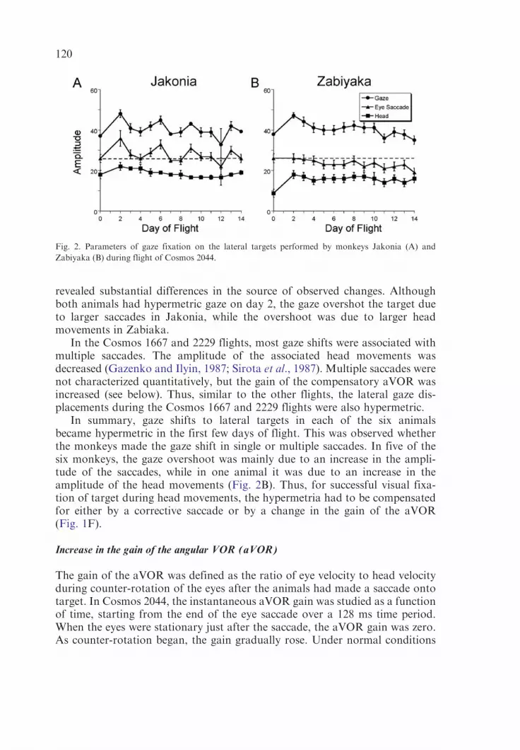
revealed substantial differences in the source of observed changes. Althoughboth animals had hypermetric gaze on day 2, the gaze overshot the target dueto larger saccades in Jakonia, while the overshoot was due to larger headmovements in Zabiaka.
In the Cosmos 1667 and 2229 flights, most gaze shifts were associated withmultiple saccades. The amplitude of the associated head movements wasdecreased (Gazenko and Ilyin, 1987; Sirota et al., 1987). Multiple saccades werenot characterized quantitatively, but the gain of the compensatory aVOR wasincreased (see below). Thus, similar to the other flights, the lateral gaze dis-placements during the Cosmos 1667 and 2229 flights were also hypermetric.
In summary, gaze shifts to lateral targets in each of the six animalsbecame hypermetric in the first few days of flight. This was observed whetherthe monkeys made the gaze shift in single or multiple saccades. In five of thesix monkeys, the gaze overshoot was mainly due to an increase in the ampli-tude of the saccades, while in one animal it was due to an increase in theamplitude of the head movements (Fig. 2B). Thus, for successful visual fixa-tion of target during head movements, the hypermetria had to be compensatedfor either by a corrective saccade or by a change in the gain of the aVOR(Fig. 1F).
Increase in the gain of the angular VOR (aVOR)
The gain of the aVOR was defined as the ratio of eye velocity to head velocityduring counter-rotation of the eyes after the animals had made a saccade ontotarget. In Cosmos 2044, the instantaneous aVOR gain was studied as a functionof time, starting from the end of the eye saccade over a 128 ms time period.When the eyes were stationary just after the saccade, the aVOR gain was zero.As counter-rotation began, the gain gradually rose. Under normal conditions
Fig. 2. Parameters of gaze fixation on the lateral targets performed by monkeys Jakonia (A) and
Zabiyaka (B) during flight of Cosmos 2044.
120

the aVOR gain is 1.0, so the eye velocity is equal to head velocity over thecounter-rotation period.
Nine superimposed gain curves obtained before flight in monkey Zabiakaare shown in Fig. 3A. The aVOR gain increased to unity within the first 30 msafter the saccade and then remained stable while the head was in motion.In most cases, the head movements ceased after 80 ms (Fig. 3E). Consequently,the aVOR gains were calculated as average values over the time interval from32–64 ms after the initial saccade. Average instantaneous gain curves based on30 responses before flight (Fig. 3B) were similar to those in Fig. 3A.
In flight there were substantial changes in aVOR gains. As shown in Fig. 3C,the gain of the aVOR increased to about 1.5 on the first recording of day 2(Fig. 3C) and remained at this level until day 8 (Fig. 3D), when the increasebecame even larger (� 2.0). The gain was still about 1.5 on day 14 (Fig. 3E).Changes in the gains of the aVOR were similar during gaze shifts in eitherdirection and were combined to obtain the average gain changes for thismonkey (Fig. 3F, filled circles). The aVOR gain was also increased when theother animal of this flight, Jakonia, was tested for the first time in space onday 3 (Fig. 3F, filled squares). The gain increase was smaller subsequently, butwas present over the entire flight.
The average gain values obtained from the instantaneous gain curves werecompared to the aVOR gains obtained from taking the ratio of eye and headvelocities at arbitrary points during the counter-rotation. The average aVORgains were the same when measured with either method (Sirota et al., 1991b).Thus, it was possible to compare the aVOR gain measurements in the differentCosmos projects. Individual gain curves for each of the monkeys in this reportare shown in Fig. 3F. There were individual differences, but as a group, therewas a substantial increase in the aVOR gains that persisted over the entire flight(Fig. 3G).
In summary, the general conclusion is that gaze became hypermetric uponentry into space on each of the Cosmos flights and that the positional errorsgenerated by this gaze overshoot were compensated for by an increased gainof the aVOR. Although both of these changes could have occurred indepen-dently, since they occurred in parallel, it is more likely that one was primary,while the other was an adaptive response to the primary change.
The angular acceleration that activates the semicircular canals is the sameon Earth and in space. Accordingly, the gain of the passive aVOR inducedby steps of velocity and by voluntary sinusoidal head oscillation in darknesswere not affected in microgravity (Benson et al., 1986; Cohen et al., 1992;Clarke et al., 2000). If the hypermetric gaze was primarily due to exposure tomicrogravity, then the alteration in aVOR gain was a simple compensationfor the positional error, driven by the visual feedback. However, there arereasons to question this explanation. First, the source of the hypermetric gazevaried among animals. In one, it was due to an increase in the amplitude of headmovements (Fig. 2B), while in the others, it was due to the increase in the
121
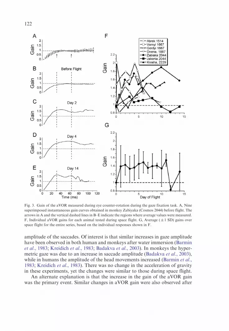
amplitude of the saccades. Of interest is that similar increases in gaze amplitudehave been observed in both human and monkeys after water immersion (Barminet al., 1983; Kreidich et al., 1983; Badakva et al., 2003). In monkeys the hyper-metric gaze was due to an increase in saccade amplitude (Badakva et al., 2003),while in humans the amplitude of the head movements increased (Barmin et al.,1983; Kreidich et al., 1983). There was no change in the acceleration of gravityin these experiments, yet the changes were similar to those during space flight.
An alternate explanation is that the increase in the gain of the aVOR gainwas the primary event. Similar changes in aVOR gain were also observed after
Fig. 3. Gain of the aVOR measured during eye counter-rotation during the gaze fixation task. A, Nine
superimposed instantaneous gain curves obtained in monkey Zabiyaka (Cosmos 2044) before flight. The
arrows in A and the vertical dashed lines in B–E indicate the regions where average values were measured.
F, Individual aVOR gains for each animal tested during space flight. G, Average (� 1 SD) gains over
space flight for the entire series, based on the individual responses shown in F.
122

water immersion (Barmin et al., 1983; Kreidich et al., 1983; Badakva et al.,2003), and as in the Cosmos 1667 flight, the first gain changes were observedafter only two hours of immersion. Since changes in gravity were not a factor inthese experiments, changes in proprioceptive inputs could have contributed toor been responsible for producing the gain changes. If the aVOR gain changeswere the primary even, then parameters of the GFR should be different if theeye saccade was not accompanied by head motion. Kozlovskaya and colleaguesdemonstrated in cosmonauts tested before and after space flight that there aretwo types of the adaptive changes in GFR. One group had changes in theamplitude of the saccades and in aVOR gains, similar to those observed in themonkeys. The second group was comprised of spacecraft commanders, e.g.,professional pilots. They performed gaze shifts in two stages: first by a saccadeonto target and then by a head movement, which followed the saccade. In thepilots, changes in their aVOR gains were similar to those described above. Sincethe amplitude of the saccades during the gaze shift was accurate within � 3�,there was no visual/vestibular mismatch that could have driven the changes inthe aVOR gains (Kozlovskaya et al., 1985).
An alternate mechanism could also have been responsible for the changes inthe gain of the aVOR. It was recently demonstrated that adaptive changes ofthe aVOR gain are a function of head position with regard to gravity (Tiliketet al., 1993; Yakushin et al., 2000a, 2003a,b, 2005). Thus, ‘‘normal’’ aVOR gainsduring active head movements may only exist under the level of gravity and/orproprioception in which the aVOR gains were adapted, and a change in gain ofthe aVOR due to insertion into microgravity could have been the primarychange that produced the changes that were observed in microgravity.
If there were changes in the gain of the aVOR, there should be associatedchanges in the cellular activity in the vestibular nuclei and/or flocculus, whichproduced the adaptive changes in aVOR gain ((Lisberger, 1994; Lisberger et al.,1994a,b), see Ito (1984) for review). Unit activity was recorded in the vestibularnuclei and flocculus during each space flight and is considered in the nextsection.
Neural activity in the vestibular nuclei and flocculus during space flight
Activity of a single lateral canal-related neuron in the right MVN of monkeyVernyi (COSMOS 1667) during sinusoidal head/body oscillation before flight isshown in Fig. 4A. The discharge rate increased in phase with ipsilateral headvelocity (Fig. 4B). Therefore, this cell was a type I unit.1 When this unit was well
1The vertical canals contribute to the neural response to head movement in the yaw plane. Therefore,
vertical canal-related units would also be modulated with this stimulus, and it is possible that some of
the activated neurons were actually more closely related to the vertical canals. Activity of the unit shown
in Fig. 4 was modulated during horizontal but not vertical eye movements, and it is likely that this unit
was a lateral canal-related neuron.
123

isolated from the background, the electrode was fixed at this location. Overtime, this electrode recorded several neurons simultaneously. The multiunitactivity recorded before and after the flight increased with ipsilateral rotationand had a similar phase relationship to head velocity (Fig. 4D) as the neuron inFig. 4A. The electrodes over the various Cosmos flights were implanted inMVNin the region of type I units.
Multiunit activity recorded from three of four of the implanted electrodes inthe two animals in Cosmos 1667 was of good quality. An example of a recordingduring a gaze shift to the right during flight is shown in Fig. 5A. Single neurons,shown as standard pulses, could be separated from the background (Fig. 5B).The quality of separation was similar for each flight day, although it was notcertain that the same neurons were analyzed throughout the flight. Beforeflight, the peak activation occurred approximately at the time when ipsilateralhead velocity reached a maximum (Fig. 5C, Before). The unit activity did not
Fig. 4. Activity of the lateral canal-related type I neuron recorded the right medial vestibular nuclei in
the monkey Vernyi (Cosmos 1667). A, Single unit recording at the site where one of the MVN electrodes
was installed. Head position down is rotation to the right (ipsilateral). B, Poststimulus histogram for the
unit shown in A. Traces below show head position and head velocity during sinusoidal rotation. C–D,
Integrated unit activity recorded from the same electrode before (C) and after (D) seven days of the space
flight. Note that the modulation in unit activity increased peaking just before the ipsilateral (rightward)
head velocity had reached maximum. The head oscillation shown in A–D was driven manually with
approximate frequencies 0.45 Hz (A, B), 0.5 Hz (C) and 0.4 Hz (D).
124

Fig. 5. A, Sample of multiunit activity recorded in the right vestibular nuclei inmonkey Vernyi (Cosmos 1667) during the gaze fixation reaction onto the target
on the right. The recording was obtained from the same electrode at the same location as in Fig. 4. B, Activity of the largest neuron from a sample similar to
that shown in A and converted to standard pulses. C, Poststimulus histograms of this unit recorded at different times in flight. Thirty head movements of
similar profile and amplitude were identified for each day. Headmotion to the right and back to the center was divided into eight bins. An additional eight bins
were used to characterize the data from before and after head motion. The idealized head position and head velocity are shown under each histogram. 125

decrease relative to the resting discharge rate (white dashed line) in associationwith contralateral head velocity. The profile of activation by ipsilateral headvelocity was the same over all flight days. The amplitude of peak activationbefore flight (27� 4 imp s�1) was comparable to the peak activation recordedduring the second hour of space flight (23� 6 imp s�1, day 1). This activationbecame larger on flight days 2–4 (50� 8 imp s�1, day 2) and then decreased tonormal on days 5–6 (25� 3 and 22� 3 imp s�1, respectively). Thus, theactivation was twice the normal value after 26 h in space (day 2). The changeswere substantial over the next 2 days and then normalized. Data from thisneuron are summarized in Fig. 6F (filled symbols). There was no significantchange in the activity recorded during gaze shifts to the right after 2 h of spaceflight (day 1), but the activity gradually increased to a maximum on day 4, andreturned back to normal for the rest of the flight.
The profile of the integrated multiunit activity was similar when multiunitactivity recorded by the same electrode was integrated (Fig. 6A–C). That is,activation before flight (Fig. 6A) was similar to that on day 7 (Fig. 6C), and theincrease in unit activity was larger on day 2 (Fig. 6B). The integrated activityfrom the multiunit recordings over the entire flight is shown in Fig. 6D (opensymbols). Activation on flight day 1 after 2 h was the same as the preflightactivity. This activity significantly increased on flight day 2 and then returned tonormal by days 5–6 (Fig. 6F). The difference between the single and integratedmultiunit recordings was presumably due to variation in response of theneurons in the vicinity of the recording electrode.
The multiunit activity in MVN was also studied in four other animals. Thetwo animals in Cosmos 1887 had two electrodes implanted in MVN (Fig. 6D)and the two animals in Cosmos 2044 flight (Fig. 6E) each had one electrode inMVN. The sensitivities recorded in Cosmos 1887 flight were maximal on day 3for all four electrodes and then decreased to normal in three of the fourelectrodes (Fig. 6E, filled circles). Changes in the sensitivities of the MVNneurons in the monkeys of Cosmos 2044 flight were more robust. Thesensitivities increased significantly on day 3 and stayed high until day 8 whenthey began to normalize. In the animal Jakonia, the sensitivity stayed above thenormal even on day 13 (Fig. 6F, filled symbols).
Multiunit activity was recorded in the flocculus from one electrode in each ofthe same four animals. In all cases the change in the sensitivity was maximal onthe first day of flight (Fig. 6G, H). In two animals, the activity normalized by themiddle of the flight (filled symbols), while in other two, it stayed above the normuntil the last day of flight (Fig. 6G, H, open symbols).
A summary of all multineuronal recordings from the Cosmos flights isshown in Fig. 6I. Overall the sensitivity of the neural activity in both thevestibular nuclei and flocculus was maximal in the earlier days of flight, with therise in activity occurring earlier in the flocculus than the MVN neurons. Therewas a striking similarity between the rise in activity of the flocculus (Fig. 6I,dashed line) and MVN multineuronal activity (Fig. 6I, solid line) and the
126
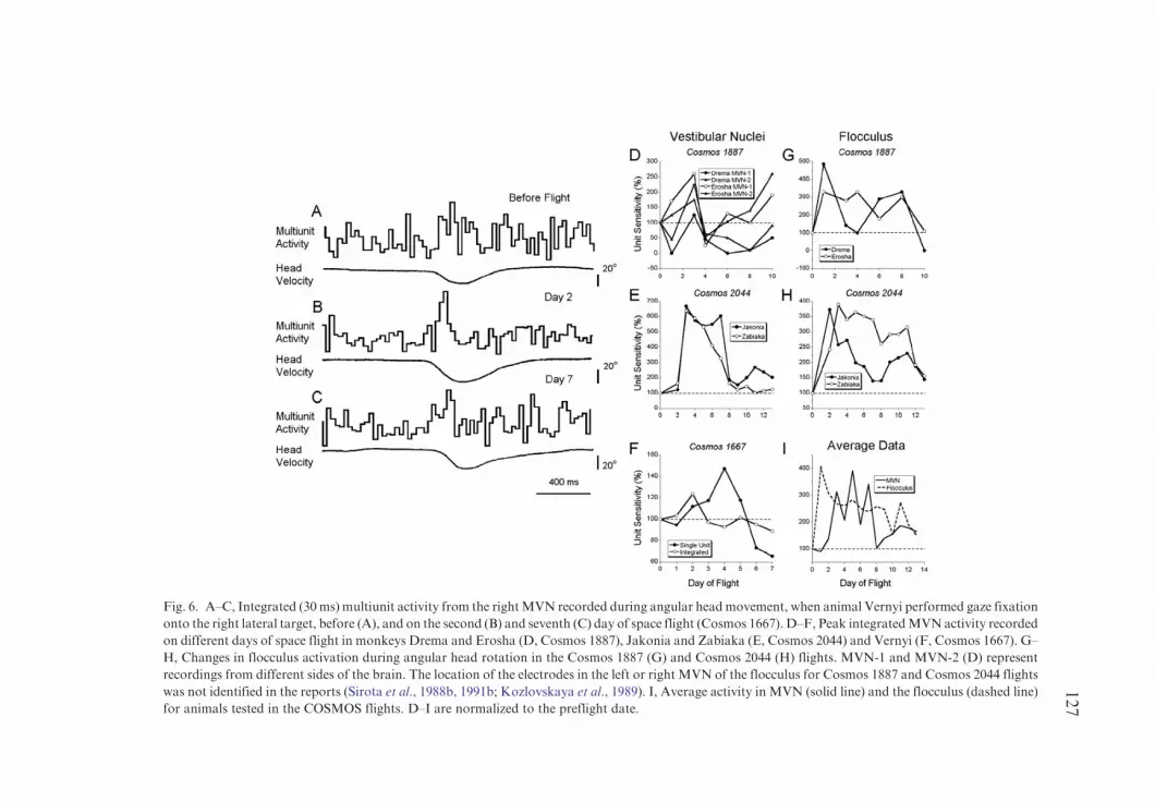
Fig. 6. A–C, Integrated (30ms) multiunit activity from the rightMVN recorded during angular headmovement, when animal Vernyi performed gaze fixation
onto the right lateral target, before (A), and on the second (B) and seventh (C) day of space flight (Cosmos 1667). D–F, Peak integratedMVNactivity recorded
on different days of space flight in monkeys Drema and Erosha (D, Cosmos 1887), Jakonia and Zabiaka (E, Cosmos 2044) and Vernyi (F, Cosmos 1667). G–
H, Changes in flocculus activation during angular head rotation in the Cosmos 1887 (G) and Cosmos 2044 (H) flights. MVN-1 and MVN-2 (D) represent
recordings from different sides of the brain. The location of the electrodes in the left or right MVN of the flocculus for Cosmos 1887 and Cosmos 2044 flights
was not identified in the reports (Sirota et al., 1988b, 1991b; Kozlovskaya et al., 1989). I, Average activity in MVN (solid line) and the flocculus (dashed line)
for animals tested in the COSMOS flights. D–I are normalized to the preflight date.
127

increases in aVOR sensitivity shown in Fig. 3G. The flocculus activity tended torise earlier and persist longer than the MVN activity, but both were increasedover the two weeks of flight. Thus, the changes in neural activity in MVN andflocculus during space flight mirrored the changes in aVOR sensitivity duringthe gaze fixation reaction. Presumably, the flocculus units were involved inthe learning that changed the gain of the aVOR, while the increase in unitsensitivity in MVN to head rotation reflected the actual changes in gain in thecompensatory processes.
Response of MVN units to linear acceleration in space
The first inflight recordings of central vestibular neurons during otolith stimu-lation in the Cosmos 1667 project utilized the same population of neurons in theright MVN of monkey Vernyi that was studied during angular rotation (Figs. 4,5, 6A–C, F). A sample multiunit activity during otolith testing on day 1 is shownin Fig. 7A, and associated single unit activity in Fig. 7B. The stimulus cycle wasdivided into four phases for analysis. The spontaneous discharge rate (Phase 1)was obtained from a 1 s time period before each elevation of the chair. Unitactivity during the elevation (Phase 2) and rapid drop (Drop, Phase 3)were taken as average values.2 Additionally, activity was also utilized over a1 s period immediately after the drop had terminated (Stop, Phase 4). Datawere first expressed in imp s�1 and then normalized to activity in Phase 1 of eachday. The bottom traces (Chair) in Figs. 7A, B, show the time intervals foreach phase.
The resulting histograms for each flight day are shown in Fig. 7C. The firstsignificant changes in the drop phase occurred on the first day after 2 hours ofspace flight. This activity normalized on days 2 and 3, but then increased again,reaching a much higher level (Fig. 7C, D). Sensitivity of the activity for the Dropin Phase 3 for each day is shown in Fig. 7D. The first significant changes wereobserved in the first day of flight. Activity was near normal on flight days 2and 3 and then increased again on days 4–5. The direction of linear accelerationwas opposite during the Drop and the Stop phases, but there was no differencebetween the two responses on any flight day. Since the head was not fixed duringthe Stop, it is not known whether the head continued to pitch at the time of theStop, which could explain this apparent disparity. Therefore, the activityassociated with the Stop Phase will not be considered further in the analysis.
Integrated multiunit activity from the second animal in the same flight(Gordyi) is shown in Fig. 8A–C. Before the flight, there was a brief increase inactivity during the Drop (Fig. 8A). On the second day of flight there was a
2The acceleration in the preflight testing was higher during the rapid drop than inflight because
gravity added a downward pull to that provided by the spring mechanism (see Methods). As described in
Methods, the linear acceleration was only significantly above threshold for detection during the rapid
drop in Phase 3 and the stop-reaction in Phase 4.
128
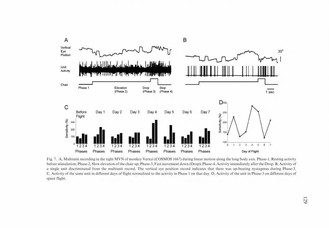
Fig. 7. A, Multiunit recording in the right MVN of monkey Vernyi (COSMOS 1667) during linear motion along the long body axis. Phase-1, Resting activity
before stimulation; Phase-2, Slow elevation of the chair up; Phase-3, Fast movement down (Drop); Phase-4, Activity immediately after theDrop. B, Activity of
a single unit discriminated from the multiunit record. The vertical eye position record indicates that there was up-beating nystagmus during Phase-3.
C, Activity of the same unit in different days of flight normalized to the activity in Phase 1 on that day. D, Activity of the unit in Phase-3 on different days of
space flight.
129

Fig. 8. A–C, Integrated (30 ms) multiunit activity recorded in monkey Gordyi during linear stimulation along the longitudinal body axis recorded before (A)
on the second (B) and seventh (C) day of flight. D–E, Average multiunit activity in MVN in Phase-3 (see Fig. 7) on different days of the Cosmos 1667 (D) and
Cosmos 2044 (E) flights. Multiunit activity in Phase-3 normalized to the activity observed before stimulation in Cosmos 1667 (D) and to the level of activation
in Phase-3 recorded before flight in Cosmos 2044 experiment (E). Note thatmultiunit activity recorded in the rightMVNofmonkey Vernyi was notmodulated
in this test, but the single unit that was selected from this record (Fig. 7) was well modulated by the linear acceleration.
130

significant increase in activity during the Drop (Fig. 8B). The activity increasedin response to the acceleration phase and then decreased to normal during thechair deceleration. The activity during the entire period on day 7 was moreirregular than the other days (Fig. 8C), and the response to the Drop was againbrief. Integrated activity recorded from both animals in the Cosmos 1667 flightis shown in Fig. 8D. The sensitivity of these MVN units was not changed onday 1, but was increased in two of the three electrodes on day 2. The activityincreased further up to day 5, and then normalized.
Changes in the sensitivities of the MVN neurons in the monkeys Jakonia(Fig. 8E, open circles) and Zabiaka (Fig. 8E, open squares) from Cosmos2044 were similar to those shown in Fig. 8D. The sensitivities graduallyincreased reaching maximal values on day 6 and then declined, although theywere still above the baseline on the last day of flight (day 13). Thus, changesin the sensitivity of the MVN neurons to vertical linear acceleration mediatedby the otoliths were similar in animals from the Cosmos 1667 and Cosmos2044 flights. The sensitivity to linear acceleration gradually increased in space,reaching a maximum at days 5–6 and then decreasing toward normal values.The peak increases in sensitivity for the MVN neurons in Fig. 6D, E inresponse to angular acceleration during the gaze fixation reaction werebetween 300 and 600%, whereas the increases in sensitivity during linearacceleration were smaller, within 200–250%. Thus, the general pattern ofresponse was similar to that obtained from angular acceleration, but themagnitude of the response to linear acceleration was smaller. This couldrepresent a difference in response to angular and linear accelerations or couldreflect a difference in the magnitude of convergence of the otolith and canalsignals onto these neurons.
The responses to linear acceleration produced by sinusoidal oscillationalong the body vertical axis in the Bion 11 Project were very similar to thoseobserved in the Bion 9 (Cosmos 2044) flight. The multiunit activity of theneurons recorded in MVN in each animal was in phase with upward accele-ration in monkey #484 and with downward acceleration in monkey #357(Badakva et al., 2000). In both cases, the sensitivity to linear accelerationincreased only slightly on the first recording on Day 2, and reached a maximumon Days 4–6. Sensitivities then decreased but were still above normal up to thelast day of flight.
Discussion
The data from the Cosmos flights show that the sensitivity of central vestibularneurons in MVN to both angular head rotation and vertical linear motion wasaffected by microgravity. In general, the sensitivity of these neurons increased inthe earlier days in Space and then gradually normalized. In some instanceschanges were similar for several populations of neurons recorded on both sidesof the brainstem from the same animal (Fig. 6D, E). In other cases, although
131
average activity of the recorded neurons had similar changes in sensitivityduring adaptation to microgravity (Fig. 6F, open symbols), the changes insensitivity of a single unit could differ from the changes observed in theremaining population of units. A striking finding was that the observed changesin neuronal sensitivity were qualitatively similar to the changes observed in theaVOR gains and occurred over the same time course. Changes in sensitivity ofthe floccular neurons to the angular rotation were more uniform than thechanges in MVN activity, presumably, because the flocculus was responsible forproducing these adaptive changes.
Changes in sensitivity of the neuronal populations in the vestibular nuclei tothe otolith stimulation were also relatively uniform and had approximately thesame time course as the sensitivity to angular acceleration. The sensitivityto linear acceleration gradually increased, reaching a maximum by the end ofthe first week in space (Fig. 7D and Fig. 8D, E). This was different from thechanges in sensitivity of the same neuronal population to angular rotation,which increased earlier in flight and was normalized by the time the otolithsensitivity had reached a maximum (Fig. 6I). This was somewhat surprising,since it would be expected that microgravity would affect otolith and notcanal sensitivity. A majority of direct projections from otolith afferents go to thelateral and descending vestibular nuclei (Buettner-Ennever, 1999), but there arecanal-sensitive neurons with some otolith sensitivity in MVN (Markhamand Curthoys, 1972), which could serve a different purpose than maintainingbalance or supporting the linear VOR. Otolith information is not necessary toinduce changes in the angular VOR gain (Crane and Demer, 1999). As recentlyshown, however, the head position with respect to gravity in which the aVORgain was adapted is expressed subsequently in changes in aVOR gain inevery head position (Yakushin et al., 2000a, 2003a,b, 2005). Therefore, theotolith signals could serve as a gravitational context for adaptation of theaVOR gain in specific head positions, and the changes in otolith sensitivitycould be a reflection of changes in this gravitational context.
SECTION 3: NASA–RUSSIAN MONKEY EXPERIMENTS ON THE
ANGULAR AND LINEAR VESTIBULO-OCULAR REFLEX
Abstract
The angular and linear vestibulo-ocular reflexes (aVOR and lVOR) of fourrhesus monkeys were recorded before and after the 1988 and 1992–1993 CosmosSpace Flights 2044 and 2229 (Table 1). Two animals flew in each mission forapproximately two weeks. Eye movements, induced by rotation with stepsof velocity about a vertical axis, by constant velocity rotation about axestilted from the vertical (off-vertical axis rotation, OVAR), and by horizontaland vertical translation were recorded binocularly with scleral search coils in
132

two- and three-dimensions. Single unit recordings were also taken fromsemicircular canal afferents before and after flight.
Compensatory eye movements produced by the angular VOR (aVOR). Gainsof semicircular canal-induced horizontal and vertical aVOR were unaffected inboth flights, although the gain of the roll aVOR was diminished. Up/downasymmetries of vertical nystagmus present before flight were reduced for sevendays after flight.
Activity of primary lateral canal afferents after space flight. The mean gain fornine different horizontal canal afferents, tested on the first postflight dayof Cosmos 2044 with steps of velocity and sinusoidal rotation, was nearlytwice that of 20 horizontal canal afferents similarly tested during preflight andpostflight control studies. Adaptation of the afferent response to passive yawrotation on the first postflight day was also greater. After the Cosmos 2229,however, afferent gains were reduced.
Spatial orientation of the aVOR. Spatial orientation of the aVOR was alteredin two of the four monkeys after flight. In one, the time constants ofpostrotatory nystagmus, which had been shortened by head tilts with regardto gravity before flight (‘‘tilt dumping’’), was unaffected by the same head tiltsafter flight. In another animal, eye velocity, which tended to align with gravitybefore flight, moved closer toward a body axis after flight. This shift of orien-tation had disappeared by seven days after landing.
Compensatory eye movements produced by the linear VOR (lVOR). The gainof the high frequency compensatory lVOR was reduced for naso-occipital linearacceleration in one monkey, but maintained in a second monkey. Gain changesin the first animal lasted for 17 days after landing.
Orienting eye movements produced by the lVOR. The gain of the lowfrequency lVOR was tested using OVAR. Ocular counter-rolling (OCR) wasreduced by about 70% during both the dynamic tilts experienced during OVARand in response to static tilts. Similarly, modulation in vergence, in response tolow frequency, naso-occipital linear acceleration during OVAR was reducedby over 50%. These changes in orienting eye reflexes persisted for 11 days afterrecovery.
Orientation of eye velocity induced by velocity storage. Steady state yawaxis horizontal eye velocities induced by OVAR were unaffected by space flight.The orientation of optokinetic after nystagmus (OKAN) and of vestibularnystagmus was altered, moving closer to a body than a spatial axis when testedshortly after landing in one animal.
Conclusion. There were both short and long term changes in otolith-ocularreflexes after adaptation to microgravity in both monkeys in the Cosmos 2229flight, although horizontal and vertical semicircular canal-induced responses ofthe angular VOR to rotation were largely unaffected. The roll aVOR gain wasalso reduced. All of the reductions were greater in one animal (7906) acrossall tests. A comparison with data from astronauts suggests that maintenance ofgain of both compensatory and orienting otolith ocular reflexes may depend on
133
continuous exposure to linear acceleration during flight. Presumably, in futurelong duration space flights, this could be provided by centrifugation.
Introduction
The vestibular system is composed of two subsystems. One, comprised of thesemicircular canals, senses angular acceleration of the head and generatescompensatory eye movements that stabilize gaze during head and body move-ment over the angular vestibulo-ocular reflex (aVOR) (see Raphan and Cohen,2002; Cohen and Gizzi, 2003; Cohen and Raphan, 2004 for review). Since thenatural angular motion that excites the aVOR is essentially turning on a head-or body-centric axis, the horizontal aVOR would not be expected to changedramatically in a different gravitational environment. The second component ofthe vestibular system, however, the otolith organs, comprised of the sacculesand utricles, sense both head orientation and head linear translation. Thealtered acceleration profiles experienced in microgravity should induce verydifferent activation profiles in otolith afferents that could initiate morphologicaland behavioral adaptation of the vestibulo-ocular and vestibulo-spinal reflexesthat depend on gravity. Thus, it would be expected that there might be changesin ocular counter-roll (OCR), vergence and spatial orientation after adaptationto microgravity but that the angular VOR would be less affected.
The linear vestibulo-ocular reflex (lVOR) can be further separated into highand low frequency components, using approximately 0.3 Hz as the dividebetween the two. The compensatory reflex provides ocular compensationagainst high frequency head translations (Schwarz et al., 1989; Hess andDieringer, 1991; Paige and Tomko, 1991a,b; Schwarz and Miles, 1991; Raphanand Cohen, 2002), and is used to maintain fixation on near targets duringtranslation (Paige and Tomko, 1991a; Schwarz and Miles, 1991; Maruta et al.,2001) or in response to centripetal acceleration generated by turning corners(Imai et al., 2001). Vergence in response to high frequency linear accelerationalong the naso-occipital axis is also compensatory, and supports fixation ofnear targets when moving forward (Paige, 1991; Dai et al., 1996). There arealso low frequency orienting otolith-ocular responses that tend to maintain theposition of the retina in relation to the spatial vertical (Cohen et al., 2001). Theseinclude horizontal, vertical and torsional shifts of the eyes, OCR, and sustainedvergence in response to head tilts with regard to gravity (see Dai et al., 1996 forreview). Finally, angular eye velocity induced through activation of a centralvestibular system known as ‘‘velocity storage’’ by the visual, vestibular and/orsomatosensory systems, tends to orient to gravity or to the GIA when the GIAis tilted with respect to the head (Dai et al., 1991, 1992).
In the pre- and postflight experiments described in this section, scleralsearch coil measurements of eye movements in response to controlled vestibularand visual stimulation were utilized in pre- and postflight experiments. Thepurpose was to demonstrate the effects of adaptation to microgravity on return
134

to the 1 g environment of Earth. Experiments were also done on semicircularcanal afferents to determine if the exposure to microgravity had significantlychanged afferent activity after adaptation to microgravity.
Methods
Nineteen juvenile rhesus monkeys (Macaca mulatta) were candidates for the twoCosmos Biosatellite Flights. Of these, four animals were chosen for flight; theothers served as controls. Monkeys 782 and 2483 flew in Cosmos Flight 2044.They were launched on 9/15/89 and recovered on 9/29/89 (Table 1). Animals6151 and 7906 flew in Cosmos Flight 2229. These animals were launched on 12/29/92 and recovered on 1/10/93. Testing extended for 5 and 11 days postflight,respectively. In space, the monkeys sat in a capsule, 60 cm in diameter, infur-lined chairs. The compartments that held the primate chairs were separatedfrom each other, but the animals were within sight of one another. Their trunkswere restrained, but their heads, arms and legs were free. The monkeys per-formed behavioral testing in space, moving their head and eyes toward lateralvisual targets (see Section 2). The status of the animals was monitored by down-linked video, along with continuous recording of a wide range of other controlsignals from the capsule, including temperature, humidity, and food and waterintake. Monkeys not chosen to fly were housed in comparable quarters onground and served as controls. The experiments conformed to the Principles ofLaboratory Animal Care (NIH Publication 85-23, Revised 1985), and wereapproved by the appropriate Institutional Animal Care and Use Committees.
Pre- and postflight testing
During experiments, the monkeys sat in a primate chair with their heads fixedto a plastic frame that held a square field coil, 25.4 cm on a side. The sameprimate chair and coil box was used in studies of high frequency linearoscillation. Yaw (horizontal), pitch (vertical) and roll (torsional) eye movementswere recorded through two search coils, which were attached to the front andthe top of the left eye. A frontal plane coil was also implanted on the right eyeof the Cosmos 2229 monkeys. Voltages associated with eye position and withthe position and velocity of the various axes were recorded through analogfilters with a bandwidth of DC to 40 Hz. Eye positions and velocities werecalibrated by assuming that the animals accurately tracked visual surroundmovement during rotation in light at 30�/s. Movements to the right and upcaused upward trace deflections. Roll velocities were assumed to have a gain of0.6. Eye movements were not recorded in the studies of afferent activity in thevestibular nerve.
Two multi-axis vestibular stimulators and three vestibular/oculomotorlaboratories were transported from the United States to the Institute ofBiomedical Problems in Moscow for these experiments. The apparatus and
135

experiments are fully described in previous publications and reports (Cohenet al., 1992; Correia et al., 1992; Tomko et al., 1993; Dai et al., 1994, 1996).In brief, the apparatus shown in Fig. 9A was used to study the angular VOR(aVOR), optokinetic nystagmus (OKN), optokinetic after-nystagmus (OKAN),and the response to off-vertical axis rotation (OVAR). The response to angularacceleration was given by rotating the animals positioned in separate tests sothat their yaw, pitch and roll axes were aligned with the spatial vertical.Optokinetic nystagmus (OKN) was induced by rotating the light-tight OKNshell around the animal’s yaw axis with the yaw axis upright or in tiltedpositions. Off-vertical axis rotation (OVAR) was given by rotating the animalsaround a tilted yaw axis. The response to static tilts was tested by incrementallyrotating the circular spine around the horizontal axis.
For tests of high frequency linear acceleration, the animals were oscillatedon a linear sled, specially constructed at the NASA-Ames Research Center(Fig. 9B). Animals sat in a Specimen Test Container and moved along alinear track in Earth-horizontal (Fig. 9Ba), oblique (9Bb) or dorsoventral(9Bc) directions. In Ba, the animals were upright and in Bc they were eitherprone or supine. Motion was delivered along the interaural, naso-occipital,and dorsoventral axes as well as along intermediate oblique axes. lVORs were
Fig. 9. A, Four axis vestibular and optokinetic stimulator for testing the aVOR, and for inducing OKN
and OKAN and OVAR. Also shown are the primate chair and coil field apparatus used to activate the
scleral search coils when recording eye position and eye movement. The same coil box and primate chair
was also utilized in the linear oscillation apparatus. B, Portable linear sled for high frequency linear
oscillation. A gimbaled, light-tight Specimen Test Container sat on a carriage supported by air bearings
and ‘‘floated’’ on ceramic rails. The linear track was situated on a gantry that could be repositioned to give
translation along earth-horizontal (Ba), oblique (Bb), or earth-vertical (Bc) axes. Stimuli were given in
darkness, in a subject-stationary lighted visual surround (VSLVOR), or while viewing the visual sur-
round that could move relative to the subject (VLVOR).
136

studied during 1.0 and 5.0 Hz Earth-horizontal head motion in darkness andwhile viewing a head-fixed (VSLVOR) or an earth-fixed (VLVOR) visual scene.
Afferent neural responses from the horizontal semicircular canals weretested by rotating the animals sinusoidally or at a constant velocity about aspatial vertical axis (not shown). To test potential sensitivity of the canalunits to tilt before and after flight, the animals were also pitched along thenaso-occipital axis.
Results
Compensatory eye movements from the angular VOR (aVOR), horizontal aVOR
For studying the gain of the aVOR, monkeys were tested with short (5 s) steps ofconstant velocity rotation from 30�/s to 180�/s in 30�/s intervals in darkness(Fig. 10A). For studying the time constant of the aVOR, monkeys were rotatedat 60� /s in darkness to produce per-rotatory nystagmus. When the nystagmusdied away, the animals were stopped to produce postrotatory nystagmus(Fig. 10A, B). The animals were subsequently rotated in the opposite direc-tion and stopped. The slow phase eye velocities of the two per- and twopostrotatory nystagmus profiles were fitted by single exponentials to extract thecentral vestibular time constants, and were averaged to generate a mean timeconstant. The gains (eye velocity/head velocity) of the response to short stepsmeasured the status of the high frequency aVOR, and the time constants gavethe state of the central velocity storage mechanism, which provides the lowfrequency characteristics of the aVOR and counters postrotatory nystagmus(see Raphan and Cohen, 2002 for review).
The induced horizontal aVOR slow phase velocities were close to stimulusvelocities up to 120�/s for control monkeys, and fell slightly for stimulusvelocities of 150�/s and 180�/s (Fig. 10B, C). Linear, least square regressionswere used to characterize each set of pre- and postflight data. Slopes, interceptsand correlation coefficients were similar for the pre- and postflight data for both782 (Fig. 10A) and for 2483 (Fig. 10B), and the data were not significantlydifferent from the group means of the control pool. During the Cosmos 2229Flight, horizontal aVOR gains were pooled for rotation to the right and left,since there was no difference between them. The preflight and postflight gainswere the same (Dai et al., 1994). Similarly, horizontal aVOR time constantswere similar before and after flight. The mean aVOR time constant of 782 onthree days of testing, about two months before flight was 22.6� 7.9 s. Pooledpostflight day 1 and day 4 time constants were 22.6� 9.2 s and 18.8� 2.1 s.These means did not significantly differ from each other. Though notsignificant, the reduction in time constant on postflight day 4 was likely dueto habituation due to repeated testing. These findings confirm findings inhumans in a NASA–ESA Spacelab Flight that indicated that 14 days ofexposure to microgravity had not affected the aVOR gain (Benson and Vieville,
137

1986). Similarly, the time constant of the horizontal aVOR was unaffected in theNASA IML Shuttle Flight in 1992 (Oman and Balkwill, 1993).
Vertical and ROLL aVOR
Both flight and control monkeys in the Cosmos 2229 mission had an asymmetryof the vertical aVOR before flight when they were rotated on their side in pitcharound a vertical axis (Fig. 11A). This stimulus only activates the vertical canalsin contrast to pitch about a horizontal interaural axis while upright, whichexcites both the vertical canals and otolith organs. The mean gain for upwardslow phases of vertical eye velocity before flight was 0.96� 0.03, and the mean
A
B C
Fig. 10. A, Horizontal eye position (H POS) and eye velocity (H VEL) in response to 5 s steps of velo-
city (rotation about a vertical axis) from monkey 2483 after recovery in the Cosmos 2044 flight.
Eye positions and eye velocities to the right are up in this and subsequent figures. The changes in yaw
position of the animal, which were recorded by a potentiometer that reset every 360�, are shown in the
third (YAW POS) trace. Downward movement of the trace is rotation to the left. The rotations were
in darkness. The surround was lighted (PHOTO CELL) between velocity steps to extinguish any
nystagmus that remained after the rotations. B, C, Horizontal slow phase velocities induced by steps of
velocity as in A. The induced velocities were close to the velocity of rotation up to 180�/s and were not
different before and after flight.
138

downward gain was 0.75� 0.04 over a velocity range of 30 to 90�/s (Dai et al.,1994). In the first postflight test, there was a 7.3% decrease in the gain of theupward aVOR to 0.90� 0.03, and a 7% increase in the gain of the downwardaVOR to 0.82� 0.03 (Fig. 3A). These differences were small but statisticallysignificant. Thus, the increase in the gain of the downward aVOR was at theexpense of a decrease in gain for the upward aVOR.
By 7 days after landing, the upward aVOR gains had begun to return topreflight levels, and there was no significant difference between pre- andpostflight values in both monkeys. The downward aVOR gain dropped to itsoriginal level in 6151, but not in 7906. There was no difference in the pitch
Fig. 11. A, Averaged gains of the vertical VOR before and after Cosmos flight 2229 for monkeys 6151
and 7906. The animals were rotated about a vertical axis with the left ear down in steps of velocity from
30 to 90�/s in 15�/s increments (accelerations >200�/s2). Postflight data were taken one day after flight
for 7906 and 3 days after flight for 6151. Before flight, the upward VOR (PreUP) gain was higher, and the
gain dropped after flight (PostUp). The downward VOR had a lower gain before flight (PreDown) that
was increased after flight (PostDown). Accordingly, the rather striking preflight Up/Down asymmetry
of the vertical VOR was decreased after space flight. B, C, Gains of the roll VOR induced by velocity
steps between 30 and 90�/s (B), and by sinusoidal oscillation at frequencies between 0.02–0.13 Hz (C).
Animals were rotated about a vertical axis in the prone position. Means for preflight testing (� 2 SD) are
shown by the heavy solid lines (PRE-MEAN). There was a decrease in roll VOR gains for both 6151 (open
symbols) and 7906 (filled symbols) after flight. The decrease was greater for 7906 (50%; filled symbols)
than for 6151; open symbols).
139

aVOR gain before and after flight if gains for upward slow phases anddownward slow phases were averaged. The pitch aVOR was also measured byrotating the animals sinusoidally at 0.1 Hz, peak velocity 60�/s, about aninteraural horizontal axis. This stimulus activates both the vertical canals andthe otolith organs. The vertical gain was slightly reduced after landing (6% for7906 and 9% for 6151), and there was a reduction in the offset of mean velo-city in the upward direction that had been present before flight. By the 11th day,the vertical gains had returned so that they were the same or slightly less thanthe preflight levels for both monkeys. Thus, there was a slight reduction inthe gains of the vertical aVOR with otolith activation measured with sinusoids1–3 days after landing, which had largely returned to the preflight level by the11th day after recovery. Since the overall vertical gains that were the averages ofthe up- and down-responses, measured with steps, were unaffected by space-flight, it is likely that the difference noted in the sinusoidal analysis was due toa change in the otolith contribution to the reflex.
The postflight upward and downward time constants in 782 were 21.3� 2.9 sand 10.1� 2.4 s, respectively, in response to rotation about a vertical axis whilelying on their sides. This is consistent with the vertical time constants in othernormal monkeys (Matsuo et al., 1979; Matsuo and Cohen, 1984). Thus, thepostflight change in vertical spontaneous nystagmus was not reflected in achange in the dominant time constants of the vertical aVOR.
The roll aVOR was measured in two experimental paradigms: Velocity stepswere given with the monkeys in a prone position by rotating them about avertical naso-occipital axis (Fig. 11B), and the monkeys were sinusoidallyoscillated about a horizontal axis while upright (Fig. 11C). Both inducetorsional nystagmus, but the first paradigm elicits pure activation of the verticalcanals, whereas the second activates both the vertical canals and the otolithorgans. The gain of the roll aVOR was reduced in both modes of stimulation,on average by 50% in 7906 and by 15% in 6151 (Fig. 11B, C). This implies thatthere had been adaptation in central pathways for torsion for activityoriginating in both the vertical canals and otolith organs during flight, as inhumans during 1992 MIR mission (Clarke et al., 1993, 2000).
Spontaneous vertical nystagmus
There was upward spontaneous nystagmus of about 5�/s in both monkeysbefore the second Cosmos flight, which is common in normal rhesus monkeys.This spontaneous nystagmus was reduced when the animals were first tested,three days after reentry.
High frequency linear vestibulo-ocular reflex
During interaural translation (5 Hz, 0.5 g) postflight, 7906 had an approximate2/3 reduction in the slope of the function relating lVOR sensitivity to vergence
140

after flight that had not recovered by 16 h after recovery (R+16 h; Figs. 12,13A). Under the same conditions, 6151 had almost identical responses pre- andpostflight (not shown). During dorsoventral translation (5 Hz, 0.5 g) 7906had from 35–60% reduction in the slope of the function relating the verticallVOR sensitivity to vergence that had not recovered by R+16 h (Fig. 13B).Under the same conditions, 6151 had responses immediately postflight thatwere almost identical to the preflight values. Pre- and postflight responsesduring naso-occipital motion were similar to one another for 6151, but theresponses of 7906 were smaller and more variable postflight. This was also thecase for motion along oblique axes between naso-occipital and dorsoventral.
Low frequency lVOR – off-vertical axis rotation (OVAR)
In preflight testing, steady state eye velocity during OVAR increased as afunction of stimulus velocity and of tilt angle for the eight control monkeys,
Fig. 12. Five-second sample of 5 Hz, 0.5 g peak inter-aural oscillation (ACC) in monkey 6151. In the first
traces, ACC is the oscillation of the sled, and HE 1 & HE 2 are right and left horizontal eye position,
respectively. In the center traces, HE 1 VSM and VE 1 VSM are right eye horizontal and vertical eye
velocity. In the third trace, TE 1 and TE 1 VSM are torsional eye position and eye velocity and VER is
vergence position. Note that when vergence was larger, at the left side of the third trace, the oscillations
in horizontal eye velocity (large sinusoidal oscillations, middle traces) were larger than when vergence
decreased toward the right side of the recording. Vergence had no effect on vertical eye velocity (middle
traces). There was a small effect of vergence on torsional eye velocity (third traces).
141
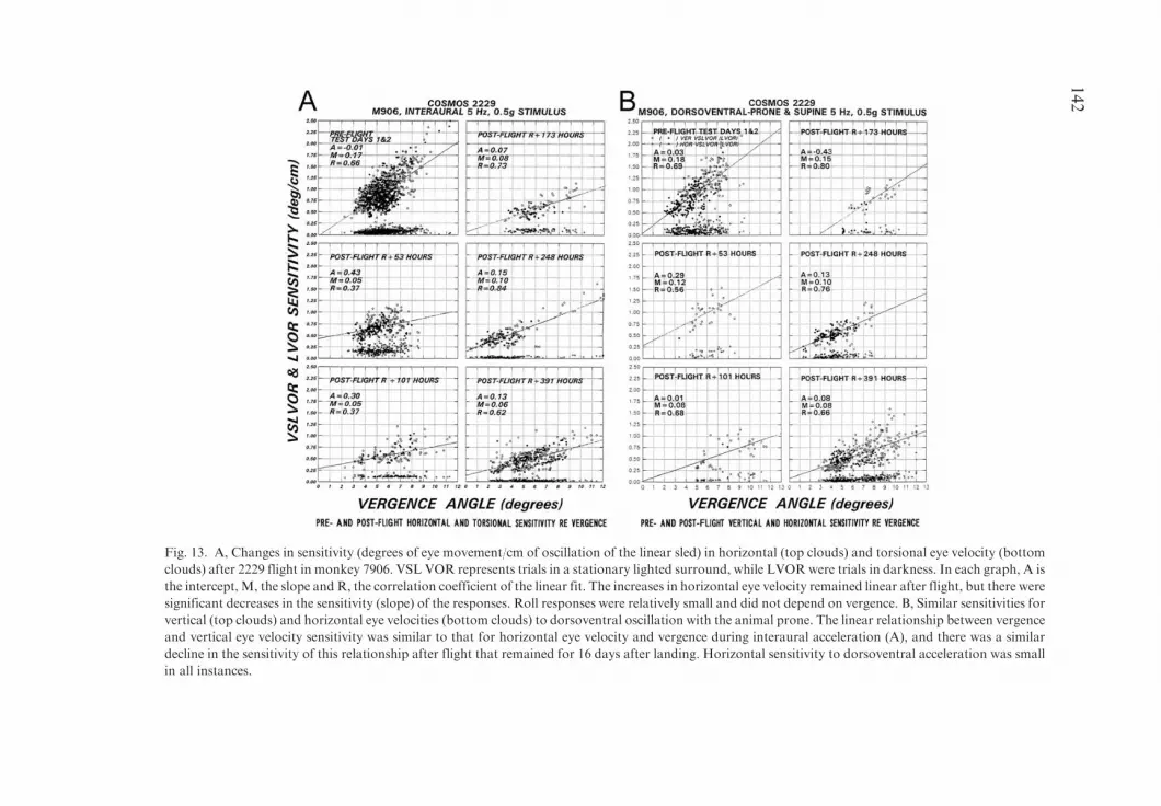
Fig. 13. A, Changes in sensitivity (degrees of eye movement/cm of oscillation of the linear sled) in horizontal (top clouds) and torsional eye velocity (bottom
clouds) after 2229 flight in monkey 7906. VSL VOR represents trials in a stationary lighted surround, while LVORwere trials in darkness. In each graph, A is
the intercept,M, the slope and R, the correlation coefficient of the linear fit. The increases in horizontal eye velocity remained linear after flight, but there were
significant decreases in the sensitivity (slope) of the responses. Roll responses were relatively small and did not depend on vergence. B, Similar sensitivities for
vertical (top clouds) and horizontal eye velocities (bottom clouds) to dorsoventral oscillation with the animal prone. The linear relationship between vergence
and vertical eye velocity sensitivity was similar to that for horizontal eye velocity and vergence during interaural acceleration (A), and there was a similar
decline in the sensitivity of this relationship after flight that remained for 16 days after landing. Horizontal sensitivity to dorsoventral acceleration was small
in all instances.
142

saturating at about 45�/s (Fig. 14A). Steady state horizontal eye velocitiesinduced by OVAR were the same after flight in the monkeys from both the2044 flight and from the 2229 flight (Fig. 14B), and the phases of the modu-lations in horizontal slow phase eye velocity were also approximately the samebefore and after flight. There was an approximate doubling of the amplitude ofmodulation of horizontal slow phase velocity after the first flight, but notafter the second flight. Thus, the animals appeared to be able to sense gravitythrough the otolith system normally after space-flight and to generate the samesteady state level of horizontal slow phase eye velocity after, as before, flight.This implies that the neural mechanism for generating horizontal eye move-ments had not been substantially altered by adaptation to microgravity.
Ocular counter-rolling
In microgravity, there is no otolith-induced compensatory torsion of the eyes(ocular counter-rolling, OCR) in response to sustained head on body tilt (Clarkeet al., 1993, 2000). OCR, elicited by linear acceleration along the interaural axisand measured by an after-image method, was reduced in two cosmonauts for upto 14 days after landing (Yakovleva et al., 1982), and recovered only at the nexttest point of 36 days. There was also anti-compensatory torsion (‘‘paradoxicalcounter-rolling’’) in the direction of head tilt in some subjects after long-duration missions (Clarke et al., 2000). In the Spacelab-1 Mission, OCR wasmeasured in four subjects using a photographic technique (Young et al., 1981).Expressed as a gain ratio, OCR in humans was reduced to one side by 28 to 56%one day after landing. OCR, measured by an after-image technique wasreduced by 57% for five days in one astronaut after the 1992 Russian–GermanMIR Space Mission (Hofstetter-Degen et al., 1993). The extensive literature isreviewed in Dai et al., 1994; Clarke et al., 2000; Moore et al., 2001. Thus, therehas been fragmentary evidence to indicate that there may be a reduction in themagnitude of OCR. On the other hand, there was no change in OCR inastronauts who flew in the 1998 Neurolab Mission (STS-90) (Moore et al.,2001). The results, however, were quite different for the monkeys that flew onCosmos 2229.
Dynamic OCR was assessed using OVAR. The OCR elicited by OVARbefore and after space flight is shown in Fig. 14A (3rd trace) and Fig. 14B(1st trace). In both instances, OCR was induced by the response to a projectionof the gravity vector that rotated relative to the head along the coronal plane.The maximum torsion was induced during OVAR when the gravity vector wasaligned with the interaural axis. Responses of monkeys 6151 and 7906 beforeand after flight for all angles of tilt of the axis of rotation are summarized inFigs. 14C, D. After flight, the mean magnitude of OCR in the flight monkeyswas 2.2� 0.7� for tilt angles between 60 and 90�. This can be contrasted to the6.3� 0.7� of ocular torsion when the axis of rotation was tilted 60 to 90� in thecontrol monkeys. The mean reduction in dynamic OCR after flight was about
143

Fig. 14. A, B, Nystagmus induced by off-vertical axis rotation (OVAR) in darkness at 60�/s about tilted axes before (A) and after (B) COSMOS 2229 space
flight for monkey 6151. In both A and B the axis of rotation (A, top trace) was tilted from 0-90� in the dark in 15� increments inducing OVAR nystagmus. The
position of the animal about the yaw axis, recorded with a potentiometer that reset each 360�, is shown in the second trace (YAWPOS). Upward spontaneous
nystagmus was reduced after flight (VER VEL). The amplitude of the horizontal slow phase velocity (HOR VEL) was unchanged after flight, but the
modulation in slow phase velocity before flight was increased. There was a prominent modulation in roll eye position (ROLL POS) before flight in response
to the lateral tilts during OVAR (A, 3rd trace). After flight the modulation in roll position was decreased (B, 1st trace). C, D, Ocular Counter-Roll (OCR)
induced during OVAR by tilts of the axis of rotation from 15 to 90� in 6151 (C) and 7906 (D), before (PRE-MEAN) and after flight (days 1, 7, 11). E,
Comparison of postflight OCR in the two flight monkeys and six control monkeys and in the two flight monkeys. As above, PRE-MEAN is the mean of the
tests before flight; D1–D11 indicate the tests done on recovery days 1 to 11. The OCR of 6151 and 7906 fell more than 2 D from the preflight means.
144
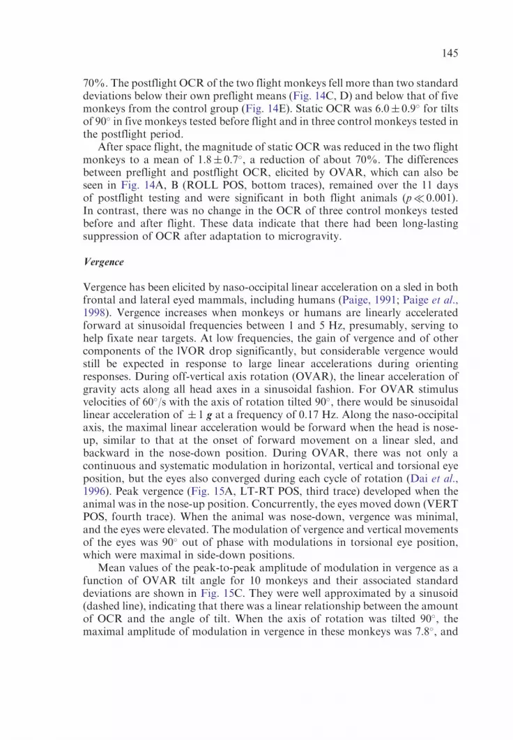
70%. The postflight OCR of the two flight monkeys fell more than two standarddeviations below their own preflight means (Fig. 14C, D) and below that of fivemonkeys from the control group (Fig. 14E). Static OCR was 6.0� 0.9� for tiltsof 90� in five monkeys tested before flight and in three control monkeys tested inthe postflight period.
After space flight, the magnitude of static OCR was reduced in the two flightmonkeys to a mean of 1.8� 0.7�, a reduction of about 70%. The differencesbetween preflight and postflight OCR, elicited by OVAR, which can also beseen in Fig. 14A, B (ROLL POS, bottom traces), remained over the 11 daysof postflight testing and were significant in both flight animals (p� 0.001).In contrast, there was no change in the OCR of three control monkeys testedbefore and after flight. These data indicate that there had been long-lastingsuppression of OCR after adaptation to microgravity.
Vergence
Vergence has been elicited by naso-occipital linear acceleration on a sled in bothfrontal and lateral eyed mammals, including humans (Paige, 1991; Paige et al.,1998). Vergence increases when monkeys or humans are linearly acceleratedforward at sinusoidal frequencies between 1 and 5 Hz, presumably, serving tohelp fixate near targets. At low frequencies, the gain of vergence and of othercomponents of the lVOR drop significantly, but considerable vergence wouldstill be expected in response to large linear accelerations during orientingresponses. During off-vertical axis rotation (OVAR), the linear acceleration ofgravity acts along all head axes in a sinusoidal fashion. For OVAR stimulusvelocities of 60�/s with the axis of rotation tilted 90�, there would be sinusoidallinear acceleration of � 1 g at a frequency of 0.17 Hz. Along the naso-occipitalaxis, the maximal linear acceleration would be forward when the head is nose-up, similar to that at the onset of forward movement on a linear sled, andbackward in the nose-down position. During OVAR, there was not only acontinuous and systematic modulation in horizontal, vertical and torsional eyeposition, but the eyes also converged during each cycle of rotation (Dai et al.,1996). Peak vergence (Fig. 15A, LT-RT POS, third trace) developed when theanimal was in the nose-up position. Concurrently, the eyes moved down (VERTPOS, fourth trace). When the animal was nose-down, vergence was minimal,and the eyes were elevated. The modulation of vergence and vertical movementsof the eyes was 90� out of phase with modulations in torsional eye position,which were maximal in side-down positions.
Mean values of the peak-to-peak amplitude of modulation in vergence as afunction of OVAR tilt angle for 10 monkeys and their associated standarddeviations are shown in Fig. 15C. They were well approximated by a sinusoid(dashed line), indicating that there was a linear relationship between the amountof OCR and the angle of tilt. When the axis of rotation was tilted 90�, themaximal amplitude of modulation in vergence in these monkeys was 7.8�, and
145
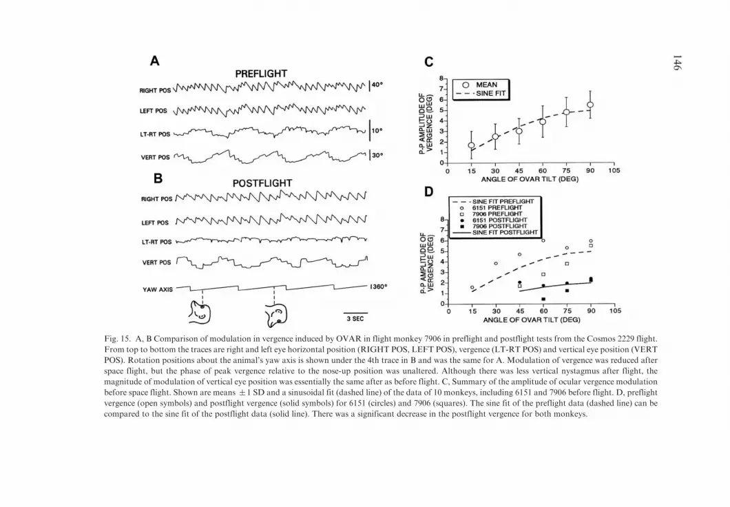
Fig. 15. A, B Comparison of modulation in vergence induced by OVAR in flight monkey 7906 in preflight and postflight tests from the Cosmos 2229 flight.
From top to bottom the traces are right and left eye horizontal position (RIGHT POS, LEFT POS), vergence (LT-RT POS) and vertical eye position (VERT
POS). Rotation positions about the animal’s yaw axis is shown under the 4th trace in B and was the same for A. Modulation of vergence was reduced after
space flight, but the phase of peak vergence relative to the nose-up position was unaltered. Although there was less vertical nystagmus after flight, the
magnitude of modulation of vertical eye position was essentially the same after as before flight. C, Summary of the amplitude of ocular vergence modulation
before space flight. Shown are means � 1 SD and a sinusoidal fit (dashed line) of the data of 10 monkeys, including 6151 and 7906 before flight. D, preflight
vergence (open symbols) and postflight vergence (solid symbols) for 6151 (circles) and 7906 (squares). The sine fit of the preflight data (dashed line) can be
compared to the sine fit of the postflight data (solid line). There was a significant decrease in the postflight vergence for both monkeys.
146

the minimum was 3.2�. The mean modulation of the peak-to-peak amplitudeof vergence for all 10 animals at a 90� OVAR tilt angle was 5.5� 1.3�. Becausethe peak GIA occurred in the nose-up position, phases of peak vergence weredetermined relative to a nose-up position during OVAR. All animals had peakvergence when their heads were rotated close to the nose-up position. The meanphase was �0.9� 26.6�. This supports the contention that the naso-occipitalacceleration was the factor responsible for the vergence.
After space flight, the modulation in vergence was strikingly reduced in thetwo flight monkeys. Eye position data from the preflight and first postflight testfor 7906 (Fig. 15B), demonstrates the reduction in the amplitude of vergence(LT-RTPOS). The magnitude of vertical eye modulation, which accompaniesvergence during naso-occipital linear acceleration (VERT POS), was virtuallythe same before and after flight. Spontaneous vertical nystagmus was reducedduring OVAR after space flight, and the beats of nystagmus were replacedby saccades (Fig. 15B, 4th trace). There was also a reduction in the nystagmusfrequency in 7906 that was not found in 6151 or in the previous monkeysafter recovery.
Results of postflight testing are summarized in Fig. 15D. As before flight, thedata were fit by a sinusoid (Fig. 15D, solid line). Vergence fell at every tilt angleat which a modulation was measurable after flight, with peak vergence beingreduced by over 50%. These reductions in modulation of vergence weresignificant. Test values throughout the postflight period were similar to thoseon postflight day 1. On postflight day 11, the peak modulation in vergence for7906 was 1.8� with a phase of 16� with respect to the nose-up position for anOVAR tilt angle of 90�. Under the same condition, the vergence modulation for6151 was 2.2� with a phase of 6.3�. By contrast, there was neither reductionin vergence modulation nor a significant phase change in two control monkeysthat were available for testing in the postflight period.
Spatial orientation of velocity storage
Eye velocity during per- and postrotatory nystagmus, optokinetic nystagmus(OKN) and optokinetic after-nystagmus (OKAN) and the nystagmus inducedby centrifugation tends to align with the direction of gravitoinertial acceleration(GIA) (Dai et al., 1991; Wearne et al., 1999; Moore et al., 2004). A similartendency has also been demonstrated during rapid reorientation of the headduring postrotatory nystagmus and OKAN (Dai et al., 1992; Fetter et al., 1996).The process of alignment has three components: (1) The horizontal timeconstant is reduced according to the projection of the GIA onto the head yawaxis; (2) The vertical and/or roll time constants are increased according to theprojection of the GIA onto the head vertical or roll axes; and (3) Vertical and/ortorsional cross-coupled components appear, which are not aligned with theangular stimulus vector when the stimulus is given about the yaw axis.Optimally, the amount of axis shift can be assessed during OKAN, recorded
147
in three dimensions with the head in tilted positions, or from vestibularnystagmus during centrifugation when the gravito-inertial acceleration vector istilted with regard to the head and body, but there are also manifestations of theorientation of eye velocity to the GIA during OKN (Moore et al., 2004). Partialtesting can be used to characterize the spatial orientation of the aVOR, speci-fically, the rapid reduction in yaw eye velocity during postrotatory nystagmus(tilt-dumping) (Waespe et al., 1985). On Earth, this results in a shorter hori-zontal aVOR time constant than in upright position. If the horizontal aVORtime constant were not to respond to head tilt with regard to gravity afterspace flight, it would indicate a reduction in sensitivity to spatial orientationof the aVOR.
Evidence for a change in spatial orientation of velocity storage came fromtwo animals that were tested shortly after return from microgravity, one fromCosmos 2044 and the second from Cosmos 2229. Monkey 782 lost the ability toshorten its horizontal time constant when tilted with regard to gravity after theCosmos 2044 flight (Fig. 15B). During postrotatory nystagmus, the animal wastilted 50� at the upward arrow. Time constants measured from the onset of tiltor at a comparable time 10 s after the onset of the postrotatory nystagmus were6.7 s after tilt as against 28 s without tilt (Fig. 16A). After flight, there was noeffect of tilt on postrotatory nystagmus in this monkey (Fig. 16B), and the timeconstant of each response, was the same as when it was upright in darkness dur-ing perrotatory nystagmus (19.3� 1.8 s (n=6) vs 22.1� 8.9 s (n=6). Findingswere the same 2 and 4 days after landing in this monkey. Control time constantswere 17.6 s (� 2.3, n=10) vs tilt time constants of 16.6 s (� 3.0, n=7) on thesedays. This indicates that the loss of the ability to dump stored slow phase eyevelocity persisted for several days after landing in this monkey. This deficit wasnot present in the second monkey from the same flight, however.
More complete testing for spatial orientation was performed after theCosmos 2229 flight. Trajectories of eye velocity during OKAN in a normalmonkey, upright and tilted with respect to gravity are shown in Fig. 16A–D.The angle of the eye velocity vector should be close to gravity in a phase planeplot at the end of OKAN when eye velocity approaches zero (Dai et al., 1991).Before flight, the axis of eye rotation of 7906 for upward coupling fromhorizontal OKAN at a 90� tilt angle approached the spatial vertical at an angleof 5� (Fig. 17A). Immediately after the flight at R+22 hours, the eye velocityvector was shifted 28� away from gravity toward a body orientation (Fig. 17B).Seven days later, the orientation of eye velocity had returned closer to gravity(Fig. 17C, 7�). Vectors calculated for OKAN with the animals tilted 30, 60 and90� showed a similar pattern. That is, the spatial orientation of velocity storagetowards gravity was altered after space flight and recovered after 7 days, suggest-ing that monkey 7906 had shifted the orientation of its eye velocity toward thebody in Space. A similar shift in spatial orientation of eye velocity was notfound in monkey 6151, but this animal was not tested until the third day, 72 hafter recovery, by which time there may have been recovery of this function.
148

Fig. 16. A, B, Slow phase velocity of per- and postrotatory nystagmus induced by steps of velocity in
darkness in monkey 782 before (A) and after (B) the Cosmos 2044 flight. In each set, the animal was
rotated in darkness about a vertical axis, inducing per- and postrotatory nystagmus. After the eye velocity
had declined to zero, the animal was stopped and 10 s later it was tilted 50� (TILT POS). Before flight (A),
this maneuver caused a prompt decline in slow phase velocity. After flight (B), the tilt did not shorten the
time course of decline in slow phase velocity. Per- and postrotatory time constants are shown below each
response. C–F, Optokinetic nystagmus (OKN), optokinetic after nystagmus (OKAN) and trajectory of
axis of eye velocity in horizontal and vertical dimensions on the right. C, D, Horizontal OKN and OKAN
were induced with a normal monkey was upright. After the lights were extinguished, OKAN was
horizontal and decayed to zero with its characteristic time course. D, The points in the phase plane plots
start at the onset of OKAN, which are farthest from the origin, and proceed beat by beat toward the origin
where the velocity was again zero. With the animal upright, the eye velocity vector was aligned with both
the animal’s yaw axis and the spatial vertical. E, F, When the animal was tilted on its side and given the
same stimulus as in C, the time constant of the horizontal component of OKAN was shorter, and
the OKAN had a cross-coupled vertical component, which rose and decayed. F, In the phase plane plot,
the trajectory of OKAN was initially along the animal’s yaw axis, but then curved toward the animal’s
pitch axis. This brought the eye velocity vector close to the spatial vertical at the end of OKAN.
149

Studies of lateral semicircular canal afferents
Single unit recordings were made from horizontal semicircular canal afferentsin four rhesus monkeys within the first 48 h following space flight recoveryand for 8–11 days thereafter. Monkeys were exposed to step and sinusoidal
Fig. 17. A–C, Trajectory (phase plane) plots of vertical (ordinate) and horizontal (abscissa) eye velocity
during cross-coupling of horizontal OKAN, similar to that shown in Fig. 16 F. The animal (7906) was in a
90� tilted, side-down position, and OKN was induced by yaw axis rotation of the visual surround at
60�/s. OKAN slow phase velocities began in each graph on the right, and the velocities progressed toward
zero to the left in a curved fashion. Each circle represents the velocity of one slow phase. The solid curved
line represents the fit of the data using amodified Levenberg–Marquardt algorithm. The straight line is the
trajectory of the last part of the decaying OKAN, showing the slope of the fitted curve as the data
approached zero. A, Before flight, the yaw axis eye velocity vector had an angle of 5� from the vertical.
B, Twenty-two hours after flight, the eye velocity vector had shifted toward the body axis and was now 28�
from the vertical. C, Seven days later, the eye velocity vector had returned to close to its original position
and now was deviated 7� from the vertical. D, Analysis of eye velocity vectors (elgenvectors) obtained
during OKAN cross-coupling, before (open circles) and 22 h (filled circles) and 7 days (open square) after
flight. Values on the ordinate are deviations from the spatial vertical, which is represented by the
horizontal line through zero. The abscissa shows the angle of tilt of the axis of rotation during the OKN
and OKAN. Animals were tested at 30, 60 and 90� of tilt. Before flight and 7 days after flight, the eye
velocity vectors lay close to the spatial vertical. Twenty two hours after recovery, there was a linear
increase in the angle of deviation of the eye velocity vectors, representing deviations of the vectors from the
spatial vertical. The deviation was maximal (28�) at 90� of tilt. Adapted from Dai et al., Exp. Brain Res.
102, 45–56, 1994, with permission.
150
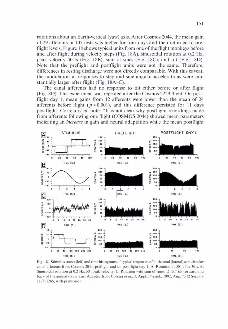
rotations about an Earth-vertical (yaw) axis. After Cosmos 2044, the mean gainof 29 afferents in 107 tests was higher for four days and then returned to pre-flight levels. Figure 18 shows typical units from one of the flight monkeys beforeand after flight during velocity steps (Fig. 18A), sinusoidal rotation at 0.2 Hz,peak velocity 50�/s (Fig. 18B), sum of sines (Fig. 18C), and tilt (Fig. 18D).Note that the preflight and postflight units were not the same. Therefore,differences in resting discharge were not directly comparable. With this caveat,the modulation in responses to step and sine angular accelerations were sub-stantially larger after flight (Fig. 18A–C).
The canal afferents had no response to tilt either before or after flight(Fig. 8D). This experiment was repeated after the Cosmos 2229 flight. On post-flight day 1, mean gains from 12 afferents were lower than the mean of 24afferents before flight ( p<0.001), and this difference persisted for 11 dayspostflight. Correia et al. note: ‘‘It is not clear why postflight recordings madefrom afferents following one flight (COSMOS 2044) showed mean parametersindicating an increase in gain and neural adaptation while the mean postflight
Fig. 18. Stimulus traces (left) and time histograms of typical responses of horizontal (lateral) semicircular
canal afferents from Cosmos 2044, preflight and on postflight day 1. A, Rotation at 50�/s for 30 s. B,
Sinusoidal rotation at 0.2 Hz, 50� peak velocity. C, Rotation with sum of sines. D, 20� tilt forward and
back of the animal’s yaw axis. Adapted from Correia et al., J. Appl. Physiol., 1992, Aug. 73 (2 Suppl.):
1125–1265, with permission.
151

recordings made from a second flight (COSMOS 2229) show mean parametersindicating a decrease in gain and no change in neural adaptation. But for bothflights, the horizontal semicircular canals were statistically significantly differentfrom those of preflight and synchronous controls. These differences werepresent while concurrent tests of the horizonal VOR, using the same stepparadigms, revealed no differences in slow phase eye velocity between controlanimals and those that flew in Space. These results suggest that compensationfor the altered input from the vestibular sensory receptor (the semicircularcanal) probably occurred within the CNS vestibular pathways.’’
Discussion
These results show that exposure to microgravity resulted in changes in thelinear VOR, the angular VOR, and in the spatial orientation of slow phasevelocity produced by the velocity storage component of the angular VOR. Thegain of compensatory ocular movements produced by the high frequencysinusoidal oscillation was reduced in one monkey, and the gains of ocularcounter-rolling (OCR) and vergence produced by low frequency, interaural andnaso-occipital linear acceleration were also substantially lower in 2 monkeysafter than before flight. Associated with this, there was a reversal of theasymmetry in vertical nystagmus, and a reduction in the gain of the roll aVOR.Finally, in the monkey that had the largest reduction in response to linearacceleration (7906), the orientation of slow phase eye velocity during OKANand vestibular nystagmus shifted from a spatial toward a body axis. In contrast,high frequency, horizontal and vertical canal-related responses, generated byangular acceleration in a head coordinate frame of reference, were unaffected bymicrogravity in post-flight testing. Since the vertical canals had a normal gainafter flight when tested with rotation in pitch, it is not likely that a change in theafferent input from the vertical canals was the cause for the reduction in the rollaVOR gain. Rather, there must have been changes in excitability in centralpathways for torsion. The adaptive changes in otolith reflexes, in the roll aVORand in the spatial orientation of eye velocity in the aVOR are likely to contributeto postural, locomotor and gaze instability on reentry after space flight.
The data in these experiments were taken from monkeys whose bodies wererestrained. Therefore, these monkeys had only a very limited exposure to linearacceleration during flight. In Space, the otoliths are not activated by staticlateral head tilt, and neck receptors do not contribute to OCR (Clarke et al.,1993). Moreover, lateral translations, which are associated with OCR onEarth, mainly induce a horizontal component in Space (Clarke et al., 2000). Therelative absence of exposure to linear acceleration, therefore, could have beenthe cause for the reduction in the ocular/spatial reactions to linear accelera-tion upon return to Earth. Lateral translation when turning corners wasalso absent in Space, which might explain the 15–50% reduction in gain of theroll aVOR.
152
Of interest in this regard are recent results from the NASANeurolabMission(STS-90), which demonstrated that these otolith-ocular orienting responseswere well maintained during space flight (Clement et al., 2001; Moore et al.,2001, 2004). It is possible that this difference in response to microgravitybetween the monkeys in the COSMOS flights and the astronauts in theNeurolab Mission was due to a species difference. More likely, however, is thatthe preservation of the otolith-ocular orienting and compensatory reflexeswas due to the active movement and frequent centrifugation experienced by theastronauts in Space on STS-90. If this is correct, then a valuable lesson hascome from the monkey experiments. Namely, that a program of active headmovement with exposure to linear acceleration is an appropriate counter-measure for the reduction in the gain of OCR, vergence and the rollaVOR. Such a countermeasure could also help maintain the gain of vestibulo-sympathetic reflexes, which serve as a first line of defense in maintainingblood pressure upon standing (Kaufmann et al., 2002). A drop in the gain ofsuch reflexes could contribute to the orthostatic hypotension that commonlyfollows space flight. Thus, the data in these monkeys suggest that the gain ofroll and vergence may be much more malleable than has commonlybeen assumed. Unfortunately, the only way to test this would be in Space,where there is a prolonged absence of gravity, and the opportunities for suchexperiments are few.
Our results also show that there is considerable variation between individualsexposed to essentially similar conditions in Space. Microgravity was associatedwith a striking reduction in the high frequency lVOR gain in 7906, whereasthe compensatory lVOR was essentially normal in 6151, although 6151 wasnot tested until the third day after recovery. This mirrors similar variabilityin humans exposed to microgravity. On the other hand, there was a strikingsimilarity in the increased sensitivity of the aVOR during active head turns, inthe saccadic hypermetria during lateral gaze shirts, and in the increase in thesensitivity of units in MVN and flocculus to head turns in some of thesesame monkeys. Afferent activity from the lateral semicircular canals also variedconsiderably between Cosmos flights 2044 and 2229 (Correia et al., 1992;Correia, 1998) in some of these same monkeys, although the passive aVORgains were unaffected (Cohen et al., 1992; Dai et al., 1994). Some of thesechanges may have been a consequence of the behavioral state or theexperimental conditions, interacting with exposure to microgravity, but therealso seems to be an individual difference of response to essentially similarconditions.
Eye velocity during nystagmus due to excitation of the slow component ofthe aVOR tends to align with the spatial vertical when the head is tilted withregard to the GIA (Dai et al., 1991, 1992). In both flights, this spatialorientation was affected in one of the two monkeys available for testingshortly after landing. The shift in orientation disappeared over a time courseof several days after reentry into a gravitational environment (Cohen et al.,
153
1992; Dai et al., 1994). The reduction in the direct orienting response of OCRand vergence persisted for a longer period than the indirect orienting responseof OKAN cross-coupling, which is produced through a different mechanism,i.e., through velocity storage. An important aspect of re-adaptation upon thereentry is to reorient to gravity so that the body posture is maintained duringmotion. This may explain why the reorientation of velocity storage, asreflected in OKAN cross-coupling, occurred sooner than the recovery of otherocular orienting reactions produced by the lVOR.
In this chapter, we have summarized experiments that provide aconsolidated body of new data that demonstrate changes that can be expectedin the vestibulo-ocular reflex and the central vestibular system after spaceflight. The experiments in Sections 2 and 3 also show the unique value of themonkey as a model for studying effects of human vestibular adaptation inSpace, wherein behavior and single unit recordings can be combined in asingle animal that has many similarities to humans. It is hoped that the workdescribed in this chapter will aid in understanding the complex phenomenathat accompany adaptation to microgravity, especially in relation to long-duration space flight. There is a larger reason to promote the use of experi-mental animals in Space. There is no other way to understand the critical roleof gravity on the formation and structure of the nervous system. Conse-quently, an essential component of future space-related experiments should bedirected toward determining how afferent input from the otoliths and semi-circular canals is altered and how central processing is changed in micro-gravity in experimental animals.
Acknowledgment
We thank Jun Maruta for assistance. This work was supported by NASAGrants NAG 2-573 (BC), NAG 2-703 (BC), NASA Contract NAS9-19441(BC), NCC2-1173 (GRH), NAG 2-946 (GRH), NIH Grants:DC02451 (GRH), DC04996 (SY), DC05204, and EY01867.
References
Aizikov, G.S. and Markin, A.S. Posture, movement and balance in rats afterBiosatellite flights. Space Biol. Aviaspace Med. 15(6), 33–38 (in Russian).
Aizikov, G.S., Markin, A.S. and Mokrousova, A.V. (1981) Specificity of thelift-reaction in white rats after prolonged spaceflight (effects of microgravity andhypergravity). Ann. Exp. Biol. Med. 12, 722–725 (in Russian).
Aizikov, G.S., Markin, A.S. and Shipov, A.A. (1992) Flipping-reaction in rats afterthe ‘‘Cosmos 936’’ spaceflight. Aviaspace Ecol. Med. 26(1), 62–64 (in Russian).
Anken, R., Kappel, T. and Rahmann, H. (1998) Morphometry of fish inner earotoliths after development at 3g hypergravity. Acta Oto-Laryngol. 118, 534–539.
154

Apanasenko, Z.I. (1977) Vestibulo-tonic reflexes of rat’s legs after the ‘‘Cosmos605’’ spaceflight. Space Biol. Aviaspace Med. 11(4), 28–34 (in Russian).
Badakva, A.M., Miller, N.V. and Eron, J.N. (2003) Effects of head-out waterimmersion on eye-head coordination in rhesus monkey. J. Gravitat. Physiol. 10,P5–P8.
Badakva, A.M., Zalkind, D.V., Miller, N.V. and Riazansky, S.N. (2000) Headpitch movement and vestibular neuronal activity in response to otolithstimulation of monkeys in space. J. Gravit. Physiol. 7, S99–S105.
Badakva, A.M., Zalkind, D.V., Miller, N.V., Sirota, M.G., Beloozerova, I.N.,Kozlovskaya, I.B. (1993) The Final Report on Cosmos 2229 Experiments.Chapter 4. Effects of microgravity on neuronal circuitry involved in forming ofthe vestibular and oculomotor responses: results of primate studies in BIONprojects. In Institute of Biomedical Problems, pp. 92–104, Russian Academy ofScience, Moscow.
Balaban, E., Centini, C. and Pompeiano, O. (2002) Tonic gravity changes altergene expression in the efferent vestibular nucleus. NeuroReport 13, 187–190.
Barmin, V.A., Kreidich, Y.V. and Kozlovskaya, I.B. (1983) Influence ofoptokinetic stimulation and immersion on eye-head coordination in man.Physiologist 26, S83–S85.
Bauerle, A., Anken, R., Hilbig, R. and Rahmann, H. (2004) Histology of the utriclein kinetotically swimmiing fish: a parabolic aircraft flight study. Acta Oto-Laryngol. 124, 19–22.
Beier, M., Anken, R. and Rahmann, H. (2002) Influence of hypergravity on fishinner ear otoliths: II. Incorporation of calcium and kinetotic behaviour. Adv.Space Res. 30, 727–731.
Benson, A.J., Kass, J.R. and Vogel, H. (1986) European vestibular experiments onthe Spacelab-1 mission: 4. Thresholds of perception of whole-body linearoscillation. Exp. Brain Res. 64, 264–271.
Benson, A.J. and Vieville, T. (1986) European vestibular experiments on theSpacelab-1 mission: 6. Yaw axis vestibulo-ocular reflex. Exp. Brain Res. 64,279–283.
Bizzi, E., Kalil, R.E. and Morasso, P. (1972) Two modes of active eye-headcoordination in monkeys. Brain Research 40, 45–48.
Bizzi, E., Kalil, R.E. and Tagliasco, V. (1971) Eye-head coordination in monkeys:evidence for centrally patterned organization. Science 173, 452–454.
Boyle, R., Mensinger, A.F., Yoshida, K., Usui, S., Intravaia, A., Tricas, T. andHighstein, S.M. (2001) Neural readaptation to Earth’s gravity following returnfrom space. J. Neurophysiol. 86, 2118–2122.
Buettner-Ennever, J.A. (1999) A review of otolith pathways to brainstem andcerebellum. Ann. N. Y. Acad. Sci. 871, 51–64.
Chubb, C.M., Fuchs, A.F. and Scudder, C.A. (1984) Neuron activity in monkeyvestibular nuclei during vertical vestibular stimulation and eye movements.J. Neurophysiol. 52, 724–742.
155

Clarke, A.H., Grigull, J., Mueller, R. and Scherer, H. (2000) The three-dimensionalvestibulo-ocular reflex during prolonged microgravity. Exp. Brain Res. 134,322–334.
Clarke, A.H., Teiwes, W. and Scherer, H. (1993) Vestibulo-oculomotor testingduring the course of a space flight mission. Clinical Investigations 71, 740–748.
Clement, G., Moore, S.T., Raphan, T. and Cohen, B. (2001) Perception of tilt(somatogravic illusion) in response to sustained linear acceleration during spaceflight. Exp. Brain Res. 138, 410–418.
Cohen, B. and Gizzi, M. (2003) The physiology of the vestibulo-ocular reflex.Chapter 41. In Luxon, L., Martini, A. and Furman, J.D.S. (eds.). Textbook ofAudiological Medicine, pp. 701–716. Oxford University Press, New York.
Cohen, B., Kozlovskaya, I., Raphan, T., Solomon, D., Helwig, D., Cohen, N.,Sirota, M. and Yakushin, S. (1992) Vestibuloocular reflex of rhesus monkeysafter space flight. J. Appl. Physiol. 73, 121S–131S.
Cohen, B., Maruta, J. and Raphan, T. (2001) Orientation of the eyes to gravito-inertial acceleration. N. Y. Acad. Sci. 942, 241–258.
Cohen, B. and Raphan, T. (2004) The physiology of the vestibulo-ocular reflex(VOR). Anatomy and physiology of the central and peripheral vestibular system.In Highstein, S., Popper, A. and Fay, R.R. (eds.). Springer Handbook ofAuditory Research. Springer Verlag, New York.
Correia, M.J. (1998) Neuronal pasticity: adaptation and readaptation to theenvironment of space. Brain Res. Rev. 28, 61–65.
Correia, M.J., Perachio, A.A., Dickman, J.D., Kozlovskaya, I.B., Sirota, M.G.,Yakushin, S.B. and Beloozerova, I.N. (1992) Changes in monkey hori-zontal semicircular canal afferents after space flight. J. Appl. Physiol. 73,112S–120S.
Crane, B.T. and Demer, J.L. (1999) A linear canal-otolith interaction model todescribe the human vestibulo-ocular reflex. Biol. Cybern 81, 109–118.
Dai, M., McGarvie, L., Kozlovskaya, I.B., Raphan, T. and Cohen, B. (1994)Effects of space flight on ocular counterrolling and spatial orientaion of thevestibular system. Exp. Brain Res. 102, 45–56.
Dai, M., Raphan, T. and Cohen, B. (1991) Spatial orientation of the vestibularsystem: dependence of optokinetic after nystagmus on gravity. J. Neurophysiol.66, 1422–1438.
Dai, M., Raphan, T., Kozlovskaya, I. and Cohen, B. (1996) Modulation ofvergence by off-vertical yaw axis rotation in the monkey: normal characteristicsand effects of space flight. Exp. Brain Res. 111, 21–29.
Dai, M.J., Raphan, T. and Cohen, B. (1992) Characterization of yaw to roll cross-coupling in the three-dimensional structure of the velocity storage integrator.Ann N.Y. Acad. Sci. 656, 829–831.
d’Ascanio, P., Balaban, E., Pompeiano, M., Centini, C. and Pompeiano, O. (2003)Fos and FRA protein expression in rat precerebellar structures during theNeurolab Space Mission. Brain Res. Bull. 62, 203–221.
156

Dichgans, J., Bizzi, E., Morasso, P. and Tagliasco, V. (1973) Mechanismsunderlying recovery of eye-head coordination following bilateral labyrinthect-omy in monkeys. Exp. Brain Research 18, 548–562.
Duflo, S.G.D., Gestreau, C. and Lacour, M. (2000) Fos expression in the rat brainafter exposure to gravito-inertial force changes. Brain Res. 861, 333–344.
Edgerton, V.R., Roy, R.R., Hodgson, J.A., Day, M.K., Weiss, J., Harkema, S.J.,Dobkin, B., Garfinkel, A., Konigsberg, E. and Koslovskaya, I. (2000) How thescience and engineering of space flight contribute to understanding the plasticityof spinal cord injury. Acta Astronautica 47, 51–62.
Einstein, A. (1911) Ueber den Einfluss der Schwerkraft auf die Ausbreitung desLichtes. Annalen der Physik 35, 898–908.
Fetter, M., Heimberger, J., Black, R.A., Hermann, W., Sievering, F. andDichgans, J. (1996) Otolith-semicircular canal interaction during postrotatorynystagmus in humans. Exp. Brain Res. 108, 463–473.
Fox, R.A., Daunton, N., Corcoran, M. and Wu, L. (1992) Time-course ofbehavioral adaptation to hypergravity. In Woollacott, M. and Horak, F. (eds.).Posture and Gait: Control Mechanisms. Vol. I, pp. 178–181. University ofOregon Books, Portland.
Fuchs, A.F. and Kimm, J. (1975) Unit activity in vestibular nucleus of the alertmonkey during horizontal angular acceleration and eye movement. J.Neurophysiol. 38, 1140–1161.
Gazenko, O.G., Grigorian, R.A., Kitaev-Smik, L.A. and Klochkov, A.I. (1965)Increase of extensor muscular tonus in microgravity in cats with complete andpartial cerebellum removal. Ann. Exp. Biol. Med. 7, 7–12 (in Russian).
Gazenko, O.G. and Ilyin, E.A. (1987) Physiological investigations of primatesonboard biosatellites Cosmos-1514 and Cosmos-1667. The Physiologist 30,S31–S35.
Grigaryan, R.A., Gazenko, O.G., Kozlovskaya, I.B., Barmin, V.A. andKreidich, Y.V. (1986) The vestibulo-cerebellar regulation of oculomotorreactions in microgravitational conditions. In Keller, S.L. and Zee, DS (eds.).Adaptive Processes in Visual and Oculomotor Systems. Vol. 57, pp. 121–127.Pergamon Press, New York.
Guedry, F.E. (1974) Psychophysics of vestibular sensation. In Kornhuber, H.H.(ed.). Handbook of Sensory Physiology. Vol. 6, pp. 3–154. Springer Verlag,Berlin.
Hess, B.J.M. and Dieringer, N. (1991) Spatial organization of linear vestibulo-ocular reflexes of the rat: Responses during horizontal and vertical linearacceleration. J. Neurophysiol. 66, 1805–1818.
Hofstetter-Degen, K., Weizig, J. and von Baumgarten, R. (1993) Oculovestibularinteractions under microgravity. Clinical Investigations 10, 749–756.
Holstein, G.R., Kukielka, E. and Martinelli, G.P. (1999) Anatomical observa-tions of the rat cerebellar nodulus after 24 hr of space flight. J. Grav. Physiol. 6,47–50.
157

Ibsch, M., Anken, R.H. and Rahmann, H. (2000) Weightlessness during spaceflight results in enhanced synapse formation in a fish brain vestibular nucleus.Neurosci. Letts. 296, 13–16.
Imai, T., Moore, S.T., Raphan, T. and Cohen, B. (2001) Interaction of the body,head and eyes during walking and turning. Exp. Brain Res. 136, 1–18.
Ito, M. (1984) Cerebellum and Neural Control. Raven Press, New York.Kaufman, G.D., Anderson, J.H. and Beitz, A.J. (1991) Activation of a specific
vestibulo-olivary pathway following centripetal acceleration in the rat. BrainRes. 562, 311–317.
Kaufman, G.D., Anderson, J.H. and Beitz, A.J. (1992) Fos-defined activity in ratbrainstem following centripetal acceleration. J. Neurosci. 12, 4489–4500.
Kaufman, G.D., Anderson, J.H. and Beitz, A.J. (1993) Otolith-brainstemconnectivity: evidence for differential neural activation by vestibular hair cellsbased on quantification of Fos expression in unilateral labyrinthectomized rats.J. Neurophysiol. 70, 117–127.
Kaufman, G.D. and Perachio, A.A. (1994) Translabyrinth electrical stimulation forthe induction of immediate-early genes in the gerbil brainstem. Brain Res. 646,345–350.
Kaufmann, H., Biaggioni, I., Voustaniuk, A., Diedriich, A., Costa, F., Clark, R.,Gizzi, M., Raphan, T. and Cohen, B. (2002) Vestibular control of sympatheticactivity: An otolth-sympathetic reflex in humans. Exp. Brain Res. 143, 463–469.
Keller, E.L. and Daniels, P.D. (1975) Oculomotor related interaction of vestibularand visual stimulation in vestibular nucleus cells in alert monkey. Exp. Neurol.46, 187–198.
Kitaev-Smik, L.A. (1963) Animal posture in microgravity. In Aviation and SpaceMedicine, pp. P247–250. Moscow (in Russian).
Kozlovskaya, I.B., Barmin, V.A., Kreidich, Y.V. and Repin, A.A. (1985) Theeffects of real and simulated microgravity on vestibulo-oculaomotor interaction.The Physiologist 28, 51–56.
Kozlovskaya, I.B., Ilyin, E.A., Sirota, M.G., Korolkov, V.I., Babayev, B.M.,Beloozerova, I.N. and Yakushin, S.B. (1989) Studies of space adaptationsyndrome in experiments on primates performed on board of Soviet biosatellite‘‘Cosmos-1887’’. The Physiologist 32, 45–48.
Kozlovskaya, I.B., Sirota, M.B., Yakushin, S.B., Beloozerova, I.I., Babayev, B.M.,Badakva, A.M., and Zalkind, D.V. (1994) The influence of weightlessness onparameters of the horizontal vestibulo-ocular reflex (HVOR) in monkeys. (InRussian). In Space Biology and Aviacosmic Medicine. Report Abstracts of XConference, 7-10/VI, pp. 113–114. Slovo, Moscow.
Kozlovskaya, I.B., Sirota, M.G., Beloozerova, I.N., and Yakushin, S.B. (1991)Vestibulo-motor interaction inmicrogravity. In InternationalMeeting ‘‘BiosatelliteCosmos’’. p. 52. (In Russian) IMBP,Moscow, Leningrad 12–15 August.
Krasnov, I.B. (1975) Quantitative cyto- and histochemical studies of the Deiter’snucleus and nodular cortex of cerebellum in rats exposed to weightlessness.Aviat. Space Environ. Med. 46, 1119–1122.
158

Krasnov, I.B. (1987) Increase in the sensibility of otolith apparatus in weight-lessness. Morphological evidence. Intercosmos Space Biol. Med. Grp. Mtng.10, 127.
Krasnov, I.B. (1991) The otolith apparatus and cerebellar nodulus in ratsdeveloped under 2-G gravity. The Physiologist 34, S206–S207.
Krasnov, I.B. and Dyachkova, L.N. (1986) Ultrastructure of the cortex ofthe cerebellar nodulus in rats after a flight on the biosatellite KOS 1514. Kosm.Biol. Aviakosm. Med. 20, 45–48.
Krasnov, I.B. and Dyachkova, L.N. (1990) The effect of space flight on theultrastructure of the rat cerebellar and hemisphere cortex. The Physiologist 33,29–30.
Kreidich, Y.V., Repin, V.A., Barmin, V.A. and Kozlovskaya, I.B. (1983) Effect ofimmersion hypokinesia on man’s eye and head movements during the gazefixation reachion. (In Russian) Space Biol. and Aerospace Med. 5, 41–45.
Lim, D.J., Stith, J.A., Stockwell, C.W. and Oyama, J. (1974) Observations onsacculus of rats exposed to long-term hypergravity. Aerospace Med. 45, 705–710.
Lisberger, S.G. (1994) Neural basis for motor learning in the vestibuloocular reflexof primates. III. Computational and behavioral analysis of the sites of learning.J. Neurophysiol. 72, 974–998.
Lisberger, S.G. and Fuchs, A.F. (1978a) Role of primate flocculus during rapidbehavioral modification of vestibuloocular reflex. I. Purkinje cell activity duringvisually guided horizontal smooth-pursuit eye movements and passive headrotation. J. Neurophysiol. 41, 733–763.
Lisberger, S.G. and Fuchs, A.F. (1978) Role of primate flocculus during rapidbehavioral modification of vestibuloocular reflex. II. Mossy fiber firing patternsduring horizontal head rotation and eye movement. J. Neurophysiol. 41,764–777.
Lisberger, S.G., Pavelko, T.A., Bronte-Stewart, H.M. and Stone, L.S. (1994a)Neural basis for motor learning in the vestibuloocular reflex of primates. II.Changes in the responses of horizontal gaze velocity Purkinje cells in thecerebellar flocculus and ventral paraflocculus. J. Neurophysiol. 72, 954–973.
Lisberger, S.G., Pavelko, T.A. and Broussard, D.M. (1994b) Neural basis formotor learning in the vestibuloocular reflex of primates. I. Changes in theresponses of brain stem neurons. J. Neurophysiol. 72, 928–953.
Lychakov, D.V., Bojajieva-Mikhailova, A., Christov, I., Pashchinin, A.N.,Evdokimov, I.I. and Markov, A.A. (1988) Changes in the otolith apparatus ofrats and fish after prolonged exposure to acceleration. (In Russian) Kosm. Biol.Aviakosm. Med. 22, 27–33.
Lychakov, D.V., Pashchinin, A.N., Bojajieva-Mikhailova, A. and Khristov, I.(1989) Study of receptor organs of the vestibular apparatus of Cosmos-1667 rats.(In Russian) Kosm. Biol. Aviakosm. Med. 23, 27–33.
Lychakov, D.V. and Lavrov, E.A. Study of the vestibular end organ structure andionic composition of the body of young tadpoles (Xenopus Laevis) afterspaceflight. Space Biol. Aviaspace Med. 19(3), 48–52.
159

Markham, C.H. and Curthoys, I.S. (1972) Convergence of labyrinthine influenceson units in the vestibular nuclei of the cat. II. Electrical stimulation. Brain Res.43, 383–396.
Maruta, J., Simpson, J.I., Raphan, T. and Cohen, B. (2001) Orienting otolith-ocular reflexes in the rabbit during static and dynamic tilts and off-vertical axisrotation. Vision Research 41, 3255–3270.
Mashburn, T.H., Kaufman, G.D., Purcell, I.M. and Perachio, A.A. (1997) Sacculecontribution to immediate-early genes production in the gerbil brainstem withposterior galvanic or hypergravity stimulation. Brain Res. 761, 51–58.
Matsuo, V. and Cohen, B. (1984) Vertical optokinetic nystagmus and vestibularnystagmus in the monkey: Up-down asymmetry and effects of gravity. Exp.Brain Res. 53, 197–216.
Matsuo, V., Cohen, B., Raphan, T., de Jong, V. and Henn, V. (1979) Asymmetricvelocity storage for upward and downward nystagmus. Brain Res. 176, 159–164.
Miles, F.A. (1974) Single unit firing patterns in the vestibular nuclei related tovoluntary eye movements and passive body rotation in conscious monkeys.Brain Res. 71, 215–224.
Moore, S.T., Clement, G., Raphan, T. and Cohen, B. (2001) Ocular counter-rolling(OCR) induced by centrifugation during orbital space flight. Exp. Brain Res.137, 323–335.
Moore, S.T., Cohen, B., Raphan, T., Berthoz, A. and Clement, G. (2004) Spatialorientation of optokinetic nystagmus and ocular pursuit during orbital spaceflight. Exp. Brain Res, In press.
Oman, C.M. and Balkwill, M.D. (1993) Horizontal angular VOR, nystagmusdumping, and sensation duration in Spacelab SLS-1 crewmembers. J. Vesti. Res.3, 315–330.
Paige, G.D. (1991) Linear vestibulo-ocular reflex (LVOR) and modulation byvergence. Acta Otolaryngol. (Stockh.) 481 (Suppl.), 282–286.
Paige, G.D., Telford, L., Seidman, S.H. and Barnes, G.R. (1998) Humanvestibuloocular reflex and its interactions with vision and fixation distanceduring linear and angular head movement. J. Neurophysiol. 80, 2391–2404.
Paige, G.D. and Tomko, D.L. (1991a) Eye movement responses to linear headmotion in the squirrel monkey. I. Basic Characteristics. J. Neurophysiol. 65,1170–1182.
Paige, G.D. and Tomko, D.L. (1991b) Eye movement responses to linear headmotion in the squirrel monkey. II. Visual-vestibular interactions and kinematicconsiderations. J. Neurophysiol. 65, 1183–1196.
Pedrozo, H.A. and Wiederhold, M.L. (1994) Effects of hypergravity on statocystdevelopment in embryonic Aplysia californica. Hearing Res. 79, 137–146.
Phillips, J.O., Ling, L., Fuchs, A.F., Siebold, C. and Plorde, J.J. (1995) Rapidhorizontal gaze movement in the monkey. J. Neurophysiology 73, 1632–1652.
Pompeiano, O., d’Ascanio, P., Centini, C., Pompeiano, M. and Balaban, E. (2002)Gene expression in rat vestibular and reticular structures during and after spaceflight. Neurosci. 114, 135–155.
160

Pompeiano, O., d’Ascanio, P., Balaban, E., Centini, C. and Pompeiano, M. (2004)Gene expression in autonomic areas of the medulla and the central nucleus ofthe amygdala in rats during and after space flight. Neurosci. 124, 53–69.
Rahmann, H., Slenzka, K., Kortje, H. and Hilbig, R. (1992) Synaptic plasticity andgravity: ultrastructural, biochemical and physico-chemical fundamentals. Adv.Space Res. 12, 63–72.
Raphan, T. and Cohen, B. (2002) The vestibulo-ocular reflex (VOR) in threedimensions. Exp. Brain Res. 145, 1–27.
Razumeev, A.N. and Grigorian, R.A. (1976) Cerebellum and Gravity, P453, Nauka,Moscow (in Russian).
Ross, M.D. (1987) Implications of otoconial changes in microgravity. The Physiol.30, 90–93.
Ross, M.D. (1993) Morphological changes in rat vestibular system followingweightlessness. J. Vestib. Res. 3, 241–251.
Ross, M.D. (1994) A space flight study of synaptic plasticity in adult rat vestibularmaculas. Acta Oto-Laryngol Suppl. 516, 3–14.
Ross, M.D. (1997) Morphological evidence for local microcircuits in rat vestibularmaculae. J. Comp. Neurol. 379, 333–346.
Ross, M.D. (2000) Changes in ribbon synapses and rough endoplasmic reticulusof rat utricular macular hair cells in weightlessness. Acta Otolaryngol. 120,490–499.
Ross, M.D., Donovan, K. and Chee, O. (1985) Otoconial morphology in space-flown rats. The Physiol. 28, 219–220.
Ross, M.D. and Tomko, D.L. (1998) Effect of gravity on vestibular neuraldevelopment. Brain Res. Revs. 28, 44–51.
Schwarz, C., Busettini, C. and Miles, F.A. (1989) Ocular responses to linear motionare inversely proportional to viewing distance. Science 245, 1394–1396.
Schwarz, C. and Miles, F.A. (1991) Ocular responses to translation and theirdependence on viewing distance. I. Motion of the observer. J. Neurophysiol. 66,851–864.
Shipov, A.A. and Aizikov, G.S. (1992) Physical state, equilibrium function andvestibulo-motor reflexes in the rats after space flights on board biosatellitesof Cosmos series. Physiologist Suppl., 35(1), S206–207.
Shipov, A.A. and Ovechkin, V.G. (1980) Rat semicircular canal function after the‘‘Cosmos 936’’ spaceflight. Space Biol. AviaspaceMed. 14(2), 25–30 (in Russian).
Shipov, A.A., Sirota, M.G., Beloozerova, I.N., Babaev, B.M. andKozlovskaya, I.B. (1986) Results of tests on the primate vestibulo-visualmotorreactions in biocosmos experiments. In Keller, S.L. and Zee, D.S. (eds.).Adaptive Processes in Visual and Oculomotor Systems, Vol. 57, pp. 129–132.Pergamon Press, New York.
Shipov, A.A. and Tabakova, L.A. (1982) Specificity of nystagmus in rats after the‘‘Cosmos 1129’’ spaceflight. Space Biol. Aviaspace Med. 16(6), (in Russian).
Sirota, M.G., Babayev, B.M., Beloozerova, I.B., Nyrova, A.N., Yakushin, S.B. andKozlovskaya, I.B. (1987) Characteristics of vestibular reactions to canal
161
and otolith stimulation at an early stage of exposure to microgravity. ThePhysiologist 30, S82–S84.
Sirota, M.G., Babayev, B.M., Beloozerova, I.N., Nyrova, A.N., Yakushin, S.B.and Kozlovskaya, I.B. (1988a) Neuronal activity of nucleus vestibularis duringcoordinated movement of eyes and head in microgravitation. The Physiologist31, 8–9.
Sirota, M.G., Beloozerova, I.N., Babaev, B.M., Yakushin, S.B., Ivanov, A.M.,Nyrova, A.N., Kozlovskaya, I.B. (1988b) The final report on Cosmos 1887experiments. Chapter 7.2. The vestibular function and vestibulo-oculomotorinteractions in microgravity. In Institute of Biomedical Problems, pp. 1–50.Ministry of Health of the USSR, Moscow.
Sirota, M.G., Beloozerova, I.N., Babaev, B.M., Kozlovskaya, I.B. (1984) The finalreport on Cosmos 1514 experiments. Chapter 2.4. Study of the vestibularfunction and vestibulo-oculomotor interaction in monkey. In Institute ofBiomedical Problems, pp. 1–31. Ministry of Health of the USSR, Moscow.
Sirota, M.G., Beloozerova, I.N., Babaev, B.M., Yakushin, S.B., Ivanov, A.M.,Kozlovskaya, I.B. (1986) The final report on Cosmos 1667 experiments.Chapter 7. The vestibular function and vestibulo-oculomotor interaction inmicrogravity. In Institute of Biomedical Problems, pp. 119–162. Ministry ofHealth of the USSR, Moscow.
Sirota, M.G., Beloozerova, I.N., Babaev, B.M., Yakushin, S.B., Kozlovskaya, I.B.(1989a) Effects of microgravity on vestibular function in monkey. Results ofspace flight studies. In Eleventh Ann. Meeting IUPS Commission on GravitationalPhysiology, 2 p, Lyon, France, 25–27 Sept.
Sirota, M.G., Beloozerova, I.N., Yakushin, S.B., Kozlovskaya, I.B. (1989b)Eye-head coordination during adaptation to microgravity. (In Russian). InRegulation of Sensor-Motor Functions, pp. 140. Vinniza, USSR.
Sirota, M.G., Beloozerova, I.N., Yakushin, S.B., Babaev, B.M., Kozlovskaya, I.B.(1990a) Changes in neuronal activity of vestibular nuclei during active headmovements in microgravity. In XII International Meeting of GravitationalPhysiology, 63 p (In Russian). IMBP, Moscow, Leningrad, 14–19 October.
Sirota, M.G., Beloozerova, I.N., Yakushin, S.B., Babaev, B.M., Kozlovskaya, I.B.(1990b) Influence of microgravity to vestibular function of monkey. In XIIInternational Meeting of Gravitational Physiology, pp. 63–64. (In Russian)IMBP, Moscow, Leningrad, 14–19 October.
Sirota, M.G., Beloozerova, I.N., Yakushin, S.B., Ivanov, A.M., Kozlovskaya, I.B.(1990c) Vestibulo-motor interaction of monkey in microgravity. Eye-headcoordination test. In XI Meeting in Space Biology and Medicine, pp. 342–343.(In Russian) IMBP, Kaluga, Moscow.
Sirota, M,G., Beloozerova, I.N., Babaev, B.M., Yakushin, S.B., Kozlovskaya, I.B.(1991a) The final report on Cosmos 2044 experiments: Chapter 6. Effectsof the space flight on primate’s motor and vestibular functions. In Instituteof Biomedical Problems, pp. 223–295. Ministry of Health of the USSR,Moscow.
162

Sirota, M.G., Beloozerova, I.N., Yakushin, S.B., Ivanov, A.M., Kozlovskaya, I.B.(1991b) Gain of vestibulo-ocular reflex of monkey in microgravity. Experimenton biosatellite ‘‘Cosmos-2044’’ boarad. (In Russian) In International Meeting‘‘Biosatellite Cosmos’’, 114 p, IMBP, Moscow, Leningrad, 12–15 August.
Sirota, M.G., Beloozerova, I.N., Yakushin, S.B., Ivanov, A.M., Kozlovskaya, I.B.(1991c) Kinematic of saccades and head movement in eye-head coordinationtest of monkey. Experiment on biosatellite ‘‘Cosmos-2044’’ board. (In Russian)In International Meeting ‘‘Biosatellite Cosmos’’, 113 p, IMBP, Moscow,Leningrad, 12–15 August.
Tiliket, C., Shelhamer, M., Tan, H.S. and Zee, D.S. (1993) Adaptation of thevestibulo-ocular reflex with the head in different orientations and positionsrelative to the axis of body rotation. J. Vestib. Res. 3, 181–195.
Tomko, D., Kozlovskaya, I.B., Paige, G. and Badakva, A.M. (1993) Adaptationto Micro-Gravity of Oculomotor Reflexes (AMOR): Otolith-Ocular Reflexes, 25p, National Aeronautics and Space Agency (NASA), Washington, D.C.
Tomlinson, D. and Bahra, P.S. (1986) Combined eye-head shifts in the primate I.Metrics. J. Neurophysiol. 56, 1542–1557.
Uganov, E.M. (1974) Vestibular testing of cosmonauts during space flight of‘‘Voshod’’ spacecraft. Microgravity, Vol. 1 (In Russian), pp. 83–88. Moscow.
Uno, T., Horii, A., Umemoto, M., Hasegawa, T., Doi, K., Uno, A., Takemura, T.and Kubo, T. (2000) Effects of hypergravity on morphology and osteopontinexpression in the rat otolith organs. J. Vestib. Res. 10, 283–289.
Uno, T., Horii, A., Uno, A., Fuse, Y., Fukushima, M., Doi, K. and Kubo, T.(2002) Quantitative changes in mRNA expression of glutamate receptors inthe rat peripheral and central vestibular systems following hypergravity.J. Neurochem. 81, 1308–1317.
Vinnikov, J.A., Lychakov, D.V., Palmbach, L.R., Govardovsky, V.I.,Adanina, V.O., Allachverdov, V.L. and Pogorelov, A.G. (1980) Study of thevestibular apparatus in the frog xenopus laevix and rats exposed to long-termweightlessness. Zh. Evol. Biochem. Phyziol. 16, 574–579.
Vinnikov, Y.A., Lychakov, D.V., Palmbax, L.R, Govardovsky, V.I., Adanin, V.O.,Allaxverdov, B.L. and Pogorelov, A.G. (1980) Study of the vestibular apparatusof frogs (Xenopus Laevis) and rats in prolonged microgravity. J. Evolut.Biochem. Physiol. 26(6), 574–579 (in Russian).
von Baumgarten, R.J., Baldrighi, G. and Shillinger, G. (1972) Vestibular behavior offish during diminished G-force and weightlessness. AerospaceMed. 43, 626–632.
von Baumgarten, R.J., Simmonds, R.C. and Boyd, J.F. (1975) Effects of prolongedweightlessness on the swimming pattern of fish aboard Skylab-3. Aviat. SpaceEnviron. Med. 46, 902–906.
Waespe, W., Cohen, B. and Raphan, T. (1985) Dynamic modification of thevestibulo-ocular reflex by the nodulus and uvula. Science 228, 199–201.
Wearne, S., Raphan, T. and Cohen, B. (1999) Effects of tilt of the gravito-inertialacceleration vector on the angular vestibuloocular reflex during centrifugation.J. Neurophysiol. 81, 2175–2190.
163

Weiderhold, M.L., Harrison, J.L., Parker, K. and Nomura, H. (2000) Otolithsdeveloped in microgravity. J. Grav. Physiol. 7, 39–42.
Weiderhold, M.L., Pedrozo, H.A., Harrison, J.L., Hejl, R. and Gao, W. (1997)Development of gravity-sensing organs in altered gravity conditions: Oppositeconclusions from an amphibian and a molluscan preparation. J. Grav. Physiol.4, 51–54.
Yakovleva, I.Y., Kornilova, L.N., Serix, G.D., Tarasov, I.K. and Alekseev, V.N.(1982) Results of vestibular function and spatial perception of the cosmonauts forthe 1st and 2nd exploitation on station of salut 6. Space Biol (Russia) 1, 19–22.
Yakushin, S.B., Beloozerova, I.N., Sirota, M.G. and Kozlovskaya, I.B. (1989)Cerebellar and brain stem structures activity during semicircular canalsstimulation in microgravity. Regulation of sensomotor functions, (In Russian),179 p, Vinniza, USSR.
Yakushin, S.B., Beloozerova, I.N., Sirota, M.G. and Kozlovskaya, I.B. (1990)Neuronal activity of brain stem vestibular structures and cerebellum of monkeyin space flight. (In Russian). In IX Meeting in Space Biology and Medicine,Vol. 1, 359 p, Vinniza, Moscow.
Yakushin, S.B., Raphan, T. and Cohen, B. (2000a) Context-specific adaptation ofthe vertical vestibuloocular reflex with regard to gravity. J. Neurophysiol. 84,3067–3071.
Yakushin, S.B., Reisine, H., Buttner-Ennever, J., Raphan, T. and Cohen, B. (2000b)Functions of the nucleus of the optic tract (NOT). I. Adaptation of the gain ofthe horizontal vestibulo-ocular reflex. Exp. Brain Res. 131, 416–432.
Yakushin, S.B., Raphan, T. and Cohen, B. (2003a) Gravity specific adaptation ofthe vertical angular vestibulo-ocular reflex; dependence on head orientation withregard to gravity. J. Neurophysiol. 89, 571–586.
Yakushin, S.B., Palla, A., Haslwanter, T., Bokisch, C.J., Straumann, D. (2003)Dependence of adaptation of the human vertical angular vestibulo-ocularreflex on gravity. Exp. Brain Res. 152, 137–142.
Yakushin, S.B., Xiang, Y., Raphan, T., Cohen, B. (2004) Spatial distribution ofgravity dependent gain changes in the vestibulo-ocular reflex. J. Neurophysiol.Article in Press, DOI, 10.1152/jn. 01269.
Yakushin, S.B., Sirota, M.G., Beloozerova, I.N., Babayev, B.M., Kozlovskaya,I.B. (1992) Eye-head coordination of monkey (Macaca mulatta) in gaze fixationreaction during ‘‘Cosmos’’ biosatellites flight. In XVIIth Barany Society Meeting,Czechoslovakia, June 1–5.
Young, L.R., Lichtenberg, B.K., Arrott, A.P., Crites, T.A., Oman, C.M. andEdelman, E.R. (1981) Ocular torsion on earth and in weightlessness. Ann. N.Y.Acad. Sci. 374, 80–92.
164